Article

Characterization and Biological Activities of Yeasts Isolated from Marine Environments

Department of Microbial Resources, National Marine Biodiversity Institute of Korea, Seocheon 33662, Republic of Korea
*
Author to whom correspondence should be addressed.
Microbiol. Res. 2023, 14(4), 1984-1999; https://doi.org/10.3390/microbiolres14040134
Submission received: 31 October 2023 / Revised: 16 November 2023 / Accepted: 20 November 2023 / Published: 23 November 2023

Abstract

:
Marine yeasts have versatile applications in the industrial, medical, and environmental fields. However, they have received little attention compared to terrestrial yeasts and filamentous fungi. In this study, a phylogenetic analysis of 11 marine-derived yeasts was conducted using internal transcribed spacers and nuclear large subunit rDNA, and their bioactivities, such as antioxidant, antibacterial, and tyrosinase inhibition activities, were investigated. The 11 marine-derived yeasts were identified to belong to seven species including Geotrichum candidum, Metschnikowia bicuspidata, Papiliotrema fonsecae, Rhodotorula mucilaginosa, Vishniacozyma carnescens, Yamadazyma olivae, and Yarrowia lipolytica, and three strains of these were candidates for new species of the genera Aureobasidium, Rhodotorula, and Vishniacozyma. Most extracts showed antioxidant activity, whereas seven strains exhibited antibacterial activities against Bacillus subtilis. Only Aureobasidium sp. US-Sd3 among the 11 isolates showed tyrosinase inhibition. Metschnikowia bicuspidata BP-Up1 and Yamadazyma olivae K2-6 showed notable radical-scavenging activity, which has not been previously reported. Moreover, Aureobasidium sp. US-Sd3 exhibited the highest antibacterial and tyrosinase inhibitory activities. These results demonstrate the potential of marine-derived yeasts as a source of bioactive compounds for improving industrial applications.

1. Introduction

Yeasts include ascomycetous or basidiomycetous fungi [1], and their most common mode of vegetative growth occurs through budding or fission. They have many applications in the fermentation, food, agricultural, biofuel, and medical industries [2].
Marine yeasts are defined as those isolated from marine environments, such as sea-water, sediment, marine animals, and plants. The first marine yeasts were recorded by Bernhard Fischer in 1894 from Atlantic Ocean seawater, and identified as Torula sp. and Mycoderma sp. [3]. Marine yeasts survive longer in seawater than in freshwater [4]. Some marine yeasts, known as “facultative marine yeasts”, originate from terrestrial habitats and survive in marine environments. The other marine yeasts, designated as “obligate or indigenous marine yeasts”, are confined to marine environments [5]. Marine yeasts play important roles in nutrient recycling and biodegradation in marine environments [6].
Marine yeasts produce diverse bioactive substances, such as enzymes, biofuels, amino acids, proteins, and vitamins. These products have potential applications in the food, cosmetics, pharmaceutical, and chemical industries. Marine yeasts have several unique features compared to terrestrial yeasts, such as tolerance to high salinity and higher chemical productivity [5,7].
In the pharmaceutical industry, marine yeasts, such as Candida membranifaciens, Rhodotorula glutinis, Yarrowia lipolytica, and Debaryomyces hansenii, can be used to produce riboflavin, astaxanthin, antimicrobial silver nanoparticles, and anticancer copper–zinc superoxide dismutase, respectively [5,8,9,10]. In the cosmetics industry, astaxanthin produced by several yeasts belonging to the genera Rhodotorula and Xanthophyllomyces (with Phaffia as its anamorphic state) can play roles in skin photo-protection and the inhibition of adverse processes induced by solar UV radiation [11].
Few natural products isolated from marine-derived microbes have been used in the cosmetics industry [12], and the bioactive compounds produced by marine microorganisms remain relatively unexplored. Because fungi are diverse in marine environments, from coastal to deep sea habitats, based on molecular and metagenomic analyses, their diversity has been studied from these habitats and fungal sequences from the known fungal taxa have been added [11,13].
In the present study, we investigated the diversity of yeasts isolated from marine environments using morphological and molecular analyses. In addition, marine yeasts were evaluated as prospective bioresource agents by investigating the antioxidant, antimicrobial, and tyrosinase activities of yeast-derived extracts.

2. Materials and Methods

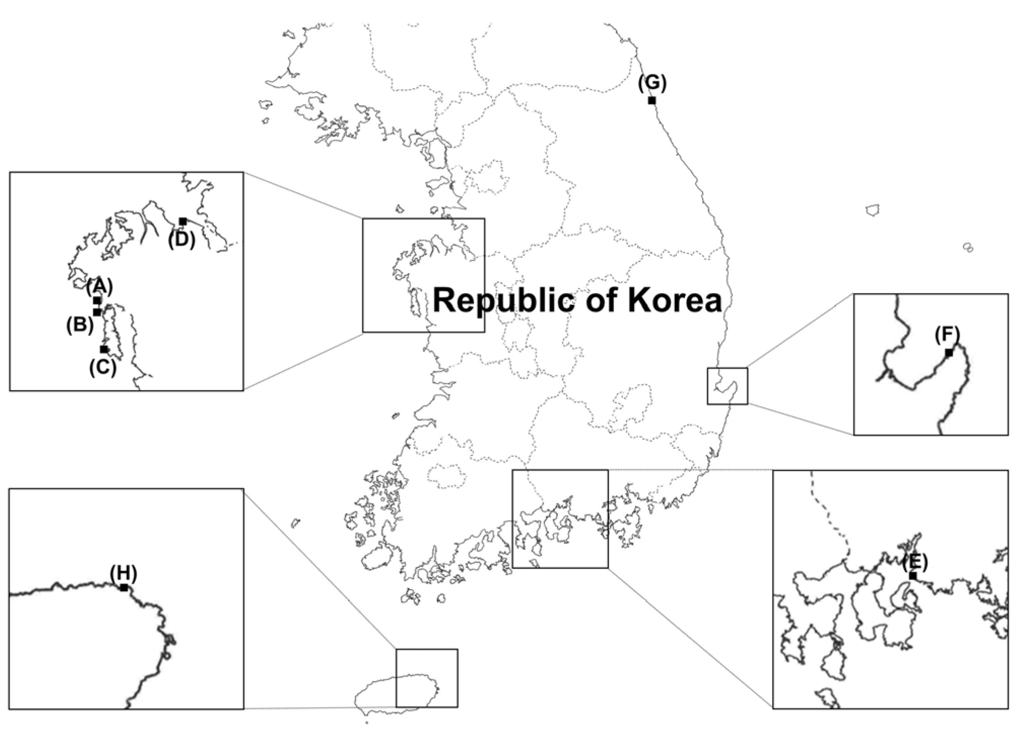
2.1. Fungal Isolation

A list of yeast strains with general and sample collection information is presented in Figure 1 and Table 1. The marine-derived yeasts used in this study were isolated from marine environments using the following procedure. Samples were collected from marine sediments, seawater, a clam (Venerupis philippinarum), the gut of a fish (Pagrus major), and sea algae (Rumex crispus and Ulva australis). The marine sediments were diluted with sterilized saline. Seawater was filtered using a 0.45 µm sterile membrane filter (Hyundai Micro Co., Ltd., Seoul, Republic of Korea). The clam was crushed, homogenized, and diluted in sterilized saline. Fish gut tissues were homogenized and diluted in sterilized saline. Sea algae were washed three times with sterile distilled water and cut into 0.5 or 1 cm pieces. Samples diluted in sterilized saline were spread, and the filters of seawater samples and pieces of sea algae were placed on potato dextrose agar (PDA; BD, Franklin Lakes, NJ, USA) supplemented with 0.01% (w/v) ampicillin (Sigma-Aldrich, St. Louis, MO, USA) and 0.01% (w/v) streptomycin (Sigma-Aldrich) to prevent bacterial growth. The samples were subsequently incubated at 25 °C for seven days. During this period, individual yeast colonies were picked and transferred to fresh PDA to isolate pure cultures. After isolation, yeast strains were cultured on yeast extract–peptone–dextrose (YPD) agar (BD) at 25 °C, unless described otherwise. Yeast strains were suspended in 20% glycerol (v/v) and stored at −80 °C.

2.2. DNA Extraction, PCR, and Identification

Genomic DNA was extracted from yeast cultures grown in YPD broth at 25 °C for three days on a 150 rpm rotary shaker. Yeast cells were collected through centrifugation at 10,000× g for 10 min, frozen, ground using liquid nitrogen, and suspended in lysis buffer, following previously established protocols [14]. PCR reactions were performed with two primer sets, ITS1F (5′-CTTGGTCATTTAGAGGAAGTAA-3′) and ITS4 (5′-TCCTCCGCTTATTGATATGC-3′), which were used to amplify the internal transcribed spacer (ITS) region [15], and LROR (5′-ACCCGCTGAACTTAAGC-3′) and LR5 (5′-TCCTGAGGGAAACTTCG-3′) were used to amplify the partial D1/D2 domain of a large subunit (LSU, 28S) of rDNA [16]. PCR conditions were as follows: Initial denaturation at 95 °C for 4 min, followed by 30 cycles of denaturation at 95 °C for 30 s, annealing at 55 °C for 30 s, extension at 72 °C for 30 s, and elongation at 72 °C for 5 min to amplify ITS and LSU regions. PCR products were purified using a Gel Extraction Kit (Qiagen, Hilden, Germany). DNA sequencing was conducted by Macrogen Inc. (Seoul, Republic of Korea) in both the forward and reverse directions with the two primer sets using the Sanger method.
The sequences generated in the forward and reverse directions were aligned and assembled using MEGA version 11 software to obtain full-length gene sequences [17]. The gene sequences of closely related taxa were searched for in the GenBank database. Phylogenetic analyses based on the concatenated sequences of the ITS regions and D1/D2 domains of the LSU rRNA gene were constructed using neighbor joining (NJ) and maximum likelihood (ML) methods. Kimura-2 parameter and general time reversible models were used for the NJ and ML methods, respectively, followed by 1000 bootstrap replicates in MEGA 11. The strains isolated in this study were deposited in the Marine Microbial BioBank (MMBB) culture collection of the National Biodiversity Institute of Korea (MABIK) (Table 1).

2.3. Culture Conditions

The growth of marine yeasts in different media and NaCl concentrations were evaluated by inoculating 10 µL of cell suspensions (optical density of 0.5 measured at a wavelength of 600 nm) in the center of Petri dishes. Morphological characteristics of the yeast strains were studied on PDA, malt extract agar (MEA; BD), and YPD agar plates with different NaCl concentrations (0, 2, 4, 6, 8, 10, 12, 16, and 20% [w/v]). Experiments were performed in three replicates for each plate. Plates inoculated with yeasts were incubated for seven days at 25 °C.

2.4. Microscopic Observation

For morphological characterization, isolates from marine environments were cultured on YPD agar (BD) at 25 °C for three days and observed under a Leica CTR6000 microscope (Leica, Wetzlar, Germany). The microscope was equipped with a Leica DMC2900 camera and LAS V4.5 software for image acquisition.

2.5. Preparation of Fungal Extracts

All yeast isolates were cultured on 50 mL PDA at 25 °C in the dark for a week. The cultures were extracted with 200 mL methanol (Sigma-Aldrich) for 24 h, followed by filtration using Whatman No. 1 filter paper. The filtrates were evaporated at 32 °C under a vacuum, and the condensed extracts were dissolved in 20 mL distilled water and 20 mL ethyl acetate (Sigma-Aldrich). After 6 h, the partitioned ethyl acetate fraction was collected and evaporated under the same conditions described above. The extracts were dissolved in dimethyl sulfoxide (DMSO; Sigma-Aldrich) to a concentration of 10 mg/mL and stored at 4 °C. All fungal cultures and extracts were prepared in triplicates.

2.6. Antioxidant Assays

2.6.1. ABTS Radical-Scavenging Assay

The 2,2′-azino-bis-3-ethylbenzothiazoline-6-sulfonic acid (ABTS; Sigma-Aldrich) radical-scavenging assay was performed based on previously established methods [18]. The ABTS was dissolved to a concentration of 7 mM in phosphate-buffered saline (PBS; pH 7.4). The solution was subsequently mixed with potassium persulfate dissolved to 2.45 mM in PBS. The mixture was stored in the dark at room temperature for 24 h to form ABTS•+. The solution was diluted with PBS to an absorbance of 0.70 (±0.02) at a wavelength of 734 nm. Then, 198 µL ABTS•+ solution and 2 µL fungal extract (10 mg/mL in DMSO) were mixed in a 96-well plate. Absorbance was measured at 734 nm after 6 min using a spectrophotometer (Hidex, Turku, Finland). Ascorbic acid (Sigma-Aldrich) was used as the positive control.

2.6.2. DPPH Radical-Scavenging Assay

The 2,2-diphenyl-1-picrylhydrazyl (DPPH; Sigma-Aldrich) radical-scavenging assay was analyzed following established methods [19]. The DPPH was dissolved in 80% methanol at 150 µM. Then, 200 µL of DPPH solution was mixed with 22 µL fungal extract (10 mg/mL in DMSO) in a 96-well plate. The mixture was stored in the dark at room temperature for 30 min, and absorbance was measured at 540 nm using a spectrophotometer. Ascorbic acid was used as the positive control.

2.7. Antimicrobial Activity

A microplate-based growth assay was performed using the microtiter broth dilution method to determine antimicrobial activity [20]. The target species were Bacillus subtilis (KCTC 3135/ATCC 6051), Escherichia coli (KCTC 2441/ATCC 11775), Candida albicans (KCCM 12555/ATCC 42266), Aspergillus flavus (KCCM 60330/ATCC 22546) and A. niger (KCCM 60332/ATCC 16888) purchased from the Korean Collection for Type Cultures (KCTC; Jeongeup, Republic of Korea) and the Korean Culture Center of Microorganisms (KCCM; Seoul, Korea). Fifty microliters of microbial suspensions (1 × 10⁶ cells/mL for bacteria, 5 × 10³ cells/mL for yeast, and 4 × 10⁵ conidia/mL spore suspensions for molds) were added to each well containing media (Mueller Hinton broth [BD] for bacteria and potato dextrose broth [BD] for yeast and molds). The yeast extracts were added to each well at a final concentration of 100 µg/mL. The 96-well plates were incubated at 25 °C for 2–3 days (two days for bacteria and three days for fungi). Extracts with lower concentrations (50, 25, and 12.5 µg/mL) were tested to determine the minimum inhibitory concentration (MIC).

2.8. Tyrosinase Inhibition Assay

Tyrosinase inhibitory activity was determined as previously described [21], with modifications. Here, 70 µL of 0.1 M potassium phosphate buffer (pH 6.8), 40 µL yeast extract (2.5 mg/mL diluted in 50% DMSO), and 30 µL of 0.02 mg/mL tyrosinase from mushroom (Sigma-Aldrich) were mixed in each well of the 96-well plates. The mixtures were heated in a water bath at 30 °C for 5 min, and 100 µL of 2.5 mM L-DOPA (Sigma-Aldrich) was added. After 30 min, the plates were put in ice to complete the reaction, and absorbance was measured at 492 nm using a spectrophotometer. Kojic acid (Sigma-Aldrich) was used as the positive control, and each mixture except L-DOPA was regarded as a blank.

2.9. Statistical Analysis

Experimental data were analyzed using one-way ANOVA followed by Tukey’s multiple comparison test (GraphPad Prism software 5.0). All experiments were conducted in biological triplicates unless otherwise described.

3. Results and Discussion

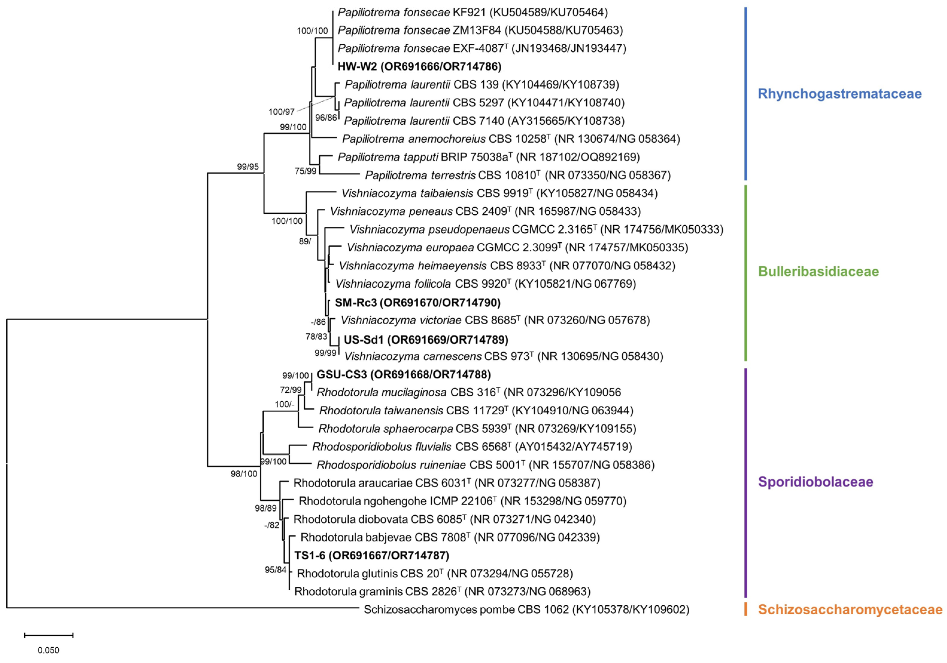
3.1. Identification and Phylogeny

Yeast identification through molecular analysis is generally conducted based on both ITS and the D1/D2 domain [22]. In this study, 11 strains of marine yeasts were identified by sequencing the ITS region and the D1/D2 domain of the LSU rRNA gene. Sequence information of the ITS regions and D1/D2 domains of the LSU rRNA gene were deposited in GenBank (accession numbers are presented in the phylogenetic trees). The phylogenetic placement of the strains is shown in Figure 2 (Ascomycota) and Figure 3 (Basidiomycota). From these phylogenetic analyses, the trees derived from the NJ and ML method were similar. The marine-derived yeasts represented seven species and three new candidate species distributed into four fungal lineages: Ascomycota, Saccharomycotina (five strains); Ascomycota, Pezizomycotina (one strain); Basidiomycota, Agricomycotina (three strains); and Basidiomycota, Pucciniomycotina (two strains) (Figure 2 and Figure 3).
A phylogenetic analysis conducted using combined ITS and D1/D2 domain sequences assigned the Ascomycota species to five genera (Figure 2). GH-W2 was closely related to Geotrichum candidum CBS 11628 (100% identity) and 11616 (100%). GH-W2 was identified as G. candidum with relatively high support (bootstrap value: NJ, 84%; ML, 82%). HW-W3 formed a monophyletic clade with Yarrowia lipolytica (bootstrap value: 100%). As BP-Up1 and US-Sd4 were grouped with Metschnikowia bicuspidata, and both strains were identified as M. bicuspidata. K2-6 was placed in the Yamadazyma olivae clade as it was closely related to Y. olivae FMCC Y-1T and CBS 1171, with high bootstrap values (NJ, 98%; ML, 100%). US-Sd3 in the Aureobasidium clade was not assigned to a clade with close references. A comparison of the ITS regions and D1/D2 domains of strain US-Sd3 with its related species showed that this strain was not grouped with a distinct species: A. namibiae CBS 147.97T (96.68% identity in ITS regions and 99.43% identity in D1/D2 domains), A. subglaciale CBS 123387T (99.45% in ITS regions and 98.87% in D1/D2 domains), and A. leucospermi CBS 130593T (97.78% in ITS regions and 98.68% in D1/D2 domains). Therefore, it was considered a new candidate species.
Basidiomycota species were assigned to three genera: Papiliotrema, Vishniacozyma, and Rhodotorula (Figure 3). A BLAST search conducted using the ITS and D1/D2 domain sequences revealed a high degree of similarity between HW-W2 and Papiliotrema fonsecae KF921 (100% identity), ZM13F84 (100%), and EXF-4087T (100%). HW-W2 belonged to the same clade as the three strains with a 100% degree of confidence. US-Sd1 was identified as Vishniacozyma carnescens with a high degree of similarity to V. carnescens CBS 973T (100% identity). Although SM-Rc3 was placed in the Vishniacozyma clade, it was not assigned to a clade through close references. Therefore, it was considered a new candidate species and designated as Vishniacozyma sp. SM-Rc3. GSU-CS3 and TS1-6 were grouped with Rhodotorula spp. GSU-CS3 was placed in a monophyletic clade with R. mucilaginosa (bootstrap value: NJ, 99%; ML, 100%). As TS1-6 was grouped with the three distinct species, R. glutinis CBS 20T (99.49% identity in ITS regions and 99.87% identity in D1/D2 domain), R. graminis CBS 2826T (99.15% in ITS regions and 100% in D1/D2 domain), and R. babjevae CBS 7808T (99.66% in ITS regions and 99.82% in D1/D2 domain), it was not identified at the species level and was thus designated as Rhodotorula sp. TS1-6.

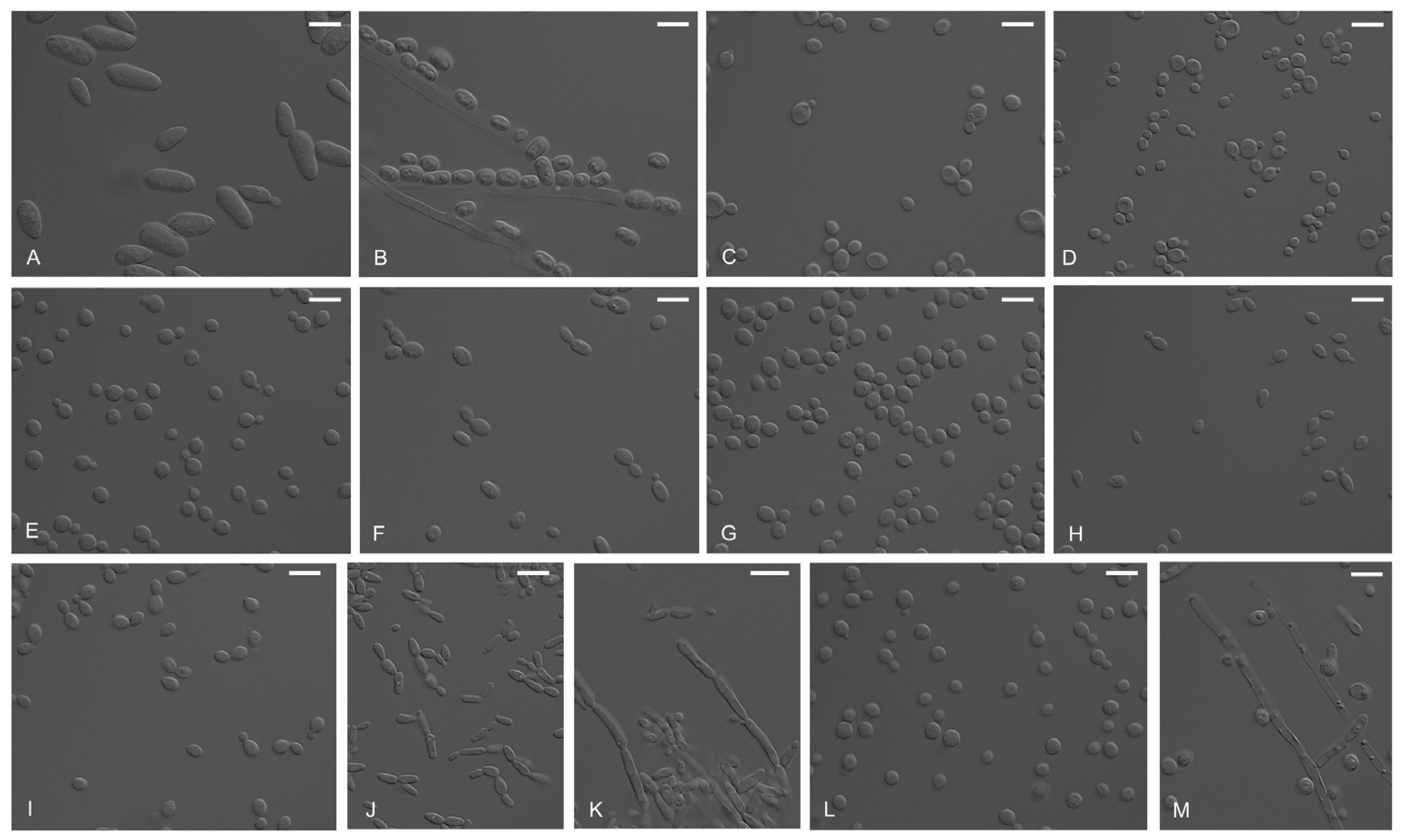
3.2. Morphological Observation

Colonies of US-Sd3 on PDA were smooth and black with white fimbriate margins. Colonies on MEA were felty and brown with white fimbriate margins. The colonies on both media initially appeared cream and later became blackish or brownish. Colonies on YPD were raised, smooth, and light pink (Figure 4). Colonies of Aureobasidium species appeared cream, yellow, light brown, and light pink at the beginning of cultivation and became blackish, owing to the production of a dark pigment, which was a melanin-like compound [23]. Conidia were hyaline, aseptate, smooth-walled, ellipsoidal-to-ovoid, and often had polar buds (Figure 5A).
Strain GH-W2, identified as G. candidum, was characterized by an intermediate morphology of mold- and yeast-like, white-felted, and non-greasy colonies (Figure 4) with abundant production of arthrospores (Figure 5B). As G. candidum is a filamentous yeast-like fungus, its strains exhibit phenotypic variability as yeast-like, intermediate, or mold-like morphotypes [24].
Strains BP-Up1 and US-Sd4 (identified as M. bicuspidata) formed smooth, entire mar-gins, and cream-colored colonies on PDA. Colonies grown on MEA and YPD media were smooth and white. These two strains showed slower growth on MEA than PDA and YPD agar (Figure 4). Their cells were globose or subglobose under microscopic observation. In addition, polar buds were observed (Figure 5C,D). Cellular morphological differences between the two strains were found in regard to cell size and salt tolerance (described below). The size of BP-Up1 was approximately 2.5–5.5 µm in width × 4.0–8.0 µm in length (N = 10). In contrast, the size of US-Sd4 was approximately 2.0–4.0 µm in width × 2.2–4.5 µm in length (N = 10).
Colonies of HW-W2, identified as P. fonsecae, were cream-beige in color, with smooth surfaces and entire margins (Figure 4). Cells were globose to oval (3.0–4.6 × 3.0–5.8 µm, N = 10) with bud formation (Figure 5E).
Strain TS1-6, identified as Rhodotorula sp., formed soft, smooth, and moist orange-coral colonies (Figure 4). Under the microscope, the cells were subglobose or elliptical in shape and formed buds (Figure 5F). Strain GSU-CS3, identified as R. mucilaginosa, produced coral-colored, smooth, and wet colonies (Figure 4). In addition, the cells were globose or subglobose (Figure 5G). Rhodotorula species are typically pink-to-orange in color owing to the production of carotenoids as intracellular lipid droplets [25].
Colonies of strain US-Sd1, identified as V. carnescens, were smooth, moist, and cream-colored with entire margins (Figure 4). Under microscopic observation, the cells were subglobose and ellipsoidal, with budding observed (Figure 5H). Strain SM-Rc3, identified as Vishniacozyma sp., formed beige-colored, smooth, and flat colonies on PDA and MEA, whereas they formed raised colonies on YPD media (Figure 4). Cells were oval or tear-shaped (3.0–4.2 × 4.6–6.2 µm, N = 10), and bud formation was observed (Figure 5I).
Colonies of strain K2-6, identified as Y. olivae, were cream-colored and moderately raised. The cells were ellipsoid and found either singly or in mother-bud pairs. Short chains of elongated cells and pseudohyphae were observed (Figure 5J,K).
Strain HW-W3, identified as Y. lipolytica, produced colonies with wrinkles, ridged surfaces, and fimbriate margins on PDA and YPD agar. On MEA, the colonies were flat with spreading edges (Figure 4). Unlike most other Saccharomycetes yeasts, this strain did not typically produce radical and smooth colonies. Cells at the microscopic level were globose or subglobose with bud formation (Figure 5L). Hyphae were also observed (Figure 5M). The yeast Y. lipolytica is a model for dimorphism, as it can undergo a yeast-to-hyphal transition [26].

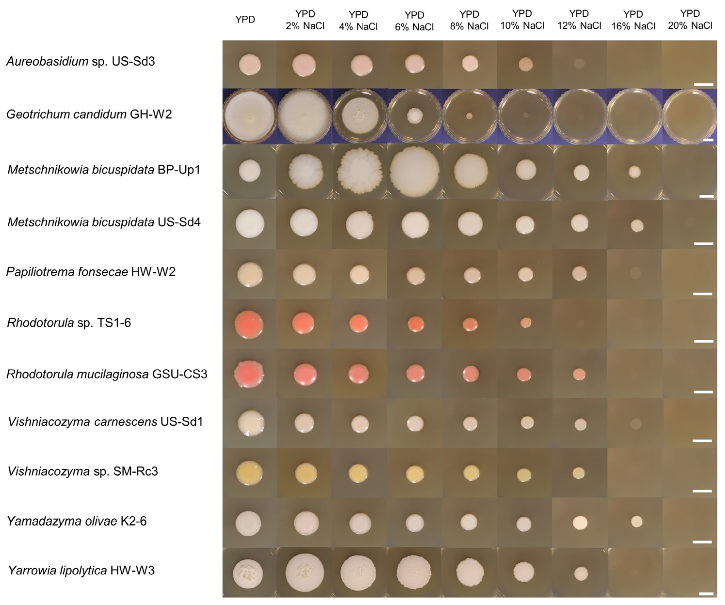
3.3. Salt-Tolerant Ability

As the isolates were derived from marine environments, the effects of NaCl concentration in YPD media were investigated (Figure 6). Different strains showed varying growth and responses to NaCl concentration. All tested marine-derived yeast strains could grow with up to 8% (w/v) NaCl, whereas no growth was observed at 20% NaCl. Unlike other strains, GH-W2 growth did not occur at 10% NaCl concentration. Two strains (US-Sd3 and TS1-6) could not grow completely at 12% of salt exposure. The growth of five strains (HW-W2, GSU-CS3, US-Sd1, SM-Rc3, and HW-W3) was completely inhibited at 16% NaCl. Three strains (BP-Up1, US-Sd4, and HW-W3) did not grow at 20% NaCl. Furthermore, growth of the GH-W2, HW-W2, TS1-6, GSU-CS3, US-Sd1, SM-Rc3, and K2-6 strains was inhibited by the addition of NaCl to the medium. The colonies of BP-Up1 and HW-W3 became bigger by 6% and 2% NaCl, respectively.
M. bicuspidata and Y. lipolytica were previously isolated from hypersaline waters such as salt lakes and ponds [27]. Y. lipolytica, with generally recognized as safe status, is considered an excellent microorganism with multiple biotechnological applications [28]. Halophiles and halotolerants have recently shown promise because fermentation processes are free under non-sterile conditions [29].
Although we observed the different responses of individual strains to NaCl concentrations, salinity tolerance generally does not distinguish between marine and terrestrial yeasts. Most yeasts are able to grow under NaCl concentrations of marine environments [3]. Yeasts employ distinct metabolic strategies associated with environmental distribution, and their phenotypes in diverse environments are driven by environmental constraints [30]. Therefore, the different growth of BP-Up1 from US-Sd4 (both M. bicusbidata) might be caused by the distinct isolation sources, sea algae and marine sediment, respectively.

3.4. Antioxidant Activity

Reactive oxygen species and free radical-mediated reactions can damage biomolecules such as proteins, lipids, and nucleic acids and contribute to aging, cancer, and various diseases [31]. Molecules of microbial origin showing antioxidant and radical-scavenging activities contain carbohydrates, phenolic compounds, carotenoids, anthraquinone, xanthones, indole derivatives, and alkaloids [11,32].
In this study, the antioxidant activities of crude fungal extracts of marine yeasts were evaluated using ABTS and DPPH radical-scavenging assays. The extracts of G. candidum GH-W2, M. bicuspidata BP-Up1, US-Sd4, R. mucilaginosa GSU-CS3, and Y. olivae K2-6 showed over 60% inhibition rates, whereas the other extracts exhibited lower inhibition rates from 9.78% to 50.94% ABTS radical-scavenging activity (Table 2). These strains exhibited high ABTS radical-scavenging activity against DPPH radicals. All 11 marine yeast extracts showed under 40% DPPH radical-scavenging activity. As ABTS and DPPH reaction buffers (PBS and 80% methanol, respectively) are different, the solubility of antioxidant compounds in each buffer affects the results [33].
Many antioxidants have been discovered, and marine fungi are one of their promising sources [11]. Carotenoids are one of the molecules shown to exert antioxidant and scavenging activities and play a key role in protecting cells by scavenging oxygen and peroxyl radicals [34]. Heterotrophic bacteria and fungi, especially pigmented yeasts, as well as photosynthetic organisms, are potentially important sources of carotenoids [35]. Several yeast species isolated from marine environments belonging to the genera Xanthophyllomyces (with Phaffia as its anamorphic state) and Rhodotorula have been used to synthesize astaxanthin [36]. In the case of the marine yeast R. mucilaginosa isolated from sea weed, it produces carotenoids, lycopene, ß-carotene, and astaxanthin [37]. Additionally, the marine yeast R. glutinis produces astaxanthin [32]. The G. candidum isolated from roots of the genus Sophora has been reported to produce matrine. Martrine, a tetracyclic quinolizine alkaloid, has anticancer, antiviral, anti-inflammatory, and antioxidant effects [38]. However, there are no reports of marine-derived G. candidum. Contrary to our results, there are no reports of antioxidants from M. bicuspidata and Y. olivae that exhibit high radical-scavenging activity. In particular, the crude extract of M. bicuspidata BP-Up1 isolated from Ulva australis of green algae exhibited the highest ABTS radical-scavenging activity (85.88%). The M. bicuspidata yeast is an opportunistic pathogen that causes diseases in many aquatic animal species in freshwater and marine environments [39]. Therefore, transmission experiments and genomic analyses of its pathogenicity have been conducted [39,40]. However, only a few of its bioactive compounds and their bioactivities have been studied. To the best of our knowledge, this is the first report of the radical-scavenging activity of M. bicuspidata and Y. olivae.

3.5. Antibacterial Activity

Antimicrobial experiments were conducted using the marine yeast extracts on bacteria (B. subtilis and E. coli) and fungi (C. albicans, A. flavus, and A. niger). In this study, antagonistic activity against B. subtilis was demonstrated (Table 2). However, no antimicrobial activity against E. coli, C. albicans, A. flavus, and A. niger was detected. Some strains of B. subtilis cause foodborne diseases resulting from the spoilage of canned vegetables, seafoods, and bakery products. Endospore-forming Bacillus species are difficult to manage because they are resistant to heat, desiccation, and UV light [41].
Seven strains exhibited remarkable activity against B. subtilis: Aureobasidium sp. US-Sd3, G. candidum GH-W2, R. mucilaginosa GSU-CS3, V. carnescens US-Sd1, Vishniacozyma sp. SM-Rc3, Y. olivae K2-6, and Y. lipolytica HW-W3 (Table 2). In particular, Aureobasidium sp. US-Sd3 extracts had the highest inhibitory activity among all the marine yeast extracts tested as they inhibited the growth of B. subtilis at 100 µg/mL. The antifouling compound aureobasidin isolated from the marine-derived Aureobasidium sp. has antibacterial activity against B. subtilis, E. coli, and Staphylococcus aureus [42].
Antimicrobial activity has been reported in yeast and yeast-like fungi. For example, the liamocin oil produced by A. pullulans is selective with antibacterial activity against Streptococcus species [43]. Similarly, the antibacterial activity of ethyl acetate extracts of G. candidum isolated from the root biome of date palm trees has been previously reported [44]. Dieuleveux et al. [45] described the purification and characterization of anti-Listeria compounds from G. candidum. Pigments from R. mucilaginosa exhibit antibacterial activity against S. aureus and antibiotic-resistant bacteria [46,47]. Moreover, the V. victoriae yeast isolated from pear fruits during cold postharvest storage has shown biocontrol effects against Penicillium expansum and Botrytis cinerea, which are major causes of postharvest pear disease [48]. The biotechnologically important yeast, Y. lipolytica, synthesizes silver nanoparticles, which are kwon antimicrobial agents [9]. Organic acids, including succinate, kynurenic acid, and α-ketoglutarate, were synthesized by Y. lipolytica in beverage-like substrates. Succinate has multiple applications in the food industry as an antimicrobial agent, flavor enhancer, and acidifier [49]. In contrast, there are no reports on the antimicrobial compounds or activities of Y. olivae.
Fungal endophytes support plant defense systems related to symbiotic associations [50] and are responsible for host plant adaptation to abiotic and biotic stresses [51]. Endophytes promote plant growth and enhance nutrient uptake in plant rhizospheres. Fungal endophytes produce compounds with antifungal activity. Thus, bioactive compounds synthesized by endophytes can be used as biocontrol agents against plant diseases [52]. Similarly, the antibacterial activity results in the present study suggest that antibacterial compounds from marine-derived yeasts could also support a symbiotic relationship with marine algae.

3.6. Tyrosinase Inhibition Activity

Only one (Aureobasidium sp. US-Sd3) of the 11 marine-derived yeast extracts showed tyrosinase inhibitory activity (Table 2). Although several tyrosinase inhibitory activities in yeasts have been reported [53], they were rarely detected in the isolates tested in the present study. Tyrosinase oxidizes tyrosine to dihydroxyphenylalanine (DOPA) and then dopaquinone through a series of oxidative reactions. Dopaquinone is eventually converted to pheomelanin. Tyrosinase plays an important role in mammalian melanogenesis and the enzymatic browning of fruits and fungi. Although melanin protects the human skin from UV radiation, the darkening of skin tone caused by melanin biosynthesis is an esthetic problem [53,54]. Tyrosinase inhibitors can prevent melanogenesis in the skin. Therefore, research interest in these compounds has increased, owing to their skin-whitening effects. Arbutin, hydroquinone, gentisic acid, deoxyarbutin, aloesin, 4-n-butylresorcinol, ascorbic acid, kojic acid, and azelaic acid are used in the cosmetics industry [54]. In particular, kojic acid, a well-known tyrosinase inhibitor, was identified from acetone extracts through a screening study of 600 marine fungi [55]. Apart from kojic acid that is produced by various Aspergillus and Penicillium spp., there are diverse compounds with anti-tyrosinase activity, such as metallothioneins from A. niger [56], homothallin II from Trichoderma viride [57], and 6-n-pentyl-α-pyrone and myrothenone A isolated from the marine-derived fungi, Myrothecium sp. [58].
There are no reports of single tyrosinase inhibitors from the genus Aureobasidium. However, there have been reports that exopolymers from A. pullulans reduce melanin production in melanoma cells and mushroom tyrosinase activity [59]. Some antioxidants inhibit tyrosinase by scavenging quinone products [54]. In the present study, the Aureobasidium sp. US-Sd3 extract exhibited tyrosinase inhibition and low-radical-scavenging (9.78% ABTS and 20.90% DPPH radical-scavenging activity) activities. These results indicate that strain US-Sd3 may have other functional mechanisms. For instance, suicide inactivators can inhibit tyrosinase by inducing conformational changes in the enzyme. Alternatively, competitive inhibitors, such as copper chelators, can inhibit tyrosinase, which is a metalloenzyme [60].
Tyrosinase inhibitors also have antibacterial activity, and some have more potent antibacterial activity than antibiotics such as ampicillin. However, tyrosinase inhibition may not always be associated with antibacterial activity [61]. The crude extract of strain US-Sd3 exhibited antibacterial activity against B. subtilis. This indicates that Aureobasidium species can likely produce tyrosinase inhibitors that regulate melanin biosynthesis, and it is possible that tyrosinase inhibitors are related to antibacterial activity.

4. Conclusions

In conclusion, this study provided information on the identification of marine-derived yeasts and their exploitable biological activities. Phylogenetic analyses and morphological observations revealed that 11 marine yeasts isolated from various marine sources were classified into 10 taxa, including three new candidate species. Isolates were tested for salt tolerance ability using 0% to 20% NaCl concentrations. The M. bicuspidata and Y. olivae strains were able to grow at 16% NaCl and adapt to conditions of higher salt stress than that observed for the other strains. Among these, three species (Aureobasidium sp., M. bicuspidata, and Y. olivae) showed notable bioactivities. Overall, the results of this study suggest that marine-derived yeasts are good sources of potential bioactive compounds. The identification of microorganisms is valuable for biodiversity and bioresources. The further purification and characterization of their potent bioactive compounds should be conducted to enable further applications.

Author Contributions

Conceptualization, W.-J.Y. and D.C.; Methodology, S.S.B. and Y.M.K.; Investigation, W.-J.Y. and E.-S.C.; Writing—Original Draft, W.-J.Y.; Writing—Review and Editing, D.C. and G.C.; Supervision and Project Administration, G.C. All authors have read and agreed to the published version of the manuscript.

Funding

This research was funded by an in-house grant from the National Marine Biodiversity Institute of Korea (grant number MABIK 2023M00600).

Institutional Review Board Statement

Not applicable.

Informed Consent Statement

Not applicable.

Data Availability Statement

The data presented in this study are openly available in online repositories. The names of repositories, strain ID, and accession numbers can be found below: https://www.mbris.kr/biobank/, MABIK FU00001121, 1179, and 1254-1262 (strain identities); https://www.ncbi.nlm.nih.gov/genbank/, OR691662-OR691672 and OR714782-OR714792 (accession numbers).

Conflicts of Interest

The authors declare no conflict of interest.

References

  1. Kwon, Y.M.; Choi, H.S.; Lim, J.Y.; Jang, H.S.; Chung, D. Characterization of amylolytic activity by a marine-derived yeast Sporidiobolus pararoseus PH-Gra1. Mycobiology 2020, 48, 195–203. [Google Scholar] [CrossRef] [PubMed]
  2. Chi, Z.M.; Liu, G.; Zhao, S.; Li, J.; Peng, Y. Marine yeasts as biocontrol agents and producers of bio-products. Appl. Microbiol. Biotechnol. 2010, 86, 1227–1241. [Google Scholar] [CrossRef] [PubMed]
  3. Kutty, S.N.; Philip, R. Marine yeasts—A review. Yeast 2008, 25, 465–483. [Google Scholar] [CrossRef] [PubMed]
  4. Kurtzman, C.; Fell, J.W.; Boekhout, T. The Yeasts: A Taxonomic Study, 5th ed.; Elsevier: Amsterdam, The Netherlands, 2011. [Google Scholar]
  5. Zaky, A.S.; Tucker, G.A.; Daw, Z.Y.; Du, C. Marine yeast isolation and industrial application. FEMS Yeast Res. 2014, 14, 813–825. [Google Scholar] [CrossRef] [PubMed]
  6. Hyde, K.D.; Jones, E.G.; Leaño, E.; Pointing, S.B.; Poonyth, A.D.; Vrijmoed, L.L. Role of fungi in marine ecosystems. Biodivers. Conserv. 1998, 7, 1147–1161. [Google Scholar] [CrossRef]
  7. Chi, Z.; Chi, Z.; Zhang, T.; Liu, G.; Li, J.; Wang, X. Production, characterization and gene cloning of the extracellular enzymes from the marine-derived yeasts and their potential applications. Biotechnol. Adv. 2009, 27, 236–255. [Google Scholar] [CrossRef]
  8. Wang, L.; Chi, Z.; Wang, X.; Ju, L.; Chi, Z.; Guo, N. Isolation and characterization of Candida membranifaciens subsp. flavinogenie W14-3, a novel riboflavin-producing marine yeast. Microbiol. Res. 2008, 163, 255–266. [Google Scholar] [CrossRef]
  9. Apte, M.; Sambre, D.; Gaikawad, S.; Joshi, S.; Bankar, A.; Kumar, A.R.; Zinjarde, S. Psychrotrophic yeast Yarrowia lipolytica NCYC 789 mediates the synthesis of antimicrobial silver nanoparticles via cell-associated melanin. AMB Express 2013, 3, 1–8. [Google Scholar] [CrossRef] [PubMed]
  10. Hernández-Saavedra, N.Y.; Ochoa, J.L. Copper–zinc superoxide dismutase from the marine yeast Debaryomyces hansenii. Yeast 1999, 15, 657–668. [Google Scholar] [CrossRef]
  11. Corinaldesi, C.; Barone, G.; Marcellini, F.; Dell’Anno, A.; Danovaro, R. Marine microbial-derived molecules and their potential use in cosmeceutical and cosmetic products. Mar. Drugs 2017, 15, 118. [Google Scholar] [CrossRef]
  12. Ding, J.; Wu, B.; Chen, L. Application of Marine Microbial Natural Products in Cosmetics. Front. Microbiol. 2022, 13, 892505. [Google Scholar] [CrossRef] [PubMed]
  13. Manohar, C.S.; Raghukumar, C. Fungal diversity from various marine habitats deduced through culture-independent studies. FEMS Microbiol. Lett. 2013, 341, 69–78. [Google Scholar] [CrossRef]
  14. Chung, D.; Baek, K.; Bae, S.S.; Jung, J. Identification and characterization of a marine-derived chitinolytic fungus, Acremonium sp. YS2-2. J. Microbiol. 2019, 57, 372–380. [Google Scholar] [CrossRef]
  15. Gardes, M.; Bruns, T.D. ITS primers with enhanced specificity for basidiomycetes-application to the identification of mycorrhizae and rusts. Mol. Ecol. 1993, 2, 113–118. [Google Scholar] [CrossRef]
  16. Vilgalys, R.; Hester, M. Rapid genetic identification and mapping of enzymatically amplified ribosomal DNA from several Cryptococcus species. J. Bacteriol. 1990, 172, 4238–4246. [Google Scholar] [CrossRef]
  17. Tamura, K.; Stecher, G.; Kumar, S. MEGA11: Molecular evolutionary genetics analysis version 11. Mol. Biol. Evol. 2021, 38, 3022–3027. [Google Scholar] [CrossRef]
  18. Re, R.; Pellegrini, N.; Proteggente, A.; Pannala, A.; Yang, M.; Rice-Evans, C. Antioxidant activity applying an improved ABTS radical cation decolorization assay. Free Radic. Biol. Med. 1999, 26, 1231–1237. [Google Scholar] [CrossRef]
  19. Fukumoto, L.R.; Mazza, G. Assessing antioxidant and prooxidant activities of phenolic compounds. J. Agric. Food Chem. 2000, 48, 3597–3604. [Google Scholar] [CrossRef]
  20. Balouiri, M.; Sadiki, M.; Ibnsouda, S.K. Methods for in vitro evaluating antimicrobial activity: A review. J. Pharm. Anal. 2016, 6, 71–79. [Google Scholar] [CrossRef]
  21. Lai, H.Y.; Lim, Y.Y.; Tan, S.P. Antioxidative, tyrosinase inhibiting and antibacterial activities of leaf extracts from medicinal ferns. Biosci. Biotechnol. Biochem. 2009, 73, 1362–1366. [Google Scholar] [CrossRef]
  22. Nutaratat, P.; Boontham, W.; Khunnamwong, P. A novel yeast genus and two novel species isolated from pineapple leaves in Thailand: Savitreella phatthalungensis gen. nov., sp. nov. and Goffeauzyma siamensis sp. nov. J. Fungi 2022, 8, 118. [Google Scholar] [CrossRef] [PubMed]
  23. Li, Y.; Chi, Z.; Wang, G.Y.; Wang, Z.P.; Liu, G.L.; Lee, C.F.; Ma, Z.C.; Chi, Z.M. Taxonomy of Aureobasidium spp. and biosynthesis and regulation of their extracellular polymers. Crit. Rev. Microbiol. 2015, 41, 228–237. [Google Scholar] [CrossRef] [PubMed]
  24. Perkins, V.; Vignola, S.; Lessard, M.H.; Plante, P.L.; Corbeil, J.; Dugat-Bony, E.; Frenette, M.; Labrie, S. Phenotypic and genetic characterization of the cheese ripening yeast Geotrichum candidum. Front. Microbiol. 2020, 11, 737. [Google Scholar] [CrossRef]
  25. Li, Z.; Li, C.; Cheng, P.; Yu, G. Rhodotorula mucilaginosa—Alternative sources of natural carotenoids, lipids, and enzymes for industrial use. Heliyon 2022, 8, e11505. [Google Scholar] [CrossRef]
  26. Nicaud, J.M. Yarrowia lipolytica. Yeast 2012, 29, 409–418. [Google Scholar] [CrossRef]
  27. Butinar, L.; Santos, S.; Spencer-Martins, I.; Oren, A.; Gunde-Cimerman, N. Yeast diversity in hypersaline habitats. FEMS Microbiol. Lett. 2005, 244, 229–234. [Google Scholar] [CrossRef]
  28. Mamaev, D.; Zvyagilskaya, R. Yarrowia lipolytica: A multitalented yeast species of ecological significance. FEMS Yeast Res. 2021, 21, foab008. [Google Scholar] [CrossRef]
  29. Yin, J.; Chen, J.C.; Wu, Q.; Chen, G.Q. Halophiles, coming stars for industrial biotechnology. Biotechnol. Adv. 2015, 33, 1433–1442. [Google Scholar] [CrossRef]
  30. Camarasa, C.; Sanchez, I.; Brial, P.; Bigey, F.; Dequin, S. Phenotypic landscape of Saccharomyces cerevisiae during wine fermentation: Evidence for origin-dependent metabolic traits. PLoS ONE 2011, 6, e25147. [Google Scholar] [CrossRef] [PubMed]
  31. Huang, W.Y.; Cai, Y.Z.; Xing, J.; Corke, H.; Sun, M. A potential antioxidant resource: Endophytic fungi from medicinal plants. Econ. Bot. 2007, 61, 14–30. [Google Scholar] [CrossRef]
  32. Vitale, G.A.; Coppola, D.; Palma Esposito, F.; Buonocore, C.; Ausuri, J.; Tortorella, E.; de Pascale, D. Antioxidant molecules from marine fungi: Methodologies and perspectives. Antioxidants 2020, 9, 1183. [Google Scholar] [CrossRef] [PubMed]
  33. Bartasiute, A.; Westerink, B.H.; Verpoorte, E.; Niederländer, H.A. Improving the in vivo predictability of an on-line HPLC stable free radical decoloration assay for antioxidant activity in methanol–buffer medium. Free Radic. Biol. Med. 2007, 42, 413–423. [Google Scholar] [CrossRef] [PubMed]
  34. Galasso, C.; Corinaldesi, C.; Sansone, C. Carotenoids from marine organisms: Biological functions and industrial applications. Antioxidants 2017, 6, 96. [Google Scholar] [CrossRef]
  35. Mata-Gómez, L.C.; Montañez, J.C.; Méndez-Zavala, A.; Aguilar, C.N. Biotechnological production of carotenoids by yeasts: An overview. Microb. Cell Fact. 2014, 13, 1–11. [Google Scholar] [CrossRef] [PubMed]
  36. Ambati, R.R.; Phang, S.M.; Ravi, S.; Aswathanarayana, R.G. Astaxanthin: Sources, extraction, stability, biological activities and its commercial applications—A review. Mar. Drugs 2014, 12, 128–152. [Google Scholar] [CrossRef]
  37. Leyton, A.; Flores, L.; Mäki-Arvela, P.; Lienqueo, M.E.; Shene, C. Macrocystis pyrifera source of nutrients for the production of carotenoids by a marine yeast Rhodotorula mucilaginosa. J. Appl. Microbiol. 2019, 127, 1069–1079. [Google Scholar] [CrossRef]
  38. Cao, K.; Chen, J.; Lu, X.; Yao, Y.; Huang, R.; Li, L. Matrine-producing endophytic fungus Galactomyces candidum TRP-7: Screening, identification, and fermentation conditions optimization for Matrine production. Biotechnol. Lett. 2023, 45, 209–223. [Google Scholar] [CrossRef]
  39. Jiang, H.; Bao, J.; Xing, Y.; Li, X.; Chen, Q. Comparative genomic analyses provide insight into the pathogenicity of Metschnikowia bicuspidata LNES0119. Front. Microbiol. 2022, 13, 939141. [Google Scholar] [CrossRef]
  40. Jiang, H.; Bao, J.; Cao, G.; Xing, Y.; Feng, C.; Hu, Q.; Li, X.; Chen, Q. Experimental transmission of the yeast, Metschnikowia bicuspidata, in the Chinese mitten crab, Eriocheir sinensis. J. Fungi 2022, 8, 210. [Google Scholar] [CrossRef]
  41. Kubo, I.; Fujita, K.I.; Nihei, K.I.; Nihei, A. Antibacterial activity of akyl gallates against Bacillus subtilis. J. Agric. Food Chem. 2004, 52, 1072–1076. [Google Scholar] [CrossRef]
  42. Abdel-Lateff, A.; Elkhayat, E.S.; Fouad, M.A.; Okino, T. Aureobasidin, new antifouling metabolite from marine-derived fungus Aureobasidium sp. Nat. Prod. Commun. 2009, 4, 389–394. [Google Scholar] [CrossRef] [PubMed]
  43. Bischoff, K.M.; Leathers, T.D.; Price, N.P.; Manitchotpisit, P. Liamocin oil from Aureobasidium pullulans has antibacterial activity with specificity for species of Streptococcus. J. Antibiot. 2015, 68, 642–645. [Google Scholar] [CrossRef]
  44. Mefteh, F.B.; Daoud, A.; Chenari Bouket, A.; Alenezi, F.N.; Luptakova, L.; Rateb, M.E.; Kadri, A.; Gharsallah, N.; Belbahri, L. Fungal root microbiome from healthy and brittle leaf diseased date palm trees (Phoenix dactylifera L.) reveals a hidden untapped arsenal of antibacterial and broad spectrum antifungal secondary metabolites. Front. Microbiol. 2017, 8, 307. [Google Scholar] [CrossRef] [PubMed]
  45. Dieuleveux, V.; Van Der Pyl, D.; Chataud, J.; Gueguen, M. Purification and characterization of anti-Listeria compounds produced by Geotrichum candidum. Appl. Environ. Microbiol. 1998, 64, 800–803. [Google Scholar] [CrossRef]
  46. Vidya, P.; Kutty, S.N.; Sebastian, C.D. Extraction, characterization and antimicrobial properties of pigments from yeast, Rhodotorula mucilaginosa isolated from the mangrove sediments of North Kerala, India. Asian J. Biol. Life Sci. 2021, 10, 559. [Google Scholar] [CrossRef]
  47. Yoo, A.Y.; Alnaeeli, M.; Park, J.K. Production control and characterization of antibacterial carotenoids from the yeast Rhodotorula mucilaginosa AY-01. Process Biochem. 2016, 51, 463–473. [Google Scholar] [CrossRef]
  48. Gramisci, B.R.; Lutz, M.C.; Lopes, C.A.; Sangorrín, M.P. Enhancing the efficacy of yeast biocontrol agents against postharvest pathogens through nutrient profiling and the use of other additives. Biol. Control 2018, 121, 151–158. [Google Scholar] [CrossRef]
  49. Sørensen, A.B.; Harholt, J.; Arneborg, N. Application of Yarrowia lipolytica in fermented beverages. Front. Food Sci. Technol. 2023, 3, 1190063. [Google Scholar] [CrossRef]
  50. Vega, F.E.; Posada, F.; Aime, M.C.; Pava-Ripoll, M.; Infante, F.; Rehner, S.A. Entomopathogenic fungal endophytes. Biol. Control 2008, 46, 72–82. [Google Scholar] [CrossRef]
  51. Singh, L.P.; Gill, S.S.; Tuteja, N. Unraveling the role of fungal symbionts in plant abiotic stress tolerance. Plant Signal. Behav. 2011, 6, 175–191. [Google Scholar] [CrossRef]
  52. Kumar, V.; Jain, L.; Kaushal, P.; Soni, R. Fungal endophytes and their applications as growth promoters and biological control agents. In Fungi Bio-Prospects in Sustainable Agriculture, Environment and Nano-Technology, 1st ed.; Sharma, V.K., Shah, M.P., Parmar, S., Kumar, A., Eds.; Elsevier: Amsterdam, The Netherlands, 2021; Volume 1, pp. 315–337. [Google Scholar] [CrossRef]
  53. Fernandes, M.S.; Kerkar, S. Microorganisms as a source of tyrosinase inhibitors: A review. Ann. Microbiol. 2017, 67, 343–358. [Google Scholar] [CrossRef]
  54. Chang, T.S. An updated review of tyrosinase inhibitors. Int. J. Mol. Sci. 2009, 10, 2440–2475. [Google Scholar] [CrossRef]
  55. Balboa, E.M.; Conde, E.; Soto, M.L.; Pérez-Armada, L.; Domínguez, H. Cosmetics from Marine Sources. In Springer Handbook of Marine Biotechnology, 1st ed.; Springer Handbooks; Kim, S.K., Ed.; Springer: Berlin/Heidelberg, Germany, 2015; pp. 1015–1042. [Google Scholar] [CrossRef]
  56. Goetghebeur, M.; Kermasha, S. Inhibition of polyphenol oxidase by copper-metallothionein from Aspergillus niger. Phytochemistry 1996, 42, 935–940. [Google Scholar] [CrossRef]
  57. Tsuchiya, T.; Yamada, K.; Minoura, K.; Miyamoto, K.; Usami, Y.; Kobayashi, T.; Hamada-Sato, N.; Imada, C.; Tsujibo, H. Purification and determination of the chemical structure of the tyrosinase inhibitor produced by Trichoderma viride strain H1-7 from a marine environment. Biol. Pharm. Bull. 2008, 31, 1618–1620. [Google Scholar] [CrossRef] [PubMed]
  58. Li, X.; Kim, M.K.; Lee, U.; Kim, S.K.; Kang, J.S.; Choi, H.D.; Son, B.W. Myrothenones A and B, cyclopentenone derivatives with tyrosinase inhibitory activity from the marine-derived fungus Myrothecium sp. Chem. Pharm. Bull. 2005, 53, 453–455. [Google Scholar] [CrossRef] [PubMed]
  59. Kim, K.H.; Park, S.J.; Lee, J.E.; Lee, Y.J.; Song, C.H.; Choi, S.H.; Ku, S.K.; Kang, S.J. Anti-skin-aging benefits of exopolymers from Aureobasidium pullulans SM2001. J. Cosmet. Sci. 2014, 65, 285–298. [Google Scholar] [PubMed]
  60. Zolghadri, S.; Bahrami, A.; Hassan Khan, M.T.; Munoz-Munoz, J.; Garcia-Molina, F.; Garcia-Canovas, F.; Saboury, A.A. A comprehensive review on tyrosinase inhibitors. J. Enzym. Inhib. Med. Chem. 2019, 34, 279–309. [Google Scholar] [CrossRef]
  61. Yuan, Y.; Jin, W.; Nazir, Y.; Fercher, C.; Blaskovich, M.A.; Cooper, M.A.; Ross, T.B.; Ziora, Z.M. Tyrosinase inhibitors as potential antibacterial agents. Eur. J. Med. Chem. 2020, 187, 111892. [Google Scholar] [CrossRef]
Figure 1. The map of sampling sites. (A) Mudflat and (B) seaside in Nam-myeon, and (C) Anmyeon-eup, Taean-gun; and (D) Seokmun-myeon, Dangjeon-si, Chungcheongnam-do; (E) Daebang-dong, Sacheon-si, Gyeongsangnam-do; (F) East sea of South Korea; (G) Toseong-myeon, Goseong-gun, Gangwon-do; (H) Gujwa-eup, Jeju-si, Jeju-do, Republic of Korea.
Microbiolres 14 00134 g001
Figure 2. The phylogenetic tree of ascomycetous yeasts (Phylum Ascomycota) based on the concatenated sequences of the internal transcribed spacer (ITS) regions and D1/D2 domains of the large subunit (LSU) rRNA gene. The tree backbone was constructed using the neighbor-joining (NJ) method. Numbers on the branches represent percentages of bootstrap values of NJ and maximum likelihood (ML) analyses, derived from 1000 random replicates. Bootstrap values lower than 70 are not shown. Our strains are shown in bold. GenBank accession numbers of the ITS and LSU regions are in parentheses.
Microbiolres 14 00134 g002
Figure 3. The phylogenetic tree of basidiomycetous yeasts (Phylum Basidiomycota) based on the concatenated sequences of the ITS regions and the D1/D2 domains of the LSU rRNA gene. The tree backbone was constructed with the NJ method. Numbers on the branches represent percentages of bootstrap values of NJ and ML analyses, derived from 1000 random replicates. Bootstrap values lower than 70 are not shown. Our strains are shown in bold. GenBank accession numbers of the ITS and LSU regions are in parentheses.
Microbiolres 14 00134 g003
Figure 4. Growth and macro-morphological characteristics of marine-derived yeasts. Isolates were inoculated in center positions on potato dextrose agar (PDA), malt extract agar (MEA), and yeast extract–peptone–dextrose (YPD) plates. The inoculated plates were incubated at 25 °C for 7 days. Scale bars = 1 cm.
Microbiolres 14 00134 g004
Figure 5. Microscopic images of isolates. (A) Conidia of Aureobasidium sp. US-Sd3; (B) hyphae and arthrospores of G. candidum GH-W2; budding yeast cells of (C) M. bicuspidata BP-Up1, (D) M. bicuspidata US-Sd4, (E) P. fonsecae HW-W2, (F) Rhodotorula sp. TS1-6, (G) R. mucilaginosa GSU-CS3, (H) V. carnescens US-Sd1, and (I) Vishniacozyma sp. SM-Rc3; (J) short chains of cells, and (K) pseudohyphae of Y. olivae K2-6; (L) budding cells and (M) hyphae of Y. lipolytica HW-W3. Scale bars = 10 µm.
Microbiolres 14 00134 g005
Figure 6. The spot plate assay for the growth of isolates on YPD medium supplemented with 0, 2, 4, 6, 8, 10, 12, 16, and 20% (w/v) NaCl for 7 days at 25 °C. Scale bars = 1 cm.
Microbiolres 14 00134 g006
Table 1. General information of the 11 marine-derived yeasts.
IdentityIDSampling Site *Isolation SourceCulture ID in MMBB of MABIK
Aureobasidium sp.US-Sd3Nam-myeon, Taean-gun, Chungcheongnam-do ASedimentMABIK FU00001254
Geotrichum candidumGH-W2Daebang-dong, Sacheon-si, Gyeongsangnam-do ESeawaterMABIK FU00001121
Metschnikowia bicuspidataBP-Up1Toseong-myeon, Goseong-gun, Gangwon-do GUlva australisMABIK FU00001255
US-Sd4Nam-myeon, Taean-gun, Chungcheongnam-do ASedimentMABIK FU00001256
Papiliotrema fonsecaeHW-W2Gujwa-eup, Jeju-si, Jeju-do HSeawaterMABIK FU00001257
Rhodotorula sp.TS1-6Anmyeon-eup, Taean-gun, Chungcheongnam-do CSeawaterMABIK FU00001258
Rhodotorula mucilaginosaGSU-CS3Nam-myeon, Taean-gun, Chungcheongnam-do BVenerupis philippinarumMABIK FU00001259
Vishniacozyma carnescensUS-Sd1Nam-myeon, Taean-gun, Chungcheongnam-do ASedimentMABIK FU00001260
Vishniacozyma sp.SM-Rc3Seokmun-myeon, Dangjeon-si, Chungcheongnam-do DRumex crispusMABIK FU00001179
Yamadazyma olivaeK2-6East sea of South Korea FGuts of Pagrus majorMABIK FU00001261
Yarrowia lipolyticaHW-W3Gujwa-eup, Jeju-si, Jeju-do HSeawaterMABIK FU00001262
* The sampling sites in Figure 1 are presented by A–H.
Table 2. Biological activities of the marine-derived yeast extracts.
Fungal NameIDRadical-Scavenging Activity (%)Antibacterial Activity (MIC 3, µg/mL)Tyrosinase Inhibition (%)
ABTS 1DPPH 2B. subtilis
Aureobasidium sp.US-Sd39.78 ± 0.40 a,A20.90 ± 1.65 ab,B100 a27.65 ± 0.26
Geotrichum candidumGH-W267.89 ± 1.93 d,A22.34 ± 2.72 ab,B>100 bN.D. 4
Metschnikowia bicuspidataBP-Up185.88 ± 0.42 e,A27.28 ± 1.61 bc,BN.D.N.D.
US-Sd465.26 ± 3.74 d,A27.47 ± 0.74 bc,BN.D.N.D.
Papiliotrema fonsecaeHW-W237.44 ± 0.84 b,A36.04 ± 0.83 c,AN.D.N.D.
Rhodotorula sp.TS1-617.23 ± 0.89 a,A15.14 ± 1.62 a,AN.D.N.D.
Rhodotorula mucilaginosaGSU-CS363.67 ± 1.86 d,A25.23 ± 2.98 b,B>100 bN.D.
Vishniacozyma carnescensUS-Sd145.15 ± 3.61 bc,A17.30 ± 1.43 a,B>100 bN.D.
Vishniacozyma sp.SM-Rc350.94 ± 0.63 c,A28.65 ± 1.43 bc,B>100 cN.D.
Yamadazyma olivaeK2-679.19 ± 1.68 e,A26.31 ± 0.62 b,B>100 dN.D.
Yarrowia lipolyticaHW-W340.10 ± 1.11 bc,A26.85 ± 2.18 b,B>100 bN.D.
Ascorbic acid 13.70 ± 0.06 *6.80 ± 0.27 *
Kojic acid 49.32 ± 0.35 *
Biological activities were represented as mean values ± standard deviation for three biological replicates. Significant differences between strains and radical-scavenging methods are indicated by different letters (one-way ANOVA, Tukey’s test, p < 0.05). The lowercase letters indicate difference between the tested strains in ABTS and DPPH radical-scavenging assays. The uppercase letters indicate differences between ABTS and DPPH radical-scavenging assays for each strain. For antibacterial activity data analysis, optical density values at 600 nm for bacterial growth at yeast extracts of 100 µg/mL concentration were used. Significant differences between strains are indicated by different letters (one-way ANOVA, Tukey’s test, p < 0.05). 1 2,2′-azino-bis-3-ethylbenzothiazoline-6-sulfonic acid; 2 2,2-diphenyl-1-picrylhydrazyl; 3 minimum inhibitory concentration; 4 not detected; positive control; * half maximal inhibitory concentration (IC50, µg/mL).
Disclaimer/Publisher’s Note: The statements, opinions and data contained in all publications are solely those of the individual author(s) and contributor(s) and not of MDPI and/or the editor(s). MDPI and/or the editor(s) disclaim responsibility for any injury to people or property resulting from any ideas, methods, instructions or products referred to in the content.

