Review

The Different Types of Metallophores Produced by Salmonella enterica: A Review

1
Department of Medical Laboratories Technology, College of Health and Medical Technologies, Al-Ayen Iraqi University, Nasiriyah 64001, Iraq
2
Faculty of Pedagogy I, Lebanese University, Unesco, Beirut P.O. Box 6573/14, Lebanon
3
Laboratory Sciences Department, Faculty of Public Health, Islamic University of Lebanon (IUL), Khalde P.O. Box 30014, Lebanon
4
Faculty of Sciences, Lebanese University, Beirut P.O. Box 6573/14, Lebanon
5
Department of Biochemistry, School of Arts and Sciences, Lebanese International University, Tyre P.O. Box 146404, Lebanon
*
Authors to whom correspondence should be addressed.
These authors contributed equally to this work.
Microbiol. Res. 2023, 14(3), 1457-1469; https://doi.org/10.3390/microbiolres14030099
Submission received: 29 August 2023 / Revised: 9 September 2023 / Accepted: 15 September 2023 / Published: 19 September 2023

Abstract

:
Salmonella enterica (S. enterica) serovars Enteritidis and Typhimurium are the main causes of bacterial gastroenteritis worldwide. This Gram-negative rods bacterium possesses several virulence factors that enable it to survive the host’s nutritional immunity. Toxins and metallophores are among these factors. Heavy metals, in particular, are essential for the survival of all living organisms including bacteria. During infection, S. enterica competes with the host for the available heavy metals by secreting metallophores, which are secondary metabolites. Once produced in the extracellular medium, metallophores complex heavy metals thus allowing Salmonella to acquire metal ions through importing them via channels embedded in their membranes. This review highlights the biosynthesis, export, import, and genetic regulation of different metallophores synthesized by this germ.

1. Introduction

The Gram-negative bacterium Salmonella enterica (S. enterica) is a rod-shaped, intracellular pathogen. This zoonotic pathogen poses a serious threat to both human and animal health worldwide [1]. It is a major factor in both morbidity and mortality for people all around the world. Salmonella species can infect a diverse range of birds, reptiles, and mammals, including humans [2]. Salmonella Typhi (S. Typhi) and S. Paratyphi cause typhoid fever, a systemic febrile illness only affecting humans. The other numerous NTS serovars such as S. Typhimurium and S. Enteritidis infect many different hosts and result in diarrheal disease. NTS also causes severe, extra-intestinal, invasive bacteremia, referred to as invasive NTS (iNTS) disease [3]. Annually, Salmonella causes ~200 million to over 1 billion infections worldwide, with 93 million cases of gastroenteritis and 155,000 deaths, and 85% of illnesses which are food-linked [4]. Following ingestion, S. enterica (S. enterica serovar Typhimurium and S. enterica serovar Enteritidis) invade the intestinal epithelium in the colon and ileum, thus causing sepsis or spreading to systemic locations and creating a neutrophilic gastroenteritis. In normal adult hosts most serovars do not spread hematogenously, creating sepsis.
Salmonella is spread via the ingestion of contaminated food or water (fecal–oral transmission) [5]. The symptoms of enteric salmonellosis include fever, nausea, vomiting, and diarrhea, and they are typically self-limiting. Treatment is required at all ages for enteric fever caused by S. Typhi, which usually does not cause gastroenteritis. Treatment should also be given to infants, malnourished people, and immunosuppressed individuals, to prevent complications of hematogenous dissemination.
Typhoid fever symptoms include fever, headache, lethargy, and anorexia, with intestinal symptoms only occurring in about one third of cases [6]. Sub-Saharan Africa and people with impaired immune systems are experiencing the emergence of an invasive nontyphoidal type of illness (iNTS) [7]. Invasive cases may cause potentially fatal bloodstream infections. Since antibiotic therapy can be life-saving for certain systemic infections, antimicrobial resistance is a serious issue. Resistance to antibiotics develops in both pathogenic and commensal bacteria within the treated host as a result of antibiotics abuse in both people and animals [8]. When it comes to zoonotic germs that are found in food, humans get infected when they consume poorly cooked meat from infected animals, and poorly cooked eggs or from foods that have been contaminated during processing or retail [9]. Only a subset of antibiotics is effective for treating S. enterica infections, as those that do not achieve adequate concentrations of macrophages are ineffective. Resistance can also spread through environmental factors like water or wildlife or through direct animal contact (as with pets) [10]. The potential of antimicrobial resistance (AMR) is a significant issue in clinical practice. Antimicrobial resistance renders conventional therapies ineffective and causes infections to linger in the host organism, making pathogens exceedingly dangerous for the patient’s survival and raising the risk of transmission to others [11,12]. This is why several nations are striving to implement the usage of antimicrobial drugs monitoring [13].
As cofactors in metalloproteins or structural elements for enzymes, metal ions are known to be crucial for the physiology of all organisms. Metals are necessary for the function of about 30% of all proteins [14,15]. However, both a low and a high metal concentration inside the cell could be stressful, that is why metal ions are essential for the pathogen’s survival and operate as a part of their virulence factors [16]. By acquiring micronutrients from the local microenvironment while colonizing a host organism, pathogenic microbes like bacteria and fungi can satiate the physiological metals’ demand. By withholding and limiting these resources through an inherent immune response known as “nutritional” immunity, the host restricts access to necessary metal micronutrients in order to avoid infections [17]. This phenomenon is the first line of defense set up by vertebrate species to reduce the effectiveness of microbial invaders. In fact, it has been found that the concentrations of non-bound trace minerals, such as iron and zinc, significantly decrease during inflammation, which may eventually lead to the development of human diseases [18,19]. Pathogens disturb the host’s metal homeostasis by using sophisticated systems to sequester metal micronutrients and circumvent their low bioavailability. Bacteria use a variety of processes, including both active and passive transport through their membranes, to receive the essential metal nutrients. In the first scenario, pathogens produce proteins that specifically serve as metal scavengers in order to increase the efficiency of metal recruitment and compensate for host-mediated metal restrictions [20]. These so-called “metallophores” are supposed to be specialized extracytoplasmic (extracellular or periplasmic) chelators that function as metal shuttles. They are able to capture the metal ions from the host microenvironmental niche and transfer them to the proper target protein, typically a transmembrane transporter [21,22]. These metallophores are present in different bacterial species and can be narrow- or broad-spectrum [23]. The study of metallophores is important for the development of new antibiotics with a different mode of action such as metalloantibiotics that are metal complexes with antimicrobial activity. Metalloantibiotics have attracted attention recently, but unfortunately the mechanism of only a few has been studied in detail such as the Trojan horse strategy. The latter depends on deceiving the target bacteria into actively internalizing a metallophore–antibiotic conjugate. Once inside the cell, the antibiotic portion of the compound can act on its target [24,25]. In this review, we highlight how Salmonella sequesters metal ions via metallophores and we attempt to summarize and characterize the major systems involved in metal ions uptake from the periplasmic and/or extracellular region.

2. Siderophores

Because iron is a cofactor needed for vital processes including energy production and DNA replication, iron sequestration provides an efficient antimicrobial defense. Salmonella-infected macrophages enhance iron export, thus Salmonella multiplication is restricted by low iron levels in macrophages, emphasizing the significance of defining Salmonella’s iron acquisition mechanisms in these iron-starved environments. Enterobactin and salmochelin, two catecholate siderophores, are secreted by Salmonella to obtain iron [26].

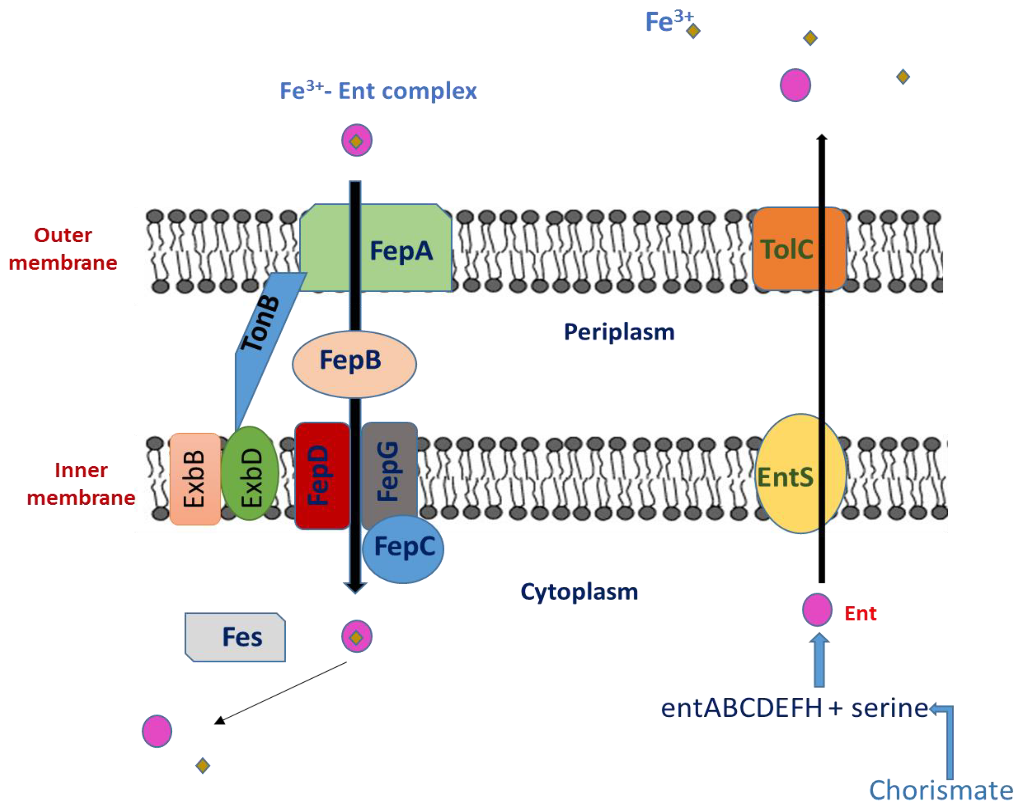
2.1. Enterochelin (Enterobactin)

It is known that macrophages increase a variety of iron-regulatory proteins (such as ferroportin) to fight bacterial infection [26]. These proteins prevent pathogens from accessing the labile iron pool [27]. To sequester iron from the host iron-binding proteins, Salmonella is prompted by the lack of iron to express enterobactin (Ent), a catecholate siderophore with a high affinity for ferric iron (Kd of 10−49 M) [23,24]. Siderophores are small iron chelators that facilitate iron transport into the bacterial cells. A triscatechol derivative of a cyclic triserine lactone gives rise to the siderophore enterobactin [28]. The structure of Ent is illustrated in Figure 1.

The Biosynthesis of Enterobactin

Thirteen proteins that are present in numerous species of Gram-negative enteric bacteria, including E. coli, S. enterica, Shigella dysenteriae (S. dysenteriae), and Klebsiella pneumoniae (K. pneumoniae) are encoded by a 24-kb gene cluster [29]. These thirteen proteins work together to produce, transport, and process the siderophore enterobactin. The operon entABCDEFH is involved in the biosynthesis of enterobactin from chorismic acid, and EntS and TolC are involved in Ent export through the cytoplasmic membrane. The two-module nonribosomal peptide synthetase (NRPS), which consists of EntE, EntB, and EntF, produces Ent (2,3-dihydroxybenzoylserine trilactone) from 2,3-dihydroxybenzoic acid (DHB) and serine [30]. EntD catalyzes the post-translational 4′-phosphopantetheinylation of EntB and EntF, a process necessary for the covalent attachment of assembly line intermediates during the production of Ent [31,32]. EntC, EntB, and EntA operate sequentially to divert the principal metabolite chorismate to DHB. Ent is exported across the inner membrane by EntS [33], a significant facilitator-subfamily exporter, and through the outer membrane by a TolC-dependent mechanism after being synthesized in the cytoplasm [34].
Given its very high affinity for Fe3+ (Kd = 10−35 M at physiological pH, [35]), Ent may successfully compete for Fe3+ binding outside of the cell with all known protein and small-molecule ligands. The Fe3+–Ent complex is then transported into the cell through the TonB-dependent outer membrane Ent-specific porin FepA [36,37], escorted through the periplasm by FepB, and pumped into the cytoplasm through the two-protein inner membrane channel FepDG into the cytoplasm via FepC-catalyzed ATP hydrolysis [38] (Figure 2). Before the tightly bound ferric iron can be transported to intracellular iron carriers, Fe3+ from Ent must first be released by the esterase Fes activity [39], which must also enzymatically degrade its trilactone scaffold to three equivalents of DHB-Ser. The iron-dependent repressor Fur (Fe uptake regulation), which acts as a sensor for intracellular iron by dissociating from its DNA-binding site when iron is deficient [40], regulates transcription of the entire Ent system. As a result, when iron levels are low, the transcription of the Ent synthesis, export, and import genes is stimulated, and it is suppressed when iron levels are high.

2.2. Interaction between Ent and the Immune System

2.2.1. Effect of Ent on Macrophages

Iron is essential for the redox activity of heme proteins expressed in both immune and non-immune cells. A study was conducted by Yeoh et al. to investigate the potential impact of enterobactin’s iron chelation on macrophage nitrosative and immunological responses. Ent was found to reduce the LPS-induced release of cytokines (such as serum amyloid A, IL-6, and lipocalin 2) and nitrite in macrophages in a dose-dependent manner [41]. Ent also suppressed the mRNA and protein expression of inducible nitric oxide synthase (iNOS; a heme protein) that is stimulated by LPS [26]. They also investigated if Ent might shield the intracellular pathogen Salmonella enterica spp. typhimurium in the gentamycin protection assay to show the physiological significance of these results. Ent increased the expression of iNOS (mRNA, protein) and Arginase-1 (mRNA), but it decreased the nitrite levels in Salmonella-infected macrophages. More critically, Salmonella eradication by macrophages treated with Ent was compromised. The addition of exogenous Ent significantly increased the longevity of both strains, and Ent-sufficient Salmonella also outlived their isogenic Ent-deficient counterparts. However, Ent’s inhibitory activities were negated when saturated with iron (1:1 ratio), demonstrating that Ent must be in its iron-free form to effectively inhibit macrophages. These data support the idea that Ent protects bacteria against immunological reactions from macrophages in addition to facilitating bacterial iron absorption [42,43].

2.2.2. The Interaction between Siderocalin and Enterobactin
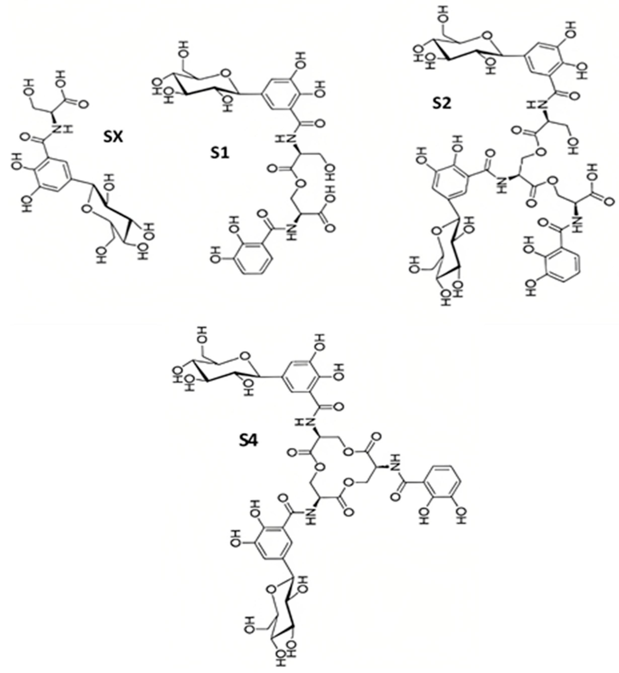
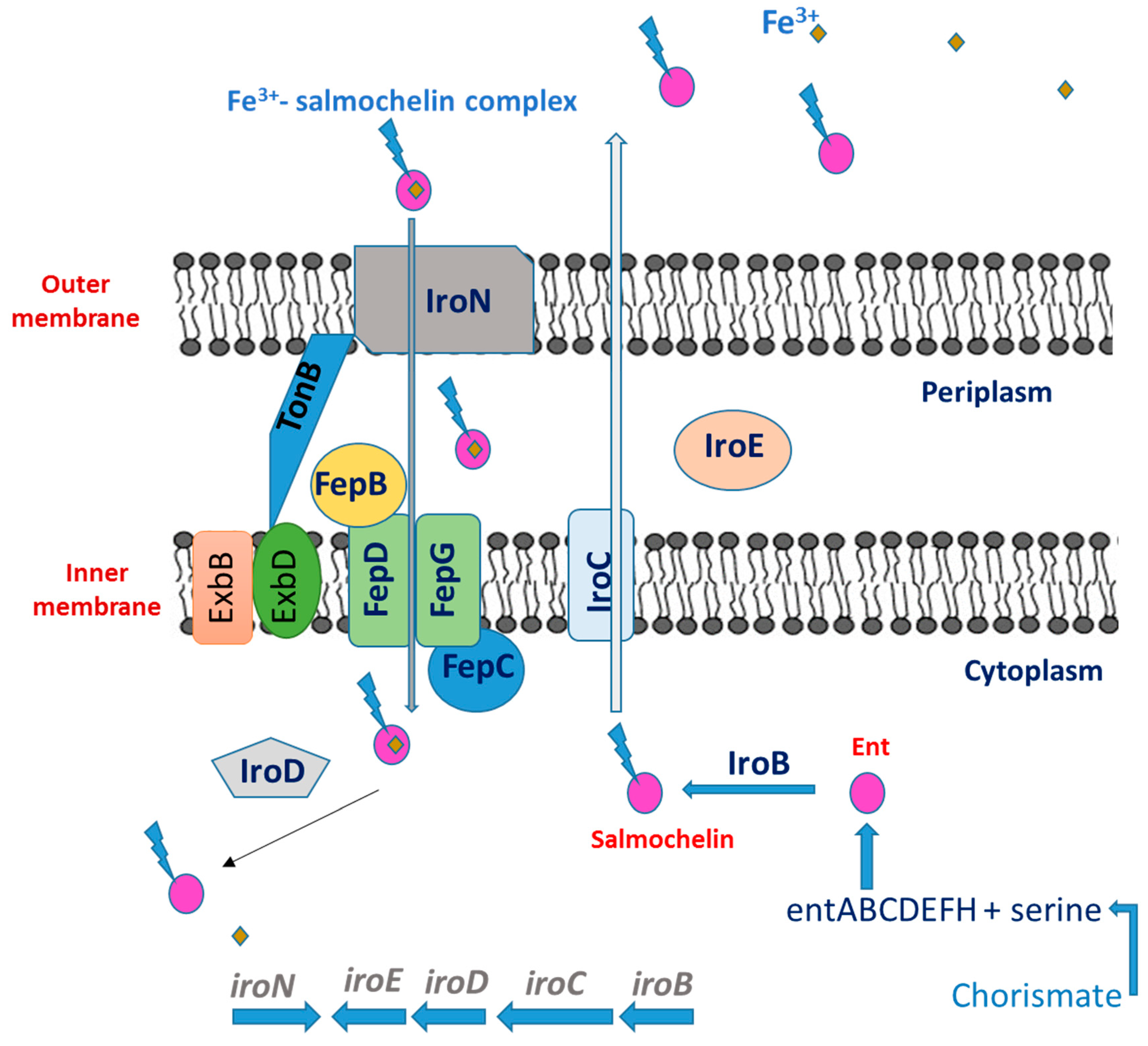
Withholding necessary iron from invading germs has long been recognized as a critical host defensive mechanism [44]. Increasing the expression of transferrin, lactoferrin receptors, and ferritin and decreasing the level of extracellular iron in serum are common bacteriostatic responses. The mammalian protein Siderocalin (Scn), also known as Lcn2, neutrophil-gelatinase-associated lipocalin, 24p3, or uterocalin, is used in a more focused approach [36]. Scn is generated and released in response to the activation of innate immune receptors, such as Toll-like receptors, in a variety of cell types. Scn is naturally present in neutrophil granules. Successful pathogens generate alternate or modified siderophores that Scn does not bind toin order to circumvent this defense. This supplements the overall antibacterial iron-depletion response and prevents major early bacteremia [45]. Numerous microbes produce the classic 2,3-catecholate siderophore enterobactin (Ent), which was the first recognized target of Scn [45]. The protein binds to and sequesters the ferric siderophore complex with an affinity for [Fe3(Ent)]3−, acting as a growth inhibitor of pathogens that solely rely on Ent-mediated iron uptake similar to that of FepA, the cognate outer membrane receptor [46]. Scn is distinct from other host iron-binding proteins in that it is selective for iron intended for bacterial usage as a ferric siderophore complex and does not chelate iron directly. Scn has been linked to innate immunity as well as kidney development [47]. As an iron-donating molecule, it transports iron to the cytoplasm, where it may activate or repress genes that respond to iron [48]. Therefore, Scn may be a substitute for transferrin in the transport of iron and be crucial for the development of tissues and organs. Scn may also function at a stage of development before transferrin circulation and the expression of transferrin receptors are established [49].

2.3. Salmochelins

In addition to Ent, Salmonella secretes other siderophores salmochelins (SX, S1, S2 and S4). Lipocalin-2, a siderophore-capturing protein, is secreted by macrophages in response to gamma interferon (IFN-γ) during infection [50,51]. Enterobactin is bound by lipocalin-2, while salmochelin is not. The latter is a glycosylated enterobactin derivative that favors Salmonella’s ability to grow specifically in the intestine [49]. Moreover, Ent is bound to serum albumin thus its effectiveness decreases [52]. In addition, the hydrophobicity of its three catechol acyl ‘arms’, which allow Ent to partition into lipid bilayers, further reduces the amount of Fe3+–Ent available for bacterial cell import [53].

Salmochelin Biosynthesis

Salmochelins are enterobactin-related substances that contain a 5-C-glucosylated 2,3-dihydroxybenzoyl residue (DHB) (Figure 3). A twofold β-C-glucosylated enterobactin analogue is the main substance of salmochelin S4 [54]. Salmochelin S2 has one unglycosylated DHB-serine moiety at the C-terminal end. Additionally, salmochelin SX is a monomeric DHB(glucosyl)-serine molecule, and salmochelin S1 is a dimer with a DBH(glucosyl)-seryl-DHB-serine constitution [54].
The enterobactin biosynthesis machinery genes and the iro genes, iroBCDE and iroN [54,55], are needed for salmochelins’ synthesis and transport. IroC is involved in the cell’s production of salmochelins, while IroB is the glucosyl transferase that attaches the glucose moieties to enterobactin [56]. Salmochelins must be transported into the cell via the outer membrane receptor IroN since the enterobactin receptor, FepA, is unable to identify them [56]. Both the FepBCDG system [57] and IroC [58] have been found to be involved in the uptake of salmochelin across the cytoplasmic membrane, suggesting that they may be involved in both the export and import of salmochelin. The siderophore is hydrolyzed by IroD and IroE [59]. While the periplasmic IroE cleaves the apo-siderophore to yield linear versions of the salmochelins, the cytosolic IroD prefers the ferric-salmochelins as a substrate [60].
The ent genes are arranged in six clusters with three sets of divergent promoters. These promoter regions include several Fur binding sites, and iron adversely regulates the operons. The iroBCDE operon [57] and iroN [61] both have overlapping 10-sequence binding sites for Fur. Additional regulators connect the expression of these genes to the bacterial cell’s overall metabolism. Between fepA and fes, the oxygen-controlled regulator FNR has a binding site, and it is anticipated that this site will function as a positive regulator. This region and the promoter region between fepB and entC both contain Crp binding sites, which connect expression to carbon metabolism [62].
The ferric uptake regulator (Fur) has control over the expression of the iroB gene. The promoter region of iroBCDE contains the Fur box. An in vitro transcription test revealed that Fur inhibited the expression of iroB in the presence of iron. As a result, Salmonella’s iron uptake system is controlled in a Fur-dependent manner [63].
To summarize, the iroA gene cluster, which consists of five genes called iroB, iroC, iroD, iroE, and iroN, is necessary for the conversion of Ent into salmochelins like S1, S2, and S4. The IroN protein was discovered to be an outer membrane receptor for the absorption of Fe3+-bound salmochelins and shares similarity with the FepA protein. IroN is also capable of recognizing a number of additional siderophores, just like FepA and Cir. IroC is believed to be an inner membrane transporter that aids in the apo siderophores’ export (Figure 4). It has been demonstrated that IroB catalyzes Ent’s C-glucosylation. IroD and IroE, the two remaining proteins, are similar to Fes. IroE is thought to be periplasmic, whereas IroD has been anticipated to be cytoplasmic. IroD and IroE are likely salmochelin esterases since bacteria with an iroA cluster produce less of the hydrolyzed salmochelins (S1, S2, and SX) than bacteria without them [64]. Salmochelin can be linearized in vitro to the following compounds: linear trimer (linearized TGE/S3), linear dimer (DGE/S2), MGE trimer, linear C-glycosylated (DBS)2 (S1), and linear monomer (SX) [41]. Additionally, the researchers demonstrated that the cytoplasmic esterase IroD can break down salmochelin forms into their constituent parts (DBS), releasing the iron into the bacterial cytoplasm [42].

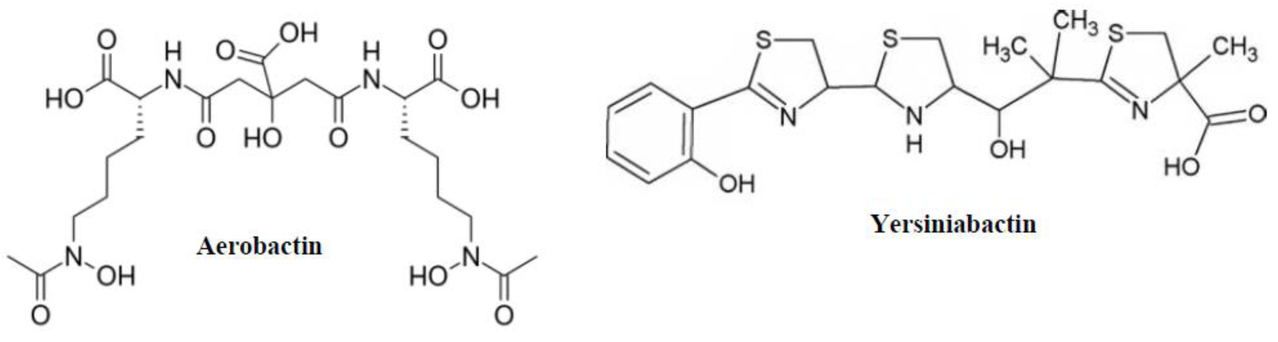
2.4. Aerobactin and Yersiniabactin

Some non-typhoidal Salmonella serovars (Salmonella enterica subsp. enterica serovar Kentucky) produce aerobactin, a mixed siderophore of the citrate-hydroxamate type (Figure 5). Enzymes from the iucABCD operon are used in an NRPS (nonribosomal peptide synthesis) pathway to produce aerobactin. L-lysine is first transformed into N6-acetyl-N6-hydroxy-l-lysine during synthesis, and then it is complexed into a citric acid backbone [43]. Aerobactin has a lower iron complex formation constant (Kf = 1023) than enterobactin (Kf = 1049) [58]. Similar to catecholate-type siderophores, aerobactin is taken up by the Iut receptor and transported into the bacterial periplasm in a TonB-dependent manner. Aerobactin is transported by the binding-protein-dependent ABC transport system FhuBCD [65] once it has reached the periplasm. Additionally, FhuBCD mediates the environment’s energy-dependent absorption of ferrichromes and coprogen.
Phenolate-type siderophores, including yersiniabactin (Ybt) (Figure 5), are a less frequent class of siderophores that can be detected in Salmonella serovars. The genomic island known as high pathogenicity island 1 (HPI) encodes Ybt, which is widely generated by Yersinia species [66]. Its distribution in Salmonella serovars is minimal since HPI 1 is absent from the majority of Salmonella enterica serovar subspecies 1 [66]. In Yersinia species, the synthesis of Ybt from the precursor isochorismic acid has been characterized as involving seven proteins (HMWP1, HMWP2, YbtD, YbtE, YbtS, YbtT, and YbtU). Salicylate, one thiazolidine, and two thiazoline rings make up the end product’s four rings. Ybt is a powerful iron chelator because it exhibits a higher anisotropy for Fe3+ (Kf = 1036) than aerobactin does. Yersiniabactin is picked up by the Psn/FyuA receptor in the outer membrane and then transported across the inner membrane by the YbtPQ ABC transporter once it has been loaded with iron [67].

2.5. The Function of Siderophores
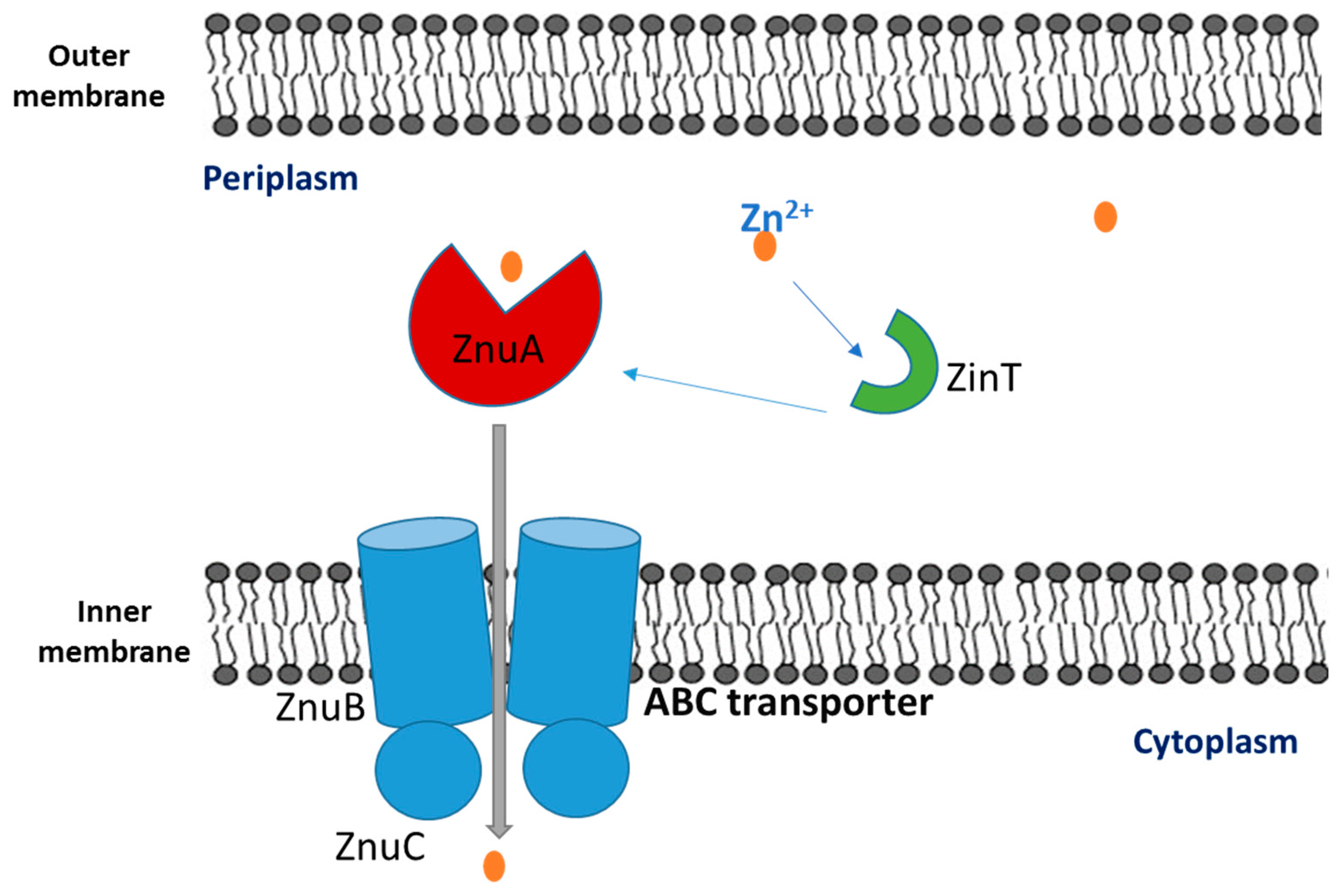
One of the most effective defenses used by innate immune cells to combat bacterial infection is the production of reactive oxygen species (ROS) by the NADPH membrane oxidase complex 2 (Nox2). Reactive oxygen species are produced by this enzyme complex and are discharged by phagocytes into phagosomes as well as the extracellular environment [68]. Phagocytes create hypochlorous acid, superoxide, and hydrogen peroxide, which all have the ability to kill microbes [69], though the exact methods by which they do so are still unclear. Superoxide converts to hydrogen peroxide, which is antibacterial and can pass bacterial membranes. In the Fenton reaction, H2O2 interacts with intracellular iron sulfur clusters to create hazardous free radicals, such as hydroxyl radicals [70], which can harm cellular constituents such as proteins, lipids, and DNA. Bacterial pathogens have a variety of defenses against and responses to ROS damage. Recently, it was discovered that catecholate siderophores helped shield S. Typhimurium from oxidative stress [71]. In addition to serving as an iron chelator, siderophores also assist bacterial colonization, quorum sensing, and the development of biofilms, among other noncanonical roles [72].

3. Zincophores

Because zinc is essential for numerous biological processes, bacteria have developed a number of strategies to deal with zinc shortages and obtain this metal from their hosts. The transcriptional regulator Zur in Gram-negative bacteria regulates the expression of a select few genes (znuABC and zupT) needed to help the cell cope with extreme zinc deficiency [73]. The zinc-containing version of Zur firmly binds to the promoter region of the aforementioned genes under zinc-replete circumstances, suppressing their expression. Conversely, the zinc-deficient version of Zur ceases to inhibit transcription when the intracellular zinc concentration falls below a crucial threshold [73].
Zur-regulated genes vary between different species of bacteria, but they are always found to encode one or more paralogs of zinc-containing ribosomal proteins as well as the various subunits of a high affinity zinc importer (ZnuABC in Gram-negative bacteria) [74,75]. Under various environmental circumstances with low availability of this metal, ZnuABC considerably improves the capacity of bacteria to recruit zinc. The discovery that ZnuABC is essential for bacterial pathogenicity suggests that zinc availability in infected hosts is constrained [76]. A periplasmic-binding protein, an ATPase, and an integral membrane protein are all encoded by the znuA-C operons, respectively. The Zur protein, a member of the Fur family of metalloregulatory proteins, binds to a nearly complete palindrome in this region when Zn is present, preventing the transcription of both znuA and znuCB [77].
Salmonella enterica offers secondary metal binding sites that can bind Zn2+ [78,79]. The N-terminal histidine-rich loop of ZnuA has been shown to play a critical role in the formation of the protein binary complex between SenZnuA and the periplasmic zinc-binding protein SenZinT (a 216-amino acid periplasmic protein), where it embeds into a structural cavity of the partner protein [77]. The loop can also be involved in specific protein–protein interactions and potential Zn2+ acquisition from additional metallochaperones [80].
Under zinc-limiting conditions, ZinT transports Zn2+ ions to the ZnuABC transporter. It is possible that metal transfer from ZinT to ZnuA (Figure 6) occurs during the interaction of the two proteins through the formation of a binary complex since ZnuA, a periplasmic component of the ZnuABC transporter, has a higher affinity for Zn2+ than ZinT [81].
ZnuA gene-deficient strains of S. Typhimurium cause systemic and gastrointestinal infections in mice that are much less severe [82]. As no additional attenuation is shown in mutant strains where the full znuABC operon is removed, deletion of the znuA gene, which encodes for the periplasmic component ZnuA, is sufficient to impair the function of the ZnuABC transporter [82]. S. Typhimurium avoids the antimicrobial effects of calprotectin, a zinc-sequestering protein secreted by neutrophils during infection in the intestinal lumen, by generating the ZnuABC transporter [82]. A strain that lacks a functional ZnuABC transporter cannot compete with the microbiota in this host environment and is destroyed by the inflammatory reaction [83].

4. Up Taking Fungal Siderophores

Both the mycobiota (the fungal gut microbiota) and the fungus in food can provide siderophores. A recent study revealed that Salmonella strains expressing the fungus siderophore receptors FhuA or FhuE, in vitro and in a mouse model, showed a competitive growth advantage due to their capacity to utilize fungus siderophores such ferrichrome and coprogen. The importance of these little-studied components of the gut ecosystem during bacterial infection elucidates the role of inter-kingdom cross-feeding between fungus and Salmonella [84,85].

5. Conclusions

For the majority of bacteria, iron is an important micronutrient. Depending on the availability and source, different Salmonella enterica strains, that cause human and animal infections have developed methods, sequester iron from the environment. Since Fe3+ is insoluble, it is frequently bound in complexes (Fe(OH)3) outside the host or sequestered by host proteins (such as lactoferrin, transferrin, and hemoglobin). Salmonella secretes siderophores, high-anity iron-binding molecules, to sequester Fe3+ from the extracellular medium. Enterobactin and salmochelin are two well-characterized siderophores of the catecholate type. The ferric ion and enterobactin form an extraordinarily stable combination. IroB, a glycosyl transferase enzyme, can glycosylate enterobactin to produce salmochelin which is considered a better iron chelator than enterobactin in presence of serum albumin. Salmonella serovars also produce yersiniabactin which is another type of siderophore but less common than enterobactin and salmochelin. Concerning zinc ions, Salmonella produces periplasmic proteins for zinc uptake through its inner membrane. Finally, some Salmonella species are capable of taking up fungal siderophores.

Author Contributions

G.G. conceived this work and managed the manuscript writing; Z.E. wrote the review; Y.M. and N.T. conducted the literature search; R.B. participated in the writing process; R.G. and I.K. edited the paper. All authors have read and agreed to the published version of the manuscript.

Funding

This research was performed without any external funding.

Institutional Review Board Statement

Not applicable.

Informed Consent Statement

Not applicable.

Data Availability Statement

No data were reported in this study.

Acknowledgments

This work was carried out with the support of the Islamic University of Lebanon, Center of Research and Development.

Conflicts of Interest

The authors declare no conflict of interest.

References

  1. Fardsanei, F.; Dallal, M.M.S.; Douraghi, M.; Memariani, H.; Bakhshi, B.; Salehi, T.Z.; Nikkhahi, F. Antimicrobial resistance, virulence genes and genetic relatedness of Salmonella enterica serotype Enteritidis isolates recovered from human gastroenteritis in Tehran, Iran. J. Glob. Antimicrob. Resist. 2018, 12, 220–226. [Google Scholar] [CrossRef] [PubMed]
  2. Lou, L.; Zhang, P.; Piao, R.; Wang, Y. Salmonella Pathogenicity Island 1 (SPI-1) and Its Complex Regulatory Network. Front. Cell. Infect. Microbiol. 2019, 9, 270. [Google Scholar] [CrossRef]
  3. Takaya, A.; Yamamoto, T.; Tokoyoda, K. Humoral Immunity vs. Salmonella. Front. Immunol. 2020, 10, 3155. [Google Scholar] [CrossRef]
  4. He, Y.; Wang, J.; Zhang, R.; Chen, L.; Zhang, H.; Qi, X.; Chen, J. Epidemiology of foodborne diseases caused by Salmonella in Zhejiang Province, China, between 2010 and 2021. Front. Public Health 2023, 11, 1127925. [Google Scholar] [CrossRef]
  5. Foley, S.L.; Johnson, T.J.; Ricke, S.C.; Nayak, R.; Danzeisen, J. Salmonella pathogenicity and host adaptation in chicken-associated serovars. Microbiol. Mol. Biol. Rev. 2013, 77, 582–607. [Google Scholar] [CrossRef]
  6. Herekar, F.; Sarfaraz, S.; Imran, M.; Ghouri, N.; Shahid, S.; Mahesar, M. Clinical spectrum and outcomes of patients with different resistance patterns of Salmonella enterica. Pak. J. Med. Sci. 2022, 38, 356–361. [Google Scholar] [CrossRef]
  7. Rabsch, W.; Tschäpe, H.; Bäumler, A.J. Non-typhoidal salmonellosis: Emerging problems. Microbes Infect. 2001, 3, 237–247. [Google Scholar] [CrossRef] [PubMed]
  8. Uche, I.V.; MacLennan, C.A.; Saul, A. A systematic review of the incidence, risk factors and case fatality rates of invasive nontyphoidal Salmonella (iNTS) disease in Africa (1966 to 2014). PLoS Negl. Trop. Dis. 2017, 11, e0005118. [Google Scholar] [CrossRef] [PubMed]
  9. Ezzeddine, Z.; Ghssein, G. Towards new antibiotics classes targeting bacterial metallophores. Microb. Pathog. 2023, 29, 06221. [Google Scholar] [CrossRef]
  10. Awada, R.; Ghssein, G.; Roz, A.; Farhat, M.; Nehme, N.; Hassan, H.F. Prevalence of Campylobacter spp. in broilers in North Lebanon. Vet. World 2023, 16, 322–328. [Google Scholar] [CrossRef]
  11. McDermott, P.F.; Zhao, S.; Wagner, D.D.; Simjee, S.; Walker, R.D.; White, D.G. The food safety perspective of antibiotic resistance. Anim. Biotechnol. 2002, 13, 71–84. [Google Scholar] [CrossRef] [PubMed]
  12. Rello, J.; Kalwaje Eshwara, V.; Conway-Morris, A.; Lagunes, L.; Alves, J.; Alp, E.; Zhang, Z.; Mer, M.; Investigators, T.S. Perceived differences between intensivists and infectious diseases consultants facing antimicrobial resistance: A global cross-sectional survey. Eur. J. Clin. Microbiol. Infect. Dis. 2019, 38, 1235–1240. [Google Scholar] [CrossRef]
  13. Bloom, D.E.; Cadarette, D. Infectious disease threats in the twenty-first century: Strengthening the global response. Front. Immunol. 2019, 10, 549. [Google Scholar] [CrossRef] [PubMed]
  14. Hancock, R.E.W.; Sahl, H.-G. Antimicrobial and host-defense peptides as new anti-infective therapeutic strategies. Nat. Biotechnol. 2006, 24, 1551–1557. [Google Scholar] [CrossRef] [PubMed]
  15. Majowicz, S.E.; Musto, J.; Scallan, E.; Angulo, F.J.; Kirk, M.; O’Brien, S.J.; Jones, T.F.; Fazil, A.; Hoekstra, R.M. International Collaboration on Enteric Disease ‘Burden of Illness’ Studies. The global burden of nontyphoidal Salmonella gastroenteritis. Clin. Infect. Dis. 2010, 50, 882–889. [Google Scholar] [CrossRef] [PubMed]
  16. Porcheron, G.; Garénaux, A.; Proulx, J.; Sabri, M.; Dozois, C.M. Iron, copper, zinc, and manganese transport and regulation in pathogenic Enterobacteria: Correlations between strains, site of infection and the relative importance of the different metal transport systems for virulence. Front. Cell. Infect. Microbiol. 2013, 3, 90. [Google Scholar] [CrossRef]
  17. Ma, Z.; Jacobsen, F.E.; Giedroc, D.P. Coordination chemistry of bacterial metal transport and sensing. Chem. Rev. 2009, 109, 4644–4681. [Google Scholar] [CrossRef]
  18. Fukada, T.; Yamasaki, S.; Nishida, K.; Murakami, M.; Hirano, T. Zinc homeostasis and signaling in health and diseases: Zinc signaling. J. Biol. Inorg. Chem. 2011, 16, 1123–1134. [Google Scholar] [CrossRef]
  19. Hood, M.I.; Skaar, E.P. Nutritional immunity: Transition metals at the pathogen-host interface. Nat. Rev. Microbiol. 2012, 10, 525–537. [Google Scholar] [CrossRef]
  20. Grüngreiff, K.; Reinhold, D.; Wedemeyer, H. The role of zinc in liver cirrhosis. Ann. Hepatol. 2016, 15, 7–16. [Google Scholar] [CrossRef]
  21. Abbaspour, N.; Hurrell, R.; Kelishadi, R. Review on iron and its importance for human health. J. Res. Med. Sci. 2014, 19, 164–174. [Google Scholar] [PubMed]
  22. Capdevila, D.A.; Edmonds, K.A.; Giedroc, D.P. Metallochaperones and metalloregulation in bacteria. Essays Biochem. 2017, 61, 177–200. [Google Scholar]
  23. Ghssein, G.; Ezzeddine, Z. The Key Element Role of Metallophores in the Pathogenicity and Virulence of Staphylococcus aureus: A Review. Biology 2022, 11, 1525. [Google Scholar] [CrossRef] [PubMed]
  24. Ghssein, G.; Matar, S.F. Chelating Mechanisms of Transition Metals by Bacterial Metallophores “Pseudopaline and Staphylopine”: A Quantum Chemical Assessment. Computation 2018, 6, 56. [Google Scholar] [CrossRef]
  25. Chaaban, T.; Mohsen, Y.; Ezzeddine, Z.; Ghssein, G. Overview of Yersinia pestis Metallophores: Yersiniabactin and Yersinopine. Biology 2023, 12, 598. [Google Scholar] [CrossRef] [PubMed]
  26. Saha, P.; Xiao, X.; Yeoh, B.S.; Chen, Q.; Katkere, B.; Kirimanjeswara, G.S.; Vijay-Kumar, M. The bacterial siderophore enterobactin confers survival advantage to Salmonella in macrophages. Gut Microbes 2019, 10, 412–423. [Google Scholar] [CrossRef]
  27. Fischbach, M.A.; Lin, H.; Liu, D.R.; Walsh, C.T. How pathogenic bacteria evade mammalian sabotage in the battle for iron. Nat. Chem. Biol. 2006, 2, 132–138. [Google Scholar] [CrossRef]
  28. Loomis, L.D.; Raymond, K.N. Solution equilibria of enterobactin and metal enterobactin complexes. Inorg. Chem. 1991, 30, 906–911. [Google Scholar] [CrossRef]
  29. Raymond, K.N.; Dertz, E.A.; Kim, S.S. Enterobactin: An archetype for microbial iron transport. Proc. Natl. Acad. Sci. USA 2003, 100, 3584–3588. [Google Scholar] [CrossRef]
  30. Gehring, A.M.; Mori, I.; Walsh, C.T. Reconstitution and characterization of the Escherichia coli enterobactin synthetase from EntB, EntE, and EntF. Biochemistry 1998, 37, 2648–2659. [Google Scholar] [CrossRef]
  31. Walsh, C.; Liu, J.; Rusnak, F.; Sakaitani, M. Molecular studies on enzymes in chorismate metabolism and the enterobactin biosynthetic pathway. Chem. Rev. 1990, 90, 1105–1129. [Google Scholar] [CrossRef]
  32. Lambalot, R.H.; Gehring, A.M.; Flugel, R.S.; Zuber, P.; LaCelle, M.; Marahiel, M.A.; Walsh, C.T. A new enzyme superfamily—The phosphopantetheinyl transferases. Chem. Biol. 1996, 3, 923–936. [Google Scholar] [CrossRef]
  33. Furrer, J.L.; Sanders, D.N.; Hook-Barnard, I.G.; McIntosh, M.A. Export of the siderophore enterobactin in Escherichia coli: Involvement of a 43 kDa membrane exporter. Mol. Microbiol. 2002, 44, 1225–1234. [Google Scholar] [CrossRef]
  34. Bleuel, C.; Große, C.; Taudte, N.; Scherer, J.; Wesenberg, D.; Krauß, G.J.; Grass, G. TolC is involved in enterobactin efflux across the outer membrane of Escherichia coli. J. Bacteriol. 2005, 187, 6701–6707. [Google Scholar] [CrossRef] [PubMed]
  35. Crosa, J.H.; Mey, A.R.; Payne, S.M. Iron Transport in Bacteria; ASM Press: Washington, DC, USA, 2004. [Google Scholar]
  36. Annamalai, R.; Jin, B.; Cao, Z.; Newton, S.M.; Klebba, P.E. Recognition of ferric catecholates by FepA. J. Bacteriol. 2004, 186, 3578–3589. [Google Scholar] [CrossRef]
  37. Buchanan, S.K.; Smith, B.S.; Venkatramani, L.; Xia, D.; Esser, L.; Palnitkar, M.; Deisenhofer, J. Crystal structure of the outer membrane active transporter FepA from Escherichia coli. Nat. Struct. Biol. 1999, 6, 56–63. [Google Scholar] [PubMed]
  38. Faraldo-Gomez, J.D.; Sansom, M.S. Acquisition of siderophores in gram-negative bacteria. Nat. Rev. Mol. Cell Biol. 2003, 4, 105–116. [Google Scholar] [CrossRef] [PubMed]
  39. Lin, H.; Fischbach, M.A.; Liu, D.R.; Walsh, C.T. In vitro characterization of salmochelin and enterobactin trilactone hydrolases IroD, IroE, and Fes. J. Am. Chem. Soc. 2005, 127, 11075–11084. [Google Scholar] [CrossRef] [PubMed]
  40. Hantke, K. Iron and metal regulation in bacteria. Curr. Opin. Microbiol. 2001, 4, 172–177. [Google Scholar] [CrossRef]
  41. Yeoh, B.S.; Saha, P.; Xiao, X.; Singh, V.; Vijay-Kumar, M. Enterobactin, a metallophore, mitigates the immune responses of macrophages. J. Immunol. 2017, 198 (Suppl. S1), 113–121. [Google Scholar] [CrossRef]
  42. Ong, S.T.; Ho, J.Z.S.; Ho, B.; Ding, J.L. Iron-withholding strategy in innate immunity. Immunobiology 2006, 211, 295–314. [Google Scholar] [CrossRef] [PubMed]
  43. Devireddy, L.R.; Teodoro, L.R.; Richard, F.A.; Green, M.R. Induction of apoptosis by a secreted lipocalin that is transcriptionally regulated by IL-3 deprivation. Science 2001, 293, 829–834. [Google Scholar] [CrossRef]
  44. Goetz, D.H.; Holmes, M.A.; Borregaard, N.; Bluhm, M.E.; Raymond, K.N.; Strong, R.K. The neutrophil lipocalin NGAL is a bacteriostatic agent that interferes with siderophore-mediated iron acquisition. Mol. Cell 2002, 10, 1033–1043. [Google Scholar] [CrossRef] [PubMed]
  45. Yang, J.; Mori, K.; Li, J.Y.; Barasch, J. Iron, lipocalin, and kidney epithelia. Am. J. Phys. Renal Phys. 2003, 285, F9–F18. [Google Scholar]
  46. Yang, J.; Goetz, D.H.; Li, J.Y.; Wang, W.; Mori, K.; Setlik, D.; Du, T.; Erdjument-Bromage, H.; Tempst, P.; Strong, R.K.; et al. An iron delivery pathway mediated by a lipocalin. Mol. Cell 2002, 10, 1045–1056. [Google Scholar] [CrossRef]
  47. Kaplan, J. Mechanisms of cellular iron acquisition: Another iron in the fire. Cell 2002, 111, 603–606. [Google Scholar] [CrossRef]
  48. Flo, T.H.; Smith, K.D.; Sato, S.; Rodriguez, D.J.; Holmes, M.A.; Strong, R.K.; Akira, S.; Aderem, A. Lipocalin 2 mediates an innate immune response to bacterial infection by sequestrating iron. Nature 2004, 432, 917–921. [Google Scholar] [CrossRef]
  49. Nairz, M.; Fritsche, G.; Brunner, P.; Talasz, H.; Hantke, K.; Weiss, G. Interferon-gamma limits the availability of iron for intramacrophage Salmonella typhimurium. Eur. J. Immunol. 2008, 38, 1923–1936. [Google Scholar] [CrossRef]
  50. Raffatellu, M.; George, M.D.; Akiyama, Y.; Hornsby, M.J.; Nuccio, S.P.; Paixao, T.A.; Butler, B.P.; Chu, H.; Santos, R.L.; Berger, T.; et al. Lipocalin-2resistance confers an advantage to Salmonella enterica serotype Typhimurium for growth and survival in the inflamed intestine. Cell Host Microbe 2009, 5, 476–486. [Google Scholar] [CrossRef]
  51. Hantke, K.; Nicholson, G.; Rabsch, W.; Winkelmann, G. Salmochelins, siderophores of Salmonella enterica and uropathogenic Escherichia coli strains, are recognized by the outer membrane receptor IroN. Proc. Natl. Acad. Sci. USA 2003, 100, 3677–3682. [Google Scholar] [CrossRef]
  52. Konopka, K.; Neilands, J.B. Effect of serum albumin on siderophore-mediated utilization of transferrin iron. Biochemistry 1984, 23, 2122–2127. [Google Scholar] [CrossRef] [PubMed]
  53. Luo, M.; Lin, H.; Fischbach, M.A.; Liu, D.R.; Walsh, C.T.; Groves, J.T. Enzymatic tailoring of the bacterial siderophore enterobactin alters membrane partitioning and iron acquisition. ACS Chem. Biol. 2006, 1, 29–32. [Google Scholar] [CrossRef]
  54. Bister, B.; Bischoff, D.; Nicholson, G.J.; Valdebenito, M.; Schneider, K.; Winkelmann, G.; Hantke, K.; Süssmuth, R.D. The structure of salmochelins: C-glucosylated enterobactins of Salmonella enterica. Biometals 2004, 17, 471–481. [Google Scholar] [CrossRef] [PubMed]
  55. Bäumler, A.J.; Tsolis, R.M.; van der Velden, A.W.M.; Stojiljkovic, I.; Anic, S.; Heffron, F. Identification of a new iron regulated locus of Salmonella typhi. Gene 1996, 183, 207–213. [Google Scholar] [CrossRef] [PubMed]
  56. Müller, S.I.; Valdebenito, M.; Hantke, K. Salmochelin, the long-overlooked catecholate siderophore of Salmonella. Biometals 2009, 22, 691–695. [Google Scholar] [CrossRef] [PubMed]
  57. Fischbach, M.A.; Lin, H.; Liu, D.R.; Walsh, C.T. In vitro characterization of IroB, a pathogen-associated C-glycosyltransferase. Proc. Natl. Acad. Sci. USA 2005, 102, 571–576. [Google Scholar] [CrossRef]
  58. Crouch, M.L.V.; Castor, M.; Karlinsey, J.E.; Kalhorn, T.; Fang, F.C. Biosynthesis and IroC-dependent export of the siderophore salmochelin are essential for virulence of Salmonella enterica serovar Typhimurium. Mol. Microbiol. 2008, 67, 971–983. [Google Scholar] [CrossRef]
  59. Bäumler, A.J.; Norris, T.L.; Lasco, T.; Voight, W.; Reissbrodt, R.; Rabsch, W.; Heffron, F. IroN, a novel outer membrane siderophore receptor characteristic of Salmonella enterica. J. Bacteriol. 1998, 180, 1446–1453. [Google Scholar] [CrossRef]
  60. Zhu, M.; Valdebenito, M.; Winkelmann, G.; Hantke, K. Functions of the siderophore esterases IroD and IroE in iron-salmochelin utilization. Microbiology 2005, 151, 2363–2372. [Google Scholar] [CrossRef]
  61. Lim, D.; Kim, K.; Song, M.; Jeong, J.H.; Chang, J.H.; Kim, S.R.; Hong, C.; Im, S.S.; Park, S.H.; Lee, J.C.; et al. Transcriptional regulation of Salmochelin glucosyltransferase by Fur in Salmonella. Biochem. Biophys. Res. Commun. 2020, 529, 70–76. [Google Scholar] [CrossRef]
  62. Balbontín, R.; Villagra, N.; Pardos de la Gándara, M.; Mora, G.; Figueroa-Bossi, N.; Bossi, L. Expression of IroN, the salmochelin siderophore receptor, requires mRNA activation by RyhB small RNA homologues. Mol. Microbiol. 2016, 100, 139–155. [Google Scholar] [CrossRef]
  63. Zhang, Z.; Gosset, G.; Barabote, R.; Gonzalez, C.S.; Cuevas, W.A.; Saier, M.H. Functional interactions between the carbon and iron utilization regulators, Crp and Fur, in Escherichia coli. J. Bacteriol. 2005, 187, 980–990. [Google Scholar] [CrossRef]
  64. Oves-Costales, D.; Kadi, N.; Challis, G.L. The long-overlooked enzymology of a nonribosomal peptide synthetase-independent pathway for virulence-conferring siderophore biosynthesis. Chem. Commun. 2009, 43, 6530–6541. [Google Scholar] [CrossRef]
  65. De Lorenzo, V.; Martinez, J. Aerobactin production as a virulence factor: A reevaluation. Eur. J. Clin.Microbiol. Infect. Dis. 1988, 7, 621–629. [Google Scholar] [CrossRef]
  66. Köster, W. ABC transporter-mediated uptake of iron, siderophores, heme and vitamin B12. Res. Microbiol. 2001, 152, 291–301. [Google Scholar] [CrossRef]
  67. Oelschlaeger, T.; Zhang, D.; Schubert, S.; Carniel, E.; Rabsch, W.; Karch, H.; Hacker, J. The high-pathogenicity island is absent in human pathogens of Salmonella enterica subspecies I but present in isolates of subspecies III and VI. J. Bacteriol. 2003, 185, 1107–1111. [Google Scholar] [CrossRef] [PubMed]
  68. Wellawa, D.H.; Allan, B.; White, A.P.; Köster, W. Iron-Uptake Systems of Chicken-Associated Salmonella Serovars and Their Role in Colonizing the Avian Host. Microorganisms 2020, 8, 1203. [Google Scholar] [CrossRef] [PubMed]
  69. Bogomolnaya, L.M.; Tilvawala, R.; Elfenbein, J.R.; Cirillo, J.D.; Andrews-Polymenis, H.L. Linearized Siderophore Products Secreted via MacAB Efflux Pump Protect Salmonella enterica Serovar Typhimurium from Oxidative Stress. mBio 2020, 11, e00528-20. [Google Scholar] [CrossRef] [PubMed]
  70. Winterbourn, C.C.; Kettle, A.J.; Hampton, M.B. Reactive oxygen species and neutrophil function. Annu. Rev. Biochem. 2016, 85, 765–792. [Google Scholar] [CrossRef]
  71. Imlay, J.A. Pathways of oxidative damage. Annu. Rev. Microbiol. 2003, 57, 395–418. [Google Scholar] [CrossRef]
  72. Ghssein, G.; Ezzeddine, Z. A Review of Pseudomonas aeruginosa Metallophores: Pyoverdine, Pyochelin and Pseudopaline. Biology 2022, 11, 1711. [Google Scholar] [CrossRef]
  73. Saha, P.; Yeoh, B.S.; Xiao, X.; Golonka, R.M.; Abokor, A.A.; Wenceslau, C.F.; Shah, Y.M.; Joe, B.; Vijay-Kumar, M. Enterobactin induces the chemokine, interleukin-8, from intestinal epithelia by chelating intracellular iron. Gut Microbes 2020, 12, 1841548. [Google Scholar] [CrossRef]
  74. Patzer, S.I.; Hantke, K. The ZnuABC high-affinity zinc uptake system and its regulator Zur in Escherichia coli. Mol. Microbiol. 1998, 28, 1199–1210. [Google Scholar] [CrossRef]
  75. Panina, E.M.; Mironov, A.A.; Gelfand, M.S. Comparative genomics of bacterial zinc regulons: Enhanced ion transport, pathogenesis, and rearrangement of ribosomal proteins. Proc. Natl. Acad. Sci. USA 2003, 100, 9912–9917. [Google Scholar] [CrossRef]
  76. Lewis, D.A.; Klesney-Tait, J.; Lumbley, S.R.; Ward, C.K.; Latimer, J.L.; Ison, C.A.; Hansen, E.J. Identification of the znuA-encoded periplasmic zinc transport protein of Haemophilus ducreyi. Infect. Immun. 1999, 67, 5060–5068. [Google Scholar] [CrossRef]
  77. Campoy, S.; Jara, M.; Busquets, N.; Pérez De Rozas, A.M.; Badiola, I.; Barbé, J. Role of the high-affinity zinc uptake znuABC system in Salmonella enterica serovar typhimurium virulence. Infect. Immun. 2002, 70, 4721–4725. [Google Scholar] [CrossRef] [PubMed]
  78. Wei, B.; Randich, A.M.; Bhattacharyya-Pakrasi, M.; Pakrasi, H.B.; Smith, T.J. Possible regulatory role for the histidine-rich loop in the zinc transport protein, ZnuA. Biochemistry 2007, 46, 8734–8743. [Google Scholar] [CrossRef] [PubMed]
  79. Yatsunyk, L.A.; Easton, J.A.; Kim, L.R.; Sugarbaker, S.A.; Bennett, B.; Breece, R.M.; Vorontsov, I.I.; Tierney, D.L.; Crowder, M.W.; Rosenzweig, A.C. Structure and metalbinding properties of ZnuA, a periplasmic zinc transporterfrom Escherichia coli. J. Biol. Inorg. Chem. 2008, 13, 271–288. [Google Scholar] [CrossRef]
  80. Ilari, A.; Alaleona, F.; Tria, G.; Petrarca, P.; Battistoni, A.; Zamparelli, C.; Verzili, D.; Falconi, M.; Chiancone, E. The Salmonella enterica ZinT structure, zinc affinity and interactionwith the high-affinity uptake protein ZnuA provideinsight into the management of periplasmic zinc. Biochim. Biophys. Acta 2014, 1840, 535–544. [Google Scholar] [CrossRef]
  81. Bellotti, D.; Rowińska-Żyrek, M.; Remelli, M. Novel insights into the metal binding ability of ZinT periplasmic protein from Escherichia coli and Salmonella enterica. Dalton Trans. 2020, 49, 9393–9403. [Google Scholar] [CrossRef] [PubMed]
  82. Petrarca, P.; Ammendola, S.; Pasquali, P.; Battistoni, A. The Zur-regulated ZinT protein is an auxiliary component of the high-affinity ZnuABC zinc transporter that facilitates metal recruitment during severe zinc shortage. J. Bacteriol. 2010, 192, 1553–1564. [Google Scholar] [CrossRef] [PubMed]
  83. Pesciaroli, M.; Aloisio, F.; Ammendola, S.; Pistoia, C.; Petrucci, P.; Tarantino, M.; Francia, M.; Battistoni, A.; Pasquali, P. An attenuated Salmonella enterica serovar Typhimurium strain lacking the ZnuABC transporter induces protection in a mouse intestinal model of Salmonella infection. Vaccine 2011, 29, 1783–1790. [Google Scholar] [CrossRef] [PubMed]
  84. Liu, J.Z.; Jellbauer, S.; Poe, A.J.; Ton, V.; Pesciaroli, M.; Kehl-Fie, T.E.; Restrepo, N.A.; Hosking, M.P.; Edwards, R.A.; Battistoni, A.; et al. Zinc sequestration by the neutrophil protein calprotectin enhances Salmonella growth in the inflamed gut. Cell Host Microbe 2012, 11, 227–239. [Google Scholar] [CrossRef]
  85. Mey, A.R.; Gómez-Garzón, C.; Payne, S.M. Iron Transport and Metabolism in Escherichia, Shigella, and Salmonella. EcoSal Plus 2021, 9, eESP00342020. [Google Scholar] [CrossRef] [PubMed]
Figure 1. Enterobactin’s structural illustration displaying the catechol, amide linkage, and triserine ring components.
Microbiolres 14 00099 g001
Figure 2. Ent export and Ent–Fe3+ complex import system in Salmonella.
Microbiolres 14 00099 g002
Figure 3. The structures of different salmochelins SX, S1, S2, and S4 secreted by S. enterica.
Microbiolres 14 00099 g003
Figure 4. Schematic illustration of the systems for salmochelin production, secretion, and uptake.
Microbiolres 14 00099 g004
Figure 5. The structures of aerobactin and yersiniabactin.
Microbiolres 14 00099 g005
Figure 6. Zn2+ uptake by ABC transporter-related zincophores in Salmonella enterica.
Microbiolres 14 00099 g006
Disclaimer/Publisher’s Note: The statements, opinions and data contained in all publications are solely those of the individual author(s) and contributor(s) and not of MDPI and/or the editor(s). MDPI and/or the editor(s) disclaim responsibility for any injury to people or property resulting from any ideas, methods, instructions or products referred to in the content.

