Article

Isolation and Characterization of Fungal Endophytes from Petiveria alliacea and Their Antimicrobial Activities in South Florida

by
Ganesh Khadka
1,
Thirunavukkarasu Annamalai
2,
Kateel G. Shetty
1,
Yuk-Ching Tse-Dinh
2 and
Krish Jayachandran
1,*
1
Department of Earth and Environment, Florida International University, Miami, FL 33199, USA
2
Department of Chemistry and Biochemistry, Florida International University, Miami, FL 33199, USA
*
Author to whom correspondence should be addressed.
Microbiol. Res. 2023, 14(3), 1470-1482; https://doi.org/10.3390/microbiolres14030100
Submission received: 31 July 2023 / Revised: 21 August 2023 / Accepted: 1 September 2023 / Published: 19 September 2023

Abstract

:
Microorganisms associated with medicinal plants are of great interest as they are the producers of important bioactive compounds effective against common and drug-resistant pathogens. The characterization and biodiversity of fungal endophytes of the Petiveria alliacea plant and their antimicrobial production potential are of great interest as they are known for their antimicrobial and anticancer properties. In this study, we investigated the endophytic fungal microbiome associated with P. alliacea, and the endophytic fungal isolates were classified into 30 morphotypes based on their cultural and morphological characteristics. Ethyl acetate extract of fungal endophytes was obtained by liquid–liquid partitioning of culture broth followed by evaporation. The crude extract dissolved in dimethyl sulfoxide was screened for antimicrobial activity against three bacterial strains (Escherichia coli ATTC 25902, Staphylococcus aureus ATTC 14775, Bacillus subtilis NRRL 5109) and two fungal strains (Candida albicans ATTC 10231 and Aspergillus fumigatus NRRL 5109). Among the crude extracts from endophytes isolated from leaves, 65% of them showed antimicrobial activity against the bacteria tested. Similarly, 71 and 88% of the fungal crude extracts from endophytes isolated from root and stem, respectively, showed inhibitory activities against at least one of the bacterial strains tested. Crude extracts (at a concentration of 10 mg/mL) from ten of the fungal isolates have shown a zone of inhibition of more than 12 mm against both Gram-positive and negative bacteria tested. Sequenced data from isolates showing strong inhibitory activity revealed that Fusarium solani, F. proliferatum, and Fusarium oxysporium are the major endophytes responsible for bioactive potential. These results indicate that Petiveria alliacea harbors fungal endophytes capable of producing antimicrobial metabolites. Future studies need to focus on testing against drug-resistant bacteria (ESKAPE group) and other pathogenic bacteria and fungi.

1. Introduction

Since ancient times, medicinal plants have been used as a primary source of medicine [1,2]. Even in these modern times, plant remedies are still the most important therapeutics to treat diseases [3]. According to the survey report of the World Health Organization, “Almost 80% of the world’s population from developing countries still rely on traditional medicine that involves the use of plant extract for their primary health care” [4,5]. Endophytes are an important component of the plant microbiome and are known to be highly abundant and diverse.
Endophytic fungi live asymptomatically within the host plant tissues [6,7]. They can be found in a wide range of host plants, and there is at least one endophyte in all the plants studied so far, and hundreds or thousands of endophytic fungus species may be present [7,8].
Endophytes are known to produce a multitude of secondary metabolites and are considered a natural reservoir of novel bioactive compounds of medical importance [9,10]. Currently, there is a growing interest in the bioprospecting of endophytes for bioactive compounds and their potential application in biotechnology and biomedicine.
Endophytic fungi produce some of the widely used antibiotic and cancer treatment drugs. Some examples of these antibiotics and anticancer drugs include Penicillenols extracted from Penicillium species and Taxol extracted from Taxomyces andreanae, which are the most effective and popular cancer drugs extracted from endophytic fungi. Similarly, Clavatol from Torreya mairei, Sordaricin from Fusarium spp. Jeserone from Pestalotipsis jester and Javanicin from Chloridium spp. are well known for possessing strong antifungal and antibacterial properties against several infectious disease-causing agents. Pestacin was isolated from Pestalotiopsis microspora and also possesses strong antioxidant properties [11]. This evidence suggests that endophytic fungi provide a promising source of novel drugs.
As they are an important source of novel antibiotics, only about a tenth of the estimated one million terrestrial fungal endophytes have been studied, and a lot more remain unknown [12]. The spread of antibiotic-resistant bacteria is a growing problem worldwide; it is critical to find new sources of antimicrobials to address this problem. Recently, there has been a significant interest in endophyte research due to their immense potential as a source of novel bioactive compounds. The choice of the plant to be used for exploring endophytes for alternative sources of bioactive compounds is important [13]. Therefore, medicinal plants are a valuable source for bioprospecting endophytes. Traditional ethnobotanical knowledge may assist in narrowing down the plants as targets for investigating the production of novel antimicrobial compounds. However, in certain cases, plant availability for commercial production can be a limiting factor. Many bioactive compounds in medicinal plants are produced by their endophytes. Hence, it is more appropriate to explore the endophytes associated with those medicinal plants [13].
South Florida also plays a crucial part in the ethnobotanical sector of US history. Tropical and sub-tropical medicinal plants present in South Florida provided an important source of remedy for Native Americans and early settlers in Florida [14]. Different varieties of medicinal plants have adapted to grow in this climatic condition. In addition to that, native people who live in South Florida, such as the Seminole and Mikasuki tribes, still use traditional herbs passed down by their ancestors. Scientists have started researching medicinal plants for therapeutic purposes, and they have been successful in finding some important chemicals from South Florida’s native medicinal plants. These include chemicals from Saw Palmetto fruits to reduce swelling associated with prostate cancer [15].
Although the potential to discover new antimicrobial compounds still exists in South Florida, the rapid loss of natural habitats and biodiversity of medicinal plants threaten the chances of discovering novel antimicrobial products from these plants. So, immediate screening of medicinal plants for their valuable compounds from endophytes is needed to fight against emerging pathogens and would be an important reason to conserve these valuable plant resources in the region.
Petiveria alliacea, which is also known by its common name Guinea Hen Weed, belongs to the family Phytolaccaceace, which is the most primitive family in the order of Caryophyllales [16]. This family has 17 genera and 120 tropical and sub-tropical species that are found across South and North America. Herbs, shrubs, and, on rare occasions, trees with little flowers and alternating leaves are included in this family [17,18]. Research conducted on plant part extracts has demonstrated important information about its bioactive potential. Petiveria alliacea leaves contain important chemicals such as antifungal, antiviral, and anti-inflammatory compounds [19]. It is used as an antirheumatic, a soothing agent, and for restorative purposes [20,21].
The endophytic fungal communities of Petiveria alliacea from South Florida and their bioactive potential have not been examined before. The purpose of this study was to investigate the endophytic fungal microbiomes of the native medicinal plant Petiveria alliacea in South Florida as a source of novel antimicrobials to combat the emerging new biothreat and antibiotic resistance problem.

2. Materials and Methods

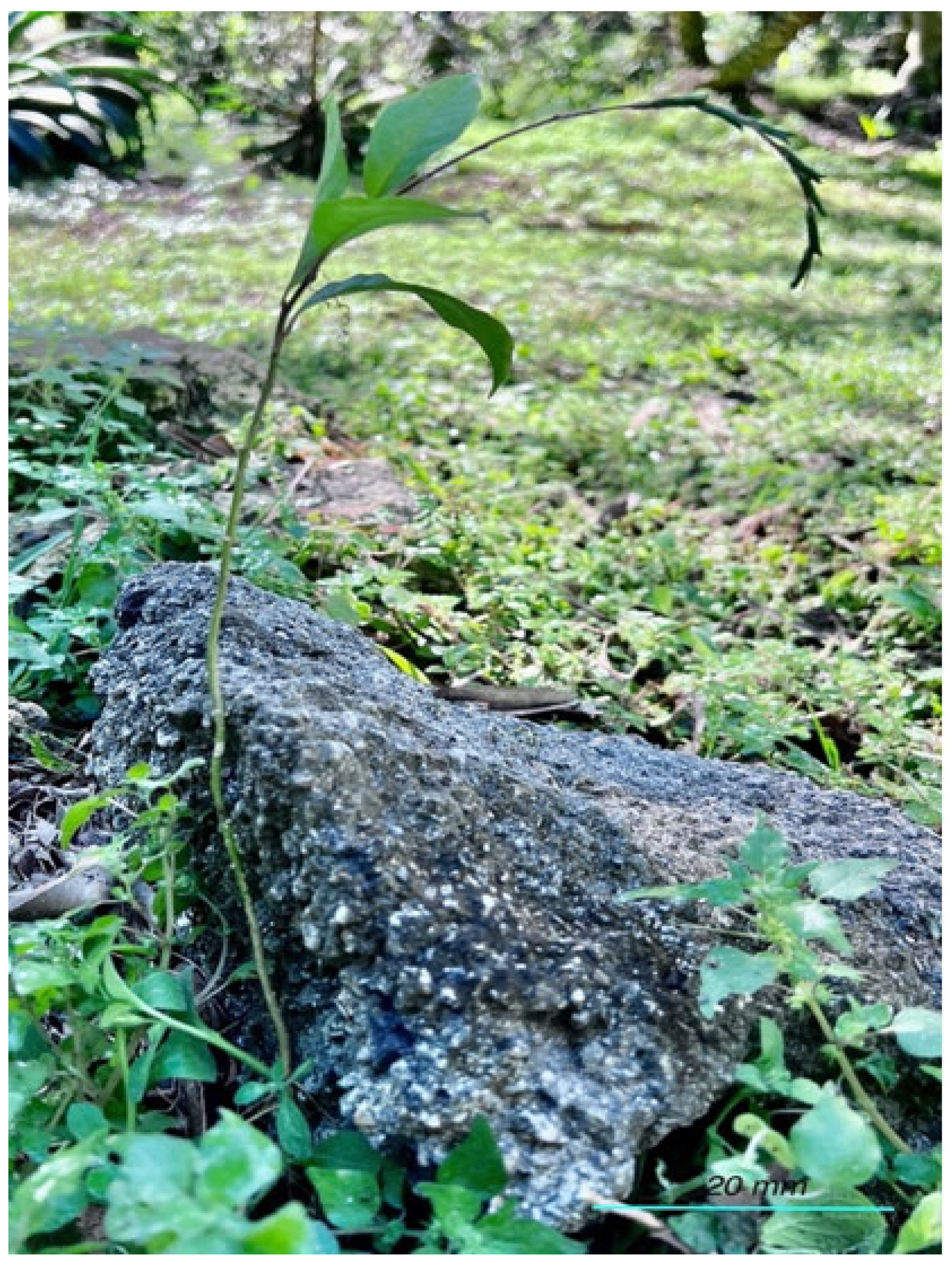
2.1. Collection of Host Medicinal Plants

The healthy plant samples of Pativeria alliacea were collected from Possum Trot Tropical Fruit Nursery (25°32′7″ N; 80°28′37″ W) located in Miami’s Redland Agricultural district, Florida. Samples of fresh, healthy leaves, stems, and roots of Pativeria alliacea were collected in separate zipper lock bags, labeled, stored in a cooler on ice, transported to the laboratory, and stored at 4 °C until processing as described previously by Tan et al., 2018 [22]. Samples were processed within 24 h of collection. Specimens were identified by Robert L. Burnum from Possum Trot farm, who is a taxonomist in native medicinal plants in South Florida.

2.2. Isolation of Endophytic Fungi

Isolation and cultivation of endophytic fungi were carried out from the leaf, stem, and root of the sampled plants (Figure 1). Surface sterilization of the plant’s parts was performed by a modified protocol from Tan et al., 2018. For surface sterilization, the segments were soaked in 70% ethanol for 30 s, then sodium hypochlorite (2.5% available chlorine) for 5 min, followed by three successive rinses with sterile distilled water and blotted dry using sterile filter paper. Sterile segments were immediately aseptically cut into smaller fragments (0.5 cm × 0.5 cm), and individual sample fragments were placed horizontally on potato dextrose agar (PDA) supplemented with chloramphenicol (200 µg/mL) and streptomycin sulfate (100 µg/mL). Nine stem/leaf/root fragments per plant sample were seeded onto agar plates. The effectiveness of the surface sterilization was confirmed by the absence of microbial growth on the triplicate PDA plates inoculated with 100 μL aliquots of the final rinse water and on the triplicate PDA plates with a surface imprint of surface sterilized sample segment. Plates were incubated at room temperature (28 °C) for more than a week. Inoculated plates were observed every day for fungal growth from plant tissue. Fungal hyphal tips from the growth were transferred onto the fresh PDA, and the sub-culturing of the fungal isolate was continued until a pure culture was obtained. Observations on the morphological characteristics (colony color, surface, margin, pattern, etc.) of the fungal isolates were recorded. Cultures of individual endophytic fungal isolates were stored in 2.0 mL cryovials containing 15% glycerol in PDB medium at −80 °C.

2.3. Crude Extract Preparation for Secondary Metabolites

Individual endophytic fungal isolates from Petiveria alliacea were grown on PDA medium for 7 days at 28 °C. After that, three discs (5 mm) of each isolate were picked and inoculated individually into a 250 mL Erlenmeyer flask containing 80 mL of potato dextrose broth (PDB) and incubated at 28 °C for 60 days in an orbital shaker set at 125 rpm. Endophytic extract preparation was carried out following a modified protocol from Pansanit and Pripdeevech, 2018 and Sharma et al., 2016 [23,24]. The fungal broth was centrifuged for 5 min at 4000× g to obtain a cell-free supernatant (CFS). The supernatant was decanted into a separate vial, and the CFS and the pellet were extracted using 50 mL and 30 mL of ethyl acetate, respectively, at room temperature for 3 days. The resulting decanted organic phase from CFS and pellet were combined, filtered, and concentrated by using a Buchi R-300 vacuum rotary evaporator (BUCHI Corporation, New Castle, DE, USA) at reduced pressure at 45 °C, resulting in 10 mL of fungal crude extract of endophyte. Later, the solvent from the crude extract was further evaporated using a vacuumed rotary evaporator (Buchi R-300) at 45 °C to yield the crude metabolite. The dry solid was dissolved in dimethyl sulphoxide (DMSO) to obtain a final concentration of 10 mg/mL and stored at 4 °C. The crude extract and DMSO extract were used for the initial and final screening of antimicrobial activity, respectively.

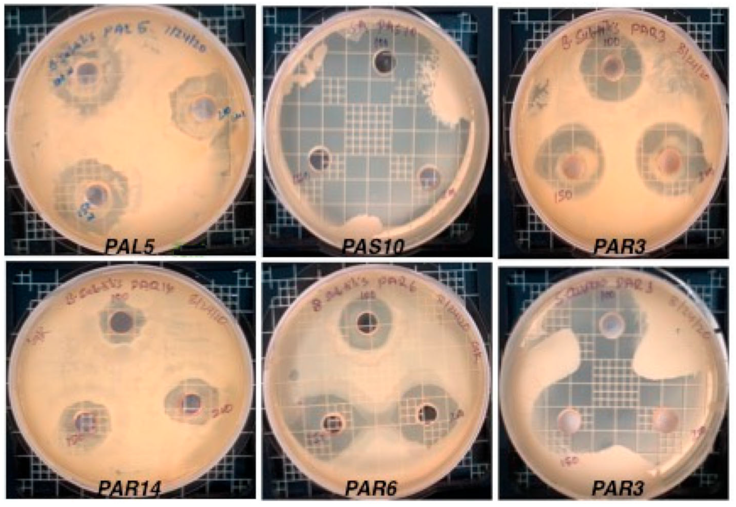
2.4. Testing of Antibacterial and Antifungal Activity

The endophytic fungal crude extracts were screened for their antimicrobial activity using the agar diffusion method against Gram-negative (Escherichia coli ATTC 25902), Gram-positive (Staphylococcus aureus ATTC 14775, Bacillus subtilis NRRL 5109), unicellular fungus (Candida albicans ATTC 10231), and multicellular filamentous fungus (Aspergillus fumigatus NRRL 5109). Tested bacteria and fungi were seeded on Muller–Hinton agar (MHA) and Potato Dextrose Agar (PDA) plates, respectively (Figure 2). On each plate, three equally spaced wells were cut into the surface of agar using a sterile cork-borer (6 mm), and 100, 150, and 200 µL of endophytic fungal crude extract or 10 mg/mL DMSO were then dispensed into three separate wells then incubated at 28 °C [23,24]. Antimicrobial activity was assessed by measuring the inhibition diameter (mm) zones at 24 and 48 h. Ampicillin sodium 100 µg/mL (positive control 1) and Fluconazole 30 µg/mL (positive control 2) were employed as positive controls, while 10% DMSO was used as a negative control. The experiment was performed in triplicates.

2.5. Sequencing of Endophytic Isolates

The isolates that showed positive antimicrobial activity were identified using molecular methods from Azenta Life Science. Briefly, the colony samples were lysed using a crude NaOH lysis technique to be directly used in PCR. To amplify ribosomal internal transcribed spacers (ITS), the primers ITS1 (5′TCCGTAGGTGAACCTGCGG3′) and ITS4 (5′TCCTCCGCTTATTGATATGC-3′) were employed in a polymerase chain reaction (PCR) assay [25]. Amplified samples were spot-checked using gel electrophoresis to check for robust amplification, along with a negative control to check for contamination. Following amplification, enzymatic cleanup was performed according to Azenta Life Sciences SOP using Exonuclease I—Shrimp Alkaline Phosphatase (ExoSAP), and dye-terminator sequencing was performed by Azenta Life Sciences Inc. (South Plainfield, NJ, USA) using Applied Biosystems BigDye version 3.1. Sequencing-specific primers were used to generate bidirectional reads. The reactions were then run on Applied Biosystem’s 3730xl DNA analyzer. Sequences were then compared with the National Center for Biotechnology Information (NCBI) database (http://www.ncbi.nlm.nih.gov), accessed on 5 January 2023, and accession numbers were assigned to each sequence. The assigned accession numbers for each isolate were submitted and published to the NCBI GenBank database. Phylogenetic analysis was performed for isolates showing strong inhibition (>20 mm) based on sequence data from ITS gene using Mega software version 11 using a Neighbor-joining phylogenetic tree. The bootstrap value was chosen to be 100 for the percentage of bootstrap replications supporting the branch [26].

2.6. Statistical Analysis

The Shannon–Weiner Index (H′) was used to calculate the fungal diversity of endophytic fungi using the formula below.
H′= −Σ(Pi × In Pi)
where Pi was calculated as Pi = n i N , and ni represents numbers of the individual segment from which fungal endophytic isolates were isolated, and N is the total number of segments incubated [27,28].
Similarly, the Colonization rates (CR%) of the fungal isolates from Petiveria alliacea were calculated as follows: CR% = Nsc/Nss × 100 where Nsc represents the number of segments infected by fungal isolates and Nss represents the total numbers of plant segments investigated. The isolation ratio (IR%) of the fungal strains was calculated as follows: (IR% = Ni/Nt × 100) where Ni is the number of segments from which fungal isolates were isolated, and Nt is the total number of plant segments incubated [29].
CR % = N o .   o f   i n f e c t e d   s e g m e n t N o .   o f   s e g m e n t   s t u d i e d   ×   100 ;   IR % = N o .   o f   s e g m e n t   f r o m   w h i c h   f u n g i   i s o l a t e d N o .   o f   s e g m e n t   i n c u b a t e d   ×   100
The diversity index was analyzed by two-way ANOVA. SPSS version 20.0 was used to perform all statistical analyses (SPSS Inc., Chicago, IL, USA).

3. Results

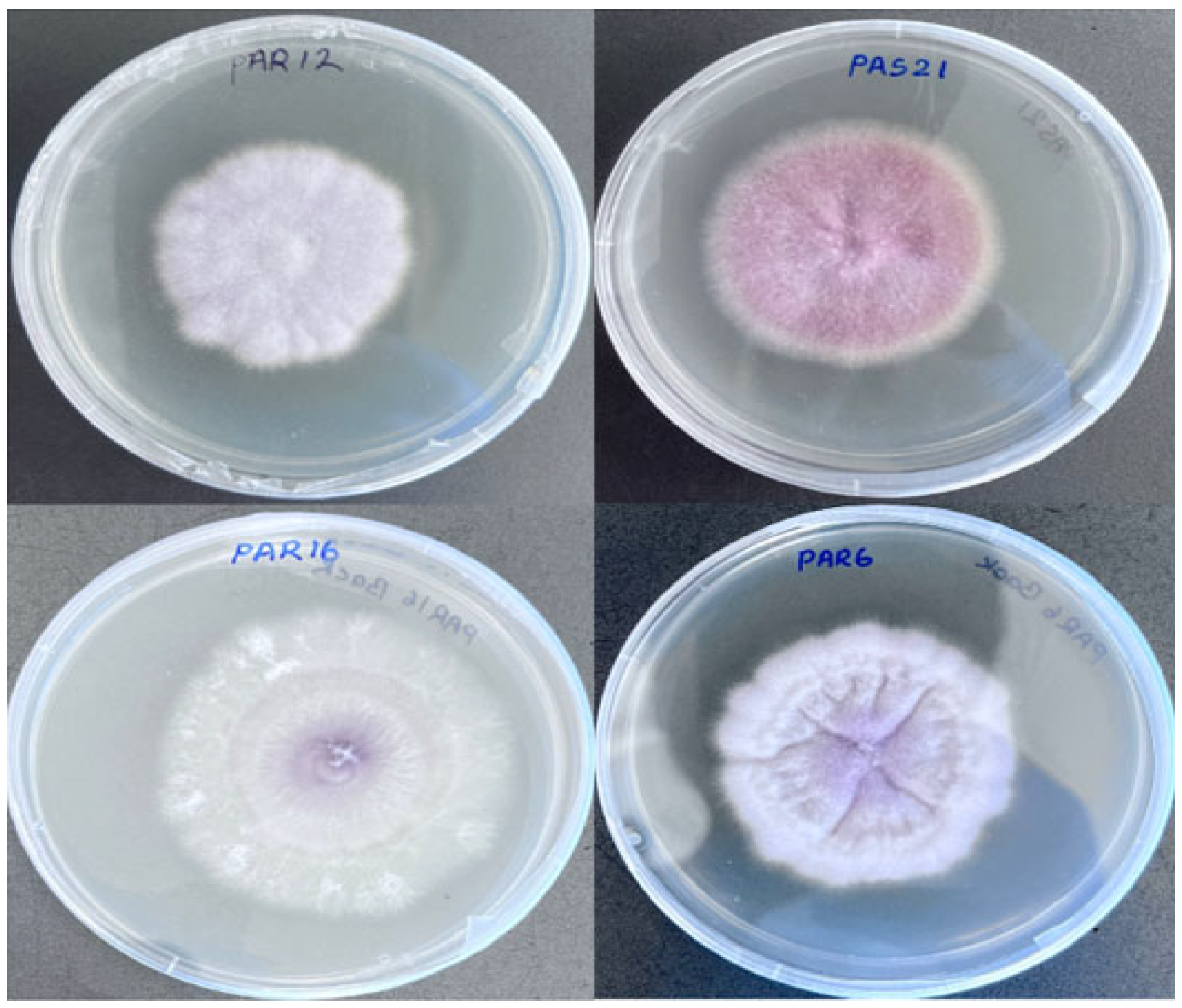
3.1. Collection and Pure Culture Isolation

From the 289 sampled plant parts (representing a total of 99 roots, 99 stems, and 99 leaves), a total of 279 isolates were counted and are categorized as 30 morphotypes based on their morphological appearances (Figure 3). Eleven endophytic fungi were morphologically different from each other and isolated from the root sample; 9 were from the stem, and 10 were isolated from the leaf sample (Table 1) [30].
The highest percentage of the colonization rate (97%) was found in the stem sample, and the highest percentage of the isolation rate (98%) was also found in the stem sample. Similarly, the highest Shannon–Weiner diversity index was found in the root sample from Petiveria alliacea (Table 1).

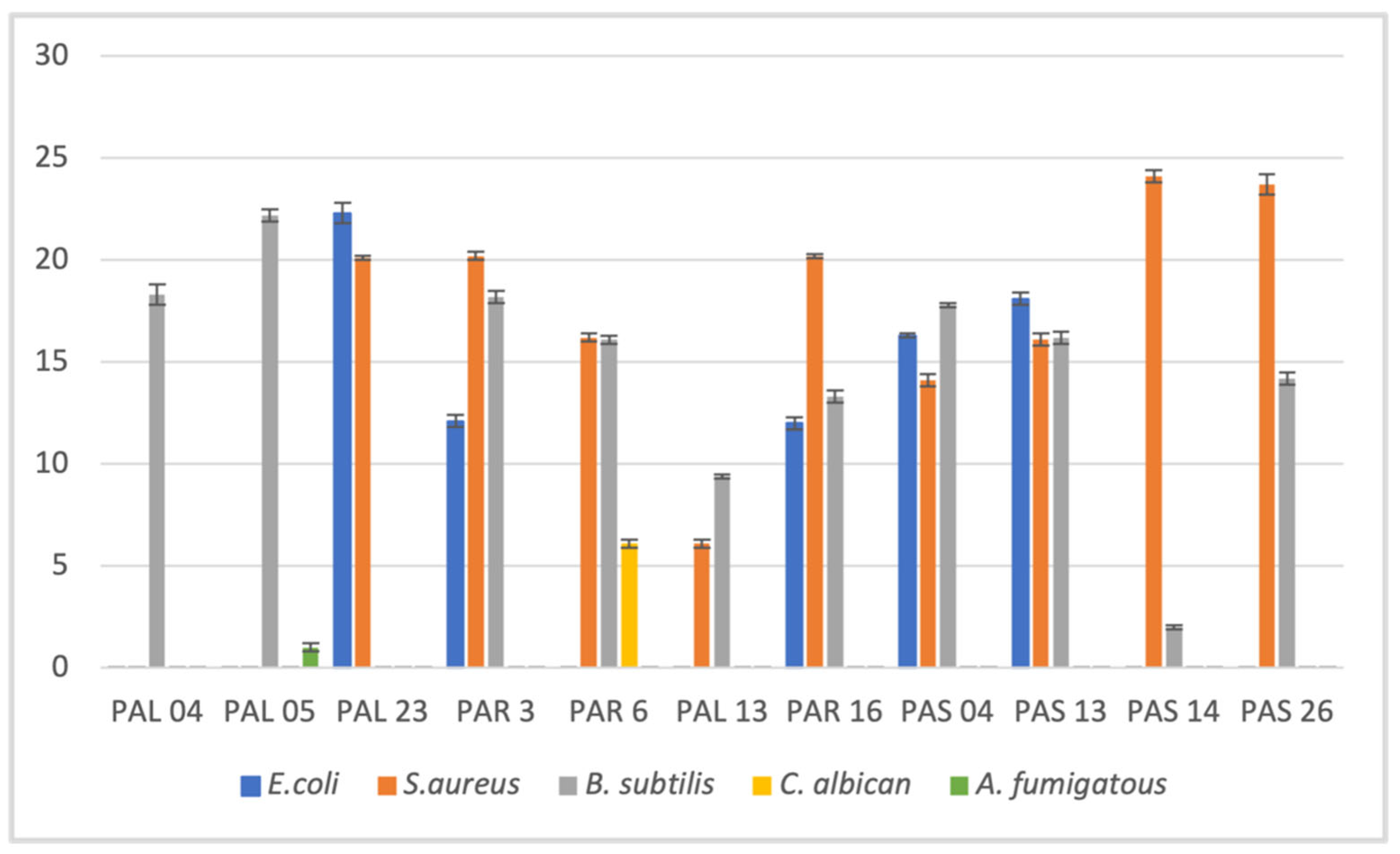
3.2. Antimicrobial Activity of Crude Fungal Extract Using Agar Well Diffusion Method

The antimicrobial activities of crude extracts from 30 endophytic fungal isolates were evaluated against test organisms. Crude extracts from the majority (83%) of the isolates showed positive antimicrobial activity in the initial screening (Table 2 and Table 3). Out of the fungal endophyte crude extracts that showed antimicrobial activity, 65% of the leaf endophyte extracts, 88% of the stem endophyte extracts, and 71% of the root endophyte extracts showed activity against at least one of the bacterial strains tested. Only 10% of the fungal crude extracts prepared from leaf endophytes demonstrated activity against at least one of the fungal strains tested. None of the crude fungal extracts prepared from the stem and root samples showed activity against the fungal strains tested. The largest zone of inhibition was observed against S. aureus (27.8 ± 0.5 mm) inhibited by the PAS-14 isolate, E. coli (27 ± 0.1 mm) inhibited by the PAL-23 isolate, and B. subtilis (25.8 ± 0.1 mm) inhibited by the PAL-05 isolate crude extracts. Crude extracts from two endophytes, PAL-05 from the leaf and PAR-06 from the root, showed broad-spectrum activity against both bacteria and fungi. Isolate Fusarium solani PAL-05 showed a maximum inhibition zone of 25.8 ± 0.1 mm and 4.1 ± 0.2 mm against B. subtilis and A. fumigatus, respectively. Similarly, the isolate Fusarium oxysporium PAR-06 showed a maximum inhibition zone of 20.0 ± 0.2 mm, 22.2 ± 0.1 mm, and 9.8 ± 0.3 mm against S. aureus, B. subtilis, and C. albicans, respectively. Three of the endophytic isolates, Fusarium oxysporum PAR-16, Fusarium oxysporum PAS-04, and Fusarium solani PAS-13, demonstrated antimicrobial activity against both Gram-negative and Gram-positive bacteria but did not show any activity against the fungal strains tested. None of the isolates examined showed inhibition against all the test organisms.
Following initial antimicrobial activity screening, the metabolites of endophytes in DMSO extract (10 mg/mL) of selected fungal crude extracts showing promising bioactive activities against test organisms were further evaluated (Table 4). The endophytic fungal isolates PAL-4 and PAL-5 showed inhibition zones of 5.1 ± 0.2 mm and 6.0 ± 0.1 mm against B. subtilis only. The extracts from isolates PAR-8 and PAS-7, in contrast, demonstrated inhibitory activity against all three bacterial strains tested. The fungal endophytic isolate PAR-14 demonstrated the largest zone of inhibition of 8.2 ± 0.2 mm against E. coli and 14.2 ± 0.2 mm against S. aureus compared to the ampicillin control with inhibition zones of 16 ± 0.2 mm and 16.3 ± 0.2 mm, respectively. Among the endophyte isolates tested against B. subtilis, only DMSO extracts from isolate PAS-07 showed the largest zone of inhibition of 13.0 ± 0.1 mm, with the ampicillin control showing a 21.1 ± 0.2 mm zone of inhibition. None of the isolates showed significant antimicrobial activity against fungal stains tested at the concentration of 10 mg/mL in DMSO.

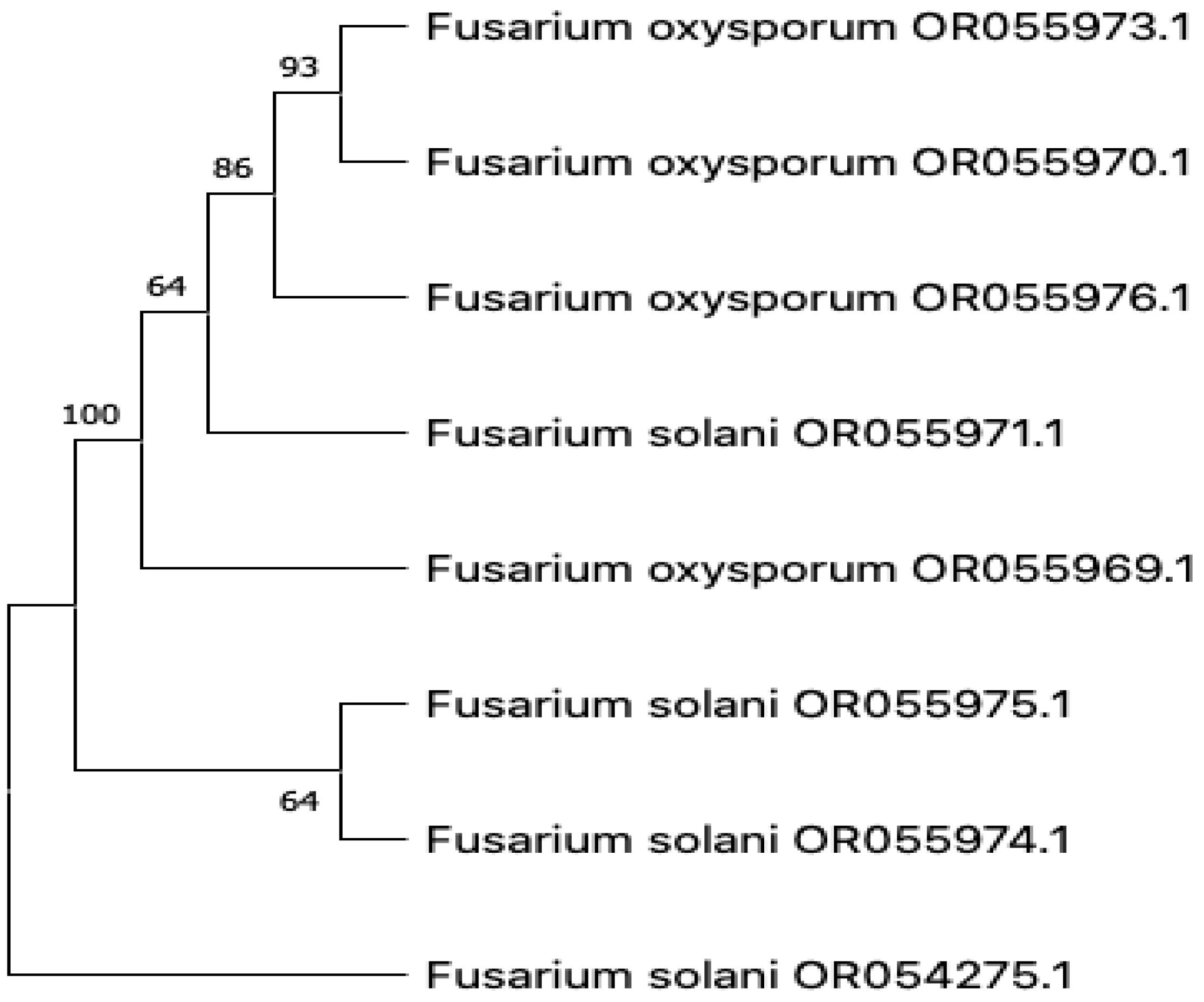
3.3. Molecular Characterization

The eight isolates showing strong antimicrobial activity (>20 mm) from fungal crude extracts were chosen to sequence the internal transcribed spacer region (ITS) of ribosomal DNA. Sequenced data revealed, as shown in Table 5, that Fusarium solani, F. oxysporium, and F. proliferatum are the main endophytic fungi that are responsible for antimicrobial activity. All the sequenced fungal isolates were Fusarium, which belongs to the Phylum Ascomycota, Class Sordariomycetes, and Order Hypocreales. Phylogenetic tree analysis demonstrated that Fusarium oxysporium OR055973.1 formed the group with F. oxysporium OR055970.1 with a strong bootstrap support of 93%. Similarly, F. solani OR055975.1 and F. solani OR055974.1 form a group with a bootstrap value of 64%. Results indicate that all the sequenced isolates belong to the same genus, Fusarium.

3.4. Statistical Analysis

The result of the two-way Manova test for maximum value showed that there is a statistically significant difference between the means of the dependent variables: concentration of extract tested (M 100, M 150, and M 200) and the two factors (factor 1: Bacterial spp. and factor 2: Endophytic fungal spp.), i.e., p ≤ 0.01. The individual ANOVA tests and Wilk’s Lambda test for the dependent variables M 100, M 150, and M 200 also demonstrated that both factors are statistically significant (p ≤ 0.01).

4. Discussion

The endophytic fungus possesses unique and diverse antimicrobial activity [31]. The diversity of fungi is unknown; the number of endophytic fungi is estimated to be approximately one million based on the 6:1 ratio of fungal–plant species [32]. However, in tropical and subtropical regions, the ratio could be up to five times higher [33]. In this study, endophytic fungi isolated from the medicinal plant Petiveria alliacea were evaluated to investigate the potential for producing novel bioactive compounds.
The medicinal herb Petiveria alliacea has gotten a lot of attention in recent years, and its extracts possess many metabolites that have the potential to kill or inhibit many types of disease-causing pathogens [34,35]. Previous studies have shown that the benzyl-containing thiosulfinates from P. alliacea exhibited the broadest spectrum of antimicrobial activity against bacteria and fungi [36]. The present study was initiated to understand the association of fungal endophytes with Petiveria alliacea in South Florida, as there was no such research conducted previously.
Many studies have been undertaken and established that endophytic fungal crude extracts have antibacterial properties [34,35]. These results might be attributed to the fact that the extract may contain a high concentration of bioactive metabolites. The fungal extract that showed very low antimicrobial activity in the bioassay might have contained active bioactive metabolites in low or very low concentrations or might have yielded more active compounds if it was further purified [34]. Various factors might influence the qualitative and quantitative aspects of bioactive metabolite production by endophytic fungi under laboratory conditions. The spectrum and degree of inhibitory activity of endophytic fungal metabolites can also be affected by the type of solvent used [37].
The fungal endophytes isolated from Petiveria alliacea were tested against E. coli, S. aureus, B. subtilis, C. albicans, and A. fumigatous to test their production of antimicrobial compounds by using a dual culture assay. These initial results hinted that those isolates might produce bioactive compounds. They were then tested using agar well diffusion assay techniques (Figure 4). The zone of inhibition calculations was calculated based on agar well diffusion techniques as it was more reliable than the dual culture bioassay technique.
Sequencing data from this study demonstrated that the endophytic fungal strain sequenced is a species of Fusarium (Figure 5). Endophytic strains of Fusarium are known for producing many different types of useful bioactive compounds. The antimicrobial drug PTOX (podophyllotoxin) was produced by the endophytes F. oxysporum isolated from Juniperus recurve [38]. Another natural product produced by the endophytic fungi Fusarium is Taxol, which is the first billion-dollar drug produced by F. proliferatum from the Taxol plant [39,40]. Crude extracts of endophyte F. equiseti isolated from Mikania cordata showed a significant broad-spectrum of antimicrobial activity against Bacillus cereus, Staphylococcus aureus, Escherichia coli, and Pseudomonas aeruginosa [41]. A polyketide fusaequisin A extracted from F. equiseti was found to be inhibitory to S. aureus and P. aeruginosa [42]. Fusarium spp. are also known to produce antimicrobial compounds such as PTOX, beauvericin, and subglutinol A and B [22].
An additional antibacterial substance produced by endophytes Fusarium spp. identified from medicinal plants are 2-methyl butyraldehyde-substituted α-pyrone, beauvericin, and subglutinol A and B [27,43,44]. Endophytic fungi such as F. solani, F. oxysporium, and F. proliferatum have the potential to produce different types of bioactive metabolites and are supported by studies of other medicinal plants all around the world [45,46,47,48,49].
The antimicrobial activities of different species of Fusarium have been attributed to the production of bioactive secondary metabolites (SMs) such as terpenoids, polyketides, steroids, quinones, flavonoids, alkaloids, peptides, and these metabolites [50,51,52]. Fusarium sp. has been reported to produce anti-Gram-negative bacterial SMs, anti-Gram-positive bacterial SMs, both anti-Gram-positive and anti-Gram-negative bacterial SMs, antifungal SMs, and both antibacterial and antifungal SMs [53].
The metabolites produced by endophytic fungi isolated from medicinal plants have antibacterial, antifungal, anticancer, and antioxidant properties, and it might provide opportunities to explore more endophytic fungi for discoveries in the field of antimicrobials [52,54]. However, more studies are needed to address the dynamical changes of endophytic communities [54] and unculturable fungal communities [55].

5. Conclusions

The present data showed that P. alliacea root contains the highest diversity of fungal endophytes. This study demonstrated that 37% of morphologically different fungal isolates were isolated from the root, whereas 33% were from the leaf and 30% were from the stem sample. Isolates that have the strongest antimicrobial activities in the three tissue samples also have similar genetic makeup as per the results from the fungal isolates that we sequenced.
This is the first study to explore endophytic fungi of Petiveria alliacea and evaluate their potential in vitro antimicrobial activities. Antimicrobial activity was tested on a total of 30 morphologically diverse fungal isolates. The isolates with a good zone of inhibition were sequenced to determine their identity. The crude extracts of endophytic fungal isolates contain promising antimicrobial compounds and deserve further purification and characterization for their chemical constituents. Future studies need to investigate all the culturable isolates, as well as unculturable isolates, using molecular methods for their identification. Expanding the testing of identified isolates for more pathogens and drug-resistant strains would help to solve the global problem of antibiotic resistance.

Author Contributions

Conceptualization: K.J., K.G.S., T.A., Y.-C.T.-D. and G.K.; Methodology: G.K., K.J., K.G.S., T.A. and Y.-C.T.-D.; Lab work: G.K. completed the lab work; Funding acquisition: K.J., Y.-C.T.-D. and K.G.S.; Resources managements: K.J., K.G.S., Y.-C.T.-D. and T.A.; Data analysis and validation: G.K., K.G.S., T.A., Y.-C.T.-D. and K.J.; Original draft preparation: G.K.; Manuscript writing—review and editing: K.J., Y.-C.T.-D., T.A. and K.G.S. reviewed and finalized manuscript. All authors have read and agreed to the published version of the manuscript.

Funding

This study was supported by the Florida International University, College of Arts, Sciences and Education. This project was also supported by Florida International University through a graduate teaching assistantship as well as from the Department of Earth and Environment at Florida International University.

Institutional Review Board Statement

Not applicable.

Informed Consent Statement

Not applicable.

Data Availability Statement

The original contributions for this study are included in the article, and further inquiries can be directed to the corresponding author.

Acknowledgments

We would like to thank Diego Salazar Amoretti for the technical resources and equipment used at the plant chemical ecology lab at Florida International University. Also, we would like to thank Amir Khoddamzadeh for providing valuable suggestions.

Conflicts of Interest

While conducting the research, all the authors declare that they have no conflict of interest. The final decision on the manuscript preparation and submission of the findings for publication had been reached by all the authors.

References

  1. Sen, S.; Chakraborty, R. Revival, Modernization and Integration of Indian Traditional Herbal Medicine in Clinical Practice: Importance, Challenges and Future. J. Tradit. Complement. Med. 2017, 7, 234–244. [Google Scholar] [CrossRef] [PubMed]
  2. Hussain, S.; Hussain, W.; Nawaz, A.; Badshah, L.; Ali, A.; Ullah, S.; Ali, M.; Hussain, H.; Bussmann, R.W. Quantitative Ethnomedicinal Study of Indigenous Knowledge on Medicinal Plants Used by the Tribal Communities of Central Kurram, Khyber Pakhtunkhwa, Pakistan. Ethnobot. Res. Appl. 2022, 23, 1–31. [Google Scholar] [CrossRef]
  3. Bussmann, R.W. The Globalization of Traditional Medicine in Northern Perú: From Shamanism to Molecules. Evid.-Based Complement. Altern. Med. 2013, 2013, 291903. [Google Scholar] [CrossRef] [PubMed]
  4. Patwardhan, B.; Vaidya, A.; Science, M.C. Ayurveda and Natural Products Drug Discovery. Curr. Sci. 2004, 86, 789–799. [Google Scholar]
  5. Singh, D.B.; Pathak, R.K.; Rai, D. From Traditional Herbal Medicine to Rational Drug Discovery: Strategies, Challenges, and Future Perspectives. Rev. Bras. Farmacogn. 2022, 32, 147–159. [Google Scholar] [CrossRef]
  6. Carroll, G. Fungal Endophytes in Stems and Leaves: From Latent Pathogen to Mutualistic Symbiont. Ecology 1988, 69, 2–9. [Google Scholar] [CrossRef]
  7. Faeth, S.H.; Hammon, K.E. Fungal Endophytes in Oak Trees: Long-Term Patterns of Abundance and Associations with Leafminers. Ecology 1997, 78, 810–819. [Google Scholar] [CrossRef]
  8. Saikkonen, K.; Faeth, S.H.; Helander, M.; Sullivan, T.J. Fungal Endophytes: A Continuum of Interactions with Host Plants. Annu. Rev. Ecol. Syst. 1998, 29, 319–343. [Google Scholar] [CrossRef]
  9. Newman, D.J.; Cragg, G.M. Plant Endophytes and Epiphytes: Burgeoning Sources of Known and “Unknown” Cytotoxic and Antibiotic Agents? Planta Med. 2020, 86, 891–905. [Google Scholar] [CrossRef]
  10. Pasrija, P.; Girdhar, M.; Kumar, M.; Arora, S.; Katyal, A. Endophytes: An Unexplored Treasure to Combat Multidrug Resistance. Phytomed. Plus 2022, 2, 100249. [Google Scholar] [CrossRef]
  11. Gouda, S.; Das, G.; Sen, S.K.; Shin, H.S.; Patra, J.K. Endophytes: A Treasure House of Bioactive Compounds of Medicinal Importance. Front. Microbiol. 2016, 7, 1538. [Google Scholar] [CrossRef] [PubMed]
  12. Ganley, J.C.; Thomas, F.S.; Seebauer, E.G.; Masel, R.I. A Priori Catalytic Activity Correlations: The Difficult Case of Hydrogen Production from Ammonia. Catal. Lett. 2004, 96, 117–122. [Google Scholar] [CrossRef]
  13. Alvin, A.; Miller, K.I.; Neilan, B.A. Exploring the Potential of Endophytes from Medicinal Plants as Sources of Antimycobacterial Compounds. Microbiol. Res. 2014, 169, 483–495. [Google Scholar] [CrossRef]
  14. Beare, N. Pirates, Pineapples, and People: A History, Tales, and Legends of the Upper Florida Keys; Papamoa Press: London, UK, 2019. [Google Scholar]
  15. Sarma, S.N.; Siwach, D.; Hasan, A.; Mittal, P.; Paul, P. Systematic Review on Safety and Efficacy of Saw Palmetto as a Health Supplement for Prostate Health in Adult Males. J. Curr. Med. Res. Opin. 2022, 5, 1252–1270. [Google Scholar]
  16. Cronquist, A. The Evolution and Classification of Flowering Plants. New York Botanical Garden, Bronx. 1988. Available online: https://www.scirp.org/(S(lz5mqp453edsnp55rrgjct55))/reference/ReferencesPapers.aspx?ReferenceID=1564967 (accessed on 18 August 2023).
  17. Duarte, M.; Fitoterapia, J.L. Leaf and Stem Morphoanatomy of Petiveria alliacea. Fitoterapia 2005, 76, 599–607. [Google Scholar] [CrossRef]
  18. Joly, A. Botânica: Introdução à Taxonomia Vegetal; Companhia Editora Nacional: São Paulo, Brasil, 1979; Available online: https://books.google.co.jp/books/about/Botanica.html?id=SBnKwgEACAAJ&redir_esc=y (accessed on 3 January 2023).
  19. Arogbodo, J.; Igbe, F.; Osho, I.; Adebayo, I. Processing Yield Percentage and Phytochemical Appraisement of the Leaf, Stem-Bark, Inflore-Infructescence, and Root of Petiveria alliacea (Linneaus). Int. J. Sci. Res. Biol. Sci. 2021, 8, 65–70. [Google Scholar]
  20. Williams, L.A.D.; The, T.L.; Gardner, M.T.; Fletcher, C.K.; Naravane, A.; Gibbs, N.; Fleishacker, R. Immunomodulatory Activities of Petiveria alliacea L. Phytother. Res. Int. J. Devoted Med. Sci. Res. Plants Plant Prod. 1997, 11, 251–253. [Google Scholar]
  21. Marini, S.; Jovicevic, L.; Milanese, C. Effects of Petiveria alliacea L. on Cytokine Production and Natural Killer Cell Activity. Pharmacol. Res. 1993, 27, 107–109. [Google Scholar] [CrossRef]
  22. Tan, X.; Zhou, Y.; Zhou, X.; Xia, X.; Wei, Y.; He, L.; Tang, H.; Yu, L. Diversity and Bioactive Potential of Culturable Fungal Endophytes of Dysosma Versipellis; A Rare Medicinal Plant Endemic to China. Sci. Rep. 2018, 8, 5929. [Google Scholar] [CrossRef]
  23. Sharma, D.; Pramanik, A.; Agrawal, P.K. Evaluation of Bioactive Secondary Metabolites from Endophytic Fungus Pestalotiopsis neglecta BAB-5510 Isolated from Leaves of Cupressus torulosa D.Don. 3 Biotech 2016, 6, 210. [Google Scholar] [CrossRef]
  24. Pansanit, A.; Pripdeevech, P. Antibacterial Secondary Metabolites from an Endophytic Fungus, Arthrinium Sp. MFLUCC16-1053 Isolated from Zingiber cassumunar. Mycology 2018, 9, 264–272. [Google Scholar] [CrossRef] [PubMed]
  25. White, T.J.; Bruns, T.; Lee, S.; Taylor, J. Amplification and Direct Sequencing of Fungal Ribosomal RNA Genes for Phylogenetics. PCR Protoc. Guide Methods Appl. 1990, 18, 315–322. [Google Scholar]
  26. Shabana, A.M.I.; Shetaia, Y.M.; Abdelwahed, N.A.M.; Esawy, M.A.; Alfarouk, O.R. Optimization, Purification and Antitumor Activity of Kodamaea ohmeri ANOMY L-Asparaginase Isolated from Banana Peel. Curr. Pharm. Biotechnol. 2021, 22, 654–671. [Google Scholar] [CrossRef] [PubMed]
  27. Sun, Y.; Wang, Q.; Lu, X.D.; Okane, I.; Kakishika, M. Endophytic Fungi Associated with Two Suaeda Species Growing in Alkaline Soil in China. Mycosphere 2011, 2, 239–248. [Google Scholar]
  28. Husna; Budi, S.W.; Mansur, I.; Kusmana, D.C. Diversity of Arbuscular Mycorrhizal Fungi in the Growth Habitat of Kayu Kuku (Pericopsis mooniana Thw.) in Southeast Sulawesi. Pak. J. Biol. Sci. 2015, 18, 1–10. [Google Scholar] [CrossRef]
  29. Hata, K.; Futai, K. Endophytic Fungi Associated with Healthy Pine Needles and Needles Infested by the Pine Needle Gall Midge, Thecodiplosis japonensis. Can. J. Bot. 1995, 73, 384–390. [Google Scholar] [CrossRef]
  30. Wang, Q.-X.; Li, S.-F.; Zhao, F.; Dai, H.-Q.; Bao, L.; Ding, R.; Gao, H.; Zhang, L.-X.; Wen, H.-A.; Liu, H.-W. Chemical Constituents from Endophytic Fungus Fusarium oxysporum. Fitoterapia 2011, 82, 777–781. [Google Scholar] [CrossRef]
  31. Pelaez, F. Biological Activities of Fungal Metabolites. Handb. Ind. Mycol. 2004, 22, 41–92. [Google Scholar]
  32. Sun, X.; Mycology, L.G. Endophytic Fungal Diversity: Review of Traditional and Molecular Techniques. Mycopathologia 2012, 3, 65–76. [Google Scholar]
  33. Gamboa, M.A.; Laureano, S.; Bayman, P. Measuring Diversity of Endophytic Fungi in Leaf Fragments: Does Size Matter? Mycopathologia 2003, 156, 41–45. [Google Scholar] [CrossRef]
  34. Garcia, A.; Rhoden, S.A.; Bernardi-Wenzel, J.; Orlandelli, R.C.; Azevedo, J.L.; Pamphile, J.A. Antimicrobial Activity of Crude Extracts of Endophytic Fungi Isolated from Medicinal Plant Sapindus saponaria L. J. Appl. Pharm. Sci. 2012, 2, 35. [Google Scholar] [CrossRef]
  35. Idris, A.; Al-tahir, I.; Idris, E. Antibacterial Activity of Endophytic Fungi Extracts from the Medicinal Plant Kigelia africana. Egypt. Acad. J. Biol. Sci. G. Microbiol. 2013, 5, 1–9. [Google Scholar] [CrossRef]
  36. Musah, R.A.; Kim, S.; Kubec, R. Antibacterial and Antifungal Activity of Sulfur-Containing Compounds from Petiveria alliacea L. Phosphorus Sulfur. Silicon Relat. Elem. 2005, 180, 1455–1456. [Google Scholar] [CrossRef]
  37. Elghaffar, R.Y.A.; Amin, B.H.; Hashem, A.H.; Sehim, A.E. Promising Endophytic Alternaria alternata from Leaves of Ziziphus spina-Christi: Phytochemical Analyses, Antimicrobial and Antioxidant Activities. Appl. Biochem. Biotechnol. 2022, 194, 3984–4001. [Google Scholar] [CrossRef] [PubMed]
  38. Vasundhara, M.; Kumar, A.; Reddy, M.S. Molecular Approaches to Screen Bioactive Compounds from Endophytic Fungi. Front. Microbiol. 2016, 7, 1774. [Google Scholar] [CrossRef] [PubMed]
  39. Tan, X.M.; Wang, C.L.; Chen, X.M.; Zhou, Y.Q.; Wang, Y.Q.; Luo, A.X.; Liu, Z.H.; Guo, S.X. In Vitro Seed Germination and Seedling Growth of an Endangered Epiphytic Orchid, Dendrobium Officinale, Endemic to China Using Mycorrhizal Fungi (Tulasnella sp.). Sci. Hortic. 2014, 165, 62–68. [Google Scholar] [CrossRef]
  40. Xiong, Z.Q.; Yang, Y.Y.; Zhao, N.; Wang, Y. Diversity of Endophytic Fungi and Screening of Fungal Paclitaxel Producer from Anglojap Yew, Taxus x Media. BMC Microbiol. 2013, 13, 71. [Google Scholar] [CrossRef]
  41. Jayatilake, P.L.; Munasinghe, H. Antimicrobial Activity of Cultivable Endophytic and Rhizosphere Fungi Associated with “Mile-a-Minute,” Mikania cordata (Asteraceae). Biomed. Res. Int. 2020, 2020, 5292571. [Google Scholar] [CrossRef]
  42. Shiono, Y.; Shibuya, F.; Murayama, T.; Koseki, T.; Poumale Poumale, H.M.; Tchaleu Ngadjui, B. A Polyketide Metabolite from an Endophytic Fusarium equiseti in a Medicinal Plant. Z. Naturforschung—Sect. C J. Biosci. 2013, 68B, 289–292. [Google Scholar] [CrossRef]
  43. Brady, S.F.; Clardy, J. CR377, a New Pentaketide Antifungal Agent Isolated from an Endophytic Fungus. J. Nat. Prod. 2000, 63, 1447–1448. [Google Scholar] [CrossRef]
  44. Lee, J.C.; Lobkovsky, E.; Pliam, N.B.; Strobel, G.; Clardy, J. Subglutinols A and B: Immunosuppressive Compounds from the Endophytic Fungus Fusarium subglutinans. J. Org. Chem. 1995, 60, 7076–7077. [Google Scholar] [CrossRef]
  45. Sogra Fathima, B.; Balakrishnan, R.M. Biosynthesis and Optimization of Silver Nanoparticles by Endophytic Fungus Fusarium solani. Mater. Lett. 2014, 132, 428–431. [Google Scholar] [CrossRef]
  46. Farhat, H.; Urooj, F.; Tariq, A.; Sultana, V.; Ansari, M.; Ahmad, V.U.; Ehteshamul-Haque, S. Evaluation of Antimicrobial Potential of Endophytic Fungi Associated with Healthy Plants and Characterization of Compounds Produced by Endophytic Cephalosporium and Fusarium solani. Biocatal. Agric. Biotechnol. 2019, 18, 101043. [Google Scholar] [CrossRef]
  47. Ginting, R.C.B.; Sukarno, N.; Widyastuti, U.T.U.T.; Darusman, L.K.; Kanaya, S. Diversity of Endophytic Fungi from Red Ginger (Zingiber officinale Rosc.) Plant and Their Inhibitory Effect to Fusarium oxysporum Plant Pathogenic Fungi. Hayati 2013, 20, 127–137. [Google Scholar] [CrossRef]
  48. Wei, F.; Zhang, Y.; Shi, Y.; Feng, H.; Zhao, L.; Feng, Z.; Zhu, H. Evaluation of the Biocontrol Potential of Endophytic Fungus Fusarium solani CEF559 against Verticillium dahliae in Cotton Plant. Biomed. Res. Int. 2019, 2019, 3187943. [Google Scholar] [CrossRef] [PubMed]
  49. Tayung, K.; Bp, B.; Dk, J.; Dc, D. Identification and Characterization of Antimicrobial Metabolite from an Endophytic Fungus, Fusarium solani Isolated from Bark of Himalayan Yew. Mycosphere 2011, 2, 203–213. [Google Scholar]
  50. Latz, M.A.C.; Jensen, B.; Collinge, D.B.; Jørgensen, H.J.L. Endophytic Fungi as Bio-Control Agents: Elucidating Mechanisms in Disease Suppression. Plant Ecol. Divers. 2018, 11, 555–567. [Google Scholar] [CrossRef]
  51. Wani, S.H.; Kumar, V.; Shriram, V.; Sah, S.K.; Kumar, S. Phytohormones and Their Metabolic Engineering for Abiotic Stress Tolerance in Crop Plants. Crop J. 2016, 4, 162–176. [Google Scholar] [CrossRef]
  52. Strobel, G.; Daisy, B.; Castillo, U.; Harper, J. Natural Products from Endophytic Microorganisms. J. Nat. Prod. 2004, 67, 257–268. [Google Scholar] [CrossRef]
  53. Xu, M.; Huang, Z.; Zhu, W.; Liu, Y.; Bai, X.; Zhang, H. Fusarium-Derived Secondary Metabolites with Antimicrobial Effects. Molecules 2023, 28, 3424. [Google Scholar] [CrossRef]
  54. Cui, J.-L.; Guo, T.-T.; Ren, Z.-X.; Zhang, N.-S.; Wang, M.-L. Diversity and Antioxidant Activity of Culturable Endophytic Fungi from Alpine Plants of Rhodiola crenulata, R. angusta, and R. sachalinensis. PLoS ONE 2015, 10, e0118204. [Google Scholar] [CrossRef] [PubMed]
  55. Tejesvi, M.V.; Kajula, M.; Mattila, S.; Pirttilä, A.M. Bioactivity and Genetic Diversity of Endophytic Fungi in Rhododendron tomentosum Harmaja. Fungal Divers. 2011, 47, 97–107. [Google Scholar] [CrossRef]
Figure 1. Adult plant of Petiveria alliacea herb with visible seed. (Bar = 20 mm).
Microbiolres 14 00100 g001
Figure 2. Antibacterial activity of the extracts of fungal endophytes against selected bacterial strains.
Microbiolres 14 00100 g002
Figure 3. Colony morphology of endophytic fungi isolated from Petiveria alliacea on potato dextrose agar.
Microbiolres 14 00100 g003
Figure 4. Mean zone of inhibition in mm of the crude extracts (y-axis) of the fungal endophyte isolates against bacterial and fungal strains (x-axis) using 100 µL.
Microbiolres 14 00100 g004
Figure 5. Phylogenetic (Neighbor-joining) tree based on sequence data from ITS gene. The value on each branch is the percentage of bootstrap replications supporting the branch.
Microbiolres 14 00100 g005
Table 1. Endophytic fungal isolates from Petiveria alliacea.
TissueSegment StudiedInfected SegmentsTotal IsolatesEndophytic spp.CR%IR%Shanon_H
Root998386118386.52.366
Stem999597996982.189
Leaf9994961095972.282
Table 2. Antibacterial activities of the crude extracts of endophytic fungi isolated from Petiveria alliacea against selected bacterial pathogens (the numbers are the diameters of the zones of inhibition in mm; mean ± SD; n = 3).
EndophytesE. coliS. aureusB. subtilis
μL100150200100150200100150200
PAL 04------18.3 ± 0.522.2 ± 0.324.0 ± 0.1
PAL 05------22.2 ± 0.323.1 ± 0.425.8 ± 0.1
PAL 13---6.1 ± 0.29.0 ± 0.210.0 ± 0.29.4 ± 0.110.2 ± 0.111.3 ± 0.2
PAL 2322.3 ± 0.524.5 ± 0.227.0 ± 0.120.1 ± 0.123.3 ± 0.225.7 ± 0.1---
PAR 0312.1 ± 0.314.2 ± 0.118.1 ± 0.320.2 ± 0.222.0 ± 0.323.9 ± 0.218.2 ± 0.320.1 ± 0.122.1 ± 0.2
PAR 06---16.2 ± 0.218.1 ± 0.320.0 ± 0.216.1 ± 0.219.3 ± 0.222.2 ± 0.1
PAR 1612.0 ± 0.314.0 ± 0.116.2 ± 0.320.2 ± 0.122.2 ± 0.324.1 ± 0.213.3 ± 0.316.1 ± 0.118.0 ± 0.2
PAS 0416.3 ± 0.118.2 ± 0.320.1 ± 0.214.1 ± 0.316.1 ± 0.118.2 ± 0.117.8 ± 0.120.1 ± 0.222.2 ± 0.3
PAS 1318.1 ± 0.320.2 ± 0.322.0 ± 0.316.1 ± 0.317.2 ± 0.317.5 ± 0.516.2 ± 0.318.2 ± 0.220.0 ± 0.1
PAS 14---24.1 ± 0.325.9 ± 0.427.8 ± 0.52.0 ± 0.13.9 ± 0.15.8 ± 0.3
PAS 26---23.7 ± 0.526.0 ± 0.327.5 ± 0.414.2 ± 0.316.1 ± 0.317.5 ± 0.3
Positive control 115.0 ± 0.116.00 ± 0.216.9 ± 0.216.0 ± 0016.3 ± 0.216.9 ± 0.220.0 ± 0.120.1 ± 0.220.5 ± 0.2
Positive control 2---------
Negative control---------
Table 3. Antifungal activities of the crude extracts of endophytic fungi isolated from Petiveria alliacea against selected fungal pathogens (the numbers are the diameters of the zones of inhibition in mm; mean ± SD; n = 3); Positive control: Fluconazole (30 µg/mL); Negative control: 10% DMSO).
EndophytesC. albicansA. fumigatus
μL100150200100150200
PAL 04------
PAL 05---1.0 ± 0.22.0 ± 0.24.1 ± 0.2
PAL 13------
PAL 23------
PAR 03------
PAR 066.1 ± 0.28.1 ± 0.19.8 ± 0.3---
PAR 16------
PAS 04------
PAS 13------
PAS 14------
PAS 26------
Positive control24.1 ± 0.324.1 ± 0.324.6 ± 0.317.1 ± 0.317.4 ± 0.117.8 ± 0.3
Negative control------
Table 4. Antimicrobial activity of the DMSO extracts of endophytic fungi against selected microbial pathogens (the numbers are the diameters of the zones of inhibition in mm; mean ± SD; n = 3); Positive control 1: Ampicillin sodium (100 µg/mL); Positive control 2: fluconazole (30 µg/mL); Negative control: 10% DMSO).
IsolatesE. coliS. aureusB. subtilisC. albicansA. fumigatous
PAL04--5.1 ± 0.2--
PAL05--6.0 ± 0.1--
PAL234.2 ± 0.12.2 ± 0.2---
PAR088.1 ± 0.211 ± 0.28.1 ± 0.1--
PAR148.2 ± 0.214.2 ± 0.2---
PAR167.2 ± 0.213.2 ± 0.1---
PAS04-10.1 ± 0.2---
PAS078 ± 0.211.3 ± 0.313.0 ± 0.1--
PAS21-11.1 ± 0.1---
Positive control 116 ± 0.216.3 ± 0.221.1 ± 0.2--
Positive control 2---24.1 ± 0.317.4 ± 0.1
Negative control-----
Table 5. Sequencing results of selected isolates based on their antimicrobial activities.
SampleTissueITS IdentityClassificationAccession No.
PAL05Leaf99.62%Fusarium solaniOR054275.1
PAL23Leaf92.68%Fusarium oxysporumOR055969.1
PAS04Stem99.62%Fusarium oxysporumOR055970.1
PAS13Stem100%Fusarium solaniOR055971.1
PAR03Root99.82%Fusarium solaniOR055974.1
PAR06Root100%Fusarium oxysporumOR055973.1
PAR13Root99.64%Fusarium solaniOR055975.1
PAR16Root100%Fusarium oxysporumOR055976.1
Disclaimer/Publisher’s Note: The statements, opinions and data contained in all publications are solely those of the individual author(s) and contributor(s) and not of MDPI and/or the editor(s). MDPI and/or the editor(s) disclaim responsibility for any injury to people or property resulting from any ideas, methods, instructions or products referred to in the content.

