Article

Melanin Pathway Determination in Sclerotium cepivorum Berk Using Spectrophotometric Assays, Inhibition Compound, and Protein Validation

by
Luis M. Salazar-García
1,
Rocío Ivette Ortega-Cuevas
1,
José A. Martínez-Álvarez
1,
Sandra E. González-Hernández
1,
Román Antonio Martínez-Álvarez
1,
Diana Mendoza-Olivares
2,
Miguel Ángel Vázquez
2,
Alberto Flores-Martínez
1 and
Patricia Ponce-Noyola
1,*
1
Departamento de Biología, División de Ciencias Naturales y Exactas, Campus Guanajuato, Universidad de Guanajuato, Guanajuato 36050, Mexico
2
Departamento de Química, División de Ciencias Naturales y Exactas, Campus Guanajuato, Universidad de Guanajuato, Guanajuato 36050, Mexico
*
Author to whom correspondence should be addressed.
Microbiol. Res. 2022, 13(2), 152-166; https://doi.org/10.3390/microbiolres13020013
Submission received: 22 February 2022 / Revised: 25 March 2022 / Accepted: 26 March 2022 / Published: 29 March 2022

Abstract

:
Sclerotium cepivorum Berk is the etiological agent of white rot disease that affects plants of the genus Allium. This fungus produces resistance structures called sclerotia that are formed by a rolled mycelium with a thick layer of melanin and it can remain dormant for many years in the soil. Current interest in S. cepivorum has arisen from economic losses in Allium crops in the agricultural sector. Melanin is a component that protects the sclerotia from adverse environmental conditions In many organisms, it plays an important role in the infectious process; in S. cepivorum, the pathway by which this component is synthetized is not fully described. By using infrared spectrophotometric assays applied direct to the sclerotia and a melanin extract followed by an NMR analysis and a tricyclazole melanin inhibition experiment, it allowed us to determine the dihydroxynaphthalene (DHN)-melanin pathway by which S. cepivorum performs its melanin synthesis. Moreover, we focused on studying scytalone dehydratase (SDH) as a key enzyme of the DHN-melanin synthesis. We obtained the recombinant SDH enzyme and tested its activity by a zymogram assay. Thereby, the S. cepivorum melanogenic route was established as a DHN pathway.

1. Introduction

Sclerotium cepivorum is a pathogenic fungus of plants from the Allium genus such as garlic and onion that causes the disease known as white rot [1]. S. cepivorum usually grows on moldy soils generating hyphae and sclerotia [2]. Despite the relevance of Allium cultures and their losses because of white rot, advances in the biological knowledge of S. cepivorum are limited, especially those directed toward the study of sclerotia.
Sclerotia are nutrient-rich structures produced by many fungi that allow them to remain dormant or quiescent during unfavorable conditions. When conditions improve, the sclerotia germinate in order to propagate the pathogenic fungus [3]. Sclerotia structures comprise a rolled mycelium covered by an outer layer of melanin, forming a ring over the hyphae tissue [4]; this helps the sclerotia to survive on the soil for several years [2]. S. cepivorum produces sclerotia as a resistance form and for that reason, controlling white rot disease has been difficult. Physical, chemical, and biological methods have not been completely effective in reducing attacks of S. cepivorum on Allium crops [5].
S. cepivorum sclerotia are the initial source of the infective inoculum and can remain viable for up to 20 years in the soil; this resistance is attributed to the blackish layer of melanin that covers it [6]. Melanin in fungi protects against physical and chemical environmental factors such as oxidative stress, UV radiation, desiccation, cold, high humidity, heat, and antagonistic micro-organisms [7,8]. In a related organism, Magnaporthe oryzae (Pyricularia oryzae), a mutant lacking a melanin expression was reported to be more sensitive to stress factors [9]. In Sclerotinia sclerotiorum, a fungus with a high evolutionary similarity to S. cepivorum [10,11] and notable for its wide host range and environmental persistence, a melanin deficiency affected the sclerotium development but not its pathogenicity [7,12].
Melanin in fungi has been divided into three different categories: dihydroxyphenylalanine, also called DOPA-melanin, eumelanin, or the tyrosine route [13,14]; a variant named pyomelanin produced from the precursor glutaminyl-hidroxybenzene (GBH-melanin) [14]; and dihydroxynaphthalene-melanin, also known as DHN-melanin, allomelanin, the DHN pathway, or the polyketide pathway [13,15]. Although a feasible strategy for combating white rot disease is to inhibit S. cepivorum melanization, this pathway is not well-established and alternatives for its control are still limited [4,16]. The study of the basic biology of S. cepivorum and several biological processes such as the development of the sclerotia and the melanization pathway will help to understand the mechanisms responsible for white rot disease and thus lay the foundations for the development of strategies that allow the control of the infection [2].
Here, we present chemical and molecular evidence that allow us to suggest that the melanin synthesis in S. cepivorum is by the DHN pathway. We used infrared spectrophotometric assays and nuclear magnetic resonance (NMR) to produce an initial proposal of the produced melanin. These studies were supported by the use of tricyclazole, which specifically interferes with DHN melanization [17]; the treated strains showed a delayed melanin formation. In the annotation of the S. cepivorum genome, we identified several genes that take part in the DHN-melanin synthesis pathway, of which scytalone dehydratase (SDH) is a key enzyme. To complete this study, the identification of the open reading frame (ORF) of the gene encoding of the SDH protein was carried out, expressed in a yeast system, and the activity of the recombinant protein was measured. This protein has important roles in the development and morphology of fungi related to S. cepivorum [12,18] and in this research we demonstrate the importance it plays in the synthesis of melanin. In addition, the SDH protein could be considered a potential target for the control of white rot disease in plants of the Allium genus.

2. Materials and Methods

2.1. Micro-Organisms and Culture Media

S. cepivorum SC strain (isolated by Dr. D. Guzmán-de-Peña, CINVESTAV-Irapuato, Guanajuato, Mexico) was used in this research. The strain was grown on potato dextrose agar (PDA) plates at 18 °C; for the sclerotia recovery, the time of incubation was 13–17 days. The sclerotia were harvested by mechanical means and kept at room temperature until used. The mycelium was obtained by incubating the sclerotia on PDA plates with a cellophane membrane for 3–7 days at 18 °C. It was then harvested by mechanical means and used immediately or kept at −20 °C until used. Pichia pastoris X-33 strain cells were grown in a yeast extract peptone dextrose (YPD) medium (1% w/v yeast extract, 2% w/v gelatin peptone, and 3% w/v dextrose) or YPDS (YPD plus 1 M Sorbitol); for the Magnaporthe grisea 70-15 strain (Orbach M., University of Arizona), the strain cells were propagated in a potato dextrose broth medium (PDB) for 5 days at 120 rpm in an orbital incubator at 28 °C. An Escherichia coli DH5α strain for cloning was grown on a Luria Bertani (LB) medium with appropriate antibiotics [19].

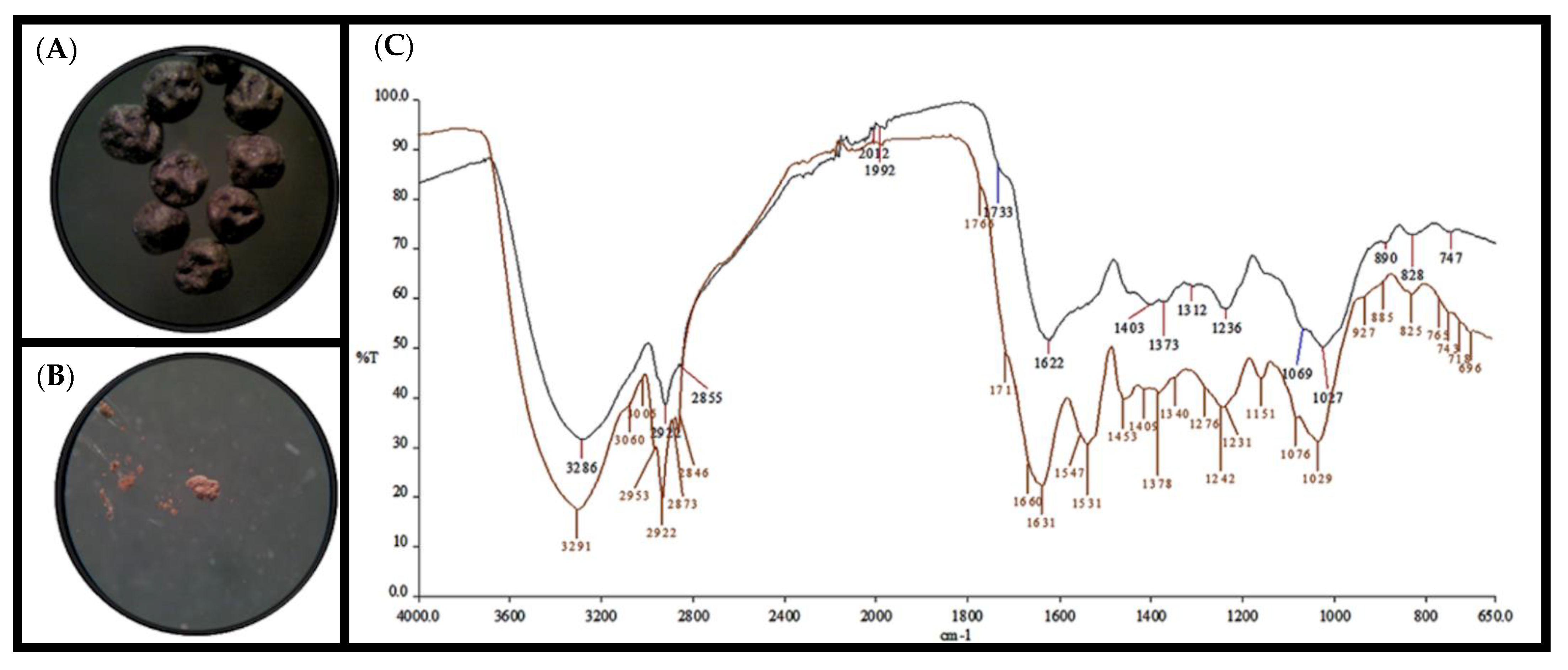
2.2. Sclerotia Melanin Extraction

S. cepivorum was grown for 13–17 days on PDA plates to promote the sclerotia development. The sclerotia were harvested by mechanical means and collected in a sterile container. Two equivalent volumes of distilled water were then added and strongly shaken in a vortex for 10 min to remove any mycelium residue from the surface of the sclerotia. The suspension was left to stand until the sedimentation of the sclerotia and the supernatant was removed. The sclerotia were washed twice in the same way and dried for 48 h at 40 °C. The recovered sclerotia were mixed with an equivalent volume of chloroform and strongly shaken in a vortex for 10 min. The sedimented sclerotia were recovered and solvent evaporation was promoted by a laminar flow. The dry sclerotia were directly used (direct sample) for the melanin extraction or kept at room temperature until use. The melanin extraction was performed as described by Gadd [20] but with slight modifications. The sclerotia obtained from the direct sample were ground in a mortar, resuspended in three equivalent volumes of 3 M NaOH, mixed thoroughly in a vortex for 10 min and incubated for 48 h at 60 °C. The homogenate was centrifuged at room temperature for 10 min at 12,000× g and the supernatant containing the melanin was recovered. The melanin was precipitated by adding 1 M HCl until a pH of 2.0 was obtained; this was then centrifuged for 20 min at 15,000× g. The supernatant was removed, and the precipitate was washed 4 times with distilled water and centrifuged for 10 min at 12,000× g. The melanin pellet was washed with chloroform, stirred slightly, and placed in a laminar flow until solvent evaporation; once dried, the purified preparation was kept at room temperature.

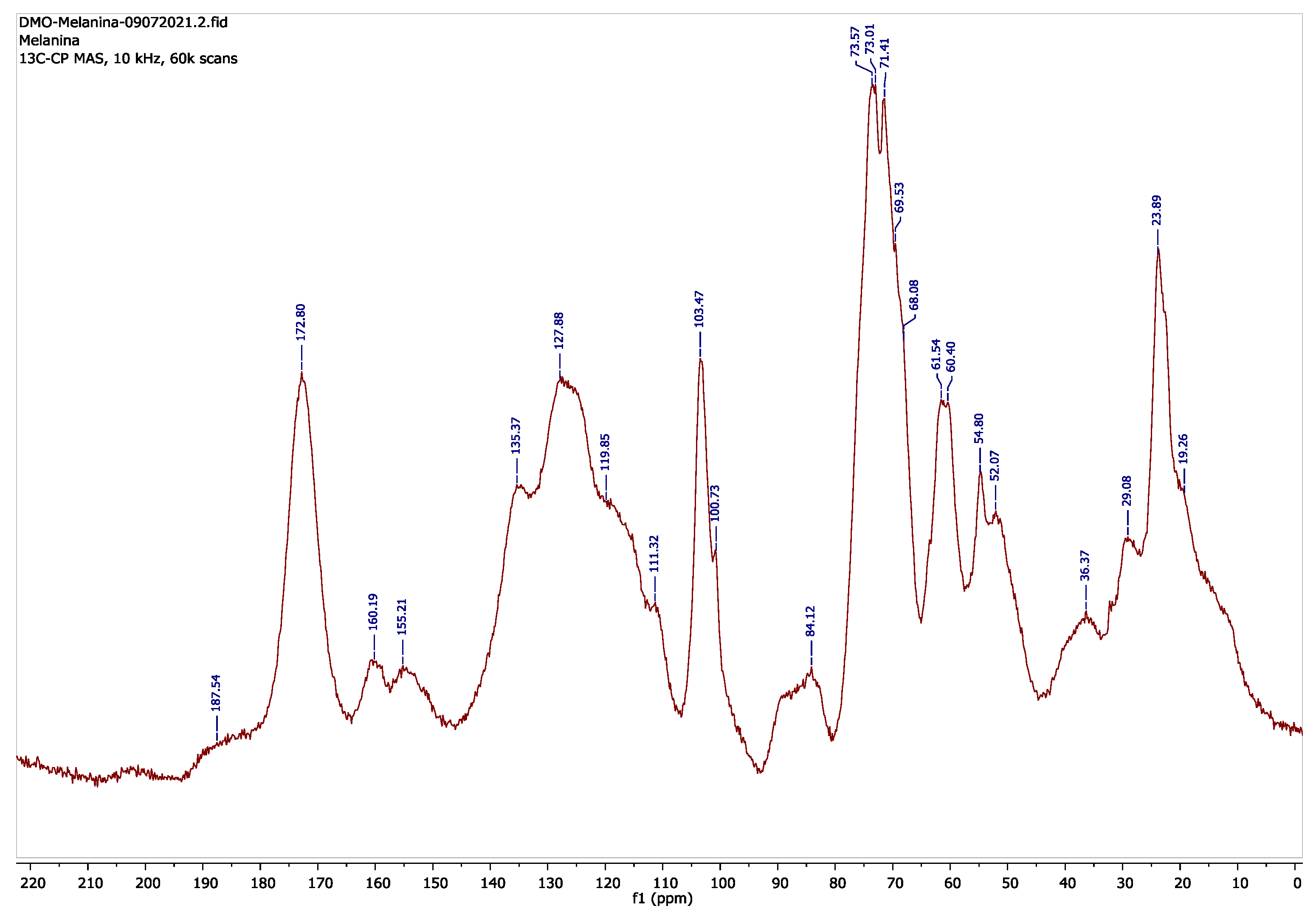
2.3. Analysis of the Melanin Composition

Direct samples of the sclerotia and the purified extracted melanin were analyzed by a Fourier transform infrared spectrophotometer (FTIR) in a Perkin Elmer SPECTRUM 100 infrared spectrophotometer by using a diamond attenuated total reflectance (ATR) device at a resolution of 4 cm−1 in the wavenumber region of 4000–650 cm−1. The purified melanin was analyzed by 13C NMR in a solid state by using a Bruker Avance III HD 400 MHz instrument operating at a frequency of 100.613 MHz employing ZrO2 rotors of 4 mm OD and 50 μL. The cross-polarization (CP) technique was applied during the magic-angle spinning (MAS) of the rotor at 10 kHz; 60,000 scans were accumulated (26 h) and the data were processed in MestreNova software version 6.

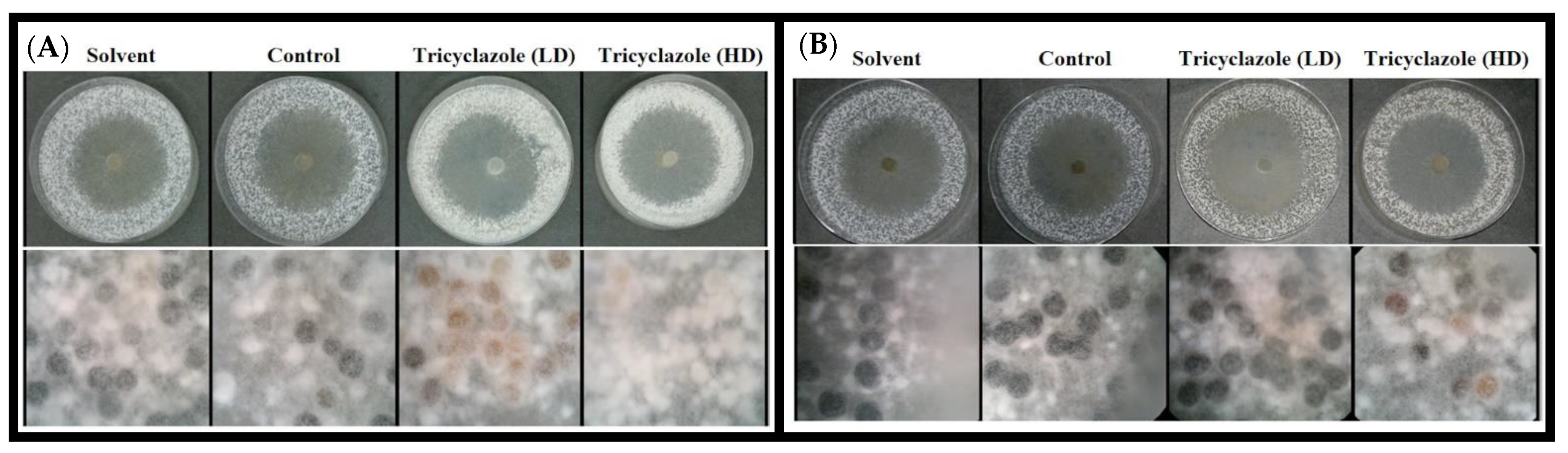
2.4. Susceptibility of S. cepivorum to Tricyclazole

The S. cepivorum susceptibility test was developed as previously described by Froyd et al. but with slight modifications [21]. Briefly, PDA discs (6 mm) containing the S. cepivorum mycelium were inoculated on PDA plates with two doses of tricyclazole (SIGMA-ALDRICH™, CAS number 41814-78-2, Burlington, MA, USA), one high (400 μg/mL) and one low (100 μg/mL) using a concentrated tricyclazole solution (10 mg/mL in absolute ethanol). Two control samples were used; the first contained absolute ethanol and the other contained neither alcohol nor tricyclazole. The number of sclerotia produced was counted at day 13 post-inoculation by using a Stratagene Eagle Eye II Imaging SystemTM. Three independent experiments with duplicates were performed and an ANOVA statistical analysis was used to describe the data.

2.5. Isolation of S. cepivorum DNA, RNA, and cDNA

DNA was isolated as reported by Raeder and Broda [22], using the mycelium grown on PDA plates for four days. The DNA obtained was washed with 70% (v/v) ethanol and kept at −70 °C until used. The total RNA was isolated with TriZolTM and the cDNA was synthesized following a protocol previously standardized in a laboratory [23].

2.6. Expression of S. cepivorum SDH Protein in P. pastoris (X-33) Cells

SDH cDNA was amplified by using the D4 forward primer 5′ CTGCAGCATCCACTATGGCTCAAGACAGAATTTCTTTTG-3 and the R6 reverse primer 5′-GCGGCCGCGTACTGCTTAAAACCCCCTTAAAAACC-3′ (underlined bases indicate PstI and NotI sites, respectively; see extended data in Supplementary Table S1) to yield a 522 bp amplicon. The amplicon was cloned into pCR2.1-TOPO (InvitrogenTM), Waltham, MA, USA) and then subcloned into the PstI and NotI sites of pPICZαB (InvitrogenTM), generating pPICZαB/sdh. As the vector conferred a resistance to ZeocinTM, P. pastoris transformant cells were selected in a YPD medium supplemented with 100 μg/mL of ZeocinTM. The production of the recombinant protein was achieved by growing the methylotrophic yeast cells in a buffered methanol-complex medium (BMMY) (1% yeast extract, 2% casein peptone, 100 mM potassium phosphate, pH 6.0, 1.34% bacto-yeast nitrogen base (YNB) w/v, 4 × 10⁻⁵% biotin w/v, 1% glycerol v/v, 4 × 10⁻³% histidine w/v) and the expression was induced with methanol to a final concentration of 1% following Pichia Expression Kit (ThermoFisher EasySelectTM, Waltham, MA, USA).The purification of the SDH recombinant was performed by using Ni-NTA Sepharose (1 mL column, eluted by gravity) for the purification of the poly histidine-containing recombinant proteins (Life Technologies™, Grand Island, NY, USA) following the instructions provided by the manufacturer. The protein was analyzed by 12% SDS-PAGE stained with Coomassie brilliant blue as reported by Laemmli [24].

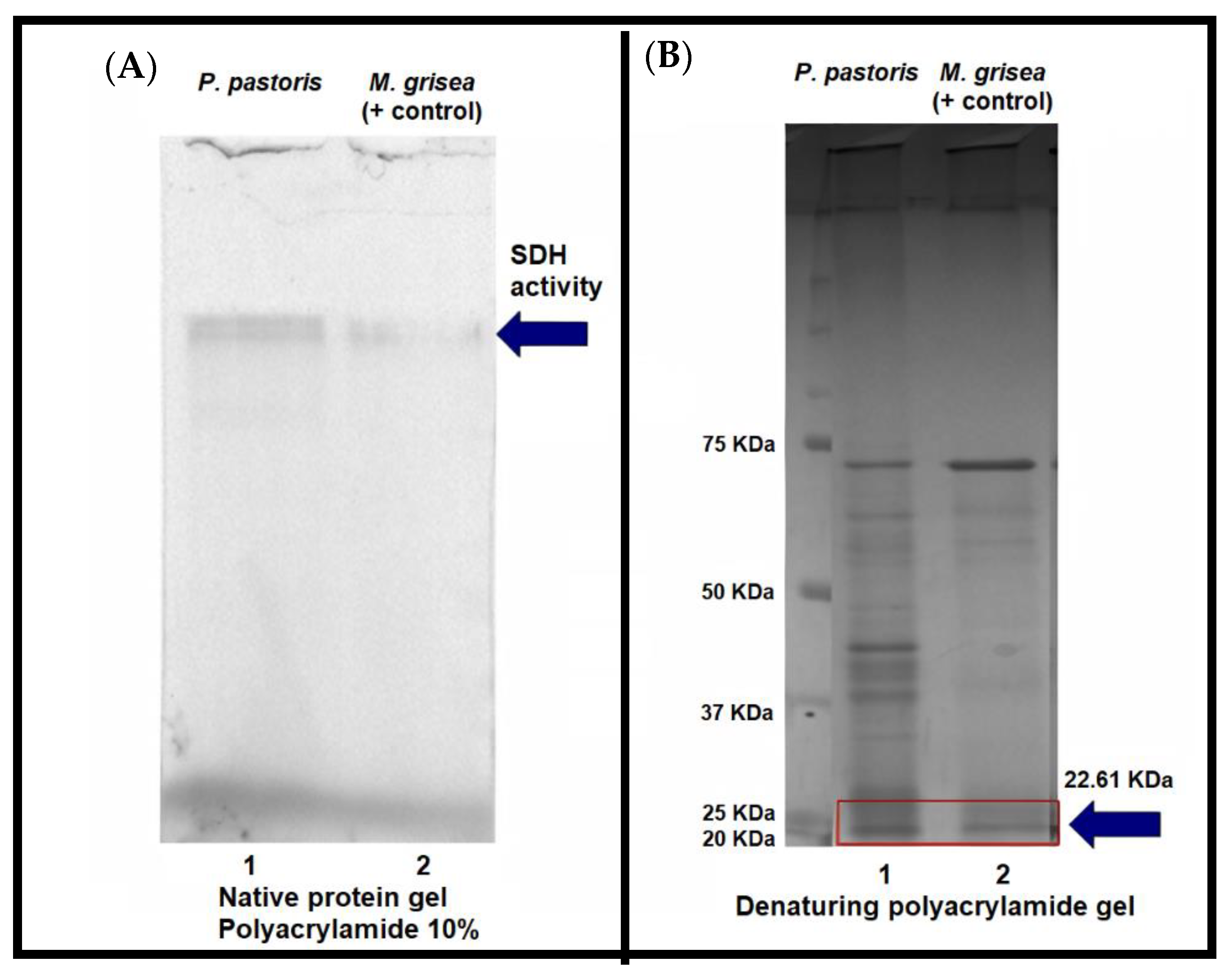
2.7. Zymogram Assay for Recombinant SDH Activity

Native polyacrylamide gel electrophoresis was performed by using 10% acrylamide gel in a Tris–boric–EDTA (8.9 mM Tris base, 8.9 mM boric acid, 0.2 mM EDTA) buffer with a pH of 8.0 as reported by Laemmli [24]. Once the electrophoresis finished, the gel was incubated for 30 min with 25 μM fluorinated coumarin, an SDH substrate (Ethyl-2-hydroxy-2-(trifluoromethyl)-2H-chromene-3-carboxylate; see Supplementary Material File S1 and Supplementary Figures S1–S3). The SDH activity was registered by using Chemi Doc (BIORADTM) equipment with a fluorescence filter for SyBr GreenTM with 2 s of exposure. The cell extracts of M. grisea 70-15 were used as a positive control for the enzyme activity.

2.8. Cell extract Obtention from Magnaporthe grisea

The protocol was based on Wheeler [25] with slight modifications. M. grisea mycelium growth of 7 days in PDA was collected, transferred to 100 mL of PDB, and incubated for 72 h at 28 °C with stirring at 180 rpm. The mycelium was harvested by filtration, washed with Na3PO4 0.1 N, pH 7, mixed with glass beads and placed in an MSK Braun homogenizer. The bottle was filled to one third of the volume with glass beads, a second third with the mycelium in a 0.1 N Na3PO4 buffer, pH 7, and subjected to disruption under the following conditions: 5 s with CO2 and 15 s without CO2. This was repeated 6 times. The sample was centrifuged at 3000× g for 5 min to recover the cell-free extract (supernatant).

2.9. Microscopy Images

The fungal cells and sclerotia were examined by microscopy using a ZeissTM microscope and an Axiocam MRc camera. All images were captured by using the same exposure settings (white light).

3. Results

3.1. Spectroscopy-Based Analysis of Pigment Extracted from S. cepivorum

An FTIR was used to analyze the S. cepivorum melanin and to provide an initial insight into its pathway. The IR analysis by diamond ATR was used to facilitate the handling of these solid compounds and we obtained the sclerotia- and melanin-extracted spectra; both were compared by overlapping, obtaining similar spectra where the most important signals were present in both samples (Figure 1).
The spectra were analyzed, and their peaks were described to characterize the specified bonds and chemical groups (Table 1). The different products generated during the melanin synthesis made it possible to realize an initial suggestion to differentiate between the melanogenic pathways involved [26] among the signals; those located at 3300 cm−1–3000 cm−1 could help to make a proposal because in the DOPA-melanin route, a sharp peak of medium intensity is characteristic of nitrogen functional groups [27] and this did not appear and the wide and high-intensity peak could be associated with the hydroxyl group in the DHN pathway (Table 1) [26,28,29].
A 13C RMN technique was used to complement our FTIR results and support the initial proposal about the DHN-melanin pathway. The purified melanin was ground again, and 75 mg was placed and compacted in a rotor (ZrO2, 4 mm OD); the spectrum is depicted in Figure 2. The 13C spectrum data are described in Table 2 and the prediction of the chemical shifts of the carbons presented in the dimers of 1,8 dihydroxynaphthalene was made according to Manini and collaborators [30] by using MestreNova software version 6. The prediction was made because the literature did not report a 13C NMR spectrum in the solid state of melanin produced by a fungus through the 1,8 DHN source that was free of other components of the cell wall such as the residues of proteins, chitin, polysaccharides, or lipids.

3.2. S. cepivorum Melanization Is Affected by Treatment with Tricyclazole

Tricyclazole has been used against rice blast caused by M. grisea [34]; this compound acts as a competitive inhibitor of the reduction complex formed between the 3-hydroxynaphthalene reductase, a specific enzyme in the DHN pathway, with nicotinamide adenine dinucleotide phosphate (NADPH) in the melanin synthesis [17,25].
In laboratory conditions, the sclerotia development started at 9 days on a PDA plate at 18 °C. The effect of tricyclazole on the S. cepivorum growth showed atypical sclerotia development, unlike that observed in the controls (Figure 3A). When 100 µg/mL of tricyclazole was used, the sclerotia presented an uncommon coloration and when a concentration of 400 µg/mL of the tricyclazole was used, the sclerotia did not melanize. On day 13, the sclerotia pigmentation was not completely present in the assays with 100 μg/mL and 400 µg/mL of tricyclazole (Figure 3B).
The quantification of the sclerotia number was performed on day 13 by using a Stratagene Eagle Eye II Imaging System TM based on the opacity and spherical shape of the samples; therefore, we associated the inhibition caused by tricyclazole when the sclerotia showed an effect in the melanization, delayed pigmentation time, and reduced number of pigmented sclerotia formations compared with the controls (Figure 4). In this way, the effect of tricyclazole on the melanization of the sclerotia from S. cepivorum was evaluated. The results showed that tricyclazole delayed the melanization of the sclerotia, suggesting—together with the FTIR and NMR data—that the melanization of the S. cepivorum sclerotia was through the DHN-melanin pathway as well as S. sclerotiorum [7].

3.3. Identification and Obtainment of a S. cepivorum Scytalone Dehydratase Gene

Once the possible DHN melanization pathway in Sclerotium cepivorum was established, we focused on the enzymatic pathway reported for this molecule [35]. We were interested in the scytalone dehydratase protein (SDH) because of its two-step participation in the melanization pathway [35]. We obtained the open reading frame (ORF) of the protein by sequencing the strain genome (S. cepivorum UGTO-Sc1) and comparing this with the reported genome (S. cepivorum GCA_014898415 and GCA_002162485); SDH ORF was deposited (GenBank: MK965546.1).
We compared the reported sequences of SDH and found high conserved regions in 5′ but a variable length in the 3′ regions even for other SDHs of Sclerotinia sclerotiorum strains (Supplementary Figure S4), indicating variations in this protein among the same species. We also compared our SDH sequence with several other SDHs reported in S. cepivorum strains and obtained a high alignment score in the nucleotide sequences (Supplementary Figure S5). A distance tree using the aa sequence indicated a relationship with Sclerotinia sclerotiorum, with an SDH of 167 amino acids (GenBank: APA08377.1) and a relationship with the SDH of the Botrytis species (Supplementary Figure S6). Furthermore, a phylogenetic tree based on our SDH aa sequence (GenBank: QJQ72301.1) compared with the reported sequences was constructed, revealing the relation with other fungi SDHs (Supplementary Figure S7).
A bioinformatics analysis specifically indicated a cluster of genes involved in the melanin synthesis such as the SDH, 1,3,8-naftalen thiol reductase, transcription factor Cys6, a transcription factor with C6 finger domains, and an alpha-beta hydrolase, which were also reported in Alternaria alternata, Botrytis cinerea, and S. sclerotiorum [36]. We cloned the cDNA of the sdh gene into the pPICZαB vector to generate the construct pPICZαB/sdh. The transformed P. pastoris X-33 cells were analyzed to verify the correct integration of the construct pPICZαB/sdh into the yeast genome by using different pairs of primers (Supplementary Table S1). A 1115 bp amplicon (AOX-F and AOX-R primers were used) confirmed the complete integration of the construct in the yeast genome. The sdh amplicon of 522 pb was verified with D4 and R6 primers (Supplementary Figure S8).
Finally, to ensure the protein expression and to corroborate that the cDNA of sdh was in phase, the amplicon obtained with the AOX primers was sequenced, validating the correct insertion of the sdh cDNA. Once the positive clone was selected, the expression of the SDH was performed (Figure 5). We annotated the SDH protein in the GenBank database (accession number MK965546.1).

3.4. S. cepivorum Scytalone Dehydratase Recombinant Activity

The SDH has a key role in melanin synthesis because it catalyzes two steps in the DHN-melanin pathway [37,38]. We induced the SDH heterologous expression in P. pastoris [39]. The recombinant SDH protein was analyzed by electrophoretic mobility under native conditions and its activity was revealed by using a fluorinated coumarin (Supplementary Material File S1). A zymogram assay (Figure 6) showed the activity of the recombinant SDH. M. grisea SDH was used as a positive control for the enzymatic activity [37,40].

4. Discussion

Melanin is an important compound in fungi protection against several factors such as UV radiation and dehydration as well as chemical and mechanical stressors [10,41]. It also protects against lytic enzymes produced by soil microbes [42] and it can be a determinant of virulence [43]. As aforementioned, melanin is a critical compound for fungi cells; for instance, its presence is necessary to develop appressorium in M. grisea [43] and for lifespan in S. sclerotiorum [7,12]. It has been inferred that Sclerotium cepivorum could have the same melanin synthesis route as S. sclerotiorum because of its evolutionary closeness [17]. Nevertheless, the lifecycle of S. sclerotiorum and its plant pathogenesis is different from the life cycle and plant pathogenesis of S. cepivorum. Melanin deficiency affects sclerotium development but not its pathogenicity [7,12]. However, for S. cepivorum, a deficiency in melanin production will affect the maturation of the sclerotia, impacting on its ability to resist changes in the environment and remain viable in the soil for long periods [44]. S. cepivorum is an exclusive pathogen of plant of the genus Allium (such as garlic, onion, scallion, shallot, leek, and chives) because of the gemination signals that the sclerotia detect from plant-secreted sulfur compounds [1], hence the importance of knowing the melanin biosynthesis pathway in S. cepivorum as a possible control mechanism for white rot disease in Allium plants.
SDH is a key enzyme in melanin biosynthesis by the DHN pathway and SDH has been studied in different fungi such as M. grisea [43], Bipolaris oryzae [45], and S. sclerotiorum [7,12]. There are no specific reports of SDH in S. cepivorum, a fungus that belongs to the sclerotinacea family. There is a study by Wheeler [25] where the effect of tricyclazole on the melanin synthesis of mycelium homogenates of S. cepivorum was determined; a strong in vitro inhibition of 1,3,6,8-THN to scytalone was obtained and an accumulation of other melanin intermediates (using 159 μM of tricyclazole) established that the dehydration reaction of scytalone and vermelone was not affected by tricyclazole. Melanin synthesis inhibition in Monilia fructicola causes the sclerotia, mycelia, and spores to be less environmentally persistent [46]. In M. grisea, the presence of inhibitors of the DHN pathway such as tricyclazole could not penetrate the host [47] and it also lost its pathogenicity and its virulence decreased [43]. Given the importance of melanin and the poorly studied melanin synthesis pathway in S. cepivorum, we were interested to know the route to improve the knowledge of this phytopathogenic fungus. IR spectroscopy was used to identify the functional groups of compounds and we used this to obtain initial insights to enable us to discriminate between the two melanins. The DOPA-melanin pathway is more common in Basidiomycetes whereas Ascomycetes use the DHN-melanin pathway and the final compounds have differential signal groups [48]. We used an FTIR to analyze the sclerotia pigment, taking advantage of the properties of melanin such as being soluble in alkali solutions, insoluble in water, and being able to be extracted from S. cepivorum by precipitation in an HCl solution [49,50]. The sclerotia and purified pigment were analyzed and, when comparing the spectrum data, we confirmed that the pigment covering the sclerotia and the purified pigment were the same (Figure 1).
The samples were analyzed at a resolution of 4 cm−1 (4000–650 cm−1), obtaining the signals of the functional groups present in the extracted melanin (Table 1). Similar data were reported for Phyllosticta capitalensis, an endophytic fungus [51]. The signals between 3300–3000 cm−1 helped to provide initial data about the type of melanin; if the DOPA-melanin route was carried out by S. cepivorum, we would have expected a sharp peak of medium intensity instead of the wide and high-intensity obtained peak (Figure 1C). Furthermore, in the DOPA-melanin route, nitrogen-linked carbon elongation strong intensity signals would have been expected for aromatic amine between 1340–1260 cm−1; these were not observed. The signals presented between 1410–1310 cm−1 and 1260–1180 cm−1 were characteristic for phenol derivatives and referred to C-O stretching and H in plane deformations [28], giving a proposal of the DHN-melanin pathway. The DHN-melanin extracted from Aspergillus fumigatus [29] was analyzed by IR and compared with synthetic melanin. The same characteristic signals were reported for both samples; signals that were observed in our FTIR spectra. Regarding the possibility that it may have been pyomelanin, another substitution pattern would have been observed in the deformation signals in the in plane and outside this for the aromatic nucleus different from the 1:2:3 substitution. The characteristic signals of phenols and their derivatives also would not appear; therefore, the pyomelanin pathway was doubtful as the pyomelanin pathway is reported only in a few mushrooms and basidiomycetes [52]. Here, we were capable of assigning a molecular and bound functional group description to each peak (Table 1) and when comparing our data with the signals reported for A. fumigatus [29], we found similar peaks describing DHN-melanin. Furthermore, our IR melanin spectra differed from the eumelanin-reported spectra, specifically for the sharp shape in the 3200–3000 cm−1 region [53]. At this point, the FTIR only partially suggested a tendency of DHN-melanin. 13C RMN was performed to support the FTIR data and the proposed DHN-melanin pathway. 13C RMN showed a chemical shift at 160.19 ppm and 135.77–111.32 ppm (Figure 2 and Table 2) as was reported by Raman and Ramasamy [29] for an Aspergillus fumigates DHN-extracted melanin. Table 2 also describes the chemical shifts corresponding with the chemical groups with nitrogen that are associated with chitin and related glucans but not with DOPA-melanin; therefore, an inhibition test was performed to support the data.
We performed the melanin inhibition test by using tricyclazole, a known inhibitor of the enzymatic reduction of trihydroxynaphthalene to vermelone, which are DHN-melanin pathway intermediates [50,54]. The inhibition assays showed that tricyclazole disturbed the normal development of the sclerotia, retarding the pigmentation process (Figure 3) and suggesting that tricyclazole affected the S. cepivorum melanogenic pathway, as reported in Verticillium dahlia with the same antifungal drug [54]. The observed delayed pigmentation was comprehensible because tricyclazole is a competitive inhibitor [55] and the fungus will produce the enzyme until the complete maturation of the sclerotia. Furthermore, the ANOVA analysis (Figure 4) indicated a significant difference between the treatments compared with the controls; therefore, a notorious effect existed on the development and melanization of S. cepivorum because of the tricyclazole. Additionally, we did not find in the genome of our S. cepivorum UGTO-Sc1 strain any genes related to the DOPA-melanin pathway such as tyrosinase (TYR), tyrosinase-related protein (TRP1), or dopachrome tautomerase (TRP2). At this point, we discarded the DOPA-melanin pathway and suggested that S. cepivorum used the DHN-melanin pathway for sclerotia melanization. Although tricyclazole has been used to control M. grisea, the side effects that it produces on the host such as accumulating on the plant leaves [56] makes the need to search for new strategies for the control of the disease urgent. The SDH enzyme represents a target for the development of new antifungal drugs.
The central role played by the genes that take part in the DHN melanogenic pathway of fungi makes them attractive candidates to study for the development of strategies that allow the control of phytopathogenic fungi. We identified the sdh gene from S. cepivorum in a typical biosynthetic gene cluster [57,58] and the arrangement of the genes was like that found in S. sclerotiorum (Ortega-Cuevas, [59]). The ORF sdh gene bioinformatic analysis indicated that the SDH was a protein of 167 aa with a molecular weight of 22.61 kDa (Figure 5). The sdh amino acid sequence was very similar between S. cepivorum and S. sclerotiorum (Supplementary Figure S4). Modeling on the platform swissmodel.expasy.org (data not shown) presented a similar structure to other reported fungi scytalone dehydratases [58]. The S. cepivorum sdh gene was analyzed and compared with the sdh gene of S. sclerotiorum, showing the differences between them such as the number and size of the introns of each gene as well as the size of the exons that comprise them (GenBank: SS1G_13314 and MK965546.1).
In the absence of effective methods to transform S. cepivorum, we carried out the experiments with a recombinant enzyme as previously reported for P. oryzae [60]. Interestingly, the recombinant enzyme, expressed in P. pastoris X-33, showed activity against a fluorinated coumarin substrate (Figure 6). In addition, the recombinant protein had in its sequence the amino acid valine 75, which is responsible for the enzymatic activities of converting scytalone to 1,3,8-trihydroxynaphthalene, as reported in M. oryzae [50].
Here, the gene identification, expression, and activity of the SDH added relevant information for the melanin synthesis pathway of S. cepivorum and the members of this family of phytopathogens fungi. Finally, our data suggested that the S. cepivorum SDH took part in the melanization pathway. Knowledge of the enzymes involved in melanin biogenesis could be used to select an appropriate target for developing strategies against white rot plant disease.

5. Conclusions

The 13C NMR and IR results, the bioinformatic results obtained, the determination of the presence of cDNA in the extracted RNA, and the expression of the enzymatic activity together with the results of the retardation of melanization by tricyclazole strongly suggested that the melanogenic route involved in S. cepivorum was the DHN pathway.

Supplementary Materials

The following are available online at https://www.mdpi.com/article/10.3390/microbiolres13020013/s1. File S1. Obtaining the Ethyl 2-hydroxy-2-(trifluoromethyl)-2H-chromene-3-carboxylate [61,62]. Supplementary Figure S1. Ethyl 2-hydroxy-2-(trifluoromethyl)-2H-chromene-3-carboxylate 1H NMR spectrum (500 MHz, CDCl3) δ: 1.42 (t, J = 7.2 Hz, 3H, CH3), 4.39 (q, J = 7.2 Hz, 2H, CH2), 7.02-7.06 (m, 2H), 7.26 (dd, J = 7.9, 1.7 Hz, 1H), 7.39 (m, 1H), 7.45 (s, 1H, OH), 7.79 (s, 1H). Supplementary Figure S2. Ethyl 2-hydroxy-2-(trifluoromethyl)-2H-chromene-3-carboxylate 13C-NMR spectrum (CDCl3, 125 MHz) δ: 14.1 (CH3), 62.4 (CH2), 95.3 (q, JC,F = 34.8 Hz, CCF3), 114.8, 116.0, 117.6, 121.5 (q, JC,F = 290 Hz, CF3), 122.7, 129.5, 133.9, 139.3, 152.7, 166.8 (C=O). Supplementary Figure S3. Ethyl 2-hydroxy-2-(trifluoromethyl)-2H-chromene-3-carboxylate HMQS spectrum of 2D-NMR (CDCl3). Supplementary Figure S4. Sclerotinia sclerotiorum alignment. Comparison between SDH of Sclerotinia sclerotiorum (Sequence ID: EDN98455.1) and another SDH of different size. NCBI alignment. Supplementary Figure S5. Multiple alignment of the Scytalone dehydratase gene from Sclerotium cepivorum in Gene Bank; NGKD01000004.1 genome sequenced by Kohn., RCTD01000004.1 genome sequenced by Valero-Jimenez et al. [63] and MK965546.1 complete SDH cds. Supplementary Figure S6. Distance tree SDH of S. cepivorum UGTO-Sc1 (GenBank: MK965546.1) [64]. Supplementary Figure S7. Phylogenetic tree of S. cepivorum UGTO-Sc1 SDH [65,66,67,68,69,70,71,]. Supplementary Figure S8. Sclerotinia sclerotiorum alignment. Comparison between SDH of Sclerotinia sclerotiorum (Sequence ID: EDN98455.1) and another SDH of different size. NCBI alignment. Supplementary Table S1. Primers used for the expression of the SDH heterologous protein. Expression performed in Pichia pastoris by using a pPICZα plasmid (InvitrogenTM).

Author Contributions

Conceptualization, L.M.S.-G. and P.P.-N.; methodology, L.M.S.-G., R.I.O.-C., R.A.M.-Á., J.A.M.-Á. and S.E.G.-H.; analytical experiments, L.M.S.-G., D.M.-O. and M.Á.V.; formal analysis, A.F.-M., L.M.S.-G., D.M.-O., M.Á.V. and P.P.-N.; investigation, A.F.-M., L.M.S.-G. and P.P.-N.; resources, A.F.-M. and P.P.-N.; data curation, L.M.S.-G., R.I.O.-C., A.F.-M. and P.P.-N.; writing-original draft preparation, L.M.S.-G.; writing-review and editing, L.M.S.-G., A.F.-M., M.Á.V. and P.P.-N.; supervision, S.E.G.-H., J.A.M.-Á., P.P.-N. and A.F.-M.; project administration, S.E.G.-H. and P.P.-N.; funding acquisition, A.F.-M. and P.P.-N. All authors contributed to the article and approved the submitted version. All authors have read and agreed to the published version of the manuscript.

Funding

This work was supported by funding from CONACYT-SAGARPA (2005-11919) and Dirección de Apoyo a la Investigación y al Posgrado (DAIP-UG: 2019,2020), Universidad de Guanajuato.

Institutional Review Board Statement

Not applicable.

Informed Consent Statement

Not applicable.

Acknowledgments

Luis M. Salazar-García (788756) and Rocío I. Ortega-Cuevas (520959) acknowledge support from CONACYT scholarships. We are grateful to Fernando Amézquita López and Laboratorio de Análisis Instrumental, DCNE, UGTO, for the analytical equipment time provided, and to Daniel Ruiz Plaza from Laboratorio Nacional de Caracterización de Propiedades Fisicoquímicas y de Estructura Molecular (LACAPFEM) de la Universidad de Guanajuato as well as Sebastien Santini (CNRS/AMU IGS UMR7256) and the PACA Bioinfo platform (supported by IBISA) for the availability and management of the phylogeny.fr website used for the phylogenetic tree development.

Conflicts of Interest

The authors declare that the research was conducted in the absence of any commercial or financial relationships that could be construed as a potential conflict of interest.

References

  1. Coley-Smith, J.; King, J. The production by species of Allium of alkyl sulphides and their effect on germination of sclerotia of Sclerotium cepivorum Berk. Ann. Appl. Biol. 1969, 64, 289–301. [Google Scholar] [CrossRef]
  2. Coley-Smith, J. White rot disease of Allium: Problems of soil-borne diseases in microcosm. Plant Pathol. 1990, 39, 214–222. [Google Scholar] [CrossRef]
  3. Willetts, H.; Bullock, S. Developmental biology of sclerotia. Mycol. Res. 1992, 96, 801–816. [Google Scholar] [CrossRef]
  4. Coley-Smith, J.R.; Parfitt, D. Some effects of diallyl bisulphide on sclerotia of Sclerotium cepivorum: Possible novel control method for white rot disease of onions. Pestic. Sci. 1986, 17, 587–594. [Google Scholar] [CrossRef]
  5. Fullerton, R.; Stewart, A. Chemical control of onion white rot (Sclerotium cepivorum Berk.) in the Pukekohe district of New Zealand. N. Z. J. Crop Hortic. Sci. 1991, 19, 121–127. [Google Scholar] [CrossRef]
  6. Entwistle, A. Allium white rot and its control. Soil Use Manag. 1990, 6, 201–208. [Google Scholar] [CrossRef]
  7. Butler, M.J.; Gardiner, R.B.; Day, A.W. Melanin synthesis by Sclerotinia sclerotiorum. Mycologia 2009, 101, 296–304. [Google Scholar] [CrossRef]
  8. Cordero, R.J.; Casadevall, A. Functions of fungal melanin beyond virulence. Fungal Biol. Rev. 2017, 31, 99–112. [Google Scholar] [CrossRef]
  9. Arazoe, T.; Miyoshi, K.; Yamato, T.; Ogawa, T.; Ohsato, S.; Arie, T.; Kuwata, S. Tailor-made CRISPR/Cas system for highly efficient targeted gene replacement in the rice blast fungus. Biotechnol. Bioeng. 2015, 112, 2543–2549. [Google Scholar] [CrossRef]
  10. Carbone, I.; Kohn, L.M. Ribosomal DNA sequence divergence within internal transcribed spacer 1 of the Sclerotiniaceae. Mycologia 1993, 85, 415–427. [Google Scholar] [CrossRef]
  11. Xu, Z.; Harrington, T.C.; Gleason, M.L.; Batzer, J.C. Phylogenetic placement of plant pathogenic Sclerotium species among teleomorph genera. Mycologia 2010, 102, 337–346. [Google Scholar] [CrossRef] [PubMed] [Green Version]
  12. Liang, Y.; Xiong, W.; Steinkellner, S.; Feng, J. Deficiency of the melanin biosynthesis genes SCD1 and THR1 affects sclerotial development and vegetative growth, but not pathogenicity, in Sclerotinia sclerotiorum. Mol. Plant Pathol. 2018, 19, 1444–1453. [Google Scholar] [CrossRef] [PubMed] [Green Version]
  13. Eisenman, H.C.; Casadevall, A. Synthesis and assembly of fungal melanin. Appl. Microbiol. Biotechnol. 2012, 93, 931–940. [Google Scholar] [CrossRef] [PubMed] [Green Version]
  14. Mattoon, E.R.; Cordero, R.J.; Casadevall, A. Fungal Melanins and Applications in Healthcare, Bioremediation and Industry. J. Fungi 2021, 7, 488. [Google Scholar] [CrossRef]
  15. Langfelder, K.; Streibel, M.; Jahn, B.; Haase, G.; Brakhage, A.A. Biosynthesis of fungal melanins and their importance for human pathogenic fungi. Fungal Genet. Biol. 2003, 38, 143–158. [Google Scholar] [CrossRef]
  16. Miñambres, G.G.; Conles, M.Y.; Lucini, E.I.; Verdenelli, R.A.; Meriles, J.M.; Zygadlo, J.A. Application of thymol and iprodione to control garlic white rot (Sclerotium cepivorum) and its effect on soil microbial communities. World J. Microbiol. Biotechnol. 2010, 26, 161. [Google Scholar] [CrossRef]
  17. Wheeler, M.H.; Bell, A.A. Melanins and their importance in pathogenic fungi. In Current Topics in Medical Mycology; Springer: Berlin/Heidelberg, Germany, 1988; pp. 338–387. [Google Scholar]
  18. Saitoh, Y.; Izumitsu, K.; Morita, A.; Tanaka, C.; Shimizu, K. Cloning of Sal1, a scytalone dehydratase gene involved in melanin biosynthesis in Cochliobolus heterostrophus. Mycoscience 2012, 53, 330–334. [Google Scholar] [CrossRef]
  19. Sezonov, G.; Joseleau-Petit, D.; d’Ari, R. Escherichia coli physiology in Luria-Bertani broth. J. Bacteriol. 2007, 189, 8746–8749. [Google Scholar] [CrossRef] [Green Version]
  20. Gadd, G. Effects of media composition and light on colony differentiation and melanin synthesis in Microdochium bolleyi. Trans. Br. Mycol. Soc. 1982, 78, 115–122. [Google Scholar] [CrossRef]
  21. Froyd, J.; Paget, C.; Guse, L.; Dreikorn, B.; Pafford, J. Tricyclazole: A new systemic fungicide for control of Pyricularia oryzae on rice. Phytopathology 1976, 66, 1135–1139. [Google Scholar] [CrossRef]
  22. Raeder, U.; Broda, P. Rapid preparation of DNA from filamentous fungi. Lett. Appl. Microbiol. 1985, 1, 17–20. [Google Scholar] [CrossRef]
  23. Trujillo-Esquivel, E.; Franco, B.; Flores-Martínez, A.; Ponce-Noyola, P.; Mora-Montes, H.M. Purification of single-stranded cDNA based on RNA degradation treatment and adsorption chromatography. Nucleosides Nucleotides Nucleic Acids 2016, 35, 404–409. [Google Scholar] [CrossRef] [PubMed]
  24. Laemmli, U.K. Cleavage of structural proteins during the assembly of the head of bacteriophage T4. Nature 1970, 227, 680–685. [Google Scholar] [CrossRef] [PubMed]
  25. Wheeler, M.H. Comparisons of fungal melanin biosynthesis in ascomycetous, imperfect and basidiomycetous fungi. Trans. Br. Mycol. Soc. 1983, 81, 29–36. [Google Scholar] [CrossRef]
  26. Simon, W.; Clerc, T. Elucidación Estructural de Compuestos Orgánicos por Métodos Espectroscópicos; Alhambra: Madrid, Spain, 1970. [Google Scholar]
  27. Rajagopal, K.; Kathiravan, G.; Karthikeyan, S. Extraction and characterization of melanin from Phomopsis: A phellophytic fungi isolated from Azadirachta indica A. Juss. Afr. J. Microbiol. Res. 2011, 5, 762–766. [Google Scholar]
  28. Pretsch, E.; Clerc, T.; Seibl, J.; Simon, W. Tables of Spectral Data for Structure Determination of Organic Compounds; Springer Science & Business Media: Berlin/Heidelberg, Germany, 2013. [Google Scholar]
  29. Raman, N.M.; Ramasamy, S. Genetic validation and spectroscopic detailing of DHN-melanin extracted from an environmental fungus. Biochem. Biophys. Rep. 2017, 12, 98–107. [Google Scholar] [CrossRef]
  30. Manini, P.; Lino, V.; D’Errico, G.; Reale, S.; Napolitano, A.; De Angelis, F.; d’Ischia, M. “Blackness” is an index of redox complexity in melanin polymers. Polym. Chem. 2020, 11, 5005–5010. [Google Scholar] [CrossRef]
  31. Chatterjee, S.; Prados-Rosales, R.; Itin, B.; Casadevall, A.; Stark, R.E. Solid-state NMR reveals the carbon-based molecular architecture of Cryptococcus neoformans fungal eumelanins in the cell wall. J. Biol. Chem. 2015, 290, 13779–13790. [Google Scholar] [CrossRef] [Green Version]
  32. Kang, X.; Kirui, A.; Muszyński, A.; Widanage, M.C.D.; Chen, A.; Azadi, P.; Wang, P.; Mentink-Vigier, F.; Wang, T. Molecular architecture of fungal cell walls revealed by solid-state NMR. Nat. Commun. 2018, 9, 1–12. [Google Scholar] [CrossRef] [Green Version]
  33. Di Pietro, M.E.; Mannu, A.; Mele, A. NMR determination of free fatty acids in vegetable oils. Processes 2020, 8, 410. [Google Scholar] [CrossRef] [Green Version]
  34. Zhang, C.-Q.; Huang, X.; Wang, J.-X.; Zhou, M.-G. Resistance development in rice blast disease caused by Magnaporthe grisea to tricyclazole. Pestic. Biochem. Physiol. 2009, 94, 43–47. [Google Scholar] [CrossRef]
  35. Butler, M.; Day, A. Fungal melanins: A review. Can. J. Microbiol. 1998, 44, 1115–1136. [Google Scholar] [CrossRef]
  36. Amselem, J.; Cuomo, C.A.; van Kan, J.A.; Viaud, M.; Benito, E.P.; Couloux, A.; Coutinho, P.M.; de Vries, R.P.; Dyer, P.S.; Fillinger, S. Genomic analysis of the necrotrophic fungal pathogens Sclerotinia sclerotiorum and Botrytis cinerea. PLoS Genet. 2011, 7, e1002230. [Google Scholar] [CrossRef] [Green Version]
  37. Jordan, D.B.; Basarab, G.S.; Steffens, J.J.; Lundqvist, T.; Pfrogner, B.R.; Schwartz, R.S.; Wawrzak, Z. Catalytic mechanism of scytalone dehydratase from Magnaporthe grisea. Pestic. Sci. 1999, 55, 277–280. [Google Scholar] [CrossRef]
  38. Tsuji, G.; Takeda, T.; Furusawa, I.; Horino, O.; Kubo, Y. Carpropamid, an Anti-Rice Blast Fungicide, Inhibits Scytalone Dehydratase Activity and Appressorial Penetration in Colletotrichum lagenarium. Pestic. Biochem. Physiol. 1997, 57, 211–219. [Google Scholar] [CrossRef]
  39. Liachko, I.; Youngblood, R.A.; Tsui, K.; Bubb, K.L.; Queitsch, C.; Raghuraman, M.; Nislow, C.; Brewer, B.J.; Dunham, M.J. GC-rich DNA elements enable replication origin activity in the methylotrophic yeast Pichia pastoris. PLoS Genet. 2014, 10, e1004169. [Google Scholar] [CrossRef] [PubMed]
  40. Lundqvist, T.; Rice, J.; Hodge, C.N.; Basarab, G.S.; Pierce, J.; Lindqvist, Y. Crystal structure of scytalone dehydratase—A disease determinant of the rice pathogen, Magnaporthe grisea. Structure 1994, 2, 937–944. [Google Scholar] [CrossRef] [Green Version]
  41. Allam, N.G.; El-Zaher, E.A. Protective role of Aspergillus fumigatus melanin against ultraviolet (UV) irradiation and Bjerkandera adusta melanin as a candidate vaccine against systemic candidiasis. Afr. J. Biotechnol. 2012, 11, 6566–6577. [Google Scholar]
  42. Potgieter, H.; Alexander, M. Susceptibility and resistance of several fungi to microbial lysis. J. Bacteriol. 1966, 91, 1526–1532. [Google Scholar] [CrossRef] [Green Version]
  43. Chumley, F.G.; Valent, B. Genetic analysis of melanin-deficient, nonpathogenic mutants of Magnaporthe grisea. Mol. Plant-Microbe Interact 1990, 3, 135–143. [Google Scholar] [CrossRef]
  44. Mahmoud, Y.A.; Sammour, R.H.; Mustafa, A.-Z.M.; Alhozeim, R.A. Role of sclerotia in the aggressiveness and pathogenicity of Sclerotium cepivorum. Asian J. Microbiol. Biotechnol. Environ. Sci. 2021, 23, 16–23. [Google Scholar]
  45. Moriwaki, A.; Kihara, J.; Kobayashi, T.; Tokunaga, T.; Arase, S.; Honda, Y. Insertional mutagenesis and characterization of a polyketide synthase gene (PKS1) required for melanin biosynthesis in Bipolaris oryzae. FEMS Microbiol. Lett. 2004, 238, 1–8. [Google Scholar] [PubMed]
  46. Rehnstrom, A.; Free, S. The isolation and characterization of melanin-deficient mutants of Monilinia fructicola. Physiol. Mol. Plant Pathol. 1996, 49, 321–330. [Google Scholar] [CrossRef]
  47. Thines, E.; Heidrun, A.; Weber, R.W. Fungal secondary metabolites as inhibitors of infection-related morphogenesis in phytopathogenic fungi. Mycol. Res. 2004, 108, 14–25. [Google Scholar] [CrossRef]
  48. Bell, A.A.; Wheeler, M.H. Biosynthesis and functions of fungal melanins. Annu. Rev. Phytopathol. 1986, 24, 411–451. [Google Scholar] [CrossRef]
  49. De la Rosa, J.M.; Martin-Sanchez, P.M.; Sanchez-Cortes, S.; Hermosin, B.; Knicker, H.; Saiz-Jimenez, C. Structure of melanins from the fungi Ochroconis lascauxensis and Ochroconis anomala contaminating rock art in the Lascaux Cave. Sci. Rep. 2017, 7, 13441. [Google Scholar] [CrossRef] [Green Version]
  50. Pal, A.K.; Gajjar, D.U.; Vasavada, A.R. DOPA and DHN pathway orchestrate melanin synthesis in Aspergillus species. Med. Mycol. 2014, 52, 10–18. [Google Scholar]
  51. Suryanarayanan, T.S.; Ravishankar, J.P.; Venkatesan, G.; Murali, T.S. Characterization of the melanin pigment of a cosmopolitan fungal endophyte. Mycol. Res. 2004, 108, 974–978. [Google Scholar] [CrossRef]
  52. Solano, F. Melanins: Skin pigments and much more—Types, structural models, biological functions, and formation routes. New J. Sci. 2014, 2014, 498276. [Google Scholar] [CrossRef] [Green Version]
  53. Correa, N.; Covarrubias, C.; Rodas, P.I.; Hermosilla, G.; Olate, V.R.; Valdés, C.; Meyer, W.; Magne, F.; Tapia, C.V. Differential antifungal activity of human and cryptococcal melanins with structural discrepancies. Front. Microbiol. 2017, 8, 1292. [Google Scholar] [CrossRef]
  54. Tokousbalides, M.C.; Sisler, H. Site of inhibition by tricyclazole in the melanin biosynthetic pathway of Verticillium dahliae. Pestic. Biochem. Physiol. 1979, 11, 64–73. [Google Scholar] [CrossRef]
  55. Andersson, A.; Jordan, D.; Schneider, G.; Lindqvist, Y. Crystal structure of the ternary complex of 1, 3, 8-trihydroxynaphthalene reductase from Magnaporthe grisea with NADPH and an active-site inhibitor. Structure 1996, 4, 1161–1170. [Google Scholar] [CrossRef] [Green Version]
  56. Liu, J.; Min, H.; Ye, L. The negative interaction between the degradation of phenanthrene and tricyclazole in medium, soil and soil/compost mixture. Biodegradation 2008, 19, 695–703. [Google Scholar] [CrossRef]
  57. Keller, N.P. Fungal secondary metabolism: Regulation, function and drug discovery. Nat. Rev. Microbiol. 2019, 17, 167–180. [Google Scholar] [CrossRef] [PubMed]
  58. Jia, S.-L.; Chi, Z.; Chen, L.; Liu, G.-L.; Hu, Z.; Chi, Z.-M. Molecular evolution and regulation of DHN melanin-related gene clusters are closely related to adaptation of different melanin-producing fungi. Genomics 2021, 113, 1962–1975. [Google Scholar] [CrossRef] [PubMed]
  59. Ortega-Cuevas, R.I. Clonación y Expresón Heteróloga de la Scitalona Deshidratasa de Sclerotium Cepivorum Berk. Master’s Thesis, Department of Biology, University of Guanajuato, Guanajuato, Mexico, 2015. [Google Scholar]
  60. Yamada, N.; Motoyama, T.; Nakasako, M.; Kagabu, S.; Kudo, T.; Yamaguchi, I. Enzymatic characterization of scytalone dehydratase Val75Met variant found in melanin biosynthesis dehydratase inhibitor (MBI-D) resistant strains of the rice blast fungus. Biosci. Biotechnol. Biochem. 2004, 68, 615–621. [Google Scholar] [CrossRef] [PubMed]
  61. Chizhov, D.L.; Sosnovskikh, V.Y.; Pryadeina, M.V.; Burgart, Y.V.; Saloutin, V.I.; Charushin, V.N. The first synthesis of 4-unsubstituted 3-(trifluoroacetyl) coumarins by the Knoevenagel condensation of salicylaldehydes with ethyl trifluoroacetoacetate followed by chromene-coumarin recyclization. Synlett 2008, 19, 281–285. [Google Scholar] [CrossRef]
  62. Xu, C.; Yang, G.; Wang, C.; Fan, S.; Xie, L.; Gao, Y. An efficient solvent-free synthesis of 2-hydroxy-2-(trifluoromethyl)-2H-chromenes using silica-immobilized L-proline. Molecules 2013, 18, 11964–11977. [Google Scholar] [CrossRef] [Green Version]
  63. Valero-Jiménez, C.A.; Steentjes, M.B.; Slot, J.C.; Shi-Kunne, X.; Scholten, O.E.; van Kan, J.A. Dynamicsin Secondary Metabolite Gene Clustersin Otherwise Highly Syntenic and Stable Genomes in the Fungal Genus. Genome Biol. Evol. 2020, 12, 2491–2507. [Google Scholar]
  64. Zhang, Z.; Schwartz, S.; Wagner, L.; Miller, W. A greedy algorithm for aligning DNA sequences. J. Comput. Biol. 2000, 7, 203–214. [Google Scholar] [CrossRef]
  65. Dereeper, A.; Audic, S.; Claverie, J.M.; Blanc, G. BLAST-EXPLORER helps you building datasets for phylogenetic analysis. BMC Evol. Biol. 2010, 10, 8. [Google Scholar] [CrossRef] [Green Version]
  66. Dereeper, A.; Guignon, V.; Blanc, G.; Audic, S.; Buffet, S.; Chevenet, F.; Dufayard, J.F.; Guindon, S.; Lefort, V.; Lescot, M.; et al. Phylogeny.fr: Robust phylogenetic analysis for the non-specialist. Nucleic Acids Res. 2008, 36, W465–W469. [Google Scholar] [CrossRef] [PubMed]
  67. Edgar, R.C. MUSCLE: Multiple sequence alignment with high accuracy and high throughput. Nucleic Acids Res. 2004, 32, 1792–1797. [Google Scholar] [CrossRef] [PubMed] [Green Version]
  68. Castresana, J. Selection of conserved blocks from multiple alignments for their use in phylogenetic analysis. Mol. Biol. Evol. 2000, 17, 540–552. [Google Scholar] [CrossRef] [PubMed] [Green Version]
  69. Guindon, S.; Gascuel, O. A simple, fast, and accurate algorithm to estimate large phylogenies by maximum likelihood. Syst. Biol. 2003, 52, 696–704. [Google Scholar] [CrossRef] [PubMed] [Green Version]
  70. Anisimova, M.; Gascuel, O. Approximate likelihood ratio test for branchs: A fast, accurate and powerful alternative. Syst. Biol. 2006, 55, 539–552. [Google Scholar] [CrossRef]
  71. Chevenet, F.; Brun, C.; Banuls, A.L.; Jacq, B.; Chisten, R. TreeDyn: Towards dynamic graphics and annotations for analyses of trees. BMC Bioinform. 2006, 7, 439. [Google Scholar] [CrossRef] [Green Version]
Figure 1. Overlapping FTIR spectra. (A) Direct sample and (B) purified melanin compound obtained from S. cepivorum sclerotia. The images were obtained by using a stereoscope. (C) Spectra obtained by FTIR in diamond ATR of the direct sample (black) and of the purified melanin compound (brown); the principal signals are annotated. Spectra were performed with a diamond ATR attachment Perkin Elmer SPECTRUM 100 infrared spectrophotometer (resolution of 4 cm−1 in the number region of 650 cm−1 to 4000 cm−1).
Microbiolres 13 00013 g001
Figure 2. Purified melanin 13C NMR spectrum of S. cepivorum Berk (400 MHz).
Microbiolres 13 00013 g002
Figure 3. Melanization delay by tricyclazole in S. cepivorum. (A) Sclerotia at day 9 of growth and (B) sclerotia at day 13 of growth; top panels represent the Petri dishes, and the bottom panels represent the stereoscopic photographs of sclerotia. LD = 100 μg/mL tricyclazole; HD = 400/mL μg tricyclazole.
Microbiolres 13 00013 g003
Figure 4. Effect of tricyclazole on S. cepivorum sclerotia melanization. Box and whiskers graph; the sclerotia were analyzed by using a Stratagene Eagle Eye II Imaging System TM on day 13 post-inoculation. ANOVA statistical analyses from 3 independent experiments were performed in duplicate. *** p < 0.05.
Microbiolres 13 00013 g004
Figure 5. SDH S. cepivorum expression. (A) Scytalone dehydratase ORF, a protein with 167 amino acids and a tail of six histidines and C-Myc = cutting site. (B) SDH expression 1: P. pastoris wt; 2: P. pastoris with empty vector; 3 and 4: P. pastoris with SDH gene vector. SDH = 22.61 kDa (SDS-PAGE 12%, Coomassie staining).
Microbiolres 13 00013 g005
Figure 6. Scytalone dehydratase activity. Zymogram and SDS-PAGE of the recombinant scytalone dehydratase expressed in P. pastoris. (A) Zymogram, line 1: crude culture supernatant from P. pastoris transformed with pPICZαB/sdh; line 2: positive activity control, M. grisea protein extracts, SDH detected activity is indicated. (B) SDS-PAGE: Proteins recovered from zymogram analysis; line 1, recombinant SDH protein (22.61 kDa); line 2, positive control from M. grisea.
Microbiolres 13 00013 g006
Table 1. Melanin S. cepivorum IR signals. Spectra signals of the purified compound and the direct preparation, both from S. cepivorum sclerotia. Identification based on Pretsch tables of spectral data for structure determination of organic compounds [28].
Purified CompoundDirect Sample
Frequency (cm−1)Signal Generating GroupFrequency (cm−1)Signal Generating Group
3291-O-H Elongation vibration. Polymeric association.3286-O-H Elongation vibration. Polymeric association.
3060
3005
2953
=C-H Elongation vibration. Aromatic compound.2922=C-H Elongation vibration. Aromatic compound.
1631
1547
1531
1453
-C=C- Deformation (in plane) aromatic compound.1622-C=C- Deformation (in plane) aromatic compound.
1409
1378
-C-O Elongation vibration. Characteristic of phenol and derivatives.1403
1373
-C-O Elongation vibration. Characteristic of phenol and derivatives.
1242-O-H Deformation (in plane). Characteristic of phenol and derivatives.1236-O-H Deformation (in plane). Characteristic of phenol and derivatives.
1151
1076
1029
-C-H Deformation (in plane) and aromatic ring substitution 1:2:3.1069
1027
-C-H Deformation (in plane) and aromatic ring substitution 1:2:3.
885
765
718
-C-H Deformation (out plane) and aromatic ring substitution 1:2:3.890
828
747
-C-H Deformation (out plane) and aromatic ring substitution 1:2:3.
Table 2. 13C NMR resonance assignments for S. cepivorum-extracted melanin.
Chemical Shift, ppmChemical GroupingReference MaterialsLiterature Refs.
172.80-CONHChitin[31,32]
160.19Aromatic -C-, O-naphthylPoly 1,8 DHN[31]
135.77–111.32Aromatic -C-, -C=C-Poly 1,8 DHN, likely oleoyl[31,33]
103.47, 100.73Aromatic –C-, -O-CH-O-Chitin, glucan[32]
84.12-CHOHα-1.3 glucan[32]
73.57–71.41-CHOH, -CHO-Chitin, α-1.3 glucan[32]
61.54-CH2OHChitin[32]
60.40-CH2OHα-1.3 glucan[32]
54.80-CH-NH-Chitin[32]
29.08-(CH2)- (C4–C7, C2–C16)Likely oleoyl[33]
23.89-C(=O)-CH3, -(CH2)- (C3)Chitin, likely oleoyl[32,33]
Publisher’s Note: MDPI stays neutral with regard to jurisdictional claims in published maps and institutional affiliations.

