Abstract
Several human pathogenic fungi produce melanin. One of its properties during parasitism is the protection against antifungal drugs. This occurs with the agents of chromoblastomycosis, in which DHN-melanin reduces antifungal susceptibility to terbinafine and itraconazole. Since these agents are resistant to some antifungal drugs, we investigated the role of DHN-melanin on the Fonsecaea susceptibility to amphotericin B, micafungin, fluconazole, and flucytosine, drugs that usually present high minimal inhibitory concentrations (MIC) to this genus. Seven strains from three Fonsecaea human pathogenic species were treated with tricyclazole, a DHN-melanin inhibitor, and the MIC of the treated and untreated cells were compared. A survival assay was performed to confirm the alterations in the susceptibility of strains with reduced melanization, and the chitin levels of the strains were estimated by fluorescence. Tricyclazole did not affect fluconazole and flucytosine MIC, while melanin inhibition increased susceptibility to amphotericin B. Surprisingly, DHN-melanin inhibition decreased the susceptibility to micafungin. Survival assays confirmed this result on five strains. Cell wall chitin levels of the strains were not associated with the decrease in micafungin susceptibility. The results show that DHN-melanin does not have a role in the intrinsic resistance of Fonseacaea spp. to amphotericin B, fluconazole, and flucytosine, and its inhibition may promote micafungin resistance.
1. Introduction
The denomination “dematiaceous fungi” is given to those producing high amounts of a brown-to-black pigment called melanin in their cell wall [1]. Melanin is a complex, hydrophobic, negatively charged macromolecule composed of phenolic and/or indolic subunits [2]. Several pathogenic fungi, such as Cryptococcus neoformans [3], Trichosporon asahii [4], Aspergillus fumigatus [5], Sporothrix schenckii [6], Paracoccidioides brasiliensis [7], Histoplasma capsulatum [8], and Candida glabrata [9] produce melanin. This production is usually associated with a resistance to some harsh conditions encountered during parasitism, which makes melanin an important virulence factor of these fungi. The dematiaceous fungi that cause chromoblastomycosis and/or phaeohyphomycosis, including Exophiala dermatitidis [10] and Fonsecaea pedrosoi [11], also produce melanin in their cell wall [12].
Fonsecaea spp. constitutively produce and secrete melanin, which acts as a scavenger of oxidative oxygen radicals making these fungi more resistant to host defense mechanisms. It also provides resistance and integrity to the cell wall against several chemical compounds, such as antifungal drugs, thus acting as an important virulence factor [13]. Melanin interferes with complement activation, reducing the susceptibility of melanized fungi to antifungal agents. This pigment protects the fungus against oxidative damage, acting as a trap for nitric oxide. This mechanism does not allow macrophages or other phagocytes to eliminate fungal elements and may explain, in part, the recalcitrant and chronic course of chromoblastomycosis [14].
In Fonsecaea spp., melanin synthesis occurs through the dihydroxynaphthalene (DHN) pathway [15,16]. DHN-derived melanin is constitutively synthesized through polyketides in a pathway that starts with the conversion of acetyl (or malonyl) coenzyme A into 1,3,6,8-tetrahydroxynaphthalene [17]. It is important to note that Fonsecaea spp. and other dematiaceous fungi also have the genetic machinery necessary to produce at least two other types of melanin: eumelanin and pyomelanin [18].
Tricyclazole (5-Methyl-1,2,4-Triazolo[3,4-B][1,3]Benzothiazole) is a pesticide commercially known as BIM 750 (Dow Agrosciences, Indianapolis, IN, USA). The specific target of this compound is in the DHN pathway of melanin biosynthesis, having little or no inhibitory effect on the fungus growth in culture [19]. Inhibition of this specific biochemical process with tricyclazole can be used to induce morphological changes in the cell wall of F. pedrosoi, leading to a decreased fungal resistance to mechanical lysis and macrophage killing [15,20]. It also decreases minimal inhibitory concentrations (MIC) of itraconazole, posaconazole, and terbinafine for Fonsecaea species [21].
Itraconazole and terbinafine are the most commonly used drugs in the treatment of chromoblastomycosis. Itraconazole doses of 200 to 400 mg per day are usually recommended for adults and adolescents. The length of treatment varies; however, most cases improve within 8 to 10 months [22]. In general, terbinafine doses recommended are 250 to 500 mg per day until mycological cure or resolution of skin lesions [23,24,25]. Other drugs used in the treatment include amphotericin B and flucytosine, but hospitalization is needed for the administration of the former [25]. In addition, the nephrotoxic effect of this drug and adverse reactions must be considered, which makes treatment difficult [26]. In general, terbinafine and voriconazole are the drugs with the best in vitro activities against Fonsecaea spp., with MIC lower than 0.25 mg/L. On the other hand, fluconazole, flucytosine, amphotericin B, and micafungin usually show high MIC to these fungi [27]. Since melanin protects Fonsecaea spp. against drugs with effective antifungal activity against them, we investigated if this pigment was also associated with the increased MIC (>1 mg/L) observed for drugs with reduced antifungal efficacy in this genus.
2. Materials and Methods
2.1. Strains
Seven Fonsecaea strains isolated from skin lesions of patients with chromoblastomycosis were included in this study. They were previously identified through internal transcriber spacer (ITS) sequencing [27] as Fonsecaea monophora (38833 and 41080), Fonsecaea nubica (16451, 34113, and 34242), or F. pedrosoi (19889 and 38437).
2.2. DHN-Melanin Inhibition
After growth on potato dextrose agar (PDA) medium (HiMedia Laboratories Pvt. Ltd., India) for seven days at 30 °C, the study isolates were cultured in PDA medium supplemented with tricyclazole (Sigma Chemical Corporation, St. Louis, MO, USA) solutions at final concentrations of 1 mg/L, 2 mg/L, 4 mg/L, 8 mg/L, and 16 mg/L to determine the minimum concentration of tricyclazole capable of inhibiting their DHN-melanin production. Tricyclazole was dissolved in ethanol and added to the cultures, ensuring that the final solvent concentration in the PDA did not exceed 0.6% to avoid cytotoxic effects on the strains [15]. As controls, the strains were grown in the absence of tricyclazole. Visual reading of cultures was performed after 7 days of incubation at 30 °C and 35 °C to verify color changes in macroscopic colonies in comparison to the controls. For subsequent experiments after this initial test, cultures were performed in PDA supplemented with the lowest tricyclazole concentration capable of inducing melanin production inhibition. Cultures were maintained at 30 °C or 35 °C for seven days and used to prepare inoculum for susceptibility tests.
2.3. Antifungal Susceptibility Test
In vitro antifungal susceptibility tests were performed following the guideline of the Clinical and Laboratory Standards Institute (CLSI), M38-A2 protocol [28], with a few modifications in order to evaluate dematiaceous fungi of slow growth. Amphotericin B, fluconazole, flucytosine (all from Sigma-Aldrich, St. Louis, MO, USA), and micafungin (Astellas Pharma Tech Corporation, Takaoka, Toyama, Japan) were tested. The inocula of the seven Fonsecaea strains were prepared from seven-day-old PDA cultures, supplemented or not with tricyclazole as described above. Cells were harvested in RPMI 1640 medium (Sigma-Aldrich) buffered with MOPS (Sigma-Aldrich), pH 7.0, and diluted in 96-well plates to approximately 0.4–5 × 10⁴ cells/mL. Plates were incubated at 35 °C for five days [21,27]. The MIC for amphotericin B, fluconazole, and flucytosine, as well as the minimal effective concentration (MEC) for micafungin, were determined according to the CLSI M38-A2 protocol. The reference strains Aspergillus flavus (ATCC 204304), Aspergillus fumigatus (ATCC 204305), Candida krusei (ATCC 6258), and Candida parapsilosis (ATCC 22019) were used for quality control.
2.4. Survival Assay
The test was performed as previously described by our group [29] with a few modifications to adapt the protocol to dematiaceous fungi. In brief, inocula were prepared as described above, and cells were diluted in RPMI-1640 medium at a 5 × 10⁴ conidia/mL concentration. The cultures were supplemented with the antifungal drugs at ½ MIC, as determined previously. Colony-forming units (CFU)/mL were determined immediately after the addition of antifungal drugs. Cultures were incubated in a rotary shaker at 150 rpm at 35 °C. After 2 and 24 h of incubation, CFU/mL counts were performed again to verify the early and late killing of the antifungal drugs, respectively. The control conditions consisted of RPMI-1640 cultures of each strain without the addition of antifungal drugs and RPMI-1640 medium without antifungal drugs or fungal inoculum. The same procedures were performed for both test and control cultures. Percent survival for each time point was determined by comparing the CFU/mL counts for each strain/antifungal combination with the CFU/mL for each strain cultured in RPMI-1640 alone. This experiment was performed in triplicate.
2.5. Chitin Quantification
Conidial suspensions of the seven strains were prepared as described for the inoculum for the antifungal susceptibility test. Conidia were treated with Uvitex2B (Polysciences Inc., Warrington, PA, USA) to detect chitin in their cell walls. In brief, 1 µL of 10 µg/mL was added to 1 mL of the conidial suspension. This mixture was incubated at 35 °C for 60 min. Then, conidia were washed three times with phosphate-buffered saline and observed in the Axio Observer fluorescence microscope (Zeiss, Jena, Germany). The images were analyzed with the ImageJ software bundled with Java 1.8.0_172 to quantify the signal intensity in the conidia using the mean fluorescence intensity (MFI) method [30].
2.6. Statistical Analyses
Comparisons between MIC values and survival rates between strains with inhibited and regular DHN-melanin production were performed using the Wilcoxon non-parametric test with the software GraphPad Prism 8.4 (GraphPad software, San Diego, CA, USA). Comparisons between MFI of tricyclazole treated and untreated conidia were performed using the Student’s t-test. A significance level of 0.05 was used in the analyses.
3. Results
3.1. High Tricyclazole Concentration Is Necessary to Partially Inhibit Fonsecaea spp. Melanization
The control Fonsecaea spp. colonies in PDA were dark green to black. Complete melanin inhibition was not achieved with any of the tested tricyclazole concentrations. The addition of 8 mg/L or 16 mg/L tricyclazole to the culture medium yielded colonies with lower black pigmentation than controls for all strains (Figure 1). Since 16 mg/L also inhibited fungal growth (Supplementary Figure S1), a tricyclazole concentration of 8 mg/L was used in subsequent experiments.
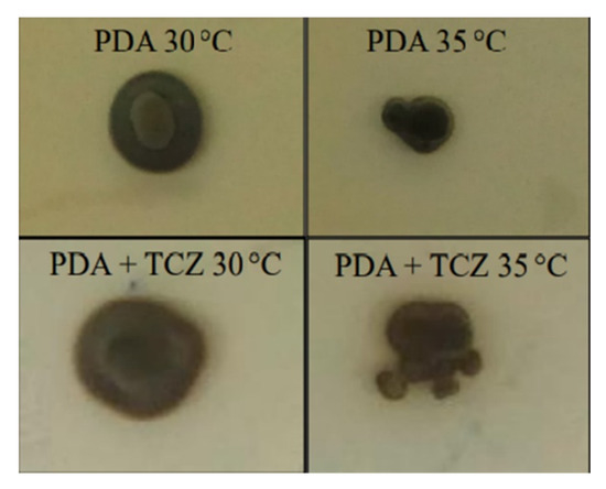
Figure 1.
Melanin inhibition in the presence of tricyclazole. The strain Fonsecaea pedrosoi 19889 was grown on potato dextrose agar (PDA, upper panels) and on PDA supplemented with 8 mg/L tricyclazole (TCZ, lower panels), at 30 °C (left panels) or 35 °C (right panels). The other strains present similar growth under these conditions.
3.2. Tricyclazole Affects Differently the MIC of Drugs with Low Antifungal Activity against Fonsecaea spp.
Table 1 presents MIC values of seven Fonsecaea strains with regular or inhibited DHN-melanin production against amphotericin B, fluconazole, and flucytosine, as well as their MEC values against micafungin. High MIC/MEC values were observed for all drugs with regular DHN-melanin production. DHN-melanin inhibition did not influence MIC values observed for fluconazole and flucytosine (p = 0.3458 and p = 0.5248, respectively). The inhibition of this melanin type increased susceptibility to amphotericin B (p = 0.0335). Surprisingly, DHN-melanin inhibition decreased susceptibility to micafungin; that is, tricyclazole-treated Fonsecaea cells presented higher MEC values than control cells (p = 0.0437).

Table 1.
Minimal inhibitory (or effective) concentrations of four antifungal drugs against seven Fonsecaea strains with regular or inhibited DHN-melanin production.
3.3. Most Fonsecaea spp. Strains with Inhibited DHN-Melanin Production Are Resistant to Micafungin Killing
The survival assay of the seven Fonsecaea spp. strains (Figure 2) revealed variations in the killing effects of micafungin, depending on the Fonsecaea strain studied. The killing effect for one strain (F. monophora 41080) was similar in both conditions. For another strain (F. nubica 16451), DHN-melanin protected against both early and late micafungin killing, but four strains with inhibited DHN-melanin production (F. pedrosoi 38437, F. monophora 38833, F. nubica 34113, and F. nubica 34242) were the more resistant to both the early and late killing while another strain (F. pedrosoi 19889) was more resistant only to micafungin late killing.
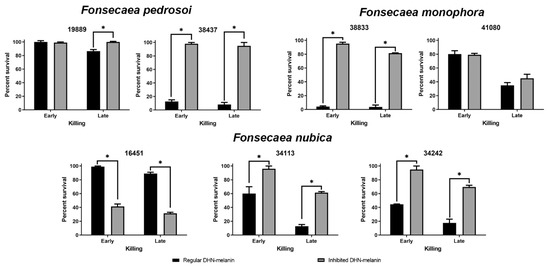
Figure 2.
Survival rates of seven Fonsecaea spp. isolates against micafungin. Black bars indicate the survival of strains with regular DHN-melanin production. Gray bars indicate the survival of strains with DHN-melanin production inhibited by tricyclazole. The * indicates a significant difference (p < 0.05) between regular and inhibited DHN-melanin production.
3.4. Chitin Is not Associated with Micafungin Resistance of Tricyclazole Treated Fonsecaea spp. Cells
Although the seven strains were treated with Uvitex 2B to estimate chitin levels on the conidial cell wall, two of them (F. monophora 38833 and F. nubica 34242) did not yield a fluorescence signal with the protocol used. Figure 3 presents the MFI values of the remaining five strains tested. F. monophora 41080 and F. nubica 34133 presented lower chitin levels in tricyclazole treated conidia (p = 0.0256 and 0.0151, respectively).
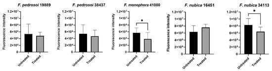
Figure 3.
Chitin contents in five Fonsecaea spp. strains. Graphs present the mean fluorescence intensity of untreated (black bars) and treated (gray bars) conidia of five Fonsecaea spp. strains after Uvitex 2B incubation. * indicates significant difference (p < 0.05).
4. Discussion
Due to the vastly explored ability of melanin to increase fungal virulence [31] and to promote resistance to antifungal drugs [32], it has been proposed that fungal melanin biosynthesis may be an innovative target candidate for the development of new antifungal drugs [9,33,34]. However, these propositions may not be accurate in all situations. In fact, when two Sporothrix brasiliensis strains with different melanin profiles isolated from a single lesion from a human patient with sporotrichosis were tested in an experimental murine model of infection, the albino strain presented as more virulent than the melanized strain [35]. Here, we provide evidence showing that, at least for some Fonsecaea strains, inhibition of DHN-melanin synthesis may increase resistance to the antifungal effects of the echinocandin micafungin.
Recently, a reduction in the MIC values of terbinafine, posaconazole, and itraconazole after Fonsecaea spp. tricyclazole exposure has been shown [21]. An older study, however, showed no influence of DHN-melanin on the antifungal susceptibility of F. monophora to eight antifungal drugs [36], suggesting an unpredictable role of this melanin type in the antifungal susceptibility of this genus. Our results showed that tricyclazole increased Fonsecaea spp. susceptibility to amphotericin B, which was not observed by another group [21], confirming the erraticism of antifungal protection conferred by DHN-melanin in this genus.
Fluconazole and flucytosine MIC values were not affected by the tricyclazole treatment of the cells. Similar results were found with Exophiala (formerly Wangiella) dermatitidis [37], another member of the Herpotrichiellaceae family presenting high levels of melanin in the cell wall. The heavy melanization of members of this family could be an explanation for the low susceptibility to these drugs, which was not the case. Flucytosine is described as an option for chromoblastomycosis treatment, especially in combination with amphotericin B [38]. Therefore, further studies on the resistance mechanisms of chromoblastomycosis agents against flucytosine may improve the research about new therapies for this neglected disease.
An unexpected result of the current study was the increased resistance of most Fonsecaea strains with inhibited DHN-melanin production against micafungin. It has been shown that melanin inhibition increases Histoplasma capsulatum and Cryptococcus neoformans susceptibility to caspofungin [8], another echinocandin with a similar mechanism of action. Alternaria infectoria, another dematiaceous fungus, increases melanization in response to caspofungin, probably as a protection mechanism against its effects [39]. Since microdilution procedures are not always effective in showing melanin influence on MIC/MEC determinations [8], we performed a survival assay of the strains in the presence of micafungin. Results of this assay with strains F. pedrosoi 19889, F. pedrosoi 38437, F. monophora 38833, F. nubica 34113, and F. nubica 34242 corroborate the individual MEC values observed for tricyclazole treated and untreated cells. To the best of our knowledge, there is no description in other fungi that DHN-melanin supports echinocandin activity.
Echinocandin resistance is associated with the inexistence of β-1,3-glucan in the cell wall [40], which is not the case for Fonsecaea spp. that present this polysaccharide in cell walls of conidia, hyphae, and muriform cells [41]. Other resistance mechanisms involve FKS point mutations and an increase in chitin cell wall levels to compensate for the lack of β-1,3-glucan [42]. It is unlikely that a seven-day exposure to tricyclazole causes specific mutations in the FKS genes. Chitin and melanin are probably bounded since as reactions of the saccharide units with melanin monomers can occur, covalent bonds are formed. We are probably dealing with melanin-chitin complexes, and this may be a reason for conflicting results of the seven strains and different antifungal drugs. A possible increase in chitin levels in tricyclazole treated Fonsecaea cells would explain the micafungin resistance. Unfortunately, the chitin levels were not determined on two of the strains, probably due to chitin levels being lower than the detection limit of the methodology. However, for most strains, chitin levels were not associated with micafungin resistance in tricyclazole-treated cells since only two strains presented significant variations. Moreover, one strain presented lower MEC in treated cells, while the other in untreated cells. The other three strains, all of them presenting higher MEC in treated cells, did not present significant differences in chitin levels of tricyclazole treated and untreated conidia.
A limitation of this study is the fact that we were unable to produce true albino phenotypes of the studied strains. Tricyclazole is a specific inhibitor of DHN-melanin [19], and members of the Fonsecaea genus have the genes necessary to also produce eumelanin and pyomelanin [18]. This may be the explanation for the fact that tricyclazole was unable to inhibit the melanization of the strains completely. Studies on eumelanin and pyomelanin in Fonsecaea spp. are scarce. These types of melanin could have distinct locations and roles. This could be essential to modifying drug susceptibility. For instance, eumelanin contains nitrogen and carboxyl groups, but DHN-melanin does not, which may impact their chelating ability.
It has been described that fungal melanins can bind to amphotericin B and caspofungin [8]. Therefore, we hypothesize that the DHN-melanin present in Fonsecaea cell walls binds amphotericin B, preventing it from reaching the ergosterol present in the cell membrane. On the other hand, micafungin binding by Fonsecaea DHN-melanin present in the cell wall increases its levels precisely at the drug site of action.
5. Conclusions
DHN-melanin does not have a role in the intrinsic resistance of Fonseacaea spp. to amphotericin B, fluconazole, and flucytosine, and its inhibition may promote micafungin resistance. Although micafungin is not recommended for chromoblastomycosis treatment, this study provides theoretical concepts about melanization and antifungal susceptibility and resistance. Future studies aiming to discover new antifungal drugs targeting melanin biosynthesis pathways need to evaluate the effects of this pigment on the susceptibility and resistance against other antifungals that may be used in combined therapies. In addition, these potential new antifungals need to inhibit several pathways for complete inhibition of melanin production by the fungus.
Supplementary Materials
The following supporting information can be downloaded at: https://www.mdpi.com/article/10.3390/microbiolres13020017/s1, Figure S1: Growth of two representative strains (Fonsecaea nubica 16451 and Fonsecaea pedrosoi 19889) on potato dextrose agar supplemented with different tricyclazole concentrations.
Author Contributions
Conceptualization, R.A.-P.; methodology, R.A.C., D.C.-J. and R.A.-P.; validation, R.A.C., M.H.G.F.-C. and D.C.-J.; formal analysis, S.F. and R.A.-P.; investigation, R.A.C., M.H.G.F.-C., J.V.d.S.S., D.C.-J.; resources, S.F., R.M.Z.-O., D.F.S.F.; writing—original draft preparation, R.A.C. and R.A.-P.; writing—review and editing, M.H.G.F.-C., D.C.-J., S.F., R.M.Z.-O., D.F.S.F.; visualization, R.A.C., D.C.-J. and R.A.-P.; supervision, S.F. and R.A.-P.; project administration, D.F.S.F. and R.A.-P.; funding acquisition, R.M.Z.-O. and R.A.-P. All authors have read and agreed to the published version of the manuscript.
Funding
This research was funded by Fundação de Amparo à Pesquisa do Estado do Rio de Janeiro (FAPERJ), grant number E-26/201.441/2021 and Inova Fiocruz/VPPCB, grant number VPPCB-008-FIO-18-2-49. R.M.Z-O. was supported in part by Conselho Nacional de Desenvolvimento Científico e Tecnológico [CNPq 302796/2017-7] and Fundação Carlos Chagas Filho de Amparo à Pesquisa do Estado do Rio de Janeiro [FAPERJ E-26/202.527/2019].
Institutional Review Board Statement
The study was approved by the Institutional Research Ethics Committee of the Evandro Chagas National Institute of Infectious Diseases/Fiocruz (protocol code CAAE: 52247016.0.0000.5262).
Informed Consent Statement
Patient consent was waived due to the fact that no patient data or images were provided within the manuscript.
Data Availability Statement
The data presented in the study are contained within the article and its supplementary material.
Acknowledgments
We are thankful to the staff of Fiocruz Platform RPT01A that performed the DNA sequencing for the reliable identification of the strains used in this study.
Conflicts of Interest
The authors declare no conflict of interest. The funders had no role in the design of the study; in the collection, analyses, or interpretation of data; in the writing of the manuscript, or in the decision to publish the results.
References
- Revankar, S.G. Dematiaceous fungi. Mycoses 2007, 50, 91–101. [Google Scholar] [CrossRef]
- Revankar, S.G.; Sutton, D.A. Melanized fungi in human disease. Clin. Microbiol. Rev. 2010, 23, 884–928. [Google Scholar] [CrossRef]
- Zaragoza, O. Basic principles of the virulence of Cryptococcus. Virulence 2019, 10, 490–501. [Google Scholar] [CrossRef]
- Figueiredo-Carvalho, M.H.G.; dos Santos, F.B.; Nosanchuk, J.D.; Zancope-Oliveira, R.M.; Almeida-Paes, R. L-dihydroxyphenylalanine induces melanin production by members of the genus Trichosporon. FEMS Yeast Res. 2014, 14, 988–991. [Google Scholar] [CrossRef]
- Chamilos, G.; Carvalho, A. Aspergillus fumigatus DHN-melanin. Curr. Top. Microbiol. Immunol. 2020, 425, 17–28. [Google Scholar] [CrossRef]
- Almeida-Paes, R.; Borba-Santos, L.P.; Rozental, S.; Marco, S.; Zancopé-Oliveira, R.M.; da Cunha, M.M.L. Melanin biosynthesis in pathogenic species of Sporothrix. Fungal Biol. Rev. 2017, 31, 50–59. [Google Scholar] [CrossRef]
- Taborda, C.P.; da Silva, M.B.; Nosanchuk, J.D.; Travassos, L.R. Melanin as a virulence factor of Paracoccidioides brasiliensis and other dimorphic pathogenic fungi: A minireview. Mycopathologia 2008, 165, 331–339. [Google Scholar] [CrossRef]
- van Duin, D.; Casadevall, A.; Nosanchuk, J.D. Melanization of Cryptococcus neoformans and Histoplasma capsulatum reduces their susceptibilities to amphotericin B and caspofungin. Antimicrob. Agents Chemother. 2002, 46, 3394–3400. [Google Scholar] [CrossRef]
- Almeida-Paes, R.; Figueiredo-Carvalho, M.H.; Da Silva, L.B.; Gerfen, G.; SAraújo, G.R.D.; Frases, S.; Zancopé-Oliveira, R.M.; Nosanchuk, J.D. Candida glabrata produces a melanin-like pigment that protects against stress conditions encountered during parasitism. Future Microbiol. 2021, 16, 509–520. [Google Scholar] [CrossRef]
- Paolo, W.F.; Dadachova, E.; Mandal, P.; Casadevall, A.; Szaniszlo, P.J.; Nosanchuk, J.D. Effects of disrupting the polyketide synthase gene WdPKS1 in Wangiella [Exophiala] dermatitidis on melanin production and resistance to killing by antifungal compounds, enzymatic degradation, and extremes in temperature. BMC Microbiol. 2006, 6, 55. [Google Scholar] [CrossRef]
- Alviano, C.S.; Farbiarz, S.R.; De Souza, W.; Angluster, J.; Travassos, L.R. Characterization of Fonsecaea pedrosoi melanin. J. Gen. Microbiol. 1991, 137, 837–844. [Google Scholar] [CrossRef] [PubMed]
- Franzen, A.J.; Cunha, M.M.L.; Miranda, K.; Hentschel, J.; Plattner, H.; da Silva, M.B.; Salgado, C.G.; de Souza, W.; Rozental, S. Ultrastructural characterization of melanosomes of the human pathogenic fungus Fonsecaea pedrosoi. J. Struct. Biol. 2008, 162, 75–84. [Google Scholar] [CrossRef] [PubMed]
- Rozental, S.; Alviano, C.S.; de Souza, W. The in vitro susceptibility of Fonsecaea pedrosoi to activated macrophages. Mycopathologia 1994, 126, 85–91. [Google Scholar] [CrossRef] [PubMed]
- Alviano, D.S.; Franzen, A.J.; Travassos, L.R.; Holandino, C.; Rozental, S.; Ejzemberg, R.; Alviano, C.S.; Rodrigues, M.L. Melanin from Fonsecaea pedrosoi induces production of human antifungal antibodies and enhances the antimicrobial efficacy of phagocytes. Infect. Immun. 2004, 72, 229–237. [Google Scholar] [CrossRef] [PubMed]
- Cunha, M.M.L.; Franzen, A.J.; Alviano, D.S.; Zanardi, E.; Alviano, C.S.; De Souza, W.; Rozental, S. Inhibition of melanin synthesis pathway by tricyclazole increases susceptibility of Fonsecaea pedrosoi against mouse macrophages. Microsc. Res. Tech. 2005, 68, 377–384. [Google Scholar] [CrossRef] [PubMed]
- Cunha, M.M.L.; Franzen, A.J.; Seabra, S.H.; Herbst, M.H.; Vugman, N.V.; Borba, L.P.; de Souza, W.; Rozental, S. Melanin in Fonsecaea pedrosoi: A trap for oxidative radicals. BMC Microbiol. 2010, 10, 80. [Google Scholar] [CrossRef]
- Cao, W.; Zhou, X.; McCallum, N.C.; Hu, Z.; Ni, Q.Z.; Kapoor, U.; Heil, C.M.; Cay, K.S.; Zand, T.; Mantanona, A.J.; et al. Unraveling the structure and function of melanin through synthesis. J. Am. Chem. Soc. 2021, 143, 2622–2637. [Google Scholar] [CrossRef]
- Vicente, V.A.; Weiss, V.A.; Bombassaro, A.; Moreno, L.F.; Costa, F.F.; Raittz, R.T.; Leão, A.C.; Gomes, R.R.; Bocca, A.L.; Fornari, G.; et al. Comparative genomics of sibling species of Fonsecaea associated with human chromoblastomycosis. Front. Microbiol. 2017, 8, 1924. [Google Scholar] [CrossRef]
- Romero-Martinez, R.; Wheeler, M.; Guerrero-Plata, A.; Rico, G.; Torres-Guerrero, H. Biosynthesis and functions of melanin in Sporothrix schenckii. Infect. Immun. 2000, 68, 3696–3703. [Google Scholar] [CrossRef]
- Franzen, A.J.; Cunha, M.M.L.; Batista, E.J.O.; Seabra, S.H.; De Souza, W.; Rozental, S. Effects of tricyclazole (5-methyl-1,2,4-triazol[3,4]benzothiazole), a specific DHN-melanin inhibitor, on the morphology of Fonsecaea pedrosoi conidia and sclerotic cells. Microsc. Res. Tech. 2006, 69, 729–737. [Google Scholar] [CrossRef]
- Heidrich, D.; Pagani, D.M.; Koehler, A.; Alves, K.D.O.; Scroferneker, M.L. Effect of melanin biosynthesis inhibition on the antifungal susceptibility of chromoblastomycosis agents. Antimicrob. Agents Chemother. 2021, 65, e0054621. [Google Scholar] [CrossRef] [PubMed]
- Queiroz-Telles, F.; Purim, K.S.; Fillus, J.N.; Bordignon, G.F.; Lameira, R.P.; Van Cutsem, J.; Cauwenbergh, G. Itraconazole in the treatment of chromoblastomycosis due to Fonsecaea pedrosoi. Int. J. Derm. 1992, 31, 805–812. [Google Scholar] [CrossRef] [PubMed]
- Silva-Rocha, W.P.; Cardoso, F.J.R.; Colalto, W.; Melo, A.S.A.; Chaves, G.M. Clinical improvement of chromoblastomycosis refractory to itraconazole successfully treated with high dose of terbinafine. J. Derm. 2013, 40, 775–776. [Google Scholar] [CrossRef] [PubMed]
- Esterre, P.; Andriantsimahavandy, A.; Ramarcel, E.R.; Pecarrere, J.L. Forty years of chromoblastomycosis in Madagascar: A review. Am. J. Trop. Med. Hyg. 1996, 55, 45–47. [Google Scholar] [CrossRef]
- Esterre, P.; Queiroz-Telles, F. Management of chromoblastomycosis: Novel perspectives. Curr. Opin. Infect. Dis. 2006, 19, 148–152. [Google Scholar] [CrossRef]
- Sawaya, B.P.; Briggs, J.P.; Schnermann, J. Amphotericin B nephrotoxicity: The adverse consequences of altered membrane properties. J. Am. Soc. Nephrol. 1995, 6, 154–164. [Google Scholar] [CrossRef]
- Coelho, R.A.; Brito-Santos, F.; Figueiredo-Carvalho, M.H.G.; Silva, J.V.D.S.; Gutierrez-Galhardo, M.C.; do Valle, A.C.F.; Zancopé-Oliveira, R.M.; Trilles, L.; Meyer, W.; Freitas, D.F.S.; et al. Molecular identification and antifungal susceptibility profiles of clinical strains of Fonsecaea spp. isolated from patients with chromoblastomycosis in Rio de Janeiro, Brazil. PLoS Negl. Trop. Dis. 2018, 12, e0006675. [Google Scholar] [CrossRef]
- CLSI Reference Method for Broth Dilution Antifungal Susceptibility Testing of Filamentous Fungi 2008; Clinical and Laboratory Standards Institute: Wayne, PA, USA, 2008.
- Almeida-Paes, R.; Figueiredo-Carvalho, M.H.G.; Brito-Santos, F.; Almeida-Silva, F.; Oliveira, M.M.E.; Zancopé-Oliveira, R.M. Melanins protect Sporothrix brasiliensis and Sporothrix schenckii from the antifungal effects of terbinafine. PLoS ONE 2016, 11, e0152796. [Google Scholar] [CrossRef]
- Shihan, M.H.; Novo, S.G.; Le Marchand, S.J.; Wang, Y.; Duncan, M.K. A simple method for quantitating confocal fluorescent images. Biochem. Biophys. Rep. 2021, 25, 100916. [Google Scholar] [CrossRef]
- Smith, D.F.Q.; Casadevall, A. The role of melanin in fungal pathogenesis for animal hosts. Curr. Top. Microbiol. Immunol. 2019, 422, 1–30. [Google Scholar] [CrossRef]
- Gómez, B.L.; Nosanchuk, J.D. Melanin and fungi. Curr. Opin. Infect. Dis. 2003, 16, 91–96. [Google Scholar] [CrossRef] [PubMed]
- Coelho, C.; Casadevall, A. Cryptococcal therapies and drug targets: The old, the new and the promising. Cell Microbiol. 2016, 18, 792–799. [Google Scholar] [CrossRef] [PubMed]
- Santos, L.A.; Grisolia, J.C.; Burger, E.; de Araujo Paula, F.B.; Dias, A.L.T.; Malaquias, L.C.C. Virulence factors of Paracoccidioides brasiliensis as therapeutic targets: A review. Antonie Van Leeuwenhoek 2020, 113, 593–604. [Google Scholar] [CrossRef] [PubMed]
- Oliveira, M.M.E.; Almeida-Paes, R.; Corrêa-Moreira, D.; Borba, C.D.M.; Menezes, R.C.; Freitas, D.F.S.; Do Valle, A.C.F.; Schubach, A.D.O.; Barros, M.B.D.L.; Nosanchuk, J.D.; et al. A case of sporotrichosis caused by different Sporothrix brasiliensis strains: Mycological, molecular, and virulence analyses. Mem. Inst. Oswaldo Cruz 2019, 114, e190260. [Google Scholar] [CrossRef]
- Sun, J.; Zhang, J.; Najafzadeh, M.J.; Badali, H.; Li, X.; Xi, L.; de Hoog, G.S. Melanization of a meristematic mutant of Fonsecaea monophora increases tolerance to stress factors while no effects on antifungal susceptibility. Mycopathologia 2011, 172, 373–380. [Google Scholar] [CrossRef]
- Polak, A.; Dixon, D.M. Loss of melanin in Wangiella dermatitidis does not result in greater susceptibility to antifungal agents. Antimicrob. Agents Chemother. 1989, 33, 1639–1640. [Google Scholar] [CrossRef]
- Queiroz-Telles, F.; Esterre, P.; Perez-Blanco, M.; Vitale, R.G.; Salgado, C.G.; Bonifaz, A. Chromoblastomycosis: An overview of clinical manifestations, diagnosis and treatment. Med. Mycol. 2009, 47, 3–15. [Google Scholar] [CrossRef]
- Fernandes, C.; Prados-Rosales, R.; Silva, B.M.A.; Nakouzi-Naranjo, A.; Zuzarte, M.; Chatterjee, S.; Stark, R.E.; Casadevall, A.; Gonçalves, T. Activation of melanin synthesis in Alternaria infectoria by antifungal drugs. Antimicrob. Agents Chemother. 2015, 60, 1646–1655. [Google Scholar] [CrossRef]
- Nakai, T.; Uno, J.; Ikeda, F.; Tawara, S.; Nishimura, K.; Miyaji, M. In vitro antifungal activity of micafungin (FK463) against dimorphic fungi: Comparison of yeast-like and mycelial forms. Antimicrob. Agents Chemother. 2003, 47, 1376–1381. [Google Scholar] [CrossRef][Green Version]
- Breda, L.C.D.; Menezes, I.G.; Paulo, L.N.M.; de Almeida, S.R. Immune sensing and potential immunotherapeutic approaches to control chromoblastomycosis. J. Fungi 2020, 7, 3. [Google Scholar] [CrossRef]
- Perlin, D.S. Mechanisms of echinocandin antifungal drug resistance. Ann. N. Y. Acad. Sci. 2015, 1354, 1–11. [Google Scholar] [CrossRef] [PubMed]


Publisher’s Note: MDPI stays neutral with regard to jurisdictional claims in published maps and institutional affiliations. |
© 2022 by the authors. Licensee MDPI, Basel, Switzerland. This article is an open access article distributed under the terms and conditions of the Creative Commons Attribution (CC BY) license (https://creativecommons.org/licenses/by/4.0/).