Review

High-Quality Colonoscopy: A Review of Quality Indicators and Best Practices

1
Albany Medical College, Albany, NY 12208, USA
2
Department of Internal Medicine, University of Connecticut, Farmington, CT 06030, USA
3
Department of Internal Medicine, Albany Medical College, Albany, NY 12208, USA
4
Department of Gastroenterology and Hepatology, Albany Medical College, Albany, NY 12208, USA
*
Author to whom correspondence should be addressed.
Gastroenterol. Insights 2022, 13(2), 162-172; https://doi.org/10.3390/gastroent13020017
Submission received: 30 March 2022 / Revised: 27 April 2022 / Accepted: 27 April 2022 / Published: 1 May 2022
(This article belongs to the Special Issue Management and Treatment of Digestive Disorders)

Abstract

:
Colorectal cancer (CRC) continues to be the third leading cause of cancer-related deaths in the US. Colonoscopy remains the best preventative tool against the development of CRC. As a result, high-quality colonoscopy is becoming increasingly important. Specifically, recent guidelines have highlighted pre-procedural, peri-procedural, and post-procedural practices, which promise to improve patient outcomes and reduce the mortality and interval cancer rates in patients undergoing colonoscopies. Despite the guidelines and advances in modern endoscopy, the procedure remains provider-dependent, which results in differences in outcomes. As a result, incorporating high-quality colonoscopy approaches early in training is key to improving patient outcomes. Additionally, ensuring that high-quality colonoscopy is practiced widely by endoscopists can lead to the most cost-effective care.

1. Introduction

As of 2022, colorectal cancer (CRC) remains the third most common cancer and the third leading cause of cancer-related deaths in the United States [1]. Colonoscopy is still the most effective and widely adopted method of reducing CRC incidence and mortality [2]. Despite advances in modern endoscopy, it remains provider-dependent, leading to differences in outcomes [3]. Rex et al. observed different endoscopists performing back-to-back colonoscopies on the same patient and noted large differences in the adenoma detection miss rates, ranging from 17% to 48% [4]. Incorporating high-quality colonoscopy approaches early in training is key to improving patient outcomes. A high-quality colonoscopy leads to an improved adenoma detection rate (ADR), lower rates of cancer incidence, and lower mortality amongst patients [5]. In addition to improving the adenoma detection rate, high-quality colonoscopy contributes to cost-effectiveness. The total annual medical cost of colorectal cancer care exceeds USD 14.1 billion in the United States [6]. The average Medicare health care spending for patients recently diagnosed with colorectal cancer ranges from USD 40,000 to USD 80,000, depending on the stage of cancer at diagnosis. There is also a new emphasis on cost-effective approaches to health care delivery, including value-based payments for providers. As a result, colonoscopy, with its innumerable quality metrics (Table 1), seems a likely candidate to transition to a pay-for-performance model for endoscopist reimbursement [7].

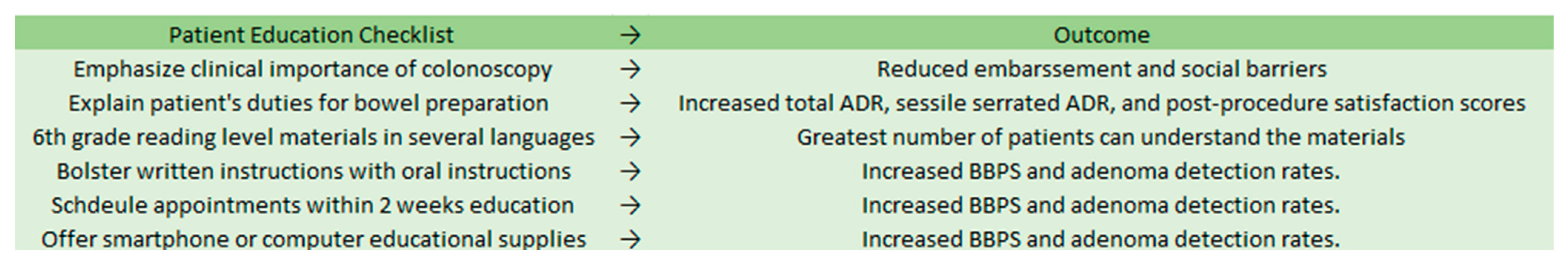
2. High-Quality Colonoscopy Component 1: Patient Education

Patient education and an understanding of the pre-procedure preparation and the procedure itself are key for better colonoscopies. Providers should educate their patients on the importance of routine colonoscopy and how bowel preparation contributes to a successful procedure. Failure to educate patients causes worse bowel preparation and less adherence to routine colonoscopy schedules.
Patients’ motivation to receive a routine colonoscopy may be lower if they do not understand the clinical importance, subsequently leading to delayed cancer diagnosis. A 2020 survey by Amlani et al. on colonoscopy across Europe showed that 72% of responders were receptive to colonoscopy if their doctor advised undergoing one, yet only 45% understood its importance in preventing colorectal cancer [8]. Studies have shown that patient education can increase patient satisfaction and ADR. A 2020 meta-analysis by Tian et al. reviewed studies comparing outcomes in colonoscopy patients who received little or no patient education to outcomes of patients who received enhanced patient education. They found that enhanced patient education materials were associated with a significant increase in the polyp detection rate (PDR), ADR, and the sessile serrated adenoma detection rate (SSADR) [9].
The readability of educational materials increases patient understanding. A 6th-grade reading level is recommended to assure comprehension by the greatest number of patients [10]. Furthermore, language barriers must be assessed and addressed. All offices should provide written instructions in English, Spanish, and other commonly spoken local languages. Greater education interventions may be required in elderly patients, those with impaired cognitive functioning, and those with hearing problems. Recurrent reminders and involving caretakers are effective ways to increase adherence to bowel preparation routines in elderly patients and patients with cognitive difficulties.
Enhancing written patient instructions with verbal instructions from a medical professional significantly improves patient adherence to bowel preparation routines. One study showed that intensive patient educational programs led by pharmacists improved patient compliance, tolerability, and acceptability of a split-dose bowel regimen, leading to greater rates of optimally prepared colons (n = 300, p < 0.001) [11].
A 2018 study by Lee et al. demonstrated that shorter waiting times from patient education to colonoscopy improved the quality of bowel preparation [12]. The Total Boston Bowel Preparation Scale (BBPS) scores for patients whose procedures were performed within 2 weeks of education were significantly higher than those of patients whose procedures took place more than 2 weeks after education (n = 130, p = 0.017).
Computer-based educational materials are also methods to improve bowel preparation and overcome social barriers. Although sample size continues to be an issue in studies on computer-based bowel preparation education materials, these educational materials have been shown to be non-inferior to traditional written instructions when measuring bowel preparation [13,14,15]. A 2021 multicenter randomized controlled trial by Veldhuijzen et al. analyzed 684 patients educated by nurses or computer-based education [16]. Adequate bowel cleansing was seen in 93.2% of computer-educated patients compared to 94.0% of nurse-educated patients. Computer-based education can be an efficient and cost-effective educational tool to improve bowel preparation (Figure 1).

3. High-Quality Colonoscopy Component 2: Bowel Preparation

Validated scales should always be used for documenting bowel preparation. Without using validated scales, comparing bowel preparation between patients is unreliable and biased. The five most widely used scales are the Aronchick Scale, Ottawa Bowel Preparation Scale, BBPS, Harefield Cleansing Scale, and the Chicago Bowel Preparation Scale [17]. Of these scales, the BBPS has the largest amount of reliability and validation data supporting its efficacy.
The BBPS, designed in 2009, is a 10-point scale that assesses bowel preparation after all cleansing maneuvers are completed [18]. The scale removes subjective terms, including “excellent, fair, or poor”, from its criteria and instead grades each section of the colon on a scale from 0 to 3. The sections that are scored include the right colon, transverse colon, and left colon. These scores are summated for a total score of 0 to 9, with a higher score indicating that more mucosa is visualized because less stool is covering it. Providers should aim for a total score greater than or equal to 6, with a score of 2 or more per segment.
Proper bowel preparation requires adherence to a low-residue or clear liquid diet. A low-residue diet may be favored since it is frequently preferred by patients over the clear liquid diet [19]. A 2019 randomized clinical trial comparing a low-fiber diet to a clear liquid diet found that patients who consumed a low-fiber diet the day before a procedure had a better perception of hunger and felt less hungry compared with those who had a clear liquid diet [20]. Adequate bowel preparation was achieved in 89.1% and 95.7% of patients in the clear liquid diet and low-fiber diet groups, respectively, showing both noninferiority and superiority (p = 0.04) of the low-fiber diet [20]. There were no significant differences in cecal intubation rate, whole-polyp detection rate, proximal colon polyp ADR, or distal colon ADR.
When determining the appropriate number of days to follow a low-residue diet, there was no significant difference in bowel preparation quality between the 1-day versus the 2-day diet. A 2020 study by Jiao et al. found comparable BBPS scores between the two groups (p > 0.05) [21]. There were similar colonoscopy insertion times, withdrawal times, and PDRs. It should be noted that patients following a 1-day diet reported significantly easier compliance than those in the 2-day group. In conclusion, 1-day low-residue diets produce statistically similar bowel preparedness scores compared with 2-day low-residue diets while increasing patient compliance and satisfaction.
Osmotic laxatives, such as polyethylene glycol (PEG), are used as a standard component of bowel preparation in the US. Randomized clinical trials assessing the effectiveness of split-dose versus single-dose preparations have shown that split-dose produces more adequately empty and cleanse bowels [22]. PEG 4L solution, oral sulfate solution, 2L PEG and ascorbate, and magnesium citrate and sodium picosulfate are four of the most commonly used laxatives for bowel preparation. A 2021 prospective randomized study by Kmochova et al. showed no significant difference in Boston Bowel Prep Scores between the four groups [23]. The best-tolerated solution was magnesium citrate and sodium picosulfate, with lower rates of nausea and higher rates of palatability [23].
Intolerable taste or texture can deter patients from completing bowel preparation. Oral sulfate tablets can be used for patients with sensitivities to textures and tastes. Randomized control trials have shown oral sulfate tablets to be just as safe and efficacious as PEG and ascorbate [24].
Slowed gastric mobility can interfere with bowel preparation. This could occur in patients with diabetes, chronic constipation, opioid use, and older age. Chronically constipated patients with rectal pain during defecation and start-to-defecation intervals of over 4 h were shown to have significantly higher rates of inadequate bowel preparation than other chronic constipation patients lacking these symptoms [25]. For diabetic patients, opioid users, and the elderly, higher doses of laxatives may be required.

4. High-Quality Colonoscopy Component 3: Proper Scoping Equipment and Contrast

Advances in endoscopy have allowed for higher-definition imaging. Traditional standard-definition endoscopes typically operate in the range of 100,000 to 400,000 total pixels displayed in a 4:3 aspect ratio [26]. High-definition endoscopes operate at over 1 million total pixels, providing aspect ratios beyond 4:3 to accommodate screens of larger widths. Furthermore, high-definition monitors can have frame rates of over 60 times per second, meaning that the image is redisplayed rapidly to create a more realistic and accurate image of the colon. Lastly, high-definition scopes have increased magnification abilities, allowing for a far more detailed image than is possible with standard-definition endoscopy. Standard-definition endoscopes are being replaced by high-definition endoscopes that produce better resolution, making it less likely to miss adenomas. In a 2019 study by Roelandt et al. comparing standard-definition white light endoscopy with high-definition white-light endoscopy, high-definition coloscopy resulted in significantly higher detection rate of sessile serrated adenomas (8.25% vs. 3.8%; p = 0.01) and adenocarcinomas (2.6% vs. 0.5%; p < 0.05) [27]. However, it should be noted that no significant difference in ADR or adenoma per colonoscopy rate (APCR) was seen between high-definition and standard-definition colonoscopy. Overall, high-definition colonoscopy equipment should be used to better detect important diagnoses.
Increasing mucosal contrast increases adenoma detection rates. Several methods are available for increasing mucosal contrast, including traditional dyes and computerized electronic virtual chromoendoscopy. These chromoendoscopy techniques allow neoplasia to be detected more easily by the endoscopist. Pan-colonic chromoendoscopy using a dye, such as a 0.4% indigo carmine spray, is one of the more commonly utilized approaches. Computerized virtual chromoendoscopy utilizes post-processing filter algorithms or a rotating filter in front of the light source to create real-time contrast and enhanced visualization of tissue vasculature and surface neoplasia [28]. A 2010 study by Pohl et al. of 1008 patients performed standard endoscopy followed by dye-enhanced colonoscopy. The proportion of patients with at least one adenoma was significantly greater in the group that received dye (46.2% vs. 36.3%; p = 0.002). Furthermore, chromoendoscopy patients had an increased overall detection rate for adenomas (0.95 vs. 0.66), flat adenomas (0.56 vs. 0.28), and serrated lesions (1.19 vs. 0.49) (p < 0.001) [28]. A 2018 randomized trial by Iacucci et al. demonstrated that virtual computerized chromoendoscopy neoplastic detection rates were non-inferior to dye-based chromoendoscopy detection rates [29]. Therefore, so long as the endoscopist uses a contrast of any sort, the outcomes are better for the patient.
Although computer-assisted detection, such as GI Genius, is not available to most endoscopists, these technologies increase adenoma detection rates and decrease missed colonic neoplasia rates. Randomized control trials have shown increased ADR when utilizing artificial intelligence rather than using high-definition white light colonoscopy alone [30]. Computer-aided detection (CADe) has also been shown to have higher detection rates of sessile serrated lesions. CADe also does not cause a significant increase in withdrawal times compared with other techniques. This technology should become more widespread and accessible in the coming years.

5. High-Quality Colonoscopy Component 4: Improving Cecal Intubation

The entire right side of the colon must be viewed to ensure maximum ADR. Cecal intubation is completed when the tip of the colonoscope passes just proximal to the ileocecal valve, allowing a full view of the cecal caput and medial wall of the cecum. Cecal intubation ensures that the endoscopist has examined the entire area capable of neoplastic growth, especially considering the substantial fraction of colorectal cancers located in the proximal colon [31]. Furthermore, first-time cecal intubation reduces the need for more expensive radiographic procedures or repeat examinations.
A 2011 study by Baxter et al., which included over 14,000 colonoscopies, showed that lower cecal intubation rates were associated with higher rates of interval proximal colon cancer [32]. Therefore, even though quality guidelines state that providers should aim to intubate the cecum in 90% of all colonoscopies and 95% of all screening cases, proper cecal intubation should be the goal of every colonoscopy [33].
Providers should be aware of factors that decrease cecal intubation, including inadequate colon cleansing, endoscopists’ lack of expertise in endoscopy, a patient body weight of under 60 kg or age over 71 years, and the need for active intervention by the anesthesiologist [34]. Improving cecal intubation rates has been a difficult issue in the field of gastroenterology. This can be attributed to the fact that cecal intubation is a demanding and technically challenging maneuver. A vast set of individual characteristics of the endoscopist, including dexterity, length of practice, and patience, contribute greatly to cecal intubation rates [35]. Some of these individual characteristics are innate to the provider and may not be transferable to the student in a master–apprentice training. Competent colonoscopy and cecal intubation are a culmination of high-quality training, continued practice, and years of experience.
The innate skill and performance of an individual endoscopist are the most important factors contributing to the cecal intubation rate. However, besides continued practice and mentorship, a variety of tools may aid in improving cecal intubation. Variable-stiffness colonoscopes allow the endoscopist to stiffen the insertion tube. This provides greater control of looping after straightening and has been demonstrated to increase cecal intubation rates [36]. Furthermore, pediatric colonoscopes may be more maneuverable through angulated and tighter portions of the colon because of their smaller diameter. These have also been shown to increase cecal intubation rates [37]. Lastly, magnetic endoscopic imaging allows the visualization of scope configuration three-dimensionally in real time. This has been shown to increase cecal intubation rates while also increasing procedural efficiency and minimizing patient discomfort [38].

6. High-Quality Colonoscopy Component 5: Second Look into the Right Colon

After cecal intubation, the scope is removed, and the evaluation is continued with a standard withdrawal colonoscopy. Upon reaching the hepatic flexure, repeating the standard forward view of the right colon is advised to ensure that right-sided adenomas are seen. Below is a proposed bumper sticker to help raise awareness of the importance of the second forward view (Figure 2).
A 2020 international multicenter randomized control trial by Tang et al. examined the differences in ADR between endoscopists performing a second look into the right colon and those who did not. Second-view colonoscopy was associated with a significantly higher ADR than colonoscopy with no second view (27.1% vs. 26.6%; p = 0.042) [39]. The median overall withdrawal time was slightly greater in the second-view group (12.0 min vs. 10.5 min; p < 0.001). However, providers who spend additional time performing a second look into the right colon are more likely to not miss colonic neoplasia.

7. High-Quality Colonoscopy Component 6: Lesion Detection Rate

Endoscopists should aim for an adenoma detection rate of 25% in males and 15% in females, as recommended by the American Society for Gastrointestinal Endoscopy (ASGE) and the US Multi-Society Task Force on Colorectal Cancer [40]. Higher ADRs and APCRs have been shown to have lower risks of advanced adenomas at follow-up appointments [40]. A 2019 prospective study of factors predicting colorectal adenoma detection rates also showed that ADR and APCR were significantly higher when withdrawal times exceeded 7.63 min (n = 261, p < 0.001) [41]. ADR and APCR can differ greatly among providers. Some metrics worth tracking could include mean withdrawal time, total lifelong case volume, weekly hours dedicated to performing colonoscopy, and educational activities undertaken to improve skills [42].

8. High-Quality Colonoscopy Component 7: Proper Resection

Providers must safely and completely remove polyps to decrease future occurrences of colorectal cancer [43]. This means taking proper margins while mitigating bleeding and excessive damage to the surrounding mucosa. Some polyps may be easily identifiable as hyperplastic and benign. Typically, these are smaller polyps in the lower bowel or rectum. These polyps are best left unresected, as resection could cause greater harm than benefit. All other noninvasive, nonmalignant polyps should be removed endoscopically.
The proper resection of polyps is a major determinant of the post-procedural incidence of colorectal cancer. A 2013 study by Pohl et al. performed a prospective study on 1426 colonoscopy patients that had at least one nonpedunculated polyp removed [43]. After the endoscopist considered the polyp removal complete on a macroscopic level, resection margins were biopsied. The study showed that 10.1% of all resected polyps were incompletely resected. These rates increased as the polyp size grew and if the polyps were serrated. Leaving behind cancerous tissue increases the rate of interval colorectal cancer after the procedure.
The first step in improving the polyp removal technique is to choose the correct approach. Choosing between polyp removal techniques depends on the polyp size and shape. Polyps that are 1 to 2 cm in size should be removed by snare polypectomy [44]. The goal of snare polypectomy is to transect the polyp along with a thin rim of surrounding healthy mucosa to ensure that the entire affected area is resected [45].
Standard snare polypectomy can be cold or hot (electrosurgical currents). Hot snare uses electrosurgical current to burn through the polyp for removal. However, both bleeding times and post-procedural pain are greater using hot snare compared with cold snare [45,46,47]. Cold snare polypectomy maximizes resection rates while limiting these complications. No additional costs or machinery are needed for cold snare polypectomy. When used in conjunction with a submucosal lift, the procedure is referred to as cold-snare endoscopic mucosal resection (EMR).
Polyps larger than 2 cm that lack the appearance of invasive cancer can be removed by EMR. When the polyp raises suspicion of early invasive cancer, en bloc removal is best performed via endoscopic submucosal dissection (ESD) [46]. However, most endoscopists refer patients with adenomas that exceed 3 cm to surgeons. ESD utilizes a small needle-like knife to carefully incise around the perimeter of the affected area [45,46,47]. ESD for larger polyps has been shown to have a lower rate of recurrence [48,49]. ESD is not indicated for smaller polyps, as the procedure takes upwards of 3 h. EMR can remove these smaller polyps in 30 min.
When performing these procedures, lifting the submucosa away from the deeper layers helps elevate and remove the polyp more easily. In addition, a greater safety margin is created as the submucosa is pulled away from the muscular layer and serosa [50]. The traditional method for lifting the submucosa was the injection of an inexpensive and readily available saline solution. However, newer methods use a combination of blue dyes and viscous agents that maintain the submucosal lift longer than the 1 to 2 min provided by simple saline solutions. The blue dye marks the raised area clearly to ensure that the provider removes all of the affected tissue. Eleview is a good choice for this procedure, as it consists of methylene blue for dyeing and medium-chain triglycerides to increase the viscosity and holding time [51].

9. High-Quality Colonoscopy Component 8: Documentation, Self-Analysis, and Quality Metrics

Validated quality metrics associated with decreased post-procedural cancers and mortality should be tracked. A gastroenterology practice should compare its own quality metrics with those of its peers locally and nationally to ensure that its methodology and practice perform at a comparable rate. Quality colonoscopy requires the provider to continually review current medical literature and update their practices.

10. High-Quality Colonoscopy Component 9: Proper Screening Timelines for Patients in the US

Adhering to proper screening timelines is critical for the early detection of colorectal cancers. The American Cancer Society recommends regular screening starting at age 45 [52]. QR code of updated ACS colonoscopy timelime can be found below (Figure 3) [52]. Colonoscopy should be performed every 10 years until age 75. Beyond that, the decision to be screened should be based on a combination of life expectancy, overall health, ability to undergo the procedure, results of prior screenings, and the patients’ preferences. Patients beyond the age of 85 generally gain no preventative health benefits from undergoing colonoscopy.
Individuals with a family history of colorectal cancer and individuals with inflammatory bowel disease, familial adenomatous polyposis, or Lynch syndrome should see a gastroenterologist on a regular basis and have colonoscopies more frequently. Individuals with a history of radiation to the abdomen or pelvis from previous cancer treatments may need increased screening as well.

11. Other Considerations and Utilizations

In light of the global COVID-19 pandemic, endoscopic procedures have been defined as procedures carrying a high risk of COVID-19 infection. As a result, significantly fewer endoscopic procedures have been performed at centers with pre-endoscopic restrictions, leading to fewer total cancer diagnoses [53]. Endoscopic restriction based on pre-endoscopic diagnosis should be carefully balanced with the potential harm of delaying colorectal cancer diagnosis. Hospitals must constantly reassess restrictions placed on endoscopic procedures, which provide great preventative utility.
It should be noted that colonoscopy serves as an effective tool in the diagnosis and evaluation of diarrheal disorders as well. Bloody diarrhea, mucoid diarrhea, urgency, and systemic inflammatory symptoms all warrant colonoscopy with biopsy to test for irritable bowel disorder (IBD) [54]. IBD must be ruled out before exploring other causes of diarrhea in these patients.

12. Conclusions

Continued technological advancements, improved mentorship, and increased research have greatly contributed to the decline in colorectal cancer incidence and mortality since 1999. However, the wide discrepancies in measurable quality metrics that exist amongst practicing endoscopists remain high. Endoscopy is a technically advanced skill that remains provider-dependent and relies heavily on the patient’s participation. As a result, many of its quality metrics are variables that could be improved. Improving colonoscopy techniques can be accomplished through the mentor–apprentice relationship, which facilitates the growth of both the mentor and the apprentice. Future studies on high-quality colonoscopy should be aimed at improving patient knowledge, the mentor–apprentice relationship, and procedure standardization to decrease discrepancies between providers. Improving colonoscopy can decrease national healthcare expenditure, and quality metrics may one day be incorporated into value-based payments for gastroenterologists.

Author Contributions

M.S., A.T. and L.R. drafted the manuscript; M.T. provided critical revision of the manuscript. All authors have read and agreed to the published version of the manuscript.

Funding

This research received no external funding.

Institutional Review Board Statement

Not applicable.

Informed Consent Statement

Not applicable.

Data Availability Statement

Not applicable.

Conflicts of Interest

The authors declare no conflict of interest.

References

  1. Colorectal Cancer Statistics. C. f. D. C. a. Prevention. Available online: https://www.cdc.gov/cancer/colorectal/statistics/index.htm (accessed on 1 April 2022).
  2. Winawer, S.J.; Zauber, A.G.; Ho, M.N.; O’Brien, M.J.; Gottlieb, L.S.; Sternberg, S.S.; Waye, J.D.; Schapiro, M.; Bond, J.H.; Panish, J.F.; et al. Prevention of colorectal cancer by colonoscopic polypectomy. The national polyp study workgroup. N. Engl. J. Med. 1993, 329, 1977–1981. [Google Scholar] [CrossRef] [PubMed]
  3. Chokshi, R.V.; Hovis, C.E.; Hollander, T.; Early, D.S.; Wang, J.S. Prevalence of missed adenomas in patients with inadequate bowel preparation on screening colonoscopy. Gastrointest. Endosc. 2012, 75, 1197–1203. [Google Scholar] [CrossRef] [PubMed]
  4. Rex, D.K.; Cutler, C.S.; Lemmel, G.T.; Rahmani, E.Y.; Clark, D.W.; Helper, D.J.; Lehman, G.A.; Mark, D.G. Colonoscopic miss rates of adenomas determined by back-to-back colonoscopies. Gastroenterology 1997, 112, 24–28. [Google Scholar] [CrossRef]
  5. Lee, R.H. Quality colonoscopy: A matter of time, technique or technology? World J. Gastroenterol. 2013, 19, 1517–1522. [Google Scholar] [CrossRef]
  6. Cost-Effectiveness of Colorectal Cancer Interventions. 2021. Available online: https://www.cdc.gov/chronicdisease/programs-impact/pop/colorectal-cancer.htm#:~:text=The%20High%20Cost%20of%20Colorectal%20Cancer&text=Average%20Medicare%20health%20care%20spending,b)%20depending%20on%20the%20stage (accessed on 27 February 2022).
  7. Sullivan, K. AGA Offers Bundled Payment Model for Gastroenterologists. The American Journal of Managed Care. 2014. Available online: https://www.ajmc.com/view/aga-offers-bundled-payment-model-for-gastroenterologists (accessed on 1 April 2022).
  8. Amlani, B.; Radaelli, F.; Bhandari, P. A survey on colonoscopy shows poor understanding of its protective value and widespread misconceptions across Europe. PLoS ONE 2020, 15, e0233490. [Google Scholar] [CrossRef]
  9. Tian, X.; Xu, L.-L.; Liu, X.-L.; Chen, W.-Q. Enhanced patient education for colonic polyp and adenoma detection: Meta-analysis of randomized controlled trials. JMIR mHealth uHealth 2020, 8, e17372. [Google Scholar] [CrossRef]
  10. Stossel, L.M.; Segar, N.; Gliatto, P.; Fallar, R.; Karani, R. Readability of patient education materials available at the point of care. J. Gen. Intern. Med. 2012, 27, 1165–1170. [Google Scholar] [CrossRef] [Green Version]
  11. Janahiraman, S.; Tay, C.Y.; Lee, J.M.; Lim, W.L.; Khiew, C.H.; Ishak, I.; Onn, Z.Y.; Ibrahim, M.R.; Chew, C.K. Effect of an intensive patient educational programme on the quality of bowel preparation for colonoscopy: A single-blind randomised controlled trial. BMJ Open Gastroenterol. 2020, 7, e000376. [Google Scholar] [CrossRef]
  12. Lee, J.; Kim, T.O.; Seo, J.W.; Choi, J.H.; Heo, N.-Y.; Park, J.; Park, S.H.; Yang, S.Y.; Moon, Y.S. Shorter waiting times from education to colonoscopy can improve the quality of bowel preparation: A randomized controlled trial. Turk. J. Gastroenterol. 2018, 29, 75–81. [Google Scholar] [CrossRef] [Green Version]
  13. Back, S.Y.; Kim, H.G.; Ahn, E.M.; Park, S.; Jeon, S.R.; Im, H.H.; Kim, J.-O.; Ko, B.M.; Lee, J.S.; Lee, T.H.; et al. Impact of patient audiovisual re-education via a smartphone on the quality of bowel preparation before colonoscopy: A single-blinded randomized study. Gastrointest. Endosc. 2018, 87, 789–799.e4. [Google Scholar] [CrossRef]
  14. Zander, Q.E.W.v.d.; Reumkens, A.; van de Valk, B.; Winkens, B.; Masclee, A.A.M.; de Ridder, R.J.J. Effects of a personalized smartphone app on bowel preparation quality: Randomized controlled trial. JMIR mHealth uHealth 2021, 9, e26703. [Google Scholar] [CrossRef] [PubMed]
  15. Wen, M.-C.; Kau, K.; Huang, S.-S.; Huang, W.-H.; Tsai, L.-Y.; Tsai, T.-Y.; Tsay, S.-L. Smartphone education improves embarrassment, bowel preparation, and satisfaction with care in patients receiving colonoscopy: A randomized controlled trail. Medicine 2020, 99, e23102. [Google Scholar] [CrossRef] [PubMed]
  16. Veldhuijzen, G.; Klemt-Kropp, M.; Droste, J.S.T.S.; van Balkom, B.; van Esch, A.A.J.; Drenth, J.P.H. Computer-based patient education is non-inferior to nurse counselling prior to colonoscopy: A multicenter randomized controlled trial. Endoscopy 2020, 53, 254–263. [Google Scholar] [CrossRef] [PubMed]
  17. Kastenberg, D.; Bertiger, G.; Brogadir, S. Bowel preparation quality scales for colonoscopy. World J. Gastroenterol. 2018, 24, 2833–2843. [Google Scholar] [CrossRef] [PubMed]
  18. Calderwood, A.H.; Jacobson, B. Comprehensive validation of the Boston Bowel Preparation Scale. Gastrointest. Endosc. 2010, 72, 686–692. [Google Scholar] [CrossRef] [Green Version]
  19. Stolpman, D.R.; Solem, C.A.; Eastlick, D.; Adlis, S.; Shaw, M.J. A randomized controlled trial comparing a low-residue diet versus clear liquids for colonoscopy preparation: Impact on tolerance, procedure time, and adenoma detection rate. J. Clin. Gastroenterol. 2014, 48, 851–855. [Google Scholar] [CrossRef]
  20. Alvarez-Gonzalez, M.A.; Pantaleon, M.; Roux, J.A.F.-L.; Zaffalon, D.; Amorós, J.; Bessa, X.; Seoane, A.; Pedro-Botet, J. Randomized clinical trial: A normocaloric low-fiber diet the day before colonoscopy is the most effective approach to bowel preparation in colorectal cancer screening colonoscopy. Dis. Colon Rectum 2019, 62, 491–497. [Google Scholar] [CrossRef]
  21. Jiao, L.; Wang, J.; Zhao, W.; Zhu, X.; Meng, X.; Zhao, L. Comparison of the effect of 1-day and 2-day low residue diets on the quality of bowel preparation before colonoscopy. Saudi J. Gastroenterol. 2020, 26, 137. [Google Scholar] [CrossRef]
  22. Mohamed, R.; Hilsden, R.J.; Dube, C.; Rostom, A. Split-Dose Polyethylene glycol is superior to single dose for colonoscopy preparation: Results of a randomized controlled trial. Can. J. Gastroenterol. Hepatol. 2016, 2016, 3181459. [Google Scholar] [CrossRef] [Green Version]
  23. Kmochova, K.; Grega, T.; Ngo, O.; Vojtechova, G.; Majek, O.; Urbanek, P.; Zavoral, M.; Suchanek, S. Comparison of four bowel cleansing agents for colonoscopy and the factors affecting their efficacy. A prospective, randomized study. J. Gastrointest. Liver Dis. 2021, 30, 213–220. [Google Scholar] [CrossRef]
  24. Di Palma, J.A.; Bhandari, R.; Cleveland, M.V.; Mishkin, D.S.; Tesoriero, J.; Hall, S.; McGowan, J. A safety and efficacy comparison of a new sulfate-based tablet bowel preparation versus a peg and ascorbate comparator in adult subjects undergoing colonoscopy. Am. J. Gastroenterol. 2020, 116, 319–328. [Google Scholar] [CrossRef] [PubMed]
  25. Guo, X.; Shi, X.; Kang, X.; Luo, H.; Wang, X.; Jia, H.; Tao, Q.; Wang, J.; Zhang, M.; Wang, J.; et al. Risk factors associated with inadequate bowel preparation in patients with functional constipation. Dig. Dis. Sci. 2019, 65, 1082–1091. [Google Scholar] [CrossRef] [PubMed]
  26. Subramanian, V.; Ragunath, K. Advanced endoscopic imaging: A review of commercially available technologies. Clin. Gastroenterol. Hepatol. 2014, 12, 368–376.e361. [Google Scholar] [CrossRef] [PubMed]
  27. Roelandt, P.; Demedts, I.; Willekens, H.; Bessissow, T.; Braeye, L.; Coremans, G.; Cuyle, P.-J.; Ferrante, M.; Gevers, A.-M.; Hiele, M.; et al. Impact of endoscopy system, high definition, and virtual chromoendoscopy in daily routine colonoscopy: A randomized trial. Endoscopy 2019, 51, 237–243. [Google Scholar] [CrossRef]
  28. Pohl, J.; Schneider, A.; Vogell, H.; Mayer, G.; Kaiser, G.; Ell, C. Pancolonic chromoendoscopy with indigo carmine versus standard colonoscopy for detection of neoplastic lesions: A randomised two-centre trial. Gut 2010, 60, 485–490. [Google Scholar] [CrossRef]
  29. Iacucci, M.; Kaplan, G.G.; Panaccione, R.; Akinola, O.; Lethebe, B.C.; Lowerison, M.; Leung, Y.; Novak, K.L.; Seow, C.H.; Urbanski, S.; et al. A randomized trial comparing high definition colonoscopy alone with high definition dye spraying and electronic virtual chromoendoscopy for detection of colonic neoplastic lesions during IBD surveillance colonoscopy. Am. J. Gastroenterol. 2018, 113, 225–234. [Google Scholar] [CrossRef]
  30. Spadaccini, M.; Iannone, A.; Maselli, R.; Badalamenti, M.; Desai, M.; Chandrasekar, V.T.; Patel, H.K.; Fugazza, A.; Pellegatta, G.; Galtieri, P.A.; et al. Computer-aided detection versus advanced imaging for detection of colorectal neoplasia: A systematic review and network meta-analysis. Lancet Gastroenterol. Hepatol. 2021, 6, 793–802. [Google Scholar] [CrossRef]
  31. Hoff, G.; Holme, Ø.; Bretthauer, M.; Sandvei, P.; Darre-Næss, O.; Stallemo, A.; Wiig, H.; Høie, O.; Noraberg, G.; Moritz, V.; et al. Cecum intubation rate as quality indicator in clinical versus screening colonoscopy. Endosc. Int. Open 2017, 5, E489–E495. [Google Scholar] [CrossRef] [Green Version]
  32. Baxter, N.N.; Sutradhar, R.; Forbes, S.; Paszat, L.F.; Saskin, R.; Rabeneck, L. Analysis of administrative data finds endoscopist quality measures associated with postcolonoscopy colorectal cancer. Gastroenterology 2011, 140, 65–72. [Google Scholar] [CrossRef]
  33. European Guidelines for Quality Assurance in Colorectal Cancer Screening and Diagnosis; Publications Office of the European Union: Luxembourg, 2013.
  34. Cardin, F.; Minicuci, N.; Andreotti, A.; Pinetti, E.; Campigotto, F.; Donà, B.M.; Martella, B.; Terranova, O. Maximizing the general success of cecal intubation during propofol sedation in a multi-endoscopist academic centre. BMC Gastroenterol. 2010, 10, 123. [Google Scholar] [CrossRef] [Green Version]
  35. Garborg, K.; Bretthauer, M. Cecal intubation failure: Refer or change technique? Gastrointest. Endosc. 2016, 83, 1245–1247. [Google Scholar] [CrossRef] [PubMed] [Green Version]
  36. Garborg, K.; Wiig, H.; Hasund, A.; Matre, J.; Holme, Ø.; Noraberg, G.; Løberg, M.; Kalager, M.; Adami, H.; Bretthauer, M. Gradual stiffness versus magnetic imaging-guided variable stiffness colonoscopes: A randomized noninferiority trial. United Eur. Gastroenterol. J. 2017, 5, 128–133. [Google Scholar] [CrossRef] [PubMed] [Green Version]
  37. Shah, S.G.; Brooker, J.C.; Williams, C.B.; Thapar, C.; Suzuki, N.; Saunders, B.P. The variable stiffness colonoscope: Assessment of efficacy by magnetic endoscope imaging. Gastrointest. Endosc. 2002, 56, 195–201. [Google Scholar] [CrossRef]
  38. Mark-Christensen, A.; Brandsborg, S.; Iversen, L.H. Magnetic endoscopic imaging as an adjuvant to elective colonoscopy: A systematic review and meta-analysis of randomized controlled trials. Endoscopy 2014, 47, 251–261. [Google Scholar] [CrossRef] [PubMed]
  39. Tang, R.S.; Lee, J.W.; Chang, L.-C.; Ong, D.E.; Chiu, H.-M.; Matsuda, T.; Kim, H.-S.; Sekiguchi, M.; Leong, R.W.; Ho, A.M.; et al. Two vs. one forward view examination of right colon on adenoma detection: An international multicenter randomized trial. Clin. Gastroenterol. Hepatol. 2020, 20, 372–380.e372. [Google Scholar] [CrossRef] [PubMed]
  40. Rex, D.K.; Schoenfeld, P.S.; Cohen, J.; Pike, I.M.; Adler, D.G.; Fennerty, M.B.; Lieb, J.G., II; Park, W.G.; Rizk, M.K.; Sawhney, M.S.; et al. Quality indicators for colonoscopy. Gastrointest. Endosc. 2014, 81, 31–53. [Google Scholar] [CrossRef] [PubMed]
  41. Wang, H.; Wang, P.; Liu, X.; Li, L.; Xiao, X.; Liu, P.; Zhang, D.; Li, Y.; Xu, G.; Tu, M.; et al. Factors predicting the colorectal adenoma detection rate in colonoscopic screening of a Chinese population: A prospective study. Medicine 2019, 98, e15103. [Google Scholar] [CrossRef]
  42. Mangas-Sanjuan, C.; Zapater, P.; Cubiella, J.; Murcia, Ó.; Bujanda, L.; Hernández, V.; Martínez-Ares, D.; Pellisé, M.; Seoane, A.; Lanas, Á.; et al. Importance of endoscopist quality metrics for findings at surveillance colonoscopy: The detection-surveillance paradox. United Eur. Gastroenterol. J. 2018, 6, 622–629. [Google Scholar] [CrossRef]
  43. Pohl, H.; Srivastava, A.; Bensen, S.P.; Anderson, P.; Rothstein, R.I.; Gordon, S.R.; Levy, L.C.; Toor, A.; Mackenzie, T.A.; Rosch, T.; et al. Incomplete polyp resection during colonoscopy—Results of the Complete Adenoma Resection (CARE) study. Gastroenterology 2013, 144, 74–80.e71. [Google Scholar] [CrossRef] [Green Version]
  44. Wallace, M.B. New strategies to improve polypectomy during colonoscopy. Gastroenterol. Hepatol. 2017, 13, 1–12. [Google Scholar]
  45. Piraka, C.; Saeed, A.; Waljee, A.K.; Pillai, A.; Stidham, R.; Elmunzer, B.J. Cold snare polypectomy for non-pedunculated colon polyps greater than 1 cm. Endosc. Int. Open 2017, 5, E184–E189. [Google Scholar] [CrossRef] [PubMed] [Green Version]
  46. Choksi, N.; Elmunzer, B.J.; Stidham, R.W.; Shuster, D.; Piraka, C. Cold snare piecemeal resection of colonic and duodenal polyps ≥1 cm. Endosc. Int. Open 2015, 3, E508–E513. [Google Scholar] [CrossRef] [PubMed] [Green Version]
  47. Tutticci, N.J.; Hewett, D.G. Cold EMR of large sessile serrated polyps at colonoscopy (with video). Gastrointest. Endosc. 2018, 87, 837–842. [Google Scholar] [CrossRef] [PubMed] [Green Version]
  48. Holmes, I.; Friedland, S. Endoscopic mucosal resection versus endoscopic submucosal dissection for large polyps: A western colonoscopist’s view. Clin. Endosc. 2016, 49, 454–456. [Google Scholar] [CrossRef] [PubMed] [Green Version]
  49. Moss, A.; Williams, S.J.; Hourigan, L.F.; Brown, G.; Tam, W.; Singh, R.; Zanati, S.; Burgess, N.; Sonson, R.; Byth, K.; et al. Long-term adenoma recurrence following wide-field endoscopic mucosal resection (WF-EMR) for advanced colonic mucosal neoplasia is infrequent: Results and risk factors in 1000 cases from the Australian Colonic EMR (ACE) study. Gut 2014, 64, 57–65. [Google Scholar] [CrossRef] [Green Version]
  50. Uraoka, T.; Saito, Y.; Yamamoto, K.; Fujii, T. Submucosal injection solution for gastrointestinal tract endoscopic mucosal resection and endoscopic submucosal dissection. Drug Des. Dev. Ther. 2008, 2, 131–138. [Google Scholar] [CrossRef] [Green Version]
  51. Rex, D.; Wallace, M.B.; Sharma, P.; Lollo, G.; Maselli, R.; Repici, A. 689 A Randomized, double-blind trial of a new injectable solution (Sic 8000) for endoscopic resection of colonic polyps larger than 2 cm: An interim report. Gastrointest. Endosc. 2017, 85, AB101. [Google Scholar] [CrossRef]
  52. What Should I Know About Screening? 2022. Available online: https://www.cdc.gov/cancer/colorectal/basic_info/screening/index.htm (accessed on 1 April 2022).
  53. Bresky, E.; Bresky, G.; Lancellotti, D.; Madariaga, J.; Licuime, S.; Palma, P.; Saez, F.; Rojas, M.J.; Seijas, L. Should the endoscopic restrictions during COVID-19 pandemic remain unchanged? Gastroenterol. Insights 2021, 12, 34. [Google Scholar] [CrossRef]
  54. Magi, L.; Rinzivillo, M.; Panzuto, F. What gastroenterologists should know about carcinoid syndrome. Gastroenterol. Insights 2022, 13, 14. [Google Scholar] [CrossRef]
Figure 1. Patient education checklist and expected outcomes.
Gastroent 13 00017 g001
Figure 2. Proposed bumper sticker to raise awareness of the importance of second forward view.
Gastroent 13 00017 g002
Figure 3. Scannable QR code of updated ACS colonoscopy timeline guidelines.
Gastroent 13 00017 g003
Table 1. Components of high-quality colonoscopy.
Quality MetricsDescription
Patient EducationEmphasis on importance of colonoscopy
Explanation of need for bowel preparation
Simple language, 6th-grade reading level materials
Materials available in different languages
Scheduling of appointments within 2 weeks of education
Bowel PreparationUse of validated scales to document bowel preparation
Adherence to low-residue or clear liquid diet
Adjustment of bowel preparation dose for patients with chronic constipation or diabetes, patients who use opioids, and the elderly.
Proper Scoping Equipment and ContrastUsing the best available modality for diagnostic purposes
Improving Cecal IntubationEndoscopists improvement of skills, as it is a technically challenging maneuver
Improvement of modifiable factors (bowel preparation)
Application of variable stiffness colonoscopes
Second Look into the Right ColonRepeat forward view upon reaching hepatic flexure
Lesion Detection RateEndoscopists should aim for an adenoma detection rate of 25% in males and 15% in females
Proper ResectionComplete removal with proper margin while minimizing damage to surrounding mucosa
Removal technique guided by size and shape of polyp
Documentation, Self-Analysis, and Quality MetricsTracking of validated quality metrics
Comparison of quality metrics amongst peers, ongoing review of new medical literature
Proper Screening Timelines for Patients in the USFor average patients, CRC screening should be started at age 45 and continued every 10 years until age 75
Screening beyond that age is based on a combination of factors and is individualized
Patients with family history of CRC and patients with inflammatory bowel, familial adenomatous polyposis, and Lynch syndrome should start screening before age 45
Publisher’s Note: MDPI stays neutral with regard to jurisdictional claims in published maps and institutional affiliations.

