Therapeutic Advances in Major NBIA Disorders: Current Strategies and Translational Challenges


Abstract
1. Introduction
2. Neurodegeneration with Brain Iron Accumulation (NBIA): Genetic and Clinical Overview
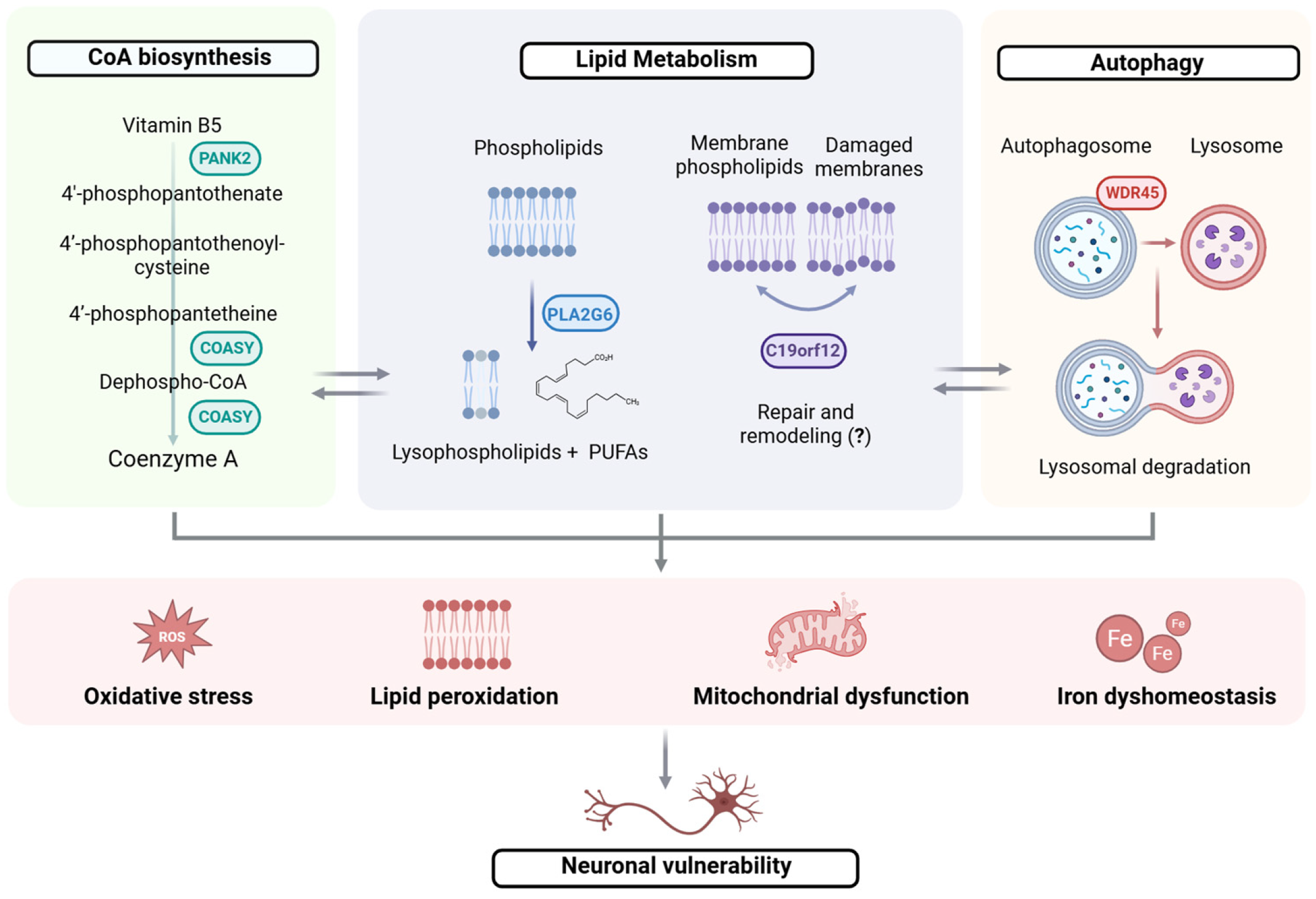
3. NBIA Due to Coenzyme A Metabolism Defects
3.1. PKAN
3.2. CoPAN
4. NBIA Due to Lipid Metabolism Defects
4.1. PLAN
4.2. MPAN
5. NBIA Due to Autophagy Defects
BPAN
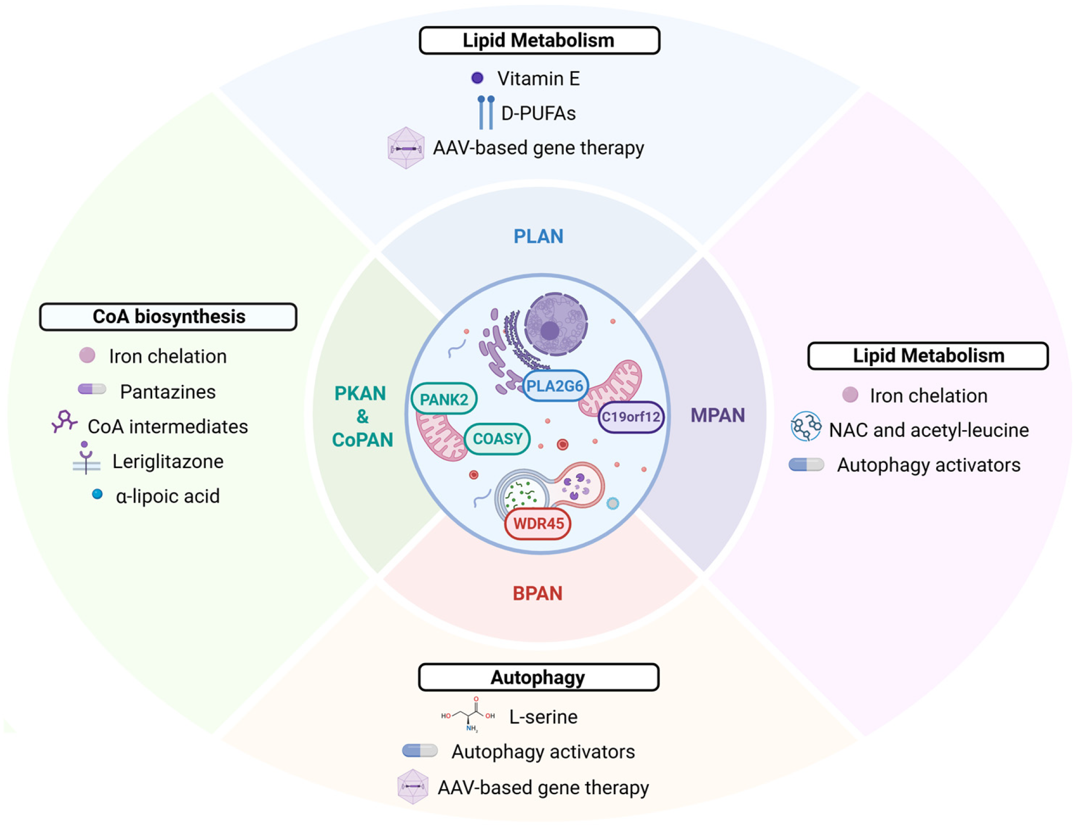
6. From Pathogenesis to Therapy: Emerging Treatment Strategies in Major NBIA Forms
6.1. Symptomatic Management and Iron-Targeting Strategies
6.2. Metabolic and Pathway-Directed Small-Molecule Strategies
6.2.1. CoA Pathway Restoration in PKAN and CoPAN
6.2.2. PPARγ Activation and Mitochondrial Support in PKAN and CoPAN
6.2.3. Autophagy Modulation in BPAN and MPAN
6.3. Antioxidant and Anti-Ferroptotic Strategies
6.3.1. α-Lipoic Acid as an Antioxidant Strategy in PKAN
6.3.2. D-PUFAs and Vitamin E for Lipid-Peroxidation Control in PLAN
6.3.3. NAC and Acetyl-Leucine as Exploratory Strategies in MPAN
6.4. Gene-Based and Precision Genetic Strategies
AAV-Mediated Gene Replacement
7. Translational Gaps and Future Perspective
8. Conclusions
Author Contributions
Funding
Data Availability Statement
Conflicts of Interest
Abbreviations
| NBIA | Neurodegeneration with brain iron accumulation |
| CP | Ceruloplasmin |
| FTL | Ferritin light chain |
| FTH1 | Ferritin heavy chain |
| PKAN | Pantothenate kinase-associated neurodegeneration |
| CoPAN | COASY protein-associated neurodegeneration |
| BPAN | β-propeller-associated neurodegeneration |
| MPAN | mitochondrial membrane protein-associated neurodegeneration |
| PLAN | PLA2G6-associated neurodegeneration |
| INAD | Infantile neuroaxonal dystrophy |
| ANAD | Atypical neuroaxonal dystrophy |
| EOPD | Early-onset Parkinson’s disease |
| MECR | Mitochondrial trans-2-enoyl-CoA reductase |
| GP | Globus pallidus |
| SN | Substantia nigra |
| PPCS | Phosphopantothenoylcysteine synthetase |
| PPCDC | Phosphopantothenoylcysteine decarboxylase |
| PPAT | 4′-phosphopantetheine adenyltransferase |
| DPCK | Dephospho-CoA kinase |
| PUFA | Polyunsaturated fatty acids |
| MAM | Mitochondria-associated membrane |
| DFO | Deferoxamine |
| DFS | Deferasirox |
| DFP | Deferiprone |
| TfR1 | Transferrin receptor 1 |
| PPARγ | Peroxisome proliferator-activated receptor gamma |
| ALA | α -lipoic acid |
| NAC | N-acetyl-L-cysteine |
| AAV | Adeno-associated virus |
| HSAN1 | Hereditary sensory and autonomic neuropathy type 1 |
| ASOs | Antisense oligonucleotides |
References
- Hayflick, S.J.; Kurian, M.A.; Hogarth, P. Neurodegeneration with Brain Iron Accumulation. Handb. Clin. Neurol. 2018, 147, 293–305. [Google Scholar] [CrossRef] [PubMed]
- Arber, C.E.; Li, A.; Houlden, H.; Wray, S. Review: Insights into Molecular Mechanisms of Disease in Neurodegeneration with Brain Iron Accumulation: Unifying Theories. Neuropathol. Appl. Neurobiol. 2016, 42, 220–241. [Google Scholar] [CrossRef] [PubMed]
- Gitlin, J.D. Aceruloplasminemia. Pediatr. Res. 1998, 44, 271–276. [Google Scholar] [CrossRef] [PubMed]
- Burn, J.; Chinnery, P.F. Neuroferritinopathy. Semin. Pediatr. Neurol. 2006, 13, 176–181. [Google Scholar] [CrossRef] [PubMed]
- Shieh, J.T.; Tintos-Hernandez, J.A.; Murali, C.N.; Penon-Portmann, M.; Flores-Mendez, M.; Santana, A.; Bulos, J.A.; Du, K.; Dupuis, L.; Damseh, N.; et al. Heterozygous Nonsense Variants in the Ferritin Heavy-Chain Gene FTH1 Cause a Neuroferritinopathy. HGG Adv. 2023, 4, 100236. [Google Scholar] [CrossRef] [PubMed]
- Hayflick, S.J. Unraveling the Hallervorden-Spatz Syndrome: Pantothenate Kinase-Associated Neurodegeneration Is the Name…. Curr. Opin. Pediatr. 2003, 15, 572–577. [Google Scholar] [CrossRef] [PubMed]
- Dusi, S.; Valletta, L.; Haack, T.B.; Tsuchiya, Y.; Venco, P.; Pasqualato, S.; Goffrini, P.; Tigano, M.; Demchenko, N.; Wieland, T.; et al. Exome Sequence Reveals Mutations in CoA Synthase as a Cause of Neurodegeneration with Brain Iron Accumulation. Am. J. Hum. Genet. 2014, 94, 11–22. [Google Scholar] [CrossRef] [PubMed]
- Morgan, N.V.; Westaway, S.K.; Morton, J.E.V.; Gregory, A.; Gissen, P.; Sonek, S.; Cangul, H.; Coryell, J.; Canham, N.; Nardocci, N.; et al. PLA2G6, Encoding a Phospholipase A2, Is Mutated in Neurodegenerative Disorders with High Brain Iron. Nat. Genet. 2006, 38, 752–754, Erratum in Nat Genet. 2006, 38, 957. [Google Scholar] [CrossRef] [PubMed]
- Kruer, M.C.; Paisán-Ruiz, C.; Boddaert, N.; Yoon, M.Y.; Hama, H.; Gregory, A.; Malandrini, A.; Woltjer, R.L.; Munnich, A.; Gobin, S.; et al. Defective FA2H Leads to a Novel Form of Neurodegeneration with Brain Iron Accumulation (NBIA). Ann. Neurol. 2010, 68, 611–618. [Google Scholar] [CrossRef] [PubMed]
- Hartig, M.B.; Iuso, A.; Haack, T.; Kmiec, T.; Jurkiewicz, E.; Heim, K.; Roeber, S.; Tarabin, V.; Dusi, S.; Krajewska-Walasek, M.; et al. Absence of an Orphan Mitochondrial Protein, C19orf12, Causes a Distinct Clinical Subtype of Neurodegeneration with Brain Iron Accumulation. Am. J. Hum. Genet. 2011, 89, 543–550. [Google Scholar] [CrossRef] [PubMed]
- Kolarova, H.; Tan, J.; Strom, T.M.; Meitinger, T.; Wagner, M.; Klopstock, T. Lifetime Risk of Autosomal Recessive Neurodegeneration with Brain Iron Accumulation (NBIA) Disorders Calculated from Genetic Databases. eBioMedicine 2022, 77, 103869. [Google Scholar] [CrossRef] [PubMed]
- Drecourt, A.; Babdor, J.; Dussiot, M.; Petit, F.; Goudin, N.; Garfa-Traoré, M.; Habarou, F.; Bole-Feysot, C.; Nitschké, P.; Ottolenghi, C.; et al. Impaired Transferrin Receptor Palmitoylation and Recycling in Neurodegeneration with Brain Iron Accumulation. Am. J. Hum. Genet. 2018, 102, 266–277. [Google Scholar] [CrossRef] [PubMed]
- Travaglini, L.; Jeon, C.; Rizza, T.; Novelli, A.; Specchio, N.; Piluso, A.; Bertini, E.; Iuso, A.; Garone, G. Biallelic Variants in SLC27A3 Cause a Complex Form of Neurodegeneration with Brain Iron Accumulation. Mov. Disord. 2025, 40, 2825–2829. [Google Scholar] [CrossRef] [PubMed]
- Heimer, G.; Kerätär, J.M.; Riley, L.G.; Balasubramaniam, S.; Eyal, E.; Pietikäinen, L.P.; Hiltunen, J.K.; Marek-Yagel, D.; Hamada, J.; Gregory, A.; et al. MECR Mutations Cause Childhood-Onset Dystonia and Optic Atrophy, a Mitochondrial Fatty Acid Synthesis Disorder. Am. J. Hum. Genet. 2016, 99, 1229–1244. [Google Scholar] [CrossRef] [PubMed]
- Haack, T.B.; Hogarth, P.; Gregory, A.; Prokisch, H.; Hayflick, S.J. BPAN. In International Review of Neurobiology; Elsevier: Amsterdam, The Netherlands, 2013; Volume 110, pp. 85–90. [Google Scholar]
- Schneider, S.A.; Paisan-Ruiz, C.; Quinn, N.P.; Lees, A.J.; Houlden, H.; Hardy, J.; Bhatia, K.P. ATP13A2 Mutations (PARK9) Cause Neurodegeneration with Brain Iron Accumulation. Mov. Disord. 2010, 25, 979–984. [Google Scholar] [CrossRef] [PubMed]
- Roubertie, A.; Hieu, N.; Roux, C.-J.; Leboucq, N.; Manes, G.; Charif, M.; Echenne, B.; Goizet, C.; Guissart, C.; Meyer, P.; et al. AP4 Deficiency: A Novel Form of Neurodegeneration with Brain Iron Accumulation? Neurol. Genet. 2018, 4, e217. [Google Scholar] [CrossRef] [PubMed]
- Khan, H.; Ilyas, M.; Qasim, H.; Zeb, H.; Israr, M.; Iqbal, A.; Ullah, A.; Ullah, A.; Dominik, N.; Houlden, H.; et al. Whole Exome Sequencing Identifies a Novel Variant Causing Neurodegeneration with Brain Iron Accumulation Syndrome (NBIA) in a Consanguineous Pashtun Family. Neurogenetics 2025, 26, 60. [Google Scholar] [CrossRef] [PubMed]
- Messina, C. Woodhouse-Sakati Syndrome: A Review. Rev. Neurol. 2025, 181, 21–30. [Google Scholar] [CrossRef] [PubMed]
- Manoochehri, J.; Shiri, A.; Khoddam, S.; Aghasipour, M.; Kamal, N.; Jafari Khamirani, H.; Dastgheib, S.A.; Dianatpour, M.; Tabei, S.M.B. Jaberi-Elahi Syndrome: Exploring a Novel GTPBP2 Mutation and a Literature Review. Eur. J. Med. Genet. 2024, 70, 104953. [Google Scholar] [CrossRef] [PubMed]
- Levi, S.; Tiranti, V. Neurodegeneration with Brain Iron Accumulation Disorders: Valuable Models Aimed at Understanding the Pathogenesis of Iron Deposition. Pharmaceuticals 2019, 12, 27. [Google Scholar] [CrossRef] [PubMed]
- Cavestro, C.; Morra, F.; Legati, A.; D’Amato, M.; Nasca, A.; Iuso, A.; Lubarr, N.; Morrison, J.L.; Wheeler, P.G.; Serra-Juhé, C.; et al. Emerging Variants, Unique Phenotypes, and Transcriptomic Signatures: An Integrated Study of COASY-associated Diseases. Ann. Clin. Transl. Neurol. 2024, 11, 1615–1629. [Google Scholar] [CrossRef] [PubMed]
- Cozzi, A.; Santambrogio, P.; Moro, A.S.; Pelagatti, A.; Rubio, A.; Balestrucci, C.; Di Meo, I.; Tiranti, V.; Levi, S. Fibroblasts and hiPS-Derived Astrocytes from CoPAN Patients Showed Different Levels of Iron Overload Correlated with Senescent Phenotype. Glia 2025, 73, 1467–1482. [Google Scholar] [CrossRef] [PubMed]
- Lin, Z.-H.; Xue, N.-J.; Liu, Y.; Zhang, F.; Si, X.-L.; Zheng, R.; Gu, L.-Y.; Li, Y.-L.; Fan, Y.; Tian, J.; et al. Parkinson’s Disease-Associated PLA2G6 Protects IP3R1 Protein to Control ER-Mitochondria Tethering and Ca2+ Transfer. Nat. Commun. 2026, 17, 5338. [Google Scholar] [CrossRef] [PubMed]
- Musthafa, T.; Nizami, S.K.; Mishra, A.; Hasan, G.; Gopurappilly, R. Altered Mitochondrial Bioenergetics and Calcium Kinetics in Young-Onset PLA2G6 Parkinson’s Disease iPSCs. J. Neurochem. 2025, 169, e70059. [Google Scholar] [CrossRef] [PubMed]
- Gnutti, B.; Iuso, A.; Angelini, C.; Finazzi, D. An Update and Perspectives on Mitochondrial Membrane Protein-Associated Neurodegeneration and C19orf12 Research. Brain Sci. 2025, 15, 777. [Google Scholar] [CrossRef] [PubMed]
- Wu, F.; Siedlak, S.L.; Bhatta, S.; Khaled, S.; Shao, C.; Torres, S.; Fujioka, H.; Wang, W. Loss of Mouse C19orf12 Homolog Disturbs Tubular ER Homeostasis and Leads to Neuroaxonal Dystrophy. Acta Neuropathol. Commun. 2025, 13, 233. [Google Scholar] [CrossRef] [PubMed]
- Heger, L.M.; Kertess, L.; Kaufhold, C.; Gubinelli, F.; Cardona-Alberich, A.; Özata, G.; Müller, S.A.; Tschirner, S.K.; Stehling, O.; Schifferer, M.; et al. Patient-Derived Neurons Exhibit α-Synuclein Pathology and Previously Unrecognized Major Histocompatibility Complex Class I Elevation in Mitochondrial Membrane Protein–Associated Neurodegeneration. Mov. Disord. 2025, 40, 2811–2818. [Google Scholar] [CrossRef] [PubMed]
- Celle, M.; Aniorte, S.; Issa, A.-R.; Falabregue, M.; Jin, H.; Sanchez-Mirasierra, I.; Ding, S.; Soukup, S.-F.; Seugnet, L.; Liao, L.; et al. A Dwdr45 Knock-out Drosophila Model to Decipher the Role of Autophagy in BPAN. Hum. Mol. Genet. 2026, 35, ddaf198. [Google Scholar] [CrossRef] [PubMed]
- Shao, Y.; Hu, J.; Yan, K.; Zheng, K.; Sha, W.; Wang, J.; Wu, J.; Huang, Y. Impaired Mitochondrial Integrity and Compromised Energy Production Underscore the Mechanism Underlying CoASY Protein-Associated Neurodegeneration. Cell. Mol. Life Sci. 2025, 82, 84. [Google Scholar] [CrossRef] [PubMed]
- Gregory, A.; Kurian, M.A.; Wilson, J.; Hayflick, S. Neurodegeneration with Brain Iron Accumulation Disorders Overview. In GeneReviews®; Adam, M.P., Bick, S., Mirzaa, G.M., Pagon, R.A., Wallace, S.E., Amemiya, A., Eds.; University of Washington, Seattle: Seattle, WA, USA, 1993. [Google Scholar]
- Tonekaboni, S.H.; Mollamohammadi, M. Neurodegeneration with brain iron accumulation: An overview. Iran. J. Child. Neurol. 2014, 8, 1. [Google Scholar] [PubMed] [PubMed Central]
- Lee, J.-H.; Yun, J.Y.; Gregory, A.; Hogarth, P.; Hayflick, S.J. Brain MRI Pattern Recognition in Neurodegeneration with Brain Iron Accumulation. Front. Neurol. 2020, 11, 1024. [Google Scholar] [CrossRef] [PubMed]
- Gregory, A.; Polster, B.J.; Hayflick, S.J. Clinical and Genetic Delineation of Neurodegeneration with Brain Iron Accumulation. J. Med. Genet. 2009, 46, 73–80. [Google Scholar] [CrossRef] [PubMed]
- Kimura, Y.; Sato, N.; Sugai, K.; Maruyama, S.; Ota, M.; Kamiya, K.; Ito, K.; Nakata, Y.; Sasaki, M.; Sugimoto, H. MRI, MR Spectroscopy, and Diffusion Tensor Imaging Findings in Patient with Static Encephalopathy of Childhood with Neurodegeneration in Adulthood (SENDA). Brain Dev. 2013, 35, 458–461. [Google Scholar] [CrossRef] [PubMed]
- Czumaj, A.; Szrok-Jurga, S.; Hebanowska, A.; Turyn, J.; Swierczynski, J.; Sledzinski, T.; Stelmanska, E. The Pathophysiological Role of CoA. Int. J. Mol. Sci. 2020, 21, 9057. [Google Scholar] [CrossRef] [PubMed]
- Strauss, E. Coenzyme A Biosynthesis and Enzymology. In Comprehensive Natural Products II: Chemistry and Biology; Elsevier: Amsterdam, The Netherlands, 2010; Volume 7, pp. 351–410. [Google Scholar]
- Bravo-Alonso, I.; Morin, M.; Arribas-Carreira, L.; Álvarez, M.; Pedrón-Giner, C.; Soletto, L.; Santolaria, C.; Ramón-Maiques, S.; Ugarte, M.; Rodríguez-Pombo, P.; et al. Pathogenic Variants of the Coenzyme A Biosynthesis-Associated Enzyme Phosphopantothenoylcysteine Decarboxylase Cause Autosomal-Recessive Dilated Cardiomyopathy. J. Inherit. Metab. Dis. 2023, 46, 261–272. [Google Scholar] [CrossRef] [PubMed]
- Lok, A.; Fernandez-Garcia, M.A.; Taylor, R.W.; French, C.; MacFarland, R.; Bodi, I.; Champion, M.; Josifova, D.; Raymond, F.L.; Iuso, A.; et al. Novel Phosphopantothenoylcysteine Synthetase (PPCS) Mutations with Prominent Neuromuscular Features: Expanding the Phenotypical Spectrum of PPCS-Related Disorders. AM. J. Med. Genet. A 2022, 188, 2783–2789. [Google Scholar] [CrossRef] [PubMed]
- Cavestro, C.; Diodato, D.; Tiranti, V.; Di Meo, I. Inherited Disorders of Coenzyme A Biosynthesis: Models, Mechanisms, and Treatments. Int. J. Mol. Sci. 2023, 24, 5951. [Google Scholar] [CrossRef] [PubMed]
- Brezavar, D.; Bonnen, P.E. Incidence of PKAN Determined by Bioinformatic and Population-Based Analysis of ~140,000 Humans. Mol. Genet. Metab. 2019, 128, 463–469. [Google Scholar] [CrossRef] [PubMed]
- Zhou, B.; Westaway, S.K.; Levinson, B.; Johnson, M.A.; Gitschier, J.; Hayflick, S.J. A Novel Pantothenate Kinase Gene (PANK2) Is Defective in Hallervorden-Spatz Syndrome. Nat. Genet. 2001, 28, 345–349. [Google Scholar] [CrossRef] [PubMed]
- Wydrych, A.; Pakuła, B.; Janikiewicz, J.; Dobosz, A.M.; Jakubek-Olszewska, P.; Skowrońska, M.; Kurkowska-Jastrzębska, I.; Cwyl, M.; Popielarz, M.; Pinton, P.; et al. Metabolic Impairments in Neurodegeneration with Brain Iron Accumulation. Biochim. Biophys. Acta (BBA) -Bioenerg. 2025, 1866, 149517. [Google Scholar] [CrossRef] [PubMed]
- Rock, C.O.; Karim, M.A.; Zhang, Y.-M.; Jackowski, S. The Murine Pantothenate Kinase (Pank1) Gene Encodes Two Differentially Regulated Pantothenate Kinase Isozymesq. Gene 2002, 291, 35–43. [Google Scholar] [CrossRef] [PubMed]
- Kotzbauer, P.T.; Truax, A.C.; Trojanowski, J.Q.; Lee, V.M.-Y. Altered Neuronal Mitochondrial Coenzyme A Synthesis in Neurodegeneration with Brain Iron Accumulation Caused by Abnormal Processing, Stability, and Catalytic Activity of Mutant Pantothenate Kinase 2. J. Neurosci. 2005, 25, 689–698. [Google Scholar] [CrossRef] [PubMed]
- Leonardi, R.; Rock, C.O.; Jackowski, S.; Zhang, Y.-M. Activation of Human Mitochondrial Pantothenate Kinase 2 by Palmitoylcarnitine. Proc. Natl. Acad. Sci. USA 2007, 104, 1494–1499. [Google Scholar] [CrossRef] [PubMed]
- Reuter, S.E.; Evans, A.M. Carnitine and Acylcarnitines: Pharmacokinetic, pharmacological and clinical aspects. Clin. Pharmacokinet. 2012, 51, 553–572. [Google Scholar] [CrossRef] [PubMed]
- Ching, K.H.L.; Westaway, S.K.; Gitschier, J.; Higgins, J.J.; Hayflick, S.J. HARP Syndrome Is Allelic with Pantothenate Kinase–Associated Neurodegeneration. Neurology 2002, 58, 1673–1674. [Google Scholar] [CrossRef] [PubMed]
- Hartig, M.B.; Hörtnagel, K.; Garavaglia, B.; Zorzi, G.; Kmiec, T.; Klopstock, T.; Rostasy, K.; Svetel, M.; Kostic, V.S.; Schuelke, M.; et al. Genotypic and Phenotypic Spectrum of PANK2 Mutations in Patients with Neurodegeneration with Brain Iron Accumulation. Ann. Neurol. 2006, 59, 248–256. [Google Scholar] [CrossRef] [PubMed]
- Freeman, K.; Gregory, A.; Turner, A.; Blasco, P.; Hogarth, P.; Hayflick, S. Intellectual and Adaptive Behaviour Functioning in Pantothenate Kinase-associated Neurodegeneration. J. Intellect. Disabil. Res. 2007, 51, 417–426. [Google Scholar] [CrossRef] [PubMed]
- Pellecchia, M.T.; Valente, E.M.; Cif, L.; Salvi, S.; Albanese, A.; Scarano, V.; Bonuccelli, U.; Bentivoglio, A.R.; D’Amico, A.; Marelli, C.; et al. The Diverse Phenotype and Genotype of Pantothenate Kinase-Associated Neurodegeneration. Neurology 2005, 64, 1810–1812. [Google Scholar] [CrossRef] [PubMed]
- Lee, J.-H.; Gregory, A.; Hogarth, P.; Rogers, C.; Hayflick, S.J. Looking Deep into the Eye-of-the-Tiger in Pantothenate Kinase–Associated Neurodegeneration. AJNR AM. J. Neuroradiol. 2018, 39, 583–588. [Google Scholar] [CrossRef] [PubMed]
- Santambrogio, P.; Ripamonti, M.; Paolizzi, C.; Panteghini, C.; Carecchio, M.; Chiapparini, L.; Raimondi, M.; Rubio, A.; Di Meo, I.; Cozzi, A.; et al. Harmful Iron-Calcium Relationship in Pantothenate Kinase Associated Neurodegeneration. Int. J. Mol. Sci. 2020, 21, 3664. [Google Scholar] [CrossRef] [PubMed]
- Stoeter, P.; Roa-Sanchez, P.; Speckter, H.; Perez-Then, E.; Foerster, B.; Vilchez, C.; Oviedo, J.; Rodriguez-Raecke, R. Changes of Cerebral White Matter in Patients Suffering from Pantothenate Kinase-Associated Neurodegeneration (PKAN): A Diffusion Tensor Imaging (DTI) Study. Park. Relat. Disord. 2015, 21, 577–581. [Google Scholar] [CrossRef] [PubMed]
- Zizioli, D.; Tiso, N.; Guglielmi, A.; Saraceno, C.; Busolin, G.; Giuliani, R.; Khatri, D.; Monti, E.; Borsani, G.; Argenton, F.; et al. Knock-down of Pantothenate Kinase 2 Severely Affects the Development of the Nervous and Vascular System in Zebrafish, Providing New Insights into PKAN Disease. Neurobiol. Dis. 2016, 85, 35–48. [Google Scholar] [CrossRef] [PubMed]
- Mignani, L.; Zizioli, D.; Khatri, D.; Facchinello, N.; Schiavone, M.; De Palma, G.; Finazzi, D. Bi-Allelic Mutations in Zebrafish Pank2 Gene Lead to Testicular Atrophy and Perturbed Behavior without Signs of Neurodegeneration. Int. J. Mol. Sci. 2022, 23, 12914. [Google Scholar] [CrossRef] [PubMed]
- Varun, P.; Hagit, T.; Uriya, B.; Sagiv, S.; Sebastian, K. A New in Vivo Model of Pantothenate Kinase-Associated Neurodegeneration Reveals a Surprising Role for Transcriptional Regulation in Pathogenesis. Front. Cell. Neurosci. 2013, 7, 146, Erratum in Front. Cell. Neurosci. 2013, 7, 187. https://doi.org/10.3389/fncel.2013.00187. [Google Scholar] [CrossRef] [PubMed]
- Kuo, Y.-M.; Duncan, J.L.; Westaway, S.K.; Yang, H.; Nune, G.; Xu, E.Y.; Hayflick, S.J.; Gitschier, J. Deficiency of Pantothenate Kinase 2 (Pank2) in Mice Leads to Retinal Degeneration and Azoospermia. Hum. Mol. Genet. 2005, 14, 49–57. [Google Scholar] [CrossRef] [PubMed]
- Brunetti, D.; Dusi, S.; Morbin, M.; Uggetti, A.; Moda, F.; D’Amato, I.; Giordano, C.; d’Amati, G.; Cozzi, A.; Levi, S.; et al. Pantothenate Kinase-Associated Neurodegeneration: Altered Mitochondria Membrane Potential and Defective Respiration in Pank2 Knock-out Mouse Model. Hum. Mol. Genet. 2012, 21, 5294–5305. [Google Scholar] [CrossRef] [PubMed]
- Brunetti, D.; Dusi, S.; Giordano, C.; Lamperti, C.; Morbin, M.; Fugnanesi, V.; Marchet, S.; Fagiolari, G.; Sibon, O.; Moggio, M.; et al. Pantethine Treatment Is Effective in Recovering the Disease Phenotype Induced by Ketogenic Diet in a Pantothenate Kinase-Associated Neurodegeneration Mouse Model. Brain 2014, 137, 57–68. [Google Scholar] [CrossRef] [PubMed]
- Orellana, D.I.; Santambrogio, P.; Rubio, A.; Yekhlef, L.; Cancellieri, C.; Dusi, S.; Giannelli, S.G.; Venco, P.; Mazzara, P.G.; Cozzi, A.; et al. Coenzyme A Corrects Pathological Defects in Human Neurons of PANK 2-associated Neurodegeneration. EMBO Mol. Med. 2016, 8, 1197–1211. [Google Scholar] [CrossRef] [PubMed]
- Santambrogio, P.; Ripamonti, M.; Cozzi, A.; Raimondi, M.; Cavestro, C.; Di Meo, I.; Rubio, A.; Taverna, S.; Tiranti, V.; Levi, S. Massive Iron Accumulation in PKAN-Derived Neurons and Astrocytes: Light on the Human Pathological Phenotype. Cell Death Dis. 2022, 13, 185. [Google Scholar] [CrossRef] [PubMed]
- Ripamonti, M.; Santambrogio, P.; Racchetti, G.; Cozzi, A.; Di Meo, I.; Tiranti, V.; Levi, S. PKAN hiPS-Derived Astrocytes Show Impairment of Endosomal Trafficking: A Potential Mechanism Underlying Iron Accumulation. Front. Cell. Neurosci. 2022, 16, 878103. [Google Scholar] [CrossRef] [PubMed]
- Leoni, V.; Strittmatter, L.; Zorzi, G.; Zibordi, F.; Dusi, S.; Garavaglia, B.; Venco, P.; Caccia, C.; Souza, A.L.; Deik, A.; et al. Metabolic Consequences of Mitochondrial Coenzyme A Deficiency in Patients with PANK2 Mutations. Mol. Genet. Metab. 2012, 105, 463–471. [Google Scholar] [CrossRef] [PubMed]
- Zhyvoloup, A.; Nemazanyy, I.; Babich, A.; Panasyuk, G.; Pobigailo, N.; Vudmaska, M.; Naidenov, V.; Kukharenko, O.; Palchevskii, S.; Savinska, L.; et al. Molecular Cloning of CoA Synthase. J. Biol. Chem. 2002, 277, 22107–22110. [Google Scholar] [CrossRef] [PubMed]
- Leonardi, R.; Zhang, Y.-M.; Rock, C.O.; Jackowski, S. Coenzyme A: Back in Action. Prog. Lipid Res. 2005, 44, 125–153. [Google Scholar] [CrossRef] [PubMed]
- Mishra, R.; Kulshreshtha, S.; Mandal, K.; Khurana, A.; Diego-Álvarez, D.; Pradas, L.; Saxena, R.; Phadke, S.; Moirangthem, A.; Masih, S.; et al. COASY Related Pontocerebellar Hypoplasia Type 12: A Common Indian Mutation with Expansion of the Phenotypic Spectrum. Am. J. Med. Genet. Part A 2022, 188, 2339–2350. [Google Scholar] [CrossRef] [PubMed]
- Rosati, J.; Johnson, J.; Stander, Z.; White, A.; Tortorelli, S.; Bailey, D.; Fong, C.; Lee, B.H. Progressive Brain Atrophy and Severe Neurodevelopmental Phenotype in Siblings with Biallelic COASY Variants. Am. J. Med. Genet. Part A 2023, 191, 842–845. [Google Scholar] [CrossRef] [PubMed]
- Evers, C.; Seitz, A.; Assmann, B.; Opladen, T.; Karch, S.; Hinderhofer, K.; Granzow, M.; Paramasivam, N.; Eils, R.; Diessl, N.; et al. Diagnosis of CoPAN by Whole Exome Sequencing: Waking up a Sleeping Tiger’s Eye. Am. J. Med. Genet. Part A 2017, 173, 1878–1886. [Google Scholar] [CrossRef] [PubMed]
- Ceccatelli Berti, C.; Dallabona, C.; Lazzaretti, M.; Dusi, S.; Tosi, E.; Tiranti, V.; Goffrini, P. Modeling Human Coenzyme A Synthase Mutation in Yeast Reveals Altered Mitochondrial Function, Lipid Content and Iron Metabolism. Microb. Cell 2015, 2, 126–135. [Google Scholar] [CrossRef] [PubMed]
- Khatri, D.; Zizioli, D.; Tiso, N.; Facchinello, N.; Vezzoli, S.; Gianoncelli, A.; Memo, M.; Monti, E.; Borsani, G.; Finazzi, D. Down-Regulation of Coasy, the Gene Associated with NBIA-VI, Reduces Bmp Signaling, Perturbs Dorso-Ventral Patterning and Alters Neuronal Development in Zebrafish. Sci. Rep. 2016, 6, 37660. [Google Scholar] [CrossRef] [PubMed]
- Cavestro, C.; D’Amato, M.; Colombo, M.N.; Cascone, F.; Moro, A.S.; Levi, S.; Tiranti, V.; Di Meo, I. CoA Synthase Plays a Critical Role in Neurodevelopment and Neurodegeneration. Front. Cell. Neurosci. 2024, 18, 1458475. [Google Scholar] [CrossRef]
- Di Meo, I.; Cavestro, C.; Pedretti, S.; Fu, T.; Ligorio, S.; Manocchio, A.; Lavermicocca, L.; Santambrogio, P.; Ripamonti, M.; Levi, S.; et al. Neuronal Ablation of CoA Synthase Causes Motor Deficits, Iron Dyshomeostasis, and Mitochondrial Dysfunctions in a CoPAN Mouse Model. Int. J. Mol. Sci. 2020, 21, 9707. [Google Scholar] [CrossRef]
- Cavestro, C.; Cascone, F.; Legati, A.; Izzo, R.; Catania, M.; Vergara, C.; Rodríguez-Pascau, L.; Pizcueta, P.; Tiranti, V.; Di Meo, I. PPARγ Activation by Leriglitazone Counteracts Neurodegeneration and Neuroinflammation in a Disease-Relevant Mouse Model of COASY Dysfunction. Pharmacol. Res. 2026, 227, 108193. [Google Scholar] [CrossRef]
- Guo, Y.-P.; Tang, B.-S.; Guo, J.-F. PLA2G6-Associated Neurodegeneration (PLAN): Review of Clinical Phenotypes and Genotypes. Front. Neurol. 2018, 9, 1100. [Google Scholar] [CrossRef] [PubMed]
- Winstead, M.V.; Balsinde, J.; Dennis, E.A. Calcium-Independent Phospholipase A2: Structure and Function. Biochim. Biophys. Acta (BBA) -Mol. Cell Biol. Lipids 2000, 1488, 28–39. [Google Scholar] [CrossRef] [PubMed]
- Chu, Y.-T.; Lin, H.-Y.; Chen, P.-L.; Lin, C.-H. Genotype-Phenotype Correlations of Adult-Onset PLA2G6-Associated Neurodegeneration: Case Series and Literature Review. BMC Neurol. 2020, 20, 101. [Google Scholar] [CrossRef] [PubMed]
- Hayashi, D.; Dennis, E.A. Molecular Basis of Unique Specificity and Regulation of Group VIA Calcium-Independent Phospholipase A2 (PNPLA9) and Its Role in Neurodegenerative Diseases. Pharmacol. Ther. 2023, 245, 108395. [Google Scholar] [CrossRef] [PubMed]
- Burke, J.E.; Dennis, E.A. Phospholipase A2 Biochemistry. Cardiovasc. Drugs Ther. 2009, 23, 49–59. [Google Scholar] [CrossRef] [PubMed]
- Gregory, A.; Kurian, M.A.; Soo, A.K.; Wilson, J.L.; Hogarth, P.; Hayflick, S.J. PLA2G6-Associated Neurodegeneration. In GeneReviews®; Adam, M.P., Bick, S., Mirzaa, G.M., Pagon, R.A., Wallace, S.E., Amemiya, A., Eds.; University of Washington, Seattle: Seattle, WA, USA, 1993. [Google Scholar]
- Nardocci, N.; Zorzi, G.; Farina, L.; Binelli, S.; Scaioli, W.; Ciano, C.; Verga, L.; Angelini, L.; Savoiardo, M.; Bugiani, O. Infantile Neuroaxonal Dystrophy: Clinical Spectrum and Diagnostic Criteria. Neurology 1999, 52, 1472–1478. [Google Scholar] [CrossRef] [PubMed]
- Fusco, C.; Frattini, D.; Panteghini, C.; Pascarella, R.; Garavaglia, B. A Case of Infantile Neuroaxonal Dystrophy of Neonatal Onset. J. Child. Neurol. 2015, 30, 368–370. [Google Scholar] [CrossRef] [PubMed]
- Gregory, A.; Westaway, S.K.; Holm, I.E.; Kotzbauer, P.T.; Hogarth, P.; Sonek, S.; Coryell, J.C.; Nguyen, T.M.; Nardocci, N.; Zorzi, G.; et al. Neurodegeneration Associated with Genetic Defects in Phospholipase A2. Neurology 2008, 71, 1402. [Google Scholar] [CrossRef] [PubMed]
- Magrinelli, F.; Mehta, S.; Di Lazzaro, G.; Latorre, A.; Edwards, M.J.; Balint, B.; Basu, P.; Kobylecki, C.; Groppa, S.; Hegde, A.; et al. Dissecting the Phenotype and Genotype of PLA2G6-Related Parkinsonism. Mov. Disord. 2022, 37, 148–161. [Google Scholar] [CrossRef] [PubMed]
- Karkheiran, S.; Shahidi, G.A.; Walker, R.H.; Paisán-Ruiz, C. PLA2G6-Associated Dystonia–Parkinsonism: Case Report and Literature Review. Tremor Other Hyperkinetic Mov. 2015, 5, 317. [Google Scholar] [CrossRef] [PubMed]
- Schneider, S.A.; Dusek, P.; Hardy, J.; Westenberger, A.; Jankovic, J.; Bhatia, K.P. Genetics and Pathophysiology of Neurodegeneration with Brain Iron Accumulation (NBIA). Curr. Neuropharmacol. 2013, 11, 59–79. [Google Scholar] [CrossRef] [PubMed]
- Erskine, D.; Attems, J. Insights into Lewy Body Disease from Rare Neurometabolic Disorders. J. Neural Transm. 2021, 128, 1567–1575. [Google Scholar] [CrossRef] [PubMed]
- Kinghorn, K.J.; Castillo-Quan, J.I.; Bartolome, F.; Angelova, P.R.; Li, L.; Pope, S.; Cochemé, H.M.; Khan, S.; Asghari, S.; Bhatia, K.P.; et al. Loss of PLA2G6 Leads to Elevated Mitochondrial Lipid Peroxidation and Mitochondrial Dysfunction. Brain 2015, 138, 1801–1816. [Google Scholar] [CrossRef] [PubMed]
- Mori, A.; Hatano, T.; Inoshita, T.; Shiba-Fukushima, K.; Koinuma, T.; Meng, H.; Kubo, S.; Spratt, S.; Cui, C.; Yamashita, C.; et al. Parkinson’s Disease-Associated iPLA2-VIA/PLA2G6 Regulates Neuronal Functions and α-Synuclein Stability through Membrane Remodeling. Proc. Natl. Acad. Sci. USA 2019, 116, 20689–20699. [Google Scholar] [CrossRef] [PubMed]
- Beck, G.; Shinzawa, K.; Hayakawa, H.; Baba, K.; Yasuda, T.; Sumi-Akamaru, H.; Tsujimoto, Y.; Mochizuki, H. Deficiency of Calcium-Independent Phospholipase A2 Beta Induces Brain Iron Accumulation through Upregulation of Divalent Metal Transporter 1. PLoS ONE 2015, 10, e0141629. [Google Scholar] [CrossRef] [PubMed]
- Malik, I.; Turk, J.; Mancuso, D.J.; Montier, L.; Wohltmann, M.; Wozniak, D.F.; Schmidt, R.E.; Gross, R.W.; Kotzbauer, P.T. Disrupted Membrane Homeostasis and Accumulation of Ubiquitinated Proteins in a Mouse Model of Infantile Neuroaxonal Dystrophy Caused by PLA2G6 Mutations. Am. J. Pathol. 2008, 172, 406–416. [Google Scholar] [CrossRef] [PubMed]
- Shinzawa, K.; Sumi, H.; Ikawa, M.; Matsuoka, Y.; Okabe, M.; Sakoda, S.; Tsujimoto, Y. Neuroaxonal Dystrophy Caused by Group VIA Phospholipase A2 Deficiency in Mice: A Model of Human Neurodegenerative Disease. J. Neurosci. 2008, 28, 2212–2220. [Google Scholar] [CrossRef] [PubMed]
- Sumi-Akamaru, H.; Beck, G.; Shinzawa, K.; Kato, S.; Riku, Y.; Yoshida, M.; Fujimura, H.; Tsujimoto, Y.; Sakoda, S.; Mochizuki, H. High Expression of α-Synuclein in Damaged Mitochondria with PLA2G6 Dysfunction. Acta Neuropathol. Commun. 2016, 4, 27. [Google Scholar] [CrossRef] [PubMed]
- Villalón-García, I.; Álvarez-Córdoba, M.; Povea-Cabello, S.; Talaverón-Rey, M.; Villanueva-Paz, M.; Luzón-Hidalgo, R.; Suárez-Rivero, J.M.; Suárez-Carrillo, A.; Munuera-Cabeza, M.; Salas, J.J.; et al. Vitamin E Prevents Lipid Peroxidation and Iron Accumulation in PLA2G6-Associated Neurodegeneration. Neurobiol. Dis. 2022, 165, 105649. [Google Scholar] [CrossRef] [PubMed]
- Ke, M.; Chong, C.-M.; Zeng, H.; Huang, M.; Huang, Z.; Zhang, K.; Cen, X.; Lu, J.-H.; Yao, X.; Qin, D.; et al. Azoramide Protects iPSC-Derived Dopaminergic Neurons with PLA2G6 D331Y Mutation through Restoring ER Function and CREB Signaling. Cell Death Dis. 2020, 11, 130. [Google Scholar] [CrossRef] [PubMed]
- Angelini, C.; Durand, C.M.; Fergelot, P.; Deforges, J.; Vital, A.; Menegon, P.; Sarrazin, E.; Bellance, R.; Mathis, S.; Gonzalez, V.; et al. Autosomal Dominant MPAN: Mosaicism Expands the Clinical Spectrum to Atypical Late-Onset Phenotypes. Mov. Disord. 2023, 38, 2103–2115. [Google Scholar] [CrossRef] [PubMed]
- Hartig, M.; Prokisch, H.; Meitinger, T.; Klopstock, T. Mitochondrial Membrane Protein-Associated Neurodegeneration (MPAN). Int. Rev. Neurobiol. 2013, 110, 73–84. [Google Scholar] [CrossRef] [PubMed]
- Klingelhuber, F.; Frendo-Cumbo, S.; Omar-Hmeadi, M.; Massier, L.; Kakimoto, P.; Taylor, A.J.; Couchet, M.; Ribicic, S.; Wabitsch, M.; Messias, A.C.; et al. A Spatiotemporal Proteomic Map of Human Adipogenesis. Nat. Metab. 2024, 6, 861–879. [Google Scholar] [CrossRef] [PubMed]
- Iuso, A.; Sibon, O.C.M.; Gorza, M.; Heim, K.; Organisti, C.; Meitinger, T.; Prokisch, H. Impairment of Drosophila Orthologs of the Human Orphan Protein C19orf12 Induces Bang Sensitivity and Neurodegeneration. PLoS ONE 2014, 9, e89439. [Google Scholar] [CrossRef] [PubMed]
- Mignani, L.; Zizioli, D.; Borsani, G.; Monti, E.; Finazzi, D. The Downregulation of C19orf12 Negatively Affects Neuronal and Musculature Development in Zebrafish Embryos. Front. Cell Dev. Biol. 2020, 8, 596069. [Google Scholar] [CrossRef] [PubMed]
- Shao, C.; Zhu, J.; Ma, X.; Siedlak, S.L.; Cohen, M.L.; Lerner, A.; Wang, W. C19orf12 Ablation Causes Ferroptosis in Mitochondrial Membrane Protein-Associated with Neurodegeneration. Free Radic. Biol. Med. 2022, 182, 23–33. [Google Scholar] [CrossRef] [PubMed]
- Venco, P.; Bonora, M.; Giorgi, C.; Papaleo, E.; Iuso, A.; Prokisch, H.; Pinton, P.; Tiranti, V. Mutations of C19orf12, Coding for a Transmembrane Glycine Zipper Containing Mitochondrial Protein, Cause Mis-Localization of the Protein, Inability to Respond to Oxidative Stress and Increased Mitochondrial Ca2+. Front. Genet. 2015, 6, 185. [Google Scholar] [CrossRef] [PubMed]
- Zanuttigh, E.; Derderian, K.; Güra, M.A.; Geerlof, A.; Di Meo, I.; Cavestro, C.; Hempfling, S.; Ortiz-Collazos, S.; Mauthe, M.; Kmieć, T.; et al. Identification of Autophagy as a Functional Target Suitable for the Pharmacological Treatment of Mitochondrial Membrane Protein-Associated Neurodegeneration (MPAN) In Vitro. Pharmaceutics 2023, 15, 267. [Google Scholar] [CrossRef] [PubMed]
- Papandreou, A.; Soo, A.K.S.; Spaull, R.; Mankad, K.; Kurian, M.A.; Sudhakar, S. Expanding the Spectrum of Early Neuroradiologic Findings in β Propeller Protein-Associated Neurodegeneration. AJNR Am. J. Neuroradiol. 2022, 43, 1810–1814. [Google Scholar] [CrossRef] [PubMed]
- Gregory, A.; Kurian, M.A.; Haack, T.; Hayflick, S.J.; Hogarth, P. Beta-Propeller Protein-Associated Neurodegeneration. In GeneReviews®; Adam, M.P., Bick, S., Mirzaa, G.M., Pagon, R.A., Wallace, S.E., Amemiya, A., Eds.; University of Washington, Seattle: Seattle, WA, USA, 1993. [Google Scholar]
- Wilson, J.L.; Gregory, A.; Kurian, M.A.; Bushlin, I.; Mochel, F.; Emrick, L.; Adang, L.; BPAN Guideline Contributing Author Group; Hogarth, P.; Hayflick, S.J. Consensus Clinical Management Guideline for Beta-propeller Protein-associated Neurodegeneration. Dev. Med. Child. Neurol. 2021, 63, 1402–1409. [Google Scholar] [CrossRef] [PubMed]
- Kimura, Y.; Sato, N.; Ishiyama, A.; Shigemoto, Y.; Suzuki, F.; Fujii, H.; Maikusa, N.; Matsuda, H.; Nishioka, K.; Hattori, N.; et al. Serial MRI Alterations of Pediatric Patients with Beta-Propeller Protein Associated Neurodegeneration (BPAN). J. Neuroradiol. 2021, 48, 88–93. [Google Scholar] [CrossRef] [PubMed]
- Fleming, A.; Bourdenx, M.; Fujimaki, M.; Karabiyik, C.; Krause, G.J.; Lopez, A.; Martín-Segura, A.; Puri, C.; Scrivo, A.; Skidmore, J.; et al. The Different Autophagy Degradation Pathways and Neurodegeneration. Neuron 2022, 110, 935–966. [Google Scholar] [CrossRef] [PubMed]
- Noda, M.; Ito, H.; Nagata, K. Physiological Significance of WDR45, a Responsible Gene for β-Propeller Protein Associated Neurodegeneration (BPAN), in Brain Development. Sci. Rep. 2021, 11, 22568. [Google Scholar] [CrossRef] [PubMed]
- Zhao, Y.G.; Sun, L.; Miao, G.; Ji, C.; Zhao, H.; Sun, H.; Miao, L.; Yoshii, S.R.; Mizushima, N.; Wang, X.; et al. The Autophagy Gene Wdr45/Wipi4 Regulates Learning and Memory Function and Axonal Homeostasis. Autophagy 2015, 11, 881–890. [Google Scholar] [CrossRef] [PubMed]
- Wan, H.; Wang, Q.; Chen, X.; Zeng, Q.; Shao, Y.; Fang, H.; Liao, X.; Li, H.-S.; Liu, M.-G.; Xu, T.-L.; et al. WDR45 Contributes to Neurodegeneration through Regulation of ER Homeostasis and Neuronal Death. Autophagy 2020, 16, 531–547. [Google Scholar] [CrossRef] [PubMed]
- Shimizu, T.; Tamura, N.; Nishimura, T.; Saito, C.; Yamamoto, H.; Mizushima, N. Comprehensive Analysis of Autophagic Functions of WIPI Family Proteins and Their Implications for the Pathogenesis of β-Propeller Associated Neurodegeneration. Hum. Mol. Genet. 2023, 32, 2623–2637. [Google Scholar] [CrossRef] [PubMed]
- Lu, Q.; Yang, P.; Huang, X.; Hu, W.; Guo, B.; Wu, F.; Lin, L.; Kovács, A.L.; Yu, L.; Zhang, H. The WD40 Repeat PtdIns(3)P-Binding Protein EPG-6 Regulates Progression of Omegasomes to Autophagosomes. Dev. Cell 2011, 21, 343–357. [Google Scholar] [CrossRef] [PubMed]
- Lee, J.-H.; Nam, S.O.; Kim, E.K.; Shin, J.-H.; Oh, S.H.; Ryu, D.; Lee, H.E.; Mun, J.Y. Autophagic Defects Observed in Fibroblasts from a Patient with β-Propeller Protein-Associated Neurodegeneration. Am. J. Med. Genet. Part A 2021, 185, 3866–3871. [Google Scholar] [CrossRef] [PubMed]
- Seibler, P.; Burbulla, L.F.; Dulovic, M.; Zittel, S.; Heine, J.; Schmidt, T.; Rudolph, F.; Westenberger, A.; Rakovic, A.; Münchau, A.; et al. Iron Overload Is Accompanied by Mitochondrial and Lysosomal Dysfunction in WDR45 Mutant Cells. Brain 2018, 141, 3052–3064, Erratum in Brain 2019, 142, e10. https://doi.org/10.1093/brain/awy316. [Google Scholar] [CrossRef] [PubMed]
- Saitsu, H.; Nishimura, T.; Muramatsu, K.; Kodera, H.; Kumada, S.; Sugai, K.; Kasai-Yoshida, E.; Sawaura, N.; Nishida, H.; Hoshino, A.; et al. De Novo Mutations in the Autophagy Gene WDR45 Cause Static Encephalopathy of Childhood with Neurodegeneration in Adulthood. Nat. Genet. 2013, 45, 445–449. [Google Scholar] [CrossRef] [PubMed]
- Aring, L.; Choi, E.-K.; Kopera, H.; Lanigan, T.; Iwase, S.; Klionsky, D.J.; Seo, Y.A.J. A neurodegeneration gene, WDR45, links impaired ferritinophagy to iron accumulation. Neurochem 2022, 160, 356–375. [Google Scholar] [CrossRef] [PubMed]
- Ceraolo, G.; Spoto, G.; Consoli, C.; Modafferi, E.; Di Rosa, G.; Nicotera, A.G. Pediatric Genetic Dystonias: Current Diagnostic Approaches and Treatment Options. Life 2025, 15, 992. [Google Scholar] [CrossRef] [PubMed]
- Hong, G.; Zhang, Z.; Wang, P.; Li, G.; Zhang, W.; Zou, H.; Luo, X. Case Report: Asymmetric Bilateral Deep Brain Stimulation for the Treatment of Pantothenate Kinase-Associated Neurodegeneration in a Patient: A Unique Case of Atypical PKAN with a Novel Heterozygous PANK2 Mutation. Front. Hum. Neurosci. 2024, 18, 1448606. [Google Scholar] [CrossRef] [PubMed]
- Levi, V.; Stanziano, M.; Pinto, C.; Zibordi, F.; Fedeli, D.; Caldiera, V.; Cilia, R.; Golfrè Andreasi, N.; Braccia, A.; Carozzi, C.; et al. Bilateral Simultaneous Magnetic Resonance–Guided Focused Ultrasound Pallidotomy for Life-Threatening Status Dystonicus. Mov. Disord. 2024, 39, 1408–1412. [Google Scholar] [CrossRef] [PubMed]
- Iankova, V.; Karin, I.; Klopstock, T.; Schneider, S.A. Emerging Disease-Modifying Therapies in Neurodegeneration with Brain Iron Accumulation (NBIA) Disorders. Front. Neurol. 2021, 12, 629414. [Google Scholar] [CrossRef] [PubMed]
- Klopstock, T.; Tricta, F.; Neumayr, L.; Karin, I.; Zorzi, G.; Fradette, C.; Kmieć, T.; Büchner, B.; Steele, H.E.; Horvath, R.; et al. Safety and Efficacy of Deferiprone for Pantothenate Kinase-Associated Neurodegeneration: A Randomised, Double-Blind, Controlled Trial and an Open-Label Extension Study. Lancet Neurol. 2019, 18, 631–642. [Google Scholar] [CrossRef] [PubMed]
- Chen, S.; Lai, X.; Fu, J.; Yang, J.; Zhao, B.; Shang, H.; Huang, R.; Chen, X. A Novel C19ORF12 Mutation in Two MPAN Sisters Treated with Deferiprone. BMC Neurol. 2023, 23, 134. [Google Scholar] [CrossRef] [PubMed]
- Fonderico, M.; Laudisi, M.; Andreasi, N.G.; Bigoni, S.; Lamperti, C.; Panteghini, C.; Garavaglia, B.; Carecchio, M.; Emanuele, E.A.; Forni, G.L.; et al. Patient Affected by Beta-Propeller Protein-Associated Neurodegeneration: A Therapeutic Attempt with Iron Chelation Therapy. Front. Neurol. 2017, 8, 385. [Google Scholar] [CrossRef] [PubMed]
- Lim, S.-Y.; Tan, A.H.; Ahmad-Annuar, A.; Schneider, S.A.; Bee, P.C.; Lim, J.L.; Ramli, N.; Idris, M.I. A Patient with Beta-Propeller Protein-Associated Neurodegeneration: Treatment with Iron Chelation Therapy. J. Mov. Disord. 2018, 11, 89–92. [Google Scholar] [CrossRef] [PubMed]
- Marupudi, N.; Xiong, M.P. Genetic Targets and Applications of Iron Chelators for Neurodegeneration with Brain Iron Accumulation. ACS Bio Med. Chem. Au 2024, 4, 119–130. [Google Scholar] [CrossRef] [PubMed]
- Hanson, L.R.; Fine, J.M.; Renner, D.B.; Svitak, A.L.; Burns, R.B.; Nguyen, T.M.; Tuttle, N.J.; Marti, D.L.; Panter, S.S.; Frey, W.H. Intranasal Delivery of Deferoxamine Reduces Spatial Memory Loss in APP/PS1 Mice. Drug Deliv. Transl. Res. 2012, 2, 160–168. [Google Scholar] [CrossRef] [PubMed]
- Hanson, L.R.; Roeytenberg, A.; Martinez, P.M.; Coppes, V.G.; Sweet, D.C.; Rao, R.J.; Marti, D.L.; Hoekman, J.D.; Matthews, R.B.; Frey, W.H.; et al. Intranasal Deferoxamine Provides Increased Brain Exposure and Significant Protection in Rat Ischemic Stroke. J. Pharmacol. Exp. Ther. 2009, 330, 679–686. [Google Scholar] [CrossRef] [PubMed]
- Mähler, A.; Mandel, S.; Lorenz, M.; Ruegg, U.; Wanker, E.E.; Boschmann, M.; Paul, F. Epigallocatechin-3-Gallate: A Useful, Effective and Safe Clinical Approach for Targeted Prevention and Individualised Treatment of Neurological Diseases? EPMA J. 2013, 4, 5. [Google Scholar] [CrossRef] [PubMed]
- Zou, Z.; Shao, S.; Zou, R.; Qi, J.; Chen, L.; Zhang, H.; Shen, Q.; Yang, Y.; Ma, L.; Guo, R.; et al. Linking the Low-density Lipoprotein Receptor-binding Segment Enables the Therapeutic 5-YHEDA Peptide to Cross the Blood-brain Barrier and Scavenge Excess Iron and Radicals in the Brain of Senescent Mice. Alzheimer’s Dement. Transl. Res. Clin. Interv. 2019, 5, 717–731. [Google Scholar] [CrossRef] [PubMed]
- Ma, J.; Liu, J.; Chen, S.; Zhang, W.; Wang, T.; Cao, M.; Yang, Y.; Du, Y.; Cui, G.; Du, Z. Understanding the Mechanism of Ferroptosis in Neurodegenerative Diseases. Front. Biosci. (Landmark Ed.) 2024, 29, 291. [Google Scholar] [CrossRef] [PubMed]
- Bhowmick, S.; Lee, Y.J. ER Stress Induced by Artemisinin and Its Derivatives Determines the Susceptibility to Their Synergistic Apoptotic Killing With TRAIL. Cancer Med. 2025, 14, e71001. [Google Scholar] [CrossRef] [PubMed]
- Schmuck, G.; Roehrdanz, E.; Haynes, R.K.; Kahl, R. Neurotoxic Mode of Action of Artemisinin. Antimicrob. Agents Chemother. 2002, 46, 821–827. [Google Scholar] [CrossRef] [PubMed]
- Srinivasan, B.; Baratashvili, M.; Van Der Zwaag, M.; Kanon, B.; Colombelli, C.; Lambrechts, R.A.; Schaap, O.; Nollen, E.A.; Podgoršek, A.; Kosec, G.; et al. Extracellular 4′-Phosphopantetheine Is a Source for Intracellular Coenzyme A Synthesis. Nat. Chem. Biol. 2015, 11, 784–792. [Google Scholar] [CrossRef] [PubMed]
- Khatri, D.; Zizioli, D.; Trivedi, A.; Borsani, G.; Monti, E.; Finazzi, D. Overexpression of Human Mutant PANK2 Proteins Affects Development and Motor Behavior of Zebrafish Embryos. Neuromol. Med. 2019, 21, 120–131. [Google Scholar] [CrossRef] [PubMed]
- Rana, A.; Seinen, E.; Siudeja, K.; Muntendam, R.; Srinivasan, B.; van der Want, J.J.; Hayflick, S.; Reijngoud, D.-J.; Kayser, O.; Sibon, O.C.M. Pantethine Rescues a Drosophila Model for Pantothenate Kinase–Associated Neurodegeneration. Proc. Natl. Acad. Sci. USA 2010, 107, 6988–6993. [Google Scholar] [CrossRef] [PubMed]
- Jeong, S.Y.; Hogarth, P.; Placzek, A.; Gregory, A.M.; Fox, R.; Zhen, D.; Hamada, J.; van der Zwaag, M.; Lambrechts, R.; Jin, H.; et al. 4′-Phosphopantetheine Corrects CoA, Iron, and Dopamine Metabolic Defects in Mammalian Models of PKAN. EMBO Mol. Med. 2019, 11, EMMM201910489. [Google Scholar] [CrossRef] [PubMed]
- Di Meo, I.; Colombelli, C.; Srinivasan, B.; De Villiers, M.; Hamada, J.; Jeong, S.Y.; Fox, R.; Woltjer, R.L.; Tepper, P.G.; Lahaye, L.L.; et al. Acetyl-4′-Phosphopantetheine Is Stable in Serum and Prevents Phenotypes Induced by Pantothenate Kinase Deficiency. Sci. Rep. 2017, 7, 11260. [Google Scholar] [CrossRef] [PubMed]
- Klopstock, T.; Videnovic, A.; Bischoff, A.T.; Bonnet, C.; Cif, L.; Comella, C.; Correa-Vela, M.; Escolar, M.L.; Fraser, J.L.; Gonzalez, V.; et al. Fosmetpantotenate Randomized Controlled Trial in Pantothenate Kinase–Associated Neurodegeneration. Mov. Disord. 2021, 36, 1342–1352. [Google Scholar] [CrossRef] [PubMed]
- Bracke, M.M.G.; Polet, S.S.; Plantinga, M.; De Koning, T.J. Mitigating the Impact of Study-Start Delays in Clinical Trials for Rare Disorders: Insights and Lessons from a PKAN Trial. Orphanet J. Rare Dis. 2025, 20, 572. [Google Scholar] [CrossRef] [PubMed]
- Sharma, L.K.; Subramanian, C.; Yun, M.-K.; Frank, M.W.; White, S.W.; Rock, C.O.; Lee, R.E.; Jackowski, S. A Therapeutic Approach to Pantothenate Kinase Associated Neurodegeneration. Nat. Commun. 2018, 9, 4399. [Google Scholar] [CrossRef] [PubMed]
- Subramanian, C.; Frank, M.W.; Sukhun, R.; Henry, C.E.; Wade, A.; Harden, M.E.; Rao, S.; Tangallapally, R.; Yun, M.-K.; White, S.W.; et al. Pantothenate Kinase Activation Restores Brain Coenzyme A in a Mouse Model of Pantothenate Kinase-Associated Neurodegeneration. J. Pharmacol. Exp. Ther. 2024, 388, 171–180. [Google Scholar] [CrossRef] [PubMed]
- Coker, A.L.; Tangallapally, R.; Yun, M.-K.; Subramanian, C.; Jayasinghe, T.; Miller, K.; Edwards, A.; Frank, M.; Jackowski, S.; Rock, C.O.; et al. Discovery of Sulfonamide Pantothenate Kinase Activators and Elucidation of the Role of Isoform Selectivity in Cellular Pantothenate Kinase Activation. J. Med. Chem. 2026, 69, 6004–6013. [Google Scholar] [CrossRef] [PubMed]
- Pizcueta, P.; Vergara, C.; Emanuele, M.; Vilalta, A.; Rodríguez-Pascau, L.; Martinell, M. Development of PPARγ Agonists for the Treatment of Neuroinflammatory and Neurodegenerative Diseases: Leriglitazone as a Promising Candidate. Int. J. Mol. Sci. 2023, 24, 3201. [Google Scholar] [CrossRef] [PubMed]
- Santambrogio, P.; Cozzi, A.; Meo, I.D.; Cavestro, C.; Vergara, C.; Rodríguez-Pascau, L.; Martinell, M.; Pizcueta, P.; Tiranti, V.; Levi, S. PPAR Gamma Agonist Leriglitazone Recovers Alterations Due to Pank2-Deficiency in hiPS-Derived Astrocytes. Pharmaceutics 2023, 15, 202. [Google Scholar] [CrossRef] [PubMed]
- Myint, S.M.M.P.; Sun, L.Y. L-Serine: Neurological Implications and Therapeutic Potential. Biomedicines 2023, 11, 2117. [Google Scholar] [CrossRef] [PubMed]
- Ye, L.; Sun, Y.; Jiang, Z.; Wang, G. L-Serine, an Endogenous Amino Acid, Is a Potential Neuroprotective Agent for Neurological Disease and Injury. Front. Mol. Neurosci. 2021, 14, 726665. [Google Scholar] [CrossRef] [PubMed]
- Dunlop, R.A.; Carney, J.M. Mechanisms of L-Serine-Mediated Neuroprotection Include Selective Activation of Lysosomal Cathepsins B and L. Neurotox. Res. 2021, 39, 17–26. [Google Scholar] [CrossRef] [PubMed]
- Levine, T.D.; Miller, R.G.; Bradley, W.G.; Moore, D.H.; Saperstein, D.S.; Flynn, L.E.; Katz, J.S.; Forshew, D.A.; Metcalf, J.S.; Banack, S.A.; et al. Phase I Clinical Trial of Safety of L-Serine for ALS Patients. Amyotroph. Lateral Scler. Front. Degener. 2017, 18, 107–111. [Google Scholar] [CrossRef] [PubMed]
- Garofalo, K.; Penno, A.; Schmidt, B.P.; Lee, H.-J.; Frosch, M.P.; von Eckardstein, A.; Brown, R.H.; Hornemann, T.; Eichler, F.S. Oral L-Serine Supplementation Reduces Production of Neurotoxic Deoxysphingolipids in Mice and Humans with Hereditary Sensory Autonomic Neuropathy Type 1. J. Clin. Investig. 2011, 121, 4735–4745. [Google Scholar] [CrossRef] [PubMed]
- Lee, H.E.; Jung, M.; Choi, K.; Jang, J.H.; Hwang, S.-K.; Chae, S.; Lee, J.-H.; Mun, J.Y. L-Serine Restored Lysosomal Failure in Cells Derived from Patients with BPAN Reducing Iron Accumulation with Eliminating Lipofuscin. Free Radic. Biol. Med. 2024, 221, 273–282. [Google Scholar] [CrossRef] [PubMed]
- Talaverón-Rey, M.; Álvarez-Córdoba, M.; Villalón-García, I.; Povea-Cabello, S.; Suárez-Rivero, J.M.; Gómez-Fernández, D.; Romero-González, A.; Suárez-Carrillo, A.; Munuera-Cabeza, M.; Cilleros-Holgado, P.; et al. Alpha-Lipoic Acid Supplementation Corrects Pathological Alterations in Cellular Models of Pantothenate Kinase-Associated Neurodegeneration with Residual PANK2 Expression Levels. Orphanet J. Rare Dis. 2023, 18, 80. [Google Scholar] [CrossRef] [PubMed]
- Camiolo, G.; Tibullo, D.; Giallongo, C.; Romano, A.; Parrinello, N.L.; Musumeci, G.; Di Rosa, M.; Vicario, N.; Brundo, M.V.; Amenta, F.; et al. α-Lipoic Acid Reduces Iron-Induced Toxicity and Oxidative Stress in a Model of Iron Overload. Int. J. Mol. Sci. 2019, 20, 609. [Google Scholar] [CrossRef] [PubMed]
- Firsov, A.M.; Fomich, M.A.; Bekish, A.V.; Sharko, O.L.; Kotova, E.A.; Saal, H.J.; Vidovic, D.; Shmanai, V.V.; Pratt, D.A.; Antonenko, Y.N.; et al. Threshold Protective Effect of Deuterated Polyunsaturated Fatty Acids on Peroxidation of Lipid Bilayers. FEBS J. 2019, 286, 2099–2117. [Google Scholar] [CrossRef] [PubMed]
- Kinghorn, K.J.; Castillo-Quan, J.I. Mitochondrial Dysfunction and Defects in Lipid Homeostasis as Therapeutic Targets in Neurodegeneration with Brain Iron Accumulation. Rare Dis. 2016, 4, e1128616. [Google Scholar] [CrossRef] [PubMed]
- Adams, D.; Midei, M.; Dastgir, J.; Flora, C.; Molinari, R.J.; Heerinckx, F.; Endemann, S.; Atwal, P.; Milner, P.; Shchepinov, M.S. Treatment of Infantile Neuroaxonal Dystrophy with RT001: A Di-Deuterated Ethyl Ester of Linoleic Acid: Report of Two Cases. JIMD Rep. 2020, 54, 54–60. [Google Scholar] [CrossRef] [PubMed]
- Pereira, A.; Fischinger Moura de Souza, C.; Álvarez-Córdoba, M.; Reche-López, D.; Sánchez-Alcázar, J.A. A Therapeutic Approach to Pantothenate Kinase Associated Neurodegeneration: A Pilot Study. Orphanet J. Rare Dis. 2024, 19, 442. [Google Scholar] [CrossRef] [PubMed]
- Álvarez-Córdoba, M.; Reche-López, D.; Cilleros-Holgado, P.; Talaverón-Rey, M.; Villalón-García, I.; Povea-Cabello, S.; Suárez-Rivero, J.M.; Suárez-Carrillo, A.; Munuera-Cabeza, M.; Piñero-Pérez, R.; et al. Therapeutic Approach with Commercial Supplements for Pantothenate Kinase-Associated Neurodegeneration with Residual PANK2 Expression Levels. Orphanet J. Rare Dis. 2022, 17, 311. [Google Scholar] [CrossRef] [PubMed]
- Suárez-Carrillo, A.; Álvarez-Córdoba, M.; Romero-González, A.; Talaverón-Rey, M.; Povea-Cabello, S.; Cilleros-Holgado, P.; Piñero-Pérez, R.; Reche-López, D.; Gómez-Fernández, D.; Romero-Domínguez, J.M.; et al. Antioxidants Prevent Iron Accumulation and Lipid Peroxidation, but Do Not Correct Autophagy Dysfunction or Mitochondrial Bioenergetics in Cellular Models of BPAN. Int. J. Mol. Sci. 2023, 24, 14576. [Google Scholar] [CrossRef] [PubMed]
- Aldini, G.; Altomare, A.; Baron, G.; Vistoli, G.; Carini, M.; Borsani, L.; Sergio, F. N-Acetylcysteine as an Antioxidant and Disulphide Breaking Agent: The Reasons Why. Free Radic. Res. 2018, 52, 751–762. [Google Scholar] [CrossRef] [PubMed]
- Rahim, A.A.; Kurian, M.A.; Zhou, H.; Ferguson, R.; Tabrizi, S.J.; Lignani, G.; Aquilina, K.; Waddington, S.N. Genetic Therapies for Neurological Diseases. Pharmacol. Rev. 2026, 78, 100093. [Google Scholar] [CrossRef] [PubMed]
- Naldini, L. Gene Therapy Returns to Centre Stage. Nature 2015, 526, 351–360. [Google Scholar] [CrossRef] [PubMed]
- Scheller, E.L.; Krebsbach, P.H. Gene Therapy: Design and Prospects for Craniofacial Regeneration. J. Dent. Res. 2009, 88, 585–596. [Google Scholar] [CrossRef] [PubMed]
- Bulcha, J.T.; Wang, Y.; Ma, H.; Tai, P.W.L.; Gao, G. Viral Vector Platforms within the Gene Therapy Landscape. Signal Transduct. Target. Ther. 2021, 6, 53. [Google Scholar] [CrossRef] [PubMed]
- Lin, G.; Tepe, B.; McGrane, G.; Tipon, R.C.; Croft, G.; Panwala, L.; Hope, A.; Liang, A.J.; Zuo, Z.; Byeon, S.K.; et al. Exploring Therapeutic Strategies for Infantile Neuronal Axonal Dystrophy (INAD/PARK14). eLife 2023, 12, e82555. [Google Scholar] [CrossRef] [PubMed]
- Hordeaux, J.; Wang, Q.; Katz, N.; Buza, E.L.; Bell, P.; Wilson, J.M. The Neurotropic Properties of AAV-PHP.B Are Limited to C57BL/6J Mice. Mol. Ther. 2018, 26, 664–668. [Google Scholar] [CrossRef] [PubMed]
- Carisi, M.C.; Shamber, C.; Bishop, M.; Sangster, M.; Chandrachud, U.; Meyerink, B.; Pilaz, L.J.; Grishchuk, Y. AAV-Mediated Gene Transfer of WDR45 Corrects Neurological Deficits in the Mouse Model of Beta-Propeller Protein-Associated Neurodegeneration. Hum. Gene Ther. 2025, 36, 637–652. [Google Scholar] [CrossRef] [PubMed]
- Bisi, N.; Pinzi, L.; Rastelli, G.; Tonali, N. Early Diagnosis of Neurodegenerative Diseases: What Has Been Undertaken to Promote the Transition from PET to Fluorescence Tracers. Molecules 2024, 29, 722. [Google Scholar] [CrossRef] [PubMed]
- Selvam, S.; Ayyavoo, V. Biomarkers in Neurodegenerative Diseases: A Broad Overview. Explor. Neuroprot. Ther. 2024, 4, 119–147. [Google Scholar] [CrossRef]
- Hansson, O. Biomarkers for Neurodegenerative Diseases. Nat. Med. 2021, 27, 954–963. [Google Scholar] [CrossRef] [PubMed]
- Chang, X.; Zhang, J.; Jiang, Y.; Wang, J.; Wu, Y. Natural History and Genotype-phenotype Correlation of Pantothenate Kinase-associated Neurodegeneration. CNS Neurosci. Ther. 2020, 26, 754–761. [Google Scholar] [CrossRef] [PubMed]
- Saffari, A.; Schröter, J.; Garbade, S.F.; Alecu, J.E.; Ebrahimi-Fakhari, D.; Hoffmann, G.F.; Kölker, S.; Ries, M.; Syrbe, S. Quantitative Retrospective Natural History Modeling of WDR45 -Related Developmental and Epileptic Encephalopathy—A Systematic Cross-Sectional Analysis of 160 Published Cases. Autophagy 2022, 18, 1715–1727. [Google Scholar] [CrossRef] [PubMed]
- Gavazzi, F.; Pierce, S.R.; Vithayathil, J.; Cunningham, K.; Anderson, K.; McCann, J.; Moll, A.; Muirhead, K.; Sherbini, O.; Prange, E.; et al. Psychometric Outcome Measures in Beta-Propeller Protein-Associated Neurodegeneration (BPAN). Mol. Genet. Metab. 2022, 137, 26–32. [Google Scholar] [CrossRef] [PubMed]
- Gregory, A.; Anderson, K.M.; Loftus, H.; Baudier, R.L.; Wilson, J.L.; Hogarth, P.; Hayflick, S.J. Prospective 5-year Natural History Study of Infantile PLA2G6-associated Neurodegeneration. Dev. Med. Child. Neurol. 2026, 68, 531–540. [Google Scholar] [CrossRef] [PubMed]
- Uygun, Ö.; Özcan, A.; Aras, F.K.; Bozdemir, E.; Uğur İşeri, S.; Kırımtay, K.; Karabay, A.; Gültekin, M.; Akçakaya, N.H.; Mammadov, O.; et al. Quantitative Iron Measurements in the Basal Ganglia of NBIA Patients Using QSM: Insights from a Tertiary Center. Ann. Clin. Transl. Neurol. 2025, 12, 2305–2316. [Google Scholar] [CrossRef] [PubMed]
- Santomauro, D.F.; Miller, P.A.; Shadid, J.; Wulf Hanson, S.; Vo, A.; Roy, D.J.; Hagins, H.; Mantilla Herrera, A.M.; Scott, J.G.; Erskine, H.E.; et al. Updated Trends in the Global Prevalence and Burden of Mental Disorders, 1990–2023: A Systematic Analysis for the Global Burden of Disease Study 2023. Lancet 2026, 407, 2040–2064. [Google Scholar] [CrossRef] [PubMed]
- Hou, X.; Zaks, T.; Langer, R.; Dong, Y. Lipid Nanoparticles for mRNA Delivery. Nat. Rev. Mater. 2021, 6, 1078–1094. [Google Scholar] [CrossRef] [PubMed]
- Reche-López, D.; Romero-González, A.; Álvarez-Córdoba, M.; Suárez-Carrillo, A.; Cilleros-Holgado, P.; Piñero-Pérez, R.; Gómez-Fernández, D.; Romero-Domínguez, J.M.; López-Cabrera, A.; González-Granero, S.; et al. Biotin Induces Inactive Chromosome X Reactivation and Corrects Physiopathological Alterations in Beta-Propeller-Protein-Associated Neurodegeneration. Int. J. Mol. Sci. 2025, 26, 1315. [Google Scholar] [CrossRef] [PubMed]
- Zeng, H.; Daniel, T.C.; Lingineni, A.; Chee, K.; Talloo, K.; Gao, X. Recent Advances in Prime Editing Technologies and Their Promises for Therapeutic Applications. Curr. Opin. Biotechnol. 2024, 86, 103071. [Google Scholar] [CrossRef] [PubMed]
- Spaull, R.V.V.; Soo, A.K.S.; Hogarth, P.; Hayflick, S.J.; Kurian, M.A. Towards Precision Therapies for Inherited Disorders of Neurodegeneration with Brain Iron Accumulation. Tremor Other Hyperkinetic Mov. 2021, 11, 51. [Google Scholar] [CrossRef] [PubMed]



| Disease | Gene | Inheritance | Protein Function | Reference |
|---|---|---|---|---|
| Iron homeostasis | ||||
| Aceruloplasminaemia | CP | AR | Iron oxidation | [3] |
| Neuroferritinopathy (NF) | FTL | AD | Cellular iron storage | [4] |
| NBIA9 | FTH1 | AD | Ferroxidase activity | [5] |
| CoA biosynthesis | ||||
| Pantothenate kinase-associated neurodegeneration (PKAN) | PANK2 | AR | Coenzyme A biosynthesis | [6] |
| COASY protein-associated neurodegeneration (CoPAN) | COASY | AR | Coenzyme A biosynthesis | [7] |
| Lipid metabolism | ||||
| PLA2G6-associated neurodegeneration (PLAN) | PLA2G6 | AR | Phospholipid remodeling | [8] |
| Fatty acid hydroxylase-associated neurodegeneration (FAHN) | FA2H | AR | Hydroxylation of fatty acids, myelin formation | [9] |
| Mitochondrial membrane protein-associated neurodegeneration (MPAN) | C19orf12 | AR | Lipid metabolism | [10] |
| Leukoencephalopathy with dystonia and motor neuropathy | SCP2 | AR | Breakdown of branched-chain fatty acids | [11] |
| NBIA8 | CRAT | AR | Carnitine acetyltransferase | [12] |
| SLC27A3 deficiency | SLC27A3 | AR | Fatty acid transport | [13] |
| Mitochondrial Enoyl-CoA Reductase Protein-Associated Neurodegeneration (MEPAN) | MECR | AR | Mitochondrial fatty acid synthesis | [14] |
| Autophagy | ||||
| β-propeller-associated neurodegeneration (BPAN) | WDR45 | XL | Autophagosome formation | [15] |
| Kufor–Rakeb disease (KRS) | ATP13A2 | AR | Lysosomal cation pump, autophagosome formation | [16] |
| Spastic paraplegia 50 (SPG-50) | AP4M1 | AR | Vesicle formation | [17] |
| NBIA7 | REPS1 | AR | Endocytosis and vesicle transport | [18] |
| Other | ||||
| Woodhouse–Sakati syndrome (WSS) | DCAF17 | AR | Protein ubiquitination | [19] |
| Jaberi–Elahi syndrome | GTPBP2 | AR | Not defined | [20] |
| Therapeutic Strategy | Main Target/Pathway | NBIA Form(s) | Evidence Level | Translational Status |
|---|---|---|---|---|
| Iron chelation | Iron dyshomeostasis | Mainly PKAN; limited reports in MPAN/BPAN | Human studies/case reports | Variable clinical benefit |
| CoA pathway restoration | CoA biosynthesis | PKAN, CoPAN | Preclinical data in CoPAN and PKAN; clinical trial in PKAN | Clinical trial |
| PANK activation | CoA biosynthesis | PKAN | Preclinical | Early translational development |
| PPARγ activation/mitochondrial support | Mitochondrial dysfunction, inflammation, iron handling | PKAN, CoPAN | Preclinical | - |
| Autophagy modulation | Autophagy-lysosomal dysfunction | BPAN, MPAN | Preclinical | - |
| Antioxidant and anti-ferroptotic strategies | Lipid peroxidation, oxidative stress, ferroptosis | PKAN, PLAN, BPAN, MPAN | Preclinical, limited patient reports | Supportive/preclinical |
| AAV-mediated gene replacement | Primary genetic defect | PLAN, BPAN; conceptual for PKAN/CoPAN | Advanced preclinical for PLAN/BPAN | Translational development |
Disclaimer/Publisher’s Note: The statements, opinions and data contained in all publications are solely those of the individual author(s) and contributor(s) and not of MDPI and/or the editor(s). MDPI and/or the editor(s) disclaim responsibility for any injury to people or property resulting from any ideas, methods, instructions or products referred to in the content. |
© 2026 by the authors. Licensee MDPI, Basel, Switzerland. This article is an open access article distributed under the terms and conditions of the Creative Commons Attribution (CC BY) license.
Share and Cite
Cascone, F.; Gasparini, G.; Tiranti, V.; Di Meo, I. Therapeutic Advances in Major NBIA Disorders: Current Strategies and Translational Challenges. Neurol. Int. 2026, 18, 133. https://doi.org/10.3390/neurolint18070133
Cascone F, Gasparini G, Tiranti V, Di Meo I. Therapeutic Advances in Major NBIA Disorders: Current Strategies and Translational Challenges. Neurology International. 2026; 18(7):133. https://doi.org/10.3390/neurolint18070133
Chicago/Turabian StyleCascone, Floriana, Gemma Gasparini, Valeria Tiranti, and Ivano Di Meo. 2026. "Therapeutic Advances in Major NBIA Disorders: Current Strategies and Translational Challenges" Neurology International 18, no. 7: 133. https://doi.org/10.3390/neurolint18070133
APA StyleCascone, F., Gasparini, G., Tiranti, V., & Di Meo, I. (2026). Therapeutic Advances in Major NBIA Disorders: Current Strategies and Translational Challenges. Neurology International, 18(7), 133. https://doi.org/10.3390/neurolint18070133