Motor Nerve Transfers in Complete and Incomplete Brachial Plexus Injuries: A State-of-the-Art Review



Abstract
1. Introduction
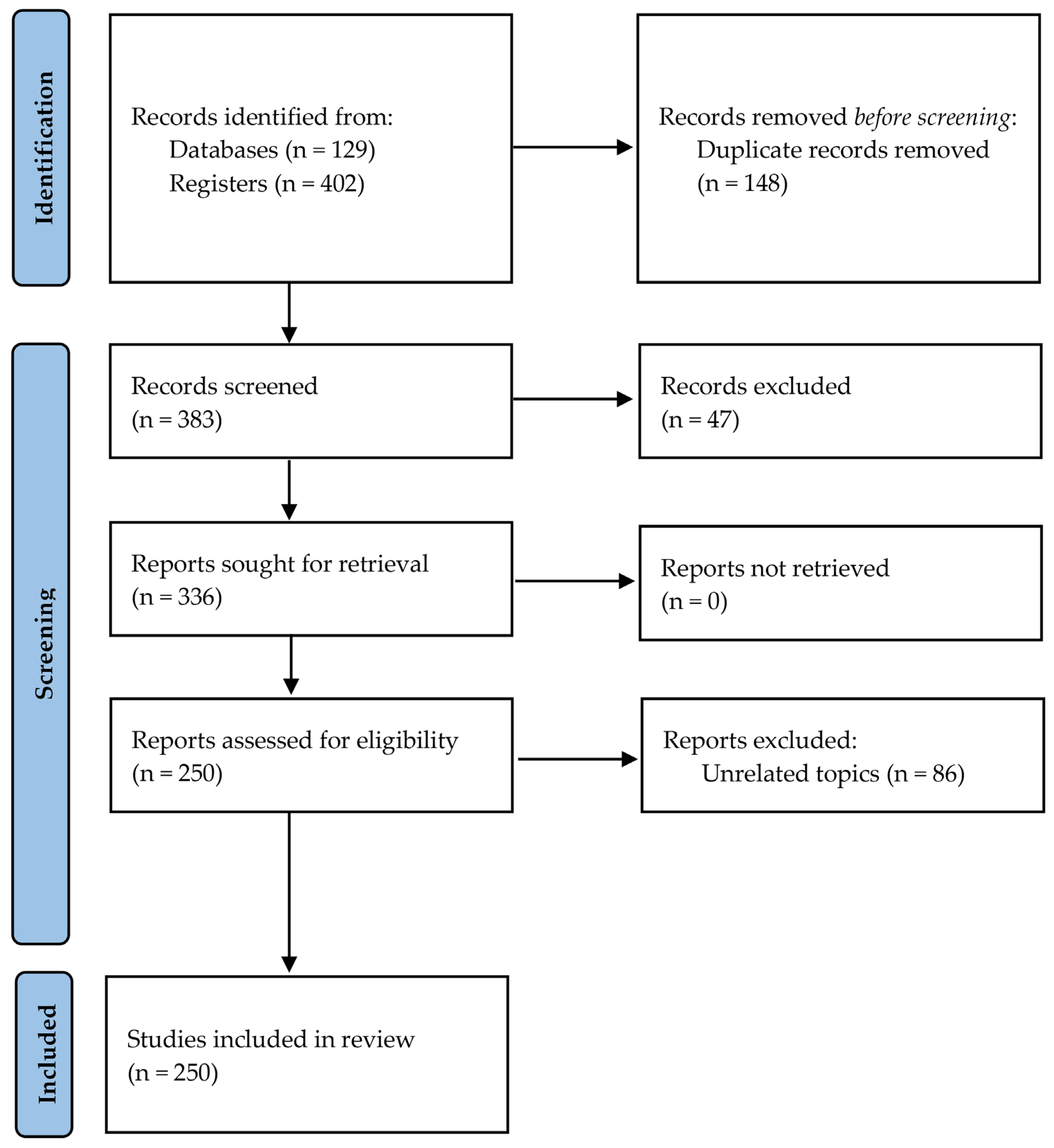
2. Materials and Methods
2.1. Search Criteria
2.2. Selection Criteria
- Articles published in English;
- Case reports, case series, randomized controlled trials, cadaveric feasibility studies, and observational cohort studies;
- Studies specifically addressing the management of BPIs through nerve transfer procedures and reporting at least one motor nerve transfer, either applied clinically or investigated in cadaveric studies.
- Studies concerning sensory nerve transfers, lower-limb nerve transfers, or tendon transfer;
- Studies addressing direct nerve repair or nerve grafting of the affected nerve;
2.3. Outcomes
- Shoulder external rotation and abduction
- Winging scapula
- Elbow, wrist and finger extension
- Elbow flexion
- Hand pronation, wrist and fingers flexion
- Intrinsic hand musculature
- Extraplexal nerves
- 37 for the first group (18 involving the suprascapular nerve and 19 involving the axillary nerve)
- 9 for the second group
- 67 for the third group
- 81 for the fourth group
- 38 for the fifth group
- 7 for the sixth group
- 11 for the seventh group (9 involving the phrenic nerve and 2 involving the spinal accessory nerve)
3. Shoulder External Rotation and Abduction
3.1. Transfer to the Suprascapular Nerve
3.1.1. Intraplexal Nerve Transfer
3.1.2. Extraplexal Nerve Transfer
3.2. Transfer to the Axillary Nerve
3.2.1. Intraplexal Nerve Transfer
3.2.2. Extraplexal Nerve Transfer
4. Winging Scapula
4.1. Transfer to Long Thoracic Nerve
4.1.1. Intraplexal Nerve Transfer
4.1.2. Extraplexal Nerve Transfer
5. Elbow, Wrist and Finger Extension
5.1. Transfer to Radial Nerve
5.1.1. Intraplexal Nerve Transfer
Elbow Extension
Wrist Extension
Finger Extension
Extraplexal Nerve Transfer
6. Elbow Flexion
6.1. Transfer to Musculocutaneous Nerve
6.1.1. Intraplexal Nerve Transfer
6.1.2. Extraplexal Nerve Transfer
7. Hand Pronation, Wrist and Fingers Flexion
7.1. Transfer to the Median Nerve
7.1.1. Intraplexal Nerve Transfer
Hand Pronation
Wrist Flexion
Finger Flexion
7.1.2. Extraplexal Nerve Transfer
8. Intrinsic Hand Musculature
8.1. Transfer to the Ulnar Nerve
8.1.1. Intraplexal Nerve Transfer
8.1.2. Extraplexal Nerve Transfer
9. Nerve Transfer for Extraplexal Nerves
9.1. Transfer to the Phrenic Nerve
Donors
9.2. Transfer to the Spinal Accessory Nerve (XI Cranial Nerve)
Donors
10. Discussion
11. Conclusions

| Group | Target Nerve | Type of Transfer | N° Articles | References |
|---|---|---|---|---|
| Shoulder external rotation and abduction | Suprascapular nerve | Intraplexual transfer | 8 | [12,13,14,15,16,17,18,19] |
| Extraplexal transfer | 10 | [26,27,28,29,30,31,32,33,34,35] | ||
| Axillary nerve | Intraplexual transfer | 14 | [38,39,40,41,42,43,44,45,46,47,48,49,50,51] | |
| Extraplexal transfer | 5 | [34,52,53,54,55] | ||
| Winging scapula | Long thoracic nerve | Intraplexual transfer | 7 | [58,59,60,61,62,63,64] |
| Extraplexal transfer | 2 | [65,66] | ||
| Elbow, wrist and finger extension | Radial nerve | Intraplexual transfer | 44 | Elbow extension [69,70,71,72,73,74,75,76,77,78,79,80,81,82] Wrist extension [83,84,85,86,87,88,89,90,91,92,93,94,95,96,97,98] Finger extension [99,100,101,102,103,104,105,106,107,108,109,110,111,112] |
| Extraplexal transfer | 23 | [54,113,114,115,116,117,118,119,120,121,122,123,124,125,126,127,128,129,130,131,132,133,134] | ||
| Elbow flexion | Musculocutaneous nerve | Intraplexual transfer | 46 | [45,136,137,138,139,140,141,142,143,144,145,146,147,148,149,150,151,152,153,154,155,156,157,158,159,160,161,162,163,164,165,166,167,168,169,170,171,172,173,174,175,176,177,178,179,180] |
| Extraplexal transfer | 35 | [21,33,149,151,181,182,183,184,185,186,187,188,189,190,191,192,193,194,195,196,197,198,199,200,201,202,203,204,205,206,207,208,209,210,211] | ||
| Hand pronation, wrist and fingers flexion | Median nerve | Intraplexual transfer | 26 | Hand pronation [103,215,216,217,218,219,220,221] Finger flexion [71,222,223,224,225,226,227,228,229,230,231,232,233,234,235,236,237,238] |
| Extraplexal transfer | 12 | [124,132,134,183,239,240,241,242,243,244,245,246] | ||
| Intrinsic hand musculature | Ulnar nerve | Intraplexual transfer | 6 | [247,248,249,250,251,252] |
| Extraplexal transfer | 1 | [253] | ||
| Extraplexal nerves | Phrenic nerve | Not applicable | 9 | [258,259,260,261,262,263,264,265,266] |
| Spinal accessory nerve | Not applicable | 2 | [268,269] |
Author Contributions
Funding
Institutional Review Board Statement
Informed Consent Statement
Data Availability Statement
Acknowledgments
Conflicts of Interest
Abbreviations
| BPIs | Brachial plexus injuries |
| SAN | Spinal accessory nerve |
| SSN | Suprascapular nerve |
| DSN | Dorsal scapular nerve |
| BPA | Brachial plexus avulsion |
| AN | Axillary nerve |
| LTN | Long thoracic nerve |
| MRC | Medical Research Council scale for muscle strength |
| ICNs | Intercostal nerves |
| PH | Phrenic nerve |
| RN | Radial nerve |
| SCI | Spinal cord injury |
| FDS | Flexor digitorum superficialis |
| ECRB | Extensor carpi radialis brevis |
| ECRL | Extensor carpi radialis longus |
| AIN | Anterior interosseous nerve |
| PQ | Pronator quadratus |
| PT | Pronator teres |
| ECU | Extensor carpi ulnaris |
| PIN | Posterior interosseous nerve |
| PL | Palmaris longus |
| FCR | Flexor carpi radialis |
| CC7 | Contralateral C7 |
| MCN | Musculocutaneous nerve |
| MN | Median nerve |
| FPL | Flexor pollicis longus |
| PCB | Palmar cutaneous branch |
| APB | Abductor pollicis brevis |
| OP | Opponens pollicis |
| FPB | Flexor pollicis brevis |
| EHL | Extensor hallucis longus |
| EDB | Extensor digitorum brevis |
| EHB | Extensor hallucis brevis |
| SPN | Superficial peroneal nerve |
| UN | Ulnar nerve |
References
- Lavorato, A.; Aruta, G.; De Marco, R.; Zeppa, P.; Titolo, P.; Colonna, M.R.; Galeano, M.; Costa, A.L.; Vincitorio, F.; Garbossa, D.; et al. Traumatic peripheral nerve injuries: A classification proposal. J. Orthop. Traumatol. 2023, 24, 20. [Google Scholar] [CrossRef]
- Noland, S.S.; Bishop, A.T.; Spinner, R.J.; Shin, A.Y. Adult traumatic brachial plexus injuries. J. Am. Acad. Orthop. Surg. 2019, 27, 705–716. [Google Scholar] [CrossRef]
- Harris, W.; Low, V.W. On the importance of accurate muscular analysis in lesions of the brachial plexus; and the treatment of Erb’s palsy and infantile paralysis of the upper extremity by cross-union of the nerve roots. Br. Med. J. 1903, 2, 1035–1038. [Google Scholar]
- Ballance, C.A.; Ballance, H.A.; Stewart, P. Remarks on the operative treatment of chronic facial palsy of peripheral origin. Br. Med. J. 1903, 1, 1009–1013. [Google Scholar] [CrossRef]
- Tuttle, H.K. Exposure of the brachial plexus with nerve-transplantation. J. Am. Med. Assoc. 1913, 61, 15–17. [Google Scholar] [CrossRef]
- Bertelli, J.A. Distal sensory nerve transfers in lower-type injuries of the brachial plexus. J. Hand Surg. Am. 2012, 37, 1194–1199. [Google Scholar] [CrossRef]
- Viterbo, F.; Amr, A.H.; Stipp, E.J.; Reis, F.J. End-to-side neurorrhaphy: Past, present, and future. Plast. Reconstr. Surg. 2009, 124, e351–e358. [Google Scholar] [CrossRef]
- Hayashi, A.; Pannucci, C.; Moradzadeh, A.; Kawamura, D.; Magill, C.; Hunter, D.A.; Tong, A.Y.; Parsadanian, A.; Mackinnon, S.E.; Myckatyn, T.M. Axotomy or compression is required for axonal sprouting following end-to-side neurorrhaphy. Exp. Neurol. 2008, 211, 539–550. [Google Scholar] [CrossRef]
- Farber, S.J.; Glaus, S.W.; Moore, A.M.; Hunter, D.A.; Mackinnon, S.E.; Johnson, P.J. Supercharge nerve transfer to enhance motor recovery: A laboratory study. J. Hand Surg. Am. 2013, 38, 466–477. [Google Scholar] [CrossRef]
- Guedes, F.; Heinen, C.; Rasulic, L.; Shin, A.Y.; Socolovsky, M. Nerve Transfers for Brachial Plexus Reconstruction After Trauma; Springer: Cham, Switzerland, 2025. [Google Scholar] [CrossRef]
- Pondaag, W.; de Boer, R.; van Wijlen-Hempel, M.S.; Hofstede-Buitenhuis, S.M.; Malessy, M.J. External rotation as a result of suprascapular nerve neurotization in obstetric brachial plexus lesions. Neurosurgery 2005, 57, 530–537. [Google Scholar] [CrossRef]
- Goubier, J.N.; Teboul, F. Transfer of the rhomboid nerve to the suprascapular nerve: An anatomical feasibility study. Chir. Main 2015, 34, 182–185. [Google Scholar] [CrossRef]
- Goubier, J.N.; Teboul, F. Rhomboid nerve transfer to the suprascapular nerve for shoulder reanimation in brachial plexus palsy: A clinical report. Hand Surg. Rehabil. 2016, 35, 363–366. [Google Scholar] [CrossRef]
- Goubier, J.N.; Echalier, C.; Dubois, E.; Teboul, F. Transfer of the rhomboid nerve for restoration of shoulder external rotation in partial brachial plexus palsy. Injury 2020, 51, S84–S87. [Google Scholar] [CrossRef]
- Augustine, H.; Choi, M.; Bain, J. Dorsal scapular nerve transfer to suprascapular nerve: A new method of restoring shoulder function. Plast. Surg. Case Stud. 2017, 3, e4614. [Google Scholar] [CrossRef]
- Malessy, M.J.; Thomeer, R.T.; Marani, E. The dorsoscapular nerve in traumatic brachial plexus lesions. Clin. Neurol. Neurosurg. 1993, 95, S17–S23. [Google Scholar] [CrossRef]
- Cambon-Binder, A.; Belkheyar, Z. Transfer of a C7 fascicle for the pectoralis major to the suprascapular nerve: A 3-year follow-up patient series. Plast. Reconstr. Surg. 2022, 149, 672–675. [Google Scholar] [CrossRef]
- Kashyap, V.H.; Singh, V.K.; Haq, A.; Kumari, A.; Sarkar, S. Outcomes in restoration of shoulder abduction in upper trunk brachial plexus injuries: A randomized study comparing ipsilateral C7 fascicle and spinal accessory nerve as donor nerves. J. Clin. Neurosci. 2025, 141, 111560. [Google Scholar] [CrossRef]
- David, S.; Balaguer, T.; Baque, P.; Lebreton, E. Transfer of pectoral nerves to suprascapular and axillary nerves: An anatomic feasibility study. J. Hand Surg. Am. 2010, 35, 92–96. [Google Scholar] [CrossRef]
- Allieu, Y.; Privat, J.M.; Bonnel, F. Les neurotisations par le nerf spinal (nerf accessorius) dans les avulsions radiculaires du plexus brachial. Neurochirurgie 1982, 28, 115–120. (In French) [Google Scholar]
- Allieu, Y.; Privat, J.M.; Bonnel, F. Paralysis in root avulsion of the brachial plexus. Neurotization by the spinal accessory nerve. Clin. Plast. Surg. 1984, 11, 133–136. [Google Scholar]
- Alnot, J.Y.; Jolly, A.; Frot, B. Traitement direct des lésions nerveuses dans les paralysies traumatiques du plex brachial chez l’adulte. Int. Orthop. 1981, 5, 151–168. (In French) [Google Scholar] [CrossRef]
- Narakas, A.O.; Hentz, V.R. Neurotization in brachial plexus injuries: Indication and results. Clin. Orthop. Relat. Res. 1988, 237, 43–56. [Google Scholar] [CrossRef]
- Midha, R. Nerve transfers for severe brachial plexus injuries: A review. Neurosurg. Focus 2004, 16, E5. [Google Scholar] [CrossRef]
- Bertelli, J.A.; Ghizoni, M.F. Transfer of the accessory nerve to the suprascapular nerve in brachial plexus reconstruction. J. Hand Surg. Am. 2007, 32, 989–998. [Google Scholar] [CrossRef]
- Bertelli, J.A.; Ghizoni, M.F. Results of spinal accessory to suprascapular nerve transfer in 110 patients with complete palsy of the brachial plexus. J. Neurosurg. Spine 2016, 24, 990–995. [Google Scholar] [CrossRef]
- Bhandari, P.S.; Deb, P. Dorsal approach in transfer of the distal spinal accessory nerve into the suprascapular nerve: Histo-morphometric analysis and clinical results in 14 cases of upper brachial plexus injuries. J. Hand Surg. Am. 2011, 36, 1182–1190. [Google Scholar] [CrossRef]
- Guan, S.B.; Hou, C.L.; Chen, D.S.; Gu, Y.D. Restoration of shoulder abduction by transfer of the spinal accessory nerve to suprascapular nerve through dorsal approach: A clinical study. Chin. Med. J. 2006, 119, 707–712. [Google Scholar] [CrossRef]
- Bertelli, J.A.; Ghizoni, M.F. Refinements in the technique for repair of the accessory nerve. J. Hand Surg. Am. 2006, 31, 1401–1406. [Google Scholar] [CrossRef]
- Zermeño-Rivera, J.; Gutiérrez-Amavizca, B.E. Contralateral spinal accessory nerve transfer: A new technique in panavulsive brachial plexus palsy. Indian J. Surg. 2015, 77, 247–250. [Google Scholar] [CrossRef]
- Durand, S.; Oberlin, C.; Fox, M.; Diverrez, J.P.; Dauge, M.C. Transfer of the first intercostal nerve to supra- and infraspinatus muscles: An anatomical study and report of the first case. J. Hand Surg. Eur. Vol. 2009, 34, 196–200. [Google Scholar] [CrossRef]
- Gu, Y.D.; Wu, M.M.; Zhen, Y.L.; Zhao, J.A.; Zhang, G.M.; Chen, D.S.; Yan, J.G.; Cheng, X.M. Phrenic nerve transfer for brachial plexus motor neurotization. Microsurgery 1989, 10, 287–289. [Google Scholar] [CrossRef]
- Gu, Y.D.; Ma, M.K. Use of the phrenic nerve for brachial plexus reconstruction. Clin. Orthop. Relat. Res. 1996, 323, 119–121. [Google Scholar] [CrossRef]
- Chuang, D.C.; Lee, G.W.; Hashem, F.; Wei, F.C. Restoration of shoulder abduction by nerve transfer in avulsed brachial plexus injury: Evaluation of 99 patients with various nerve transfers. Plast. Reconstr. Surg. 1995, 96, 122–128. [Google Scholar] [CrossRef]
- Sungpet, A.; Suphachatwong, C.; Kawinwonggowith, V. Restoration of shoulder abduction in brachial plexus injury with phrenic nerve transfer. Aust. N. Z. J. Surg. 2000, 70, 783–785. [Google Scholar] [CrossRef]
- Okwumabua, E.; Thompson, J.H. Anatomy, shoulder and upper limb, axillary nerve. In StatPearls [Internet]; StatPearls Publishing: Treasure Island, FL, USA, 2025. Available online: https://www.ncbi.nlm.nih.gov/books/NBK493212/ (accessed on 4 February 2026).
- Hangge, P.T.; Breen, I.; Albadawi, H.; Knuttinen, M.G.; Naidu, S.G.; Oklu, R. Quadrilateral space syndrome: Diagnosis and clinical management. J. Clin. Med. 2018, 7, 86. [Google Scholar] [CrossRef]
- Witoonchart, K.; Leechavengvongs, S.; Uerpairojkit, C.; Thuvasethakul, P.; Wongnopsuwan, V. Nerve transfer to deltoid muscle using the nerve to the long head of the triceps, part I: An anatomic feasibility study. J. Hand Surg. Am. 2003, 28, 628–632. [Google Scholar] [CrossRef]
- Leechavengvongs, S.; Witoonchart, K.; Uerpairojkit, C.; Thuvasethakul, P. Nerve transfer to deltoid muscle using the nerve to the long head of the triceps, part II: A report of 7 cases. J. Hand Surg. Am. 2003, 28, 633–638. [Google Scholar] [CrossRef]
- Bertelli, J.A.; Ghizoni, M.F. Reconstruction of C5 and C6 brachial plexus avulsion injury by multiple nerve transfers: Spinal accessory to suprascapular, ulnar fascicles to biceps branch, and triceps long or lateral head branch to axillary nerve. J. Hand Surg. Am. 2004, 29, 131–139. [Google Scholar] [CrossRef]
- Bertelli, J.A.; Ghizoni, M.F. Nerve transfer from triceps medial head and anconeus to deltoid for axillary nerve palsy. J. Hand Surg. Am. 2014, 39, 940–947. [Google Scholar] [CrossRef]
- Jerome, J.T. Anterior deltopectoral approach for axillary nerve neurotisation. J. Orthop. Surg. 2012, 20, 66–70. [Google Scholar] [CrossRef]
- Soldado, F.; Barrera-Ochoa, S.; Romero-Larrauri, P.; Nguyen, T.Q.; Diaz-Gallardo, P.; Knörr, J. Triceps to teres minor motor nerve transfer to restore glenohumeral external rotation after neonatal brachial plexus injury. Microsurgery 2022, 42, 533–537. [Google Scholar] [CrossRef]
- Dai, S.Y.; Lin, D.X.; Han, Z.; Zhoug, S.Z. Transference of thoracodorsal nerve to musculocutaneous or axillary nerve in old traumatic injury. J. Hand Surg. Am. 1990, 15, 36–37. [Google Scholar] [CrossRef]
- Samardzic, M.M.; Grujicic, D.M.; Rasulic, L.G.; Milicic, B.R. The use of thoracodorsal nerve transfer in restoration of irreparable C5 and C6 spinal nerve lesions. Br. J. Plast. Surg. 2005, 58, 541–546. [Google Scholar] [CrossRef]
- Janes, L.E.; Crowe, C.; Shah, N.; Sasson, D.; Ko, J.H. Alternative nerve transfer for shoulder function: Thoracodorsal and medial triceps to anterior axillary nerve. Plast. Reconstr. Surg. Glob. Open 2022, 10, e4614. [Google Scholar] [CrossRef]
- Tubbs, R.S.; Khoury, C.A.; Salter, E.G.; Acakpo-Satchivi, L.; Wellons, J.C., 3rd; Blount, J.P.; Oakes, W.J. Quantitation of the lower subscapular nerve for potential use in neurotization procedures. J. Neurosurg. 2006, 105, 881–883. [Google Scholar] [CrossRef]
- Haninec, P.; Hradecky, J.; Mencl, L. Lower subscapular nerve transfer for axillary nerve repair in upper brachial plexus palsy. Acta Neurochir. 2020, 162, 135–139. [Google Scholar] [CrossRef]
- Haninec, P.; Hradecky, J.; Ouzky, M.; Samal, F.; Mencl, L. Nerve transfers for axillary nerve repair in brachial plexus injuries: Results from 206 patients. J. Neurosurg. Spine 2025, 42, 659–666. [Google Scholar] [CrossRef]
- Rodríguez-Aceves, C.A.; Córdoba-Mosqueda, M.E. Long thoracic to axillary nerve transfer. In Nerve Transfers for Brachial Plexus Reconstruction After Trauma; Guedes, F., Heinen, C., Rasulic, L., Shin, A.Y., Socolovsky, M., Eds.; Springer: Cham, Switzerland, 2025. [Google Scholar]
- Haninec, P.; Kaiser, R. Axillary nerve repair by fascicle transfer from the ulnar or median nerve in upper brachial plexus palsy. J. Neurosurg. 2012, 117, 610–614. [Google Scholar] [CrossRef]
- Farrar, J.Q. Spinal Accessory Nerve to Axillary Nerve Transfer. In Rehabilitation for Nerve Transfers of the Upper Extremity; Stonner, L.C., Dy, M.M., Kahn, C.J., Eds.; Springer: Cham, Switzerland, 17 October 2025. [Google Scholar] [CrossRef]
- Samardzić, M.; Rasulić, L.; Grujicić, D.; Milicić, B. Results of nerve transfers to the musculocutaneous and axillary nerves. Neurosurgery 2000, 46, 93–101; discussion 101–103. [Google Scholar] [CrossRef]
- Malungpaishrope, K.; Leechavengvongs, S.; Witoonchart, K.; Uerpairojkit, C.; Boonyalapa, A.; Janesaksrisakul, D. Simultaneous intercostal nerve transfers to deltoid and triceps muscle through the posterior approach. J. Hand Surg. Am. 2012, 37, 677–682. [Google Scholar] [CrossRef]
- Nath, R.K.; Somasundaram, C. Outcomes of Brachial Plexus Neurolysis in 40 Patients with Obstetric Brachial Plexus Injury. Eplasty 2025, 25, e4. [Google Scholar]
- Lung, K.; Lui, F. Anatomy, Thorax, Long Thoracic Nerve. In StatPearls [Internet]; StatPearls Publishing: Treasure Island, FL, USA, 2025. Available online: https://www.ncbi.nlm.nih.gov/books/NBK535396/ (accessed on 4 February 2026).
- Galano, G.J.; Bigliani, L.U.; Ahmad, C.S.; Levine, W.N. Surgical treatment of winged scapula. Clin. Orthop. Relat. Res. 2008, 466, 652–660. [Google Scholar] [CrossRef] [PubMed]
- Novak, C.B.; Mackinnon, S.E. Surgical treatment of a long thoracic nerve palsy. Ann. Thorac. Surg. 2002, 73, 1643–1645. [Google Scholar] [CrossRef]
- Uerpairojkit, C.; Leechavengvongs, S.; Witoonchart, K.; Malungpaishorpe, K.; Raksakulkiat, R. Nerve transfer to serratus anterior muscle using the thoracodorsal nerve for winged scapula in C5 and C6 brachial plexus root avulsions. J. Hand Surg. Am. 2009, 34, 74–78. [Google Scholar] [CrossRef] [PubMed]
- Martinez, J.P.; Lovaglio, A.; Masi, G.D.; Mandolesi, J.; Zancolli, P.; Socolovsky, M. Thoracodorsal to long thoracic nerve transfer in a patient with traumatic injury: A case report. Surg. Neurol. Int. 2024, 15, 163. [Google Scholar] [CrossRef]
- Noland, S.S.; Krauss, E.M.; Felder, J.M.; Mackinnon, S.E. Surgical and Clinical Decision Making in Isolated Long Thoracic Nerve Palsy. Hand 2018, 13, 689–694. [Google Scholar] [CrossRef]
- Li, M.; Parekh, J.N.; Jimulia, D.; Duraku, L.S.; Chaudhry, T.; Power, D.M. Long Thoracic Nerve Decompression and Transfer: Single-Center Case Series of Clinical and Electromyographic Outcomes. Hand 2025, 16, 15589447251343240. [Google Scholar] [CrossRef]
- Tomaino, M.M. Neurophysiologic and clinical outcome following medial pectoral to long thoracic nerve transfer for scapular winging: A case report. Microsurgery 2002, 22, 254–257. [Google Scholar] [CrossRef]
- Ray, W.Z.; Pet, M.A.; Nicoson, M.C.; Yee, A.; Kahn, L.C.; Mackinnon, S.E. Two-level motor nerve transfer for the treatment of long thoracic nerve palsy. J. Neurosurg. 2011, 115, 858–864. [Google Scholar] [CrossRef]
- Yamada, T.; Doi, K.; Hattori, Y.; Hoshino, S.; Sakamoto, S.; Arakawa, Y. Long thoracic nerve neurotization for restoration of shoulder function in C5-7 brachial plexus preganglionic injuries: Case report. J. Hand Surg. Am. 2010, 35, 1427–1431. [Google Scholar] [CrossRef] [PubMed]
- Louis, R.G., Jr.; Whitesides, J.D.; Kollias, T.F.; Iwanaga, J.; Tubbs, R.S.; Loukas, M. Intercostal Nerve to Long Thoracic Nerve Transfer for the Treatment of Winged Scapula: A Cadaveric Feasibility Study. Cureus 2017, 9, e1898. [Google Scholar] [CrossRef] [PubMed]
- Krijnen, N.A.; Comerci, A.J.; Head, L.K.; Legerstee, I.W.F.; de Klerk, H.H.; Chen, N.; Teunis, T. What Is the Probability of Radial Nerve Recovery After Surgical Repair of Humerus Fractures Accounting for Time Since Injury? Clin. Orthop. Relat. Res. 2025, 484, 844–853. [Google Scholar] [CrossRef]
- Zancolli, E.A. Midcervical tetraplegia with strong wrist extension: A two-stage synergistic reconstruction of the hand. Hand Clin. 2002, 18, 481–495. [Google Scholar] [CrossRef] [PubMed]
- Bertelli, J.A.; Tacca, C.P.; Winkelmann Duarte, E.C.; Ghizoni, M.F.; Duarte, H. Transfer of axillary nerve branches to reconstruct elbow extension in tetraplegics: A laboratory investigation of surgical feasibility. Microsurgery 2011, 31, 376–381. [Google Scholar] [CrossRef]
- Bertelli, J.A.; Ghizoni, M.F. Transfer of the teres minor motor branch for triceps reinnervation in tetraplegia. J. Neurosurg. 2011, 114, 1457–1460. [Google Scholar] [CrossRef]
- Fox, I.K.; Davidge, K.M.; Novak, C.B.; Hoben, G.; Kahn, L.C.; Juknis, N.; Ruvinskaya, R.; Mackinnon, S.E. Nerve Transfers to Restore Upper Extremity Function in Cervical Spinal Cord Injury: Update and Preliminary Outcomes. Plast. Reconstr. Surg. 2015, 136, 780–792. [Google Scholar] [CrossRef]
- Bertelli, J.A.; Ghizoni, M.F. Nerve transfers for elbow and finger extension reconstruction in midcervical spinal cord injuries. J. Neurosurg. 2015, 122, 121–127. [Google Scholar] [CrossRef] [PubMed]
- Bazarek, S.; Sten, M.; Upton, S.; Magee, G.; Mandeville, R.; Brown, J.M. Axillary to Radial Nerve Transfer for Elbow Extension in the Tetraplegic Patient: A Case Series. Neurosurgery 2025, 98, 1247–1254. [Google Scholar] [CrossRef] [PubMed]
- Bazarek, S.; Thum, J.A.; Sten, M.; Magee, G.; Mandeville, R.; Brown, J.M. Axillary to Radial Nerve Transfer for Recovery of Elbow Extension After Spinal Cord Injury. Oper. Neurosurg. 2023, 25, e324–e329. [Google Scholar] [CrossRef]
- Bertelli, J.A.; Soldado, F.; Ghizoni, M.F.; Rodríguez-Baeza, A. Transfer of a Terminal Motor Branch Nerve to the Flexor Carpi Ulnaris for Triceps Reinnervation: Anatomical Study and Clinical Cases. J. Hand Surg. Am. 2015, 40, 2229–2235.e2. [Google Scholar] [CrossRef]
- Flores, L.P. Triceps brachii reinnervation in primary reconstruction of the adult brachial plexus: Experience in 25 cases. Acta Neurochir. 2011, 153, 1999–2007. [Google Scholar] [CrossRef]
- Pet, M.A.; Ray, W.Z.; Yee, A.; Mackinnon, S.E. Nerve transfer to the triceps after brachial plexus injury: Report of four cases. J. Hand Surg. Am. 2011, 36, 398–405. [Google Scholar] [CrossRef]
- Emamhadi, M.; Behzadnia, H.; Golsefid, H.N.; Emamhadi, A.; Andalib, S. Reanimation of triceps muscle using ulnar nerve fascicle transfer to the nerve to long head of the triceps muscle. Acta Neurochir. 2020, 162, 1899–1905. [Google Scholar] [CrossRef]
- Soldado, F.; Ghizoni, M.F.; Bertelli, J. Thoracodorsal nerve transfer for triceps reinnervation in partial brachial plexus injuries. Microsurgery 2016, 36, 191–197. [Google Scholar] [CrossRef] [PubMed]
- Flores, L.P. Reanimation of elbow extension with medial pectoral nerve transfer in partial injuries to the brachial plexus. J. Neurosurg. 2013, 118, 588–593. [Google Scholar] [CrossRef]
- Flores, L.P. The radio-radial nerve transfer for elbow extension restoration in C5 to C7 nerve root injury. Microsurgery 2012, 32, 55–59. [Google Scholar] [CrossRef] [PubMed]
- Flores, L.P. Outcomes of Transferring a Healthy Motor Fascicle from the Radial Nerve to a Branch for the Triceps to Recover Elbow Extension in Partial Brachial Plexus Palsy. Neurosurgery 2017, 80, 448–453. [Google Scholar] [CrossRef]
- Lurje, A.S. On the Use of N. Musculocutaneous for Neurotization of N. Radialis in Cases of Very Large Defects of the Latter. Ann. Surg. 1948, 128, 110–115. [Google Scholar] [CrossRef] [PubMed]
- Fridén, J.; Gohritz, A. Brachialis-to-extensor carpi radialis longus selective nerve transfer to restore wrist extension in tetraplegia: Case report. J. Hand Surg. Am. 2012, 37, 1606–1608. [Google Scholar] [CrossRef]
- Long Azad, C.; Orlando, N.A.; Belzberg, A.J.; Tuffaha, S.H. Surgical Technique: Brachioradialis to Extensor Carpi Radialis Longus and Brevis Nerve Transfers for Tetraplegia. Tech. Hand Up. Extrem. Surg. 2024, 28, 88–91. [Google Scholar] [CrossRef]
- Ustün, M.E.; Ogün, T.C.; Büyükmumcu, M. Neurotization as an alternative for restoring finger and wrist extension. J. Neurosurg. 2001, 94, 795–798. [Google Scholar] [CrossRef]
- Lowe, J.B., 3rd; Tung, T.R.; Mackinnon, S.E. New surgical option for radial nerve paralysis. Plast. Reconstr. Surg. 2002, 110, 836–843. [Google Scholar] [CrossRef]
- García-López, A.; Perea, D. Transfer of median and ulnar nerve fascicles for lesions of the posterior cord in infraclavicular brachial plexus injury: Report of 2 cases. J. Hand Surg. Am. 2012, 37, 1986–1989. [Google Scholar] [CrossRef] [PubMed]
- García-López, A.; Navarro, R.; Martinez, F.; Rojas, A. Nerve transfers from branches to the flexor carpi radialis and pronator teres to reconstruct the radial nerve. J. Hand Surg. Am. 2014, 39, 50–56. [Google Scholar] [CrossRef]
- Ukrit, A.; Leechavengvongs, S.; Malungpaishrope, K.; Uerpairojkit, C.; Chongthammakun, S.; Witoonchart, K. Nerve transfer for wrist extension using nerve to flexor digitorum superficialis in cervical 5, 6, and 7 root avulsions: Anatomic study and report of two cases. J. Hand Surg. Am. 2009, 34, 1659–1666. [Google Scholar] [CrossRef]
- Bertelli, J.A.; Ghizoni, M.F. Transfer of a flexor digitorum superficialis motor branch for wrist extension reconstruction in C5–C8 root injuries of the brachial plexus: A case series. Microsurgery 2013, 33, 39–42. [Google Scholar] [CrossRef] [PubMed]
- Kerver, A.L.; Carati, L.; Eilers, P.H.; Langezaal, A.C.; Kleinrensink, G.J.; Walbeehm, E.T. An anatomical study of the ECRL and ECRB: Feasibility of developing a preoperative test for evaluating the strength of the individual wrist extensors. J. Plast. Reconstr. Aesthet. Surg. 2013, 66, 543–550. [Google Scholar] [CrossRef]
- Bertelli, J.A.; Ghizoni, M.F.; Tacca, C.P. Results of wrist extension reconstruction in C5-8 brachial plexus palsy by transferring the pronator quadratus motor branch to the extensor carpi radialis brevis muscle. J. Neurosurg. 2016, 124, 1442–1449. [Google Scholar] [CrossRef]
- Bertelli, J.A.; Tacca, C.P.; Winkelmann Duarte, E.C.; Ghizoni, M.F.; Duarte, H. Transfer of the pronator quadratus motor branch for wrist extension reconstruction in brachial plexus palsy. Plast. Reconstr. Surg. 2012, 130, 1269–1278. [Google Scholar] [CrossRef]
- Bhatia, A.; Salama, M. Pronator Quadratus to Extensor Carpi Radialis Brevis Nerve Transfer in C5-C7 or C5-C8 Brachial Plexus Injuries for Independent Wrist Extension. Indian J. Plast. Surg. 2020, 53, 36–41. [Google Scholar] [CrossRef] [PubMed]
- Bertelli, J.A.; Nehete, S.; Winkelmann Duarte, E.C.; Patel, N.; Ghizoni, M.F. Distal pronator teres motor branch transfer for wrist extension restoration in radial nerve paralysis. J. Neurosurg. 2020, 134, 251–257. [Google Scholar] [CrossRef] [PubMed]
- Tian, J.; Leng, M.; Wang, K.; Huang, Q. Pronator teres nerve branch transfer to the extensor carpi radialis brevis nerve branch for wrist extension reconstruction in proximal radial nerve injury following humeral shaft fractures. BMC Musculoskelet. Disord. 2022, 23, 980. [Google Scholar] [CrossRef] [PubMed]
- Fox, I.K.; Davidge, K.M.; Novak, C.B.; Hoben, G.; Kahn, L.C.; Juknis, N.; Ruvinskaya, R.; Mackinnon, S.E. Use of peripheral nerve transfers in tetraplegia: Evaluation of feasibility and morbidity. Hand 2015, 10, 60–67. [Google Scholar] [CrossRef]
- Bertelli, J.A.; Ghizoni, M.F. Transfer of supinator motor branches to the posterior interosseous nerve in C7-T1 brachial plexus palsy. J. Neurosurg. 2010, 113, 129–132. [Google Scholar] [CrossRef]
- Lamb, D.W.; Landry, R. The hand in quadriplegia. Hand 1971, 3, 31–37. [Google Scholar] [CrossRef] [PubMed]
- Ray, W.Z.; Mackinnon, S.E. Clinical outcomes following median to radial nerve transfers. J. Hand Surg. Am. 2011, 36, 201–208. [Google Scholar] [CrossRef]
- Davidge, K.M.; Yee, A.; Kahn, L.C.; Mackinnon, S.E. Median to radial nerve transfers for restoration of wrist, finger, and thumb extension. J. Hand Surg. Am. 2013, 38, 1812–1827. [Google Scholar] [CrossRef]
- Sallam, A.A.; El-Deeb, M.S.; Imam, M.A. Useful functional outcome can be achieved after motor nerve transfers in management of the paralytic hand: An observational study. HSS J. 2016, 12, 2–7. [Google Scholar] [CrossRef]
- Bertelli, J.A.; Kechele, P.R.; Santos, M.A.; Besen, B.A.; Duarte, H. Anatomical feasibility of transferring supinator motor branches to the posterior interosseous nerve in C7-T1 brachial plexus palsies: Laboratory investigation. J. Neurosurg. 2009, 111, 326–331. [Google Scholar] [CrossRef]
- Bertelli, J.A.; Tacca, C.P.; Ghizoni, M.F.; Kechele, P.R.; Santos, M.A. Transfer of supinator motor branches to the posterior interosseous nerve to reconstruct thumb and finger extension in tetraplegia: Case report. J. Hand Surg. Am. 2010, 35, 1647–1651. [Google Scholar] [CrossRef]
- Texakalidis, P.; Liu, L.; Karras, C.L.; Alden, T.D.; Franz, C.K.; Swong, K. Supinator to posterior interosseous nerve transfer to restore hand opening in brachial plexus and spinal cord injury: A systematic review and individual patient-data meta-analysis. J. Neurosurg. Spine 2024, 41, 621–627. [Google Scholar] [CrossRef]
- Zhang, C.G.; Dong, Z.; Gu, Y.D. Restoration of hand function in C7-T1 brachial plexus palsies using a staged approach with nerve and tendon transfer. J. Neurosurg. 2014, 121, 1264–1270. [Google Scholar] [CrossRef]
- Bazarek, S.; Sten, M.; Nin, D.; Brown, J.M. Supinator to posterior interosseous nerve transfer for restoration of finger extension. Oper. Neurosurg. 2021, 21, E408–E413. [Google Scholar] [CrossRef]
- Dong, Z.; Gu, Y.D.; Zhang, C.G.; Zhang, L. Clinical use of supinator motor branch transfer to the posterior interosseous nerve in C7-T1 brachial plexus palsies. J. Neurosurg. 2010, 113, 113–117. [Google Scholar] [CrossRef]
- Xu, B.; Dong, Z.; Zhang, C.G.; Gu, Y.D. Clinical outcome following transfer of the supinator motor branch to the posterior interosseous nerve in patients with C7-T1 brachial plexus palsy. J. Reconstr. Microsurg. 2015, 31, 102–106. [Google Scholar] [CrossRef]
- Zhang, L.; Zhang, C.L.; Dong, Z.; Gu, Y.D. Outcome of finger extension after nerve transfer to repair C7-T1 brachial plexus palsy in rats: Comparative study of the supinator motor branch transfer to the posterior interosseous nerve and the contralateral C7 transfer to the lower trunk. Neurosurgery 2017, 80, 627–634. [Google Scholar] [CrossRef]
- Palazzi, S.; Palazzi, J.L.; Caceres, J.P. Neurotization with the brachialis muscle motor nerve. Microsurgery 2006, 26, 330–333. [Google Scholar] [CrossRef]
- Bulstra, L.F.; Rbia, N.; Kircher, M.F.; Spinner, R.J.; Bishop, A.T.; Shin, A.Y. Spinal accessory nerve to triceps muscle transfer using long autologous nerve grafts for recovery of elbow extension in traumatic brachial plexus injuries. J. Neurosurg. 2018, 129, 1041–1047. [Google Scholar] [CrossRef]
- Gatskiy, A.A.; Tretyak, I.B.; Tsymbaliuk, V.I.; Tsymbaliuk, Y.V. Nerve transfers in a patient with asymmetrical neurological deficit following traumatic cervical spinal cord injury: Simultaneous bilateral restoration of pinch grip and elbow extension. Illustrative case. J. Neurosurg. Case Lessons 2022, 4, CASE22301. [Google Scholar] [CrossRef]
- Flores, L.P.; Socolovsky, M. Phrenic nerve transfer for reconstruction of elbow extension in severe brachial plexus injuries. J. Reconstr. Microsurg. 2016, 32, 546–550. [Google Scholar] [CrossRef]
- Lin, H.; Hou, C.; Chen, D. Full-length phrenic nerve transfer as the treatment for brachial plexus avulsion injury to restore wrist and finger extension. Muscle Nerve 2012, 45, 39–42. [Google Scholar] [CrossRef]
- Goubier, J.N.; Teboul, F. Transfer of the intercostal nerves to the nerve of the long head of the triceps to recover elbow extension in brachial plexus palsy. Tech. Hand Up. Extrem. Surg. 2007, 11, 139–141. [Google Scholar] [CrossRef]
- Goubier, J.N.; Teboul, F.; Khalifa, H. Reanimation of elbow extension with intercostal nerves transfers in total brachial plexus palsies. Microsurgery 2011, 31, 7–11. [Google Scholar] [CrossRef]
- Kittithamvongs, P.; Leechavengvongs, S.; Malungpaishrope, K.; Pongpinyopap, W.; Anantavorasakul, N.; Uerpairojkit, C. The intercostal nerves transfer to the radial nerve branch to the long head triceps muscle: Influencing factor and outcome of 55 cases. J. Hand Surg. Am. 2023, 48, 733.e1–733.e7. [Google Scholar] [CrossRef]
- Malungpaishrope, K.; Kittithamvongs, P.; Siripoonyothai, S.; Anantavorasakul, N.; Uerpairojkit, C.; Leechavengvongs, S. Transfer of the intercostal nerves to the radial nerve branch innervating the long head of the triceps muscle for elbow extension. JBJS Essent. Surg. Tech. 2025, 15, e23.00092. [Google Scholar] [CrossRef] [PubMed]
- Malungpaishrope, K.; Leechavengvongs, S.; Uerpairojkit, C.; Anantavorasakul, N.; Kittithamvongs, P.; Densiri-Aksorn, W. Outcomes of spinal accessory nerve and intercostal nerve transfers for shoulder stabilisation and elbow extension in patients with C5–C7 root avulsion injury. J. Hand Surg. Asian Pac Vol. 2022, 27, 447–452. [Google Scholar] [CrossRef]
- Malungpaishope, K.; Leechavengvongs, S.; Ratchawatana, P.; Pitakveerakul, A.; Jindahara, S.; Uerpairojkit, C.; Putthiwara, D.; Anantavorasakul, N.; Tan, V.H. Simultaneous phrenic and intercostal nerves transfer for elbow flexion and extension in total brachial plexus root avulsion injury. J. Hand Surg. Asian Pac. Vol. 2018, 23, 496–500. [Google Scholar] [CrossRef] [PubMed]
- Senes, F.M.; Catena, N.; Dapelo, E.; Senes, J. Nerve transfer for elbow extension in obstetrical brachial plexus palsy. Ann. Acad. Med. Singap. 2016, 45, 221–224. [Google Scholar] [CrossRef]
- Gu, Y.D.; Zhang, G.M.; Chen, D.S.; Yan, J.G.; Cheng, X.M.; Chen, L. Seventh cervical nerve root transfer from the contralateral healthy side for treatment of brachial plexus root avulsion. J. Hand Surg. Br. 1992, 17, 518–521. [Google Scholar] [CrossRef] [PubMed]
- Gu, Y.D.; Chen, D.S.; Zhang, G.M.; Cheng, X.M.; Xu, J.G.; Zhang, L.Y.; Cai, P.Q.; Chen, L. Long-term functional results of contralateral C7 transfer. J. Reconstr. Microsurg. 1998, 14, 57–59. [Google Scholar] [CrossRef]
- Gu, Y.; Xu, J.; Chen, L.; Wang, H.; Hu, S. Long term outcome of contralateral C7 transfer: A report of 32 cases. Chin. Med. J. 2002, 115, 866–868. [Google Scholar] [PubMed]
- Hattori, Y.; Doi, K.; Ikeda, K.; Pagsaligan, J.M.; Watanabe, M. Restoration of prehension using double free muscle technique after complete avulsion of brachial plexus in children: A report of three cases. J. Hand Surg. Am. 2005, 30, 812–819. [Google Scholar] [CrossRef]
- Terzis, J.K.; Kokkalis, Z.T. Selective contralateral C7 transfer in posttraumatic brachial plexus injuries: A report of 56 cases. Plast. Reconstr. Surg. 2009, 123, 927–938. [Google Scholar] [CrossRef]
- Terzis, J.K.; Barmpitsioti, A. Our experience with triceps nerve reconstruction in patients with brachial plexus injury. J. Plast. Reconstr. Aesthet. Surg. 2012, 65, 590–600. [Google Scholar] [CrossRef]
- Gao, K.; Lao, J.; Zhao, X.; Gu, Y. Outcome of contralateral C7 transfer to two recipient nerves in 22 patients with the total brachial plexus avulsion injury. Microsurgery 2013, 33, 605–611. [Google Scholar] [CrossRef]
- Li, W.J.; He, L.Y.; Chen, S.L.; Lyu, Y.W.; Wang, S.F.; Yong, Y.; Tian, W.; Tian, G.L.; Gu, Y.D. Contralateral C7 nerve root transfer for function recovery in adults: A meta-analysis. Chin. Med. J. 2017, 130, 2960–2968. [Google Scholar] [CrossRef] [PubMed]
- Yang, G.; Chang, K.W.; Chung, K.C. A systematic review of contralateral C7 transfer for the treatment of traumatic brachial plexus injury: Part 1. Overall outcomes. Plast. Reconstr. Surg. 2015, 136, 794–809. [Google Scholar] [CrossRef]
- Xiao, F.; Lao, J. Comparative study of intercostal nerve and contralateral C7 nerve transfers for elbow extension after global brachial plexus avulsion. Ann. Plast. Surg. 2020, 85, 272–275. [Google Scholar] [CrossRef]
- Titolo, P.; Lavorato, A.; Isoardo, G.; Vincitorio, F.; Garbossa, D.; Battiston, B. Transfer of the peroneal component of the sciatic nerve in total brachial plexus lesion: An anatomical feasibility study. Injury 2020, 51, 2904–2909. [Google Scholar] [CrossRef] [PubMed]
- Vernon Lee, C.Y.; Cochrane, E.; Chew, M.; Bains, R.D.; Bourke, G.; Wade, R.G. The effectiveness of different nerve transfers in the restoration of elbow flexion in adults following brachial plexus injury: A systematic review and meta-analysis. J. Hand Surg. Am. 2023, 48, 236–244. [Google Scholar] [CrossRef]
- Oberlin, C.; Béal, D.; Leechavengvongs, S.; Salon, A.; Dauge, M.C.; Sarcy, J.J. Nerve transfer to biceps muscle using a part of ulnar nerve for C5-C6 avulsion of the brachial plexus: Anatomical study and report of four cases. J. Hand Surg. Am. 1994, 19, 232–237. [Google Scholar] [CrossRef]
- Sungpet, A.; Suphachatwong, C.; Kawinwonggowit, V.; Patradul, A. Transfer of a single fascicle from the ulnar nerve to the biceps muscle after avulsions of upper roots of the brachial plexus. J. Hand Surg. Br. 2000, 25, 325–328. [Google Scholar] [CrossRef]
- Leechavengvongs, S.; Witoonchart, K.; Uerpairojkit, C.; Thuvasethakul, P.; Ketmalasiri, W. Nerve transfer to biceps muscle using a part of the ulnar nerve in brachial plexus injury (upper arm type): A report of 32 cases. J. Hand Surg. Am. 1998, 23, 711–716. [Google Scholar] [CrossRef] [PubMed]
- Schreiber, J.J.; Byun, D.J.; Khair, M.M.; Rosenblatt, L.; Lee, S.K.; Wolfe, S.W. Optimal axon counts for brachial plexus nerve transfers to restore elbow flexion. Plast. Reconstr. Surg. 2015, 135, 135e–141e. [Google Scholar] [CrossRef]
- Gardenal, R.M.; Vélez, M.; Glasberg, E.; Seri, M.; Faccendini, S.; Slullitel, M.; Bichara, J.; Capomassi, M. Nerve transfers for elbow flexion in traumatic brachial plexus injuries. Rev. Asoc. Argent. Ortop. Traumatol. 2022, 87, 507–516. [Google Scholar] [CrossRef]
- Elsebaey, M.E.; Elghabaty, M.E.Y.; Nasef, T.M.; Mostafa, H.N.; Elfarsy, M.F. Surgical techniques for restoration of elbow flexion in traumatic brachial plexus injuries: A single-center experience in Egypt. Egypt J. Neurosurg. 2025, 40, 97. [Google Scholar] [CrossRef]
- Nagano, Y.; Kawamura, D.; Terkawi, A.; Urita, A.; Matsui, Y.; Iwasaki, N. Minimum ten-year outcomes of partial ulnar nerve transfer for restoration of elbow flexion in patients with upper brachial plexus injury. J. Hand Surg. Asian Pac. Vol. 2019, 24, 283–288. [Google Scholar] [CrossRef]
- Leechavengvongs, S.; Witoonchart, K.; Uerpairojkit, C.; Thuvasethakul, P.; Malungpaishrope, K. Combined nerve transfers for C5 and C6 brachial plexus avulsion injury. J. Hand Surg. Am. 2006, 31, 183–189. [Google Scholar] [CrossRef]
- Socolovsky, M.; Martins, R.S.; Di Masi, G.; Siqueira, M. Upper brachial plexus injuries: Grafts vs ulnar fascicle transfer to restore biceps muscle function. Neurosurgery 2012, 71, ons227–ons232. [Google Scholar] [CrossRef]
- Ayhan, E.; Soldado, F.; Fontecha, C.G.; Bertelli, J.A.; Leblebicioglu, G. Elbow flexion reconstruction with nerve transfer or grafting in patients with brachial plexus injuries: A systematic review and comparison study. Microsurgery 2020, 40, 79–86. [Google Scholar] [CrossRef] [PubMed]
- Bertelli, J.A.; Ghizoni, M.F. Nerve root grafting and distal nerve transfers for C5-C6 brachial plexus injuries. J. Hand Surg. Am. 2010, 35, 769–775. [Google Scholar] [CrossRef]
- Hou, Z.; Xu, Z. Nerve transfer for treatment of brachial plexus injury: Comparison study between the transfer of partial median and ulnar nerves and that of phrenic and spinal accessory nerves. Chin. J. Traumatol. 2002, 5, 263–266. [Google Scholar] [PubMed]
- Kang, G.H.; Lim, R.Q.; Yong, F.C. Elbow flexion reconstruction in brachial plexus avulsion injuries—Results with intercostal nerve and distal nerve transfers. J. Hand Surg. Asian Pac. Vol. 2020, 25, 307–314. [Google Scholar] [CrossRef]
- Genedy, M.K.A.; Salama, E.Y.; Elsaadany, M.A.; AbdelWahab, M.A.F.; Amin, A.F.; Lashin, A.A.; Sabry, A.O. Nerve transfer to musculocutaneous for elbow flexion restoration in brachial plexus injury (ulnar and/or median vs. intercostal): A systematic review and meta-analysis of comparative studies. Acta Neurochir. 2025, 167, 229. [Google Scholar] [CrossRef]
- Liu, Y.; Zhuang, Y.; Yu, H.; Xiong, H.; Lao, J. Comparative study of phrenic and partial ulnar nerve transfers for elbow flexion after upper brachial plexus avulsion: A retrospective clinical analysis. J. Plast. Reconstr. Aesthet. Surg. 2018, 71, 1245–1251. [Google Scholar] [CrossRef]
- Kim, R.B.; Bounajem, M.; Hamrick, F.; Mahan, M.A. Optimal donor nerve to restore elbow flexion after traumatic brachial plexus injury: A systematic review and meta-analysis. Neurosurgery 2022, 90, 39–50. [Google Scholar] [CrossRef]
- Al-Qattan, M.M. Oberlin’s ulnar nerve transfer to the biceps nerve in Erb’s birth palsy. Plast. Reconstr. Surg. 2002, 109, 405–407. [Google Scholar] [CrossRef]
- Shigematsu, K.; Yajima, H.; Kobata, Y.; Kawamura, K.; Maegawa, N.; Takakura, Y. Oberlin partial ulnar nerve transfer for restoration in obstetric brachial plexus palsy of a newborn: Case report. J. Brachial Plex. Peripher. Nerve Inj. 2006, 1, 3. [Google Scholar] [CrossRef] [PubMed]
- Noaman, H.H.; Shiha, A.E.; Bahm, J. Oberlin’s ulnar nerve transfer to the biceps motor nerve in obstetric brachial plexus palsy: Indications, and good and bad results. Microsurgery 2004, 24, 182–187. [Google Scholar] [CrossRef]
- Siqueira, M.G.; Socolovsky, M.; Heise, C.O.; Martins, R.S.; Di Masi, G. Efficacy and safety of Oberlin’s procedure in the treatment of brachial plexus birth palsy. Neurosurgery 2012, 71, 1156–1160; discussion 1161. [Google Scholar] [CrossRef] [PubMed]
- Al-Qattan, M.M.; Thallaj, A.; Abdelhamid, M.M. Ulnar nerve to musculocutaneous nerve transfer in an ulnar ray-deficient infant with brachial plexus birth palsy: Case report. J. Hand Surg. Am. 2010, 35, 1432–1434. [Google Scholar] [CrossRef]
- Sungpet, A.; Suphachatwong, C.; Kawinwonggowit, V. One-fascicle median nerve transfer to biceps muscle in C5 and C6 root avulsions of brachial plexus injury. Microsurgery 2003, 23, 10–13. [Google Scholar] [CrossRef] [PubMed]
- Al-Qattan, M.M.; Al-Kharfy, T.M. Median nerve to biceps nerve transfer to restore elbow flexion in obstetric brachial plexus palsy. BioMed Res. Int. 2014, 2014, 854084. [Google Scholar] [CrossRef] [PubMed]
- Mackinnon, S.E.; Novak, C.B.; Myckatyn, T.M.; Tung, T.H. Results of reinnervation of the biceps and brachialis muscles with a double fascicular transfer for elbow flexion. J. Hand Surg. Am. 2005, 30, 978–985. [Google Scholar] [CrossRef] [PubMed]
- Goubier, J.N.; Teboul, F. Technique of the double nerve transfer to recover elbow flexion in C5, C6, or C5 to C7 brachial plexus palsy. Tech. Hand Up. Extrem. Surg. 2007, 11, 15–17. [Google Scholar] [CrossRef]
- Acharya, A.M.; Hegde, N.; Bhat, A.K. The recovery and independence of elbow flexion and forearm supination after Oberlin II transfer in brachial plexus injuries: A long-term follow-up study. Musculoskelet. Surg. 2025, 109, 97–106. [Google Scholar] [CrossRef]
- Sneiders, D.; Bulstra, L.F.; Hundepool, C.A.; Treling, W.J.; Hovius, S.E.R.; Shin, A.Y. Outcomes of single versus double fascicular nerve transfers for restoration of elbow flexion in patients with brachial plexus injuries: A systematic review and meta-analysis. Plast. Reconstr. Surg. 2019, 144, 155–166. [Google Scholar] [CrossRef]
- Donnelly, M.R.; Rezzadeh, K.T.; Vieira, D.; Daar, D.; Hacquebord, J. Is one nerve transfer enough? A systematic review and pooled analysis comparing ulnar fascicular nerve transfer and double ulnar and median fascicular nerve transfer for restoration of elbow flexion after traumatic brachial plexus injury. Microsurgery 2020, 40, 361–369. [Google Scholar] [CrossRef]
- Carlsen, B.T.; Kircher, M.F.; Spinner, R.J.; Bishop, A.T.; Shin, A.Y. Comparison of single versus double nerve transfers for elbow flexion after brachial plexus injury. Plast. Reconstr. Surg. 2011, 127, 269–276. [Google Scholar] [CrossRef]
- Martins, R.S.; Siqueira, M.G.; Heise, C.O.; Foroni, L.; Teixeira, M.J. A prospective study comparing single and double fascicular transfer to restore elbow flexion after brachial plexus injury. Neurosurgery 2013, 72, 709–714; discussion 714–715; quiz 715. [Google Scholar] [CrossRef]
- Srampickal, G.M.; Mathew, A.; Raveendran, S.; Yadav, B.K.; Thomas, B.P. Restoration of elbow flexion in adult traumatic brachial plexus injury—A quantitative analysis of results of single versus double nerve transfer. Injury 2021, 52, 511–515. [Google Scholar] [CrossRef]
- Al-Mistarehi, A.H.; Khalifeh, J.M.; Foster, C.H.; Ghaith, A.K.; Khalilullah, T.; Yang, X.; Xia, Y.; Khan, M.F.; Albert, A.N.; Rajasekaran, J.; et al. Single versus double fascicular “Oberlin” nerve transfer to restore elbow flexion in brachial plexus injury. Oper. Neurosurg. 2025, 30, 836–846. [Google Scholar] [CrossRef]
- Chepla, K.J.; Bafus, B.T. Transfer of a radial nerve branch to the brachialis nerve for restoration of elbow flexion. Tech Hand Up. Extrem Surg. 2018, 22, 65–67. [Google Scholar] [CrossRef]
- Brandt, K.E.; Mackinnon, S.E. A technique for maximizing biceps recovery in brachial plexus reconstruction. J. Hand Surg. Am. 1993, 18, 726–733. [Google Scholar] [CrossRef]
- Hansasuta, A.; Tubbs, R.S.; Grabb, P.A. Surgical relationship of the medial pectoral nerve to the musculocutaneous nerve: A cadaveric study. Neurosurgery 2001, 48, 203–206; discussion 206–207. [Google Scholar] [CrossRef]
- Wellons, J.C.; Tubbs, R.S.; Pugh, J.A.; Bradley, N.J.; Law, C.R.; Grabb, P.A. Medial pectoral nerve to musculocutaneous nerve neurotization for the treatment of persistent birth-related brachial plexus palsy: An 11-year institutional experience. J. Neurosurg. Pediatr. 2009, 3, 348–353. [Google Scholar] [CrossRef]
- Zuckerman, S.L.; Allen, L.A.; Broome, C.; Bradley, N.; Law, C.; Shannon, C.; Wellons, J.C., 3rd. Functional outcomes of infants with Narakas grade 1 birth-related brachial plexus palsy undergoing neurotization compared with infants who did not require surgery. Childs Nerv. Syst. 2016, 32, 791–800. [Google Scholar] [CrossRef]
- Richardson, P.M. Recovery of biceps function after delayed repair for brachial plexus injury. J. Trauma 1997, 42, 791–792. [Google Scholar] [CrossRef]
- Soldado, F.; Ghizoni, M.F.; Bertelli, J.A. Thoracodorsal nerve transfer for elbow flexion reconstruction in infraclavicular brachial plexus injuries. J. Hand Surg. Am. 2014, 39, 1766–1770. [Google Scholar] [CrossRef]
- Novak, C.B.; Mackinnon, S.E.; Tung, T.H. Patient outcome following a thoracodorsal to musculocutaneous nerve transfer for reconstruction of elbow flexion. Br. J. Plast. Surg. 2002, 55, 416–419. [Google Scholar] [CrossRef]
- Haninec, P.; Sámal, F.; Tomás, R.; Houstava, L.; Dubovwý, P. Direct repair (nerve grafting), neurotization, and end-to-side neurorrhaphy in the treatment of brachial plexus injury. J. Neurosurg. 2007, 106, 391–399. [Google Scholar] [CrossRef] [PubMed]
- Kluemper, C.; Aversano, M.; Kozin, S.; Zlotolow, D.A. Long thoracic nerve transfer for children with brachial plexus injuries. J. Pediatr. Orthop. 2021, 41, 374–378. [Google Scholar] [CrossRef] [PubMed]
- Ferraresi, S.; Garozzo, D.; Basso, E.; Maistrello, L.; Lucchin, F.; Di Pasquale, P. The medial cord to musculocutaneous (MCMc) nerve transfer: A new method to reanimate elbow flexion after C5-C6-C7-(C8) avulsive injuries of the brachial plexus—Technique and results. Neurosurg. Rev. 2014, 37, 321–329; discussion 329. [Google Scholar] [CrossRef]
- Moissonnier, P.; Cuvilliez, V.; Klein, A.; Carozzo, C.; Escriou, C.; Gnirs, K.; Blot, S.; Hidalgo, A. Restoration of elbow flexion by performing contralateral lateral thoracic and thoracodorsal nerve transfers after experimental musculocutaneous nerve transection. J. Neurosurg. 2005, 103, 70–78. [Google Scholar] [CrossRef]
- Tubbs, R.S.; Loukas, M.; Shoja, M.M.; Shokouhi, G.; Wellons, J.C.; Oakes, W.J.; Cohen-Gadol, A.A. The contralateral long thoracic nerve as a donor for upper brachial plexus neurotization procedures: Cadaveric feasibility study. J. Neurosurg. 2009, 110, 749–753. [Google Scholar] [CrossRef]
- Seddon, H.J. Nerve grafting. J. Bone Jt. Surg. Br. 1963, 45, 447–461. [Google Scholar] [CrossRef]
- Nagano, A.; Tsuyama, N.; Ochiai, N.; Hara, T.; Takahashi, M. Direct nerve crossing with the intercostal nerve to treat avulsion injuries of the brachial plexus. J. Hand Surg. Am. 1989, 14, 980–985. [Google Scholar] [CrossRef] [PubMed]
- Ogino, T.; Naito, T. Intercostal nerve crossing to restore elbow flexion and sensibility of the hand for a root avulsion type of brachial plexus injury. Microsurgery 1995, 16, 571–577. [Google Scholar] [CrossRef] [PubMed]
- Chuang, D.C.; Yeh, M.C.; Wei, F.C. Intercostal nerve transfer of the musculocutaneous nerve in avulsed brachial plexus injuries: Evaluation of 66 patients. J. Hand Surg. Am. 1992, 17, 822–828. [Google Scholar] [CrossRef]
- Nagano, A. Intercostal nerve transfer for elbow flexion. Tech Hand Up. Extrem Surg. 2001, 5, 136–140. [Google Scholar] [CrossRef]
- Bhatia, A.; Kulkarni, A.; Zancolli, P.; Martinez, R.R.; Clifton, J.; El-Gammal, T.; Muset, A. The effect of age and the delay before surgery on the outcomes of intercostal nerve transfers to the musculocutaneous nerve: A retrospective study of 232 cases of posttraumatic total and near-total brachial plexus injuries. Indian J. Plast. Surg. 2020, 53, 260–265. [Google Scholar] [CrossRef]
- Leland, H.A.; Azadgoli, B.; Gould, D.J.; Seruya, M. Investigation into the optimal number of intercostal nerve transfers for musculocutaneous nerve reinnervation: A systematic review. Hand 2018, 13, 621–626. [Google Scholar] [CrossRef] [PubMed]
- Iwabuchi, S.; Hara, Y.; Yoshii, Y.; Yamazaki, M. A favourable suture method for size-mismatched nerve transfer: A case series of intercostal-to-musculocutaneous nerve transfer for brachial plexus injury. J. Hand Surg. Eur. Vol. 2024, 49, 267–269. [Google Scholar] [CrossRef] [PubMed]
- Vancea, C.V.; Hodea, F.V.; Bordeanu-Diaconescu, E.M.; Cacior, S.; Dumitru, C.S.; Ratoiu, V.A.; Stoian, A.; Lascar, I.; Zamfirescu, D. Functional outcomes following nerve transfers for shoulder and elbow reanimation in brachial plexus injuries: A 10-year retrospective study. J. Med. Life 2025, 18, 375–386. [Google Scholar] [CrossRef] [PubMed]
- Romeih, M.; Mazrou, I.A. Comparing the long-term results of Oberlin II versus intercostal neurotization for elbow flexion restoration: Prospective study. Hand Surg. Rehabil. 2025, 44, 102080. [Google Scholar] [CrossRef]
- Hussain, T.; Khan, I.; Ahmed, M.; Beg, M.S.A. Neurotization of musculocutaneous nerve with intercostal nerve versus phrenic nerve: A retrospective comparative study. Surg. Neurol. Int. 2022, 13, 305. [Google Scholar] [CrossRef]
- Emamhadi, M.; Behzadnia, H.; Zamanidoust, M.; Baghi, I.; Ebrahimian, R.; Emamhadi, R.; Andalib, S. Intercostal or ulnar nerve: Which donor nerve is to be used for reanimation of elbow flexion? Musculoskelet. Surg. 2021, 105, 183–188. [Google Scholar] [CrossRef]
- Lara, A.M.; Bhatia, A.; Correa, J.C.; Gammal, T.A.E. Intercostal nerve transfers to the musculocutaneous—A reliable nerve transfer for restoration of elbow flexion in birth-related brachial plexus injuries. Indian J. Plast. Surg. 2020, 53, 254–259. [Google Scholar] [CrossRef]
- Allio, N.; Druel, T.; Rutka, V.; Brenac, C.; Mathieu, L.; Gazarian, A.; Walch, A. Patient-reported outcomes following intercostal nerve transfer to musculocutaneous nerve at a median follow-up of 3.5 years. Plast. Reconstr. Surg. Glob. Open 2025, 13, e6969. [Google Scholar] [CrossRef]
- Ali, Z.S.; Heuer, G.G.; Faught, R.W.; Kaneriya, S.H.; Sheikh, U.A.; Syed, I.S.; Stein, S.C.; Zager, E.L. Upper brachial plexus injury in adults: Comparative effectiveness of different repair techniques. J. Neurosurg. 2015, 122, 195–201. [Google Scholar] [CrossRef]
- Songcharoen, P.; Mahaisavariya, B.; Chotigavanich, C. Spinal accessory neurotization for restoration of elbow flexion in avulsion injuries of the brachial plexus. J. Hand Surg. Am. 1996, 21, 387–390. [Google Scholar] [CrossRef] [PubMed]
- Thammaroj, T.; Jianmongkol, S.; Vinitpairot, C. The outcome of spinal accessory nerve transfer to the musculocutaneous nerve in birth brachial plexus palsy. J. Hand Surg. Am. 2025, 50, 881.e1–881.e6. [Google Scholar] [CrossRef]
- Allieu, Y.; Cenac, P. Neurotization via the spinal accessory nerve in complete paralysis due to multiple avulsion injuries of the brachial plexus. Clin. Orthop. Relat. Res. 1988, 237, 67–74. [Google Scholar] [CrossRef]
- Pengked, K.; Laohaprasitiporn, P.; Monteerarat, Y.; Limthongthang, R.; Vathana, T. Effect of shorter nerve graft and selective motor branch of recipient nerve on nerve transfer surgery for elbow flexion in patients with brachial plexus injury. J. Neurosurg. 2023, 139, 1405–1411. [Google Scholar] [CrossRef]
- Bhat, S.G.; Bishop, A.T.; Spinner, R.J.; Kaufman, K.R.; Shin, A.Y. Spinal accessory nerve versus intercostal nerves in gracilis free-functioning muscle transfer for elbow flexion in adult brachial plexus patients. J. Hand Surg. Am. 2025, 50, 837–845. [Google Scholar] [CrossRef]
- Cardoso, M.M.; Gepp, R.A.; Mamare, E.; Guedes-Correa, J.F. Results of phrenic nerve transfer to the musculocutaneous nerve using video-assisted thoracoscopy in patients with traumatic brachial plexus injury: Series of 28 cases. Oper. Neurosurg. 2019, 17, 261–267. [Google Scholar] [CrossRef] [PubMed]
- Socolovsky, M.; Malessy, M.; Bonilla, G.; Di Masi, G.; Conti, M.E.; Lovaglio, A. Phrenic to musculocutaneous nerve transfer for traumatic brachial plexus injuries: Analyzing respiratory effects on elbow flexion control. J. Neurosurg. 2019, 131, 165–174. [Google Scholar] [CrossRef]
- de Mendonça Cardoso, M.; Gepp, R.; Correa, J.F. Outcome following phrenic nerve transfer to musculocutaneous nerve in patients with traumatic brachial palsy: A qualitative systematic review. Acta Neurochir. 2016, 158, 1793–1800. [Google Scholar] [CrossRef] [PubMed]
- Socolovsky, M.; Cardoso, M.M.; Lovaglio, A.; Di Masi, G.; Bonilla, G.; de Amoreira Gepp, R. Comparison between supraclavicular versus video-assisted intrathoracic phrenic nerve section for transfer in patients with traumatic brachial plexus injuries: Case series. Oper. Neurosurg. 2020, 19, 249–254. [Google Scholar] [CrossRef]
- Xu, W.D.; Gu, Y.D.; Xu, J.G.; Tan, L.J. Full-length phrenic nerve transfer by means of video-assisted thoracic surgery in treating brachial plexus avulsion injury. Plast. Reconstr. Surg. 2002, 110, 104–109; discussion 110–111. [Google Scholar] [CrossRef]
- Socolovsky, M.; Di Masi, G.; Bonilla, G.; Domínguez Paez, M.; Robla, J.; Calvache Cabrera, C. The phrenic nerve as a donor for brachial plexus injuries: Is it safe and effective? Case series and literature analysis. Acta Neurochir. 2015, 157, 1077–1086; discussion 1086. [Google Scholar] [CrossRef] [PubMed]
- Zheng, M.X.; Qiu, Y.Q.; Xu, W.D.; Xu, J.G. Long-term observation of respiratory function after unilateral phrenic nerve and multiple intercostal nerve transfer for avulsed brachial plexus injury. Neurosurgery 2012, 70, 796–801; discussion 801. [Google Scholar] [CrossRef]
- Luedemann, W.; Hamm, M.; Blömer, U.; Samii, M.; Tatagiba, M. Brachial plexus neurotization with donor phrenic nerves and its effect on pulmonary function. J. Neurosurg. 2002, 96, 523–526. [Google Scholar] [CrossRef]
- Chuang, D.C.; Hernon, C. Minimum 4-year follow-up on contralateral C7 nerve transfers for brachial plexus injuries. J. Hand Surg. Am. 2012, 37, 270–276. [Google Scholar] [CrossRef] [PubMed]
- Liu, Y.; Yang, X.; Gao, K.; Yu, H.; Xiao, F.; Zhuang, Y.; Lao, J. Outcome of contralateral C7 transfers to different recipient nerves after global brachial plexus avulsion. Brain Behav. 2018, 8, e01174. [Google Scholar] [CrossRef] [PubMed]
- Brunelli, G.; Monini, L. Neurotization of avulsed roots of brachial plexus by means of anterior nerves of cervical plexus. Clin. Plast. Surg. 1984, 11, 149–152. [Google Scholar] [CrossRef]
- Murphy, K.A.; Ryan, N.; Fakoya, A.O. Anatomy, shoulder and upper limb, median nerve. In StatPearls [Internet]; StatPearls Publishing: Treasure Island, FL, USA, 2025. Available online: https://www.ncbi.nlm.nih.gov/books/NBK448084/ (accessed on 4 February 2026).
- Kapandji, A. Biomechanics of pronation and supination of the forearm. Hand Clin. 2001, 17, 111–122, vii. [Google Scholar] [CrossRef]
- Haugstvedt, J.R.; Berger, R.A.; Berglund, L.J. A mechanical study of the moment-forces of the supinators and pronators of the forearm. Acta Orthop. Scand. 2001, 72, 629–634. [Google Scholar] [CrossRef]
- Tung, T.H.; Mackinnon, S.E. Flexor digitorum superficialis nerve transfer to restore pronation: Two case reports and anatomic study. J. Hand Surg. Am. 2001, 26, 1065–1072. [Google Scholar] [CrossRef]
- Hsiao, E.C.; Fox, I.K.; Tung, T.H.; Mackinnon, S.E. Motor nerve transfers to restore extrinsic median nerve function: Case report. Hand 2009, 4, 92–97. [Google Scholar] [CrossRef]
- Maasarani, S.; Wee, C. Nerve transfers for median nerve injury. Oper. Tech. Orthop. 2025, 35, 101171. [Google Scholar] [CrossRef]
- Boutros, S.; Nath, R.K.; Yüksel, E.; Weinfeld, A.B.; Mackinnon, S.E. Transfer of flexor carpi ulnaris branch of the ulnar nerve to the pronator teres nerve: Histomorphometric analysis. J. Reconstr. Microsurg. 1999, 15, 119–122. [Google Scholar] [CrossRef] [PubMed]
- Tung, T.H.; Weber, R.V.; Mackinnon, S.E. Nerve transfers for the upper and lower extremities. Oper. Tech. Orthop. 2004, 14, 213–222. [Google Scholar] [CrossRef]
- Humphreys, D.B.; Mackinnon, S.E. Nerve transfers. Oper. Tech. Plast. Reconstr. Surg. 2002, 9, 89–99. [Google Scholar] [CrossRef]
- Weber, R.V.; Mackinnon, S.E. Nerve transfers in the upper extremity. J. Am. Soc. Surg. Hand 2004, 4, 200–213. [Google Scholar] [CrossRef]
- Vernadakis, A.J.; Humphreys, D.B.; Mackinnon, S.E. Distal anterior interosseous nerve in the recurrent motor branch graft for reconstruction of a median nerve neuroma-in-continuity. J. Reconstr. Microsurg. 2004, 20, 7–11. [Google Scholar] [CrossRef]
- Ustün, M.E.; Oğün, T.C.; Karabulut, A.K.; Büyükmumcu, M. An alternative method for restoring opposition after median nerve injury: An anatomical feasibility study for the use of neurotisation. J. Anat. 2001, 198, 635–638. [Google Scholar] [CrossRef]
- Wang, Y.; Zhu, S. Transfer of a branch of the anterior interosseus nerve to the motor branch of the median nerve and ulnar nerve. Chin. Med. J. 1997, 110, 216–219. [Google Scholar]
- Schultz, R.J.; Aiache, A. An operation to restore opposition of the thumb by nerve transfer. Arch. Surg. 1972, 105, 777–779. [Google Scholar] [CrossRef] [PubMed]
- Bertelli, J.A.; Mendes Lehm, V.L.; Tacca, C.P.; Winkelmann Duarte, E.C.; Ghizoni, M.F.; Duarte, H. Transfer of the distal terminal motor branch of the extensor carpi radialis brevis to the nerve of the flexor pollicis longus: An anatomic study and clinical application in a tetraplegic patient. Neurosurgery 2012, 70, 1011–1016; discussion 1016. [Google Scholar] [CrossRef]
- Jitpun, E.; Rojanawatsirivej, A.; Tangviriyapaiboon, T. Single-stage double motor nerve transfer for all finger flexion in iatrogenic C8-T1 spinal nerve injury: A case report and review of literature. Acta Neurochir. 2022, 164, 2683–2688. [Google Scholar] [CrossRef]
- Tadisina, K.K.; Sharma, K. Restoration of extrinsic grasp with supinator to flexor digitorum superficialis and extensor carpi radialis brevis to anterior interosseous nerve transfers for medial cord injury of the brachial plexus. Plast. Reconstr. Surg. Glob. Open 2026, 14, e7396. [Google Scholar] [CrossRef]
- Mackinnon, S.E.; Colbert, S.H. Nerve transfers in the hand and upper extremity surgery. Tech. Hand Up. Extrem. Surg. 2008, 12, 20–33. [Google Scholar] [CrossRef]
- Westrup, A.M.; Prather, K.Y.; Zhao, X.; Stephens, M.E.; Jea, A.H. Transfer of extensor carpi radialis brevis branch of radial nerve to anterior interosseous nerve for lower trunk brachial plexopathy after motor vehicle accident. Neurosurg. Focus Video 2023, 8, V11. [Google Scholar] [CrossRef]
- Bertelli, J.A. Transfer of the radial nerve branch to the extensor carpi radialis brevis to the anterior interosseous nerve to reconstruct thumb and finger flexion. J. Hand Surg. Am. 2015, 40, 323–328.e2. [Google Scholar] [CrossRef]
- Sananpanich, K.; Kraisarin, J.; Siriwittayakorn, W.; Tongprasert, S.; Suwansirikul, S. Double motor nerve transfer for all finger flexion in cervical spinal cord injury: An anatomical study and a clinical report. J. Hand Surg. Am. 2018, 43, 920–926. [Google Scholar] [CrossRef]
- Brown, J.M.; Mackinnon, S.E. Nerve transfers in the forearm and hand. Hand Clin. 2008, 24, 319–340. [Google Scholar] [CrossRef]
- Mackinnon, S.E.; Yee, A.; Ray, W.Z. Nerve transfers for the restoration of hand function after spinal cord injury. J. Neurosurg. 2012, 117, 176–185. [Google Scholar] [CrossRef]
- Bertelli, J.A.; Ghizoni, M.F. Nerve transfers for restoration of finger flexion in patients with tetraplegia. J. Neurosurg. Spine 2017, 26, 55–61. [Google Scholar] [CrossRef]
- Zhang, L.; Zhang, C.L.; Cai, T.; Zhu, K.P.; Hu, J.P.; Dong, Z. Comparative study of pronator teres branch transfer and brachialis motor branch transfer to the anterior interosseous nerve to treat lower brachial plexus injury in rats. J. Plast. Reconstr. Aesthet. Surg. 2020, 73, 231–241. [Google Scholar] [CrossRef]
- Cho, Á.B.; Ferreira, C.H.V.; Fontana, R.M.; Montano, G.A.; Kiyohara, L.Y.; Sorrenti, L. Distal neurotization of the anterior interosseous nerve to recover hand grasping. Acta Ortop. Bras. 2023, 31, e257852. [Google Scholar] [CrossRef]
- Yang, J.; Jia, X.; Yu, C.; Gu, Y. Pronator teres branch transfer to the anterior interosseous nerve for treating C8T1 brachial plexus avulsion: An anatomic study and case report. Neurosurgery 2014, 75, 375–379; discussion 379. [Google Scholar] [CrossRef]
- Takahashi, M.; Ueno, M.; Nishiura, Y.; Nakamura, K.; Uchinishi, K. Experience of intercostal nerve cross-anastomosis to the musculocutaneous and median nerves on the root avulsion type of brachial plexus injury. Seikeigeka 1991, 42, 499–505. [Google Scholar]
- Doi, K.; Muramatsu, K.; Hattori, Y.; Otsuka, K.; Tan, S.H.; Nanda, V.; Watanabe, M. Restoration of prehension with the double free muscle technique following complete avulsion of the brachial plexus: Indications and long-term results. J. Bone Jt. Surg. Am. 2000, 82, 652–666. [Google Scholar] [CrossRef]
- Jiang, Y.; Wang, L.; Lao, J.; Zhao, X. Comparative study of intercostal nerve transfer to lower trunk and contralateral C7 root transfer in repair of total brachial plexus injury in rats. J. Plast. Reconstr. Aesthet. Surg. 2016, 69, 623–628. [Google Scholar] [CrossRef]
- Zhao, X.; Lao, J.; Hung, L.K.; Zhang, G.M.; Zhang, L.Y.; Gu, Y.D. Selective neurotization of the median nerve in the arm to treat brachial plexus palsy: An anatomic study and case report. J. Bone Jt. Surg. Am. 2004, 86, 736–742. [Google Scholar] [CrossRef]
- Zhao, X.; Lao, J.; Hung, L.K.; Zhang, G.M.; Zhang, L.Y.; Gu, Y.D. Selective neurotization of the median nerve in the arm to treat brachial plexus palsy: Surgical technique. J. Bone Jt. Surg. Am. 2005, 87, 122–135. [Google Scholar] [CrossRef]
- Xu, W.D.; Lu, J.Z.; Qiu, Y.Q.; Jiang, S.; Xu, L.; Xu, J.G.; Gu, Y.D. Hand prehension recovery after brachial plexus avulsion injury by performing a full-length phrenic nerve transfer via endoscopic thoracic surgery. J. Neurosurg. 2008, 108, 1215–1219. [Google Scholar] [CrossRef] [PubMed]
- Bai, Y.; Han, S.; Guan, J.Y.; Lin, J.; Zhao, M.G.; Liang, G.B. Contralateral C7 nerve transfer in the treatment of upper-extremity paralysis: A review of anatomical basis, surgical approaches, and neurobiological mechanisms. Rev. Neurosci. 2022, 33, 491–514. [Google Scholar] [CrossRef]
- Mathews, A.L.; Yang, G.; Chang, K.W.; Chung, K.C. A systematic review of outcomes of contralateral C-7 transfer for the treatment of traumatic brachial plexus injury: An international comparison. J. Neurosurg. 2017, 126, 922–932. [Google Scholar] [CrossRef] [PubMed]
- Dehghan Marvast, A.; Aliakbari, M.; Monzavi, A.M.; Salehi, A.; Shahsavan, M. Secondary ulnar nerve reconstruction of high ulnar nerve injuries: A comparative study of sural grafting and anterior interosseous nerve transfer. Arch. Bone Jt. Surg. 2025, 13, 349–358. [Google Scholar] [CrossRef]
- Thakkar, M.; Rose, A.; King, W.; Engelman, K.; Bednarz, B. Anterior interosseous nerve to ulnar nerve transfer: A systematic review. JPRAS Open 2022, 32, 195–210. [Google Scholar] [CrossRef] [PubMed]
- Battiston, B.; Lanzetta, M. Reconstruction of high ulnar nerve lesions by distal double median to ulnar nerve transfer. J. Hand Surg. Am. 1999, 24, 1185–1191. [Google Scholar] [CrossRef]
- Bertelli, J.A.; Soldado, F.; Rodrígues-Baeza, A.; Ghizoni, M.F. Transferring the motor branch of the opponens pollicis to the terminal division of the deep branch of the ulnar nerve for pinch reconstruction. J. Hand Surg. Am. 2019, 44, 9–17. [Google Scholar] [CrossRef] [PubMed]
- Bertelli, J.A. Reconstructing pinch strength after ulnar nerve injury by transferring the opponens pollicis motor branch. Plast. Reconstr. Surg. 2024, 154, 351–361. [Google Scholar] [CrossRef]
- Titolo, P.; Crosio, A.; Fanecco, A.; Cavalli, E.; Vincitorio, F.; Teodori, J.; Colzani, G.; Bini, N.; Ronga, M.; Colonna, M.R.; et al. Nerve transfer for restoration of ulnar fingers flexion through pronator teres motor branch: A cadaveric feasibility study. J. Hand Surg. Glob. Online 2025, 8, 100844. [Google Scholar] [CrossRef]
- Isla, A.; Bejarano, B.; Morales, C.; Pérez Conde, C.; Avendaño, C. Anatomical and functional connectivity of the transected ulnar nerve after intercostal neurotization in cats. J. Neurosurg. 1999, 90, 1057–1063. [Google Scholar] [CrossRef] [PubMed]
- Oliver, K.A.; Ashurst, J.V. Anatomy, Thorax, Phrenic Nerves. In StatPearls [Internet]; StatPearls Publishing: Treasure Island, FL, USA, 2025. Available online: https://www.ncbi.nlm.nih.gov/books/NBK513325/ (accessed on 4 February 2026).
- Lurje, A. Concerning surgical treatment of traumatic injury of the upper division of the brachial plexus (Erb’s-type). Ann. Surg. 1948, 127, 317–326. [Google Scholar] [CrossRef]
- Siqueira, M.G.; Martins, R.S. Phrenic nerve transfer in the restoration of elbow flexion in brachial plexus avulsion injuries: How effective and safe is it? Neurosurgery 2009, 65, A125–A131. [Google Scholar] [CrossRef]
- Zouari, M.; Abid, I.; Mhiri, R. Diaphragmatic paralysis following open-heart surgery in an 18-month-old child. Asian Cardiovasc. Thorac. Ann. 2018, 26, 641–642. [Google Scholar] [CrossRef]
- Merav, A.D.; Attai, L.A.; Condit, D.D. Successful repair of a transected phrenic nerve with restoration of diaphragmatic function. Chest 1983, 84, 642–644. [Google Scholar] [CrossRef] [PubMed]
- Krieger, L.M.; Krieger, A.J. The intercostal to phrenic nerve transfer: An effective means of reanimating the diaphragm in patients with high cervical spine injury. Plast. Reconstr. Surg. 2000, 105, 1255–1261. [Google Scholar] [CrossRef]
- Latreille, J.; Lindholm, E.B.; Zlotolow, D.A.; Grewal, H. Thoracoscopic intercostal to phrenic nerve transfer for diaphragmatic reanimation in a child with tetraplegia. J. Spinal Cord Med. 2021, 44, 425–428. [Google Scholar] [CrossRef]
- Jia, X.; Yang, J.; Yu, C. Intercostal nerve transfer for restoration of the diaphragm muscle function after phrenic nerve transfer in total brachial plexus avulsion. Clin. Neurol. Neurosurg. 2020, 197, 106085. [Google Scholar] [CrossRef]
- Yang, M.L.; Li, J.J.; Zhang, S.C.; Du, L.J.; Gao, F.; Li, J.; Wang, Y.M.; Gong, H.M.; Cheng, L. Functional restoration of the paralyzed diaphragm in high cervical quadriplegia via phrenic nerve neurotization utilizing the functional spinal accessory nerve. J. Neurosurg. Spine 2011, 15, 190–194. [Google Scholar] [CrossRef]
- Kaufman, M.R.; Elkwood, A.I.; Aboharb, F.; Cece, J.; Brown, D.; Rezzadeh, K.; Jarrahy, R. Diaphragmatic reinnervation in ventilator-dependent patients with cervical spinal cord injury and concomitant phrenic nerve lesions using simultaneous nerve transfers and implantable neurostimulators. J. Reconstr. Microsurg. 2015, 31, 391–395. [Google Scholar] [CrossRef]
- Heredia Gutiérrez, A.; Cachón Cámara, G.E.; González Carranza, V.; Torres García, S.; Chico Ponce de León, F. Phrenic nerve neurotization utilizing half of the spinal accessory nerve for functional restoration of the paralyzed diaphragm in high spinal cord injury secondary to brain tumor resection. Childs Nerv. Syst. 2020, 36, 1307–1310. [Google Scholar] [CrossRef]
- Javeed, S.; Benedict, B.; Dibble, C.F.; Zhang, J.K.; Greenberg, J.; Kaleem, M.; Hardi, A.; Faraji, A.H.; Ray, W.Z. Restoration of respiration in high cervical spinal cord injury via phrenic nerve reinnervation: Illustrative case. J. Neurosurg. Case Lessons 2024, 8, CASE24236. [Google Scholar] [CrossRef]
- Ding, W.; Jiang, J.; Xu, L. Experimental study of nerve transfer to restore diaphragm function. World Neurosurg. 2020, 137, e75–e82. [Google Scholar] [CrossRef]
- Bordoni, B.; Varacallo, M.A. Neuroanatomy, Cranial Nerve 11 (Accessory). In StatPearls [Internet]; StatPearls Publishing: Treasure Island, FL, USA, 2025. Available online: https://www.ncbi.nlm.nih.gov/books/NBK507722/ (accessed on 4 February 2026).
- Goubier, J.N.; Teboul, F. Partial transfer from C7 root to external branch of accessory nerve for trapezius palsy. Hand Surg. Rehabil. 2016, 35, 418–419. [Google Scholar] [CrossRef]
- Mayer, J.A.; Hruby, L.A.; Salminger, S.; Bodner, G.; Aszmann, O.C. Reconstruction of the spinal accessory nerve with selective fascicular nerve transfer of the upper trunk. J. Neurosurg. Spine 2019, 31, 133–138. [Google Scholar] [CrossRef]

| Potential Donors for Transfer to the Suprascapular Nerve | |
|---|---|
| Intraplexal | Extraplexal |
| Dorsal scapular nerve | Ipsilateral spinal accessory nerve |
| Ipsilateral C7 root (branch for the pectoralis major) | Contralateral spinal accessory nerve |
| First intercostal nerve | |
| Phrenic nerve | |
| Potential Donors for Transfer to the Axillary Nerve | |
|---|---|
| Intraplexal | Extraplexal |
| Radial nerve (branch to long head of the triceps) | Ipsilateral spinal accessory nerve |
| Radial nerve (branches to medial head of the triceps and to anconeus) | Intercostal nerves |
| Thoracodorsal nerve | Phrenic nerve |
| Lower subscapular nerve | |
| Long thoracic nerve | |
| Ulnar/median nerve | |
| Potential Donors for Transfer to the Long Thoracic Nerve | |
|---|---|
| Intraplexal | Extraplexal |
| Thoracodorsal nerve | Intercostal nerves (third and fourth) |
| Medial pectoral nerve | Intercostal nerves (fifth and sixth) |
| Potential Donors for Transfer to the Radial Nerve | |
|---|---|
| Intraplexal | Extraplexal |
| Elbow extension | Ipsilateral spinal accessory nerve |
| Axillary nerve | Phrenic nerve |
| Ulnar nerve (branch to flexor carpi ulnaris) | Intercostal nerves (third, fourth, and fifth) |
| Thoracodorsal nerve | Contralateral C7 |
| Medial pectoral nerve | Common peroneal nerve (branch to peroneus brevis and peroneus longus) |
| Radial nerve (branch to extensor digitorum communis) | |
| Wrist extension | |
| Musculocutaneous nerve (branch to brachialis) | |
| Median nerve (branch to flexor digitorum superficialis) | |
| Median nerve (branch to pronator teres) | |
| Anterior interosseous nerve (branch to pronator quadratus) | |
| Radial nerve (branch to supinator) | |
| Radial nerve (branch to brachioradialis) | |
| Finger extension | |
| Median nerve (branch to palmaris longus) | |
| Median nerve (branch to flexor carpi radialis) | |
| Median nerve (branch to flexor digitorum superficialis) | |
| Radial nerve (branch to supinator) | |
| Musculocutaneous nerve (branch to brachialis) | |
| Potential Donors for Transfer to the Musculocutaneous Nerve | |
|---|---|
| Intraplexal | Extraplexal |
| Ulnar nerve (branch to flexor carpi ulnaris) | Intercostal nerves (third, fourth, and fifth) |
| Median nerve (branch to flexor carpi radialis) | Ipsilateral spinal accessory nerve |
| Ulnar and median nerve | Phrenic nerve |
| Radial nerve (branch to medial head of triceps and anconeus) | Contralateral C7 |
| Medial pectoral nerve | Cervical plexus branches |
| Ipsilateral thoracodorsal nerve | |
| Ipsilateral long thoracic nerve | |
| Medial cord | |
| Contralateral thoracodorsal/long thoracic nerve | |
| Potential Donors for Transfer to the Median Nerve | |
|---|---|
| Intraplexal | Extraplexal |
| Hand pronation | Intercostal nerves (fifth and sixth) |
| Median nerve (branch to flexor digitorum superficialis) | Phrenic nerve |
| Median nerve (branch to flexor carpi radialis) | Contralateral C7 |
| Radial nerve (branch to extensor carpi radialis brevis) | Common peroneal nerve (branch to extensor hallucis longus and branches to tibialis anterior) |
| Musculocutaneous nerve (branch to brachialis) | Common peroneal nerve (branch to extensor digitorum brevis and extensor hallucis brevis) |
| Ulnar nerve (branch to flexor carpi ulnaris) | |
| Wrist flexion | |
| - | |
| Finger flexion | |
| Anterior interosseous nerve (branch to pronator quadratus) | |
| Radial nerve (branch to extensor carpi radialis brevis) | |
| Radial nerve (branch to supinator) | |
| Musculocutaneous nerve (branch to brachialis) | |
| Median nerve (branch to pronator teres) | |
| Potential Donors for Transfer to the Ulnar Nerve | |
|---|---|
| Intraplexal | Extraplexal |
| Anterior interosseus nerve (branch to pronator quadratus) | Contralateral C7 |
| Median nerve (branch to opponens pollicis) | |
| Median nerve (branch to pronator teres) | |
| Potential Donors for Transfer to the Phrenic Nerve |
|---|
| Donors |
| Intercostal nerve (fourth) |
| Intercostal nerve (ninth) |
| Ipsilateral spinal accessory nerve |
| Contralateral phrenic nerve |
| Potential Donors for Transfer to the Spinal Accessory Nerve |
|---|
| Donors |
| Middle trunk (branch to pectoralis major) |
| Upper trunk (branch to deltoid) |
Disclaimer/Publisher’s Note: The statements, opinions and data contained in all publications are solely those of the individual author(s) and contributor(s) and not of MDPI and/or the editor(s). MDPI and/or the editor(s) disclaim responsibility for any injury to people or property resulting from any ideas, methods, instructions or products referred to in the content. |
© 2026 by the authors. Licensee MDPI, Basel, Switzerland. This article is an open access article distributed under the terms and conditions of the Creative Commons Attribution (CC BY) license.
Share and Cite
Bradaschia, L.; Heinen, C. Motor Nerve Transfers in Complete and Incomplete Brachial Plexus Injuries: A State-of-the-Art Review. Neurol. Int. 2026, 18, 103. https://doi.org/10.3390/neurolint18060103
Bradaschia L, Heinen C. Motor Nerve Transfers in Complete and Incomplete Brachial Plexus Injuries: A State-of-the-Art Review. Neurology International. 2026; 18(6):103. https://doi.org/10.3390/neurolint18060103
Chicago/Turabian StyleBradaschia, Leonardo, and Christian Heinen. 2026. "Motor Nerve Transfers in Complete and Incomplete Brachial Plexus Injuries: A State-of-the-Art Review" Neurology International 18, no. 6: 103. https://doi.org/10.3390/neurolint18060103
APA StyleBradaschia, L., & Heinen, C. (2026). Motor Nerve Transfers in Complete and Incomplete Brachial Plexus Injuries: A State-of-the-Art Review. Neurology International, 18(6), 103. https://doi.org/10.3390/neurolint18060103