ECG-Gated 4D-CTA Assessment of Intracranial Aneurysm Wall Dynamics and Longitudinal Size Change: An Exploratory Study

 , ,
, ,
Abstract
1. Introduction
2. Materials and Methods
2.1. Study Population
2.2. ECG-Gated 4D-CTA Acquisition
2.3. Image Segmentation, Automatic Neck Identification, and Generation of 3D Color Wall Displacement Maps
2.4. Geometric Analysis of the Aneurysm
2.5. Assessment of Aneurysm Size Change
2.6. Global Volumetric Pulsation (GVP)
- The measured pulsation amplitude exceeded a noise-based threshold of .
2.7. Spatial Wall Pulsation (SWP) from Surface Displacement Maps
2.8. Processing and Reliability Assessment of Phase-Resolved Geometric Signals
2.8.1. Dynamic Signal Qualification (Quality Control)
2.8.2. Decomposition of Pulsation and Reliability-Based Deformability Estimation
2.8.3. Morphology-Based Correlation Analysis
2.9. Statistics
3. Results
3.1. Assessment of Aneurysm Size Change and Pulsation
3.1.1. Study Population and Baseline Characteristics
3.1.2. Relationship Between Global Volumetric and Spatial Wall Pulsation
3.1.3. Association Between Pulsation and Aneurysm Size Change
3.1.4. Integrated Analysis of ELAPSS Score, Pulsation, and Size Change
3.1.5. Follow-Up Pulsation
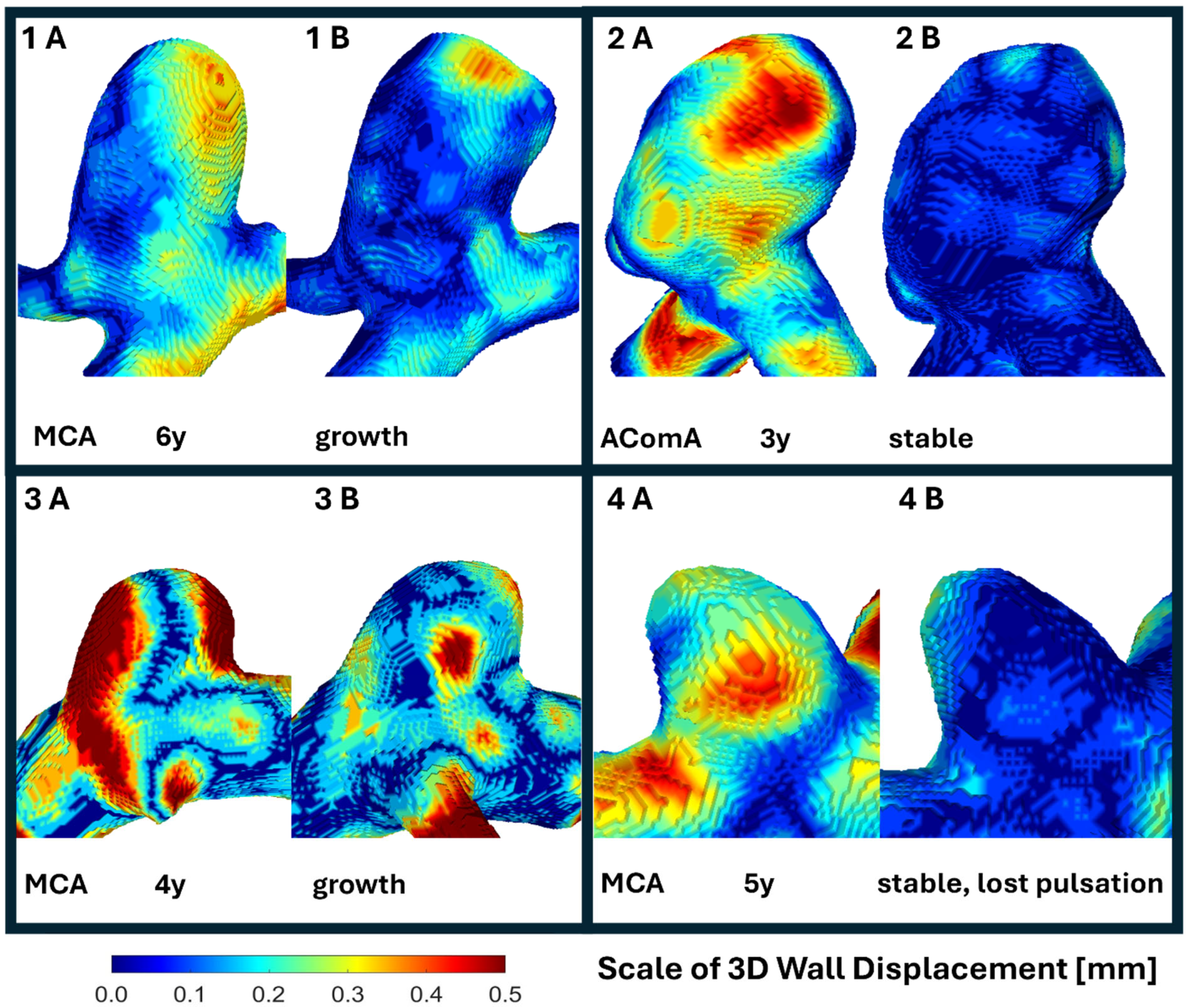
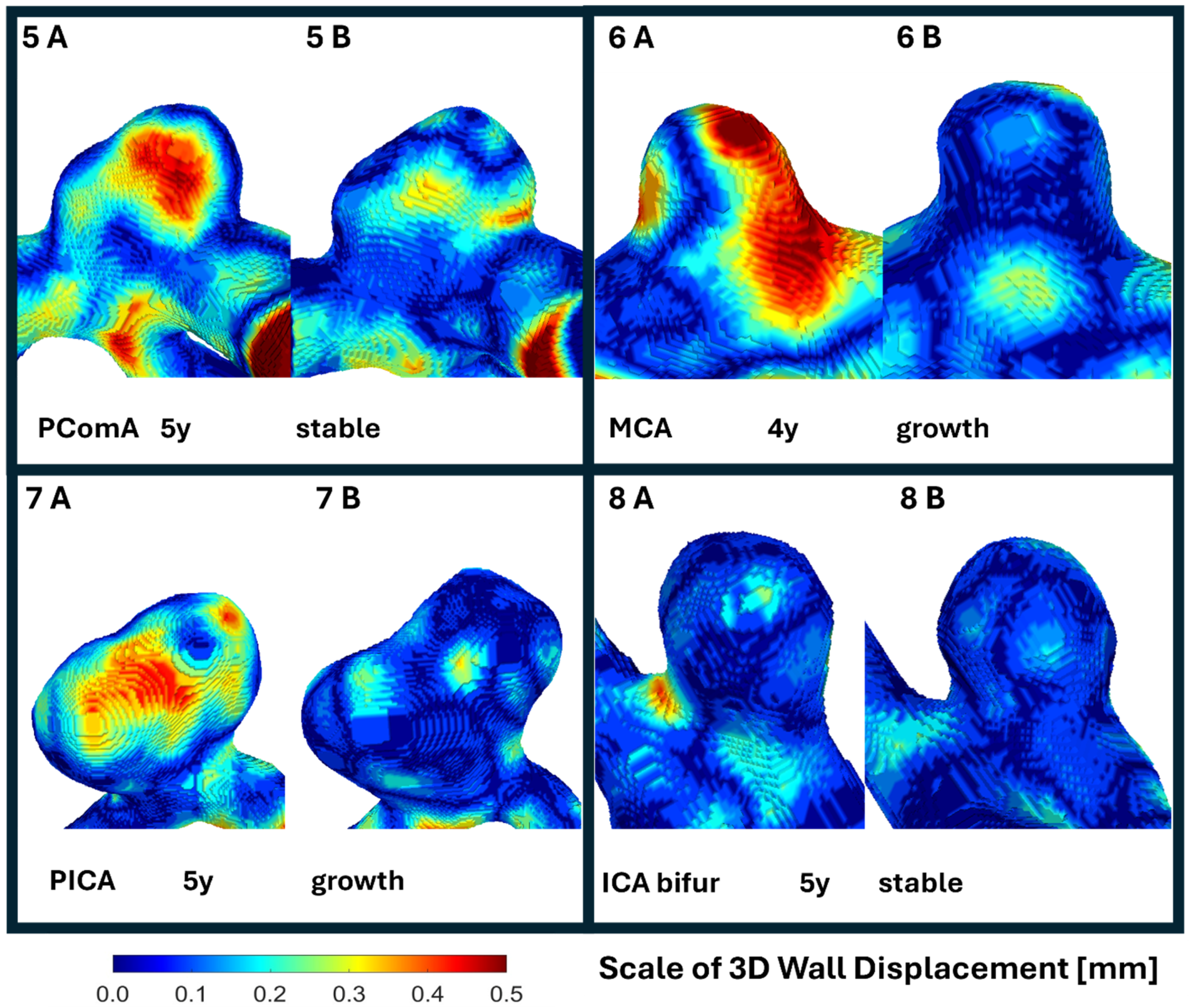
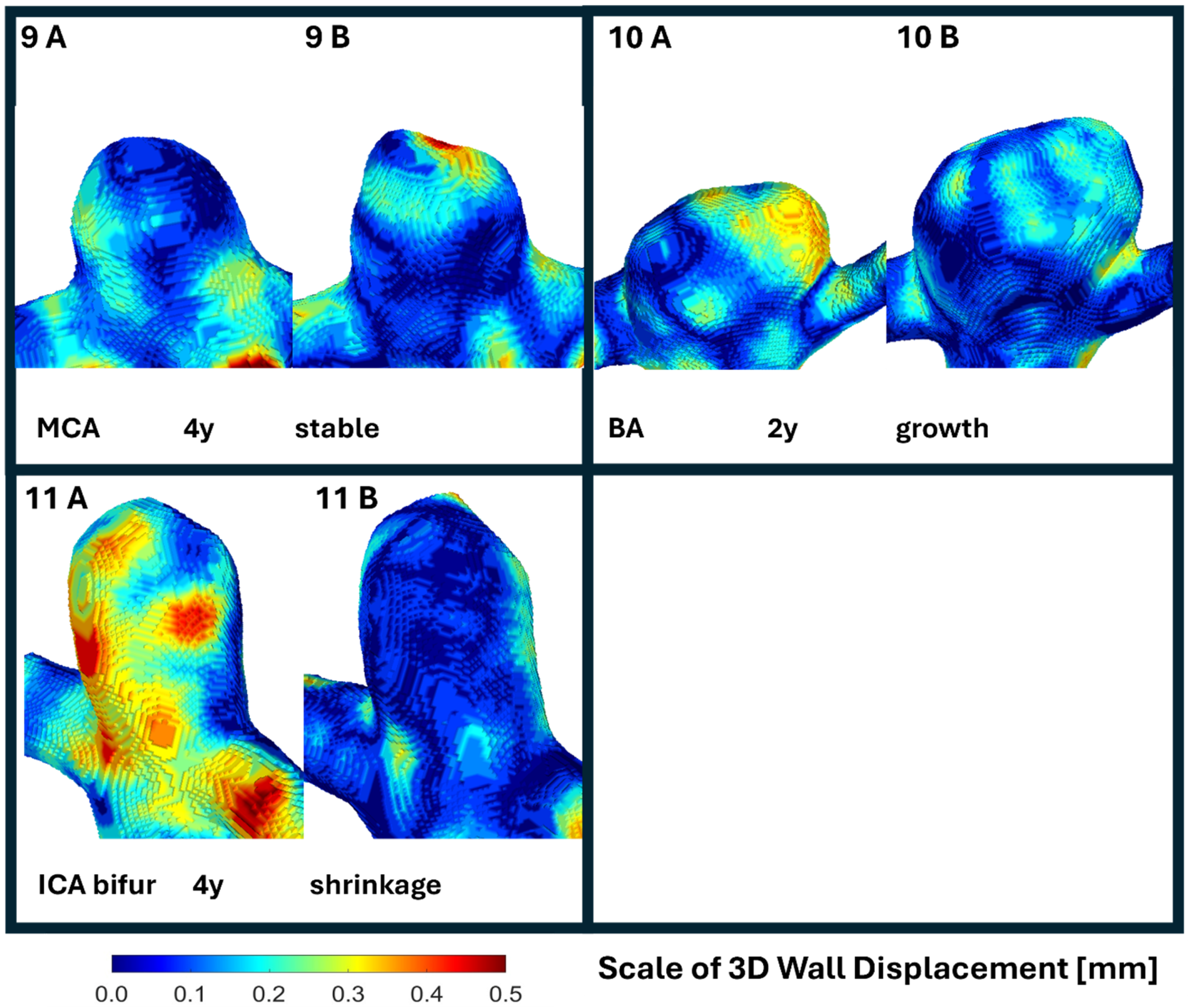
3.1.6. Visual Presentation
3.2. Signal Qualification, Deformability Estimates, and Geometric Progression
3.3. Associations Between Aneurysm Morphology and Geometric Variability
3.3.1. Linear and Neck-Based Variability
3.3.2. Surface and Volume Variability
3.3.3. Variability Versus Longitudinal Remodeling
4. Discussion
4.1. Spatial Wall Pulsation Versus Global Volumetric Pulsation
4.2. Imaging Uncertainty and Conservative Deformability Assessment
4.3. Deformability, Variability, Morphology, and Size Change
4.4. Relation to Rupture-Focused Pulsation Studies
4.5. Clinical Implications and Limitations
5. Conclusions
Supplementary Materials
Author Contributions
Funding
Institutional Review Board Statement
Informed Consent Statement
Data Availability Statement
Conflicts of Interest
Appendix A
Appendix A.1. Signal Differentiation Using First Harmonic Amplitude, Residual MAD, and Repeatability Thresholds
Appendix A.2. Harmonic Modelling of the Periodic Component
- is the geometric measurement at cardiac phase
- is the mean value of the signal
- and are harmonic coefficients
- is the angular frequency of the cardiac cycle.
Appendix A.3. Extraction of the Residual Signal
Appendix A.4. Quantification of Residual Variability
Appendix A.5. Repeatability Analysis and Minimum Detectable Change
Appendix B


References
- Etminan, N.; Brown, R.D.; Beseoglu, K.; Juvela, S.; Raymond, J.; Morita, A.; Torner, J.C.; Derdeyn, C.P.; Raabe, A.; Mocco, J.; et al. The Unruptured Intracranial Aneurysm Treatment Score: A Multidisciplinary Consensus. Neurology 2015, 85, 881–889. [Google Scholar] [CrossRef] [Scilit] [PubMed]
- Vlak, M.H.; Algra, A.; Brandenburg, R.; Rinkel, G.J. Prevalence of Unruptured Intracranial Aneurysms, with Emphasis on Sex, Age, Comorbidity, Country, and Time Period: A Systematic Review and Meta-Analysis. Lancet Neurol. 2011, 10, 626–636. [Google Scholar] [CrossRef] [Scilit] [PubMed]
- Etminan, N.; Rinkel, G.J. Unruptured Intracranial Aneurysms: Development, Rupture and Preventive Management. Nat. Rev. Neurol. 2016, 12, 699–713. [Google Scholar] [CrossRef] [Scilit] [PubMed]
- Investigators, U.J.; Morita, A.; Kirino, T.; Hashi, K.; Aoki, N.; Fukuhara, S.; Hashimoto, N.; Nakayama, T.; Sakai, M.; Teramoto, A.; et al. The Natural Course of Unruptured Cerebral Aneurysms in a Japanese Cohort. N. Engl. J. Med. 2012, 366, 2474–2482. [Google Scholar] [CrossRef] [Scilit]
- Greving, J.P.; Wermer, M.J.H.; Brown, R.D., Jr.; Morita, A.; Juvela, S.; Yonekura, M.; Ishibashi, T.; Torner, J.C.; Nakayama, T.; Rinkel, G.J.E.; et al. Development of the PHASES Score for Prediction of Risk of Rupture of Intracranial Aneurysms: A Pooled Analysis of Six Prospective Cohort Studies. Lancet Neurol. 2014, 13, 59–66. [Google Scholar] [CrossRef] [Scilit]
- Al-Khindi, T.; Macdonald, R.L.; Schweizer, T.A. Cognitive and Functional Outcome After Aneurysmal Subarachnoid Hemorrhage. Stroke 2010, 41, e519–e536. [Google Scholar] [CrossRef] [Scilit]
- van Gijn, J.; Kerr, R.S.; Rinkel, G.J. Subarachnoid Haemorrhage. Lancet 2007, 369, 306–318. [Google Scholar] [CrossRef] [Scilit]
- Kato, Y.; Hayakawa, M.; Sano, H.; Sunil, M.; Imizu, S.; Yoneda, M.; Watanabe, S.; Abe, M.; Kanno, T. Prediction of Impending Rupture in Aneurysms Using 4D-CTA: Histopathological Verification of a Real-Time Minimally Invasive Tool in Unruptured Aneurysms. Min-Minim. Invasive Neurosurg. 2004, 47, 131–135. [Google Scholar] [CrossRef] [Scilit]
- Hayakawa, M.; Katada, K.; Anno, H.; Imizu, S.; Hayashi, J.; Irie, K.; Negoro, M.; Kato, Y.; Kanno, T.; Sano, H. CT Angiography with Electrocardiographically Gated Reconstruction for Visualizing Pulsation of Intracranial Aneurysms: Identification of Aneurysmal Protuberance Presumably Associated with Wall Thinning. Am. J. Neuroradiol. 2005, 26, 1366–1369. [Google Scholar]
- Ishida, F.; Ogawa, H.; Simizu, T.; Kojima, T.; Taki, W. Congress of Neurological Surgeons/American Association of Neurological Surgeons Joint Section Chairmen. Neurosurgery 2005, 57, 471. [Google Scholar] [CrossRef] [Scilit]
- Matsumoto, M.; Sasaki, T.; Suzuki, K.; Sakuma, J.; Endo, Y.; Kodama, N. Visualizing the Dynamics of Cerebral Aneurysms with Four-Dimensional Computed Tomographic Angiography. Neurosurgery 2006, 58, E1003. [Google Scholar] [CrossRef] [Scilit] [PubMed]
- Karmonik, C.; Diaz, O.; Grossman, R.; Klucznik, R. In-Vivo Quantification of Wall Motion in Cerebral Aneurysms from 2D Cine Phase Contrast Magnetic Resonance Images. RöFo Fortschritte Auf Dem Geb. Röntgenstrahlen Bild. Verfahr. 2009, 182, 140–150. [Google Scholar] [CrossRef] [Scilit] [PubMed]
- Kuroda, J.; Kinoshita, M.; Tanaka, H.; Nishida, T.; Nakamura, H.; Watanabe, Y.; Tomiyama, N.; Fujinaka, T.; Yoshimine, T. Cardiac Cycle-Related Volume Change in Unruptured Cerebral Aneurysms. Stroke 2012, 43, 61–66. [Google Scholar] [CrossRef] [Scilit] [PubMed]
- Dissaux, B.; Ognard, J.; Aouni, M.C.E.; Nonent, M.; Haioun, K.; Magro, E.; Gentric, J.C. Volume Variation May Be a Relevant Metric in the Study of Aneurysm Pulsatility: A Study Using ECG-Gated 4D-CTA (PULSAN). J. NeuroInterv. Surg. 2020, 12, 632–636. [Google Scholar] [CrossRef] [Scilit]
- Stam, L.B.; Aquarius, R.; de Jong, G.A.; Slump, C.H.; Meijer, F.J.A.; Boogaarts, H.D. A Review on Imaging Techniques and Quantitative Measurements for Dynamic Imaging of Cerebral Aneurysm Pulsations. Sci. Rep. 2021, 11, 2175. [Google Scholar] [CrossRef] [Scilit]
- Xie, H.; Yu, H.; Wu, H.; Wang, J.; Wu, S.; Zhang, J.; Zhao, H.; Yuan, M.; Mendieta, J.B.; Anbananthan, H.; et al. Quantifying Irregular Pulsation of Intracranial Aneurysms Using 4D-CTA. J. Biomech. 2024, 174, 112269. [Google Scholar] [CrossRef] [Scilit]
- Fedorov, A.; Beichel, R.; Kalpathy-Cramer, J.; Finet, J.; Fillion-Robin, J.-C.; Pujol, S.; Bauer, C.; Jennings, D.; Fennessy, F.; Sonka, M.; et al. 3D Slicer as an Image Computing Platform for the Quantitative Imaging Network. Magn. Reson. Imaging 2012, 30, 1323–1341. [Google Scholar] [CrossRef] [Scilit]
- Koizumi, S.; Kin, T.; Shono, N.; Kiyofuji, S.; Umekawa, M.; Sato, K.; Saito, N. Patient-Specific Cerebral 3D Vessel Model Reconstruction Using Deep Learning. Med. Biol. Eng. Comput. 2024, 62, 3225–3232. [Google Scholar] [CrossRef] [Scilit]
- Hsu, W.-C.; Meuschke, M.; Frangi, A.F.; Preim, B.; Lawonn, K. A Survey of Intracranial Aneurysm Detection and Segmentation. Med. Image Anal. 2025, 101, 103493. [Google Scholar] [CrossRef] [Scilit]
- Kottner, J.; Audigé, L.; Brorson, S.; Donner, A.; Gajewski, B.J.; Hróbjartsson, A.; Roberts, C.; Shoukri, M.; Streiner, D.L. Guidelines for Reporting Reliability and Agreement Studies (GRRAS) Were Proposed. J. Clin. Epidemiol. 2011, 64, 96–106. [Google Scholar] [CrossRef] [Scilit]
- Dhar, S.; Tremmel, M.; Mocco, J.; Kim, M.; Yamamoto, J.; Siddiqui, A.H.; Hopkins, L.N.; Meng, H. Morphology parameters for intracranial aneurysm rupture risk assessment. Neurosurgery 2008, 63, 185–197. [Google Scholar] [CrossRef] [Scilit]
- Raghavan, M.L.; Ma, B.; Harbaugh, R.E. Quantified Aneurysm Shape and Rupture Risk. J. Neurosurg. 2005, 102, 355–362. [Google Scholar] [CrossRef] [Scilit] [PubMed]
- Dhar, R.; Diringer, M.N. Relationship between Angiographic Vasospasm, Cerebral Blood Flow, and Cerebral Infarction after Subarachnoid Hemorrhage. Acta Neurochir. Suppl. 2015, 120, 161–165. [Google Scholar] [CrossRef] [Scilit]
- Kleinloog, R.; Zwanenburg, J.J.M.; Schermers, B.; Krikken, E.; Ruigrok, Y.M.; Luijten, P.R.; Visser, F.; Regli, L.; Rinkel, G.J.E.; Verweij, B.H. Quantification of Intracranial Aneurysm Volume Pulsation with 7T MRI. Am. J. Neuroradiol. 2018, 39, 713–719. [Google Scholar] [CrossRef] [Scilit] [PubMed]
- Linden, S.M.L.; Stam, L.B.; Aquarius, R.; Hering, A.; de Korte, C.L.; Prokop, M.; Boogaarts, H.D.; Meijer, F.J.A.; Oostveen, L.J. Feasibility of Capturing Vessel Expansion with 4D-CTA: Phantom Study to Determine Reproducibility, Spatial and Temporal Resolution. Med. Phys. 2024, 51, 7171–7179. [Google Scholar] [CrossRef] [Scilit] [PubMed]
- Rousseeuw, P.J.; Croux, C. Alternatives to the Median Absolute Deviation. J. Am. Stat. Assoc. 1993, 88, 1273. [Google Scholar] [CrossRef]
- Leys, C.; Ley, C.; Klein, O.; Bernard, P.; Licata, L. Detecting Outliers: Do Not Use Standard Deviation around the Mean, Use Absolute Deviation around the Median. J. Exp. Soc. Psychol. 2013, 49, 764–766. [Google Scholar] [CrossRef] [Scilit]
- Shidhore, T.C.; Cohen-Gadol, A.A.; Rayz, V.L.; Christov, I.C. Comparative Assessment of Biomechanical Parameters in Subjects with Multiple Cerebral Aneurysms Using Fluid–Structure Interaction Simulations. J. Biomech. Eng. 2022, 145, 051003. [Google Scholar] [CrossRef] [Scilit]
- Backes, D.; Rinkel, G.J.E.; Greving, J.P.; Velthuis, B.K.; Murayama, Y.; Takao, H.; Ishibashi, T.; Igase, M.; terBrugge, K.G.; Agid, R.; et al. ELAPSS Score for Prediction of Risk of Growth of Unruptured Intracranial Aneurysms. Neurology 2017, 88, 1600–1606. [Google Scholar] [CrossRef] [Scilit]
- Chen, J.; Faber, T.L.; Cooke, C.D.; Garcia, E.V. Temporal Resolution of Multiharmonic Phase Analysis of ECG-Gated Myocardial Perfusion SPECT Studies. J. Nucl. Cardiol. 2008, 15, 383–391. [Google Scholar] [CrossRef] [Scilit]
- Atchley, A.E.; Trimble, M.A.; Samad, Z.; Shaw, L.K.; Pagnanelli, R.; Chen, J.; Garcia, E.V.; Iskandrian, A.E.; Velazquez, E.J.; Borges-Neto, S. Use of Phase Analysis of Gated SPECT Perfusion Imaging to Quantify Dyssynchrony in Patients with Mild-to-Moderate Left Ventricular Dysfunction. J. Nucl. Cardiol. 2009, 16, 888–894. [Google Scholar] [CrossRef] [Scilit] [PubMed]
- Giavarina, D. Understanding Bland Altman Analysis. Biochem. Med. 2015, 25, 141–151. [Google Scholar] [CrossRef] [Scilit] [PubMed]
- Bland, J.M.; Altman, D.G. Statistical Methods for Assessing Agreement between Two Methods of Clinical Measurement. Int. J. Nurs. Stud. 2010, 47, 931–936. [Google Scholar] [CrossRef] [Scilit]
- de Vet, H.C.W.; Terwee, C.B.; Knol, D.L.; Bouter, L.M. When to Use Agreement versus Reliability Measures. J. Clin. Epidemiol. 2006, 59, 1033–1039. [Google Scholar] [CrossRef] [Scilit]
- Lee, P.; Liu, C.-H.; Fan, C.-W.; Lu, C.-P.; Lu, W.-S.; Hsieh, C.-L. The Test–Retest Reliability and the Minimal Detectable Change of the Purdue Pegboard Test in Schizophrenia. J. Formos. Med. Assoc. 2013, 112, 332–337. [Google Scholar] [CrossRef] [Scilit]
- Holmes, D.T.; Buhr, K.A. Error Propagation in Calculated Ratios. Clin. Biochem. 2007, 40, 728–734. [Google Scholar] [CrossRef] [Scilit]
- Chen, S.; Zhang, W.; Cheng, Y.; Wang, G.; Lv, N. Quantification of Morpho-Hemodynamic Changes in Unruptured Intracranial Aneurysms with Irregular Pulsation during the Cardiac Cycle Using 4D-CTA. Front. Neurol. 2024, 15, 1436086. [Google Scholar] [CrossRef] [Scilit]
- Zhou, J.; Guo, Q.; Chen, Y.; Lin, B.; Ding, S.; Zhao, H.; Pan, Y.; Wan, J.; Zhao, B. Irregular Pulsation of Intracranial Aneurysm Detected by Four-Dimensional CT Angiography and Associated with Small Aneurysm Rupture: A Single-Center Prospective Analysis. Front. Neurol. 2022, 13, 809286. [Google Scholar] [CrossRef] [Scilit]
- Zhang, J.; Li, X.; Zhao, B.; Zhang, J.; Sun, B.; Wang, L.; Ding, S.; Liu, X.; Yan, J.; Mossa-Basha, M.; et al. Irregular Pulsation of Intracranial Unruptured Aneurysm Detected by Four-Dimensional CT Angiography Is Associated with Increased Estimated Rupture Risk and Conventional Risk Factors. J. NeuroInterv. Surg. 2021, 13, 854–859. [Google Scholar] [CrossRef] [Scilit]
- Firouzian, A.; Manniesing, R.; Metz, C.T.; Risselada, R.; Klein, S.; van Kooten, F.; Sturkenboom, M.C.J.M.; van der Lugt, A.; Niessen, W.J. Quantification of Intracranial Aneurysm Morphodynamics from ECG-Gated CT Angiography. Acad. Radiol. 2013, 20, 52–58. [Google Scholar] [CrossRef] [Scilit]
- Schetelig, D.; Sedlacik, J.; Fiehler, J.; Frölich, A.; Knopp, T.; Sothmann, T.; Waschkewitz, J.; Werner, R. Analysis of the Influence of Imaging-Related Uncertainties on Cerebral Aneurysm Deformation Quantification Using a No-Deformation Physical Flow Phantom. Sci. Rep. 2018, 8, 11004. [Google Scholar] [CrossRef] [Scilit]
- Hayakawa, M.; Tanaka, T.; Sadato, A.; Adachi, K.; Ito, K.; Hattori, N.; Omi, T.; Oheda, M.; Katada, K.; Murayama, K.; et al. Detection of Pulsation in Unruptured Cerebral Aneurysms by ECG-Gated 3D-CT Angiography (4D-CTA) with 320-Row Area Detector CT (ADCT) and Follow-up Evaluation Results: Assessment Based on Heart Rate at the Time of Scanning. Clin. Neuroradiol. 2014, 24, 145–150. [Google Scholar] [CrossRef] [Scilit]
- Zhang, J.; Li, X.; Zhao, B.; Zhang, J.; Sun, B.; Wang, L.; Tian, J.; Mossa-Basha, M.; Kim, L.J.; Yan, J.; et al. Irregular Pulsation of Aneurysmal Wall Is Associated with Symptomatic and Ruptured Intracranial Aneurysms. J. NeuroInterv. Surg. 2023, 15, 91–96. [Google Scholar] [CrossRef] [Scilit]
- Vanrossomme, A.E.; Eker, O.F.; Thiran, J.-P.; Courbebaisse, G.P.; Boudjeltia, K.Z. Intracranial Aneurysms: Wall Motion Analysis for Prediction of Rupture. Am. J. Neuroradiol. 2015, 36, 1796–1802. [Google Scholar] [CrossRef] [Scilit]
- Hayakawa, M.; Maeda, S.; Sadato, A.; Tanaka, T.; Kaito, T.; Hattori, N.; Ganaha, T.; Moriya, S.; Katada, K.; Murayama, K.; et al. Detection of Pulsation in Ruptured and Unruptured Cerebral Aneurysms by Electrocardiographically Gated 3-Dimensional Computed Tomographic Angiography With a 320-Row Area Detector Computed Tomography and Evaluation of Its Clinical Usefulness. Neurosurgery 2011, 69, 843–851. [Google Scholar] [CrossRef] [Scilit]
- Stam, L.B.; Linden, S.M.L.; Oostveen, L.J.; Hansen, H.H.G.; Aquarius, R.; Slump, C.H.; de Korte, C.L.; Bartels, R.H.M.A.; Prokop, M.; Boogaarts, H.D.; et al. Dynamic Computed Tomography Angiography for Capturing Vessel Wall Motion: A Phantom Study for Optimal Image Reconstruction. PLoS ONE 2023, 18, e0293353. [Google Scholar] [CrossRef] [Scilit]



| Characteristic | Value |
|---|---|
| Age, years—mean ± SD | 63.1 ± 13.2 |
| Age, years—median (range) | 68 (32–79) |
| Female sex, n (%) | 5 (45.5) |
| Male sex, n (%) | 6 (54.5) |
| Multiple aneurysms, n (%) | 7 (63.6) |
| History of SAH, n (%) | 1 (9.1) |
| Smoking, n (%) | 6 (54.5) |
| Alcohol consumption, n (%) | 1 (9.1) |
| Arterial hypertension, n (%) | 9 (81.8) |
| Interval between baseline and follow-up, years (mean ± SD, range) | 4.3 ± 1.1 (2–6) |
| Patient No. | Age (years) | Gender (M/F) | Aneurysm No. | Location | Sizemax BL (mm) | Volume BL (mm3) | Sizemax FU (mm) | Volume FU (mm3) | FU Interval (years) | ELAPSS 3 y Risk (%) |
|---|---|---|---|---|---|---|---|---|---|---|
| 1 | 70 | F | 1 | MCA | 6.64 ± 0.12 | 65.05 ± 2.41 | 6.85 ± 0.09 | 81.59 ± 1.4 | 6 | 17.5 |
| 2 | 79 | M | 2 | AComA | 7.49 ± 0.14 | 87.79 ± 1.96 | 6.92 ± 0.04 | 91.72 ± 0.76 | 3 | 17.5 |
| 3 | 58 | F | 3 | MCA | 4.60 ± 0.07 | 22.04 ± 0.44 | 5.19 ± 0.10 | 27.53 ± 0.72 | 4 | 42.7 |
| 4 | 68 | M | 4 | MCA | 3.49 ± 0.17 | 12.41 ± 1.21 | 3.87 ± 0.12 | 14.26 ± 0.85 | 5 | 11.7 |
| 4 | 68 | M | 5 | PComA | 6.71 ± 0.16 | 58.13 ± 2.02 | 6.33 ± 0.08 | 57.56 ± 1.8 | 5 | 11.7 |
| 5 | 52 | M | 6 | MCA | 4.09 ± 0.10 | 17.89 ± 0.77 | 4.42 ± 0.06 | 23.42 ± 0.49 | 4 | 17.5 |
| 6 | 32 | M | 7 | VA | 9.80 ± 0.11 | 203.73 ± 3.87 | 10.38 ± 0.14 | 220.85 ± 3.2 | 5 | 7.8 |
| 7 | 66 | F | 8 | ICA bifur | 4.91 ± 0.09 | 31.08 ± 0.59 | 4.64 ± 0.13 | 31.01 ± 1.27 | 5 | 25.8 |
| 8 | 69 | F | 9 | MCA | 5.06 ± 0.04 | 36.36 ± 1.01 | 5.06 ± 0.09 | 36.89 ± 1.41 | 4 | 17.5 |
| 9 | 51 | F | 10 | BA | 7.84 ± 0.20 | 62.2 ± 2.32 | 8.95 ± 0.07 | 164.27 ± 4.2 | 2 | 17.5 |
| 10 | 71 | M | 11 | ICA bifur | 7.99 ± 0.10 | 102.07 ± 0.98 | 8.04 ± 0.12 | 90.82 ± 0.93 | 4 | 11.7 |
| Pulsation Type | Size Change | Stable | Total |
|---|---|---|---|
| Spatial wall pulsation | |||
| Present | 6 | 3 | 9 |
| Absent | 0 | 2 | 2 |
| Global volumetric pulsation | |||
| Present | 4 | 4 | 8 |
| Absent | 2 | 1 | 3 |
| ELAPSS Category | Size Change Yes/No | Global Volumetric Pulsation n/N (%) | Spatial Wall Pulsation n/N (%) |
|---|---|---|---|
| <20% (n = 9) | yes (N = 5) | 4/5 (80%) | 5/5 (100%) |
| no (N = 4) | 4/4 (100%) | 3/4 (75%) | |
| ≥20% (n = 2) | yes (N = 1) | 0/1 (0%) | 1/1 (100%) |
| no (N = 1) | 0/1 (0%) | 0/1 (0%) | |
| Aneurysm growth detected | 4/6 (67%) | 6/6 (100%) |
| Parameter Δ | Spearman ρ | p-Value |
|---|---|---|
| Linear geometry | ||
| Height | 0.109 | 0.750 |
| Heightmax | −0.491 | 0.125 |
| Sizemax | 0.073 | 0.832 |
| Neck geometry | ||
| Neckarea | 0.345 | 0.298 |
| Neckmax | 0.473 | 0.142 |
| Neckperimet | 0.291 | 0.385 |
| Surface | ||
| Ssac | −0.209 | 0.537 |
| Ssacpar | 0.391 | 0.235 |
| Volume | ||
| Vpar | 0.264 | 0.433 |
| Vsac | −0.255 | 0.450 |
| Vsacpar | 0.400 | 0.223 |
| Ratio-Based Morphology Parameter at Baseline | Geometric Measures | Correlation (ρ) | Interpretation |
|---|---|---|---|
| Undulation Index (UI) | Ssac | 0.75–0.85 | Moderate–strong |
| Ssacpar | 0.85–0.95 | Strong | |
| Vsac | 0.70–0.85 | Moderate–strong | |
| Vsacpar | 0.85–0.95 | Strong | |
| Vpar | ≈0.90 | Strong | |
| Non-Sphericity Index (NSI) | Ssac, Ssacpar, Vsac, Vsacpar, Vpar | Weak | Weak associations |
Disclaimer/Publisher’s Note: The statements, opinions and data contained in all publications are solely those of the individual author(s) and contributor(s) and not of MDPI and/or the editor(s). MDPI and/or the editor(s) disclaim responsibility for any injury to people or property resulting from any ideas, methods, instructions or products referred to in the content. |
© 2026 by the authors. Licensee MDPI, Basel, Switzerland. This article is an open access article distributed under the terms and conditions of the Creative Commons Attribution (CC BY) license.
Share and Cite
Jankovič, P.; Chodzyński, K.J.; Vanrossomme, A.E.; Boudjeltia, K.Z.; Šteňo, A.; Wirtz, C.R.; Šulaj, J.; Paľa, A. ECG-Gated 4D-CTA Assessment of Intracranial Aneurysm Wall Dynamics and Longitudinal Size Change: An Exploratory Study. Neurol. Int. 2026, 18, 81. https://doi.org/10.3390/neurolint18050081
Jankovič P, Chodzyński KJ, Vanrossomme AE, Boudjeltia KZ, Šteňo A, Wirtz CR, Šulaj J, Paľa A. ECG-Gated 4D-CTA Assessment of Intracranial Aneurysm Wall Dynamics and Longitudinal Size Change: An Exploratory Study. Neurology International. 2026; 18(5):81. https://doi.org/10.3390/neurolint18050081
Chicago/Turabian StyleJankovič, Peter, Kamil J. Chodzyński, Axel E. Vanrossomme, Karim Zouaoui Boudjeltia, Andrej Šteňo, Christian R. Wirtz, Ján Šulaj, and Andrej Paľa. 2026. "ECG-Gated 4D-CTA Assessment of Intracranial Aneurysm Wall Dynamics and Longitudinal Size Change: An Exploratory Study" Neurology International 18, no. 5: 81. https://doi.org/10.3390/neurolint18050081
APA StyleJankovič, P., Chodzyński, K. J., Vanrossomme, A. E., Boudjeltia, K. Z., Šteňo, A., Wirtz, C. R., Šulaj, J., & Paľa, A. (2026). ECG-Gated 4D-CTA Assessment of Intracranial Aneurysm Wall Dynamics and Longitudinal Size Change: An Exploratory Study. Neurology International, 18(5), 81. https://doi.org/10.3390/neurolint18050081