Review

Desmosomal Versus Non-Desmosomal Arrhythmogenic Cardiomyopathies: A State-of-the-Art Review

1
Department of Neuroscience, Imaging and Clinical Sciences, “G. d’Annunzio” University of Chieti-Pescara, Via dei Vestini 31, 66100 Chieti, Italy
2
Department of Medicine, Banner University Medical Center, University of Arizona, 1625 N Campbell Ave, Tucson, AZ 85719, USA
3
Center for Advanced Studies and Technology (CAST), “G. d’Annunzio” University of Chieti-Pescara, 66100 Chieti, Italy
4
Department of Medical Genetics, “G. d’Annunzio” University of Chieti-Pescara, 66100 Chieti, Italy
5
Department of Cardiology, Pierangeli Hospital, 65124 Pescara, Italy
6
Barts Heart Centre, St. Bartholomew’s Hospital, Barts Health NHS Trust, West Smithfield, London EC1A 7BE, UK
7
Newham University Hospital, Barts Health NHS Trust, London EC1M 6BQ, UK
8
William Harvey Research Institute, NIHR Barts Biomedical Centre, Queen Mary University of London, Charterhouse Square, London EC1M 6BQ, UK
9
Center for Inherited Cardiovascular Diseases, Department of Cardiology, WellSpan Health, 30 Monument Rd, York, PA 17403, USA
10
Department of Cardiovascular Medicine, Mayo Clinic, 200 First Str, Rochester, MN 55905, USA
11
Institute for Advanced Biomedical Technologies, “G. d’Annunzio” University of Chieti-Pescara, 66100 Chieti, Italy
12
University Cardiology Division, Heart Department, SS. Annunziata Hospital, ASL 2 Abruzzo, 66100 Chieti, Italy
*
Authors to whom correspondence should be addressed.
These authors contributed equally to this work and share the first authorship.
These authors contributed equally to this work and share the senior authorship.
Cardiogenetics 2025, 15(3), 22; https://doi.org/10.3390/cardiogenetics15030022
Submission received: 1 June 2025 / Revised: 14 July 2025 / Accepted: 29 July 2025 / Published: 1 August 2025
(This article belongs to the Section Cardiovascular Genetics in Clinical Practice)

Abstract

Arrhythmogenic cardiomyopathies (ACMs) are a phenotypically and etiologically heterogeneous group of myocardial disorders characterized by fibrotic or fibro-fatty replacement of ventricular myocardium, electrical instability, and an elevated risk of sudden cardiac death. Initially identified as a right ventricular disease, ACMs are now recognized to include biventricular and left-dominant forms. Genetic causes account for a substantial proportion of cases and include desmosomal variants, non-desmosomal variants, and familial gene-elusive forms with no identifiable pathogenic mutation. Nongenetic etiologies, including post-inflammatory, autoimmune, and infiltrative mechanisms, may mimic the phenotype. In many patients, the disease remains idiopathic despite comprehensive evaluation. Cardiac magnetic resonance imaging has emerged as a key tool for identifying non-ischemic scar patterns and for distinguishing arrhythmogenic phenotypes from other cardiomyopathies. Emerging classifications propose the unifying concept of scarring cardiomyopathies based on shared structural substrates, although global consensus is evolving. Risk stratification remains challenging, particularly in patients without overt systolic dysfunction or identifiable genetic markers. Advances in tissue phenotyping, multi-omics, and artificial intelligence hold promise for improved prognostic assessment and individualized therapy.

1. Introduction

Arrhythmogenic cardiomyopathies (ACMs) are a phenotypically and etiologically heterogeneous group of myocardial disorders characterized by fibro-fatty replacement of ventricular myocardium, leading to electrical instability, ventricular arrhythmias (VAs), and sudden cardiac death (SCD) [1]. Initially described as arrhythmogenic right ventricular cardiomyopathy (ARVC), the condition was first identified in individuals presenting with malignant VAs and structural abnormalities confined to the right ventricle (RV) [2,3]. However, subsequent evidence from autopsy studies, genotype–phenotype correlations, and contrast-enhanced cardiac magnetic resonance (CMR) imaging demonstrated frequent involvement of the left ventricle (LV), prompting the adoption of the broader revised term ACM [4,5]. In many cases, LV involvement may equal or exceed RV disease in severity. In response to the need for greater nosological clarity across cardiomyopathy subtypes, a 2025 consensus classification, proposed by investigators in Padua, grouped cardiomyopathies into three overarching categories: hypertrophic/restrictive, dilated/hypokinetic, and scarring/arrhythmogenic [6]. Within this framework, ACM is defined as a prototypical scarring cardiomyopathy, distinguished by loss of ventricular myocardium due to myocyte death and subsequent fibro-fatty replacement [5,7,8,9,10]. The myocardial scarring predisposes to potentially lethal VAs and may cause the impairment of ventricular systolic function, regardless of the pattern of ventricular involvement. This classification emphasizes structural and pathobiological features over chamber-specific terminology and is intended to harmonize diagnosis, prognostication, and treatment strategies across the cardiomyopathy spectrum. ACM is typically inherited in an autosomal dominant pattern with variable penetrance and expressivity. Pathogenic variants have been identified in both desmosomal and non-desmosomal genes, although a substantial proportion of patients remain genetically elusive (“genotype-negative”). The estimated prevalence of ACM ranges between 1:1000 and 1:5000, though this figure is likely underestimated due to frequent misdiagnosis or subclinical presentation, particularly when SCD is the sentinel manifestation [11,12]. The condition was first recognized as part of Naxos disease, a cardiocutaneous syndrome described in families from the Greek island of Naxos and characterized by woolly hair, palmoplantar keratoderma, and ARVC [13]. A phenotypically similar disorder, Carvajal syndrome, was later reported in South America, involving biventricular or left-dominant disease and associated with desmoplakin (DSP) pathogenic variants [14]. Sporadic, non-syndromic forms of ACM have also been identified in populations from Asia [15], Africa [16], and North-Eastern Italy [8]. Despite growing recognition, there remains a lack of systematic epidemiological data delineating the distribution of desmosomal versus non-desmosomal forms. This review aims to summarize the key differences in pathophysiology, genotype–phenotype correlations, and risk stratification of ACM in relation to its genetic etiology.

2. Definitions

The first formal descriptions of ACM date back to 1982, when Marcus et al. reported 24 patients presenting with left bundle branch block (LBBB)-type ventricular tachycardia, T-wave inversion (TWI) in precordial leads V1–V4, and RV dilation with regional wall motion abnormalities, predominantly involving the infundibulum. They termed this condition arrhythmogenic right ventricular dysplasia (ARVD) [17]. In 1984, Fontaine et al. linked delayed activation in the RV free wall, secondary to fibro-fatty replacement, to re-entrant arrhythmias [18]. Subsequently, in 1988, Thiene et al. identified ARVD in twelve out of sixty SCD cases, characterizing the disease by extensive fibro-fatty myocardial replacement and RV aneurysmal dilation. Initially regarded as a congenital myocardial malformation, ARVD was reclassified following the discovery of pathogenic variants in desmosomal genes. This genetic basis, combined with the recognition that the clinical phenotype typically develops over time and is not present at birth, led to a shift in terminology from dysplasia to cardiomyopathy [8]. ACM diagnosis relies on a multiparametric approach due to the absence of a highly sensitive and specific diagnostic test. In 1994, McKenna et al. proposed the first diagnostic criteria for ARVC [19], later revised by Marcus et al. in 2010 to integrate emerging genetic and imaging modalities. These criteria encompassed six categories: (I) global/regional dysfunction and structural abnormalities (evaluated by echocardiography, CMR, or angiography); (II) tissue characterization by endomyocardial biopsy (EMB); (III) repolarization abnormalities; (IV) depolarization/conduction defects; (V) arrhythmias; and (VI) family history. Diagnosis was stratified into probable, borderline, or definite, based on the combination of major and minor criteria within each category [3]. Advances in CMR with late gadolinium enhancement (LGE) have highlighted frequent biventricular and left-dominant involvement. Thus, Corrado et al. proposed the Padua Criteria in 2020, which classified ACM into three phenotypes: classic right-dominant (ARVC), biventricular ACM (BivACM), and left-dominant arrhythmogenic left ventricular cardiomyopathy (ALVC). These criteria introduced sex-, age-, and body surface area-adjusted thresholds for ventricular size and function, recognized LGE as an equivalent structural criterion to EMB, and included post-mortem diagnosis in first-degree relatives as a major genetic criterion [20]. Further refinements to the Padua Criteria were introduced in 2024 by a European Task Force [21]. Notably, LGE was downgraded from a major to a minor structural criterion, except for “ring-like” patterns, while genetic criteria were revised, eliminating the requirement for pathogenic ACM gene variants in ALVC diagnosis and emphasizing a phenotype-based approach. These refinements highlight the evolving understanding of ACM and the importance of comprehensive, multiparametric diagnostic frameworks. Table 1 presents the different ACM definitions currently used according to the main scientific societies.

3. Genetic History

The earliest literature reports suggesting a plausible genetic etiology for ARVC originated in 1988. Nava et al. observed a high incidence of familial recurrence of ARVD in a series of families investigated for cases of SCD in youth. Seventy-two subjects across nine families were studied, revealing sixteen deaths at a young age, eleven of which showed massive replacement of myocardial tissue with fibro-fatty replacement. Notably, in eight of the nine families, at least two members were affected by the disease. From this point, the hypothesis that ARVC could be a genetic condition with autosomal dominant inheritance and variable expression and penetrance gained significant support [22]. With the advent of genetic sequencing technologies in the following decades, initial evidence emerged linking pathogenic variants in desmosomal genes. In the early 2000s, McKoy et al. studied nineteen individuals with Naxos disease, alongside unaffected relatives and unrelated subjects from the Greek islands of Naxos and Milos. A two-base pair deletion in the plakoglobin (JUP) gene was found exclusively in the nineteen affected individuals, leading to a premature termination of the protein, as demonstrated by Western blot analysis. This discovery clarified that the rare variant-induced disruption of cell–cell adhesion likely played a pivotal role in maintaining cellular integrity, providing a pathophysiological substrate for arrhythmias and SCD in patients with fibro-fatty replacement [23]. The role of desmosomal variants grew with the identification of the first variant in DSP (7901delG) in a set of families from Ecuador, subsequently named Carvajal Syndrome. This deletion was associated with generalized striate keratoderma, particularly affecting the palmoplantar epidermis, woolly hair, and dilated cardiomyopathy (DCM), which developed from adolescence [14]. With the development of genotype–phenotype correlation studies, the involvement of non-desmosomal genes was noted. As the largest mammalian protein, titin (encoded by the second-largest gene TTN), is expressed in both skeletal and cardiac muscle, connecting transitional junctions to intercalated discs. It is essential for multiple signaling pathways, so proteolysis of this protein was reported to trigger structural alterations pathognomonic for ARVC [24]. Beffagna et al. demonstrated involvement of untranslated regions of the transforming growth factor β3 (TGF-β3) gene, where overexpression could be linked to the distinctive fibrosis observed in the cardiac myocardium in ARVC [25]. Furthermore, a potentially deleterious missense mutation was observed in the transmembrane protein 43 (TMEM43) gene in fifteen families with ARVD. This gene encodes a peroxisome proliferator-activated receptor-γ (PPARγ) response element. PPARγ is an adipocyte growth factor and may be associated with the differentiation of fibro-adipocyte progenitor cells into myocardial adipocytes—thus offering an explanation for the phenomenon of fibro-fatty infiltration of the myocardium [26]. Together, these findings underscore the complex genetic basis of ACM and the need for continued investigation into both desmosomal and non-desmosomal factors that contribute to its development.

4. Pathophysiology

The pathophysiology of ACM has historically been based on the distinction between desmosomal and non-desmosomal genetic variants. However, various mechanisms converge to determine the clinical manifestation of the disease, stressing the importance of multiple triggers to uncover the condition. Desmosomes, integral components of intercalated discs, confer mechanical stability and functional integration to cardiomyocytes. These structures include transmembrane cadherins—desmogleins (DSG) and desmocollins (DSC)—linked to intermediate filaments through adapter proteins (PKG), plakophilin (PKP), and DSP [27]. The intercalated disc also incorporates adherens junctions (AJs) and gap junctions (GJs), which mediate mechanical cohesion and electrical coupling, respectively. AJs, anchored by N-cadherin and catenins, reinforce cytoskeletal integrity, while GJs, predominantly composed of connexin-43 (Cx43), facilitate electrical conduction. Intercalated disc proteins regulate key signaling pathways, including Wnt/β-catenin, MAPK, and Hippo [28]. Variants in desmosomal genes impair these interactions, contributing to structural remodeling, electrical dysfunction, and inflammatory responses. Besides desmosomal genetic defects, non-desmosomal pathogenic variants affect ion channels, autophagy, and cell adhesion, further promoting arrhythmogenesis and disease progression. Ion channel abnormalities—especially in sodium, calcium, and potassium channels—exacerbate electrical instability, while altered autophagy in fibrotic regions enhances disease development [29]. These diverse mechanisms highlight the complexity of ACM pathophysiology, requiring an integrated, multiparametric diagnostic approach.

4.1. From Histopathology to Disease Manifestation

Fibro-fatty replacement represents the hallmark histopathological finding of ACM, characterized by myocardial atrophy starting in the epicardium and progressing transmurally, particularly within the RV dysplasia triangle [1]. Histological examination reveals islands of surviving myocytes embedded in fibrous and adipose tissue, reflecting the bipotential differentiation of depleted cardiac cells into fibrogenic or adipogenic lineages. Arrhythmogenesis is driven by the dysfunction of Cx43 and Voltage-Gated Sodium Channel Alpha Subunit 5 (Nav1.5), which impair electrical conduction. Adipogenesis is regulated by the Wnt/β-catenin, Hippo–Yes-associated protein (YAP), PPARγ, and microRNA (miRNA) signaling pathways, while fibrosis is associated with the TGFβ and MAPK pathways [30,31,32,33]. Variants in desmosomal proteins alter the cellular response to mechanical stress, supporting the “wall stress” theory in ARVC pathogenesis [34]. Meanwhile, the “desmosomal reserve” theory expands on this concept, suggesting that genetic variants impair the myocardial capacity to withstand additional stress induced by intense physical exercise, accelerating disease progression [35]. The preferential RV involvement may stem from differential embryonic origins, as progenitor cells derived from the second heart field epicardium—contributing predominantly to the RV—exhibit greater adipogenic potential following JUP nuclear translocation. Furthermore, mechanical stress preferentially affects the RV wall due to its thinner structure and lower muscle mass, contributing to the regional susceptibility of ACM [30]. Inflammation triggers, including viral infections and autoimmune responses, further exacerbate cardiac damage [36,37]. Occasionally, myocarditis may represent the initial clinical manifestation of ACM, with viral infections or autoimmune responses accelerating adverse myocardial remodeling in genetically predisposed individuals. The recruitment of immune cells, particularly CD3+ lymphocytes and macrophages, perpetuates tissue damage through the release of pro-inflammatory cytokines and matrix metalloproteinases [38,39]. The interplay between genetic variants, mechanical stress, and inflammatory responses highlights the multifaceted nature of ACM pathogenesis. Another critical aspect involves the nuclear translocation of JUP and β-catenin, which represses the Wnt/β-catenin signaling pathway, promoting adipogenesis at the expense of myogenic differentiation. The Hippo–YAP signaling pathway further regulates the balance between adipogenic and myogenic differentiation, with YAP downregulation favoring adipocyte accumulation. Additionally, miRNA dysregulation, including increased expression of miR-21 and miR-146, contributes to fibrotic remodeling and inflammation [40,41,42]. The identification of these signaling pathways has provided insights into the molecular mechanisms underlying ACM and represents potential therapeutic targets. The interaction between genetic susceptibility and environmental factors reinforces the multifactorial nature of ACM pathogenesis, requiring comprehensive diagnostic and therapeutic strategies.

4.2. Translational Science Insights

Murine models have been developed to investigate ACM pathogenesis, encompassing pathogenic variants in key intercalated disc proteins, including Jup, Dsp, plakophilin2 (Pkp2), desmoglein2 (Dsg2), and desmocollin2 (Dsc2). Dysfunction of the Wnt/β-catenin signaling pathway represents a central pathogenetic mechanism, with additional contributions from Hippo, GSK3β, and PPARγ pathways [43]. Pkp2-mutant mice manifest exercise-induced cardiac dysfunction, accelerated by apoptosis and muscle mass reduction rather than immediate contractile impairment [35]. Dsg2mt/wt mice develop an ARVC-like phenotype following endurance training, despite the absence of fibrosis or inflammation [44,45]. Dsg2-W2A murine models faithfully replicate ACM features, including myocardial fibrosis and intercalated disc abnormalities, through integrin-β6/TGF-β signaling [46]. In a murine model of ACM with Dsg2 rare variants, impaired autophagy, ER/SR stress, and calcium homeostasis disruption contribute to disease progression. Lc3 and Sqstm1/p62 accumulation, increased Chop mRNA, and altered Ncx1/Ryr2 expression suggest a key role of these pathways in pathogenesis, warranting further investigation in human models [47] Human induced pluripotent stem cell-derived cardiomyocytes (hiPSC-CMs) from PKP2-mutant patients recapitulate key pathological features, including lipid accumulation, increased apoptosis, and phenotypic exacerbation under adipogenic stress. hiPSC-CMs from DSG2-mutant patients exhibit ion channel dysfunction, pro-inflammatory cytokine expression, and greater sensitivity to adrenergic stimulation. Organoid models and 3D bioprinting technologies are emerging tools for replicating the complex architecture and cellular interactions of ACM-affected myocardium, offering new platforms for drug testing and mechanistic studies. Both in vivo and in vitro models remain essential to elucidate ACM pathogenesis and explore novel therapeutic strategies [48].

5. Genotype–Phenotype Correlation

Recent genotype–phenotype correlation studies have demonstrated that the phenotypic spectrum of ACM is remarkably broad, ranging from phenotypically normal hearts to forms with pathognomonic histopathological findings—showing varying degrees of biventricular involvement [49]. Like all forms of familial cardiomyopathy, ACM is genetically heterogeneous (Figure 1). Desmosomal variants are typically associated with the classical phenotype of ARVC, characterized by RV dilatation and dysfunction, regional RV wall motion abnormalities, and fibro-fatty replacement. An exception is represented by mutations in the DSP gene, which are more frequently associated with extensive biventricular involvement and a distinctive pattern of subepicardial or intramyocardial ring-like fibrosis predominantly affecting the LV. In contrast, non-desmosomal variants are more commonly associated with BivACM or ALVC and often demonstrate overlap with other cardiomyopathies. In these cases, fibrosis predominantly involves the subepicardial and intramyocardial layers of the LV, displaying a non-ischemic pattern. Morpho-functional and structural abnormalities of the RV are generally less prominent or less frequently observed [6]. However, in contrast to other cardiomyopathies, the presence of a gene mutation serves as a diagnostic criterion [3]. Population studies have shown that ACM-related genetic variants are common, with major mutations sometimes found in control individuals [50]. Therefore, the diagnosis of ACM should primarily rely on structural, histopathological, and arrhythmic manifestations in the proband within a family [1]. Using the Clinical Genome Resource approach to gene-disease curation, only eight genes (PKP2, DSP, DSG2, DSC2, JUP, TMEM43, phospholamban (PLN), and desmin (DES) have been classified with definitive or moderate evidence for ARVC, accounting for nearly all pathogenic/likely pathogenic (P/LP) ARVC variants in ClinVar. Only P/LP variants in these eight genes should yield a major criterion for ACM diagnosis. Variants in other genes should prompt further phenotyping [51], as they may be associated with other cardiovascular conditions. Below, we provide a schematic distinction of the phenotypic manifestations associated with different ACM genetic subgroups (Table 2), with reflections on less common populations (e.g., genotype-negative/phenotype-positive patients), where polygenic risk scores (PRSs) may play a pivotal role in the future.

5.1. Desmosomal Variants

Most ARVC forms are linked to rare variants in desmosomal genes, including PKP2, DSP, DSG2, and DSC2. These variants are associated with specific clinical and electrocardiographic features, such as TWI and VAs, and often exhibit strong familial clustering [60]. PKP2 variants, the most frequent, are typically associated with the classical ARVC phenotype and are not linked to an increased risk of sustained ventricular tachycardia, ventricular fibrillation, implantable cardioverter defibrillator (ICD) intervention, or SCD [61]. In contrast, DSP variants more often lead to biventricular or left-dominant forms and are associated with a higher incidence of left ventricular dysfunction, heart failure (HF), and arrhythmic death [60]. The phenotypic implications of DSG2 mutations have been more controversial. A recent analysis of ARVC registries in Europe and North America confirmed that most P/LP desmosomal variants are inherited and recurrent, with a minority representing de novo events, often large deletions [62]. In a large multinational cohort study, Chen et al. characterized 271 individuals with DSG2 or DSC2 pathogenic variants, highlighting a high prevalence of homozygous, compound heterozygous, or digenic variants. These individuals demonstrated a distinctive phenotype marked by early onset, frequent biventricular involvement, and increased risk of end-stage heart failure and malignant arrhythmias. Outcomes were significantly worse than those seen in PKP2 carriers [59]. These data reinforce the importance of variant-specific risk stratification, particularly in patients harboring multiple desmosomal cadherin variants [59]. Further evidence reinforces the concept that not all desmosomal genes confer the same degree of clinical risk. In a comprehensive cohort study, Pergola et al. demonstrated that carriers of desmosomal gene variants exhibit markedly heterogeneous outcomes. Specifically, nonmissense variants in DSP were associated with a fivefold increase in the risk of end-stage heart failure, while missense variants located in hotspot domains—such as the spectrin repeat 4 (SR4, amino acids 272–375) and glycine–serine–arginine-rich (GSR, 2828–2844) regions in DSP and the armadillo repeat 6 (ARD6, 680–710) domain in PKP2—significantly elevated the incidence of life-threatening arrhythmic events. These findings establish that clinical interpretation of desmosomal variants must consider not only the gene involved but also the variant type and its precise topological location. This domain-based stratification refines prognostic assessment and enables more personalized management of desmosomal cardiomyopathies [63].

5.2. Non-Desmosomal Variants

Non-desmosomal gene variants have been increasingly recognized as important contributors to ACM pathogenesis, often leading to phenotypes distinct from those seen in desmosomal gene variants [64]. Among these, filamin C (FLNC) variants are frequently associated with ALVC and are primarily characterized by conduction system abnormalities, as well as an increased risk of life-threatening arrhythmias. Patients with FLNC-related ACM often develop progressive ventricular dysfunction, which can manifest as DCM-like features [65], further complicating diagnosis and risk stratification. TMEM43 variants are known for their strong association with biventricular involvement and a significantly heightened risk of SCD, particularly in male carriers. This has been most notably described in cases from Newfoundland, where a founder mutation in TMEM43 has been linked to a highly malignant form of ACM. Unlike desmosomal variants, which typically show a gradual disease progression, TMEM43-related ACM can lead to abrupt clinical deterioration [60], underscoring the necessity for early recognition and intervention. PLN variants contribute to ACM by disrupting calcium handling within cardiomyocytes, predisposing affected individuals to severe arrhythmias and progressive ventricular dysfunction. Despite its relatively small gene size, pathogenic PLN variants are among the most common non-desmosomal genetic variants implicated in ACM, particularly in European cohorts [66]. Additionally, variants in genes encoding nuclear envelope proteins, such as lamin A/C (LMNA), are associated with a wide array of phenotypic manifestations. LMNA variants can present with an overlapping phenotype that includes isolated ACM, Emery–Dreifuss muscular dystrophy, limb–girdle muscular dystrophy type 1B, familial lipodystrophy, and even Hutchinson–Gliford progeria [67]. Clinically, patients with LMNA-related ACM frequently exhibit conduction abnormalities, including atrioventricular block, atrial fibrillation, and sinus node dysfunction, in addition to life-threatening VAs. Unlike typical ACM, LMNA rare variants often result in an aggressive disease course with early onset of heart failure symptoms and a high incidence of malignant arrhythmias [68], necessitating close monitoring and consideration of early ICD implantation. Additional variants in non-desmosomal genes associated with ACM, such as sodium voltage-gated channel alpha subunit 5 (SCN5A), catenin alpha-3 (CTNNA3), Bcl2-associated athanogene 3 (BAG3), RNA binding motif protein 20 (RBM20), cadherin 2 (CDH2), integrin-linked kinase (ILK), and TTN, have been reported. However, for several of these genes, the strength of evidence linking them to ACM is currently limited and warrants further validation.

5.3. Genotype-Negative–Phenotype-Positive Individuals

Genotype-negative (G-) individuals exhibit ACM features without identifiable pathogenic or likely pathogenic mutations. Conversely, based on a recent report by ClinGen and Gene Coalition Consortium Investigators, patients are considered genotype-positive when the identified variants are classified as pathogenic or likely pathogenic with at least two stars of evidence in ClinVar [69]. As such, the definition of G- also includes individuals who carry variants of uncertain significance (VUS). Despite the absence of genetic variants, phenotypic expression is a prerequisite for malignant arrhythmic events and SCD in ACM, indicating that clinical features are critical for risk stratification, regardless of genetic status [70]. G- patients show an intermediate risk (16%) of LV dysfunction (LVEF ≤ 45%) and arrhythmic endpoints comparable to DSC2/DSG2/DSP variants carriers. Comprehensive clinical evaluation remains essential for these patients, underscoring the importance of phenotype-based diagnosis and risk stratification [71]. The underlying genetic basis of this subgroup remains elusive, and future studies leveraging next-generation sequencing and epigenetic analyses may provide further insights into their pathogenesis.

5.4. Genetically Undefined Forms and Polygenic Risk Scores

Genetically undefined forms refer to instances where no P/LP genetic variants are identified despite comprehensive genetic testing [72], including the presence of VUS—genetic alterations for which current evidence is insufficient to establish pathogenicity or benignity. VUS are subject to periodic re-evaluation through curated databases and bioinformatic resources that integrate emerging clinical, functional, and population-level data to reassess potential disease associations. As genetic knowledge evolves, many VUS are eventually reclassified, often as benign, based on population frequency data or segregation analyses. Given the increasing scale of genomic testing, laboratories are encouraged to establish clear policies regarding the reanalysis of genetic data and to specify whether additional costs may apply. In the absence of proactive updates, clinicians are advised to periodically follow up with testing laboratories to assess whether variant interpretations have changed [73]. Genetic undetermined ACM conditions account for 50–70% of cases, possibly due to undiscovered genes, genetic interactions, or environmental factors [74]. ACM typically follows an autosomal dominant inheritance pattern, but autosomal recessive, compound heterozygous (biallelic pathogenic variants in one gene), and digenic heterozygosity (pathogenic variants in more than one ACM-related gene) have been reported [75]. Emerging evidence supports a threshold model, where multiple genetic variants, environmental exposures, and exercise contribute to disease expression [76]. For instance, the 2019 ARVC risk model is effective in PKP2 variant carriers but less accurate in gene-elusive and DSP-Task Force Criteria-positive patients, especially with LV involvement. This highlights the need for genotype-specific risk algorithms and the incorporation of genetic testing into routine cardiac care for cardiomyopathies [77,78]. PRSs, which aggregate the effects of multiple common genetic variants, are increasingly recognized as valuable tools for assessing the genetic predisposition to complex diseases. In ACM, PRSs may help identify individuals at higher risk of developing the disease, particularly in G- families or those with borderline phenotypes. Although still in the early stages of clinical implementation, PRSs have the potential to refine risk stratification and guide personalized management strategies in ACM [79].

6. Therapeutic Management

Therapeutic management of ACM is guided by consensus documents from the European Task Force (2015) [80], American Heart Association (AHA) Ventricular Arrhythmias guidelines (2017) [81], the Heart Rhythm Society (HRS) consensus document (2019) [82], and the European Society of Cardiology (ESC) Cardiomyopathy guidelines (2023) [83]. Key recommendations include routine use of echocardiography and cardiac MRI in diagnosis and follow-up, genetic counseling and testing in index patients, VAs management with beta-blockers (βBs), amiodarone, catheter ablation (CA), and exercise restriction. Consensus has not been reached on family management or ICD indications. Specific management strategies for ALVC and BivACM are also lacking, with only diagnostic criteria currently available.

6.1. Exercise Prescription

Physical exercise is a well-established trigger for the phenotypic expression of ACM and a major risk factor for life-threatening VAs [84,85]. Accordingly, current guidelines recommend that both affected individuals and G-/phenotype-negative (P-) individuals from families with ARVC avoid high-intensity competitive sports [83] (>70% of maximum O2 consumption) [86]. Patients with confirmed ARVC or those at risk should refrain from high-level or endurance exercise to prevent disease progression, VAs, and SCD, due to the dose-dependent association between exercise intensity and disease onset [65]. In ARVC family members with pathogenic desmosomal variants but without phenotypic expression, exercise restriction—particularly dose limitation—is advised. Maintaining activity levels below 650 metabolic equivalent hours per year (MET-Hr/year) [87], as per the AHA minimum recommendation, significantly lowers the likelihood of developing the phenotype or experiencing sustained VAs. In contrast, no association was found between the amount of physical activity and the susceptibility to develop ARVC, DCM, VAs, or HF in a cohort of 207 PLN p.(Arg14del) carriers from the PLN registry [88]. However, it is worth noting that the median level of activity reported was of very low intensity (median of 490 MET-hours/year (approximately 9.4 MET-hours/week)—equivalent to walking for pleasure for approximately 2.5 h per week) and potentially too modest to draw general conclusions about excluding exercise as a potential trigger in PLN-related ACM.

6.2. Current Management: From Drugs to Heart Transplant

In addition to strict physical activity limitation, ACM therapeutic management primarily focuses on preventing syncope, cardiac arrest, and SCD. In patients with ARVC, annual ambulatory ECG monitoring is recommended to support diagnosis, management, and risk stratification [83]. βBs represent the first-line therapy due to their antiadrenergic effect, ability to reduce the risk of stress-induced VAs, and potential to hinder disease progression by reducing ventricular load. Despite the lack of prospective studies demonstrating efficacy in preventing SCD, βB treatment is recommended for all patients with a defined ACM diagnosis, especially in the presence of premature ventricular contractions, sustained or non-sustained ventricular tachycardias (NSVTs) [83]. However, prophylactic use in genetic variant carriers without clinical manifestations is not justified in the absence of specific evidence [80]. Antiarrhythmic drug (AAD) therapy alone does not provide adequate protection against SCD, as demonstrated in a study by Corrado et al., in which 48% of patients with ICD experienced an appropriate intervention despite AAD treatment [89]. Sotalol and amiodarone are the most effective drugs, with a low proarrhythmic risk, although long-term use of amiodarone is limited by its extracardiac toxicity [90]. New evidence suggests that the combination of flecainide and bisoprolol may offer additional benefits in some patients [91]. CA, with an epicardial approach guided by 3D electro-anatomical mapping, is indicated in patients with incessant VT, frequent appropriate ICD interventions, or VAs refractory to drug therapy [83]. Although initially effective, endocardial ablation presents a high recurrence rate due to the progression of myocardial fibrosis and the epicardial location of many re-entry circuits [92,93]. Disease progression leads to worsening fibro-fatty myocardial replacement, which, in turn, creates new scar areas and re-entry circuits over time. Moreover, since the lesion wavefront typically starts and progresses from the epicardium to the endocardium, several VT re-entry circuits are located in the epicardial layer of the RV wall and cannot be reached from the traditional endocardial side. This is why a combined endocardial and epicardial approach is more effective than endocardial ablation alone [1]. No randomized trials exist to guide ICD therapy in ACM patients, but observational data demonstrate its effectiveness in interrupting potentially fatal ventricular tachyarrhythmias. A recent meta-analysis on almost 600 ACM patients reported an annual mortality rate of 0.9%, but with significant ICD-related morbidity (device and lead complications 4.4%/year, inappropriate interventions 3.7%/year) [94]. The 2023 ESC guidelines recommend ICD for secondary prevention and in the presence of high-risk factors, such as arrhythmic syncope, NSVTs, RV ejection fraction (RVEF) < 40%, LV ejection fraction (LVEF) < 45%, or sustained ventricular tachycardia during programmed electrical stimulation [82]. Heart transplant represents the final option in patients with advanced biventricular involvement, refractory HF, or lethal VAs uncontrollable with AAD and CA [95]. Post-transplant survival rates are 94% at 1 year and 88% at 6 years, with outcomes similar to those of other non-ischemic etiologies [96].

6.3. Disease-Specific Therapies and Ongoing Trials

The complex pathogenesis of ACM opens new perspectives for targeted therapies on specific pathogenic pathways (Table 3). LX2020 is a gene therapy with adeno-associated viral (AAV) vectors that restores cardiac expression of human PKP2 [97], with preclinical studies conducted in Pkp2 mutant mouse models demonstrating dose-dependent improvements in survival. In Pkp2 mice, high-dose administration ensured 100% survival, compared to 50% in controls [98]. AAV vectors offer effective delivery of therapeutic genes to the myocardium, with low immunogenic risk and stable transgene expression [99]. Currently, a phase 1/2 clinical trial (NCT06109181) is underway to evaluate the safety and efficacy of LX2020 in suppressing VAs in patients with ACM and PKP2 mutation. The study, started in February 2024, plans to enroll 10 patients and follow them for one year, with results expected by 2027. Challenges of clinical translation include precise tissue delivery, immunogenic reactions, and long-term safety, with potential hepatotoxic and myocarditic effects [100].

7. Prognosis

ACM prevalence among SCD victims is relatively high, with ACM being one of the primary causes of sudden death in young individuals. A post-mortem investigation of sixty young sudden deaths in the Veneto region of Italy over a 7-year period revealed that twelve (20%) had ACM upon autopsy [8]. In a US-based study of one hundred individuals, thirty-one (31%) were diagnosed with post-mortem ACM [101]. The primary objective in ACM management is to prevent SCD, arrhythmic events, and heart failure, as no definitive causal therapy currently exists. ICD remains the only proven life-saving intervention, but implantation is associated with morbidity due to peri-procedural complications, infection risk, and potential inappropriate interventions [102]. Therefore, the decision regarding ICD implantation represents a complex clinical challenge. Prognostic factors for risk stratification primarily include arrhythmic burden and the degree of ventricular dysfunction. The European Task Force/International Task Force classifies ARVC patients into high-, intermediate-, and low-risk categories based on the estimated annual incidence of life-threatening arrhythmic events (≥10%, 1–10%, and <1%, respectively) [82]. This classification incorporates factors such as arrhythmia occurrence, RV or LV systolic dysfunction, history of syncope, sex, complex genotypes, and endocardial voltage mapping. Recent studies have identified genotype as an additional prognostic factor, particularly in conjunction with clinical and imaging markers such as LGE, ventricular dysfunction, and sex. The following summarizes the characteristics described in the literature for specific genotype–phenotype correlation and the associated risk scores.

7.1. Desmosomal Genes

Desmosomal variants are typically associated with the classical ARVC phenotype. Key genes include JUP, PKP2, DSP, DSG2, and DSC2, as well as adherens junction proteins such as CTNNA3 and CDH2. Also, recent evidence shows how patients with acute myocarditis and desmosomal gene variants present a higher incidence of adverse cardiovascular events compared to those without such variants, suggesting a potential role for genetic testing in refining risk stratification [103].
  • DSG2: Zhang et al. reported that DSG2 pathogenic variants lead to cardiomyocyte loss and fibrosis, with early LV involvement, extensive necrosis, and persistent immune cell infiltration [54].
  • PKP2: A study on 56 Polish patients found that PKP2 rare variant carriers had an earlier diagnosis (mean age 32 ± 11 years) compared to non-carriers (mean age 42 ± 12 years) [104].
  • DSP: Smith et al. described a DSP cardiomyopathy cohort characterized by a higher prevalence in females, an average diagnostic age of 36 ± 16 years, and a predominantly LV phenotype, with increased clinical penetrance compared to PKP2-related ACM [105]. Recent data from the DSP-ERADOS Network, involving twenty-six academic institutions across nine countries, highlighted the distinct phenotype of DSP cardiomyopathy. Patients with P/LP-DSP genetic variants exhibit higher rates of sustained VAs and heart failure hospitalizations. Key adverse outcome predictors include prior sustained or NSVTs, TWI in ≥3 leads, LVEF ≤ 50%, and myocardial injury events [106].

7.2. Non-Desmosomal Genes

Non-desmosomal genetic variants are more frequently associated with BivACM or ALVC and overlap with other cardiomyopathies, such as DCM and non-dilated LV cardiomyopathy (NDLVC). The 2023 ESC guidelines classify BivACM and ALVC under NDLVC, highlighting high-risk genotypes such as TMEM43, DES, FLNC, and PLN [83].
  • LMNA: LMNA-associated cardiomyopathy, often linked to conduction disturbances and malignant VAs, is the second most common genetic cause of DCM after TTN [107]. A study by Wahbi et al. on 444 LMNA variant carriers identified male sex, missense mutations, first-degree or higher AV block, NSVTs, and ventricular dysfunction as predictors of life-threatening arrhythmic events [108].
  • TMEM43: The TMEM43 1073C→T variant has been linked to a severe ACM phenotype with complete penetrance by the age of 63 years in males and 76 years in females, with males experiencing twice the disease risk [26]. Hodgkinson et al. reported that ARVC patients carrying the p.S358L TMEM43 variant benefited significantly from ICD implantation, particularly males. Over a median 6.3-year follow-up, sixty-five of eighty control males experienced ventricular tachycardia, fibrillation, or SCD versus thirteen of sixty-eight females, while among ICD-implanted males, the median time to first appropriate discharge was 11.1 years, whereas it was not reached for females [109].
  • FLNC truncating variants: FLNC truncating variants are primarily associated with LV involvement, regardless of the expressed cardiomyopathy phenotype. Ortiz-Genga et al. described twenty-eight unrelated patients with FLNC truncating variants presenting with DCM, ACM, or restrictive cardiomyopathy, with predominant myocardial fibrosis in the LV wall and VAs in 82% of cases, including >500 premature ventricular contractions per day and NSVTs [110]. Gigli et al. reported eighty-five FLNC truncating variant carriers, 49% with DCM and 28% with ACM (25% ALVC, 3% ARVC), characterized by LGE distribution predominantly in the LV [111].
  • PLN: The PLN gene is associated with both ACM and DCM. van der Zwaag et al. analyzed ninety-seven PLN R14del variant carriers with ARVC-like expression, noting a higher prevalence of arrhythmic events (appropriate ICD interventions or family history of SCD) in mutation carriers compared to non-carriers, although statistical significance was not reached [112].

7.3. Arrhythmic Risk Stratification

Several risk scores have been proposed to guide ICD indications. Both the HRS and the ESC recommend ICD implantation in ACM patients with LVEF ≤ 35% and high-risk genotypes, such as PLN, LMNA, and FLNC. However, their recommendations differ regarding additional SCD risk factors, including LVEF < 45%, sex, and NSVT. The ESC guidelines incorporate CMR-based LGE assessment and gene-specific risk scores for LMNA and PLN, while also recommending the 2019 ARVC Risk Calculator for individualized ICD decision-making, integrating disease-specific variables to estimate 5- and 10-year risks [83]. However, this risk score is limited by study population heterogeneity, including patients with and without ICDs, predominantly from Northern Europe and North America, making it less representative of the broader Caucasian population and other ethnicities or genotypes.

8. Gaps in Knowledge and Future Directions

The evolving landscape of ACMs reveals substantial gaps in diagnosis, risk stratification, and tailored management—especially for ALVC, BivACM, and gene-elusive forms [113]. Current diagnostic frameworks remain limited by phenotypic variability, under-recognition of “hot-phase” myocarditis-like presentations featuring acute chest pain, dynamic ECG changes, and acute myocardial injury, and a lack of externally validated risk models beyond PKP2 carriers. The absence of harmonized definitions across major guidelines further complicates clinical implementation and hinders cross-registry data aggregation. Emerging technologies offer pathways to overcome these limitations. Cardiogenomic advances have identified rare or low-penetrance variants (e.g., CDH2, CTNNA3, TGFB3, TJP1), whose interaction with major pathogenic variants may modulate clinical expression [56]. Their incorporation into polygenic risk algorithms could refine predictive models for at-risk individuals, particularly in genotype-negative/phenotype-positive cases. “Gene-first” population screening may offer an avenue for early detection and prevention of SCD, although penetrance is unknown and may be low [114]. Furthermore, the optimal timing of intervention and the lack of randomized, placebo-controlled, blinded studies on the efficacy of treatments are lacking in genotype-positive individuals [115]. Additionally, the functional role of noncoding RNAs—especially long-ncRNA/miRNA/mRNA regulatory axes—may uncover novel molecular drivers of fibrosis, adipogenesis, and inflammation, serving both as biomarkers and therapeutic targets [58,116]. Artificial intelligence (AI) is poised to redefine ACM detection and prognostication through pattern recognition in multimodal imaging and electrocardiography, integration of multi-omic datasets, and development of phenotype-driven decision-support tools. AI-based models may enhance diagnostic accuracy, particularly in borderline or overlapping phenotypes, and enable dynamic, individualized risk prediction [57]. Translational models—including human iPSC-derived cardiomyocytes and murine lines carrying desmosomal and non-desmosomal mutations—are essential for probing disease mechanisms and evaluating targeted interventions. Nonetheless, bridging preclinical insights with human application remains a critical challenge. Cell-based therapies and engineered biologics, such as exosomes and secretomes, represent a frontier in myocardial repair, potentially addressing the irreversible fibro-fatty remodeling that defines advanced disease stages [117]. Moving forward, future research should prioritize prospective, genotype–phenotype stratified cohorts, inclusion of “hot-phase” presentations in registries, and international consensus on ACM subtypes. Harmonized diagnostic criteria and risk algorithms—enriched by genetic, molecular, and imaging data—are essential to advance precision care in ACM.

9. Conclusions

ACMs represent a heterogeneous spectrum of myocardial diseases unified by a shared vulnerability to electrical instability and sudden cardiac death. The distinction between desmosomal and non-desmosomal forms is clinically meaningful, as it shapes the pattern of ventricular involvement, disease progression, and arrhythmic burden. Desmosomal variants are typically associated with right-dominant or biventricular phenotypes, often familial and variably penetrant, whereas non-desmosomal variants more frequently result in left-sided disease with rapid deterioration and a higher incidence of malignant arrhythmias. Despite significant advances in molecular genetics and imaging, substantial gaps persist. Current risk stratification tools do not adequately capture patients without a pathogenic variant or those presenting with left-dominant phenotypes. The absence of harmonized diagnostic frameworks and externally validated prediction models impairs clinical decision-making and limits access to preventive therapies. Future efforts should aim to incorporate multi-omic data, rare variant burden, and imaging biomarkers into dynamic, genotype-informed risk models. Artificial intelligence may improve diagnostic precision and guide personalized interventions. Ultimately, translating mechanistic insights into patient benefit will require coordinated, prospective efforts that align biological discovery with clinical application. Until then, care must remain anchored in rigorous clinical judgment, informed by evolving evidence, and attuned to diverse trajectories of this complex cardiomyopathy.

Author Contributions

Conceptualization, C.A.A.C. and F.R.; methodology, K.G. and F.R.; investigation, K.G., L.I., M.L.R., D.F., G.N., L.S., H.N.P. and R.G.; resources K.G., D.M., L.I., M.L.R., H.N.P. and R.G.; data curation, K.G., L.I., M.L.R., H.N.P. and R.G.; writing—original draft preparation, K.G.; writing—review and editing, K.G., L.I., M.L.R., D.F., G.N., L.S., H.N.P. and R.G.; visualization, K.G.; supervision, L.M., S.G., C.M., D.F., G.N., L.S., M.Y.K., F.R. and C.A.A.C.; project administration, K.G., F.R. and C.A.A.C. All authors have read and agreed to the published version of the manuscript.

Funding

K.G. was supported by a 2024 Research Fellowship Grant from the Italian Society of Cardiology. F.R. was supported by the European Union—Next-Generation EU, under the National Recovery and Resilience Plan (NRRP), Mission 4 Component 2—M4C2, Investment 1.5—Call No. 3277 of 30 December 2021—The Italian Ministry of University and Research (MUR), Award Number: ECS00000041, project title: “Innovation, digitalisation and sustainability for the diffused economy in Central Italy”, Concession Degree No. 1057 of 23 June 2022, adopted by the Italian Ministry of University and Research (MUR). CUP: D73C22000840006.

Informed Consent Statement

Not applicable.

Data Availability Statement

Not applicable.

Conflicts of Interest

The authors declare no conflicts of interest.

Abbreviations

AADantiarrhythmic drugHFheart failure
AAVadeno-associated viralHRSHeart Rhythm Society
ACMarrhythmogenic cardiomyopathyICDimplantable cardioverter defibrillator
AHAAmerican Heart AssociationJUPplakoglobin
AIartificial intelligenceLBBBleft bundle branch block
AJsadherens junctionsLGElate gadolinium enhancement
ALVCarrhythmogenic left ventricular cardiomyopathyLMNAlamin A/C
ARVCarrhythmogenic right ventricular cardiomyopathyLVleft ventricular
ARVDarrhythmogenic right ventricular dysplasiaMET-Hr/yearmetabolic equivalent hours per year
BivACMbiventricular arrhythmogenic cardiomyopathymiRNAmicroRNA
βBsbeta-blockersncRNAnoncoding RNA
CAcatheter ablationNDLVCnon-dilated left ventricular cardiomyopathy
CMRcardiovascular magnetic resonanceNSVTnon-sustained ventricular tachycardia
Cx43connexin-43Pphenotype
DCMdilated cardiomyopathyP/LPpathogenic/likely pathogenic
DESdesminPKP2plakophilin 2
DSC2desmocollin 2PLNphospholamban
DSG2desmoglein 2PPARγperoxisome proliferator-activated receptor-γ
DSPdesmoplakinPRSpolygenic risk score
EMBendomyocardial biopsyRVright ventricular
ESCEuropean Association of CardiologySCDsudden cardiac death
FLNCfilamin CTMEM43transmembrane protein 43
GgenotypeTWIT-wave inversion
GJsgap junctionsVAsventricular arrhythmias
hiPSC-CMshuman induced pluripotent stem cell-derived cardiomyocytesYAPYes-associated protein

References

  1. Corrado, D.; Basso, C.; Judge, D.P. Arrhythmogenic Cardiomyopathy. Circ. Res. 2017, 121, 784–802. [Google Scholar] [CrossRef]
  2. Corrado, D.; Link, M.S.; Calkins, H. Arrhythmogenic Right Ventricular Cardiomyopathy. N. Engl. J. Med. 2017, 376, 61–72. [Google Scholar] [CrossRef]
  3. Marcus, F.I.; McKenna, W.J.; Sherrill, D.; Basso, C.; Bauce, B.; Bluemke, D.A.; Calkins, H.; Corrado, D.; Cox, M.; Daubert, J.; et al. Diagnosis of arrhythmogenic right ventricular cardiomyopathy/dysplasia: Proposed modification of the task force criteria. Circulation 2010, 121, 1533–1541. [Google Scholar] [CrossRef] [PubMed]
  4. Sen-Chowdhry, S.; Syrris, P.; Prasad, S.K.; Hughes, S.E.; Merrifield, R.; Ward, D.; Pennell, D.J.; McKenna, W.J. Left-dominant arrhythmogenic cardiomyopathy: An under-recognized clinical entity. J. Am. Coll. Cardiol. 2008, 52, 2175–2187. [Google Scholar] [CrossRef] [PubMed]
  5. Corrado, D.; Basso, C.; Thiene, G.; McKenna, W.J.; Davies, M.J.; Fontaliran, F.; Nava, A.; Silvestri, F.; Blomstrom-Lundqvist, C.; Fontaine, G.; et al. Spectrum of clinicopathologic manifestations of arrhythmogenic right ventricular cardiomyopathy/dysplasia: A multicenter study. J. Am. Coll. Cardiol. 1997, 30, 1512–1520. [Google Scholar] [CrossRef] [PubMed]
  6. Corrado, D.; Graziano, F.; Bauce, B.; Bueno Marinas, M.; Calore, C.; Celeghin, R.; Cipriani, A.; De Gaspari, M.; De Lazzari, M.; Migliore, F.; et al. The ‘Padua classification’ of cardiomyopathies into three groups: Hypertrophic/restrictive, dilated/hypokinetic, and scarring/arrhythmogenic. Eur. Heart J. Suppl. 2025, 27 (Suppl. S1), i73–i82. [Google Scholar] [CrossRef]
  7. Basso, C.; Thiene, G.; Corrado, D.; Angelini, A.; Nava, A.; Valente, M. Arrhythmogenic Right Ventricular Cardiomyopathy. Circulation 1996, 94, 983–991. [Google Scholar] [CrossRef]
  8. Thiene, G.; Nava, A.; Corrado, D.; Rossi, L.; Pennelli, N. Right Ventricular Cardiomyopathy and Sudden Death in Young People. N. Engl. J. Med. 1988, 318, 129–133. [Google Scholar] [CrossRef]
  9. Basso, C.; Bauce, B.; Corrado, D.; Thiene, G. Pathophysiology of arrhythmogenic cardiomyopathy. Nat. Rev. Cardiol. 2012, 9, 223–233. [Google Scholar] [CrossRef]
  10. Corrado, D.; Basso, C.; Pilichou, K.; Thiene, G. Molecular biology and clinical management of arrhythmogenic right ventricular cardiomyopathy/dysplasia. Heart 2011, 97, 530–539. [Google Scholar] [CrossRef]
  11. Peters, S.; Trümmel, M.; Meyners, W. Prevalence of right ventricular dysplasia-cardiomyopathy in a non-referral hospital. Int. J. Cardiol. 2004, 97, 499–501. [Google Scholar] [CrossRef]
  12. Bomma, C.; Rutberg, J.; Tandri, H.; Nasir, K.; Roguin, A.; Tichnell, C.; Rodriguez, R.; James, C.; Kasper, E.; Spevak, P.; et al. Misdiagnosis of Arrhythmogenic Right Ventricular Dysplasia/Cardiomyopathy. J. Cardiovasc. Electrophysiol. 2004, 15, 300–306. [Google Scholar] [CrossRef] [PubMed]
  13. Li, G.L.; Saguner, A.M.; Fontaine, G.H. Naxos disease: From the origin to today. Orphanet J. Rare Dis. 2018, 13, 74. [Google Scholar] [CrossRef]
  14. Norgett, E.E.; Hatsell, S.J.; Carvajal-Huerta, L.; Cabezas, J.C.; Common, J.; Purkis, P.E.; Whittock, N.; Leigh, I.M.; Stevens, H.P.; Kelsell, D.P. Recessive mutation in desmoplakin disrupts desmoplakin-intermediate filament interactions and causes dilated cardiomyopathy, woolly hair and keratoderma. Hum. Mol. Genet. 2000, 9, 2761–2766. [Google Scholar] [CrossRef]
  15. Zhou, X.; Chen, M.; Song, H.; Wang, B.; Chen, H.; Wang, J.; Wang, W.; Feng, S.; Zhang, F.; Ju, W.; et al. Comprehensive analysis of desmosomal gene mutations in Han Chinese patients with arrhythmogenic right ventricular cardiomyopathy. Eur. J. Med. Genet. 2015, 58, 258–265. [Google Scholar] [CrossRef] [PubMed]
  16. Hendricks, N.; Watkins, D.A.; Mayosi, B.M. Lessons from the first report of the Arrhythmogenic Right Ventricular Cardiomyopathy Registry of South Africa. Cardiovasc. J. Afr. 2010, 21, 129–130. [Google Scholar] [CrossRef]
  17. Marcus, F.I.; Fontaine, G.H.; Guiraudon, G.; Frank, R.; Laurenceau, J.L.; Malergue, C.; Grosgogeat, Y. Right ventricular dysplasia: A report of 24 adult cases. Circulation 1982, 65, 384–398. [Google Scholar] [CrossRef]
  18. Fontaine, G.; Frank, R.; Tonet, J.L.; Guiraudon, G.; Cabrol, C.; Chomette, G.; Grosgogeat, Y. Arrhythmogenic right ventricular dysplasia a clinical model for the study of chronic ventricular tachycardia. Jpn. Circ. J. 1984, 48, 515–538. [Google Scholar] [CrossRef]
  19. McKenna, W.J.; Thiene, G.; Nava, A.; Fontaliran, F.; Blomstrom-Lundqvist, C.; Fontaine, G.; Camerini, F. Diagnosis of arrhythmogenic right ventricular dysplasia/cardiomyopathy. Task Force of the Working Group Myocardial and Pericardial Disease of the European Society of Cardiology and of the Scientific Council on Cardiomyopathies of the International Society and Federation of Cardiology. Heart 1994, 71, 215–218. [Google Scholar] [CrossRef]
  20. Corrado, D.; Perazzolo Marra, M.; Zorzi, A.; Beffagna, G.; Cipriani, A.; De Lazzari, M.; Migliore, F.; Pilichou, K.; Rampazzo, A.; Rigato, I.; et al. Diagnosis of arrhythmogenic cardiomyopathy: The Padua criteria. Int. J. Cardiol. 2020, 319, 106–114. [Google Scholar] [CrossRef] [PubMed]
  21. Corrado, D.; Anastasakis, A.; Basso, C.; Bauce, B.; Blomström-Lundqvist, C.; Bucciarelli-Ducci, C.; Cipriani, A.; De Asmundis, C.; Gandjbakhch, E.; Jimenez-Jaimez, J.; et al. Proposed diagnostic criteria for arrhythmogenic cardiomyopathy: European Task Force consensus report. Int. J. Cardiol. 2024, 395, 131447. [Google Scholar] [CrossRef] [PubMed]
  22. Nava, A.; Thiene, G.; Canciani, B.; Scognamiglio, R.; Daliento, L.; Buja, G.; Martini, B.; Stritoni, P.; Fasoli, G. Familial occurrence of right ventricular dysplasia: A study involving nine families. J. Am. Coll. Cardiol. 1988, 12, 1222–1228. [Google Scholar] [CrossRef]
  23. McKoy, G.; Protonotarios, N.; Crosby, A.; Tsatsopoulou, A.; Anastasakis, A.; Coonar, A.; Norman, M.; Baboonian, C.; Jeffrey, S.; McKenna, W.J. Identification of a deletion in plakoglobin in arrhythmogenic right ventricular cardiomyopathy with palmoplantar keratoderma and woolly hair (Naxos disease). Lancet 2000, 355, 2119–2124. [Google Scholar] [CrossRef]
  24. Paul, M.; Wichter, T.; Fabritz, L.; Waltenberger, J.; Schulze-Bahr, E.; Kirchhof, P. Arrhythmogenic right ventricular cardiomyopathy: An update on pathophysiology, genetics, diagnosis, and risk stratification. Herzschrittmacherther. Elektrophysiol. 2012, 23, 186–195. [Google Scholar] [CrossRef] [PubMed]
  25. Beffagna, G.; Occhi, G.; Nava, A.; Vitiello, L.; Ditadi, A.; Basso, C.; Bauce, B.; Carraro, G.; Thiene, G.; Towbin, J.A.; et al. Regulatory mutations in transforming growth factor-?3 gene cause arrhythmogenic right ventricular cardiomyopathy type 1. Cardiovasc. Res. 2005, 65, 366–373. [Google Scholar] [CrossRef] [PubMed]
  26. Merner, N.D.; Hodgkinson, K.A.; Haywood, A.F.M.; Connors, S.; French, V.M.; Drenckhahn, J.D.; Kupprion, C.; Ramadanova, K.; Thierfelder, L.; McKenna, W.J.; et al. Arrhythmogenic Right Ventricular Cardiomyopathy Type 5 Is a Fully Penetrant, Lethal Arrhythmic Disorder Caused by a Missense Mutation in the TMEM43 Gene. Am. J. Human Genet. 2008, 82, 809–821. [Google Scholar] [CrossRef]
  27. Delmar, M.; McKenna, W.J. The cardiac desmosome and arrhythmogenic cardiomyopathies: From gene to disease. Circ. Res. 2010, 107, 700–714. [Google Scholar] [CrossRef]
  28. Zhao, G.; Qiu, Y.; Zhang, H.M.; Yang, D. Intercalated discs: Cellular adhesion and signaling in heart health and diseases. Heart Fail. Rev. 2019, 24, 115–132. [Google Scholar] [CrossRef]
  29. Li, K.; Jiang, Y.; Zeng, Y.; Zhou, Y. Advances in Ion Channel, Non-Desmosomal Variants and Autophagic Mechanisms Implicated in Arrhythmogenic Cardiomyopathy. Curr. Issues Mol. Biol. 2023, 45, 2186–2200. [Google Scholar] [CrossRef]
  30. Vimalanathan, A.K.; Ehler, E.; Gehmlich, K. Genetics of and pathogenic mechanisms in arrhythmogenic right ventricular cardiomyopathy. Biophys. Rev. 2018, 10, 973–982. [Google Scholar] [CrossRef]
  31. Shaikh, T.; Nguyen, D.; Dugal, J.K.; DiCaro, M.V.; Yee, B.; Houshmand, N.; Lei, K.; Namazi, A. Arrhythmogenic Right Ventricular Cardiomyopathy: A Comprehensive Review. J. Cardiovasc. Dev. Dis. 2025, 12, 71. [Google Scholar] [CrossRef]
  32. Austin, K.M.; Trembley, M.A.; Chandler, S.F.; Sanders, S.P.; Saffitz, J.E.; Abrams, D.J.; Pu, W.T. Molecular mechanisms of arrhythmogenic cardiomyopathy. Nat. Rev. Cardiol. 2019, 16, 519–537. [Google Scholar] [CrossRef]
  33. Gerull, B.; Brodehl, A. Insights Into Genetics and Pathophysiology of Arrhythmogenic Cardiomyopathy. Curr. Heart Fail. Rep. 2021, 18, 378–390. [Google Scholar] [CrossRef]
  34. Hariharan, V.; Asimaki, A.; Michaelson, J.E.; Plovie, E.; MacRae, C.A.; Saffitz, J.E.; Huang, H. Arrhythmogenic right ventricular cardiomyopathy mutations alter shear response without changes in cell–cell adhesion. Cardiovasc. Res. 2014, 104, 280–289. [Google Scholar] [CrossRef]
  35. Cerrone, M.; Marrón-Liñares, G.M.; van Opbergen, C.J.M.; Costa, S.; Bourfiss, M.; Pérez-Hernández, M.; Schlamp, F.; Sanchis-Gomar, F.; Mailkani, K.; Drenkova, K.; et al. Role of plakophilin-2 expression on exercise-related progression of arrhythmogenic right ventricular cardiomyopathy: A translational study. Eur. Heart J. 2022, 43, 1251–1264. [Google Scholar] [CrossRef] [PubMed]
  36. Asatryan, B.; Asimaki, A.; Landstrom, A.P.; Khanji, M.Y.; Odening, K.E.; Cooper, L.T.; Marchlinkski, F.E.; Gelzer, A.R.; Semsarian, C.; Reichlin, T.; et al. Inflammation and Immune Response in Arrhythmogenic Cardiomyopathy: State-of-the-Art Review. Circulation 2021, 144, 1646–1655. [Google Scholar] [CrossRef]
  37. Chatterjee, D.; Fatah, M.; Akdis, D.; Spears, D.A.; Koopmann, T.T.; Mittal, K.; Rafiq, M.A.; Cattanach, B.M.; Zhao, Q.; Healey, J.S.; et al. An autoantibody identifies arrhythmogenic right ventricular cardiomyopathy and participates in its pathogenesis. Eur. Heart J. 2018, 39, 3932–3944. [Google Scholar] [CrossRef] [PubMed]
  38. Lota, A.S.; Hazebroek, M.R.; Theotokis, P.; Wassall, R.; Salmi, S.; Halliday, B.P.; Tayal, U.; Verdonschot, J.; Meena, D.; Owen, R.; et al. Genetic Architecture of Acute Myocarditis and the Overlap with Inherited Cardiomyopathy. Circulation 2022, 146, 1123–1134. [Google Scholar] [CrossRef]
  39. Lutokhina, Y.; Zaklyazminskaya, E.; Kogan, E.; Nartov, A.; Nartova, V.; Blagova, O. Incidence and Impact of Myocarditis in Genetic Cardiomyopathies: Inflammation as a Potential Therapeutic Target. Genes 2025, 16, 51. [Google Scholar] [CrossRef]
  40. Liu, X.; Liu, L.; Zhao, J.; Wang, H.; Li, Y. Mechanotransduction regulates inflammation responses of epicardial adipocytes in cardiovascular diseases. Front. Endocrinol. 2022, 13, 1080383. [Google Scholar] [CrossRef] [PubMed]
  41. Mestroni, L.; Sbaizero, O. Arrhythmogenic Cardiomyopathy. Circulation 2018, 137, 1611–1613. [Google Scholar] [CrossRef]
  42. Rouhi, L.; Fan, S.; Cheedipudi, S.M.; Braza-Boïls, A.; Molina, M.S.; Yao, Y.; Robertson, M.J.; Coarfa, C.; Gimeno, J.R.; Molina, P.; et al. The EP300/TP53 pathway, a suppressor of the Hippo and canonical WNT pathways, is activated in human hearts with arrhythmogenic cardiomyopathy in the absence of overt heart failure. Cardiovasc. Res. 2022, 118, 1466–1478. [Google Scholar] [CrossRef] [PubMed]
  43. Vencato, S.; Romanato, C.; Rampazzo, A.; Calore, M. Animal Models and Molecular Pathogenesis of Arrhythmogenic Cardiomyopathy Associated with Pathogenic Variants in Intercalated Disc Genes. Int. J. Mol. Sci. 2024, 25, 6208. [Google Scholar] [CrossRef] [PubMed]
  44. Fabritz, L.; Hoogendijk, M.G.; Scicluna, B.P.; van Amersfoorth, S.C.M.; Fortmueller, L.; Wolf, S.; Laakman, S.; Kreienkamp, N.; Piccini, I.; Breithardt, G.; et al. Load-Reducing Therapy Prevents Development of Arrhythmogenic Right Ventricular Cardiomyopathy in Plakoglobin-Deficient Mice. J. Am. Coll. Cardiol. 2011, 57, 740–750. [Google Scholar] [CrossRef] [PubMed]
  45. Fabritz, L.; Fortmueller, L.; Gehmlich, K.; Kant, S.; Kemper, M.; Kucerova, D.; Syeda, F.; Faber, C.; Leube, R.E.; Kirchhof, P.; et al. Endurance Training Provokes Arrhythmogenic Right Ventricular Cardiomyopathy Phenotype in Heterozygous Desmoglein-2 Mutants: Alleviation by Preload Reduction. Biomedicines 2024, 12, 985. [Google Scholar] [CrossRef]
  46. Schinner, C.; Xu, L.; Franz, H.; Zimmermann, A.; Wanuske, M.T.; Rathod, M.; Hanns, P.; Geier, F.; Pelczar, P.; Liang, Y.; et al. Defective Desmosomal Adhesion Causes Arrhythmogenic Cardiomyopathy by Involving an Integrin-αVβ6/TGF-β Signaling Cascade. Circulation 2022, 146, 1610–1626. [Google Scholar] [CrossRef]
  47. Pitsch, M.; Kant, S.; Mytzka, C.; Leube, R.E.; Krusche, C.A. Autophagy and Endoplasmic Reticulum Stress during Onset and Progression of Arrhythmogenic Cardiomyopathy. Cells 2021, 11, 96. [Google Scholar] [CrossRef]
  48. Caspi, O.; Huber, I.; Gepstein, A.; Arbel, G.; Maizels, L.; Boulos, M.; Gepstein, L. Modeling of Arrhythmogenic Right Ventricular Cardiomyopathy with Human Induced Pluripotent Stem Cells. Circ. Cardiovasc. Genet. 2013, 6, 557–568. [Google Scholar] [CrossRef]
  49. Bauce, B.; Basso, C.; Rampazzo, A.; Beffagna, G.; Daliento, L.; Frigo, G.; Malacrida, S.; Settimo, L.; Danieli, G.; Thiene, G.; et al. Clinical profile of four families with arrhythmogenic right ventricular cardiomyopathy caused by dominant desmoplakin mutations. Eur. Heart J. 2005, 26, 1666–1675. [Google Scholar] [CrossRef]
  50. Kapplinger, J.D.; Landstrom, A.P.; Salisbury, B.A.; Callis, T.E.; Pollevick, G.D.; Tester, D.J.; Cox, M.G.P.J.; Bhuiyan, Z.; Bikker, H.; Wiesfeld, A.C.P.; et al. Distinguishing Arrhythmogenic Right Ventricular Cardiomyopathy/Dysplasia–Associated Mutations from Background Genetic Noise. J. Am. Coll. Cardiol. 2011, 57, 2317–2327. [Google Scholar] [CrossRef]
  51. James, C.A.; Jongbloed, J.D.H.; Hershberger, R.E.; Morales, A.; Judge, D.P.; Syrris, P.; Pilichou, K.; Domingo, A.M.; Murray, B.; Cadrin-Tourigny, J.; et al. International Evidence Based Reappraisal of Genes Associated with Arrhythmogenic Right Ventricular Cardiomyopathy Using the Clinical Genome Resource Framework. Circ. Genom. Precis. Med. 2021, 14, 273–284. [Google Scholar] [CrossRef]
  52. Lombardi, R.; da Graca Cabreira-Hansen, M.; Bell, A.; Fromm, R.R.; Willerson, J.T.; Marian, A.J. Nuclear Plakoglobin Is Essential for Differentiation of Cardiac Progenitor Cells to Adipocytes in Arrhythmogenic Right Ventricular Cardiomyopathy. Circ. Res. 2011, 109, 1342–1353. [Google Scholar] [CrossRef] [PubMed]
  53. Pérez-Hernández, M.; van Opbergen, C.J.M.; Bagwan, N.; Vissing, C.R.; Marrón-Liñares, G.M.; Zhang, M.; Torres Vega, E.; Sorrentino, A.; Drici, L.; Sulek, K.; et al. Loss of Nuclear Envelope Integrity and Increased Oxidant Production Cause DNA Damage in Adult Hearts Deficient in PKP2: A Molecular Substrate of ARVC. Circulation 2022, 146, 851–867. [Google Scholar] [CrossRef] [PubMed]
  54. Zhang, B.; Wu, Y.; Yang, X.; Xiang, Y.; Yang, B. Molecular insight into arrhythmogenic cardiomyopathy caused by DSG2 mutations. Biomed. Pharmacother. 2023, 167, 115448. [Google Scholar] [CrossRef]
  55. Vite, A.; Gandjbakhch, E.; Hery, T.; Fressart, V.; Gary, F.; Simon, F.; Varnous, S.; Hidden Lucet, F.; Charron, P.; Villard, E. Desmoglein-2 mutations in propeptide cleavage-site causes arrhythmogenic right ventricular cardiomyopathy/dysplasia by impairing extracellular 1-dependent desmosomal interactions upon cellular stress. EP Eur. 2020, 22, 320–329. [Google Scholar] [CrossRef]
  56. Micolonghi, C.; Perrone, F.; Fabiani, M.; Caroselli, S.; Savio, C.; Pizzuti, A.; Germani, A.; Visco, V.; Petrucci, S.; Rabattu, S.; et al. Unveiling the Spectrum of Minor Genes in Cardiomyopathies: A Narrative Review. Int. J. Mol. Sci. 2024, 25, 9787. [Google Scholar] [CrossRef]
  57. Salavati, A.; van der Wilt, C.N.; Calore, M.; van Es, R.; Rampazzo, A.; van der Harst, P.; van Steenbeek, F.G.; van Tintelen, J.P.; Harakalova, M.; Te Riele, A.S.J.M. Artificial Intelligence Advancements in Cardiomyopathies: Implications for Diagnosis and Management of Arrhythmogenic Cardiomyopathy. Curr. Heart Fail. Rep. 2025, 22, 5. [Google Scholar] [CrossRef]
  58. Carabetta, N.; Siracusa, C.; Leo, I.; Panuccio, G.; Strangio, A.; Sabatino, J.; Torella, D.; De Rosa, S. Cardiomyopathies: The Role of Non-Coding RNAs. Noncoding RNA 2024, 10, 53. [Google Scholar] [CrossRef]
  59. Chen, L.; Hu, Y.; Saguner, A.M.; Bauce, B.; Liu, Y.; Shi, A.; Guan, F.; Chen, Z.; Bueno Marines, M.; Wu, L.; et al. Natural History and Clinical Outcomes of Patients with DSG2/DSC2 Variant-Related Arrhythmogenic Right Ventricular Cardiomyopathy. Circulation 2025, 151, 1213–1230. [Google Scholar] [CrossRef]
  60. Xu, Z.; Zhu, W.; Wang, C.; Huang, L.; Zhou, Q.; Hu, J.; Cheng, X.; Hong, K. Genotype-phenotype relationship in patients with arrhythmogenic right ventricular cardiomyopathy caused by desmosomal gene mutations: A systematic review and meta-analysis. Sci. Rep. 2017, 7, 41387. [Google Scholar] [CrossRef] [PubMed]
  61. van Tintelen, J.P.; Entius, M.M.; Bhuiyan, Z.A.; Jongbloed, R.; Wiesfeld, A.C.P.; Wilde, A.A.M.; van der Smagt, J.; Boven, L.G.; Mannens, M.M.A.M.; van Langen, I.M.; et al. Plakophilin-2 Mutations Are the Major Determinant of Familial Arrhythmogenic Right Ventricular Dysplasia/Cardiomyopathy. Circulation 2006, 113, 1650–1658. [Google Scholar] [CrossRef]
  62. van Lint, F.H.M.; Murray, B.; Tichnell, C.; Zwart, R.; Amat, N.; Lekanne Deprez, R.H.; Dittmann, S.; Stallmeyer, B.; Calkins, H.; van der Smagt, J.J.; et al. Arrhythmogenic Right Ventricular Cardiomyopathy-Associated Desmosomal Variants Are Rarely De Novo. Circ. Genom. Precis. Med. 2019, 12, e002467. [Google Scholar] [CrossRef]
  63. Pergola, V.; Trancuccio, A.; Kukavica, D.; Mazzanti, A.; Napolitano, C.; Scilabra, G.G.; Steele, K.; Memmi, M.; Gambelli, P.; Sugamiele, A.; et al. Genotype-Specific Outcomes of Desmosomal Cardiomyopathies. Circulation. Circ. 2025, 152, 233–245. [Google Scholar] [CrossRef] [PubMed]
  64. Cabrera-Borrego, E.; Bermúdez-Jiménez, F.J.; Gasperetti, A.; Tandri, H.; Sánchez-Millán, P.J.; Molina-Lerma, M.; Roca-Luque, I.; Vasquez-Calvo, S.; Compagnucci, P.; Casella, M.; et al. Electrophysiological Phenotype-Genotype Study of Sustained Monomorphic Ventricular Tachycardia in Inherited, High Arrhythmic Risk, Left Ventricular Cardiomyopathy. Circ. Arrhythm. Electrophysiol. 2024, 17, e013145. [Google Scholar] [CrossRef]
  65. Nielsen, J.C.; Lin, Y.J.; de Oliveira Figueiredo, M.J.; Sepehri Shamloo, A.; Alfie, A.; Boveda, S.; Dagres, N.; Di Toro, D.; Eckhardt, L.L.; Ellenbogen, K.; et al. European Heart Rhythm Association (EHRA)/Heart Rhythm Society (HRS)/Asia Pacific Heart Rhythm Society (APHRS)/Latin American Heart Rhythm Society (LAHRS) expert consensus on risk assessment in cardiac arrhythmias: Use the right tool for the right outcome, in the right population. Heart Rhythm 2020, 17, e269–e316. [Google Scholar]
  66. Schmitt, J.P.; Kamisago, M.; Asahi, M.; Li, G.H.; Ahmad, F.; Mende, U.; Kranias, E.G.; MacLennan, D.H.; Seidman, J.G.; Seidman, C.E. Dilated Cardiomyopathy and Heart Failure Caused by a Mutation in Phospholamban. Science 2003, 299, 1410–1413. [Google Scholar] [CrossRef]
  67. Young, S.G.; Jung, H.J.; Lee, J.M.; Fong, L.G. Nuclear Lamins and Neurobiology. Mol. Cell. Biol. 2014, 34, 2776–2785. [Google Scholar] [CrossRef]
  68. Quarta, G.; Syrris, P.; Ashworth, M.; Jenkins, S.; Zuborne Alapi, K.; Morgan, J.; Muir, A.; Pantazis, A.; McKenna, W.J.; Elliott, P.M. Mutations in the Lamin A/C gene mimic arrhythmogenic right ventricular cardiomyopathy. Eur. Heart J. 2012, 33, 1128–1136. [Google Scholar] [CrossRef] [PubMed]
  69. Josephs, K.S.; Roberts, A.M.; Theotokis, P.; Walsh, R.; Ostrowski, P.J.; Edwards, M.; Fleming, A.; Thaxton, C.; Roberts, J.D.; Care, M.; et al. Beyond gene-disease validity: Capturing structured data on inheritance, allelic requirement, disease-relevant variant classes, and disease mechanism for inherited cardiac conditions. Genome Med. 2023, 15, 86. [Google Scholar] [CrossRef] [PubMed]
  70. Zorzi, A.; Rigato, I.; Pilichou, K.; Perazzolo Marra, M.; Migliore, F.; Mazzotti, E.; Gregori, D.; Thiene, G.; Daliento, L.; Iliceto, S.; et al. Phenotypic expression is a prerequisite for malignant arrhythmic events and sudden cardiac death in arrhythmogenic right ventricular cardiomyopathy. Europace 2016, 18, 1086–1094. [Google Scholar] [CrossRef]
  71. Christensen, A.H.; Platonov, P.G.; Jensen, H.K.; Chivulescu, M.; Svensson, A.; Dahlberg, P.; Madsen, T.; Frederiksen, T.C.; Helio, T.; Haugen Lie, O.; et al. Genotype–phenotype correlation in arrhythmogenic right ventricular cardiomyopathy—Risk of arrhythmias and heart failure. J. Med. Genet. 2022, 59, 858–864. [Google Scholar] [CrossRef]
  72. Lippi, M.; Chiesa, M.; Ascione, C.; Pedrazzini, M.; Mushtaq, S.; Rovina, D.; Rigio, D.; Di Blasio, A.M.; Biondi, M.L.; Pompilio, G.; et al. Spectrum of Rare and Common Genetic Variants in Arrhythmogenic Cardiomyopathy Patients. Biomolecules 2022, 12, 1043. [Google Scholar] [CrossRef]
  73. Richards, S.; Aziz, N.; Bale, S.; Bick, D.; Das, S.; Gastier-Foster, J.; Grody, W.W.; Hedge, M.; Lyon, E.; Spector, E.; et al. Standards and guidelines for the interpretation of sequence variants: A joint consensus recommendation of the American College of Medical Genetics and Genomics and the Association for Molecular Pathology. Genet. Med. 2015, 17, 405–424. [Google Scholar] [CrossRef] [PubMed]
  74. Wilde, A.A.M.; Semsarian, C.; Márquez, M.F.; Sepehri Shamloo, A.; Ackerman, M.J.; Ashley, E.A.; Back Sternick, E.; Barajas-Martinez, H.; Behr, E.R.; Bezzina, C.R.; et al. European Heart Rhythm Association (EHRA)/Heart Rhythm Society (HRS)/Asia Pacific Heart Rhythm Society (APHRS)/Latin American Heart Rhythm Society (LAHRS) Expert Consensus Statement on the State of Genetic Testing for Cardiac Diseases. Heart Rhythm 2022, 19, e1–e60. [Google Scholar] [CrossRef] [PubMed]
  75. Xu, T.; Yang, Z.; Vatta, M.; Rampazzo, A.; Beffagna, G.; Pillichou, K.; Scherer, S.E.; Saffitz, J.; Kravitz, J.; Zareba, W.; et al. Compound and Digenic Heterozygosity Contributes to Arrhythmogenic Right Ventricular Cardiomyopathy. J. Am. Coll. Cardiol. 2010, 55, 587–597. [Google Scholar] [CrossRef]
  76. James, C.A.; Syrris, P.; van Tintelen, J.P.; Calkins, H. The role of genetics in cardiovascular disease: Arrhythmogenic cardiomyopathy. Eur. Heart J. 2020, 41, 1393–1400. [Google Scholar] [CrossRef] [PubMed]
  77. Protonotarios, A.; Bariani, R.; Cappelletto, C.; Pavlou, M.; García-García, A.; Cipriani, A.; Protonotarius, I.; Rivas, A.; Wittenberg, R.; Graziosi, M.; et al. Corrigendum to: Importance of genotype for risk stratification in arrhythmogenic right ventricular cardiomyopathy using the 2019 ARVC risk calculator. Eur. Heart J. 2022, 43, 4373. [Google Scholar] [CrossRef]
  78. Gasperetti, A.; Carrick, R.; Protonotarios, A.; Laredo, M.; van der Schaaf, I.; Syrris, P.; Murray, B.; Tichnell, C.; Cappelletto, C.; Gigli, M.; et al. Long-Term Arrhythmic Follow-Up and Risk Stratification of Patients with Desmoplakin-Associated Arrhythmogenic Right Ventricular Cardiomyopathy. JACC Adv. 2024, 3, 100832. [Google Scholar] [CrossRef]
  79. Schunkert, H.; Di Angelantonio, E.; Inouye, M.; Patel, R.S.; Ripatti, S.; Widen, E.; Sanderson, S.C.; Kaski, J.P.; McEvoy, J.W.; Vardas, P.; et al. Clinical utility and implementation of polygenic risk scores for predicting cardiovascular disease. Eur. Heart J. 2025, 46, 1372–1383. [Google Scholar] [CrossRef]
  80. Corrado, D.; Wichter, T.; Link, M.S.; Hauer, R.; Marchlinski, F.; Anastasakis, A.; Bauce, B.; Basso, C.; Brunckhorst, C.; Tsatsopoulou, A.; et al. Treatment of arrhythmogenic right ventricular cardiomyopathy/dysplasia: An international task force consensus statement. Eur. Heart J. 2015, 36, 3227–3237. [Google Scholar] [CrossRef]
  81. Al-Khatib, S.M.; Stevenson, W.G.; Ackerman, M.J.; Bryant, W.J.; Callans, D.J.; Curtis, A.B.; Deal, B.J.; Dickfeld, T.; Filed, M.E.; Fonarow, G.C.; et al. 2017 AHA/ACC/HRS Guideline for Management of Patients with Ventricular Arrhythmias and the Prevention of Sudden Cardiac Death. Circulation 2018, 138, e91–e220. [Google Scholar]
  82. Towbin, J.A.; McKenna, W.J.; Abrams, D.J.; Ackerman, M.J.; Calkins, H.; Darrieux, F.C.C.; Daubert, J.P.; de Chillou, C.; DePasquale, E.C.; Desai, M.Y.; et al. 2019 HRS expert consensus statement on evaluation, risk stratification, and management of arrhythmogenic cardiomyopathy. Heart Rhythm 2019, 16, e301–e372. [Google Scholar] [CrossRef] [PubMed]
  83. Arbelo, E.; Protonotarios, A.; Gimeno, J.R.; Arbustini, E.; Barriales-Villa, R.; Basso, C.; Bezzina, C.R.; Biagini, E.; Blom, N.A.; de Boer, R.A.; et al. 2023 ESC Guidelines for the management of cardiomyopathies. Eur. Heart J. 2023, 44, 3503–3626. [Google Scholar] [CrossRef] [PubMed]
  84. Cruz, F.M.; Sanz-Rosa, D.; Roche-Molina, M.; García-Prieto, J.; García-Ruiz, J.M.; Pizarro, G.; Jimenez-Borreguero, L.J.; Torres, M.; Bernad, A.; Ruiz-Cabello, J.; et al. Exercise Triggers ARVC Phenotype in Mice Expressing a Disease-Causing Mutated Version of Human Plakophilin-2. J. Am. Coll. Cardiol. 2015, 65, 1438–1450. [Google Scholar] [CrossRef]
  85. Corrado, D.; Thiene, G.; Nava, A.; Rossi, L.; Pennelli, N. Sudden death in young competitive athletes: Clinicopathologic correlations in 22 cases. Am. J. Med. 1990, 89, 588–596. [Google Scholar] [CrossRef]
  86. Pelliccia, A.; Sharma, S.; Gati, S.; Bäck, M.; Börjesson, M.; Caselli, S.; Collet, J.P.; Corrado, D.; Drezner, J.A.; Halle, M.; et al. 2020 ESC Guidelines on sports cardiology and exercise in patients with cardiovascular disease. Eur. Heart J. 2021, 42, 17–96. [Google Scholar] [CrossRef] [PubMed]
  87. Wang, W.; Tichnell, C.; Murray, B.A.; Agafonova, J.; Cadrin-Tourigny, J.; Chelko, S.; Tandri, H.; Calkins, H.; James, C.A. Exercise restriction is protective for genotype-positive family members of arrhythmogenic right ventricular cardiomyopathy patients. EP Eur. 2020, 22, 1270–1278. [Google Scholar] [CrossRef]
  88. van Lint, F.H.M.; Hassanzada, F.; Verstraelen, T.E.; Wang, W.; Bosman, L.P.; van der Zwaag, P.A.; Oomen, T.; Calkins, H.; Murray, B.; Tichnell, C.; et al. Exercise does not influence development of phenotype in PLN p.(Arg14del) cardiomyopathy. Neth. Heart J. 2023, 31, 291–299. [Google Scholar] [CrossRef]
  89. Corrado, D.; Leoni, L.; Link, M.S.; Bella, P.; Della Gaita, F.; Curnis, A.; Salerno, J.U.; Igidbashian, D.; Raviele, A.; Disertori, M.; et al. Implantable Cardioverter-Defibrillator Therapy for Prevention of Sudden Death in Patients with Arrhythmogenic Right Ventricular Cardiomyopathy/Dysplasia. Circulation 2003, 108, 3084–3091. [Google Scholar] [CrossRef]
  90. Marcus, G.M.; Glidden, D.V.; Polonsky, B.; Zareba, W.; Smith, L.M.; Cannom, D.S.; Mark Estes, N.A., 3rd; Marcus, F.; Scheinman, M.M. Efficacy of Antiarrhythmic Drugs in Arrhythmogenic Right Ventricular Cardiomyopathy. J. Am. Coll. Cardiol. 2009, 54, 609–615. [Google Scholar] [CrossRef]
  91. Al-Aidarous, S.; Protonotarios, A.; Elliott, P.M.; Lambiase, P.D. Management of arrhythmogenic right ventricular cardiomyopathy. Heart 2024, 110, 156–162. [Google Scholar] [CrossRef]
  92. Verma, A.; Kilicaslan, F.; Schweikert, R.A.; Tomassoni, G.; Rossillo, A.; Marrouche, N.F.; Ozduran, V.; Wazni, O.M.; Elayi, S.C.; Saenz, L.C.; et al. Short- and Long-Term Success of Substrate-Based Mapping and Ablation of Ventricular Tachycardia in Arrhythmogenic Right Ventricular Dysplasia. Circulation 2005, 111, 3209–3216. [Google Scholar] [CrossRef] [PubMed]
  93. Dalal, D.; Jain, R.; Tandri, H.; Dong, J.; Eid, S.M.; Prakasa, K.; Tichnell, C.; James, C.; Abraham, T.; Russell, S.D.; et al. Long-Term Efficacy of Catheter Ablation of Ventricular Tachycardia in Patients with Arrhythmogenic Right Ventricular Dysplasia/Cardiomyopathy. J. Am. Coll. Cardiol. 2007, 50, 432–440. [Google Scholar] [CrossRef] [PubMed]
  94. Schinkel, A.F.L. Implantable Cardioverter Defibrillators in Arrhythmogenic Right Ventricular Dysplasia/Cardiomyopathy. Circ. Arrhythm. Electrophysiol. 2013, 6, 562–568. [Google Scholar] [CrossRef]
  95. Tedford, R.J.; James, C.; Judge, D.P.; Tichnell, C.; Murray, B.; Bhonsale, A.; Philips, B.; Abraham, T.; Dalal, D.; Halushka, M.K.; et al. Cardiac Transplantation in Arrhythmogenic Right Ventricular Dysplasia/Cardiomyopathy. J. Am. Coll. Cardiol. 2012, 59, 289–290. [Google Scholar] [CrossRef]
  96. DePasquale, E.C.; Cheng, R.K.; Deng, M.C.; Nsair, A.; McKenna, W.J.; Fonarow, G.C.; Jacoby, D.L. Survival After Heart Transplantation in Patients with Arrhythmogenic Right Ventricular Cardiomyopathy. J. Card. Fail. 2017, 23, 107–112. [Google Scholar] [CrossRef]
  97. Venturiello, D.; Tiberi, P.G.; Perulli, F.; Nardoianni, G.; Guida, L.; Barsali, C.; Terrone, C.; Cianca, A.; Lustri, C.; Sclafani, M.; et al. Unveiling the Future of Cardiac Care: A Review of Gene Therapy in Cardiomyopathies. Int. J. Mol. Sci. 2024, 25, 13147. [Google Scholar] [CrossRef]
  98. Sheikh, F.; Zhang, J.; Wang, J.; Bradford, W.H.; Nair, A.; Fargnoli, A.; Selvan, N.; Gutierrez, S.; Law, K.; Fenn, T.; et al. Abstract 13599: LX2020, an Adeno Associated Viral-Based Plakophilin 2 Gene Therapy Stabilizes Cardiac Disease Phenotype in a Severe Mouse Model of Arrhythmogenic Right Ventricular Cardiomyopathy. Circulation 2022, 146 (Suppl. S1), A13599. [Google Scholar] [CrossRef]
  99. Bradford, W.H.; Zhang, J.; Gutierrez-Lara, E.J.; Liang, Y.; Do, A.; Wang, T.M.; Nguyen, L.; Mataraarachchi, N.; Wang, J.; Gu, Y.; et al. Plakophilin 2 gene therapy prevents and rescues arrhythmogenic right ventricular cardiomyopathy in a mouse model harboring patient genetics. Nat. Cardiovasc. Res. 2023, 2, 1246–1261. [Google Scholar] [CrossRef]
  100. Argiro, A.; Bui, Q.; Hong, K.N.; Ammirati, E.; Olivotto, I.; Adler, E. Applications of Gene Therapy in Cardiomyopathies. JACC Heart Fail. 2024, 12, 248–260. [Google Scholar] [CrossRef] [PubMed]
  101. Dalal, D.; Nasir, K.; Bomma, C.; Prakasa, K.; Tandri, H.; Piccini, J.; Roguin, A.; Tichnell, C.; James, C.; Russell, S.D.; et al. Arrhythmogenic Right Ventricular Dysplasia. Circulation 2005, 112, 3823–3832. [Google Scholar] [CrossRef]
  102. Bueno Marinas, M.; Cason, M.; Bariani, R.; Celeghin, R.; De Gaspari, M.; Pinci, S.; Cipriani, A.; Rigato, I.; Zorzi, A.; Rizzo, S.; et al. A Comprehensive Analysis of Non-Desmosomal Rare Genetic Variants in Arrhythmogenic Cardiomyopathy: Integrating in Padua Cohort Literature-Derived Data. Int. J. Mol. Sci. 2024, 25, 6267. [Google Scholar] [CrossRef]
  103. Ammirati, E.; Raimondi, F.; Piriou, N.; Sardo Infirri, L.; Mohiddin, S.A.; Mazzanti, A.; Shenoy, C.; Cavallari, U.A.; Imazio, M.; Aquaro, G.D.; et al. Acute Myocarditis Associated with Desmosomal Gene Variants. JACC Heart Fail. 2022, 10, 714–727. [Google Scholar] [CrossRef]
  104. Biernacka, E.K.; Borowiec, K.; Franaszczyk, M.; Szperl, M.; Rampazzo, A.; Woźniak, O.; Roszczynko, M.; Smigielski, W.; Lutynska, A.; Hoffman, P. Pathogenic variants in plakophilin-2 gene (PKP2) are associated with better survival in arrhythmogenic right ventricular cardiomyopathy. J. Appl. Genet. 2021, 62, 613–620. [Google Scholar] [CrossRef]
  105. Smith, E.D.; Lakdawala, N.K.; Papoutsidakis, N.; Aubert, G.; Mazzanti, A.; McCanta, A.C.; Agarwal, P.P.; Arscott, P.; Dellefave-Castillo, L.M.; Vorovich, E.E.; et al. Desmoplakin Cardiomyopathy, a Fibrotic and Inflammatory Form of Cardiomyopathy Distinct from Typical Dilated or Arrhythmogenic Right Ventricular Cardiomyopathy. Circulation 2020, 141, 1872–1884. [Google Scholar] [CrossRef]
  106. Gasperetti, A.; Carrick, R.T.; Protonotarios, A.; Murray, B.; Laredo, M.; van der Schaaf, I.; Lekanna, R.H.; Syrris, P.; Cannie, D.; Tichnell, C.; et al. Clinical features and outcomes in carriers of pathogenic desmoplakin variants. Eur. Heart J. 2025, 46, 362–376. [Google Scholar] [CrossRef]
  107. Wang, Z.; Wu, J.; Lv, Z.; Liang, P.; Li, Q.; Li, Y.; Guo, Y. LMNA-related cardiomyopathy: From molecular pathology to cardiac gene therapy. J. Adv. Res. 2025, in press. [Google Scholar] [CrossRef] [PubMed]
  108. Wahbi, K.; Ben Yaou, R.; Gandjbakhch, E.; Anselme, F.; Gossios, T.; Lakdawala, N.K.; Stalens, C.; Sacher, F.; Babuty, D.; Trochu, J.N.; et al. Development and Validation of a New Risk Prediction Score for Life-Threatening Ventricular Tachyarrhythmias in Laminopathies. Circulation 2019, 140, 293–302. [Google Scholar] [CrossRef] [PubMed]
  109. Hodgkinson, K.A.; Howes, A.J.; Boland, P.; Shen, X.S.; Stuckless, S.; Young, T.L.; Curtis, F.; Collier, A.; Parfrey, P.S.; Connors, S.P.; et al. Long-Term Clinical Outcome of Arrhythmogenic Right Ventricular Cardiomyopathy in Individuals with a p.S358L Mutation in TMEM43 Following Implantable Cardioverter Defibrillator Therapy. Circ. Arrhythm. Electrophysiol. Electrophysiol. 2016, 9, e003589. [Google Scholar] [CrossRef] [PubMed]
  110. Ortiz-Genga, M.F.; Cuenca, S.; Dal Ferro, M.; Zorio, E.; Salgado-Aranda, R.; Climent, V.; Padron-Barthe, L.; Duro-Aguado, I.; Jimenez-Jaimez, J.; Hidalgo-Olivares, V.M.; et al. Truncating FLNC Mutations Are Associated with High-Risk Dilated and Arrhythmogenic Cardiomyopathies. J. Am. Coll. Cardiol. 2016, 68, 2440–2451. [Google Scholar] [CrossRef]
  111. Gigli, M.; Stolfo, D.; Graw, S.L.; Merlo, M.; Gregorio, C.; Nee Chen, S.; Dal Ferro, M.; Paldino, A.; De Angelis, G.; Brun, F.; et al. Phenotypic Expression, Natural History, and Risk Stratification of Cardiomyopathy Caused by Filamin C Truncating Variants. Circulation 2021, 144, 1600–1611. [Google Scholar] [CrossRef]
  112. van der Zwaag, P.A.; van Rijsingen, I.A.W.; Asimaki, A.; Jongbloed, J.D.H.; van Veldhuisen, D.J.; Wiesfeld, A.C.P.; Cox, M.G.P.J.; van Lochem, L.T.; de Boer, R.A.; Hofstra, R.M.W.; et al. Phospholamban R14del mutation in patients diagnosed with dilated cardiomyopathy or arrhythmogenic right ventricular cardiomyopathy: Evidence supporting the concept of arrhythmogenic cardiomyopathy. Eur. J. Heart Fail. 2012, 14, 1199–1207. [Google Scholar] [CrossRef]
  113. Iezzi, L.; Sorella, A.; Galanti, K.; Gallina, S.; Chahal, A.A.; Bauce, B.; Cipriani, A.; Providencia, R.; Lopes, L.R.; Ricci, F.; et al. Arrhythmogenic cardiomyopathy diagnosis and management: A systematic review of clinical practice guidelines and recommendations with insights for future research. Eur. Heart J. Qual. Care Clin. Outcomes 2025. ahead of the print. [Google Scholar] [CrossRef]
  114. Asatryan, B.; Murray, B.; Tadros, R.; Rieder, M.; Shah, R.A.; Sharaf Dabbagh, G.; Landstrom, A.P.; Dobner, S.; Munroe, P.B.; Haggerty, C.M.; et al. Promise and Peril of a Genotype-First Approach to Mendelian Cardiovascular Disease. J. Am. Heart Assoc. 2024, 13, e033557. [Google Scholar] [CrossRef]
  115. Asatryan, B.; Shah, R.A.; Sharaf Dabbagh, G.; Landstrom, A.P.; Darbar, D.; Khanji, M.Y.; Lopes, L.R.; van Duijvenboden, S.; Muser, D.; Lee, A.M.; et al. Predicted Deleterious Variants in Cardiomyopathy Genes Prognosticate Mortality and Composite Outcomes in the UK Biobank. JACC Heart Fail. 2024, 12, 918–932. [Google Scholar] [CrossRef] [PubMed]
  116. Li, H.; Song, S.; Shi, A.; Hu, S. Identification of Potential lncRNA-miRNA-mRNA Regulatory Network Contributing to Arrhythmogenic Right Ventricular Cardiomyopathy. J. Cardiovasc. Dev. Dis. 2024, 11, 168. [Google Scholar] [CrossRef] [PubMed]
  117. Braunwald, E. Cardiac cell therapy: A call for action. Eur. Heart J. 2022, 43, 2352–2353. [Google Scholar] [CrossRef] [PubMed]
Figure 1. Desmosomal versus Non-Desmosomal Arrhythmogenic Cardiomyopathies. Representative short-axis late gadolinium enhancement cardiac magnetic resonance images in patients with genetically confirmed arrhythmogenic cardiomyopathy. The top row shows desmosomal gene pathogenic variants (DSC2, DSG2, PKP2, JUP, DSP), while the bottom row illustrates non-desmosomal variants (DES, FLNC, LMNA, TMEM43, PLN). Light orange arrowheads highlight areas of myocardial fibrosis. Desmosomal variants are typically associated with the classical phenotype of ARVC, with the exception of DSP-ACM, frequently presenting as extensive biventricular involvement and ring-like LGE. In contrast, non-desmosomal variants are more commonly associated with BivACM or ALVC and often demonstrate overlap with other cardiomyopathies. ARVC: arrhythmogenic right ventricular cardiomyopathy, ALVC: arrhythmogenic cardiomyopathy, BivACM: biventricular arrhythmogenic cardiomyopathy, DES: desmin, DSC2: desmocollin 2, DSG2: desmoglein 2, DSP: desmoplakin, FLNC: finamin C, LGE: late gadolinium enhancement; LMNA: lamin A/C, JUP: plakoglobin, PLN: phospholamban, PKP2: plakophilin 2, RV: right ventricle, TMEM43: transmembrane protein 43.
Cardiogenetics 15 00022 g001
Table 1. ACM diagnostic criteria according to the main scientific societies.
ESCAHA/ACC/HRSHRSETF/ITF
Definition of ACMARVC: presence of predominantly RV dilatation and/or dysfunction in the presence of histological involvement and/or ECG abnormalities in accordance with published criteria [19]. There is progressive myocardial atrophy with fibro-fatty replacement of the RV myocardium, but lesions can also be present in the LV myocardium.Inherited cardiomyopathy that predominantly affects the RV but can affect the LV, causing areas of myocardial replacement with fibrosis and adipose tissue that frequently causes VA and SCD.Arrhythmogenic heart muscle disorder not explained by ischemic, hypertensive, or valvular heart disease.Heart muscle disease characterized by prominent non-ischemic myocardial scarring predisposing to ventricular electrical instability, that may affect both ventricles, with variants being RV-dominant, Biv-, or LV-dominant.
ARVCARVC diagnosis should be suspected in adolescents or young adults with palpitations, syncope, or aborted sudden death; frequent VEs or VT of LBBB morphology; right precordial TWI (V1–V3) in routine ECG testing; low QRS voltages in the peripheral leads and terminal activation delay in the right precordial leads; RV dilatation on 2D echo.
Revised Task Force Criteria for the diagnosis of ARVC: [3]
-
Definite: two major OR one major + two minor OR four minor criteria from different categories.
-
Borderline: one major + one minor OR three minor criteria from different categories.
-
Possible: one major OR two minor criteria from different categories.
General endorsement of Padua criteria: [20]
No major or minor morpho-functional and/or structural LV criteria +
-
Definite: two major OR one major + two minor OR four minor RV criteria from different categories (at least one major or minor morpho-functional and/or structural RV criteria).
-
Borderline: one major + two minor OR three minor RV criteria from different categories (at least one major or minor morpho-functional and/or structural RV criteria).
-
Possible: one major OR two minor RV criteria from different categories (at least one major or minor morpho-functional and/or structural RV criteria).
Presence of clinical symptoms along with the presence of Revised Task Force Criteria for the diagnosis of ARVC) [3]:
-
Definite: two major OR one major + two minor OR four minor criteria from different categories.
-
Borderline: one major + one minor OR three minor criteria from different categories.
-
Possible: one major OR two minor criteria from different categories.
The diagnosis of ARVC should be considered in the following: patients with exercise-related palpitations and/or syncope; survivors of SCA (particularly during exercise); and individuals with frequent VEs (>500 in 24 h) and/or VT of LBBB morphology in the absence of other heart disease.
Revised Task Force Criteria for the diagnosis of ARVC [3]:
-
Definite: two major OR one major + two minor OR four minor criteria from different categories.
-
Borderline: one major + one minor OR three minor criteria from different categories.
-
Possible: one major OR two minor criteria from different categories.
European Task Force Proposed Diagnostic Criteria for the diagnosis of ACM [21]:
No major or minor morpho-functional or structural (tissue characterization) LV criteria +
-
Definite: two major OR one major + two minor OR four minor criteria for ARVC from different categories (at least one major or minor morpho-functional and/or structural RV criteria).
-
Borderline: one major + one minor OR three minor criteria for ARVC from different categories (at least one major or minor morpho-functional and/or structural RV criteria).
-
Possible: one major OR two minor criteria for ARVC from different categories (at least one major or minor morpho-functional and/or structural RV criteria).
ALVCThe NDLVC phenotype includes ALVC, left-dominant ARVC, or arrhythmogenic DCM. The term NDLVC defined by the presence of non-ischemic LV scarring or fatty replacement regardless of the presence of global or regional wall motion abnormalities, or isolated global LV hypokinesia without scarring.
General endorsement of Padua criteria: [20]
No major or minor morpho-functional and/or structural RV criteria + ≥1 major structural LV criteria + pathogenic or likely pathogenic ACM-causing gene mutation.
Not reportedNot reportedEuropean Task Force Proposed Diagnostic Criteria for the diagnosis of ACM [21]:
No major or minor morpho-functional or structural (tissue characterization) RV criteria + the following:
-
Definite: two major OR one major + two minor OR four minor criteria for ALVC from different categories (at least one major or minor structural LV criteria).
-
Borderline: one major + one minor OR three minor criteria for ALVC from different categories (at least one major or minor structural LV criteria).
-
Possible: one major OR two minor criteria for ALVC from different categories (at least one major or minor structural LV criteria).
Biventricular ACMGeneral endorsement of Padua criteria: [20]
Presence of ≥1 major or minor morpho-functional and/or structural RV criteria + ≥1 major or minor morpho-functional or structural (tissue characterization) LV criteria +
-
Definite: two major OR one major + two minor OR four minor RV and LV criteria from different categories.
-
Borderline: one major + two minor OR three minor RV and LV criteria from different categories.
-
Possible: one major OR two minor RV and LV criteria from different categories.
Not reportedNot reportedEuropean Task Force Proposed Diagnostic Criteria for the diagnosis of ACM: [21]
Presence of ≥1 major or minor morpho-functional or structural (tissue characterization) RV criteria +
Presence of ≥1 major or minor morpho-functional or structural (tissue characterization) LV criteria
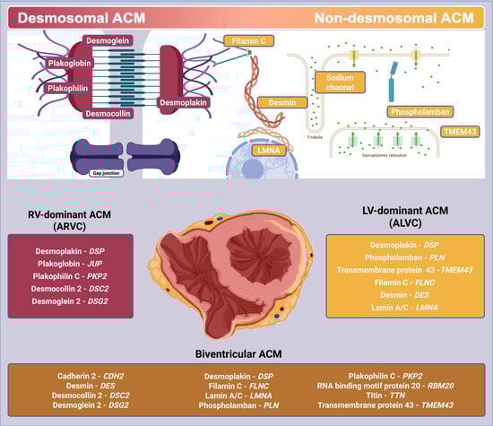
-
Definite: two major OR one major + two minor OR four minor criteria for ARVC and ALVC from different categories.
-
Borderline: one major + one minor OR three minor criteria for ARVC and ALVC from different categories.
-
Possible: one major OR two minor criteria for ARVC and ALVC from different categories.
AHA: American Heart Association, ACC: American College of Cardiology, HRS: Heart Rhythm Society, ESC: European Society of Cardiology, ETF/ITF: European/International Task Force, ARVC: arrhythmogenic right ventricular cardiomyopathy, RV: right ventricle, LV: left ventricle; Biv: biventricular; VA: ventricular arrhythmias; SCD: sudden cardiac death; TWI: T wave inversion; VE: ventricular extrasystole; VF: ventricular fibrillation; VT: ventricular tachycardia; LBBB: left bundle branch block; RBBB: right bundle branch block; RV: right ventricle; RVEDV: right ventricular end-diastolic volume; RVEF: right ventricle ejection fraction; RVOT: right ventricular outflow tract; SCA: sudden cardiac arrest.
Table 2. Genotype–phenotype ACM correlation with biochemical insights.
Desmosomal ACM
Gene VariantCell DamagePhenotype
Plakoglobin
  • β-catenin displacement and Wnt pathway suppression.
  • Adipogenesis promotion.
  • Fibro-fatty remodeling [52].
ARVC (mainly)
ALVC
Naxos disease (hair and skin)
Plakophilin C
  • Nuclear envelope disruption (chromatin disorganization).
  • DNA damage.
  • Impaired transcription of electron transport chain genes (mitochondrial dysfunction) [53].
ARVC
Desmocollin 2
  • Adipogenesis promotion.
  • Fibro-fatty remodeling [52].
ARVC
Desmoglein 2
  • Compromised desmosomal adhesion under mechanical stress.
  • Cardiomyocyte necrosis and electrical disorders [54,55].
ARVC (mainly)
ALVC
Biv-ACM
Desmoplakin
  • Adipogenesis promotion.
  • Fibro-fatty remodeling [56].
ALVC (mainly)
Biv-ACM
ARVC
Non-desmosomal ACM
Gene variantCell damagePhenotype
Phospholamban
  • Calcium channel disruption with Ca overload.
  • CaMKII and Calcineurin activation [57].
  • PLN-R1del: SERCA2a superinhibition [58].
ALVC (mainly)
Biv-ACM
ARVC
Transmembrane protein 43
  • Impaired Wnt/β-catenin signaling.
  • Promoted adipogenesis [53].
ALVC (mainly)
Biv-ACM (fast deterioration)
Filamin C
  • Impaired Wnt/β-catenin signaling.
  • Promoted adipogenesis [53].
ALVC (mainly)
Biv-ACM (fast deterioration)
Skeletal myofibrillar myopathy
Desmin
  • Impaired Wnt/β-catenin signaling.
  • Promoted adipogenesis [53].
ALVC (mainly)
Biv-ACM
Skeletal myofibrillar myopathy
Conduction system abnormalities
Lamin A/C
  • Impaired Wnt/β-catenin signaling.
  • Promoted adipogenesis [53].
ALVC (mainly)
ARVC
BivACM (fast deterioration)
Emery–Dreifuss muscular dystrophy
Limb–girdle muscular dystrophy 1B
Familial lipodystrophy
Hutchinson–Gliford progeria [59]
SCN5A
  • Impaired Nav1.5 channels function reduces excitability.
  • Impaired mechanical coupling between desmosomes and Nav1.5 [55].
ARVC
ALVC
ACM: arrhythmogenic cardiomyopathy, ARVC: arrhythmogenic right ventricular cardiomyopathy, ALVC: arrhythmogenic cardiomyopathy, BivACM: biventricular arrhythmogenic cardiomyopathy, CaMKII: calcium/Calmodulin-dependent Protein Kinase II, Nav1.5: Voltage-Gated Sodium Channel Alpha Subunit 5, PLN-R1: Phospholamban Repressor-1, SERCA2a: Sarcoplasmic/Endoplasmic Reticulum Calcium ATPase 2a, Wnt/β-catenin: Wingless/Integrated signaling pathway involving β-catenin.
Table 3. Ongoing clinical trials on gene therapy for the specific PKP2-ACM subtype.
Clinical TrialsDescription
NCT05885412
Phase 1—dose escalation
Intravenous injected recombinant AAV vector containing PKP2 (RP-A601) in subjects with high-risk PKP2-ACM
NCT06109181
Phase 1/2—open label,
dose escalating, multicentric trial
Safety and tolerability of LX2020 (AAV vector encoding PKP2 gene) in 10 adult patients with PKP2-ACM
NCT06228924—RIDGE-1
open label, phase 1
Fifteen patients across two designated dose groups who are experiencing symptomatic PKP2-ACM, each cohort receiving a single endovenous dose of TN-401 (AAV9 containing PKP2 transgene)
NCT06311708
multicentric, observational
Prevalence of pre-existing antibodies to AAV9 in a population of PKP2-ACM
AAV: adeno-associated viral, ACM: arrhythmogenic cardiomyopathy, PKP2: plakophilin2.
Disclaimer/Publisher’s Note: The statements, opinions and data contained in all publications are solely those of the individual author(s) and contributor(s) and not of MDPI and/or the editor(s). MDPI and/or the editor(s) disclaim responsibility for any injury to people or property resulting from any ideas, methods, instructions or products referred to in the content.

