Article

Mutations in MYBPC3 and MYH7 in Association with Brugada Type 1 ECG Pattern: Overlap between Brugada Syndrome and Hypertrophic Cardiomyopathy?

1
Unit of Medical Genetics, Department of Medical Sciences, University of Ferrara, 44121 Ferrara, Italy
2
Cardiology Department, University Hospital S. Anna Ferrara, 44121 Ferrara, Italy
3
Department of Pharmacy-Drug Sciences, University of Bari “Aldo Moro”, 70125 Bari, Italy
4
Cardiology Unit, Ramazzini Hospital, 41012 Carpi (Modena), Italy
5
Department of Cardiology, S. Orsola-Malpighi Hospital, 40138 Bologna, Italy
6
Maria Cecilia Hospital, GVM Care & Research, 48033 Cotignola, Italy
7
Medical Genetics Service, Department of Mother and Child, University Hospital S. Anna Ferrara, 44121 Ferrara, Italy
*
Author to whom correspondence should be addressed.
First two authors contributed equally to the paper.
Cardiogenetics 2021, 11(3), 139-147; https://doi.org/10.3390/cardiogenetics11030016
Submission received: 11 June 2021 / Revised: 1 September 2021 / Accepted: 6 September 2021 / Published: 9 September 2021
(This article belongs to the Section Cardiovascular Genetics in Clinical Practice)

Abstract

:
Brugada syndrome (BrS) is an inherited disorder with high allelic and genetic heterogeneity clinically characterized by typical coved-type ST segment elevation at the electrocardiogram (ECG), which may occur either spontaneously or after provocative drug testing. BrS is classically described as an arrhythmic condition occurring in a structurally normal heart and is associated with the risk of ventricular fibrillation and sudden cardiac death (SCD). We studied five patients with spontaneous or drug-induced type 1 ECG pattern, variably associated with symptoms and a positive family history through a Next Generation Sequencing panels approach, which includes genes of both channelopathies and cardiomyopathies. We identified variants in MYBPC3 and in MYH7, hypertrophic cardiomyopathy (HCM) genes (MYBPC3: p.Lys1065Glnfs*12 and c.1458-1G > A, MYH7: p.Arg783His, p.Val1213Met, p.Lys744Thr). Our data propose that Brugada type 1 ECG may be an early electrocardiographic marker of a concealed structural heart disease, possibly enlarging the genotypic overlap between Brugada syndrome and cardiomyopathies.

1. Introduction

A typical ECG pattern, characterized by a coved ST-segment elevation ≥ 2 mm in at least one right precordial lead followed by a concave or straight ST segment elevation and a negative symmetric T-wave [1], is associated with Brugada syndrome, an inherited disease characterized by an increased risk of ventricular arrhythmias and sudden cardiac death (SCD) in a structurally normal heart [2]. Nowadays a pathogenic genetic variant is detected in up to 30% of subjects with BrS, usually transmitted in an autosomal-dominant manner [3,4]. The gene most frequently involved, and the sole one considered as definitively associated with the syndrome [5], is SCN5A, which encodes the alpha subunit of the cardiac sodium channel Nav1.5 and is responsible for nearly 20–30% of all cases [6]. Several other genes have been proposed as underlying BrS, but with incomplete, often disputed evidence [7]; more and more evidence suggest a possible oligogenic or polygenic inheritance for inheritable cardiac disorders, including BrS [8,9,10]. Genetic counseling in BrS is complicated by many confounding factors: genetic and allelic heterogeneity, variants of uncertain significance [11], incomplete penetrance [12], phenotypic overlaps [6] and new evidence of complex inheritance [12] and mutation load [13], thus overcoming the one gene–one disease paradigm [10]. Furthermore, the concept of BrS as a pure electric condition, occurring in an otherwise structurally normal heart, has been contradicted by several observations. Indeed, the overlap between BrS and arrhythmogenic cardiomyopathy (ACM) is well known in the literature, so much that ACM and BrS could be seen as two different entities belonging to the same disease spectrum [10,14].
Sarcomeric genes, encoding for components of the contractile unit in the cardiomyocyte (the sarcomere), are characteristically associated with structural cardiomyopathies [15]. MYBPC3 and MYH7, encoding for the cardiac myosin binding protein C and the cardiac beta-myosin heavy-chain, respectively, represent the genes most frequently involved in hypertrophic cardiomyopathy (HCM), accounting for 50% and 33% of cases with a positive genetic test result, respectively [16]. In five patients with spontaneous or drug-induced type 1 ECG pattern (BrP), negative for mutations in SCN5A and other putative BrS genes, we found likely pathogenic/pathogenic variants in MYBPC3 and MYH7, structural cardiomyopathy genes. This observation adds evidence to the genotypic overlap between arrhythmic and structural heart diseases and supports Brugada type 1 ECG as an early electrical sign of an upcoming structural disease.

2. Materials and Methods

Written informed consent for genetic testing was obtained from all enrolled subjects (local ethical committee approval 26/7/2012, P. 7/2012). NGS analysis was performed on MiSeq™Dx Instrument using the commercial gene panel Trusight Cardio Sequencing Kit by Illumina (San Diego, CA, USA, www.illumina.com accessed on 21 September 2020), which includes both structural and arrhythmogenic cardiomyopathies genes. Data were analyzed and filtered using Sophia Genetics DDMR software (https://dropgen.sophiagenetics.com accessed on 21 September 2020). The minimum depth of coverage for variant calling was 20×. The Exome Variant Server (ESP), the Exome Aggregation Consortium (ExAC) and the gnomAD database (version 2.1.1) with a frequency greater than 0.1% were used to filter out common variants. Potential disease-causing missense variants were assessed using Mutation Taster, Polyphen2 and SIFT. VarSome (https://varsome.com/ accessed on 15 July 2021) and/or ClinVar (https://www.ncbi.nlm.nih.gov/clinvar/ accessed on 15 July 2021) were used as a tool to sum up actual knowledge about the variants. All identified variants were classified according to American College of Medical Genetics and Genomics (ACMG) guidelines [11]. The molecular confirmation of variants was performed by standard Sanger sequencing on an automated analyzer (ABI PRISM® 3130). The MLPA technique was used to detect deletions or duplications in SCN5A by using MRC-Holland probemix P108 kit (Amsterdam, The Netherlands, https://www.mrcholland.com/ accessed on 16 July 2021).

3. Results

3.1. Clinical Evaluation
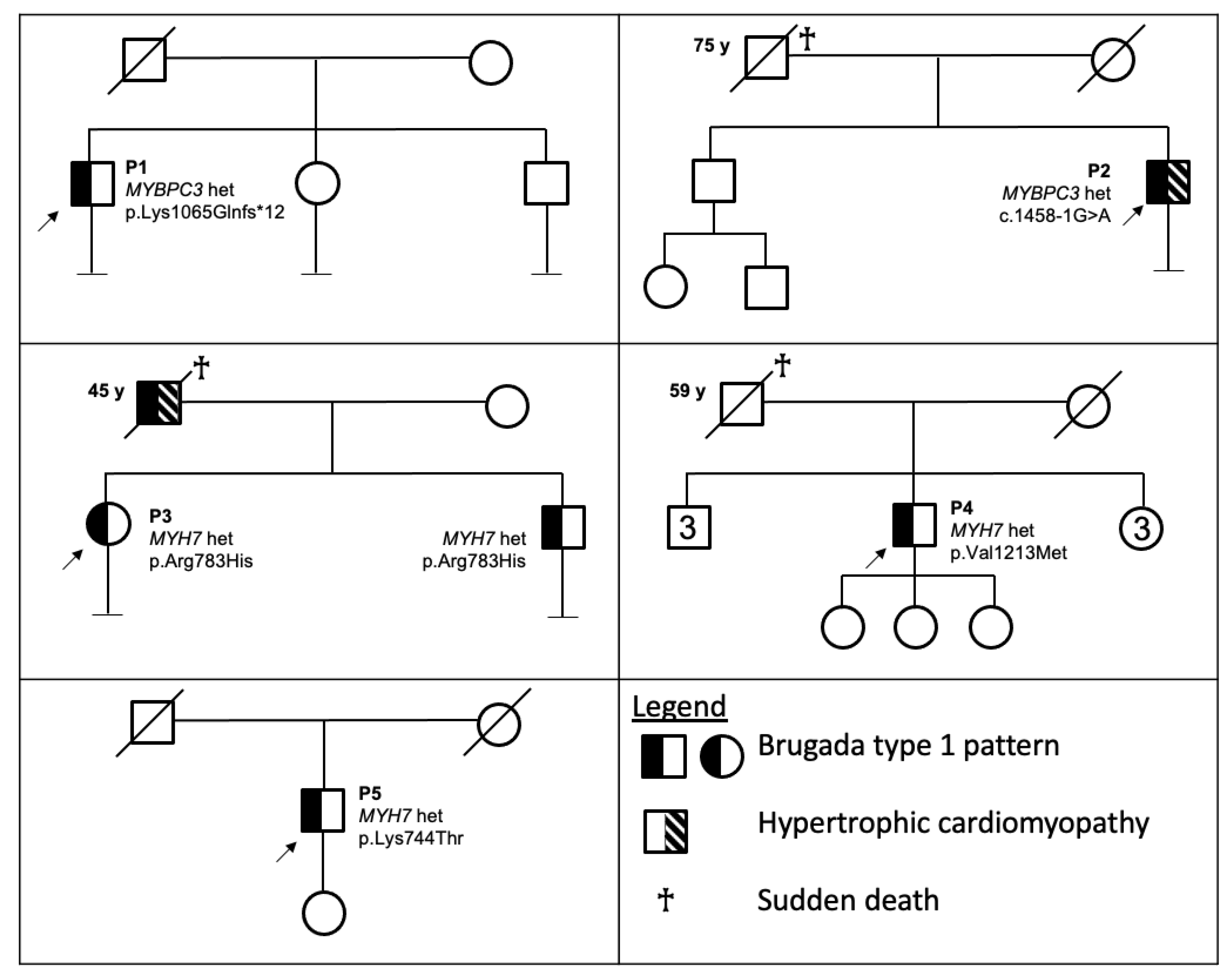
Among a cohort of 51 patients with BrP tested by the gene panel including both structural and arrhythmogenic cardiomyopathies, we identified five patients carrying a pathogenic/likely pathogenic variation in sarcomeric genes. Table 1 summarizes clinical and genetic evaluations, Figure 1 shows the pedigree of the five patients and Figure 2 shows their ECGs.

3.1.1. Patient 1

P1 is an adult male with a spontaneous intermittent type 1 BrP, which was detected at 34 years old as an incidental finding during a routine visit prior to sport activity. ECG showed: PR = 172 ms; QTc = 440 ms at a heart rate of 56 bpm. His family history was negative for cardiovascular disease (CVD) or sudden cardiac death (SCD).
He experienced recurrent palpitations (at least two times a week, duration 5 to 20 min); symptoms were documented several times by an external loop recorder which detected an atrio-ventricular nodal reentrant tachycardia that was subsequently successfully treated by radiofrequency ablation. No programmed electrical stimulation (PES) was performed.
His echocardiogram showed no signs of structural abnormalities. He performed a cardiac magnetic resonance (MR), which shows absence of right ventricle (RV) bulging, normal RV function, preserved LV volume, myocardial mass and function, no fibro-fatty involvement of both ventricles, normal perfusion scan and no abnormalities in late-gadolinium-enhancement (LGE) study.

3.1.2. Patient 2

P2 is an adult male who had an incidental finding of spontaneous type 1 BrP at 56 years old during a routine visit, without any other symptoms, except for sporadic extrasystoles. ECG showed: PR 148 ms, QTc 416 ms at a heart rate of 68 bpm.
His father died suddenly at 75 years old (no previous history of cardiac disease). He performed an echocardiogram that showed left ventricular hypertrophy (LVH), more evident in the mid-basal septum and in the posteroinferior wall (maximum thickness: 16 mm) and a cardiac MR that showed left ventricular hypertrophy with a maximum wall thickness of 18 mm in the mid basal septum (Figure 3). His HCM risk-SCD score was calculated as 1.97% at five years. No PES was performed.

3.1.3. Patient 3

P3 is a 26-year-old woman with a history of syncope and a drug-induced type 1 BrP. ECG showed: PR = 148 ms, QTc = 420 ms at a heart rate of 68 bpm. Her father, who was a known carrier of a type 1 BrP, died suddenly during sleep at 45 years old (a moderate left ventricular hypertrophy was found before during cardiac ultrasound and at the autopsy). Her echocardiogram showed no signs of structural abnormalities; she also performed a cardiac MR, which showed normal biventricular dimension, wall thickness, volume and function and negative LGE sequences. Because of her family history of SCD and the history of recurrent syncope, she received a subcutaneous implantable cardioverter defibrillator (S-ICD) in primary prevention.
P3 has an 18-year-old brother, who also had a drug-induced type 1 BrP and no sign of structural heart disease at echocardiography and MR scan. ECG showed: PR = 158 ms, QTc = 380 ms at a heart rate of 60 bpm. He also received an S-ICD in primary prevention.
The defibrillation was effective with 30 and 25 J, in P3 and her brother, respectively. No PES was performed in both siblings.

3.1.4. Patient 4

P4 is an adult male who had an ECG finding of spontaneous type 1 BrP at 61 years old during a routine visit. ECG showed: PR = 160 ms, QTc = 433 ms at a heart rate of 86 bpm. He did not have any symptoms of palpitations or syncope. He had a positive familial history of SCD, which occurred in his father at 59 years old during sleep. His echocardiogram shows no evidence of structural abnormalities and preserved biventricular function. No PES was performed.

3.1.5. Patient 5

P5 is an adult male who had an incidental finding of spontaneous type 1 BrP at 46 years old during an episode of palpitations and abdominal pain. ECG showed: PR = 160 ms, QTc = 372 ms at a heart rate of 70 bpm. His echocardiogram showed no signs of structural cardiomyopathy. His family history was negative for CVD or SCD. No PES was performed.

3.2. Genetic Evaluation

In the five patients, neither variations nor deletions/duplications were identified in SCN5A and no variations were identified in other minor BrS genes. We detected heterozygous variations in sarcomeric genes (Table 1). All of these mutations, but the last one in P5, are known in literature as associated with HCM [17,18,19,20].
P1, P2 and P3 are heterozygous, respectively, for the frameshifting mutation c.3192dupC (p.Lys1065Glnfs*12) in exon 30 in MYBPC3, the splicing mutation c.1458-1G > A in intron 16 in MYBPC3 and the missense variation c.2348G > A (p.Arg783His) in exon 21 in MYH7. All these changes are known in literature as associated with HCM [17,18,19] and are classified as pathogenic or likely pathogenic according to ACMG criteria. The MYH7 variant detected in P3 segregated in her 18-year-old brother.
P4 is heterozygous for the missense variation c.3637G > A (p.Val1213Met) in exon 27 in MYH7, classified as VUS/LP by prediction tools and databases. Nonetheless, this variant is absent from general population and it has been described several times in literature as associated with HCM [20].
P5 is heterozygous for the missense variation c.2231A > C (p.Lys744Thr) in exon 20 in MYH7. This sequence change has not been described in literature in affected subjects and is absent from controls (PM2 criterion according to ACMG). In silico prediction tools support its pathogenicity and the variant is currently classified as LP (Table 1).

4. Discussion

In BrS patients, gross structural alterations are not usually detected at routine investigation and BrS is considered a primary electrical disease. Nonetheless, there is growing evidence that patients with BrS may exhibit subtle cardiac abnormalities; thus, the idea of a ‘Brugada cardiomyopathy’ has been proposed [21]. The phenotypic and genotypic overlap between BrS and arrhythmogenic cardiomyopathy (ACM) is well-known in literature and it is supported by the identification of both conduction abnormalities and structural alterations in the right ventricular outflow tract (RVOT) in BrS patients [14,22,23,24]. Nademanee, in particular, reported fibro-fatty replacement of the RVOT in both autoptic and in vivo collected cardiac samples from BrS patients, who showed no cardiac structural abnormality at ecocardiography and/or cardiac MRI, thus suggesting that BrS pattern could be a very early sign of disease, anticipating manifest myocardium alterations [22]. By using Doppler tissue imaging (DTI), we recently demonstrated a contraction delay at the RVOT in individuals with spontaneous type 1 Brugada ECG pattern [25].
Mutations in ACM desmosomal genes have been associated with BrS and studies have shown that deficiencies in PKP2 (one of the structural components of the cardiac desmosome and a major ACM gene) determine downstream effects, both on the integrity of gap junctions and on the sodium channel function, with consequences on electrical coupling and on sodium current [10].
Further supporting type 1 BrP as a possible marker of an occult cardiomyopathy, we detected variants in sarcomeric genes in five patients presenting spontaneous or drug induced BrS pattern.
MYBPC3 encodes the cardiac myosin binding protein C and MYH7 encodes the cardiac beta-myosin heavy-chain, two proteins typically involved in the structural function of the cardiac sarcomere. Mutations in the two genes are mainly associated with hypertrophic cardiomyopathy (HCM) [15]. In recent years, some authors have suggested a possible link between BrS and HCM, although with still limited evidence. In 2015 Di Resta and colleagues detected a borderline significant association for mutations in MYH7 gene in BrS patients [5/91 (5.5%) patients vs. 0/91 controls] [26]. In 2016, a family with four members showing both HCM and BrS was described and an integrated linkage analysis and NGS approach identified a missense mutation within the sarcomeric TPM1 gene [27]. More recently, Pappone et al. reported a family in which a MYBPC3 mutation segregated in the father, who was affected by HCM, and in his daughter and son, who were affected by BrS [28]. The presence of left ventricular hypertrophy in autopsy data of BrS subjects has also been described [29].
In our case series, all patients shared a type 1 BrP (spontaneous or drug-induced) and carried a heterozygous variation in a sarcomeric gene, but only one patient (P2, carrying a pathogenic splicing mutation in MYBPC3), presented instrumental signs of HCM. The other patients showed a pure electrical phenotype, variably associated with a positive family history for BrS and/or sudden death. Nevertheless, it has to be taken into account that these patients are relatively young and HCM penetrance is incomplete and age dependent.
Despite the lack of segregation analysis, which was possible only in two individuals in one family, available data support the identified mutations in sarcomeric genes as disease causing. Indeed, except for the missense variation c.2231A > C (p.Lys744Thr) in exon 20 in MYH7 in P5, the mutations we identified have been reported in several individuals in the literature, all affected by structural cardiomyopathy [17,18,19,20], and all these variations are absent in population control databases. Based on the increasing knowledge in literature of a strong structural/electrical overlap [26,27,28,29], it may be speculated that these sarcomeric mutations may justify Brugada type 1 ECG in our patients, which is that the Brugada type 1 ECG is an early sign of a structural disease. Further evidence is needed to confirm this hypothesis and, most of all, how defects in sarcomeric genes can lead to BrS-related electrical manifestation remains to be clarified. Notably, Baudenbacher et al. have shown that increased myofilament Ca2+ sensitivity determined arrhythmia susceptibility in mice expressing troponin T mutants, even without anatomical abnormalities [30]. Furthermore, an abnormal SCN5A mRNA splicing with reduction of the full-length transcript has been observed in HCM patients [31], suggesting that a reduction in SCN5A may contribute to the arrhythmic risk in HCM.
In conclusion, data obtained from this case series provide further evidence of the genotypic overlap between BrS-related electrical phenotype and structural cardiomyopathies and support the concept that individuals presenting with either primary cardiomyopathy or arrhythmias should be tested for a comprehensive cardiomyopathy/arrhythmia panel. Based on our observation, the type 1 BrP may represent an early sign of a structural disease that would remain concealed unless investigated, with implications in clinical management and genetic counseling of BrS patients and overlapping phenotypes.

Author Contributions

M.F.: data acquisition, processing and interpretation; writing of the manuscript. F.G., C.B.: study concept and supervision, data interpretation, critical revision of manuscript. A.F.: study concept and critical revision of manuscript. C.B., E.D.M., M.B. (Mauro Biffi), C.R., P.I., M.B. (Matteo Bertini): data acquisition and interpretation, critical revision of manuscript. M.F., A.D.D., M.D.R., F.G.: data acquisition and clinical evaluation. A.M., R.S.: genetic analysis, data interpretation. All authors have read and agreed to the published version of the manuscript.

Funding

No financial assistance was received in support of the study.

Institutional Review Board Statement

Written informed consent was obtained from the patients (Ferrara local ethical committee approval 26/7/2012, P. 7/2012).

Informed Consent Statement

Informed consent was obtained from all subjects involved in the study.

Data Availability Statement

The data that support the findings of this study are available from the corresponding Authors upon reasonable request.

Acknowledgments

Thanks to the “Associazione Voglio Volare-Davide Barbi” for supporting this study.

Conflicts of Interest

The authors have no conflict of interest to declare.

References

  1. Wilde, A.A.M.; Antzelevitch, C.; Borggrefe, M.; Brugada, J.; Brugada, R.; Brugada, P.; Corrado, D.; Hauer, R.; Kass, R.; Nademanee, K.; et al. Proposed diagnostic criteria for the Brugada syndrome. Eur. Heart J. 2002, 23, 1648–1654. [Google Scholar] [CrossRef]
  2. Brugada, P.; Brugada, J. Right bundle branch block, persistent ST segment elevation and sudden cardiac death: A distinct clinical and electrocardiographic syndrome. A multicenter report. J. Am. Coll. Cardiol. 1992, 20, 1391–1396. [Google Scholar] [CrossRef]
  3. Juang, J.-M.J.; Horie, M. Genetics of Brugada syndrome. J. Arrhythm. 2016, 32, 418–425. [Google Scholar] [CrossRef] [PubMed] [Green Version]
  4. Sarquella-Brugada, G.; Campuzano, O.; Arbelo, E.; Brugada, J.; Brugada, R. Brugada syndrome: Clinical and genetic findings. Genet. Med. 2016, 18, 3–12. [Google Scholar] [CrossRef] [Green Version]
  5. ClinGen. Available online: https://search.clinicalgenome.org/kb/genes/HGNC:10593 (accessed on 15 July 2021).
  6. Brugada, J.; Campuzano, O.; Arbelo, E.; Sarquella-Brugada, G.; Brugada, R. Present Status of Brugada Syndrome: JACC State-of-the-Art Review. J. Am. Coll. Cardiol. 2018, 72, 1046–1059. [Google Scholar] [CrossRef]
  7. Hosseini, S.M.; Kim, R.; Udupa, S.; Costain, G.; Jobling, R.; Liston, E.; Jamal, S.M.; Szybowska, M.; Morel, C.F.; Bowdin, S.; et al. Reappraisal of Reported Genes for Sudden Arrhythmic Death. Circulation 2018, 138, 1195–1205. [Google Scholar] [CrossRef] [PubMed]
  8. Campuzano, O.; Sarquella-Brugada, G.; Cesar, S.; Arbelo, E.; Brugada, J.; Brugada, R. Update on Genetic Basis of Brugada Syndrome: Monogenic, Polygenic or Oligogenic? Int. J. Mol. Sci. 2020, 21, 7155. [Google Scholar] [CrossRef] [PubMed]
  9. Monasky, M.M.; Micaglio, E.; Ciconte, G.; Pappone, C. Brugada Syndrome: Oligogenic or Mendelian Disease? Int. J. Mol. Sci. 2020, 21, 1687. [Google Scholar] [CrossRef] [Green Version]
  10. Cerrone, M.; Remme, C.A.; Tadros, R.; Bezzina, C.R.; Delmar, M. Beyond the One Gene-One Disease Paradigm: Complex Genetics and Pleiotropy in Inheritable Cardiac Disorders. Circulation 2019, 140, 595–610. [Google Scholar] [CrossRef]
  11. Richards, S.; Aziz, N.; Bale, S.; Bick, D.; Das, S.; Gastier-Foster, J.; Grody, W.W.; Hegde, M.; Lyon, E.; Spector, E.; et al. Standards and guidelines for the interpretation of sequence variants: A joint consensus recommendation of the American College of Medical Genetics and Genomics and the Association for Molecular Pathology. Genet. Med. 2015, 17, 405–424. [Google Scholar] [CrossRef]
  12. Gourraud, J.-B.; Barc, J.; Thollet, A.; Le Scouarnec, S.; Le Marec, H.; Schott, J.-J.; Redon, R.; Probst, V. The Brugada Syndrome: A Rare Arrhythmia Disorder with Complex Inheritance. Front. Cardiovasc. Med. 2016, 3, 9. [Google Scholar] [CrossRef] [Green Version]
  13. Gualandi, F.; Zaraket, F.; Malagù, M.; Parmeggiani, G.; Trabanelli, C.; Fini, S.; Dang, X.; Wei, X.; Fang, M.; Bertini, M.; et al. Mutation Load of Multiple Ion Channel Gene Mutations in Brugada Syndrome. Cardiology 2017, 137, 256–260. [Google Scholar] [CrossRef]
  14. Moncayo-Arlandi, J.; Brugada, R. Unmasking the molecular link between arrhythmogenic cardiomyopathy and Brugada syndrome. Nat. Rev. Cardiol. 2017, 14, 744–756. [Google Scholar] [CrossRef]
  15. Ingles, J.; Goldstein, J.; Thaxton, C.; Caleshu, C.; Corty, E.W.; Crowley, S.B.; Dougherty, K.; Harrison, S.M.; McGlaughon, J.; Milko, L.V.; et al. Evaluating the Clinical Validity of Hypertrophic Cardiomyopathy Genes. Circ. Genom. Precis. Med. 2019, 12, e002460. [Google Scholar] [CrossRef] [Green Version]
  16. Alfares, A.A.; Kelly, M.A.; McDermott, G.; Funke, B.H.; Lebo, M.S.; Baxter, S.B.; Shen, J.; McLaughlin, H.M.; Clark, E.H.; Babb, L.J.; et al. Results of clinical genetic testing of 2,912 probands with hypertrophic cardiomyopathy: Expanded panels offer limited additional sensitivity. Genet. Med. 2015, 17, 880–888. [Google Scholar] [CrossRef] [Green Version]
  17. Girolami, F.; Olivotto, I.; Passerini, I.; Zachara, E.; Nistri, S.; Re, F.; Fantini, S.; Baldini, K.; Torricelli, F.; Cecchi, F. A molecular screening strategy based on beta-myosin heavy chain, cardiac myosin binding protein C and troponin T genes in Italian patients with hypertrophic cardiomyopathy. J. Cardiovasc. Med. 2006, 7, 601–607. [Google Scholar] [CrossRef] [PubMed]
  18. Cecconi, M.; Parodi, M.I.; Formisano, F.; Spirito, P.; Autore, C.; Musumeci, M.B.; Favale, S.; Forleo, C.; Rapezzi, C.; Biagini, E.; et al. Targeted next-generation sequencing helps to decipher the genetic and phenotypic heterogeneity of hypertrophic cardiomyopathy. Int. J. Mol. Med. 2016, 38, 1111–1124. [Google Scholar] [CrossRef] [PubMed] [Green Version]
  19. Waldmüller, S.; Müller, M.; Rackebrandt, K.; Binner, P.; Poths, S.; Bonin, M.; Scheffold, T. Array-based resequencing assay for mutations causing hypertrophic cardiomyopathy. Clin. Chem. 2008, 54, 682–687. [Google Scholar] [CrossRef]
  20. Homburger, J.R.; Green, E.M.; Caleshu, C.; Sunitha, M.S.; Taylor, R.E.; Ruppel, K.M.; Metpally, R.P.R.; Colan, S.D.; Michels, M.; Day, S.M.; et al. Multidimensional structure-function relationships in human β-cardiac myosin from population-scale genetic variation. Proc. Natl. Acad. Sci. USA 2016, 113, 6701–6706. [Google Scholar] [CrossRef] [Green Version]
  21. Bastiaenen, R.; Cox, A.T.; Castelletti, S.; Wijeyeratne, Y.D.; Colbeck, N.; Pakroo, N.; Ahmed, H.; Bunce, N.; Anderson, L.; Moon, J.C.; et al. Late gadolinium enhancement in Brugada syndrome: A marker for subtle underlying cardiomyopathy? Heart Rhythm 2017, 14, 583–589. [Google Scholar] [CrossRef] [PubMed]
  22. Corrado, D.; Basso, C.; Thiene, G. Is it time to include ion channel diseases among cardiomyopathies? J. Electrocardiol. 2005, 38, 81–87. [Google Scholar] [CrossRef] [PubMed]
  23. Nademanee, K.; Raju, H.; de Noronha, S.V.; Papadakis, M.; Robinson, L.; Rothery, S.; Makita, N.; Kowase, S.; Boonmee, N.; Vitayakritsirikul, V.; et al. Fibrosis, Connexin-43, and Conduction Abnormalities in the Brugada Syndrome. J. Am. Coll. Cardiol. 2015, 66, 1976–1986. [Google Scholar] [CrossRef] [PubMed] [Green Version]
  24. Behr, E.R.; Ben-Haim, Y.; Ackerman, M.J.; Krahn, A.D.; Wilde, A.A.M. Brugada syndrome and reduced right ventricular outflow tract conduction reserve: A final common pathway? Eur. Heart J. 2021, 42, 1073–1081. [Google Scholar] [CrossRef] [PubMed]
  25. Trevisan, F.; Bertini, M.; Balla, C.; Pestelli, G.; Luisi, A.; Smarrazzo, V.; Farnè, M.; Ferlini, A.; Gualandi, F.; Mele, D. Type 1 Brugada Pattern is associated with echocardiography-detected delayed right ventricular outflow tract contraction. JACC 2021, 77, 2865–2867. [Google Scholar] [CrossRef]
  26. Di Resta, C.; Pietrelli, A.; Sala, S.; della Bella, P.; De Bellis, G.; Ferrari, M.; Bordoni, R.; Benedetti, S. High-throughput genetic characterization of a cohort of Brugada syndrome patients. Hum. Mol. Genet. 2015, 24, 5828–5835. [Google Scholar] [CrossRef] [Green Version]
  27. Mango, R.; Luchetti, A.; Sangiuolo, R.; Ferradini, V.; Briglia, N.; Giardina, E.; Ferrè, F.; Citterich, M.H.; Romeo, F.; Novelli, G.; et al. Next Generation Sequencing and Linkage Analysis for the Molecular Diagnosis of a Novel Overlapping Syndrome Characterized by Hypertrophic Cardiomyopathy and Typical Electrical Instability of Brugada Syndrome. Circ. J. 2016, 80, 938–949. [Google Scholar] [CrossRef] [Green Version]
  28. Pappone, C.; Monasky, M.M.; Ciconte, G. Epicardial ablation in genetic cardiomyopathies: A new frontier. Eur. Heart J. Suppl. 2019, 21, B61–B66. [Google Scholar] [CrossRef] [Green Version]
  29. Chevalier, P. Brugada syndrome and myocardial histology: Where may the truth Lie? Arch. Cardiovasc. Dis. 2019, 112, 367–369. [Google Scholar] [CrossRef]
  30. Baudenbacher, F.; Schober, T.; Pinto, J.R.; Sidorov, V.Y.; Hilliard, F.; Solaro, R.J.; Potter, J.D.; Knollmann, B.C. Myofilament Ca2+ sensitization causes susceptibility to cardiac arrhythmia in mice. J. Clin. Investig. 2008, 118, 3893–3903. [Google Scholar] [CrossRef] [Green Version]
  31. Noyes, A.M.; Zhou, A.; Gao, G.; Gu, L.; Day, S.; Wasserstrom, J.A.; Dudley, S.C. Abnormal sodium channel mRNA splicing in hypertrophic cardiomyopathy. Int. J. Cardiol. 2017, 249, 282–286. [Google Scholar] [CrossRef] [PubMed]
Figure 1. Pedigrees of the five patients. The legend is shown in the figure. For clinical and familial details, see text in Results.
Cardiogenetics 11 00016 g001
Figure 2. ECG images of patients P1, P3, P4 and P5. (A) ECG of patient P1 showing type 1 BrP in V1–V2 recorded at second, third and fourth intercostal space; (B) ECG of patient P3 showing type 1 BrP in V1–V2 recorded at fourth intercostal space; (C) ECG of patient P4 showing type 1 BrP in V1–V2 recorded at fourth intercostal space; (D) ECG of patient P5 showing type 1 BrP in V1–V2 recorded at fourth intercostal space.
Cardiogenetics 11 00016 g002
Figure 3. ECG and cardiac MRI of the BrS patient P2 carrying the MYBPC3 c.1458-1G > A splicing mutation. (A) ECG of patient P2 showing type 1 BrP in V1-V2 recorded at fourth intercostal space. (B) MRI imaging of the same patient showing left ventricular hypertrophy.
Cardiogenetics 11 00016 g003
Table 1. Patients with Type 1 ECG Brugada pattern carrying heterozygous variants in hypertrophic cardiomyopathy genes.
PatientSexAge of
Detection
Type 1 ECG
Pattern
Family
History
Clinical
Evaluation
Genec.DNA ChangeProtein ChangeVarSomeClinVarACMGReference
P1M34Spontaneous-Asymptomatic except for AVNRT. No signs of HCM.MYBPC3ex 30 c.3192dupCp.Lys1065Glnfs*12
(K1065Qfs * 12)
PPPVS1-PP5-PM2-PP3 (P)[17]
P2M56Spontaneous+ *Asymptomatic except for sporadic extrasistoles. Instrumental signs of HCM.MYBPC3int 16
c.1458-1G > A
p.? PPPVS1-PP5-PM2-PP3 (P)[18]
P3F24Drug-induced+ **History of sincope and Premature Ventricular Contractions.
No signs of HCM.
MYH7ex 21 c.2348G > Ap.Arg783His
(R783H)
LPVUS/LPPM1-PM2-PM5-PP2-BP4 (LP)[19]
P4M61Spontaneous+ ***Asymptomatic.
No signs of HCM.
MYH7ex 27 c.3637G > Ap.Val1213Met
(V1213M)
VUS/LPVUSPM2-PP2-PP3 (VUS)[20]
P5M46Spontaneous-Asymptomatic.
No signs of HCM.
MYH7ex 20 c.2231A > Cp.Lys744Thr
(K744T)
LPNot reportedPM1-PM2-PP2-PP3 (LP)Present paper
F: Female, M: Male; ex: Exon; int: Intron; AVNRT: Atrio-Ventricular Nodal Reentrant Tachycardia; HCM: Hypertrophic cardiomyopathy; VUS: Variant of uncertain significance; LP: Likely pathogenic; P: Pathogenic. Data from Clinvar and VarSome tools and ACMG criteria are updated 15 July 2021. Accession numbers: MYBPC3: NG_007667.1(NM_000256.3), MYH7: NM_000257.2. Exons are numbered according to LOVD database (https://databases.lovd.nl/ accessed on 21 September 2020). * Sudden death in his father at 75 years old. ** Sudden death in her father at 45 years old, a known carrier of a type 1 Brugada pattern and with moderate left ventricular hypertrophy. Younger brother (carrying the same mutation) with drug-induced type 1 Brugada pattern and Premature Ventricular Contractions. *** Sudden death in his father at 59 years old.
Publisher’s Note: MDPI stays neutral with regard to jurisdictional claims in published maps and institutional affiliations.

