Renaissance of Traditional Mineral Drugs in Cancer: Advanced Delivery Strategies and Bioengineering Approaches


 and
and
Abstract
1. Introduction
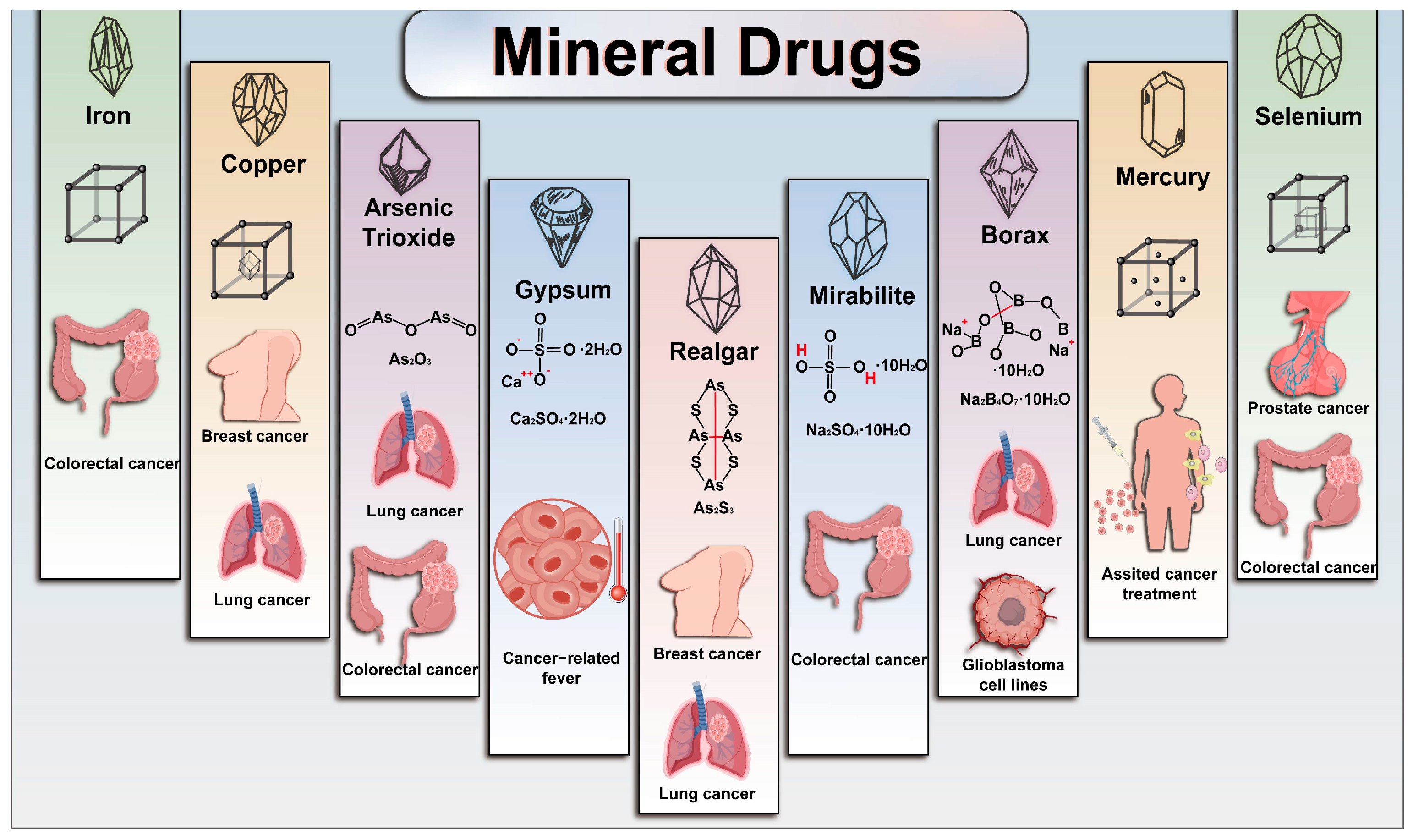
2. Applications of Mineral Drugs in Cancer Treatment
2.1. Anticancer Effects
2.2. Alleviation of Cancer-Related Complications
2.3. Synergistic Effects with Other Anticancer Therapies
3. Representative Antitumor Mineral Drugs
3.1. Realgar
3.2. Mirabilite
3.3. ATO
3.4. Gypsum
3.5. Other Mineral Drugs with Anticancer or Adjunctive Potential
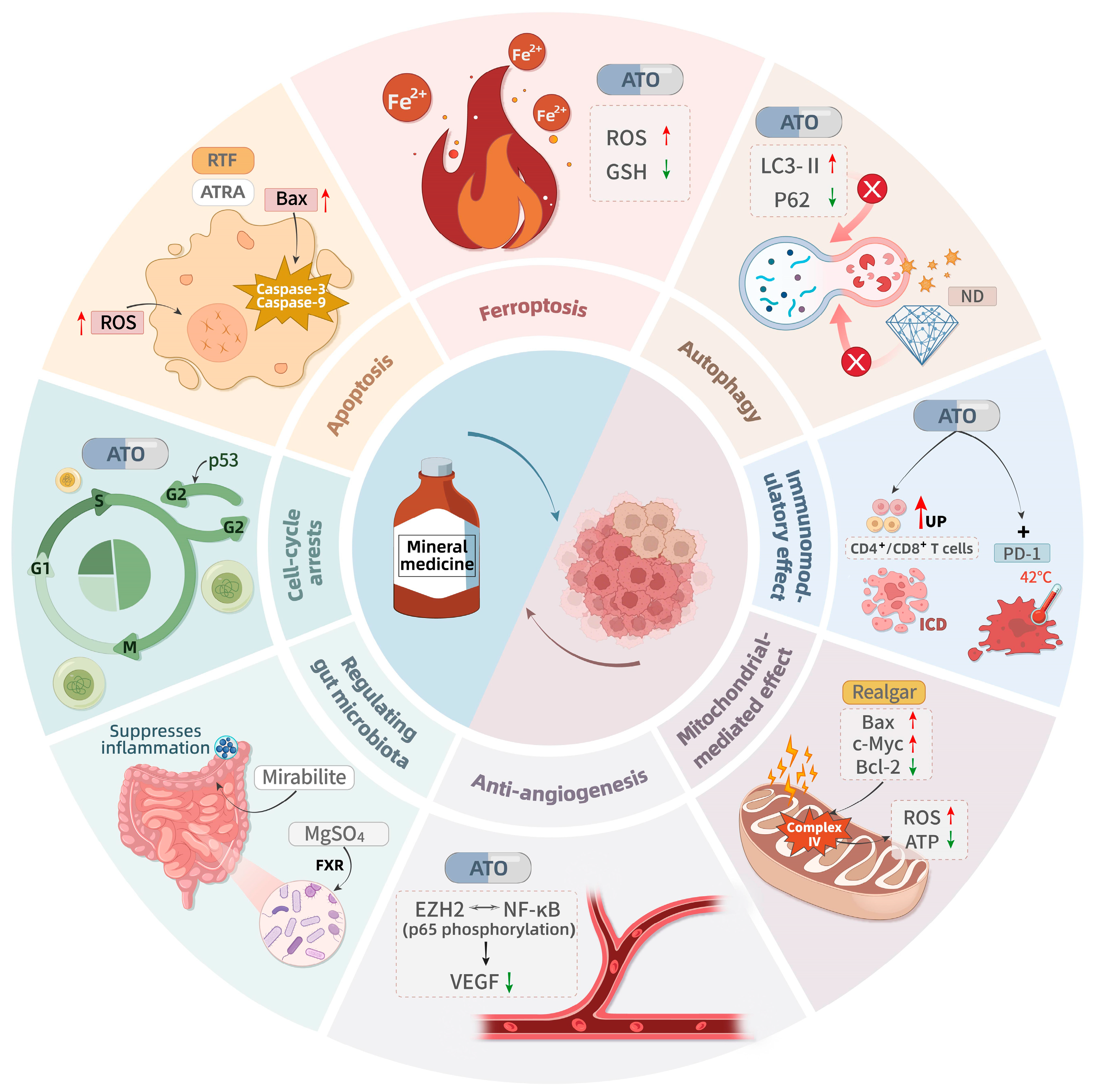
4. Mechanism of Mineral Drugs-Mediated Anticancer
4.1. Induction of Apoptosis and Cell Cycle Arrest
4.2. Induction of Ferroptosis and Modulation of Autophagy
4.3. Mitochondrial-Mediated Effects
4.4. Anti-Angiogenesis
4.5. Immunomodulatory Effects
4.6. Regulation of the Gut Microbiota
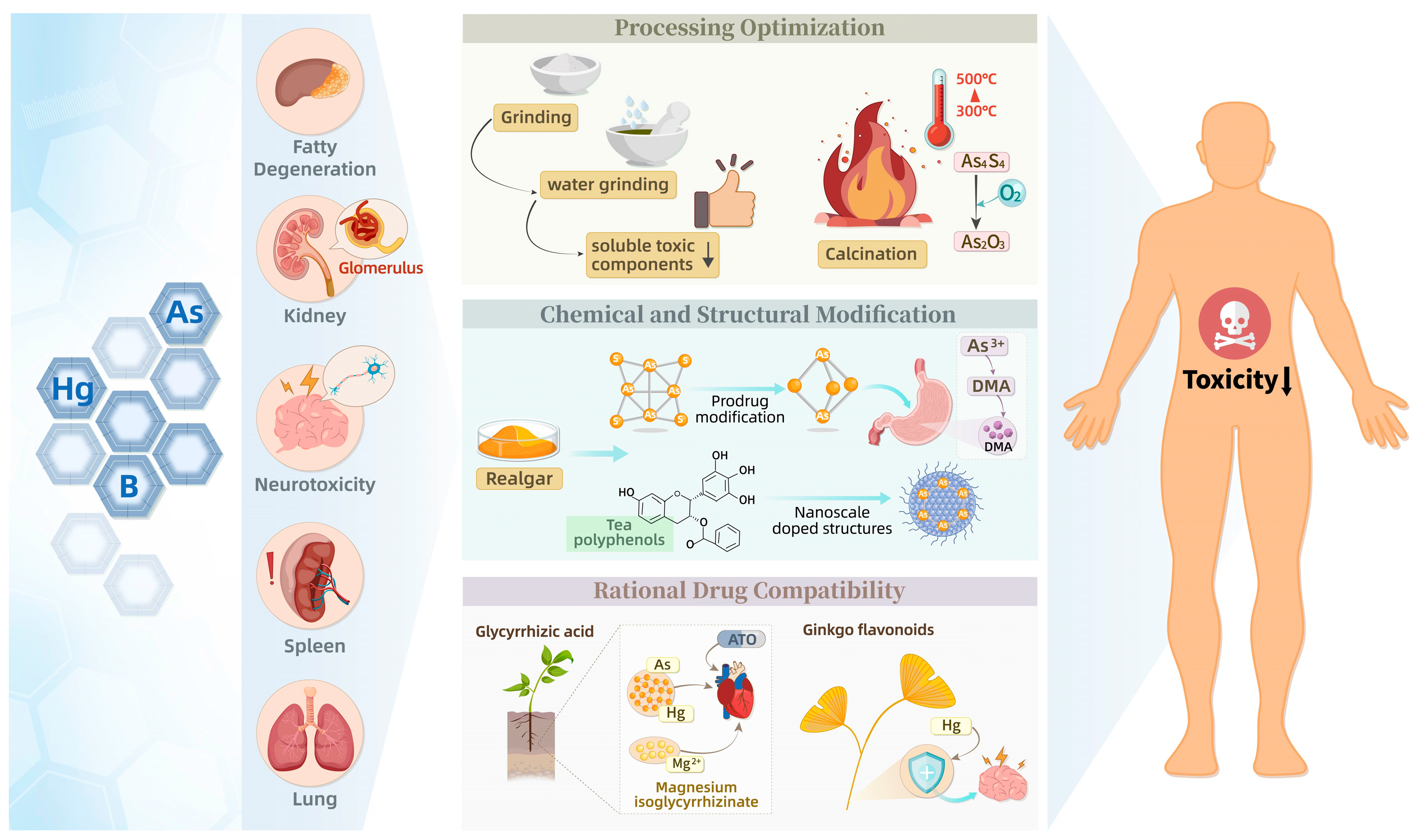
5. Toxicity of Mineral Drugs and Detoxification Strategies
5.1. Toxicity
5.2. Detoxification Strategies
5.2.1. Processing Optimization
5.2.2. Rational Drug Compatibility
5.2.3. Chemical and Structural Modification
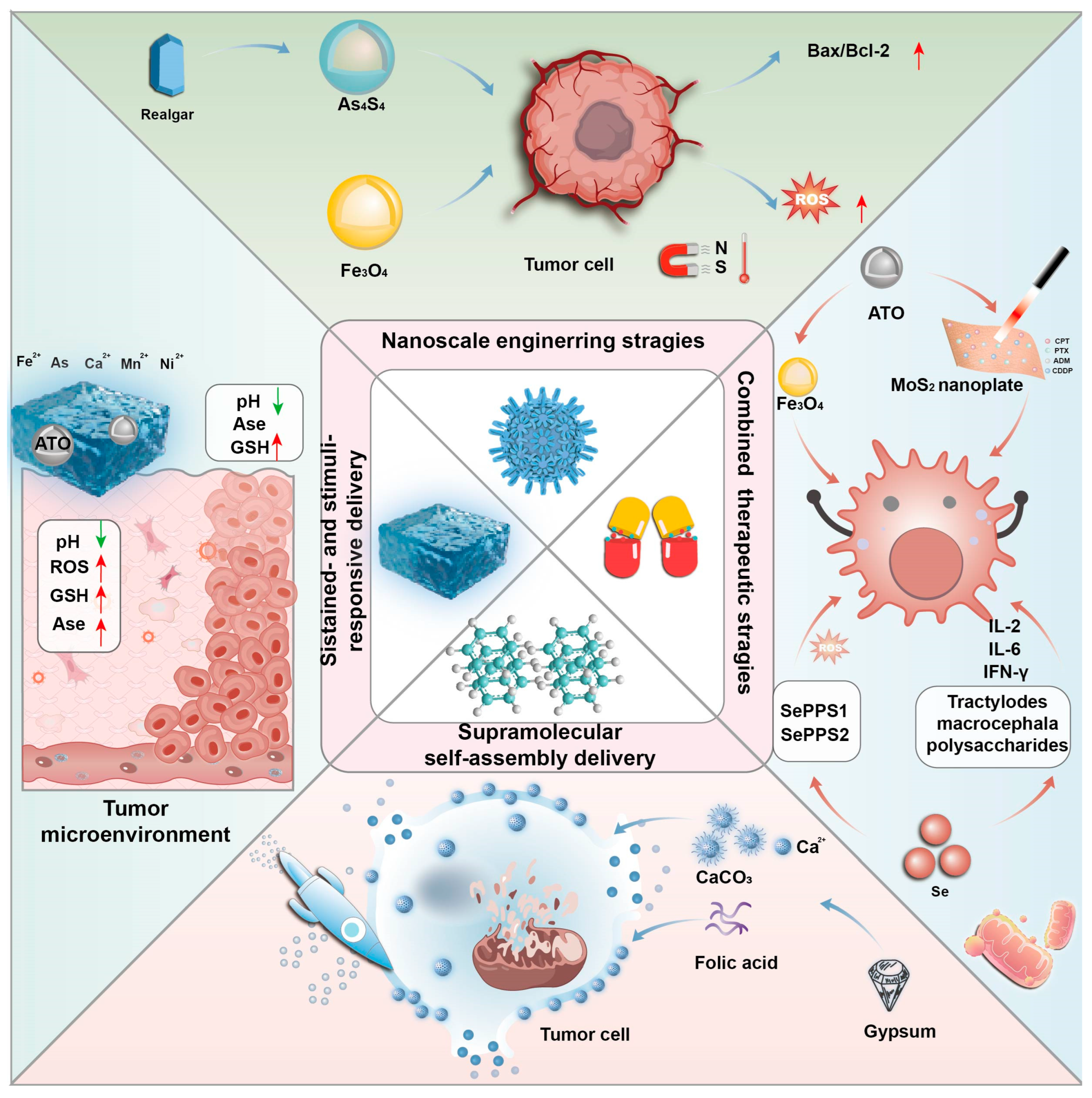
6. Innovative Delivery and Development of Antitumor Mineral Drugs
6.1. Nanoscale Engineering Strategies
6.2. Sustained- and Stimuli-Responsive Delivery
6.3. Supramolecular Self-Assembly Delivery
6.4. Combined Therapeutic Strategies
7. Conclusions, Limitations, and Future Perspectives
Author Contributions
Funding
Institutional Review Board Statement
Informed Consent Statement
Data Availability Statement
Acknowledgments
Conflicts of Interest
Abbreviations
| ALT | Alanine Aminotransferase |
| APL | Acute Promyelocytic Leukemia |
| APS | Advanced Planning and Scheduling |
| AST | Aspartate Aminotransferase |
| ATO | Arsenic Trioxide |
| ATRA | All-Trans Retinoic Acid |
| AUC | Area Under the Curve |
| CAM | Chorioallantoic Membrane |
| CHDT | Compound Huangdai Tablet |
| CI | Contrast Index |
| Cr | Platinum (Il)-based complex |
| CRC | Colorectal Cancer |
| DFS | Disease-free Survival |
| ECG | Electrocardiogram |
| EFS | Sevent-free Survival |
| EGCG-RNPs | (-)-Epigallocatechin-3-Gallate Encapsulated Realgar Nanoparticles |
| EZH2 | Enhancer of Zeste Homolog 2 |
| FoxO3a | Forkhead Box O3a |
| FXR | Farnesoid X Receptor |
| GPx | Glutathione Peroxidase |
| GPX4 | Glutathione Peroxidase 4 |
| GSH | Glutathione |
| GEM-MNP-pHLIP | G-PON Encapsulation Mode-Magnetic Nanoparticle-PH-Low Insertion Peptide |
| HCC | Hepatocellular Carcinoma |
| HgS | Mercuric Sulfide |
| IC50 | Half Maximal Inhibitory Concentration |
| ICD | Immunogenic Cell Death |
| IFN-γ | Interferon-γ |
| IL-6 | Interleukin-6 |
| KM | Kunming |
| LD50 | Median Lethal Dose |
| LRP | Lipoprotein Receptor-related Protein |
| MAPK | Mitogen-activated Protein Kinase |
| MDM2 | Mouse Double Minute 2 Homolog |
| MMP-2 | Matrix Metalloproteinase-2 |
| MPE | Malignant Pleural Effusion |
| mPTP | Mitochondrial Permeability Transition Pore |
| MRI | Magnetic Resonance Imaging |
| N/A | Not Applicable |
| NDs | Nanodiamonds |
| NR | Nanodiamonds Not Reported |
| NRA | Nano-realgar |
| PEG | Polyethylene glycol |
| PK | Pharmacokinetics |
| PLGA | Poly (lactic-co-glycolic acid) |
| PML | Promyelocytic Leukemia |
| rhG-CSF | Recombinant Human Granulocyte Colony-Stimulating Factor |
| RT | Radiotherapy |
| RTS | Realgar-containing Serum |
| RIF | Realgar-Indigo Naturalis Formula |
| ROS | Reactive Oxygen Species |
| SeNPs | Selenium Nanoparticles |
| SPIONs | Superparamagnetic Iron Oxide Nanoparticles |
| T1/2 | Half-life |
| TAR | Tumor Area Ratio |
| TCM | Traditional Chinese Medicine |
| TME | Tumor Microenvironment |
| TNF-α | Tumor Necrosis Factor-α |
| TSIIA@SeNPs-APS | TCM active ingredient-based SeNP surface decorated with APS and loaded with TSIIA |
| WHO | World Health Organization |
| ZIF-8 | Zeolitic Imidazolate Framework-8 |
| 5-FU | 5-fluorouracil |
References
- Zhong, X.; Di, Z.; Xu, Y.; Liang, Q.; Feng, K.; Zhang, Y.; Di, L.; Wang, R. Mineral medicine: From traditional drugs to multifunctional delivery systems. Chin. Med. 2022, 17, 21. [Google Scholar] [CrossRef] [Scilit] [PubMed]
- Photos-Jones, E.; Christidis, G.E.; Piochi, M.; Keane, C.; Mormone, A.; Balassone, G.; Perdikatsis, V.; Leanord, A. Testing Greco-Roman medicinal minerals: The case of solfataric alum. J. Archaeol. Sci. Rep. 2016, 10, 82–95. [Google Scholar] [CrossRef] [Scilit]
- Zhou, X.; Sun, H.; Ren, J.; Yan, G.; Yang, L.; Zhang, H.; Lu, H.; Li, X.; Makino, T.; Yin, F.; et al. Mineral crude drug mirabilite (Mangxiao) inhibits the occurrence of colorectal cancer by regulating the Lactobacillus-bile acid-intestinal farnesoid X receptor axis based on multiomics integration analysis. MedComm 2024, 5, e556. [Google Scholar] [CrossRef] [Scilit] [PubMed]
- Bai, D.; Nowak, M.; Lu, D.; Wang, Q.; Fitzgerald, M.; Zhang, H.; MacDonald, R.; Xu, Z.; Luo, L. The outcast of medicine: Metals in medicine--from traditional mineral medicine to metallodrugs. Front. Pharmacol. 2025, 16, 1542560. [Google Scholar] [CrossRef] [Scilit] [PubMed]
- Sanz, M.A.; Fenaux, P.; Tallman, M.S.; Estey, E.H.; Löwenberg, B.; Naoe, T.; Lengfelder, E.; Döhner, H.; Burnett, A.K.; Chen, S.J.; et al. Management of acute promyelocytic leukemia: Updated recommendations from an expert panel of the European LeukemiaNet. Blood 2019, 133, 1630–1643. [Google Scholar] [CrossRef] [Scilit] [PubMed]
- Xiang, K.; Wang, P.; Liu, Y.; Chen, Y.; Liu, D.; Han, Y.; Shen, A.; Xu, F.; Yu, W.; Wei, L.; et al. Ellagic Acid Alleviates Ulcerative Colitis in a GPR35-Dependent Manner Associated with SDH Restoration. Phytomedicine 2026, 155, 158130. [Google Scholar] [CrossRef] [Scilit] [PubMed]
- Su, T.H.; Cao, J.; Ding, X.X.; Guo, X.Z.; Chen, A.L.; Zhou, W.H.; Deng, P.; Luo, P.; Chen, Y.; Wang, F. Galangin Overcomes Gefitinib and Sotorasib Resistance in Non-small Cell Lung Cancer by Inhibiting Efferocytosis. J. Agric. Food Chem. 2026, 74, 12892–12905. [Google Scholar] [CrossRef] [Scilit] [PubMed]
- Ao, Q.; Tang, L.; Bao, Y.; Wang, M.; Zhu, J.; Chen, Y.; Zhu, H.; Gao, J. Plant-derived natural compounds targeting drug resistance in ovarian cancer: Molecular mechanisms and therapeutic perspectives. Phytomedicine 2026, 155, 158065. [Google Scholar] [CrossRef] [Scilit] [PubMed]
- Iyer, S.G.; Elias, L.; Stanchina, M.; Watts, J. The treatment of acute promyelocytic leukemia in 2023: Paradigm, advances, and future directions. Front. Oncol. 2022, 12, 1062524. [Google Scholar] [CrossRef] [Scilit] [PubMed]
- Luo, S.; Tian, J.; Sun, X.; Wu, F.; Liu, Y.; Wan, W.; She, Z.; Wen, C. Oral Realgar-Indigo Naturalis Formula Treatment for Acute Promyelocytic Leukemia in Children: A Randomized, Control Clinical Trial. Evid. Based Complement Altern. Med. 2022, 2022, 8314176. [Google Scholar] [CrossRef] [Scilit]
- Qian, G.; Xiong, L.; Ye, Q. Hydroxyapatite-based carriers for tumor targeting therapy. RSC Adv. 2023, 13, 16512–16528. [Google Scholar] [CrossRef] [Scilit] [PubMed]
- Yang, S.; Raza, F.; Li, K.; Qiu, Y.; Su, J.; Qiu, M. Maximizing arsenic trioxide’s anticancer potential: Targeted nanocarriers for solid tumor therapy. Colloids Surf. B Biointerfaces 2024, 241, 114014. [Google Scholar] [CrossRef] [Scilit] [PubMed]
- Wen, J.; Li, A.; Wang, Z.; Guo, X.; Zhang, G.; Litzow, M.R.; Liu, Q. Hepatotoxicity induced by arsenic trioxide: Clinical features, mechanisms, preventive and potential therapeutic strategies. Front. Pharmacol. 2025, 16, 1536388. [Google Scholar] [CrossRef] [Scilit] [PubMed]
- Zhang, M.; Zhu, J.; Bao, Y.; Ao, Q.; Mao, X.; Qiu, Z.; Zhang, Y.; Chen, Y.; Zhu, H.; Gao, J. Bevacizumab in ovarian cancer therapy: Current advances, clinical challenges, and emerging strategies. Front. Bioeng. Biotechnol. 2025, 13, 1589841. [Google Scholar] [CrossRef] [Scilit] [PubMed]
- Guan, H.; Xu, Y.; Ma, C.; Zhao, D. Pharmacology, Toxicology, and Rational Application of Cinnabar, Realgar, and Their Formulations. Evid. Based Complement Altern. Med. 2022, 2022, 6369150. [Google Scholar] [CrossRef] [Scilit]
- Miodragović, Ð.; Swindell, E.P.; Waxali, Z.S.; Bogachkov, A.; O’Halloran, T.V. Beyond Cisplatin: Combination Therapy with Arsenic Trioxide. Inorganica Chim. Acta 2019, 496, 119030. [Google Scholar] [CrossRef] [Scilit] [PubMed]
- Long, Q.; Zhao, X.; Gao, L.; Liu, M.; Pan, F.; Gao, X.; Zhan, C.; Chen, Y.; Wang, J.; Qian, J. Effects of Surface IR783 Density on the In Vivo Behavior and Imaging Performance of Liposomes. Pharmaceutics 2024, 16, 744. [Google Scholar] [CrossRef] [Scilit] [PubMed]
- Liu, J.; Fan, H.; Liang, X.; Chen, Y. Polycomb repressor complex: Its function in human cancer and therapeutic target strategy. BioMed Pharmacother. 2023, 169, 115897. [Google Scholar] [CrossRef] [Scilit] [PubMed]
- Duan, X.; Li, T.; Han, X.; Ren, J.; Chen, P.; Li, H.; Gong, S. The antitumor effect of arsenic trioxide on hepatocellular carcinoma is enhanced by andrographolide. Oncotarget 2017, 8, 90905–90915. [Google Scholar] [CrossRef] [Scilit] [PubMed]
- Hung, K.C.; Chang, L.C.; Ho, C.N.; Hsu, C.W.; Wu, J.Y.; Lin, Y.T.; Chen, I.W. Influence of Intravenous Magnesium Sulfate Infusion on the Subjective Postoperative Quality of Recovery: A Meta-Analysis of Randomized Controlled Trials. Nutrients 2024, 16, 2375. [Google Scholar] [CrossRef] [Scilit] [PubMed]
- Xie, Q.; Yu, L.; Wang, X.; Wu, Z.; Zhi, D.; Yang, J.; Guo, Z.; Wu, T.; Sun, Y.; Zhao, L.; et al. A novel realgar-indigo naturalis formula more effectively induces apoptosis in NB4 cells. Pak. J. Pharm. Sci. 2019, 32, 957–962. [Google Scholar] [PubMed]
- Krutsch, A.D.; Tudoran, C.; Motofelea, A.C. New Insights into the Assessment of Peri-Operative Risk in Women Undergoing Surgery for Gynecological Neoplasms: A Call for a New Tool. Medicina 2024, 60, 1679. [Google Scholar] [CrossRef] [Scilit] [PubMed]
- Wei, L.; Xiang, K.; Kang, H.; Yu, Y.; Fan, H.; Zhou, H.; Hou, T.; Ge, Y.; Wang, J.; Guo, Z.; et al. Development and Characterization of Fluorescent Probes for the G Protein-Coupled Receptor 35. ACS Med. Chem. Lett. 2023, 14, 411–416. [Google Scholar] [CrossRef] [Scilit] [PubMed]
- Azmoonfar, R.; Moslehi, M.; Shahbazi-Gahrouei, D. Radioprotective Effect of Selenium Nanoparticles: A Mini Review. IET Nanobiotechnol 2024, 2024, 5538107. [Google Scholar] [CrossRef] [Scilit] [PubMed]
- Zhang, H.; Jiang, M.; Gao, L.; Lin, Z. The clinical efficacy of external application of mirabilite and rhubarb combined with intrathoracic chemotherapy in treating malignant pleural effusion: A prospective, randomized, controlled clinical trial. Medicine 2021, 100, e24758. [Google Scholar] [CrossRef] [Scilit] [PubMed]
- Yang, M.; Wang, L.; Zhang, T.; Zhu, A.; Sun, Y.; Zhao, J.; Liu, D.; Wang, Q.; Zeng, K. Different proteomic profiles of cinnabar upon therapeutic and toxic exposure reveal distinctive biological manifestations. J. Ethnopharmacol. 2020, 253, 112668. [Google Scholar] [CrossRef] [Scilit] [PubMed]
- Tuncer, C.; Hacioglu, C. Borax induces ferroptosis of glioblastoma by targeting HSPA5/NRF2/GPx4/GSH pathways. J. Cell Mol. Med. 2024, 28, e18206. [Google Scholar] [CrossRef] [Scilit] [PubMed]
- Tang, S.; Shen, Y.; Wei, X.; Shen, Z.; Lu, W.; Xu, J. Olaparib synergizes with arsenic trioxide by promoting apoptosis and ferroptosis in platinum-resistant ovarian cancer. Cell Death Dis. 2022, 13, 826. [Google Scholar] [CrossRef] [Scilit] [PubMed]
- Wang, X.; Cheng, S.; Xu, Y.; Zheng, T.; Ling, C.; Du, J. Arsenic Trioxide Enhances the Efficacy of PD-1 Inhibitors in Hepatocellular Carcinoma by Inducing Immunogenic Cell Death via the ROS/ERS Pathway. Immun. Inflamm. Dis. 2025, 13, e70214. [Google Scholar] [CrossRef] [Scilit] [PubMed]
- Dai, L.; Yu, P.; Fan, H.; Xia, W.; Zhao, Y.; Zhang, P.; Zhang, J.Z.H.; Zhang, H.; Chen, Y. Identification and Validation of New DNA-PKcs Inhibitors through High-Throughput Virtual Screening and Experimental Verification. Int. J. Mol. Sci. 2024, 25, 7982. [Google Scholar] [CrossRef] [Scilit] [PubMed]
- Lu, M.; Xia, L.; Luo, D.; Waxman, S.; Jing, Y. Dual effects of glutathione-S-transferase pi on As2O3 action in prostate cancer cells: Enhancement of growth inhibition and inhibition of apoptosis. Oncogene 2004, 23, 3945–3952. [Google Scholar] [CrossRef] [Scilit] [PubMed]
- Liu, J.; Shi, J.Z.; Yu, L.M.; Goyer, R.A.; Waalkes, M.P. Mercury in traditional medicines: Is cinnabar toxicologically similar to common mercurials? Exp. Biol. Med. 2008, 233, 810–817. [Google Scholar] [CrossRef] [Scilit]
- Chen, Z.; Ye, S.Y.; Zhu, R.G. The extraordinary transformation of traditional Chinese medicine: Processing with liquid excipients. Pharm. Biol. 2020, 58, 561–573. [Google Scholar] [CrossRef] [Scilit] [PubMed]
- Sun, H.; Zhang, H.L.; Zhang, A.H.; Zhou, X.H.; Wang, X.Q.; Han, Y.; Yan, G.L.; Liu, L.; Wang, X.J. Network pharmacology combined with functional metabolomics discover bile acid metabolism as a promising target for mirabilite against colorectal cancer. RSC Adv. 2018, 8, 30061–30070. [Google Scholar] [CrossRef] [Scilit] [PubMed]
- Wang, H.Y.; Gong, S.; Li, G.H.; Yao, Y.Z.; Zheng, Y.S.; Lu, X.H.; Wei, S.H.; Qin, W.W.; Liu, H.B.; Wang, M.C.; et al. An effective and chemotherapy-free strategy of all-trans retinoic acid and arsenic trioxide for acute promyelocytic leukemia in all risk groups (APL15 trial). Blood Cancer J. 2022, 12, 158. [Google Scholar] [CrossRef] [Scilit] [PubMed]
- Thomas, X. Acute Promyelocytic Leukemia: A History over 60 Years-From the Most Malignant to the most Curable Form of Acute Leukemia. Oncol. Ther. 2019, 7, 33–65. [Google Scholar] [CrossRef] [Scilit] [PubMed]
- Sun, H.; Zhang, A.H.; Zhang, H.L.; Zhou, X.H.; Wang, X.Q.; Liu, L.; Wang, X.J. Ultra-performance liquid chromatography/mass spectrometry technology and high-throughput metabolomics for deciphering the preventive mechanism of mirabilite on colorectal cancer via the modulation of complex metabolic networks. RSC Adv. 2019, 9, 35356–35363. [Google Scholar] [CrossRef] [Scilit] [PubMed]
- Zhang, H.; Zhang, S.H.; Hu, J.L.; Wu, Y.T.; Ma, X.Y.; Chen, Y.; Yu, B.; Liao, S.; Huang, H.; Gao, S. Structural functional characterization of multiple myeloma associated cytoplasmic poly(A) polymerase FAM46C. Cancer Commun. 2021, 41, 615–630. [Google Scholar] [CrossRef] [Scilit]
- Merkel, O.; Heyder, C.; Asslaber, D.; Hamacher, F.; Tinhofer, I.; Holler, C.; Stocher, M.; Prokesch, A.; Papak, C.; Scheideler, M.; et al. Arsenic trioxide induces apoptosis preferentially in B-CLL cells of patients with unfavourable prognostic factors including del17p13. J. Mol. Med. 2008, 86, 541–552. [Google Scholar] [CrossRef] [Scilit] [PubMed]
- Chen, J.; Wen, J.; Fu, Y.; Li, X.; Huang, J.; Guan, X.; Zhou, Y. A bifunctional bortezomib-loaded porous nano-hydroxyapatite/alginate scaffold for simultaneous tumor inhibition and bone regeneration. J. Nanobiotechnol. 2023, 21, 174. [Google Scholar] [CrossRef] [Scilit] [PubMed]
- Chin, L.; Kumana, C.R.; Kwong, Y.L.; Gill, H. The Development and Clinical Applications of Oral Arsenic Trioxide for Acute Promyelocytic Leukaemia and Other Diseases. Pharmaceutics 2022, 14, 1945. [Google Scholar] [CrossRef] [Scilit] [PubMed]
- Beauchamp, E.M.; Ringer, L.; Bulut, G.; Sajwan, K.P.; Hall, M.D.; Lee, Y.C.; Peaceman, D.; Ozdemirli, M.; Rodriguez, O.; Macdonald, T.J.; et al. Arsenic trioxide inhibits human cancer cell growth and tumor development in mice by blocking Hedgehog/GLI pathway. J. Clin. Invest 2011, 121, 148–160. [Google Scholar] [CrossRef] [Scilit] [PubMed]
- Jiang, L.; Wang, L.; Chen, L.; Cai, G.H.; Ren, Q.Y.; Chen, J.Z.; Shi, H.J.; Xie, Y.H. As2O3 induces apoptosis in human hepatocellular carcinoma HepG2 cells through a ROS-mediated mitochondrial pathway and activation of caspases. Int. J. Clin. Exp. Med. 2015, 8, 2190–2196. [Google Scholar] [PubMed]
- Wang, F.; Ding, X.X.; Su, T.H.; Cao, J.; Li, F.Q.; Dong, Z.S.; Guo, X.Z.; Zhang, Y.; Chen, Y.; Yang, D.H.; et al. GPR35-mediated metabolic reprogramming promotes tumorigenesis in digestive cancers. Front. Immunol. 2025, 16, 1668471. [Google Scholar] [CrossRef] [Scilit] [PubMed]
- Hayashi, T.; Hideshima, T.; Akiyama, M.; Richardson, P.; Schlossman, R.L.; Chauhan, D.; Munshi, N.C.; Waxman, S.; Anderson, K.C. Arsenic trioxide inhibits growth of human multiple myeloma cells in the bone marrow microenvironment. Mol. Cancer Ther. 2002, 1, 851–860. [Google Scholar] [PubMed]
- Rao, Y.; Li, R.; Zhang, D. A drug from poison: How the therapeutic effect of arsenic trioxide on acute promyelocytic leukemia was discovered. Sci. China Life Sci. 2013, 56, 495–502. [Google Scholar] [CrossRef] [Scilit] [PubMed]
- Dai, M.; Wang, D. Mineral medicines of the East: An analysis of records in historical Chinese and Japanese medical texts. Front. Pharmacol. 2025, 16, 1584500. [Google Scholar] [CrossRef] [Scilit] [PubMed]
- Norn, S.; Permin, H.; Kruse, E.; Kruse, P.R. Mercury—A major agent in the history of medicine and alchemy. Dan Med. Arb. 2008, 36, 21–40. [Google Scholar]
- Sun, Y.; Wang, Z.; Gong, P.; Yao, W.; Ba, Q.; Wang, H. Review on the health-promoting effect of adequate selenium status. Front. Nutr. 2023, 10, 1136458. [Google Scholar] [CrossRef] [Scilit] [PubMed]
- Pal, A.; Dhar, A.; Shamim, M.A.; Rani, I.; Negi, R.R.; Sharma, A.; Chatterjee, N.; Goyal, A.; Kaur, B.; Tondolo, V.; et al. Selenium levels in colorectal cancer: A systematic review and meta-analysis of serum, plasma, and colorectal specimens. J. Trace Elem. Med. Biol. 2024, 84, 127429. [Google Scholar] [CrossRef] [Scilit] [PubMed]
- Chen, S.; Qin, W.; Lu, X.; Liu, L.; Zheng, Y.; Lu, X.; Wang, X.; Zhang, X.; Gong, S.; Wei, S.; et al. Arsenic trioxide versus Realgar-Indigo naturalis formula in non-high-risk acute promyelocytic leukemia: A multicenter, randomized trial. Haematologica 2025, 110, 621–628. [Google Scholar] [PubMed]
- Jimenez, J.J.; Chale, R.S.; Abad, A.C.; Schally, A.V. Acute promyelocytic leukemia (APL): A review of the literature. Oncotarget 2020, 11, 992–1003. [Google Scholar] [CrossRef] [Scilit] [PubMed]
- Kar, E.; Övenler, Z.; Hacıoğlu, C.; Kar, F. Boric Acid Induces Oxidative Damage and Apoptosis Through SEMA3A/PLXNA1/NRP1 Signalling Pathway in U251 Glioblastoma Cell. J. Cell Mol. Med. 2025, 29, e70578. [Google Scholar] [CrossRef] [Scilit] [PubMed]
- Tao, L.; Fu, J.; Wang, F.; Song, Y.; Li, Y.; Zhang, J.; Wang, Z. The application of mirabilite in traditional Chinese medicine and its chemical constituents, processing methods, pharmacology, toxicology and clinical research. Front. Pharmacol. 2023, 14, 1293097. [Google Scholar] [CrossRef] [Scilit] [PubMed]
- Westley, R.; De Fonseka, D.; Bedawi, E.O. Updates in the management of malignant pleural effusion: A clinical practice review. J. Thorac. Dis. 2025, 17, 6283–6295. [Google Scholar] [CrossRef] [Scilit] [PubMed]
- Yu, Z.; Zhao, X.; Qiu, S.; Zhou, S.; Li, P.; Liu, N. Hyperthermic Intraperitoneal Chemotherapy May Damage Renal Function and Cause Serum Electrolyte Disturbance: A Retrospective Observational Study. Surg. Laparosc. Endosc. Percutan Tech 2023, 33, 302–309. [Google Scholar] [CrossRef] [Scilit] [PubMed]
- Ferguson, H.J.; Ferguson, C.I.; Speakman, J.; Ismail, T. Management of intestinal obstruction in advanced malignancy. Ann. Med. Surg. 2015, 4, 264–270. [Google Scholar] [CrossRef] [Scilit]
- Hu, J.; Cao, J.; Huang, S.; Chen, Y. ITGAX promotes gastric cancer progression via epithelial-mesenchymal transition pathway. Front. Pharmacol. 2025, 15, 1536478. [Google Scholar] [CrossRef] [Scilit] [PubMed]
- Fan, H.; Zhao, H.; Zheng, Y.; Chen, G.; Ji, Y.; Yu, W.; Yan, J.; Yang, H.; Liang, X.; Chen, Y. Polygonati kingianum polysaccharide alleviates dextran sulfate sodium-induced colitis by modulating gut microbiota and metabolic homeostasis. Int. J. Biol. Macromol. 2025, 316, 144836. [Google Scholar] [CrossRef] [Scilit] [PubMed]
- Zou, P.; Zhang, K.; Jin, Y.; Liu, G. The Efficacy of Magnesium Sulfate (MgSO4) Wet Dressing in Reducing Eyelid Swelling and Bruising after Blepharoplasty: A Randomized, Controlled, and Observer-Blinded Assessment Study. Facial Plast. Surg. 2024, 40, 46–51. [Google Scholar] [PubMed]
- Gaur, B.; Yadav, A.; Jain, A.; Mangla, Y. Refractory scrotal oedema in a child with nephrotic syndrome subsided on magnesium sulphate with glycerine (SU-MAG) application: A rare case report. Curr. Med. Res. Pract. 2025, 15, 35–37. [Google Scholar] [CrossRef] [Scilit]
- Liu, Q.; Liu, Z.; Zhang, C.; Xu, Y.; Li, X.; Gao, H. Effects of 3% Boric Acid Solution on Cutaneous Candida albicans Infection and Microecological Flora Mice. Front. Microbiol. 2021, 12, 709880. [Google Scholar] [CrossRef] [Scilit] [PubMed]
- Luo, Y.; Feng, M.; Fan, Z.; Zhu, X.; Jin, F.; Li, R.; Wu, J.; Yang, X.; Jiang, Q.; Bai, H.; et al. Effect of Kangfuxin Solution on Chemo/Radiotherapy-Induced Mucositis in Nasopharyngeal Carcinoma Patients: A Multicenter, Prospective Randomized Phase III Clinical Study. Evid. Based Complement Altern. Med. 2016, 2016, 8692343. [Google Scholar] [CrossRef] [Scilit]
- Hazman, Ö.; Bozkurt, M.F.; Fidan, A.F.; Uysal, F.E.; Çelik, S. The Effect of Boric Acid and Borax on Oxidative Stress, Inflammation, ER Stress and Apoptosis in Cisplatin Toxication and Nephrotoxicity Developing as a Result of Toxication. Inflammation 2018, 41, 1032–1048. [Google Scholar] [CrossRef] [Scilit] [PubMed]
- Wu, Y.; Zhang, P.; Fan, H.; Zhang, C.; Yu, P.; Liang, X.; Chen, Y. GPR35 acts a dual role and therapeutic target in inflammation. Front. Immunol. 2023, 14, 1254446. [Google Scholar] [CrossRef] [Scilit] [PubMed]
- Pan, W.; Cai, S.; Latour, J.M.; Zhong, M.; Lv, M.; Li, J.; Zhang, X.; Zhang, Y. External use of Mirabilite combined with Lactulose improves postoperative gastrointestinal mobility among older patients undergoing abdominal surgery. J. Adv. Nurs. 2021, 77, 755–762. [Google Scholar] [CrossRef] [Scilit] [PubMed]
- Jansen, A.; de Jong, A.; Hoogendam, J.P.; Baeten, I.G.T.; Jürgenliemk-Schulz, I.M.; Zweemer, R.P.; Gerestein, C.G. Lymphocele following lymph node dissection in cervical and endometrial cancer: A systematic review and meta-analysis. Gynecol. Oncol. 2023, 170, 273–281. [Google Scholar] [CrossRef] [Scilit] [PubMed]
- Wu, J.; Shao, Y.; Liu, J.; Chen, G.; Ho, P.C. The medicinal use of realgar (As4S4) and its recent development as an anticancer agent. J. Ethnopharmacol. 2011, 135, 595–602. [Google Scholar] [CrossRef] [Scilit] [PubMed]
- Liu, J.; Wei, L.X.; Wang, Q.; Lu, Y.F.; Zhang, F.; Shi, J.Z.; Li, C.; Cherian, M.G. A review of cinnabar (HgS) and/or realgar (As4S4)-containing traditional medicines. J. Ethnopharmacol. 2018, 210, 340–350. [Google Scholar] [CrossRef] [Scilit] [PubMed]
- Lu, Y.F.; Yan, J.W.; Wu, Q.; Shi, J.Z.; Liu, J.; Shi, J.S. Realgar- and cinnabar-containing an-gong-niu-huang wan (AGNH) is much less acutely toxic than sodium arsenite and mercuric chloride. Chem. Biol. Interact. 2011, 189, 134–140. [Google Scholar] [CrossRef] [Scilit] [PubMed]
- Huang, D.P.; Yang, L.C.; Chen, Y.Q.; Wan, W.Q.; Zhou, D.H.; Mai, H.R.; Li, W.L.; Yang, L.H.; Lan, H.K.; Chen, H.Q.; et al. Long-term outcome of children with acute promyelocytic leukemia: A randomized study of oral versus intravenous arsenic by SCCLG-APL group. Blood Cancer J. 2023, 13, 178. [Google Scholar] [CrossRef] [Scilit] [PubMed]
- Lou, Y.; Ma, Y.; Jin, J.; Zhu, H. Oral Realgar-Indigo Naturalis Formula Plus Retinoic Acid for Acute Promyelocytic Leukemia. Front. Oncol. 2020, 10, 597601. [Google Scholar] [CrossRef] [Scilit] [PubMed]
- Jung, E.J.; Kim, H.J.; Shin, S.C.; Kim, G.S.; Jung, J.M.; Hong, S.C.; Kim, C.W.; Lee, W.S. Tetraarsenic Hexoxide Enhanced the Anticancer Effects of Artemisia annua, L. Polyphenols by Inducing Autophagic Cell Death and Apoptosis in Oxalplatin-Resistant HCT116 Colorectal Cancer Cells. Int. J. Mol. Sci. 2025, 26, 7661. [Google Scholar] [CrossRef] [Scilit] [PubMed]
- Catanzaro, E.; Feron, O.; Skirtach, A.G.; Krysko, D.V. Immunogenic Cell Death and Role of Nanomaterials Serving as Therapeutic Vaccine for Personalized Cancer Immunotherapy. Front. Immunol. 2022, 13, 925290. [Google Scholar] [CrossRef] [Scilit] [PubMed]
- Qian, X.; Wang, Y.; Liu, Z.; Fang, F.; Ma, Y.; Zhou, L.; Pan, Y.; Meng, X.; Yan, B.; Zhu, X.; et al. Establishment of XRD fourier fingerprint identification method of realgar decoction pieces and its anti-tumor activity in tumor-in-situ transplanted mice. J. Ethnopharmacol. 2024, 331, 118303. [Google Scholar] [CrossRef] [Scilit] [PubMed]
- Zhao, Q.; Chen, Y.Y.; Xu, D.Q.; Yue, S.J.; Fu, R.J.; Yang, J.; Xing, L.M.; Tang, Y.P. Action Mode of Gut Motility, Fluid and Electrolyte Transport in Chronic Constipation. Front. Pharmacol. 2021, 12, 630249. [Google Scholar] [CrossRef] [Scilit] [PubMed]
- Zhang, H.L.; Zhang, A.H.; Zhou, X.H.; Sun, H.; Wang, X.Q.; Liang, L.; Wang, X.J. High-throughput lipidomics reveal mirabilite regulating lipid metabolism as anticancer therapeutics. RSC Adv. 2018, 8, 35600–35610. [Google Scholar] [CrossRef] [Scilit] [PubMed]
- Fan, H.; Zhao, H.; Zhang, P.; Yu, P.; Ji, Y.; Chen, G.; Jin, H.; Liu, Y.; Liu, J.; Chen, Z.S.; et al. Homoisoflavanone Delays Colorectal Cancer Progression via DNA Damage-Induced Mitochondrial Apoptosis and Parthanatos-Like Cell Death. Adv. Sci. 2026, 13, e11406. [Google Scholar] [CrossRef] [Scilit]
- Qu, R.; Zhang, Y.; Kim, B.; Zeng, G.; Wang, P.; Shaoyong, W.; Huang, Y.; Guo, W.; Chen, Y.; Wang, P.; et al. Microbial riboflavin inhibits ceramide synthase 3 to lower ceramide (d18:1/26:0) and delay colorectal cancer progression. Cell Metab. 2025, 37, 1852–1869. [Google Scholar] [CrossRef] [Scilit] [PubMed]
- Lo-Coco, F.; Avvisati, G.; Vignetti, M.; Thiede, C.; Orlando, S.M.; Iacobelli, S.; Ferrara, F.; Fazi, P.; Cicconi, L.; Di Bona, E.; et al. Retinoic acid and arsenic trioxide for acute promyelocytic leukemia. N. Engl. J. Med. 2013, 369, 111–121. [Google Scholar] [CrossRef] [Scilit] [PubMed]
- Zhang, X.W.; Yan, X.J.; Zhou, Z.R.; Yang, F.F.; Wu, Z.Y.; Sun, H.B.; Liang, W.X.; Song, A.X.; Lallemand-Breitenbach, V.; Jeanne, M.; et al. Arsenic trioxide controls the fate of the PML-RARalpha oncoprotein by directly binding, PML. Science 2010, 328, 240–243. [Google Scholar] [CrossRef] [Scilit] [PubMed]
- Chen, J.; Jin, Z.; Zhang, S.; Zhang, X.; Li, P.; Yang, H.; Ma, Y. Arsenic trioxide elicits prophylactic and therapeutic immune responses against solid tumors by inducing necroptosis and ferroptosis. Cell Mol. Immunol. 2023, 20, 51–64. [Google Scholar] [PubMed]
- Li, X.; Pan, Y.F.; Chen, Y.B.; Wan, Q.Q.; Lin, Y.K.; Shang, T.Y.; Xu, M.Y.; Jiang, T.Y.; Pei, M.M.; Tan, Y.X.; et al. Arsenic trioxide augments immunogenic cell death and induces cGAS-STING-IFN pathway activation in hepatocellular carcinoma. Cell Death Dis. 2024, 15, 300. [Google Scholar] [CrossRef] [Scilit] [PubMed]
- Walker, S.R.; Jamieson, H.E.; Lanzirotti, A.; Hall, G.E.M.; Peterson, R.C. The effect of ore roasting on arsenic oxidation state and solid phase speciation in gold mine tailings. Geochem. Explor. Environ. Anal. 2015, 15, 273–291. [Google Scholar] [CrossRef] [Scilit]
- Lin, C.J.; Su, Y.C.; Lee, C.H.; Li, T.C.; Chen, Y.A.; Lin, S.J. Bai-hu-tang, ancient chinese medicine formula, may provide a new complementary treatment option for sepsis. Evid. Based Complement Altern. Med. 2013, 2013, 193084. [Google Scholar] [CrossRef] [Scilit]
- Wang, L.J.; Lu, J.Z.; Cai, B.N.; Li, M.W.; Qu, B.L. Effect of compound Zhuye Shigao Granule ( ) on acute radiation-induced esophagitis in cancer patients: A randomized controlled trial. Chin. J. Integr. Med. 2017, 23, 98–104. [Google Scholar] [PubMed]
- Gupta, H.; Pandey, A.; Agarwal, R.; Mehra, H.; Gupta, S.; Gupta, N.; Kumar, A. Application of calcium sulfate as graft material in implantology and maxillofacial procedures: A review of literature. Natl. J. Maxillofac. Surg. 2024, 15, 183–187. [Google Scholar] [CrossRef] [Scilit] [PubMed]
- Lun, D.X.; Li, S.Y.; Li, N.N.; Mou, L.M.; Li, H.Q.; Zhu, W.P.; Li, H.F.; Hu, Y.C. Limitations and modifications in the clinical application of calcium sulfate. Front. Surg. 2024, 11, 1278421. [Google Scholar] [CrossRef] [Scilit] [PubMed]
- Fukugaichi, S.; Matsue, N. One-Step Synthesis of Calcium Sulfate Hemihydrate Nanofibers from Calcite at Room Temperature. ACS Omega 2018, 3, 2820–2824. [Google Scholar] [CrossRef] [Scilit] [PubMed]
- Liu, J.; Dong, F.; Tan, H.; Zhang, H.; Zhou, L.; He, P.; Zhou, L.; Feng, C.; Li, R. Facile preparation of high-strength α-CaSO4·0.5H2O regulated by maleic acid from phosphogypsum: Experimental and molecular dynamics simulation studies. SN Appl. Sci. 2020, 2, 1645. [Google Scholar] [CrossRef] [Scilit]
- Du, M.; Wang, J.; Dong, F.; Wang, Z.; Yang, F.; Tan, H.; Fu, K.; Wang, W. The study on the effect of flotation purification on the performance of α-hemihydrate gypsum prepared from phosphogypsum. Sci. Rep. 2022, 12, 95. [Google Scholar] [CrossRef] [Scilit] [PubMed]
- Daugėlaitė, G.; Užkuraitytė, K.; Jagelavičienė, E.; Filipauskas, A. Prevention and Treatment of Chemotherapy and Radiotherapy Induced Oral Mucositis. Medicina 2019, 55, 25. [Google Scholar] [CrossRef] [Scilit] [PubMed]
- Zhao, M.; Li, Y.; Wang, Z. Mercury and Mercury-Containing Preparations: History of Use, Clinical Applications, Pharmacology, Toxicology, and Pharmacokinetics in Traditional Chinese Medicine. Front. Pharmacol. 2022, 13, 807807. [Google Scholar] [CrossRef] [Scilit] [PubMed]
- Abdullah, K.M.; Kaushal, J.B.; Takkar, S.; Sharma, G.; Alsafwani, Z.W.; Pothuraju, R.; Batra, S.K.; Siddiqui, J.A. Copper metabolism and cuproptosis in human malignancies: Unraveling the complex interplay for therapeutic insights. Heliyon 2024, 10, e27496. [Google Scholar] [CrossRef] [Scilit] [PubMed]
- Zhong, C.; Li, N.; Wang, S.; Li, D.; Yang, Z.; Du, L.; Huang, G.; Li, H.; Yeung, W.S.; He, S.; et al. Targeting osteoblastic 11beta-HSD1 to combat high-fat diet-induced bone loss and obesity. Nat. Commun. 2024, 15, 8588. [Google Scholar] [CrossRef] [Scilit] [PubMed]
- Ge, E.J.; Bush, A.I.; Casini, A.; Cobine, P.A.; Cross, J.R.; DeNicola, G.M.; Dou, Q.P.; Franz, K.J.; Gohil, V.M.; Gupta, S.; et al. Connecting copper and cancer: From transition metal signalling to metalloplasia. Nat. Rev. Cancer 2022, 22, 102–113. [Google Scholar] [PubMed]
- Vinceti, M.; Filippini, T.; Del Giovane, C.; Dennert, G.; Zwahlen, M.; Brinkman, M.; Zeegers, M.P.; Horneber, M.; D’Amico, R.; Crespi, C.M. Selenium for preventing cancer. Cochrane Database Syst. Rev. 2018, 1, Cd005195. [Google Scholar] [PubMed]
- Rao, S.; Lin, Y.; Lin, R.; Liu, J.; Wang, H.; Hu, W.; Chen, B.; Chen, T. Traditional Chinese medicine active ingredients-based selenium nanoparticles regulate antioxidant selenoproteins for spinal cord injury treatment. J. Nanobiotechnol. 2022, 20, 278. [Google Scholar] [CrossRef] [Scilit]
- Liu, R.; Rong, G.; Liu, Y.; Huang, W.; He, D.; Lu, R. Delivery of apigenin-loaded magnetic Fe2O3/Fe3O4@mSiO2 nanocomposites to A549 cells and their antitumor mechanism. Mater. Sci. Eng. C Mater. Biol. Appl. 2021, 120, 111719. [Google Scholar] [CrossRef] [Scilit] [PubMed]
- Li, L.; Ge, F.; Jiang, Y.; An, Z.; Li, N.; Zhang, Z.; Liu, H.; Li, J.; Liang, D. Research Status and Development Trends of Inorganic Salt Lake Resource Extraction Based on Bibliometric Analysis. Sustainability 2024, 17, 121. [Google Scholar] [CrossRef] [Scilit]
- Anjum, S.; Hashim, M.; Imran, M.; Babur, S.; Adnan, S.; Hano, C.; Ibrahim, W.N. Selenium Nanoparticles in Cancer Therapy: Unveiling Cytotoxic Mechanisms and Therapeutic Potential. Cancer Rep. 2025, 8, e70210. [Google Scholar] [CrossRef] [Scilit]
- Hoshyar, N.; Gray, S.; Han, H.; Bao, G. The effect of nanoparticle size on in vivo pharmacokinetics and cellular interaction. nomedicine 2016, 11, 673–692. [Google Scholar] [CrossRef] [Scilit]
- Cavalu, S.; Abdelhamid, A.M.; Saber, S.; Elmorsy, E.A.; Hamad, R.S.; Abdel-Reheim, M.A.; Yahya, G.; Salama, M.M. Cell cycle machinery in oncology: A comprehensive review of therapeutic targets. FASEB J. 2024, 38, e23734. [Google Scholar] [CrossRef] [Scilit] [PubMed]
- Wang, Z.; Chen, Y.; Huang, A.; Wen, H.; Wu, Y.; Xu, X.; Qiao, Z.; Chen, L.; Zhao, Y.; Liang, X. Design, synthesis and biological evaluation of novel beta-caryophyllene derivatives as potential anti-cancer agents through the ROS-mediated apoptosis pathway. RSC Med. Chem. 2025, 16, 3174–3189. [Google Scholar] [CrossRef] [Scilit] [PubMed]
- Hu, J.L.; Hua, Y.J.; Chen, Y.; Yu, B.; Gao, S. Structural analysis of tumor-related single amino acid mutations in human MxA protein. Chin. J. Cancer 2015, 34, 583–593. [Google Scholar] [CrossRef] [Scilit] [PubMed]
- He, Z.; Tian, T.; Guo, D.; Wu, H.; Chen, Y.; Zhang, Y.; Wan, Q.; Zhao, H.; Wang, C.; Shen, H.; et al. Cytoplasmic retention of a nucleocytoplasmic protein TBC1D3 by microtubule network is required for enhanced EGFR signaling. PLoS ONE 2014, 9, e94134. [Google Scholar] [CrossRef] [Scilit] [PubMed]
- Luo, L.; Xin, X.; Wang, Q.; Wei, M.; Huang, N.; Gao, S.; Gu, X.; Li, R. Characterization and comparation of toxicity between natural realgar and artificially optimized realgar. Front. Pharmacol. 2024, 15, 1476139. [Google Scholar] [CrossRef] [Scilit] [PubMed]
- Park, J.W.; Choi, Y.J.; Jang, M.A.; Baek, S.H.; Lim, J.H.; Passaniti, T.; Kwon, T.K. Arsenic trioxide induces G2/M growth arrest and apoptosis after caspase-3 activation and bcl-2 phosphorylation in promonocytic U937 cells. Biochem Biophys. Res. Commun. 2001, 286, 726–734. [Google Scholar] [CrossRef] [Scilit] [PubMed]
- Wang, L.; Zhou, G.B.; Liu, P.; Song, J.H.; Liang, Y.; Yan, X.J.; Xu, F.; Wang, B.S.; Mao, J.H.; Shen, Z.X.; et al. Dissection of mechanisms of Chinese medicinal formula Realgar-Indigo naturalis as an effective treatment for promyelocytic leukemia. Proc. Natl. Acad. Sci. USA 2008, 105, 4826–4831. [Google Scholar] [CrossRef] [Scilit] [PubMed]
- Qi, K.; Li, Y.; Huang, K.; Xiong, X.; Chuchu, F.; Zhang, C.; Weng, W. Pre-application of arsenic trioxide may potentiate cytotoxic effects of vinorelbine/docetaxel on neuroblastoma SK-N-SH cells. BioMed Pharmacother. 2019, 113, 108665. [Google Scholar] [CrossRef] [Scilit] [PubMed]
- Wang, Y.S.; Yin, J.Z.; Shi, X.Q.; Zhao, X.W.; Li, B.; Yang, M.H. MDM2-mediated Inhibitory Effect of Arsenic Trioxide on Small Cell Lung Cancer Cell Line by Degrading Mutant p53. Anticancer Agents Med. Chem. 2023, 23, 1829–1837. [Google Scholar] [CrossRef] [Scilit] [PubMed]
- Kumar, S.; Yedjou, C.G.; Tchounwou, P.B. Arsenic trioxide induces oxidative stress, DNA damage, and mitochondrial pathway of apoptosis in human leukemia (HL-60) cells. J. Exp. Clin. Cancer Res. 2014, 33, 42. [Google Scholar] [CrossRef] [Scilit] [PubMed]
- Sun, S.; Shen, J.; Jiang, J.; Wang, F.; Min, J. Targeting ferroptosis opens new avenues for the development of novel therapeutics. Signal Transduct. Target Ther. 2023, 8, 372. [Google Scholar] [CrossRef] [Scilit] [PubMed]
- Li, S.; Yuan, H.; Chen, Y.; Guo, Z. Metal complexes induced ferroptosis for anticancer therapy. Fundam. Res. 2023, 3, 525–528. [Google Scholar] [CrossRef] [Scilit] [PubMed]
- Su, M.; Liu, X.; Ma, Y.; Peng, X.; Xiong, X.; Weng, W.; Huang, K.; Li, Y. Arsenic trioxide induces ferroptosis in neuroblastoma by mediating GPX4 transcriptional inhibition. Clin. Transl. Sci. 2024, 17, e13716. [Google Scholar] [CrossRef] [Scilit] [PubMed]
- Wu, B.; Tan, M.; Cai, W.; Wang, B.; He, P.; Zhang, X. Arsenic trioxide induces autophagic cell death in osteosarcoma cells via the ROS-TFEB signaling pathway. Biochem Biophys. Res. Commun. 2018, 496, 167–175. [Google Scholar] [CrossRef] [Scilit] [PubMed]
- Wu, P.J.; Hsin, I.L.; Hung, W.L.; Lee, M.S.; Wang, P.H.; Ko, J.L. Combination treatment with cyclosporin A and arsenic trioxide induce synergistic cell death via non-apoptotic pathway in uterine cervical cancer cells. Chem. Biol. Interact. 2022, 368, 110177. [Google Scholar] [CrossRef] [Scilit] [PubMed]
- Lee, S.; Hwang, N.; Seok, B.G.; Lee, S.; Lee, S.J.; Chung, S.W. Autophagy mediates an amplification loop during ferroptosis. Cell Death Dis. 2023, 14, 464. [Google Scholar] [CrossRef] [Scilit] [PubMed]
- Szwed, M.; Poczta-Krawczyk, A.; Bukowski, K.; Marczak, A. Nanoparticle-Mediated Ferroptosis for Cancer Therapy: Mechanisms and Therapeutic Strategies. Nanotechnol. Sci. Appl. 2025, 18, 445–470. [Google Scholar] [CrossRef] [Scilit] [PubMed]
- Liu, X.; Li, X.; Wang, L.; Lv, X.; Chen, N.; Li, P.; Lu, K.; Wang, X. Realgar induces apoptosis in the chronic lymphocytic leukemia cell line MEC-1. Mol. Med. Rep. 2013, 8, 1866–1870. [Google Scholar] [CrossRef] [Scilit] [PubMed]
- Chen, Q.; Hao, M.; Wang, L.; Li, L.; Chen, Y.; Shao, X.; Tian, Z.; Pfuetzner, R.A.; Zhong, Q.; Brunger, A.T.; et al. Prefused lysosomes cluster on autophagosomes regulated by VAMP8. Cell Death Dis. 2021, 12, 939. [Google Scholar] [CrossRef] [Scilit] [PubMed]
- Chen, Q.; Shao, X.; Tian, Z.; Chen, Y.; Mondal, P.; Liu, F.; Wang, F.; Ling, P.; He, W.; Zhang, K.; et al. Nanoscale monitoring of mitochondria and lysosome interactions for drug screening and discovery. Nano Res. 2019, 12, 1009–1015. [Google Scholar] [CrossRef] [Scilit]
- Cao, Y.L.; Meng, S.; Chen, Y.; Feng, J.X.; Gu, D.D.; Yu, B.; Li, Y.J.; Yang, J.Y.; Liao, S.; Chan, D.C.; et al. MFN1 structures reveal nucleotide-triggered dimerization critical for mitochondrial fusion. Nature 2017, 542, 372–376. [Google Scholar] [CrossRef] [Scilit] [PubMed]
- Korotkov, S.M. Mitochondrial Oxidative Stress Is the General Reason for Apoptosis Induced by Different-Valence Heavy Metals in Cells and Mitochondria. Int. J. Mol. Sci. 2023, 24, 14459. [Google Scholar] [CrossRef] [Scilit] [PubMed]
- An, X.; Yu, W.; Liu, J.; Tang, D.; Yang, L.; Chen, X. Oxidative cell death in cancer: Mechanisms and therapeutic opportunities. Cell Death Dis. 2024, 15, 556. [Google Scholar] [CrossRef] [Scilit] [PubMed]
- Paz, G.S.; Fernandes, J. Natural compounds targeting mitochondrial metabolism in cancer therapy: A literature review. Chin. Clin. Oncol. 2025, 14, 56. [Google Scholar] [CrossRef] [Scilit] [PubMed]
- Vázquez Cervantes, G.I.; González Esquivel, D.F.; Ramírez Ortega, D.; Blanco Ayala, T.; Ramos Chávez, L.A.; López-López, H.E.; Salazar, A.; Flores, I.; Pineda, B.; Gómez-Manzo, S.; et al. Mechanisms Associated with Cognitive and Behavioral Impairment Induced by Arsenic Exposure. Cells 2023, 12, 2537. [Google Scholar] [CrossRef] [Scilit] [PubMed]
- Punshon, L.D.; Fabbrizi, M.R.; Phoenix, B.; Green, S.; Parsons, J.L. Current Insights into the Radiobiology of Boron Neutron Capture Therapy and the Potential for Further Improving Biological Effectiveness. Cells 2024, 13, 2065. [Google Scholar] [CrossRef] [Scilit] [PubMed]
- Yao, C.; Wu, S.; Kong, J.; Sun, Y.; Bai, Y.; Zhu, R.; Li, Z.; Sun, W.; Zheng, L. Angiogenesis in hepatocellular carcinoma: Mechanisms and anti-angiogenic therapies. Cancer Biol. Med. 2023, 20, 25–43. [Google Scholar] [CrossRef] [Scilit] [PubMed]
- Jiang, F.; Li, Y.; Si, L.; Zhang, Z.; Li, Z. Interaction of EZH2 and P65 is involved in the arsenic trioxide-induced anti-angiogenesis in human triple-negative breast cancer cells. Cell Biol. Toxicol. 2019, 35, 361–371. [Google Scholar] [CrossRef] [Scilit] [PubMed]
- Sun, Z.; Li, M.; Bai, L.; Fu, J.; Lu, J.; Wu, M.; Zhou, C.; Zhang, Y.; Wu, Y. Arsenic trioxide inhibits angiogenesis in vitro and in vivo by upregulating FoxO3a. Toxicol. Lett. 2019, 315, 1–8. [Google Scholar] [CrossRef] [Scilit] [PubMed]
- Song, P.; Hai, Y.; Wang, X.; Zhao, L.; Chen, B.; Cui, P.; Xie, Q.; Yu, L.; Li, Y.; Wu, Z.; et al. Realgar transforming solution suppresses angiogenesis and tumor growth by inhibiting VEGF receptor 2 signaling in vein endothelial cells. Arch. Pharm. Res. 2018, 41, 467–480. [Google Scholar] [CrossRef] [Scilit] [PubMed]
- Hu, M.; Yang, J.L.; Teng, H.; Jia, Y.Q.; Wang, R.; Zhang, X.W.; Wu, Y.; Luo, Y.; Chen, X.C.; Zhang, R.; et al. Anti-angiogenesis therapy based on the bone marrow-derived stromal cells genetically engineered to express sFlt-1 in mouse tumor model. BMC Cancer 2008, 8, 306. [Google Scholar] [CrossRef] [Scilit] [PubMed]
- Kepp, O.; Pan, H.; Liu, P.; Kroemer, G. Arsenic trioxide as an inducer of immunogenic cell death. Oncoimmunology 2023, 12, 2174723. [Google Scholar] [CrossRef] [Scilit] [PubMed]
- Alex, A.A.; Ganesan, S.; Palani, H.K.; Balasundaram, N.; David, S.; Lakshmi, K.M.; Kulkarni, U.P.; Nisham, P.N.; Korula, A.; Devasia, A.J.; et al. Arsenic Trioxide Enhances the NK Cell Cytotoxicity Against Acute Promyelocytic Leukemia While Simultaneously Inhibiting Its Bio-Genesis. Front. Immunol. 2018, 9, 1357. [Google Scholar] [CrossRef] [Scilit] [PubMed]
- Chen, C.; Su, Q.; Zi, M.; Hua, X.; Zhang, Z. Harnessing gut microbiota for colorectal cancer therapy: From clinical insights to therapeutic innovations. npj Biofilms Microbiomes 2025, 11, 190. [Google Scholar] [CrossRef] [Scilit] [PubMed]
- Cariello, M.; Zerlotin, R.; Pasculli, E.; Piccinin, E.; Peres, C.; Porru, E.; Roda, A.; Gadaleta, R.M.; Moschetta, A. Intestinal FXR Activation via Transgenic Chimera or Chemical Agonism Prevents Colitis-Associated and Genetically-Induced Colon Cancer. Cancers 2022, 14, 3081. [Google Scholar] [CrossRef] [Scilit] [PubMed]
- Li, Y.; Liu, M.; Kong, B.; Zhang, G.; Zhang, Q. The role of selenium intervention in gut microbiota homeostasis and gene function in mice with breast cancer on a high-fat diet. Front. Microbiol. 2024, 15, 1439652. [Google Scholar] [CrossRef] [Scilit] [PubMed]
- Su, Y.; Cai, X.; Fan, X.; Ning, J.; Shen, M. Effect of Trace Element Selenium on the Intestinal Microbial Community in Nude Mice with Colorectal Cancer. Microorganisms 2024, 12, 1336. [Google Scholar] [CrossRef] [Scilit] [PubMed]
- Coryell, M.; McAlpine, M.; Pinkham, N.V.; McDermott, T.R.; Walk, S.T. The gut microbiome is required for full protection against acute arsenic toxicity in mouse models. Nat. Commun. 2018, 9, 5424. [Google Scholar] [CrossRef] [Scilit] [PubMed]
- Chi, L.; Xue, J.; Tu, P.; Lai, Y.; Ru, H.; Lu, K. Gut microbiome disruption altered the biotransformation and liver toxicity of arsenic in mice. Arch. Toxicol. 2019, 93, 25–35. [Google Scholar] [PubMed]
- Lu, K.; Abo, R.P.; Schlieper, K.A.; Graffam, M.E.; Levine, S.; Wishnok, J.S.; Swenberg, J.A.; Tannenbaum, S.R.; Fox, J.G. Arsenic exposure perturbs the gut microbiome and its metabolic profile in mice: An integrated metagenomics and metabolomics analysis. Environ. Health Perspect. 2014, 122, 284–291. [Google Scholar] [CrossRef] [Scilit] [PubMed]
- Paganini, D.; Zimmermann, M.B. The effects of iron fortification and supplementation on the gut microbiome and diarrhea in infants and children: A review. Am. J. Clin. Nutr. 2017, 106, 1688s–1693s. [Google Scholar] [CrossRef] [Scilit] [PubMed]
- Kortman, G.A.; Boleij, A.; Swinkels, D.W.; Tjalsma, H. Iron availability increases the pathogenic potential of Salmonella typhimurium and other enteric pathogens at the intestinal epithelial interface. PLoS ONE 2012, 7, e29968. [Google Scholar] [CrossRef] [Scilit] [PubMed]
- Cholewińska, E.; Ognik, K.; Fotschki, B.; Zduńczyk, Z.; Juśkiewicz, J. Comparison of the effect of dietary copper nanoparticles and one copper (II) salt on the copper biodistribution and gastrointestinal and hepatic morphology and function in a rat model. PLoS ONE 2018, 13, e0197083. [Google Scholar] [CrossRef] [Scilit] [PubMed]
- Ganie, S.Y.; Javaid, D.; Hajam, Y.A.; Reshi, M.S. Arsenic toxicity: Sources, pathophysiology and mechanism. Toxicol. Res. 2024, 13, tfad111. [Google Scholar]
- Khaleda, L.; Alam, M.M.; Tasnim, Z.; Ezaj, M.M.A.; Apu, M.A.R.; Akter, R.; Bakar, M.A.; Alam, M.J.; Chowdhury, R.H.; Datta, A.; et al. Detrimental effects of chronic arsenic exposure through daily diet on hepatic and renal health: An animal model study. Toxicol. Rep. 2025, 14, 101993. [Google Scholar] [CrossRef] [Scilit] [PubMed]
- Concessao, P.L.; Prakash, J. Arsenic-induced nephrotoxicity: Mechanisms, biomarkers, and preventive strategies for global health. Vet. World 2025, 18, 2136–2157. [Google Scholar] [CrossRef] [Scilit] [PubMed]
- Li, J.; Zhao, L.; Zhang, Y.; Li, W.; Duan, X.; Chen, J.; Guo, Y.; Yang, S.; Sun, G.; Li, B. Imbalanced immune responses involving inflammatory molecules and immune-related pathways in the lung of acute and subchronic arsenic-exposed mice. Environ. Res. 2017, 159, 381–393. [Google Scholar] [CrossRef] [Scilit] [PubMed]
- Gochfeld, M.; Burger, J. Mercury interactions with selenium and sulfur and the relevance of the Se:Hg molar ratio to fish consumption advice. Environ. Sci. Pollut. Res. Int. 2021, 28, 18407–18420. [Google Scholar] [CrossRef] [Scilit] [PubMed]
- Kang, B.; Wang, J.; Guo, S.; Yang, L. Mercury-induced toxicity: Mechanisms, molecular pathways, and gene regulation. Sci. Total Env. 2024, 943, 173577. [Google Scholar] [CrossRef] [Scilit]
- Yorifuji, T. Lessons From an Early-stage Epidemiological Study of Minamata Disease. J. Epidemiol. 2020, 30, 12–14. [Google Scholar] [CrossRef] [Scilit] [PubMed]
- Hadrup, N.; Frederiksen, M.; Sharma, A.K. Toxicity of boric acid, borax and other boron containing compounds: A review. Regul. Toxicol. Pharmacol. 2021, 121, 104873. [Google Scholar] [CrossRef] [Scilit] [PubMed]
- Guo, Y.S.; Zuo, T.T.; Chen, A.Z.; Wang, Z.; Jin, H.Y.; Wei, F.; Li, P.; Ma, S.C. Progress in quality control, detection techniques, speciation and risk assessment of heavy metals in marine traditional Chinese medicine. Chin. Med. 2023, 18, 73. [Google Scholar] [CrossRef] [Scilit] [PubMed]
- Bai, K.; Hong, B.; He, J.; Hong, Z.; Tan, R. Preparation and antioxidant properties of selenium nanoparticles-loaded chitosan microspheres. Int. J. Nanomed. 2017, 12, 4527–4539. [Google Scholar] [CrossRef] [Scilit]
- Tang, H.; Xu, M.; Zhou, X.; Zhang, Y.; Zhao, L.; Ye, G.; Shi, F.; Lv, C.; Li, Y. Acute toxicity and biodistribution of different sized copper nano-particles in rats after oral administration. Mater. Sci. Eng. C Mater. Biol. Appl. 2018, 93, 649–663. [Google Scholar] [CrossRef] [Scilit] [PubMed]
- Bolt, H.M.; Başaran, N.; Duydu, Y. Effects of boron compounds on human reproduction. Arch. Toxicol. 2020, 94, 717–724. [Google Scholar] [CrossRef] [Scilit] [PubMed]
- Huang, Y.; Hsu, J.C.; Koo, H.; Cormode, D.P. Repurposing ferumoxytol: Diagnostic and therapeutic applications of an FDA-approved nanoparticle. Theranostics 2022, 12, 796–816. [Google Scholar] [CrossRef] [Scilit] [PubMed]
- Han, T.; Zhang, H.; Xu, W.; Li, C.; Wang, M.; Bai, Y.; Yang, L.; Zhang, S.; Jia, Z.; Xu, X.; et al. Study on the Mechanism of Reducing Hepatotoxicity of Water-Grinding Realgar by Metabolomics, Morphology, and Chemical Analysis. Evid. Based Complement Altern. Med. 2021, 2021, 8538287. [Google Scholar] [CrossRef] [Scilit]
- Deng, Z.; Yin, X.; Zhang, S.; Fang, H.; Gao, S.; Liu, Y.; Jiang, X.; Song, G.; Jiang, W.; Wang, L. Study on arsenic speciation, bioaccessibility, and gut microbiota in realgar-containing medicines by DGT technique and artificial gastrointestinal extraction (PBET) combine with simulated human intestinal microbial ecosystem (SHIME). J. Hazard Mater. 2024, 463, 132863. [Google Scholar] [CrossRef] [Scilit] [PubMed]
- Ollson, C.J.; Smith, E.; Scheckel, K.G.; Betts, A.R.; Juhasz, A.L. Assessment of arsenic speciation and bioaccessibility in mine-impacted materials. J. Hazard Mater. 2016, 313, 130–137. [Google Scholar] [CrossRef] [Scilit] [PubMed]
- Zheng, B.; Yang, Y.; Li, J.; Li, J.; Zuo, S.; Chu, X.; Xu, S.; Ma, D.; Chu, L. Magnesium Isoglycyrrhizinate Alleviates Arsenic Trioxide-Induced Cardiotoxicity: Contribution of Nrf2 and TLR4/NF-κB Signaling Pathway. Drug Des. Devel Ther. 2021, 15, 543–556. [Google Scholar] [CrossRef] [Scilit] [PubMed]
- Kesti, S.; Macar, O.; Kalefetoğlu Macar, T.; Çavuşoğlu, K.; Yalçın, E. Investigation of the protective role of Ginkgo biloba, L. against phytotoxicity, genotoxicity and oxidative damage induced by Trifloxystrobin. Sci. Rep. 2024, 14, 19937. [Google Scholar] [CrossRef] [Scilit] [PubMed]
- Huang, Q.; Wang, T.; Xiong, Y.; Qu, L.; Yin, Q.; Zou, W. Safety and efficacy of Compound Huangdai Tablets combined with all-trans retinoic acid for treatment of acute promyelocytic leukemia: Clinical evidence and potential mechanisms. Chin. Herb. Med. 2022, 14, 154–165. [Google Scholar] [CrossRef] [Scilit] [PubMed]
- Fang, W.; Peng, Z.L.; Dai, Y.J.; Wang, D.L.; Huang, P.; Huang, H.P. (-)-Epigallocatechin-3-gallate encapsulated realgar nanoparticles exhibit enhanced anticancer therapeutic efficacy against acute promyelocytic leukemia. Drug Deliv. 2019, 26, 1058–1067. [Google Scholar] [CrossRef] [Scilit] [PubMed]
- El-Ghiaty, M.A.; El-Kadi, A.O.S. The Duality of Arsenic Metabolism: Impact on Human Health. Annu Rev. Pharmacol. Toxicol. 2023, 63, 341–358. [Google Scholar] [CrossRef] [Scilit] [PubMed]
- Lucic, B.; Franciscato, D.S.; Nogueira, H.P.; Gallucci, L.; Silveira Junior, A.T.; Ismail, A.M.; Robinson, M.; Dallinger, T.; Gutfleisch, C.; Kurz, J.; et al. Fast pH-Driven Solubilization Method of Realgar (As4S4) to Reduce the Toxicity of Arsenic [As(III)] for Medicinal Purposes. Adv. Sci. 2025, 12, e2502740. [Google Scholar] [CrossRef] [Scilit]
- Abbas, G.; Murtaza, B.; Bibi, I.; Shahid, M.; Niazi, N.K.; Khan, M.I.; Amjad, M.; Hussain, M. Arsenic Uptake, Toxicity, Detoxification, and Speciation in Plants: Physiological, Biochemical, and Molecular Aspects. Int. J. Environ. Res. Public Health 2018, 15, 59. [Google Scholar] [CrossRef] [Scilit] [PubMed]
- Dzade, N.Y.; Roldan, A.; de Leeuw, N.H. Structures and Properties of As(OH)3 Adsorption Complexes on Hydrated Mackinawite (FeS) Surfaces: A DFT-D2 Study. Environ. Sci. Technol. 2017, 51, 3461–3470. [Google Scholar] [CrossRef] [Scilit] [PubMed]
- Wang, S.; Liu, C.; Wang, C.; Ma, J.; Xu, H.; Guo, J.; Deng, Y. Arsenic trioxide encapsulated liposomes prepared via copper acetate gradient loading method and its antitumor efficiency. Asian J. Pharm. Sci. 2020, 15, 365–373. [Google Scholar] [CrossRef] [Scilit] [PubMed]
- Kong, D.; Jiang, T.; Liu, J.; Jiang, X.; Liu, B.; Lou, C.; Zhao, B.; Carroll, S.L.; Feng, G. Chemoembolizing hepatocellular carcinoma with microsphere cored with arsenic trioxide microcrystal. Drug Deliv. 2020, 27, 1729–1740. [Google Scholar] [CrossRef] [Scilit] [PubMed]
- Wang, Y.; Wei, Y.; Wu, Y.; Zong, Y.; Song, Y.; Pu, S.; Wu, W.; Zhou, Y.; Xie, J.; Yin, H. Multifunctional Nano-Realgar Hydrogel for Enhanced Glioblastoma Synergistic Chemotherapy and Radiotherapy: A New Paradigm of an Old Drug. Int. J. Nanomed. 2023, 18, 743–763. [Google Scholar] [CrossRef] [Scilit]
- Sedky, N.K.; Fawzy, I.M.; Hassan, A.; Mahdy, N.K.; Attia, R.T.; Shamma, S.N.; Alfaifi, M.Y.; Elbehairi, S.E.; Mokhtar, F.A.; Fahmy, S.A. Innovative microwave-assisted biosynthesis of copper oxide nanoparticles loaded with platinum(ii) based complex for halting colon cancer: Cellular, molecular, and computational investigations. RSC Adv. 2024, 14, 4005–4024. [Google Scholar] [CrossRef] [PubMed]
- Dutta, G.; Chinnaiyan, S.K.; Palaniyandi, T.; Sugumaran, A.; Narayanasamy, D. Biogenic synthesized CuO nanoparticles and 5-fluorouracil loaded anticancer gel for HeLa cervical cancer cells. Discov. Nano 2024, 19, 217. [Google Scholar] [CrossRef] [Scilit] [PubMed]
- Yao, M.; Deng, Y.; Zhao, Z.; Yang, D.; Wan, G.; Xu, X. Selenium Nanoparticles Based on Morinda officinalis Polysaccharides: Characterization, Anti-Cancer Activities, and Immune-Enhancing Activities Evaluation In Vitro. Molecules 2023, 28, 2426. [Google Scholar] [CrossRef] [Scilit] [PubMed]
- Al-Duais, M.A.; El Rabey, H.A.; Mohammed, G.M.; Al-Awthan, Y.S.; Althiyabi, A.S.; Attia, E.S.; Rezk, S.M.; Tayel, A.A. The anticancer activity of fucoidan coated selenium nanoparticles and curcumin nanoparticles against colorectal cancer lines. Sci. Rep. 2025, 15, 287. [Google Scholar] [CrossRef] [Scilit] [PubMed]
- Wang, B.; Hu, S.; Teng, Y.; Chen, J.; Wang, H.; Xu, Y.; Wang, K.; Xu, J.; Cheng, Y.; Gao, X. Current advance of nanotechnology in diagnosis and treatment for malignant tumors. Signal Transduct. Target Ther. 2024, 9, 200. [Google Scholar] [CrossRef] [Scilit] [PubMed]
- Chen, Q.; Li, K.; Li, J.; Liu, X.; Li, J.; Xu, L.; Han, Y.; Zou, T.; Wang, X.; Yao, Y.; et al. Targeting Metal Ion Homeostasis for Regulated Cell Death-Amplified Tumor Nanomedicine. Int. J. Nanomed. 2025, 20, 15627–15654. [Google Scholar] [CrossRef] [Scilit]
- Luo, Z.; Chen, C.Y.; Li, S. Improving Tumor Targeting and Penetration for Nanoparticle-Mediated Cancer Therapy. Small Methods 2025, 9, e2401860. [Google Scholar] [CrossRef] [Scilit] [PubMed]
- Tian, Y.; Wang, X.; Xi, R.; Pan, W.; Jiang, S.; Li, Z.; Zhao, Y.; Gao, G.; Liu, D. Enhanced antitumor activity of realgar mediated by milling it to nanosize. Int. J. Nanomed. 2014, 9, 745–757. [Google Scholar] [CrossRef] [Scilit]
- Li, Y.; Zhang, H. Fe3O4-based nanotheranostics for magnetic resonance imaging-synergized multifunctional cancer management. Nanomedicine 2019, 14, 1493–1512. [Google Scholar] [CrossRef] [Scilit] [PubMed]
- Wang, Y.; Wu, X.; Bao, X.; Mou, X. Progress in the Mechanism of the Effect of Fe3O4 Nanomaterials on Ferroptosis in Tumor Cells. Molecules 2023, 28, 4562. [Google Scholar] [CrossRef] [Scilit] [PubMed]
- Niu, C.; Wang, Z.; Lu, G.; Krupka, T.M.; Sun, Y.; You, Y.; Song, W.; Ran, H.; Li, P.; Zheng, Y. Doxorubicin loaded superparamagnetic PLGA-iron oxide multifunctional microbubbles for dual-mode US/MR imaging and therapy of metastasis in lymph nodes. Biomaterials 2013, 34, 2307–2317. [Google Scholar] [CrossRef] [Scilit] [PubMed]
- Huang, Y.; Mao, K.; Zhang, B.; Zhao, Y. Superparamagnetic iron oxide nanoparticles conjugated with folic acid for dual target-specific drug delivery and MRI in cancer theranostics. Mater. Sci. Eng. C Mater. Biol. Appl. 2017, 70, 763–771. [Google Scholar] [CrossRef] [Scilit] [PubMed]
- Zhang, L.; Qiu, M.; Wang, R.; Li, S.; Liu, X.; Xu, Q.; Xiao, L.; Jiang, Z.X.; Zhou, X.; Chen, S. Monitoring ROSResponsive Fe3O4-based Nanoparticle Mediated Ferroptosis Immunotherapy 129Xe MRI. Angew. Chem. 2024, 63, e202403771. [Google Scholar] [CrossRef] [Scilit]
- Yu, M.; Zhang, Y.; Fang, M.; Jehan, S.; Zhou, W. Current Advances of Nanomedicines Delivering Arsenic Trioxide for Enhanced Tumor Therapy. Pharmaceutics 2022, 14, 743. [Google Scholar] [CrossRef] [Scilit] [PubMed]
- Xu, H.; Li, C.; Wei, Y.; Zheng, H.; Zheng, H.; Wang, B.; Piao, J.G.; Li, F. Angiopep-2-modified calcium arsenite-loaded liposomes for targeted and pH-responsive delivery for anti-glioma therapy. Biochem Biophys. Res. Commun. 2021, 551, 14–20. [Google Scholar] [CrossRef] [Scilit] [PubMed]
- Ettlinger, R.; Moreno, N.; Volkmer, D.; Kerl, K.; Bunzen, H. Zeolitic Imidazolate Framework-8 as pH-Sensitive Nanocarrier for “Arsenic Trioxide” Drug Delivery. Chemistry 2019, 25, 13189–13196. [Google Scholar] [CrossRef] [Scilit] [PubMed]
- Li, M.; Zhao, G.; Su, W.K.; Shuai, Q. Enzyme-Responsive Nanoparticles for Anti-tumor Drug Delivery. Front. Chem. 2020, 8, 647. [Google Scholar] [CrossRef] [Scilit]
- Zhou, W.; Jia, Y.; Liu, Y.; Chen, Y.; Zhao, P. Tumor Microenvironment-Based Stimuli-Responsive Nanoparticles for Controlled Release of Drugs in Cancer Therapy. Pharmaceutics 2022, 14, 2346. [Google Scholar] [CrossRef] [Scilit] [PubMed]
- Fan, H.; Zhao, H.; Gao, L.; Dong, Y.; Zhang, P.; Yu, P.; Ji, Y.; Chen, Z.S.; Liang, X.; Chen, Y. CCN1 Enhances Tumor Immunosuppression through Collagen-Mediated Chemokine Secretion in Pancreatic Cancer. Adv. Sci. 2025, 12, e2500589. [Google Scholar] [CrossRef] [Scilit]
- Guo, S.; Xie, X.; Chen, Y.; Liu, Y.; Luo, L. Editorial: Advances of novel approaches to enhance therapeutic efficacy and safety in human solid cold tumor. Front. Immunol. 2024, 15, 1398270. [Google Scholar] [CrossRef] [Scilit] [PubMed]
- Chen, D.Q.; Xie, Y.; Cao, L.Q.; Fleishman, J.S.; Chen, Y.; Wu, T.; Yang, D.H. The role of ABCC10/MRP7 in anti-cancer drug resistance and beyond. Drug Resist Updat 2024, 73, 101062. [Google Scholar] [CrossRef] [Scilit] [PubMed]
- Jin, Z.; Al Amili, M.; Guo, S. Tumor Microenvironment-Responsive Drug Delivery Based on Polymeric Micelles for Precision Cancer Therapy: Strategies and Prospects. Biomedicines 2024, 12, 417. [Google Scholar] [CrossRef] [Scilit] [PubMed]
- Yang, G.; Xu, L.; Chao, Y.; Xu, J.; Sun, X.; Wu, Y.; Peng, R.; Liu, Z. Hollow MnO2 as a tumor-microenvironment-responsive biodegradable nano-platform for combination therapy favoring antitumor immune responses. Nat. Commun. 2017, 8, 902. [Google Scholar] [CrossRef] [Scilit] [PubMed]
- Jin, Z.; Yi, X.; Yang, J.; Zhou, M.; Wu, P.; Yan, G. Liposome-Coated Arsenic-Manganese Complex for Magnetic Resonance Imaging-Guided Synergistic Therapy Against Carcinoma. Int. J. Nanomed. 2021, 16, 3775–3788. [Google Scholar] [CrossRef] [Scilit]
- Li, X.; Jian, M.; Sun, Y.; Zhu, Q.; Wang, Z. The Peptide Functionalized Inorganic Nanoparticles for Cancer-Related Bioanalytical and Biomedical Applications. Molecules 2021, 26, 3228. [Google Scholar] [CrossRef] [Scilit] [PubMed]
- Cai, X.; Zhu, Q.; Zeng, Y.; Zeng, Q.; Chen, X.; Zhan, Y. Manganese Oxide Nanoparticles As MRI Contrast Agents In Tumor Multimodal Imaging And Therapy. Int. J. Nanomed. 2019, 14, 8321–8344. [Google Scholar] [CrossRef] [Scilit]
- Liu, Y.; Wu, Y.; Luo, Z.; Li, M. Designing supramolecular self-assembly nanomaterials as stimuli-responsive drug delivery platforms for cancer therapy. iScience 2023, 26, 106279. [Google Scholar] [CrossRef] [Scilit] [PubMed]
- Lu, C.; Ma, Y.; Zhao, H.; Zheng, Y.; Zhao, Q.; Lin, L.; Chen, X.; Chen, L.; Li, G.; Chen, R. Exploration of the antiviral mechanism of gypsum-licorice compatibility pairing in Ma-Xing-Shi-Gan decoction from the perspective of metal-organic supramolecular interactions. Front. Med. Technol. 2025, 7, 1651390. [Google Scholar] [CrossRef] [Scilit] [PubMed]
- Zhang, Y.Z.; Pi, W.M.; Tan, X.R.; Xu, R.; Wang, X.; Xu, M.Y.; Huang, X.M.; Wang, P.L. Exploration of differences in decoction phase state, material form, and crystal form between Glycyrrhizae Radix et Rhizoma-Gypsum Fibrosum and Glycyrrhizae Radix et Rhizoma-CaSO4·2H2O based on supramolecules of traditional Chinese medicine. Zhongguo Zhong Yao Za Zhi 2025, 50, 412–421. [Google Scholar] [PubMed]
- Hattori, Y.; Nagaoka, Y.; Kubo, M.; Yamasaku, H.; Ishii, Y.; Okita, H.; Nakano, H.; Uesato, S.; Maitani, Y. Antitumor effect of liposomal histone deacetylase inhibitor-lipid conjugates in vitro. Chem. Pharm. Bull. 2011, 59, 1386–1392. [Google Scholar] [CrossRef] [Scilit]
- Wu, D.; Zhu, Z.Q.; Tang, H.X.; Shi, Z.E.; Kang, J.; Liu, Q.; Qi, J. Efficacy-shaping nanomedicine by loading Calcium Peroxide into Tumor Microenvironment-responsive Nanoparticles for the Antitumor Therapy of Prostate Cancer. Theranostics 2020, 10, 9808–9829. [Google Scholar] [CrossRef] [Scilit] [PubMed]
- Zhao, Y.; Yu, X.; Kong, W.; Kong, R.M.; Zhang, E.; Xia, L.; Zhang, J.; Qu, F.; Tan, W. Responsive calcium-derived nanoassemblies induce mitochondrial disorder to promote tumor calcification. Chem. Sci. 2023, 14, 9350–9359. [Google Scholar] [CrossRef] [Scilit] [PubMed]
- Cui, X.; Ruan, Q.; Zhuo, X.; Xia, X.; Hu, J.; Fu, R.; Li, Y.; Wang, J.; Xu, H. Photothermal Nanomaterials: A Powerful Light-to-Heat Converter. Chem. Rev. 2023, 123, 6891–6952. [Google Scholar] [CrossRef] [Scilit] [PubMed]
- Qiu, Y.; Wu, Z.; Chen, Y.; Liao, J.; Zhang, Q.; Wang, Q.; Duan, Y.; Gong, K.; Chen, S.; Wang, L.; et al. Nano Ultrasound Contrast Agent for Synergistic Chemo-photothermal Therapy and Enhanced Immunotherapy Against Liver Cancer and Metastasis. Adv. Sci. 2023, 10, e2300878. [Google Scholar] [CrossRef] [Scilit]
- Khafaji, M.; Zamani, M.; Golizadeh, M.; Bavi, O. Inorganic nanomaterials for chemo/photothermal therapy: A promising horizon on effective cancer treatment. Biophys. Rev. 2019, 11, 335–352. [Google Scholar] [CrossRef] [Scilit] [PubMed]
- Li, L.Y.; Guan, Q.Y.; Lin, Y.R.; Zhao, J.R.; Wang, L.; Zhang, Q.; Liu, H.F.; Zhao, X.H. Monosaccharide Composition and In Vitro Activity to HCT-116 Cells of Purslane Polysaccharides after a Covalent Chemical Selenylation. Foods 2022, 11, 3748. [Google Scholar] [CrossRef] [Scilit] [PubMed]
- Zhang, Z.; Dai, Q.; Zhang, Y.; Zhuang, H.; Wang, E.; Xu, Q.; Ma, L.; Wu, C.; Huan, Z.; Guo, F.; et al. Design of a Multifunctional Biomaterial Inspired by Ancient Chinese Medicine for Hair Regeneration in Burned Skin. ACS Appl. Mater. Interfaces 2020, 12, 12489–12499. [Google Scholar] [CrossRef] [Scilit] [PubMed]
- Hheidari, A.; Mohammadi, J.; Ghodousi, M.; Mahmoodi, M.; Ebrahimi, S.; Pishbin, E.; Rahdar, A. Metal-based nanoparticle in cancer treatment: Lessons learned and challenges. Front. Bioeng. Biotechnol. 2024, 12, 1436297. [Google Scholar] [CrossRef] [Scilit] [PubMed]
- Navya, P.N.; Kaphle, A.; Srinivas, S.P.; Bhargava, S.K.; Rotello, V.M.; Daima, H.K. Current trends and challenges in cancer management and therapy using designer nanomaterials. Nano Converg. 2019, 6, 23. [Google Scholar] [CrossRef] [Scilit] [PubMed]
- Murillo, M.I.; Gaiddon, C.; Le Lagadec, R. Targeting of the intracellular redox balance by metal complexes towards anticancer therapy. Front. Chem. 2022, 10, 967337. [Google Scholar] [CrossRef] [Scilit] [PubMed]
- Bhatia, S.N.; Chen, X.; Dobrovolskaia, M.A.; Lammers, T. Cancer nanomedicine. Nat. Rev. Cancer 2022, 22, 550–556. [Google Scholar] [CrossRef] [Scilit] [PubMed]
- Ghiuzeli, C.M.; Stýblo, M.; Saunders, J.; Calabro, A.; Budman, D.; Allen, S.; Devoe, C.; Dhingra, R. The pharmacokinetics of therapeutic arsenic trioxide in acute promyelocytic leukemia patients. Leuk. Lymphoma 2022, 63, 653–663. [Google Scholar] [PubMed]
- Damasco, J.A.; Ravi, S.; Perez, J.D.; Hagaman, D.E.; Melancon, M.P. Understanding Nanoparticle Toxicity to Direct a Safe-by-Design Approach in Cancer Nanomedicine. Nanomaterials 2020, 10, 2186. [Google Scholar] [CrossRef] [Scilit] [PubMed]
- Anderson, N.M.; Simon, M.C. The tumor microenvironment. Curr. Biol. 2020, 30, R921–R925. [Google Scholar] [CrossRef] [Scilit] [PubMed]
- Stylianopoulos, T.; Munn, L.L.; Jain, R.K. Reengineering the Physical Microenvironment of Tumors to Improve Drug Delivery and Efficacy: From Mathematical Modeling to Bench to Bedside. Trends Cancer 2018, 4, 292–319. [Google Scholar] [CrossRef] [Scilit] [PubMed]
- Tripathi, D.; Pandey, P.; Sharma, S.; Rai, A.K.; Prabhu, B.H.M. Advances in nanomaterials for precision drug delivery: Insights into pharmacokinetics and toxicity. Bioimpacts 2025, 15, 30573. [Google Scholar] [PubMed]
- Zhang, S.; Zhang, X.; Gao, H.; Zhang, X.; Sun, L.; Huang, Y.; Zhang, J.; Ding, B. Cell Membrane-Coated Biomimetic Nanoparticles in Cancer Treatment. Pharmaceutics 2024, 16, 531. [Google Scholar] [CrossRef] [Scilit] [PubMed]
- Hua, S.; de Matos, M.B.C.; Metselaar, J.M.; Storm, G. Current Trends and Challenges in the Clinical Translation of Nanoparticulate Nanomedicines: Pathways for Translational Development and Commercialization. Front. Pharmacol. 2018, 9, 790. [Google Scholar] [CrossRef] [Scilit] [PubMed]
- Mukherjee, A.; Paul, M.; Mukherjee, S. Recent Progress in the Theranostics Application of Nanomedicine in Lung Cancer. Cancers 2019, 11, 597. [Google Scholar] [CrossRef] [Scilit] [PubMed]
- Sun, L.; Liu, H.; Ye, Y.; Lei, Y.; Islam, R.; Tan, S.; Tong, R.; Miao, Y.B.; Cai, L. Smart nanoparticles for cancer therapy. Signal Transduct. Target Ther. 2023, 8, 418. [Google Scholar] [CrossRef] [Scilit] [PubMed]
- Mohapatra, A.; Mohanty, A.; Park, I.K. Inorganic Nanomedicine-Mediated Ferroptosis: A Synergistic Approach to Combined Cancer Therapies and Immunotherapy. Cancers 2024, 16, 3210. [Google Scholar] [CrossRef] [Scilit] [PubMed]
- Ndagi, U.; Mhlongo, N.; Soliman, M.E. Metal complexes in cancer therapy—An update from drug design perspective. Drug Des. Devel Ther. 2017, 11, 599–616. [Google Scholar] [CrossRef] [Scilit] [PubMed]




| Category | Agent/System | Components | Applications | Status | Refs. |
|---|---|---|---|---|---|
| Direct anticancer | ATO | As2O3 | APL and selected solid tumors | Clinical applications | [19,21] |
| Direct anticancer | RIF | As4S4-based formula | APL | Phase III clinical trials/clinical use in China | [22,23] |
| Direct anticancer | Realgar/nano-realgar | As4S4 | Leukemia and selected solid tumors | Preclinical studies | [24,25] |
| Direct anticancer | Cinnabar-/HgS-containing mineral drugs | HgS | Tumor-like masses | Preclinical studies | [26,27] |
| Direct anticancer | Borax and boric acid derivatives | Na2B4O7·10H2O/boric acid | Hepatocellular carcinoma, glioblastoma, and small-cell lung cancer | Preclinical studies | [28,29] |
| Direct anticancer | Selenium-containing agents | Se/sodium selenite/selenium-based nanosystems | Chemoprevention, radiosensitization, and anticancer | Phase I clinical trials/preclinical studies | [30,31] |
| Direct anticancer/delivery platform | Iron-containing mineral drugs/SPIONs | Fe3O4/γ-Fe2O3 | Magnetic hyperthermia, drug delivery, and ferroptosis-oriented tumor inhibition | Preclinical studies | [32,33] |
| Antitumor/adjunctive care | Mirabilite (Mangxiao) | Na2SO4·10H2O | Malignant pleural effusion and colorectal cancer | Clinical supportive use/preclinical studies | [34,35] |
| Adjunctive care | Gypsum/calcium sulfate | CaSO4·2H2O | Cancer-related fever and postoperative tissue | Clinical supportive use | [36,37] |
| Adjunctive care | Magnesium sulfate (Epsom salt) | MgSO4 | Postoperative gastrointestinal recovery | Clinical supportive use | [38,39] |
| Local delivery | Hydroxyapatite- and calcium salt carriers | Hydroxyapatite/calcium sulfate-based biomaterials | Bone tumor-related reconstruction | Preclinical studies | [40,41] |
| Comparison Pair | Quantitative Toxicity Comparison | Main Toxicological Concern | Key Safety Interpretation | Refs. |
|---|---|---|---|---|
| ATO vs. realgar | ATO: oral LD50 of As2O3 is 33–39 mg/kg in mice. Realgar: oral LD50 approximately 3.2 g/kg. | ATO: heart block, hepatotoxicity, encephalopathy, carcinogenicity, and embryo-fetal toxicity. Realgar: hepatic and renal toxicity. | Poorly soluble arsenic sulfides and impurity removal lower acute systemic arsenic exposure. Realgar safety depends on the speciation, soluble content, purity, dose, and duration. | [51] |
| Copper ions vs. copper nanoparticles | Copper ions: reference. Copper nanoparticles: LD50 increased approximately 2.8-fold for 30 nm, 4.9-fold for 50 nm, 5.8-fold for 80 nm, and >13.9-fold for 1 μm. | Liver toxicity and kidney toxicity. | Larger particle size and controlled copper release may reduce acute toxicity, but organ accumulation and chronic toxicity remain key translational concerns. | [155] |
| Copper ions vs. copper oxides | Copper ions: oral LD50 value used as reference. Copper oxides: oral LD50 values are >14-fold higher and >13.9- fold higher. | Gastrointestinal, hepatic, renal, and oxidative stress-related toxicities. | Poor solubility, form, and particle size reduce immediate systemic copper availability. Lower acute oral toxicity does not establish an anticancer therapeutic index. | [156] |
| Boric acid vs. borax | Boric acid: oral LD50 3450 mg/kg (male rats) and 4080 mg/kg (female rats). Borax: LD50 4550 mg/kg (male rats) and 4980 mg/kg (female rats); approximately 1.3-fold and 1.2-fold higher. | Testicular atrophy, reduced fertility, decreased fetal body weight, and skeletal abnormalities. | Both have low acute oral toxicity, yet reproductive/developmental toxicity limits safety; systemic absorption depends on exposure route and skin integrity. | [157] |
| Cinnabar/HgS-containing mineral drugs vs. soluble mercury species | NR | Mercury accumulation, renal dysfunction, neurotoxicity, and organ-specific injury. | Washing, levigation, and quality control must separate poorly soluble HgS from toxic soluble mercury or free contamination; translational feasibility then depends on stability, purity, and long-term accumulation. | [93] |
| Ferumoxytol/SPIONs formulation vs. non-clinical iron oxide systems | NR | Hypersensitivity/anaphylaxis, hypotension, iron overload, MRI interference. | Carbohydrate coating boosts stability and iron handling, but iron replacement does not prove oncology safety; tumor dosing requires separate validation. | [158] |
| Agent/Formulation | Reported Efficacy Data | Reported PK, Biodistribution, or Release Data | Calculated or Comparative Interpretation | Refs. |
|---|---|---|---|---|
| ATRA + ATO vs. ATRA + chemotherapy | ATRA-ATO: Complete remission. ATRA-chemotherapy: 2-year EFS was 97% vs. 86%. | 2-year EFS: 11 percentage points higher than that with ATRA + chemotherapy. | ATRA + ATO vs. ATRA + chemotherapy | [80] |
| ATRA + RIF vs. ATRA + intravenous ATO | ATRA-RIF: 2-year DFS was 97%. ATRA-ATO: 98%. | 2-year DFS: 1 percentage point difference vs. intravenous ATO. | ATRA + RIF vs. ATRA + intravenous ATO | [51] |
| ATO-loaded liposomes vs. free ATO/control | ATO-loaded liposomes: 61.2% tumor inhibition (S180 mice); >80% entrapment; reduced plasma clearance; increased T1/2 and AUC0-12 h. | Improved tumor inhibition and systemic exposure profiles. | ATO-loaded liposomes vs. free ATO/control | [170] |
| ATO microcrystal-loaded PLGA microspheres vs. free ATO/previous ATO nano- or microparticles | ATO microcrystal-loaded PLGA microspheres: 80% tumor inhibition (HCC model), 40.1% drug-loading efficiency, 4–20-fold higher. | Drug loading: 4–20-fold higher than that of previous ATO nano-or microparticle systems. | ATO microcrystal-loaded PLGA microspheres vs. free ATO/previous ATO nano- or microparticles | [171] |
| Nano-realgar hydrogel/NRA@DH Gel + RT vs. NRAQDs, RT alone, gel control, or saline | NRA@DH Gel + RT: 0.30 (TAR), tumor CI: 5.88 and 3.82 on days 2 and 3, pH-sensitive hydrogel enabled sustained release. Saline control group: 14.38 (TAR), RT-alone: 1.09 (TAR). | TAR: 47.9-fold Lower than that of saline control and 3.6-fold lower than that of RT alone. | Nano-realgar hydrogel/NRA@DH Gel + RT vs. NRAQDs, RT alone, gel control, or saline | [172] |
| Cr-loaded CuO NPs vs. free Cr or CuO NPs | Cr@CuO NPs: IC50 5.17 ± 0.48 μg/mL, entrapment 78.9 ± 5.9%, pH 5.5 release: 26.42% (2 h), 77.30% (12 h). Free Cr: 8.24 + 0.74 ug/mL, CuO NPs: 11.25 ± 1.04 μg/mL. | Cr@CuO NPs: IC50 reduced by approximately 37.3% vs. free Cr and by 54.0% vs. CuO NPs alone. | Cr-loaded CuO NPs vs. free Cr or CuO NPs | [173] |
| CuO NPs/5-FU-loaded Cu gel vs. free 5-FU or CuO NPs | PXFCu6 gel: IC50 11.82 ± 0.22 μg/mL, controlled/sustained release: >8 h. Free 5-FU: 19.3 ± 0.49 μg/mL, CuONPs: 42.8 ± 0.24 μg/mL. | PXFCu6 gel: IC50 reduced by approximately 38.8% vs. free 5-FU and by 72.4% vs. CuO NPs alone. | CuO NPs/5-FU-loaded Cu gel vs. free 5-FU or CuO NPs | [174] |
| Se-MOP vs. control/other cancer cell lines | Se-MOP: IC50 2 μg/mL at 48 h in HepG2 cells. Control/other cancer cell lines: NR. | NR. | Se-MOP vs. control/other cancer cell lines | [175] |
| Fu/NCur/SeNPs vs. cisplatin or individual components | Fu/NCur/SeNPs: IC50 10.35 ± 0.83 mg/L (CaCo2 cells), 19.44 ± 1.39 mg/L (HT-29 cells). Cisplatin or individual components: NR. | NR. | Fu/NCur/SeNPs vs. cisplatin or individual components. | [176] |
| Comparison Pair | Delivery Strategy/Responsive Trigger | Reported Release PK, or Biodistribution Data | Mechanistic and Translational Note | Refs. |
|---|---|---|---|---|
| ATO-loaded liposomes vs. free ATO/control | Liposomal encapsulation using a Cu(OAc)2 gradient | ATO-loaded liposomes: entrapment > 80%; reduced clearance; increased T1/2 and AUC0-12 h; 61.2% tumor inhibition (S180 mice). | Encapsulation improves arsenic retention, tumor-specific PK, biodistribution, and toxicity. | [186] |
| Angiopep-2-modified calcium arsenite-loaded liposomes vs. non-targeted/non-responsive formulations | Ligand-modified liposomes with acid-responsive release | Angiopep-2-modified calcium arsenite-loaded liposomes: high loading/entrapment; pH-responsive arsenic release; enhanced anti-glioma activity. | Angiopep-2 enables BBB/glioma targeting via LRP; acid-responsive release activates arsenic. PK and distribution data need standardization. | [187] |
| ATO microcrystal-loaded PLGA microspheres vs. free ATO/previous ATO nano-or microparticles | Polymeric microspheres for locoregional delivery/chemoembolization | ATO microcrystal-loaded PLGA microspheres: 40.1% drug-loading (4–20-fold higher), 80% tumor inhibition (HCC model). | Local retention increases tumor arsenic exposure and oxidative stress-mediated suppression. More suitable for local or interventional delivery than systemic delivery. | [171] |
| As@ZIF-8 nanoparticles vs. neutral pH release condition | pH-responsive coordination polymer | As@ZIF-8 nanoparticles: loading: 74 μg, minimal release at neutral pH, substantial under acidic conditions. | Acidic TME triggers arsenic release and tumor-selective cytotoxicity. | [188] |
| Nano-realgar hydrogel/NRA@DH gel vs. NRAQDs, RT alone, gel control, or saline | Hydrogel-based local retention and pH-sensitive release | Nano-realgar hydrogel/NRA@DH gel: tumor CI: 5.88 and 3.82 (days 2 and 3), hydrogel sustained release. | Nano-realgar sustained local exposure, radiosensitization tumor suppression (local GBM models only; systemic applicability unclear). | [172] |
| Cr-loaded CuO NPs vs. free Cr or CuO NPs | pH-associated release from copper-based nanocarrier | Cr-loaded CuO NPs: Cr entrapment 78.9 ± 5.9%; pH 5.5 release: 26.42% (2 h), 77.30% (12 h), IC50 5.17 ± 0.48 μg/mL. Free Cr: IC50 8.24 ± 0.74 μg/mL. CuO NPs: IC50 11.25 ± 1.04 μg/mL. | Acidic TME triggers release, tumor-selective cytotoxicity (preclinical). | [173] |
| CuO NPs/5-FU-loaded Cu gel vs. free 5-FU or CuO NPs | Sustained local gel delivery | CuO NPs/5-FU-loaded Cu gel: sustained release >8 h, IC50 11.82 ± 0.22 μg/mL. Free 5-FU: 19.3 ± 0.49 μg/mL, CuO NPs: IC50 42.8 ± 0.24 μg/mL. | Sustained release: local exposure/potency (limited to in vitro/local delivery). | [174] |
| Enzyme-responsive mineral-based systems vs. non-enzyme-responsive systems | Enzyme-triggered release, such as MMP- or hyaluronidase-responsive designs | Enzyme-responsive mineral-based systems: mineral-drug release and distribution data limited. | Enzyme-rich TME: potential localized release (emerging; needs quantitative data) | [189] |
Disclaimer/Publisher’s Note: The statements, opinions and data contained in all publications are solely those of the individual author(s) and contributor(s) and not of MDPI and/or the editor(s). MDPI and/or the editor(s) disclaim responsibility for any injury to people or property resulting from any ideas, methods, instructions or products referred to in the content. |
© 2026 by the authors. Licensee MDPI, Basel, Switzerland. This article is an open access article distributed under the terms and conditions of the Creative Commons Attribution (CC BY) license.
Share and Cite
Chen, A.; Luo, P.; Cao, J.; Su, T.; Ding, X.; Guo, X.; Zhou, W.; Chen, Y.; Wang, F. Renaissance of Traditional Mineral Drugs in Cancer: Advanced Delivery Strategies and Bioengineering Approaches. Pharmaceutics 2026, 18, 768. https://doi.org/10.3390/pharmaceutics18070768
Chen A, Luo P, Cao J, Su T, Ding X, Guo X, Zhou W, Chen Y, Wang F. Renaissance of Traditional Mineral Drugs in Cancer: Advanced Delivery Strategies and Bioengineering Approaches. Pharmaceutics. 2026; 18(7):768. https://doi.org/10.3390/pharmaceutics18070768
Chicago/Turabian StyleChen, Aolin, Ping Luo, Jing Cao, Taohong Su, Xinxin Ding, Xinzhi Guo, Wenhao Zhou, Yang Chen, and Fang Wang. 2026. "Renaissance of Traditional Mineral Drugs in Cancer: Advanced Delivery Strategies and Bioengineering Approaches" Pharmaceutics 18, no. 7: 768. https://doi.org/10.3390/pharmaceutics18070768
APA StyleChen, A., Luo, P., Cao, J., Su, T., Ding, X., Guo, X., Zhou, W., Chen, Y., & Wang, F. (2026). Renaissance of Traditional Mineral Drugs in Cancer: Advanced Delivery Strategies and Bioengineering Approaches. Pharmaceutics, 18(7), 768. https://doi.org/10.3390/pharmaceutics18070768