1. Introduction
Cervical cancer is one of the most prevalent gynecological malignancies worldwide, ranking fourth in both incidence and mortality among all female cancers. In 2020, there were approximately 604,000 new cases and 342,000 cancer-related deaths from cervical cancer globally, with China accounting for 18.3% (110,000 cases) and 17.6% (47,700 deaths) of the global totals, respectively [
1]. Although the human papillomavirus (HPV) vaccine has become an effective preventive measure, the high incidence and mortality of cervical cancer persist in developing countries due to inadequate medical resources and insufficient public health intervention strategies. Thus, the development of more effective and safe therapeutic strategies for cervical cancer is an urgent clinical need.
Traditional therapeutic modalities for cervical cancer include surgery, radiotherapy, and chemotherapy; yet these approaches are limited by severe off-target side effects, high recurrence rates, and poor patient compliance [
2]. In recent years, targeted therapy and immunotherapy have emerged as promising alternatives for cancer treatment, which improve therapeutic efficacy by precisely targeting tumor cells or the tumor microenvironment (TME) while reducing damage to normal tissues [
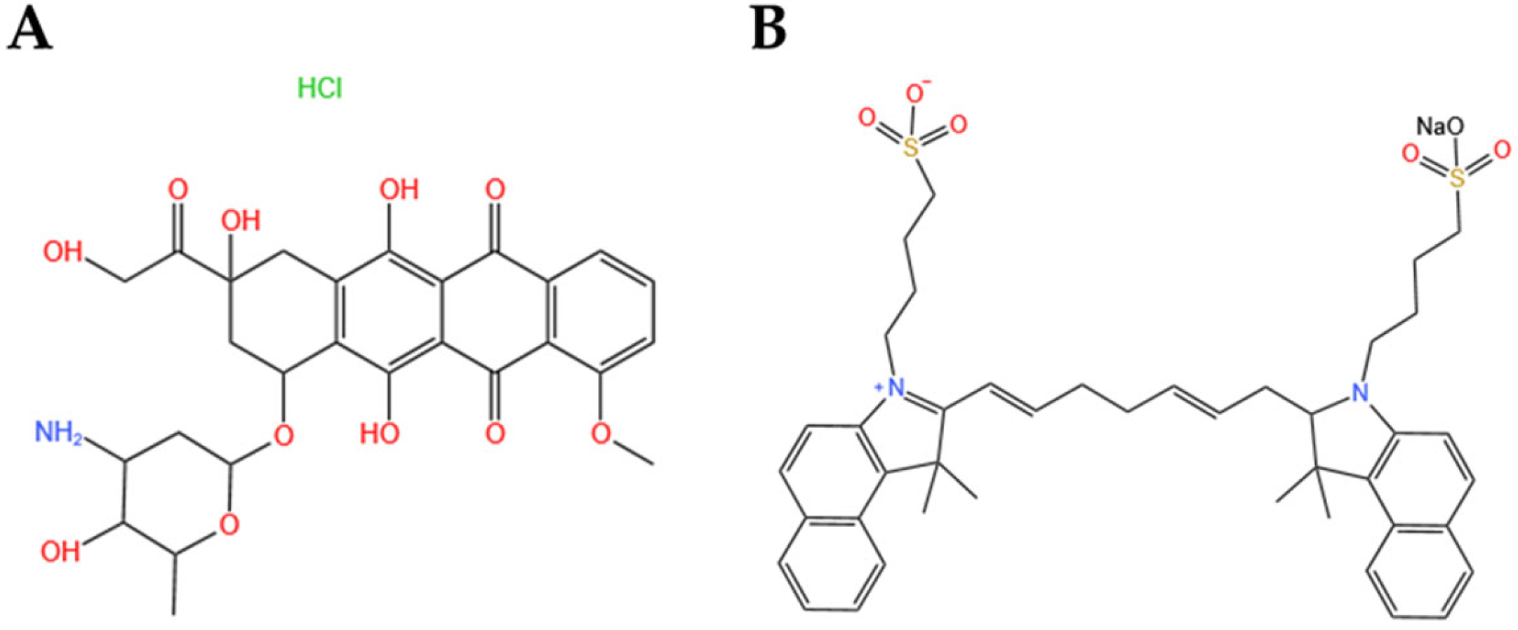
3]. As a classic anthracycline chemotherapeutic drug, doxorubicin (DOX) exerts anti-tumor activity by intercalating into DNA double strands and inhibiting topoisomerase II (
Figure 1A), thereby blocking DNA replication and transcription in rapidly proliferating tumor cells. However, the clinical application of free DOX is severely restricted by its cardiotoxicity, myelosuppression, poor water solubility, and non-specific tissue distribution. Photothermal therapy (PTT), a non-invasive therapeutic approach, converts light energy into thermal energy via photothermal agents to induce tumor cell necrosis or apoptosis, and has attracted extensive attention for its high spatial-temporal selectivity and minimal side effects. Indocyanine green (ICG), a clinically approved near-infrared (NIR) fluorescent dye, is a promising photothermal agent with good biocompatibility, strong NIR light absorption, and efficient photothermal conversion efficiency (
Figure 1B). Nevertheless, the clinical translation of free ICG is hindered by its inherent instability, rapid clearance, and potential phototoxicity to normal tissues caused by non-specific distribution. Consequently, the single application of PTT is limited by insufficient tumor targeting and poor therapeutic efficacy for deep-seated tumors, while the combination of chemotherapy and PTT can achieve synergistic anti-tumor effects and overcome the limitations of monotherapy.
Liposomes, as classic nano-drug delivery systems, have been widely applied in cancer therapy due to their excellent biocompatibility, low immunogenicity, biodegradability, and tunable surface modification [
4]. Liposomal encapsulation can improve the solubility and bioavailability of chemotherapeutic drugs, prolong their in vivo circulation time, and reduce off-target side effects [
5]. However, unmodified liposomes face challenges such as poor tumor targeting, easy clearance by the mononuclear phagocyte system (MPS), and limited penetration of the biological barrier. PEGylation is a common surface modification strategy to prolong the in vivo circulation of liposomes, but it may induce immune responses and reduce the cellular internalization efficiency of liposomes [
6,
7]. Thus, developing novel surface modification strategies to enhance tumor targeting and therapeutic efficacy of liposomes is of great significance.
The TME is a complex microenvironment composed of tumor cells, immune cells, stromal cells, and extracellular matrix. Neutrophils are the most abundant innate immune cells in peripheral blood, accounting for 50–70% of circulating white blood cells [
8,
9]. Tumor-associated neutrophils (TANs) are a subgroup of neutrophils recruited to the TME, which can be polarized into the anti-tumor N1 phenotype and the pro-tumor N
2 phenotype with plastic phenotypic and functional characteristics [
10,
11,
12,
13]. Pro-tumor N
2 neutrophils participate in all stages of tumor progression, including tumorigenesis, metastasis, and immunosuppression, and are actively recruited to the TME by cytokines secreted by tumor cells [
14,
15]. L-selectin [
16], a cell surface adhesion molecule highly expressed on neutrophils, mediates neutrophil adhesion, migration, and activation, and is a potential target for neutrophil-based tumor targeting [
17,
18]. Polysialic acid (PSA), a hydrophilic linear polysaccharide composed of N-acetylneuraminic acid (Neu5Ac) units, is the natural ligand of L-selectin [
19]. PSA with α-2,8 glycosidic linkages is a non-immunogenic, biodegradable endogenous polymer that can be degraded into non-toxic sialic acid by tissue sialidases [
20]. Compared with PEG, PSA not only has similar hydrophilicity, flexibility, and controllable molecular weight, but also can achieve active targeting to neutrophils via the specific binding of PSA and L-selectin, improve the enhanced permeability and retention (EPR) effect of nano-drugs in tumor tissues, and reduce MPS-mediated clearance [
21]. Therefore, PSA modification is an ideal strategy to enhance the tumor targeting and in vivo stability of liposomes.
Microfluidic technology, a novel nanomaterial preparation method, has significant advantages over traditional liposome preparation methods (e.g., thin-film hydration, reverse evaporation). It enables continuous and large-scale preparation of liposomes by precisely manipulating microscale fluid flow, and can accurately control the physicochemical properties of liposomes (e.g., particle size, PDI, drug loading) with high batch-to-batch reproducibility. In addition, microfluidic technology has the characteristics of low reagent consumption, high production efficiency, and rapid heat transfer, which are suitable for the preparation of functional nano-drug delivery systems.
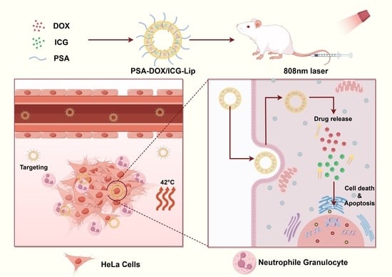
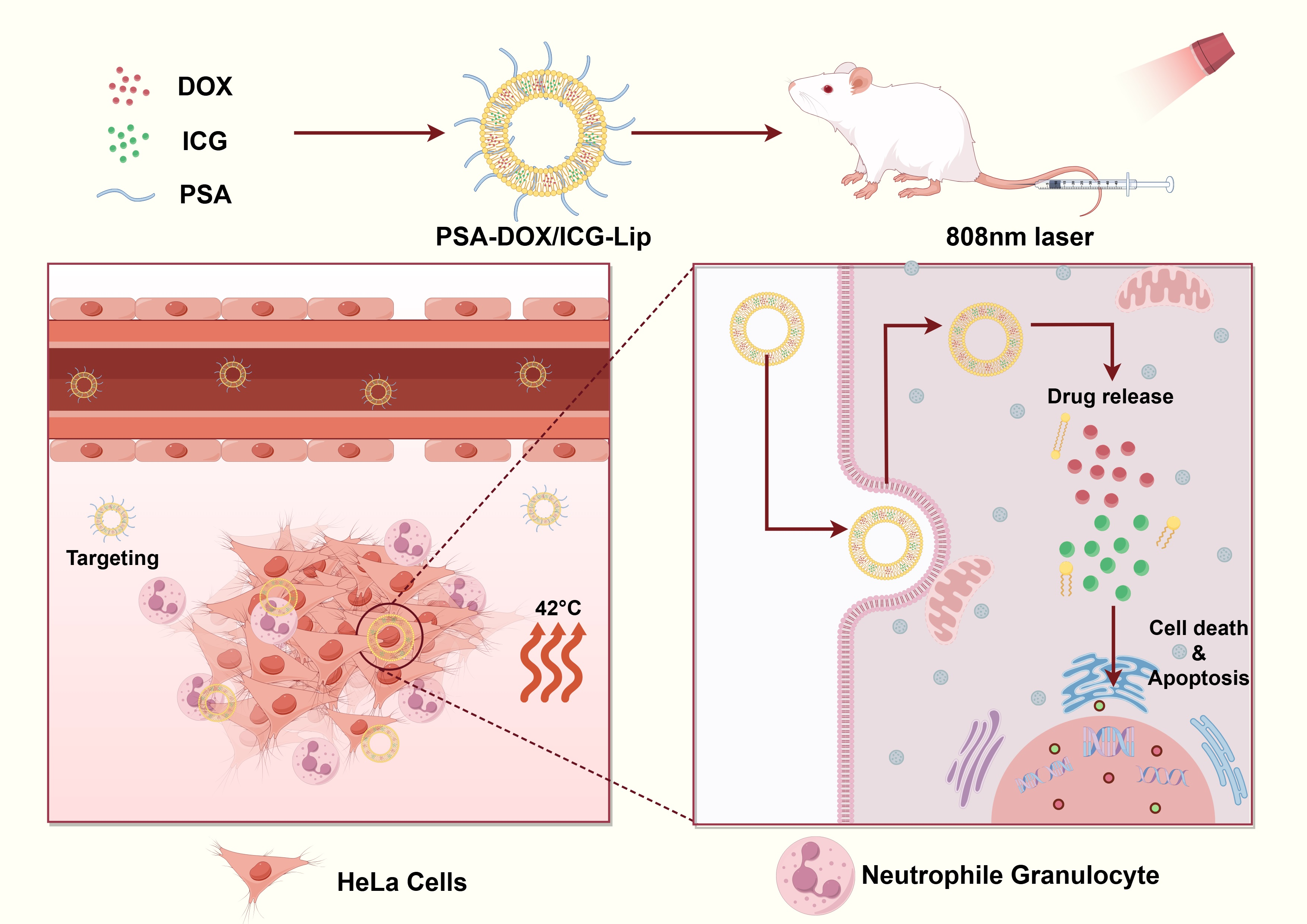
In this study, we prepared PSA-modified DOX/ICG co-loaded liposomes (PSA-DOX/ICG-Lip) by microfluidic technology for synergistic chemotherapy and PTT of cervical cancer. The physicochemical properties, stability, photothermal conversion efficiency, and pH/laser dual-responsive drug release behavior of the liposomes were systematically characterized. The in vitro anti-tumor effects (cellular uptake, ROS generation, anti-proliferation, anti-migration) were evaluated on human cervical cancer HeLa and C33a cells, and the in vivo anti-tumor efficacy and biosafety were verified using a nude mouse xenograft model of cervical cancer. We hypothesized that PSA modification could achieve active targeting of liposomes to neutrophils and tumor cells, and the combination of DOX-mediated chemotherapy and ICG-mediated PTT could exert synergistic anti-tumor effects, providing a novel and effective nano-therapeutic strategy for cervical cancer.
2. Materials and Methods
2.1. Materials
Heating magnetic stirrer (RCT Basic, IKA Corporation, Staufen, Germany); electronic balance (BP211D, Sartorius Group, Shanghai, China); heated numerical control ultrasonic cleaner (KH-300DE, Kunshan Hechuang Ultrasonic Instrument Co., Ltd., Kunshan, Jiangsu, China); constant temperature shaker (Jiangsu Jintan Medical Instrument, Changzhou, Jiangsu, China); ultrasonic cleaner (SY-360, Beijing Tianpeng Electronic New Technology Co., Ltd., Beijing, China); laser particle size and zeta potential analyzer (ZEN3600, Malvern Company, Malvern, UK); PSA (Zhongke Hongji Biotechnology Co., Ltd., Jinzhong, Shanxi, China); DOX (Shanghai Macklin Biochemical Technology Co., Ltd., Shanghai, China); ICG (Bide Pharmatech Co., Ltd., Shanghai, China); dipalmitoyl phosphatidylcholine (DPPC), hydrogenated soy phospholipids (HSPC), cholesterol, and DSPE-PEG2000-COOH were purchased from AVT Pharmaceutical Tech Co., Ltd. (Shanghai, China); DMEM medium was purchased from Gibco Life Technologies (New York, NY, USA); the CCK-8 assay kit was purchased from Life-iLab Biotechnology Co., Ltd. (Shanghai, China); the Calcein-AMPI live/dead cell dual staining kit was purchased from Solarbio (Beijing, China); the ROS assay kit and DCFH-DA was purchased from Beyotime Biotechnology Co., Ltd. (Shanghai, China); and the neutrophil isolation solution was purchased from Haoyang Biological Manufacture Co., Ltd. (Tianjin, China); syringe pump (Jiangsu Zhiyu Medical Instrument Co., Ltd., Taixing, Jiangsu, China); transmission electron microscope HT7800 (HITACHI, Tokyo, Japan); carbon-coated copper grid (Zhongjing Scientific Instruments, Beijing, China); dialysis bag (Solarbio, Beijing, China); UV-visible spectrophotometry 2700 (SHIMADZU, Kyoto, Japan); constant-temperature shaker (Shanghai Bosun Medical and Biological Instruments Co., Ltd., Shanghai, China); NIR laser (NBeT Group Corp., Beijing, China); thermal imaging camera 323pro (Fotric Inc., Shanghai, China); LCSM ECLipSE Ti (NIKON, Shanghai, China); fluorescence inverted microscope DMi8 (LEICA, Wetzlar, Germany); Human cervical cancer HeLa cells were obtained from the Wuhan Servicebio Technology Co., Ltd. (Wuhan, China); C33a cells were obtained from the laboratory (Xinjiang, China); BALB/c-nu nude mice (female, 4–6 weeks old, 18–22 g) were purchased from Beijing Vital River Laboratory Animal Technology Co., Ltd. (Beijing, China)
2.2. Preparation of PSA-DOX/ICG-Lip
PSA-DOX/ICG-Lip was prepared by microfluidic technology combined with the ammonium sulfate gradient method. Briefly, DPPC, HSPC, cholesterol, and DSPE-PEG2000-PSA were mixed at a molar ratio of 3:3:2:0.5 and dissolved in methanol to form the organic phase (Solution A). ICG was dissolved in 0.25 M ammonium sulfate solution to form the aqueous phase (Solution B). Solution A and Solution B were simultaneously injected into a microfluidic mixer using a syringe pump at a total flow rate of 12 mL/min and an organic/aqueous phase flow rate ratio of 1:2. The obtained liposome suspension was dialyzed against phosphate-buffered saline (PBS) (pH 7.4) for 4 h to remove methanol and establish the ammonium sulfate gradient. DOX solution was added to the liposome suspension at a drug-lipid mass ratio of 1:10, and the mixture was incubated at 60 °C for 30 min to load DOX into the liposomes via the ammonium sulfate gradient method. The unencapsulated free DOX was removed by dialysis against PBS (pH 7.4) for 10 h, and the final PSA-DOX/ICG-Lip was obtained and stored at 4 °C in the dark. For comparison, blank liposomes (Blank-Lip), DOX-loaded liposomes (DOX-Lip), ICG-loaded liposomes (ICG-Lip), and unmodified DOX/ICG co-loaded liposomes (DOX/ICG-Lip) were prepared using the same method without adding DSPE-PEG2000-PSA or the corresponding drugs.
2.3. Physicochemical Properties of Liposomes
2.3.1. Morphological Characterization
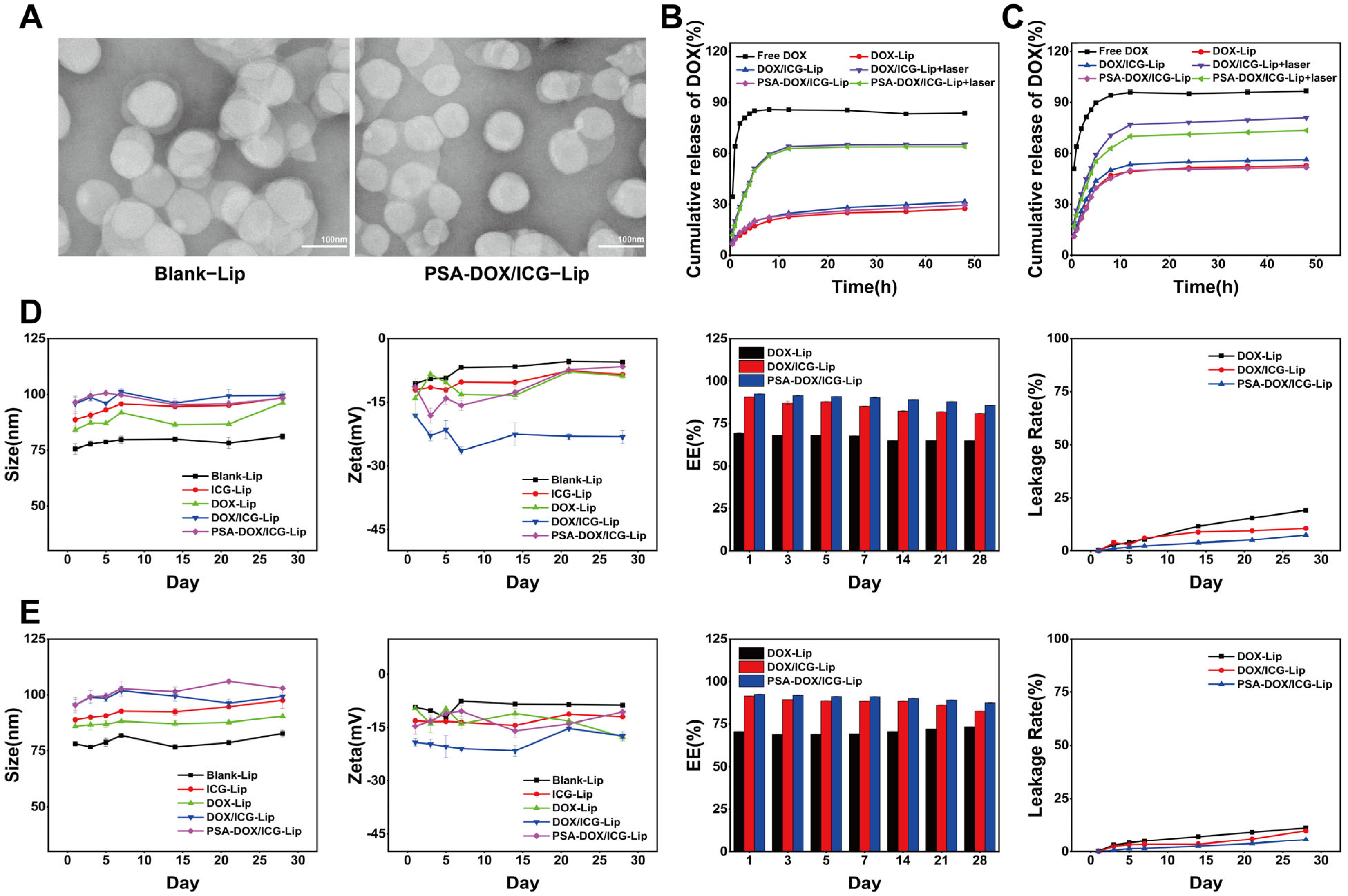
The morphology of PSA-DOX/ICG-Lip was observed by transmission electron microscope (TEM). A 10 μL aliquot of the liposome suspension (diluted 10-fold with deionized water) was placed onto a carbon-coated copper grid, air-dried at room temperature, and stained with 10% phosphotungstic acid for 10 min. The excess staining solution was blotted with filter paper, and the morphology of the liposomes was observed under TEM at an acceleration voltage of 80 kV.
2.3.2. Particle Size, PDI, and Zeta Potential
The liposome suspension was diluted 10-fold with deionized water, and the particle size, PDI, and zeta potential were measured using a laser particle size and zeta potential analyzer at 25 °C. Each sample was tested in triplicate, and the average value was calculated.
2.3.3. Encapsulation Efficiency (EE) and Drug Loading Capacity (LC)
The EE and LC of DOX and ICG in the liposomes were determined by the dialysis method combined with UV-visible spectrophotometry. Briefly, 1 mL of the liposome suspension was placed in a dialysis bag (MWCO: 8000 Da) and dialyzed against 50 mL of PBS (pH 7.4) for 24 h at 37 °C with gentle shaking to remove unencapsulated drugs. After dialysis, the liposome suspension in the dialysis bag was disrupted with methanol (liposome/methanol volume ratio = 1:9), and the concentration of DOX and ICG was measured by UV-visible spectrophotometry at 480 nm and 780 nm, respectively. For the determination of the total drug amount, 1 mL of the liposome suspension was directly disrupted with methanol, and the drug concentration was measured using the same method. The EE and LC were calculated according to the following formulas:
In the formulas, W1 represents the amount of drug encapsulated in the liposomes after dialysis, W2 represents the total amount of drug initially added before dialysis, and W3 represents the total amount of liposomal membrane material and drug.
2.3.4. In Vitro Drug Release
The pH/laser dual-responsive drug release behavior of DOX from the liposomes was evaluated by the dialysis method. Briefly, 3 mL of Free DOX, DOX/ICG-Lip, and PSA-DOX/ICG-Lip suspensions (DOX concentration: 100 μg/mL) were placed in dialysis bags (MWCO: 8000 Da), and immersed in 30 mL of release medium (PBS containing 0.5% (w/v) Tween 80, pH 7.4 or pH 5.0 (10 mM citrate)). The release system was incubated in a constant-temperature shaker at 37 °C and 200 rpm. For the laser irradiation group, the release system was irradiated with an 808 nm NIR laser (power density: 1 W/cm2) for 10 min at the beginning of the experiment, while the non-irradiation group was incubated in the dark. At predetermined time points (0.5, 1, 2, 3, 4, 5, 8, 12, 24, 36, 48 h), 1 mL of the release medium was collected, and an equal volume of fresh pre-warmed release medium was replenished. The concentration of DOX in the collected medium was measured by UV-visible spectrophotometry at 480 nm, and the cumulative drug release rate was calculated. Each sample was tested in triplicate.
2.3.5. Stability Evaluation
The stability of the liposomes was evaluated by monitoring the changes in particle size, PDI, zeta potential, and drug leakage rate during storage. Blank-Lip, DOX-Lip, ICG-Lip, DOX/ICG-Lip, and PSA-DOX/ICG-Lip were stored at 4 °C and 25 °C in the dark for 28 days. At predetermined time points (1, 3, 5, 7, 14, 21, 28 days), the particle size, PDI, and zeta potential of the liposomes were measured, and the drug leakage rate was calculated by determining the concentration of the encapsulated drug using the method described in
Section 2.3.3.
2.3.6. Photothermal Property Evaluation
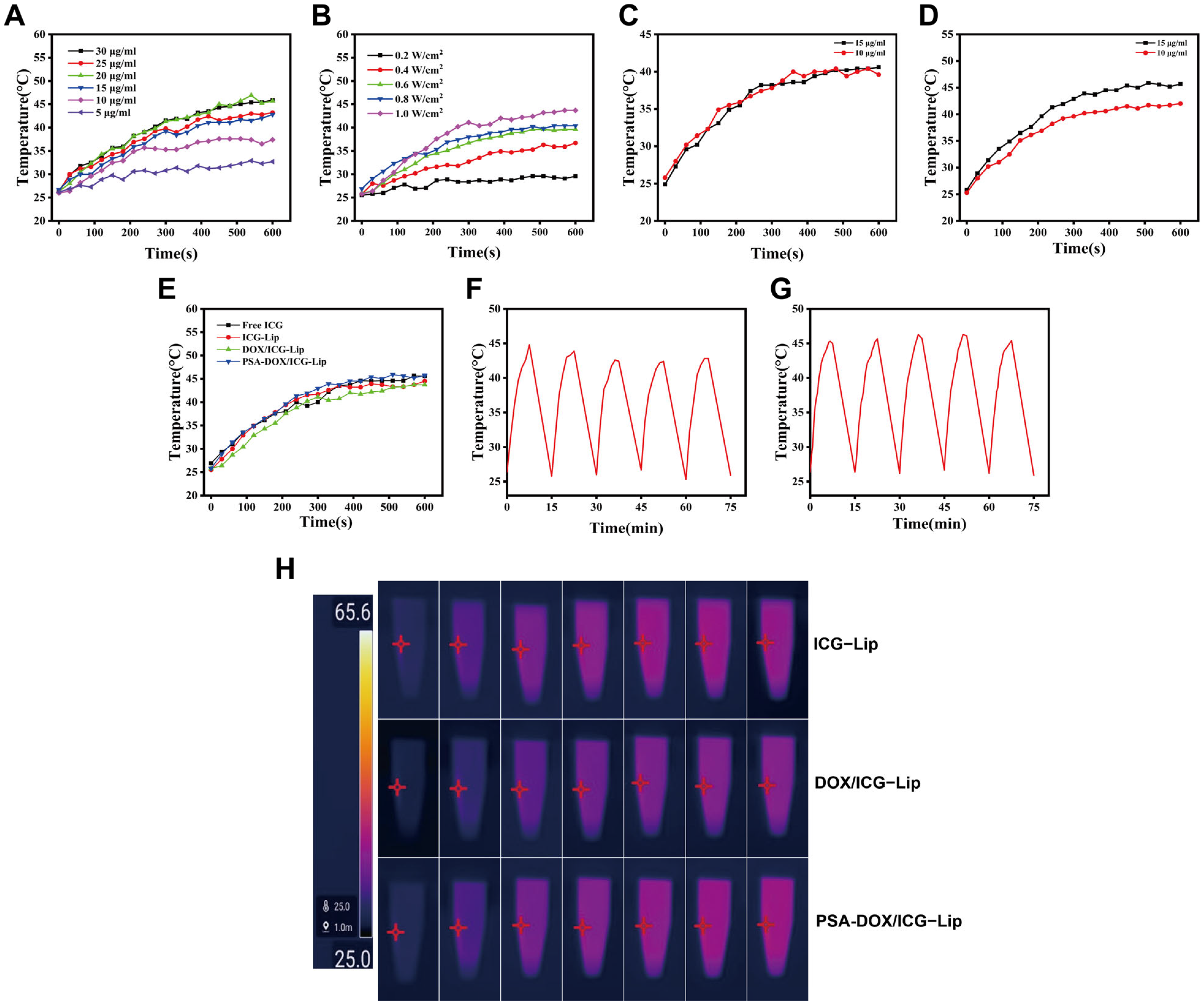
The photothermal conversion efficiency and photothermal stability of the liposomes were evaluated using an 808 nm NIR laser and a thermal imaging camera. (1) Optimal ICG concentration screening: Free ICG solutions with concentrations of 5, 10, 15, 20, 25, and 30 μg/mL were prepared, and irradiated with an 808 nm laser (power density: 0.8 W/cm
2) for 10 min. The temperature of the solution was recorded every 30 s using a thermometer, and the temperature change curve was plotted. (2) Optimal laser power density screening: DOX/ICG-Lip suspension (ICG concentration: 15 μg/mL) was irradiated with 808 nm lasers at power densities of 0.2, 0.4, 0.6, 0.8 and 1.0 W/cm
2 for 10 min, and the temperature change was recorded every 30 s. (3) Photothermal conversion efficiency of PSA-DOX/ICG-Lip: PSA-DOX/ICG-Lip suspensions with ICG concentrations of 10 and 15 μg/mL were irradiated with 808 nm lasers at power densities of 0.8 and 1.0 W/cm
2 for 10 min, and the temperature change was recorded. (4) Comparison of photothermal properties: Free ICG, ICG-Lip, DOX/ICG-Lip, and PSA-DOX/ICG-Lip suspensions (ICG concentration: 15 μg/mL) were irradiated with an 808 nm laser (power density: 1 W/cm
2) for 10 min, and the temperature change and thermal images were recorded. (5) Photothermal stability: DOX/ICG-Lip and PSA-DOX/ICG-Lip suspensions (ICG concentration: 15 μg/mL) were subjected to five freeze–thaw cycles (−20 °C/37 °C), and then irradiated with an 808 nm laser (power density: 1 W/cm
2) for 10 min. The temperature change was recorded to evaluate the photothermal stability. (6) Thermal imaging analysis: ICG-Lip, DOX/ICG-Lip, and PSA-DOX/ICG-Lip suspensions (ICG concentration: 10 μg/mL) were irradiated with an 808 nm laser (power density: 1 W/cm
2) for 15 min, and thermal images were captured every 30 s using a thermal imaging camera [
22].
2.4. In Vitro Cell Studies
2.4.1. Cytotoxicity Test
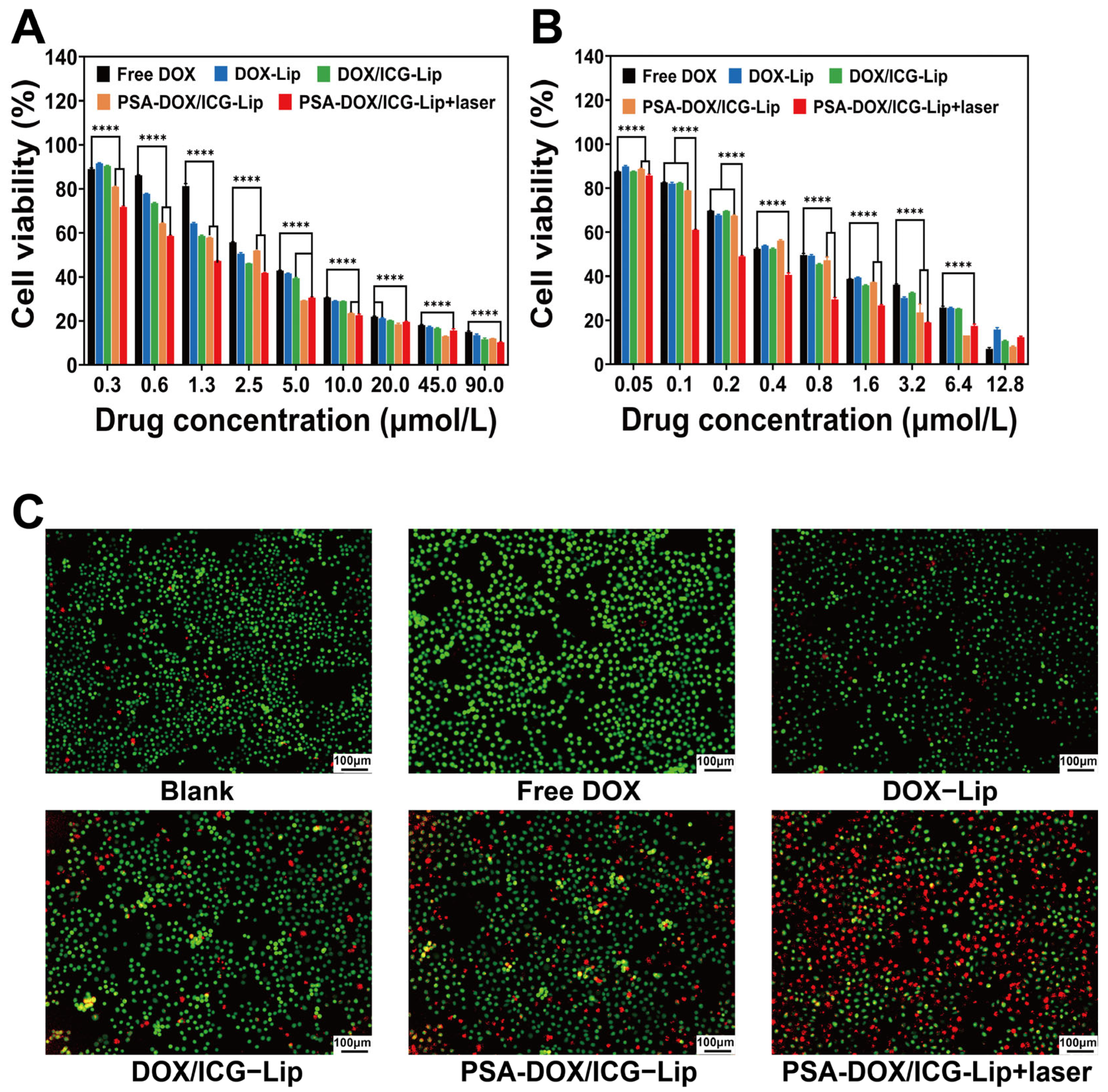
The cytotoxicity of the liposomes on HeLa and C33a cells was evaluated by the CCK-8 assay. HeLa cells (5 × 10³ cells/well) and C33a cells (8 × 10³ cells/well) were seeded into 96-well plates and cultured for 24 h to allow cell adhesion. The culture medium was replaced with fresh medium containing Free DOX, DOX-Lip, DOX/ICG-Lip, and PSA-DOX/ICG-Lip at different DOX concentrations (HeLa: 0, 0.312, 0.625, 1.25, 2.5, 5, 10, 20, 45, 90 μM; C33a: 0, 0.05, 0.1, 0.2, 0.4, 0.8, 1.6, 3.2, 6.4, 12.8 μM). The laser irradiation group was irradiated with an 808 nm laser (power density: 1 W/cm2) for 10 min after drug addition, while the non-irradiation group was incubated in the dark. After incubation for 48 h (HeLa) and 24 h (C33a), 10 μL of CCK-8 solution was added to each well, and the plates were incubated for an additional 40 min (HeLa) and 90 min (C33a). The absorbance at 450 nm was measured using a microplate reader.
2.4.2. Calcein-AM/PI Live/Dead Cell Staining
The anti-proliferative effect of the liposomes on HeLa cells was intuitively evaluated by Calcein-AM/PI double staining. HeLa cells (5 × 10
4 cells/well) were seeded into 6-well plates and cultured for 24 h. The culture medium was replaced with fresh medium containing Free DOX, DOX-Lip, DOX/ICG-Lip and PSA-DOX/ICG-Lip (DOX concentration: 5 μM). The laser irradiation group was irradiated with an 808 nm laser (power density: 1 W/cm
2) for 10 min, and the non-irradiation group was incubated in the dark. After incubation for 48 h, the culture medium was discarded, and the cells were stained with Calcein-AM/PI staining solution (Calcein-AM: 2 μM, PI: 5 μM) for 30 min in the dark. The stained cells were observed and photographed under a fluorescence inverted microscope, where live cells emit green fluorescence and dead cells emit red fluorescence [
23].
2.4.3. Laser Confocal Scanning Microscopy (LCSM) for Cellular Uptake
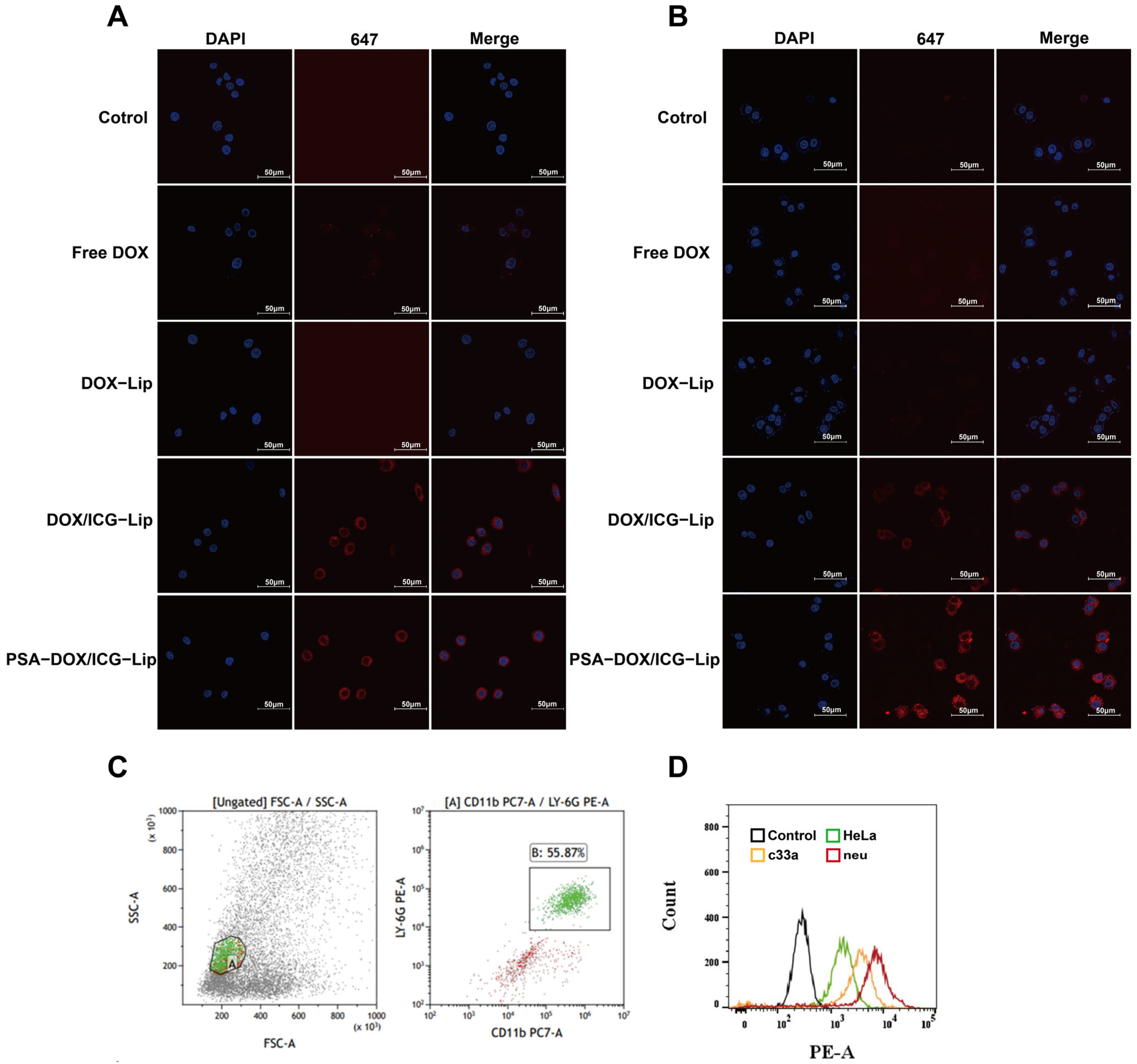
The cellular uptake efficiency of the liposomes by HeLa and C33a cells was evaluated by LCSM. HeLa cells (2.5 × 10⁵ cells/dish) and C33a cells (5 × 10⁵ cells/dish) were seeded into confocal culture dishes and cultured for 12 h. The culture medium was replaced with fresh medium containing Free DOX, DOX-Lip, DOX/ICG-Lip, and PSA-DOX/ICG-Lip (DOX concentration: 5 μM), and the cells were incubated for 3 h. The cells were washed three times with cold PBS, fixed with 4% paraformaldehyde for 15 min, and stained with 4′,6-Diamidino-2′-Phenylindole (DAPI) (2 μg/mL) for 10 min to label the cell nuclei. The cellular uptake of the liposomes was observed and photographed under LCSM, where DOX emits red fluorescence, ICG emits near-infrared fluorescence, and DAPI emits blue fluorescence. The fluorescence intensity was analyzed using ImageJ 1.54f software.
2.4.4. Flow Cytometry for Neutrophil Targeting
Neutrophils were isolated from the orbital blood of healthy BALB/c-nu mice using a Neutrophil Isolation Kit according to the manufacturer’s instructions, and the purity of neutrophils was identified by flow cytometry using Ly6g and CD11b as specific markers. HeLa cells, C33a cells, and isolated neutrophils (5 × 10⁴ cells/well) were seeded into 6-well plates and cultured for 24 h. PSA-DOX/ICG-Lip suspension (DOX concentration: 5 μM) was added to each well, and the cells were incubated for 3 h. The cells were digested with trypsin (for adherent cells) or collected by centrifugation (for neutrophils), washed three times with cold PBS, and resuspended in 500 μL of cold PBS. The fluorescence intensity of DOX in the cells was detected by flow cytometry, and the targeting ability of PSA-DOX/ICG-Lip to neutrophils was evaluated by comparing the fluorescence intensity in different cells.
2.4.5. ROS Assay
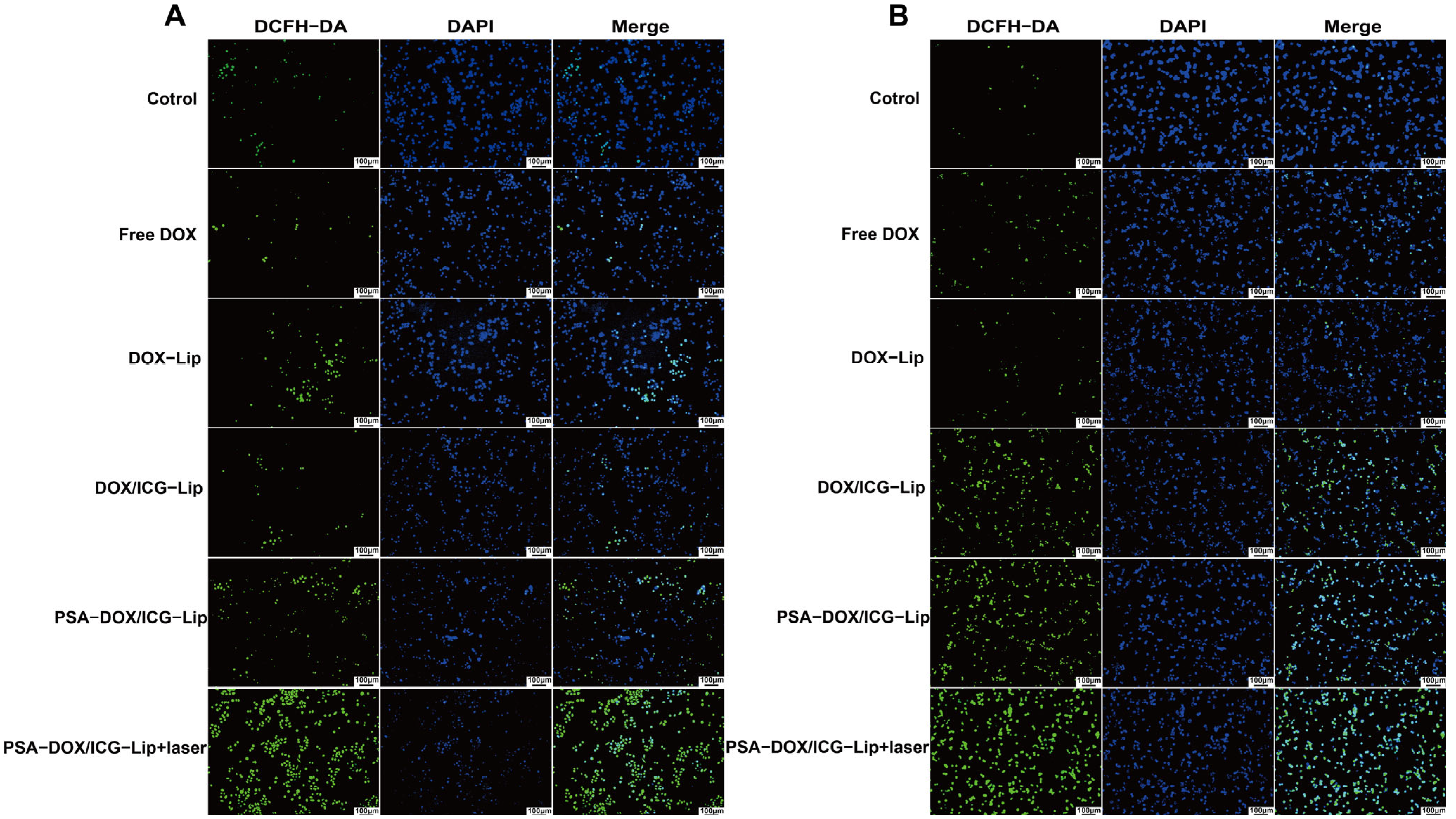
The intracellular ROS generation induced by the liposomes was evaluated using the 2′,7′ -Dichlorofluorescein Diacetate (DCFH-DA) ROS Assay Kit. HeLa cells (2.5 × 10⁵ cells/well) and C33a cells (3 × 10⁴ cells/well) were seeded into 12-well plates and cultured for 24 h. The culture medium was replaced with fresh medium containing Free DOX, DOX-Lip, DOX/ICG-Lip, and PSA-DOX/ICG-Lip (DOX concentration: 5 μM). The laser irradiation group was irradiated with an 808 nm laser (power density: 1 W/cm2) for 10 min, and the non-irradiation group was incubated in the dark. After incubation for 3 h, the cells were washed three times with cold PBS and incubated with DCFH-DA probe (10 μM) for 20 min in the dark. The cells were washed again with cold PBS, stained with DAPI (2 μg/mL) for 10 min, and observed and photographed under a fluorescence inverted microscope. The fluorescence intensity of DCF (oxidized form of DCFH-DA) was analyzed using ImageJ 1.54f software to evaluate the intracellular ROS level.
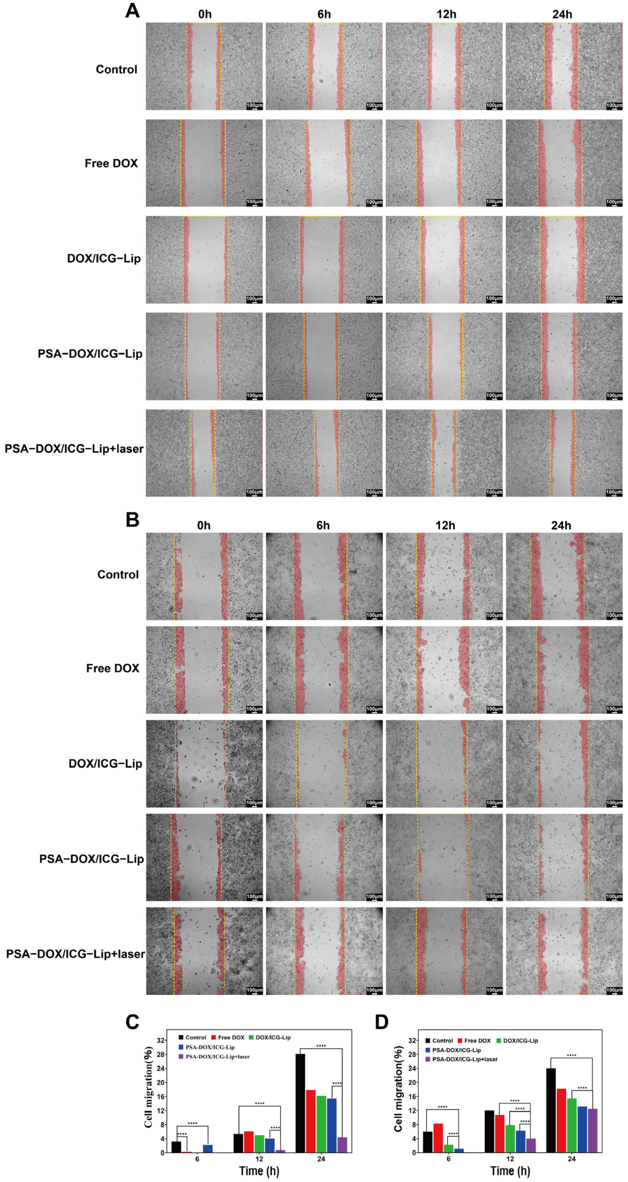
2.4.6. Cell Scratch Assay for Anti-Migration Effect
The anti-migration effect of the liposomes on HeLa and C33a cells was evaluated by the cell scratch assay. HeLa cells (2.5 × 10
5 cells/well) and C33a cells (3 × 10
5 cells/well) were seeded into 6-well plates and cultured until the cell confluency reached 90–100%. A straight scratch was made on the cell monolayer using a 200 μL pipette tip, and the detached cells were washed away with cold PBS. The culture medium was replaced with fresh serum-free medium containing Free DOX, DOX-Lip, DOX/ICG-Lip, and PSA-DOX/ICG-Lip (DOX concentration: 5 μM). The laser irradiation group was irradiated with an 808 nm laser (power density: 1 W/cm
2) for 10 min, and the non-irradiation group was incubated in the dark. The scratch area was photographed under a fluorescence inverted microscope at predetermined time points (0, 6, 12, 24 h) [
24].
2.5. In Vivo Anti-Tumor Efficacy Evaluation
2.5.1. Establishment of Cervical Cancer Xenograft Mode
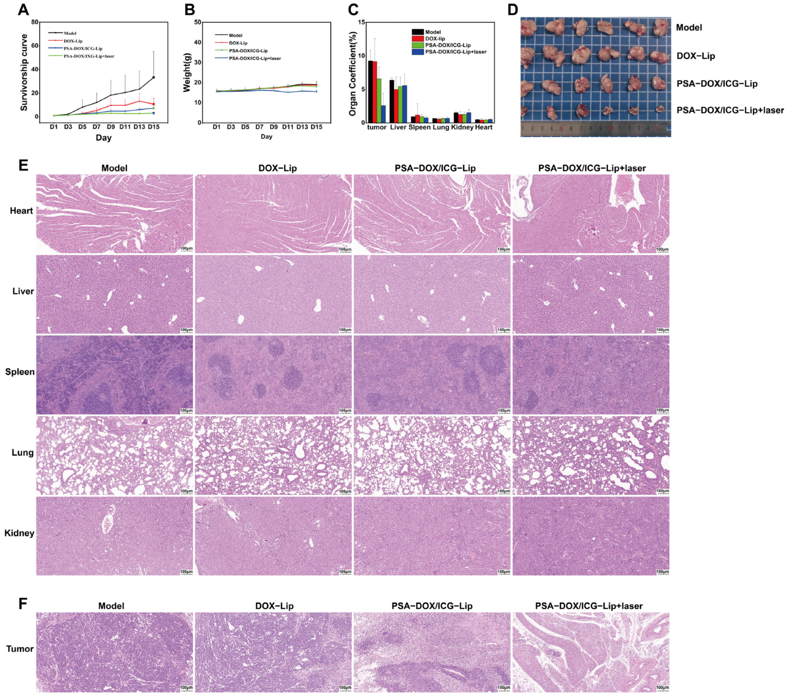
BALB/c-nu nude mice were acclimatized in a specific pathogen-free (SPF) animal facility for 1 week before the experiment. HeLa cells in the logarithmic growth phase were digested with 0.25% trypsin, resuspended in serum-free RPMI 1640 medium, and the cell concentration was adjusted to 1 × 10⁷ cells/mL. A 100 μL aliquot of the cell suspension was subcutaneously injected into the right axilla of each nude mouse. The tumor volume was measured every 2 days using a digital vernier caliper, and the tumor volume was calculated according to the formula V = ab2/2 (where a is the longest diameter and b is the shortest diameter perpendicular to a).
2.5.2. In Vivo Administration and Therapeutic Efficacy
The mice in each group were intravenously injected with the corresponding formulations via the tail vein every other day for a total of 14 days (DOX dosage: 1 mg/kg). For the PSA-DOX/ICG-Lip + laser group, the tumor site was irradiated with an 808 nm NIR laser (power density: 1 W/cm2, irradiation time: 10 min) at 4 h after each drug injection. The body weight and tumor volume of the mice were measured every 2 days during the experiment.
2.5.3. Hematoxylin–Eosin (H&E) Staining
The harvested tumors and major organs were fixed with 4% paraformaldehyde for 24 h, dehydrated with gradient ethanol, embedded in paraffin, and sectioned into 5 μm thick slices. The slices were stained with hematoxylin and eosin, and observed and photographed under an fluorescence inverted microscope to evaluate the tumor tissue necrosis and organ histopathological changes.
2.6. Statistical Analysis
All experimental data were expressed as the mean ± standard deviation (mean ± SD). Statistical analysis was performed using IBM SPSS Statistics 26.0 software and GraphPad Prism 10.1.2 software. One-way analysis of variance (ANOVA) followed by LSD post hoc test, Tukey’s HSD test, and Waller–Duncan multiple range test was used for multiple comparisons. Homogeneity of variance was tested before ANOVA. A p-value < 0.05 was considered statistically significant, and the statistical symbols were indicated as follows: * p < 0.05, ** p < 0.01, *** p < 0.001, **** p < 0.0001.



 and
and