Review

Structural and Functional Insights into Viral and Fungal Proteins Involved in Chronic Inflammation and Their Biologic Treatments

Department of Biomedical Sciences, Cooper Medical School, Rowan University, Camden, NJ 08103, USA
*
Author to whom correspondence should be addressed.
Pharmaceutics 2025, 17(11), 1466; https://doi.org/10.3390/pharmaceutics17111466
Submission received: 15 September 2025 / Revised: 7 November 2025 / Accepted: 12 November 2025 / Published: 13 November 2025
(This article belongs to the Special Issue Antibody–Drug Conjugates Therapeutics)

Abstract

Chronic inflammation constitutes a significant characteristic of sustained infections caused by viral and fungal pathogens, with a strong correlation to the development of cancer, autoimmune disorders, and tissue fibrosis. Viral proteins such as HIV-1 Tat, HBV X (HBx), HPV E6/E7, and EBV LMP1 modulate the host’s immune signaling pathways, primarily through the activation of the NF-κB signaling cascade and the disruption of cytokine equilibrium. These molecular interactions result in a pro-inflammatory microenvironment that facilitates viral persistence, immune evasion, and the process of oncogenesis. Structural investigations have elucidated the mechanisms by which these viral proteins interact with host signaling complexes, thereby highlighting their potential as viable therapeutic targets. Similarly, fungal proteins, including secreted aspartyl proteases (Saps), ribotoxin Asp f1, and chitin-binding proteins, incite chronic inflammation by activating pattern recognition receptors and triggering inflammasome activation. Despite the limited structural information of these fungal proteins, emerging models and bioinformatic analyses identified conserved motifs that are crucial for host interactions. Biologic therapies, encompassing antiviral and antifungal peptides as well as monoclonal antibodies, are currently under development to disrupt these protein-host interactions and modulate inflammatory responses. This review provides structural and functional insight into viral and fungal inflammatory proteins and evaluates the potential of biologics as targeted therapeutic interventions for chronic inflammation associated with infections. We discuss the ongoing clinical trials involving neutralizing antibodies targeting HIV, peptide vaccines aimed at HPV and other promising molecules. Finally, we discuss the current limitations of biologics and possible solutions to translate these promising therapeutics into clinical practice.

Graphical Abstract

1. Introduction

Chronic inflammation represents a sustained and dysregulated immune response that is involved in the pathogenesis of a myriad of human diseases, encompassing cancer, autoimmune disorders, and organ fibrosis [1]. Persistent infections of viral and fungal agents are significant contributors to this inflammatory condition. In such instances, microbial proteins avoid immune clearance mechanisms and actively interact with or reprogram host immune signaling pathways, resulting in increased cytokine production, immune cell recruitment, and tissue remodeling [2]. An in-depth understanding of the molecular mechanisms through which viral and fungal proteins perpetuate chronic inflammation is crucial for the advancement of targeted immunotherapies and biologics.
In the context of viral infections, proteins synthesized by HIV-1, hepatitis B virus (HBV), human papillomavirus (HPV), and Epstein–Barr virus (EBV) have been rigorously investigated for their immunomodulatory functions. Examples include the HIV-1 trans-activator of transcription (Tat), HBV X protein (HBx), HPV E6 and E7, and EBV latent membrane protein 1 (LMP1, [3,4,5]). Despite their structural and functional heterogeneity, these proteins exhibit a shared inflammatory mechanism; the activation of the nuclear factor-kappa B (NF-κB) pathway and the subsequent dysregulation of cytokine signaling. The NF-κB pathway functions as a regulator of immune and inflammatory responses, via transcriptional activation of genes involved in cytokine synthesis, cellular proliferation, and survival. The canonical NF-κB pathway is the most extensively studied mechanism of activation. It is induced by pro-inflammatory cytokines, such as tumor necrosis factor-α (TNF-α) and interleukin-1β (IL-1β), pathogen-associated molecules including lipopolysaccharide (LPS), or cellular stress signals [6]. In quiescent cells, NF-κB dimers are sequestered in the cytoplasm by inhibitory IκB proteins. Activation occurs by engagement with cell surface receptors that recruit adaptor proteins and activate the IκB kinase (IKK) complex [7]. The IKK complex subsequently phosphorylates IκB proteins, marking them for ubiquitin-mediated degradation by the proteasome. This process liberates NF-κB dimers, which then translocate to the nucleus, bind to κB DNA motifs, and initiate transcription. NF-κB-regulated genes include those encoding pro-inflammatory cytokines (TNF-α, IL-6, IL-1β), chemokines, adhesion molecules, and anti-apoptotic proteins, which enhances inflammation and promoting cellular survival. The canonical pathway is distinguished by its rapid yet transient activation, ensuring prompt responses to infectious agents or tissue damage [8]. Conversely, the non-canonical NF-κB pathway operates at a slower pace, is more stringently regulated, and is activated by a more limited array of stimuli. It is predominantly induced by members of the tumor necrosis factor receptor (TNFR) superfamily, which play critical roles in adaptive immunity, lymphoid organogenesis, and B-cell functionality. Unlike the canonical pathway, the activation of the non-canonical route does not depend on the IKK complex [9].
The dysregulation of the NF-κB pathway contributes to chronic inflammation and fosters the development of a tumor-promoting microenvironment [10]. This chronic inflammatory milieu also promotes viral persistence and immune dysfunction. In response to these challenges, a variety of biologic therapies have emerged, including antiviral peptides that inhibit viral entry or replication, and monoclonal antibodies (mAbs) that target viral envelope proteins or modulate immune responses in the context of chronic viral infections [11]. For example, broadly neutralizing antibodies against HIV-1 are currently undergoing clinical trials, including vrc01 and 3BNC117 [12,13], while therapeutic antibodies such as nivolumab and pembrolizumab are being investigated for their efficacy in virus-associated cancers, including HPV-associated cervical cancer, EBV-associated nasopharyngeal carcinoma, and HBV- or HCV-related hepatocellular carcinoma [14].
Fungal infections, particularly among immunocompromised populations, represent significant contributors to chronic inflammation. Pathogens such as Candida albicans, Aspergillus fumigatus, and Cryptococcus neoformans secrete numerous proteins that continuously engage the host immune response [15]. In contrast to viral proteins, fungal inflammatory proteins predominantly function through extracellular mechanisms, by interacting with pattern recognition receptors (PRRs) such as Dectin-1 and Toll-like receptors (TLRs), activating inflammasomes, and modulating the activities of immune cells, including macrophages and neutrophils [16]. These interactions result in excessive activation of the innate immune system and prolonged inflammatory responses. Notable fungal proteins include secreted aspartyl proteases (Saps), mannoproteins, and chitin-binding proteins [17]. Although the structural elucidation of fungal inflammatory proteins remains limited, advancements have been made in the development of antifungal peptides that disrupt fungal membranes or biofilm formation, and mAbs directed against fungal antigens, such as Candida adhesins or Aspergillus allergens. These biologics hold promise not only for achieving fungal clearance but also for the modulation of inflammation associated with persistent fungal infections [18].
In this review, we discuss the structural and functional attributes of viral and fungal proteins implicated in chronic inflammation, concentrating on two distinct mechanistic criteria: the activation of NF-κB and cytokine dysregulation for viral proteins, and PRR engagement, inflammasome activation, and immune cell modulation for fungal proteins. Furthermore, we explore the current landscape of antiviral and antifungal biologics, including peptides and mAbs that have been developed or are under investigation for their ability to target these pathogens and their associated inflammatory consequences. We will highlight both the analogous and distinct methodologies employed by viruses and fungi in the maintenance of inflammatory responses and provide guidance for the advancement of targeted immunotherapies for chronic inflammatory diseases associated with infections.

2. Viral Proteins Associated with Chronic Inflammation: Structural and Functional Analyses

Chronic viral infections are associated with prolonged immune activation and inflammation, frequently driven by specific viral proteins that disrupt host signaling pathways. Proteins derived from HIV-1, HBV, HPV, and EBV manipulate the NF-κB signaling cascade and cytokine networks, prompting a sustained pro-inflammatory microenvironment that fosters viral persistence, immune evasion, and, in numerous instances, tumorigenesis (Figure 1, [19]). Next, we discuss these viral proteins that contribute to chronic inflammation, emphasizing their structural characteristics and available three-dimensional configurations.

2.1. HIV-1 Tat

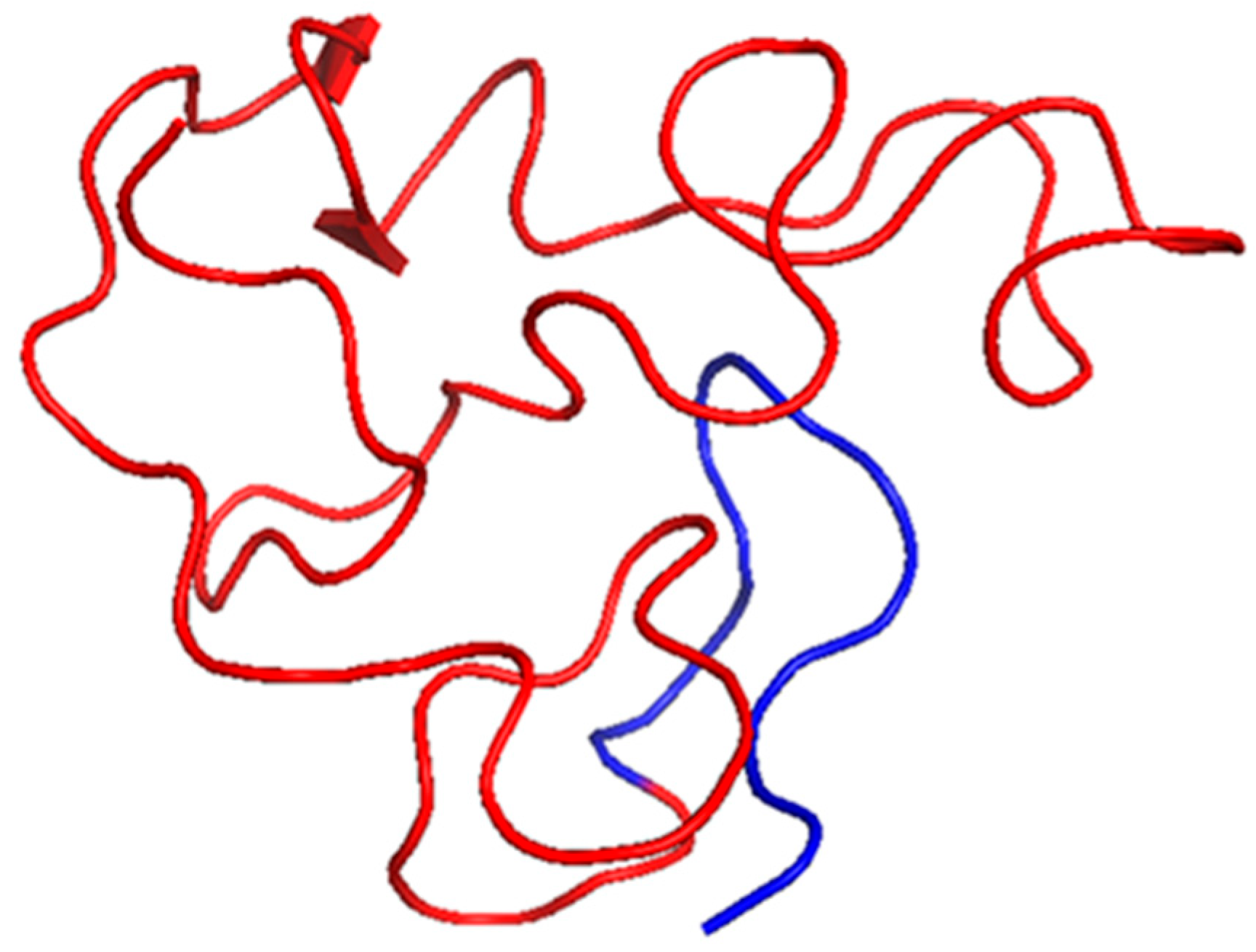
The HIV-1 Tat protein is necessary for viral transcription and concurrently functions as a secreted pro-inflammatory effector. Tat binds to p65 (RelA) of the NF- κB to activate signaling and enhance the expression of the proinflammatory cytokines (Figure 1). It also activates the IKK complex, leading to increased translocation into the nucleus [20]. From a structural perspective, Tat is intrinsically disordered but encompasses segments with defined secondary structures that promote interactions with host proteins and nucleic acids [21]. The NMR solution structure of a truncated form of Tat (residues 1–72) from subtype B is available (Table 1, Figure 2). Tat effectively crosses cell membranes and induces inflammatory responses in uninfected cells, which contributes to HIV-associated systemic and neuroinflammation. Tat also effects endothelial cells and astrocytes, which leads to HIV-associated neurocognitive disorders, and represent a defining feature of chronic inflammation during HIV infection [22].

2.2. HBV X Protein

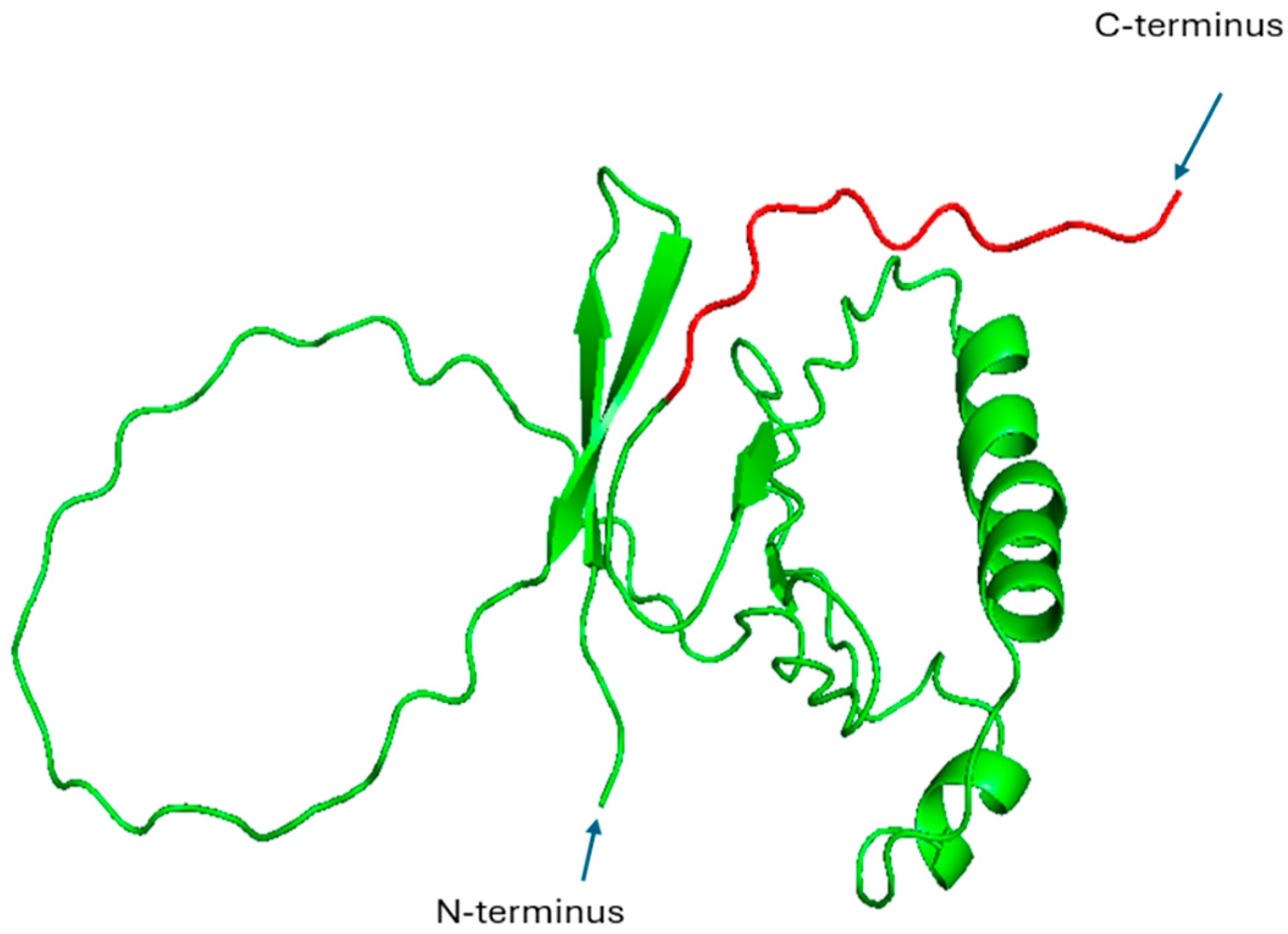
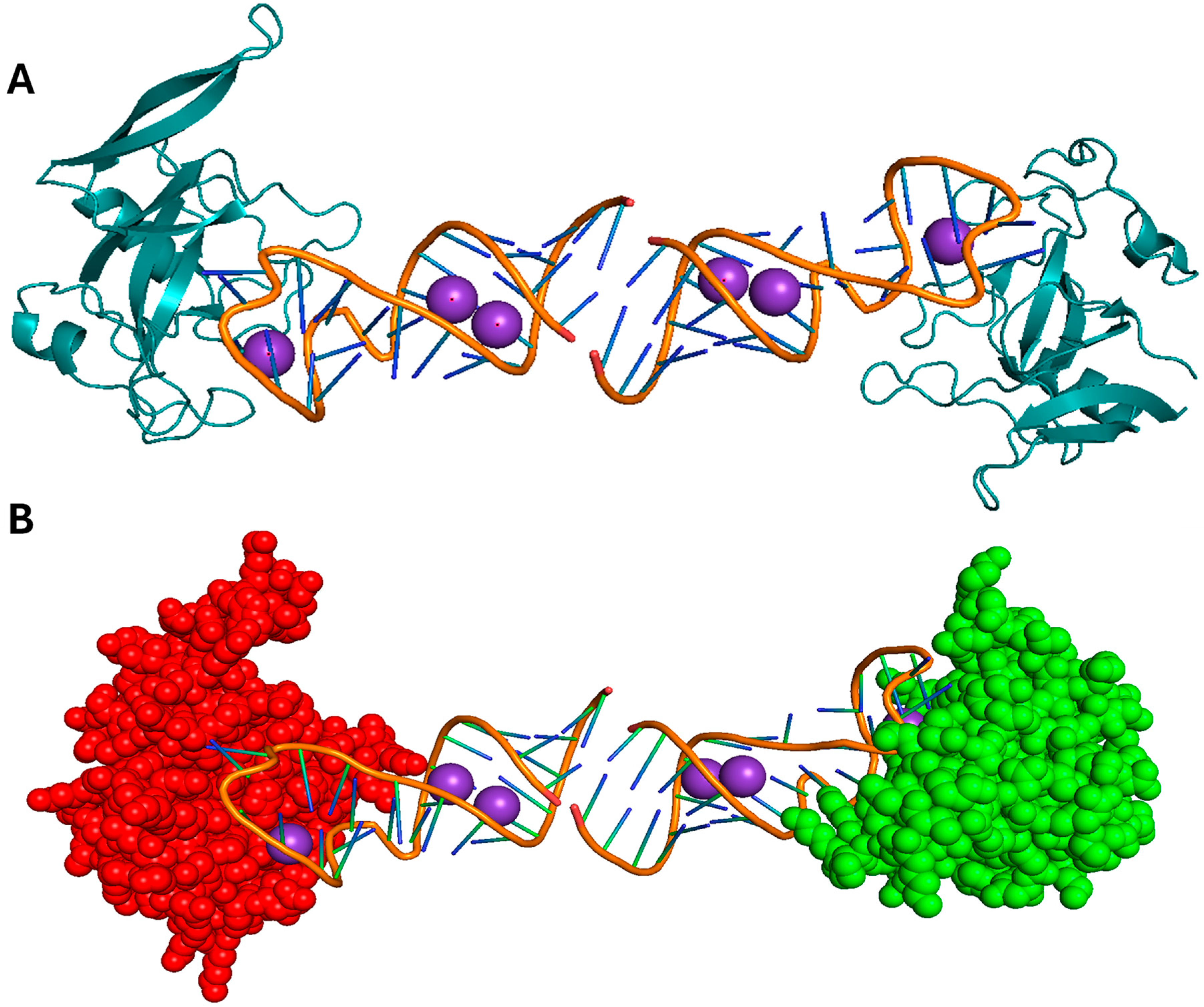
HBx is a multifunctional protein that coordinates HBV replication and modulates the signaling pathways of host cells. It activates NF-κB through direct interactions with the IKK complex and the inhibition of IκBα, culminating in the nuclear translocation of NF-κB and the subsequent expression of inflammatory genes (Figure 1). Additionally, HBx plays a role in the survival of hepatocytes, the induction of oxidative stress, and tumorigenesis within the context of chronic HBV infection [28]. Structurally, the characterization of HBx is challenging due to its partially disordered nature and propensity for aggregation. As no complete crystal or NMR structure currently exists in the Protein Data Bank (PDB), we used AlphaFold to predict its structure. This prediction shows that HBx comprises multiple helical segments and a flexible C-terminal region, which could be important for transcriptional activation by HBX of both viral and host genes (Table 1, Figure 3, [29]).

2.3. HPV E6 and E7 Proteins

The oncogenic proteins E6 and E7 of HPV are important for the viral life cycle and the underlying pathogenesis of HPV-related malignancies. E6 facilitates the degradation of p53, while E7 inactivates the retinoblastoma (Rb) tumor suppressor protein, resulting in unregulated cellular proliferation. Furthermore, both proteins have been associated with the promotion of chronic inflammation through the activation of NF-κB and the upregulation of cytokines (Figure 1, [30,31]). E6 directly influences the binding of NF-κB to promoter regions [32], while E7 activates the IKK complex, leading to Iκβ degradation and NF-κB translocation to the nucleus [33]. The structure of HPV16 E6 in association with the LxxLL motif of the E6AP ubiquitin ligase is available (Table 1, Figure 4A). This crystallographic analysis revealed the presence of two zinc-binding domains that facilitate substrate recognition and interaction with host proteins. These domains provide the mechanism of specificity toward the LxxLL motif and stability during binding to the E6AP ubiquitin ligase, which targets p53 for proteolytic degradation [34]. E6 comprises an N-terminal CR1/CR2 domain that is responsible for binding to Rb, which regulates the cell cycle, and a C-terminal zinc-binding domain (Figure 4A, [25]). There is no comprehensive, full-length structure of HPV E7, but there is a structure of the C-terminal domain (Figure 4B). The C-terminal domain contains a conserved zinc-binding motif (CxxC), where C represents cysteine residues and x represents any amino acid. This motif coordinates a zinc ion, which is needed to stabilize the three-dimensional structure, and promote interactions between proteins [35]. This structural integrity allows E7 to disrupt numerous cellular pathways, including those governing cell proliferation and immune response mechanisms [36]. A comprehensive understanding of these structural foundations not only clarifies their contributions to disease progression but also guides the development of targeted therapeutic strategies [26].

2.4. EBV LMP1

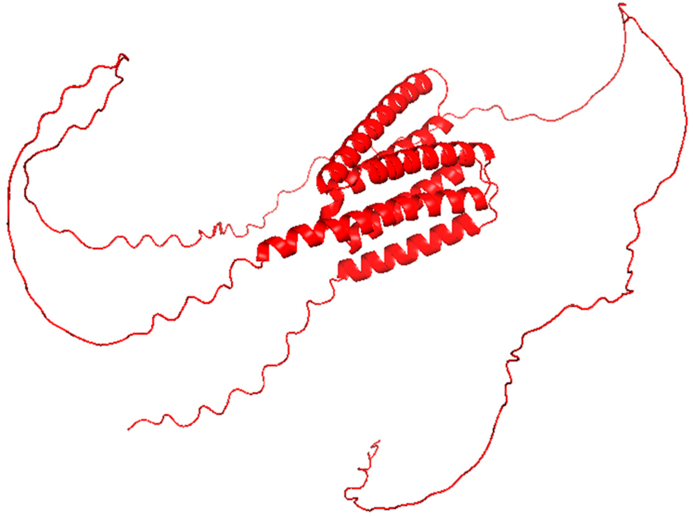
LMP1 is a transmembrane protein that operates as a constitutively active analogue of the TNFR. LMP1 interacts with the adaptor proteins TNF receptor-associated factor (TRAF) and TNFR1-associated death domain (TRADD) protein via its cytoplasmic C-terminal activation regions (CTAR1 and CTAR2), thereby connecting receptors to intracellular signaling pathways [37]. This interaction facilitates the activation of both canonical and non-canonical NF-κB signaling pathways, resulting in the upregulation of pro-inflammatory cytokines IL-6 and IL-8 (Figure 1). This results in sustained B cell activation, immune evasion, and chronic inflammatory responses, each of which contributes to viral persistence and oncogenesis [27,38]. LMP1 is associated with EBV-induced malignancies, including Hodgkin’s lymphoma and nasopharyngeal carcinoma, wherein inflammation leads to disease progression [39]. For LMP1, due to its multiple transmembrane domains and high flexibility, the full-length structure remains unresolved. We predicted a structure with AlphaFold (Table 1, Figure 5). The hypothesized structure revealed a configuration comprising six transmembrane α-helices integrated within the membrane domain, interconnected by long, unstructured intracellular and extracellular loops. Of particular interest, CTAR1 and CTAR2 protrude into the cytosol and are represented as partially disordered domains, which likely facilitates the recruitment of TRAF and TRADD.

3. Fungal Proteins Involved in Chronic Inflammation: Structural and Functional Insights

Fungal pathogens, such as C. albicans, A. fumigatus, and C. neoformans, are increasingly recognized as significant contributors to chronic inflammation, particularly in immunocompromised individuals [15,40]. Unlike viral proteins, which primarily manipulate intracellular host signaling pathways, fungal proteins exert their effects through interactions with extracellular and membrane-associated immune receptors. The shared mechanisms through which fungal proteins induce chronic inflammation involve the engagement of PRRs, including Dectin-1, TLRs, and C-type lectins, the activation of inflammasome complexes, particularly the NLRP3 inflammasome, and the modulation of immune cell functions, such as macrophage polarization and neutrophil recruitment (Figure 6 [41,42]). In the subsequent sections, we discuss several fungal proteins that are recognized for driving persistent inflammation, with a particular focus on their structural attributes and their implications in shaping the host immune response.

3.1. Saps from C. Albicans

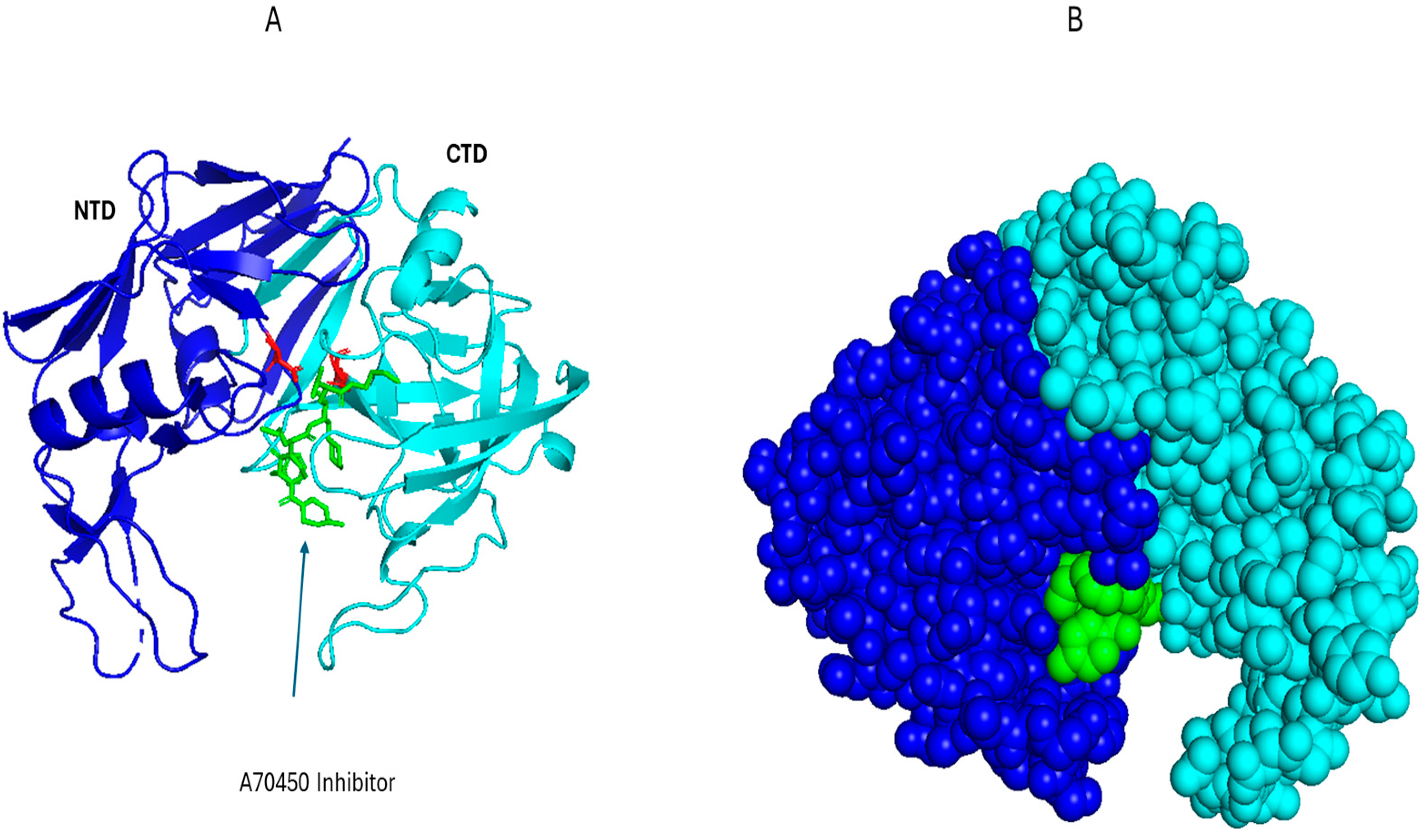
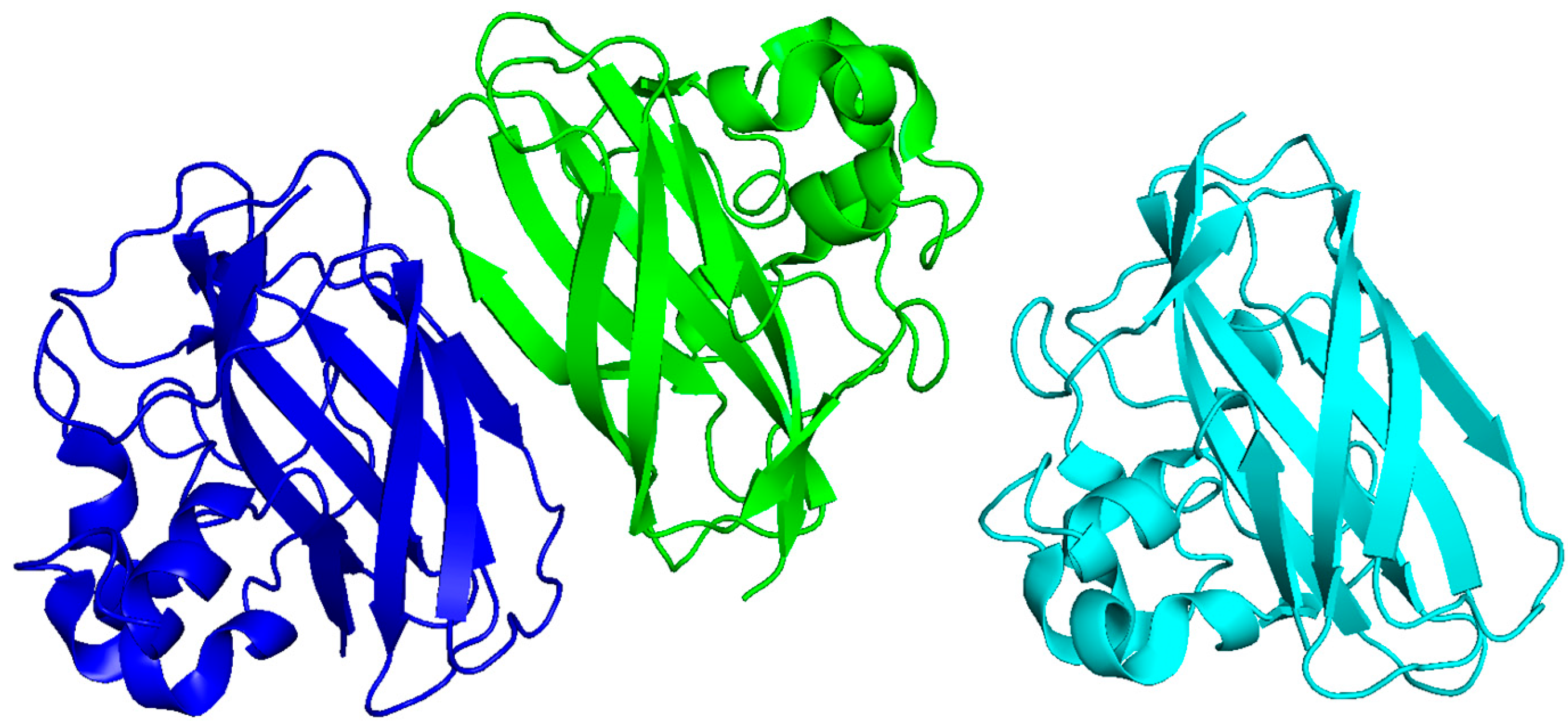
Saps represent a family of hydrolytic enzymes secreted by C. albicans that facilitate the degradation of host proteins to promote tissue invasion, including extracellular matrix (ECM) components such as collagen, laminin, and fibronectin. Among the various isoforms, Sap2 is the most investigated variant, which is involved in fungal pathogenesis and the modulation of host immune responses. The molecular architecture of Sap2 has been solved by X-ray crystallography at a resolution of 2.1 Å, which revealed a characteristic bilobed conformation typical of aspartic proteases. Sap2 contains distinct N- and C-terminal domains that have a substrate-binding cleft, with a well-conserved catalytic dyad (Asp32 and Asp218, Table 2, Figure 7A,B [43]). Such an arrangement gives it specificity and operational efficacy under acidic environments, akin to those found at mucosal surfaces and within phago-lysosomal compartments [44,45,46]. The enzymatic catabolism of ECM proteins by Sap2 not only facilitates fungal invasion into host tissues but also leads to the liberation of damage-associated molecular patterns (DAMPs). These DAMPs are recognized by PRRs including TLR4, Dectin-1, and various C-type lectin receptors (Figure 6). Moreover, the cellular stress induced by Sap2 and the degradation of host proteins leads to the activation of the NLRP3 inflammasome, a cytosolic complex that is important for the maturation and secretion of pro-inflammatory cytokines such as IL-1β and IL-18 [47,48]. Sap2 also results in the polarization of macrophages and augmentation of the recruitment of neutrophils, both of which enhance the mucosal inflammatory responses [49].
The small-molecule inhibitor A-70450 binds Sap2 with high affinity and occupies the active-site cleft, which emulates the transition state during peptide cleavage [50]. Extended exposure to Saps, particularly Sap2, is associated with chronic mucosal inflammation, as evidenced in conditions such as recurrent vulvovaginal candidiasis, where sustained immune activation and epithelial damage contribute significantly to the persistence and severity of the disease [44]. While A-70450 is potent in vitro, there was no protective effect in murine disseminated candidiasis models [51,52]. This could be due to the expression of additional, different Sap isoforms during system infection, so inhibition of Sap2 alone would be insufficient, or poor tissue distribution of the peptide. These discouraging early fundings likely contributed to the lack of further development of A-70450. However, with proper investigation, A-70450 could serve as the foundation for potent antifungal agents.
Table 2. Summary of fungal proteins that induce inflammation.
Fungal
Protein
SpeciesMechanism of
Inflammation
Structural FeaturesPDB CodeBinding Motif or
Interface
Ref.
Sap2C. albicansTLR4, Dectin-1; NLRP3 inflammasome activation; cytokine induction (IL-1β, IL-6, TNF-α)Pepsin-like fold;
active-site cleft
1EAGActive-site Asp residues + substrate-binding loops mediate TLR engagement[53]
Asp f1A. fumigatusTLR2/TLR4 activation; Th2 response; eosinophilic inflammationConserved α/β ribotoxin topology1JBSRibotoxin loop regions interact with TLR extracellular domains[54]
ChitinMultipleDectin-1, TLR9, NOD receptor engagement; IL-17 production; granuloma formationβ-sandwich fold (modelled from bacterial CBP21)2BEMGlucan-binding surface recognized by Dectin-1 CRD[55]
Als3C. albicansBinds E-cadherin and N-cadherin, triggers NF-κB and MAPK signalingβ barrel of the N-terminus4LE8β barrel of the N-terminus binds to the host cadherin receptors.[56]

3.2. Asp f1 of A. fumigatus

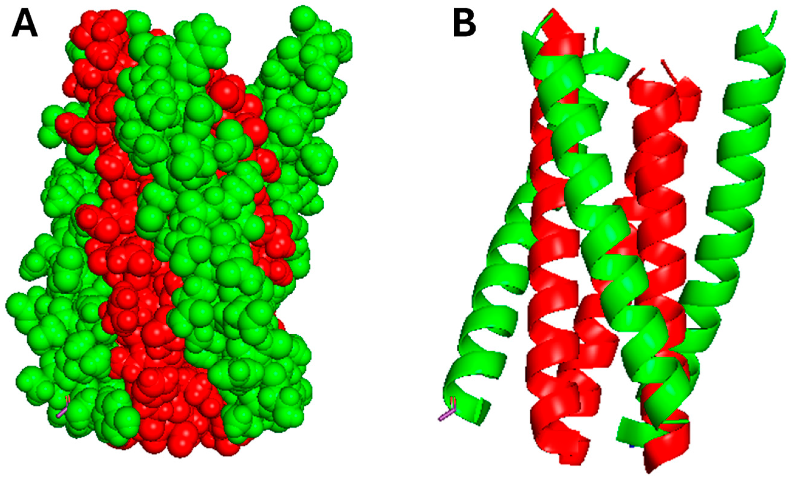
Asp f1 is a ribotoxin secreted by A. fumigatus that cleaves rRNA and can be a potent allergen in allergic bronchopulmonary aspergillosis (ABPA) [57]. Asp f1 induces TLR2 and TLR4, which results in a Th2-skewed immune responses, eosinophilia, and chronic airway inflammation (Figure 6, [58]). Despite the unknown three-dimensional conformation of Asp f1, bioinformatic analyses and sequence alignment investigations indicate that it exhibits over 60% sequence homology with restrictocin, an extensively characterized ribotoxin derived from Aspergillus restrictus (Figure 8, [54]). These homologous proteins generally adopt a conserved α/β ribotoxin topology, which includes the formation of a pronounced active-site cleft that is stabilized by disulfide linkages, thereby implying that Asp f1 is likely to exhibit a comparable structural conformation and catalytic mechanism. We predicted the three-dimensional structure of the Asp f1 protein using AlphaFold and aligned it with the experimentally determined structure of restrictocin (Table 2, Figure 9). The predicted and reference structures exhibit a high degree of structural similarity.

3.3. Chitin and Chitin-Binding Proteins

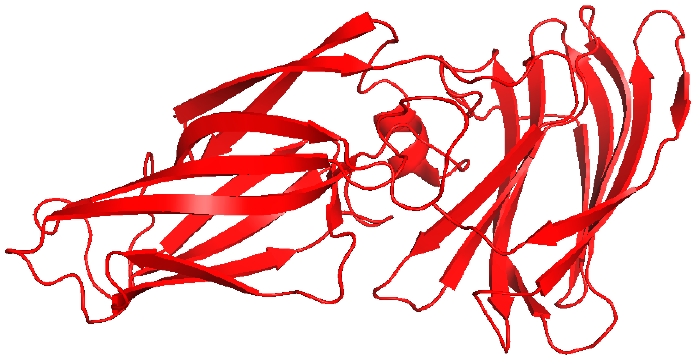
Chitin serves as a structural component of the fungal cell wall and functions as a pathogen-associated molecular pattern (PAMP). It is recognized by Dectin-1, NOD-like receptors, and TLR9, which leads to macrophage activation, IL-17 production, and, in certain instances, granulomatous inflammation (Figure 6). Binding to Dectin-1, a C-type lectin receptor, leads to receptor clustering and phosphorylation, which activates the NF-kB and MAPK pathways, leading to cytokine production and inflammation [59,60]. NOD-like receptors similarly can activate NF-kB [61]. Furthermore, fungi secrete chitin-binding proteins (CBPs) that stabilize newly synthesized cell walls and regulate chitin synthase and chitinase activity [62]. CBPs also defend against host immune responses, by sequestering the chitin and hiding it from immune detection [63]. While native chitin exhibits poor immunogenicity, chitin-binding proteins, like CBP21 from Serratia marcescens, a bacterial ortholog commonly used for fungal models, possess well-characterized structural features (Table 2, Figure 10). The trimeric form of CBP21 is stable and biologically relevant. CBPs exhibit conserved β-sandwich folds and surface residues that are important for interaction with chitin. The conserved β-sandwich folds also contribute to the stability of the structures that accommodate surface-exposed aromatic and polar residues, which enable selective interactions with chitin [64]. This structural configuration is important, since chitin is insoluble and needs specialized binding interfaces to promote enzymatic degradation [64,65]. Structural modelling of fungal CBPs suggests a parallel architecture [55].

3.4. Agglutinin-like Sequence Protein

Agglutinin-like sequence protein 3 (Als3) is a well-characterized fungal adhesin/invasin and is recognized as a virulence factor of C. albicans. Als3 interacts with E-cadherin receptors on epithelial cells and N-cadherin receptors on endothelial cells, which aids in fungal attachment and promotes endocytosis [56,66,67]. Epithelial cells that are exposed to C. albicans activate the NF-κB, MAPK, and PI3K signaling pathways, shortly after initial contact (Figure 6, [68]). A Swiss-Model was used to predict the structural conformation of Als3, which revealed that it contains an N-terminal adhesive domain that forms a cleft to accommodate host receptors [68]. The crystal structure of Als3 was also solved, and showed that it has a β-barrel fold characterized by a distinctive hydrophobic peptide-binding cleft, which facilitates interactions with host proteins, including E- and N-cadherin (Figure 11, [69]).

4. Biologics Targeting Viral and Fungal Inflammation

Biologic therapeutics, including antiviral peptides (AVPs), antifungal peptides (AFPs), and mAbs have emerged as pivotal components in the formulation of targeted interventions for chronic infections [70]. These biologics not only impede pathogen proliferation but also modulate the host immune response to mitigate subsequent inflammatory processes [71]. Chronic inflammation is distinguished by the continual activation of pro-inflammatory signaling cascades and overproduction of cytokines. Numerous pathogens utilize immune evasion mechanisms that enable their sustained presence within the host, which intensifies the inflammatory reaction [72,73,74]. Biologics have been engineered to dampen these common inflammatory pathways by neutralizing the pathogen directly or attenuating subsequent immune activation [75].
AVPs are short peptide sequences that inhibit viral entry or replication while simultaneously providing immunomodulatory benefits. Enfuvirtide (T-20) is an FDA-approved peptide that obstructs the entry of HIV-1 by specifically targeting the gp41 envelope glycoprotein (Figure 12). gp41 itself is immunogenic, which leads to antibody production and the release of pro-inflammatory cytokines. Consequently, the inhibition of gp41 not only curtails viral replication but may also mitigates inflammation, including that which is linked to Tat-mediated cytokine induction. The crystal structure of gp41 in complex with T20 was obtained using a peptide (N39) that mimics the N-terminal heptad repeat region (NHR) of gp41, which inherently forms a coiled coil. T20 occupies the hydrophobic grooves of this NHR mimic, effectively obstructing the formation of the six-helix bundle, which is needed for membrane fusion [76,77]. Investigational peptides such as C34 and HIV-1 inhibitor virus-inhibitory peptide (VIRIP) were developed to inhibit HIV-1 fusion [78,79]. C34, which is derived from the heptad repeat 2 (HR2) domain of gp41, inhibits the formation of the six-helix bundle that is essential for viral fusion. Lipid-conjugated derivatives, including sifuvirtide and albuvirtide, went through the preliminary phases of clinical trials in China, and had favorable safety profiles, tolerability, and suppression of viral replication in patients with prior treatment failure [80,81,82]. Albuvirtide received regulatory approval in China in 2018 following phase III clinical trials that demonstrated sustained reductions in HIV-1 viral load when used in conjunction with standard antiretroviral therapy [83]. VIRIP (VIR-576) is a peptide fragment derived from α1-antitrypsin that directly binds to gp41 and obstructs its insertion into the membranes of host cells [84]. In a Phase I/IIa clinical trials with 18 subjects, VIR-576 was generally tolerated and had a favorable safety profile. The use of VIR-576 as a short-term monotherapy in treatment-naïve HIV-1 patients resulted in a substantial decrease in plasma viral load exceeding one order of magnitude, indicating both its tolerability and antiviral efficacy [85].
Peptide-based vaccines targeting E6 and E7 of HPV are designed to provoke cytotoxic T cell response to reduce inflammation and viral persistence. ISA101, a synthetic long-peptide vaccine that targets E6 and E7, induced HPV-specific CD4+ and CD8+ T cell responses in preclinical models [86]. A Phase II clinical trial examined the synergistic effects of ISA101 in combination with the programmed cell death protein 1 (PD-1) inhibitor nivolumab in 26 individuals diagnosed with advanced HPV-positive oropharyngeal carcinoma. Within this cohort, 3 patients had a partial response (PR), including two individuals who had not previously exhibited a response to immune checkpoint inhibitor therapy. Booster immunizations were delivered to nine patients, and the interval to response varied between 127 and 307 days. Adverse events associated with ISA101 were mostly minor, but autoimmune events were observed in 7.7% of the cohort. These results suggest that the combination of ISA101 and nivolumab has a benefit in patients who have failed therapy. This study highlights the potential efficacy of peptide-based immunotherapeutic strategies in malignancies driven by viral pathogens [87].
Additional peptides currently under development target inflammatory viral proteins such as HBx and LMP1, to disrupt their interactions with host signaling proteins that activate NF-κB [86,88]. A TRAF6-inhibitory peptide was identified from a bead-based AlphaScreen assay that interferes with the interaction between LMP1 and TRAF6 [89]. This peptide could be used to reduce NF-κB activation in vivo. Synthetic peptides derived from the C-terminal domain of LMP1 have also been synthesized, which mimic the signaling domains of LMP1. These peptides function as decoys by competitively binding to adaptor proteins such as TRAF2 and TRADD, obstructing LMP1 interaction. This sequestration impedes NF-κB signaling and triggers apoptosis in EBV-transformed cells [90].
AFPs also have dual functionalities by targeting fungal membranes while concurrently attenuating host inflammatory responses [91]. Naturally occurring peptides, such as Histatin 5 from human saliva, exhibit antifungal activity against C. albicans and contribute to the limitation of fungal-induced epithelial inflammation [92]. LL-37, a cathelicidin, has antifungal and anti-inflammatory properties by inhibiting the release of pro-inflammatory cytokines [93]. At present, LL-37 is undergoing both preclinical and clinical development. Phase I/II clinical investigations involving the management of venous leg ulcers revealed that the administration of low-dose topical formulations significantly enhances wound healing [94]. Human defensins (hBD-1, hBD-2) also disrupt fungal membranes and alter innate immune pathways, including TLR signaling and macrophage activation [95]. The use of β-defensins, specifically hBD-2, are in preclinical development and have demonstrated encouraging anti-inflammatory and antifungal properties across various animal models. Although clinical trials have not yet commenced, the observed efficacy and in vivo safety indicate potential for further development [96]. NP339, a synthetic AFP, is presently under investigation for its therapeutic potential in treating candidiasis while minimizing immune overactivation [97]. NP339 exhibits rapid fungicidal activity against various fungal pathogens and does not promote resistance development. In addition, NP339 was effective in animal models and Phase I clinical trials will soon begin [98,99].
mAbs are effective in the regulation of inflammatory responses that are associated with chronic infections [100]. Infliximab and Adalimumab are anti-TNFα antibodies and mitigate the excessive signaling of TNFα. Excessive TNFα signaling is frequently elevated in the presence of viral proteins, and during fungal inflammation mediated through Dectin-1 or TLR signaling pathways [101,102]. Tocilizumab, an antibody that specifically targets the IL-6 receptor, was approved for autoimmune disorders and conditions characterized by cytokine storms and was recently investigated for use with severe viral infections. During the COVID-19 pandemic, large clinical trials demonstrated that tocilizumab decreased mortality rates and enhanced recovery outcomes in patients hospitalized with severe disease. Despite its lack of widespread endorsement for other viral infections, its efficacy in the context of COVID-19 demonstrates its utility in mitigating cytokine-mediated complications associated with viral diseases (Table 3, [103]). The use of tocilizumab for treatment of fungal diseases, including ABPA, is currently under exploration due to the known role of IL-6 in exacerbating the inflammatory responses. Initial research indicates that the inhibition of IL-6 signaling may alleviate pulmonary inflammation and enhance respiratory function in patients afflicted with ABPA. More stringent clinical trials are required to evaluate the safety profile, optimal dosing parameters, and overall efficacy in relation to fungal-associated immune dysregulation [104,105]. Anakinra, which acts as an IL-1 receptor antagonist, is being assessed for its capacity to inhibit IL-1β–mediated inflammation that occurs in response to fungal inflammasome activators and viral proteins that interact with innate immune sensors (Table 3 [106,107]). In preclinical models, anakinra was effective in diminishing IL-1β production and mitigating inflammation in patients with cystic fibrosis and chronic granulomatous disease, which have an increased risk for fungal infections and the activation of inflammasomes [108]. Despite compelling pre-clinical evidence, there are no clinical trials examining the use of anakinra for fungal infections. Most of the research focuses on broader inflammatory disorders or autoimmune diseases. Considering the encouraging preclinical findings, further clinical investigations to evaluate anakinra for the treatment of fungal infections characterized by inflammation should be performed [109].
In addition to biologic agents that directly target pathogens, there is an increasing focus on therapies that target the common molecular mechanisms of inflammation that are initiated by both viral and fungal infections. A pivotal pathway in this context is the activation of NF-κB, which is exploited by viral proteins and is activated through fungal PRR signaling. Bortezomib, a proteasome inhibitor that has been approved for the treatment of multiple myeloma, inhibits the degradation of IκB and consequently prevents the activation of NF-κB (Table 3). Bortezomib has anti-inflammatory effects in experimental models of both viral- and fungal-induced inflammation [110]. Likewise, dimethyl fumarate, which was originally approved for the management of multiple sclerosis, inhibits NF-κB activity and demonstrated potential in treating neuroinflammation linked to HIV [111]. Colchicine, a microtubule inhibitor that is utilized in the treatment of gout, obstructs inflammasome assembly and reduces IL-1β levels in inflammation driven by infections. Given the impact of heightened IL-1β signaling in the context of fungal infections, there is interest in the synergistic application of antifungal agents alongside targeted immunomodulatory therapies [112]. The use of colchicine or alternative inflammasome-inhibiting treatments, such as anakinra, alongside traditional antifungal regimens can become a host-directed therapeutic approach that reduces both fungal load and immune-mediated damage to tissues.

5. Limitations of Current Biologics and the Potential of Conjugated Therapeutic Proteins

Despite the potential of biologics, including mAbs, AVPs, and AFPs, in the management of chronic inflammation associated with viral and fungal infections, numerous challenges impede their sustained efficacy and clinical applicability. A principal limitation is their abbreviated plasma half-life, particularly pertinent to peptides, which frequently necessitate repeated administration or formulation modifications to sustain therapeutic concentrations [113]. Furthermore, restricted tissue penetration, particularly into immune-privileged regions such as the central nervous system or deeply embedded fungal reservoirs, constrains the accessibility of many antibodies and peptides [114]. Another significant concern is the potential for immunogenicity, where recurrent administration may elicit host immune responses against the therapeutic agent itself, which diminishes efficacy and possibly induces adverse reactions [115]. Biologics frequently exhibit a lack of specificity for the inflammatory microenvironment, operating systemically rather than accurately targeting inflamed or infected tissues. This phenomenon may result in off-target effects, including generalized immune suppression, which could render patients susceptible to secondary infections or hinder the clearance of the primary pathogen [116]. Finally, in the context of AFPs and AVPs, proteolytic degradation by host enzymes can further diminish bioavailability, necessitating chemical modifications or protective delivery methodologies [117].
To address these challenges, there is an increasing interest in engineered conjugated therapeutic proteins, which combine biologic molecules with targeting, stabilizing, or effector domains to enhance functional capacity, selectivity, and pharmacokinetics. For example, AVPs or AFPs can be conjugated to antibody scaffolds or Fc fragments to augment stability and improve tissue distribution. Such conjugates preserve antimicrobial activity while increasing the structural stability and specificity inherent to antibodies. Likewise, mAbs may be conjugated to cytokine inhibitors, signaling inhibitors, or enzymatic payloads, resulting in multifunctional therapeutics capable of neutralizing a pathogen and modulating the local immune response [118]. Recent preclinical investigations have evaluated the viability of such constructs within inflammatory and oncological contexts. For example, antibody–drug conjugates that specifically target immune cells were designed to deliver anti-inflammatory agents, such as PDE4 inhibitors or glucocorticoids, which resulted in a diminished release of cytokines in human monocytes and peripheral blood mononuclear cells [119,120]. Additionally, bifunctional constructs that integrate antigen targeting with both immunomodulatory cytokines and cytotoxic agents have also exhibited therapeutic potential in murine experimental models [120,121]. Although these multifunctional mAbs have yet to be utilized in clinical practice for infectious diseases, their success with other disease states implies promise for host-directed therapeutic strategies for inflammation induced by pathogens.
Conjugated proteins present an opportunity to administer anti-inflammatory payloads directly to inflamed tissues, such as by targeting NF-κB–active environments or cells expressing PRRs like Dectin-1 or TLRs. For example, a conjugated antibody that targets fungal β-glucan could facilitate the localized delivery of an inflammasome inhibitor, which would attenuate IL-1β secretion specifically at the infection site [118]. For viral infections, conjugates that include Tat- or HBx-targeting antibodies with cell-penetrating peptides could locally inhibit NF-κB activation without inducing systemic toxicity [122]. Delivery platforms, which include nanoparticles or liposomes, affixed with AVPs, AFPs, or mAbs, can be engineered to respond to inflammatory stimuli, such as pH variations or reactive oxygen species levels, which would release their therapeutic cargo exclusively within diseased tissues [122]. This intelligent targeting paradigm mitigates collateral effects and enhances therapeutic efficacy. Recent advancements in protein engineering and chemical conjugation methodologies now facilitate site-specific, stable, and scalable production of such biologics, which could lead to a new generation of therapeutics [123]. These advancements include the implementation of engineered antibody scaffolds, and novel conjugation chemistries, such as thiol-selective modification and enzymatic click reactions that facilitate the controlled and reproducible attachment of therapeutic payloads [121,124]. Such site-specific methodologies reduce heterogeneity, augment pharmacokinetics, and enhance safety by mitigating off-target effects [125]. Collectively, these innovations establish a robust technological framework for the translation of conjugated proteins from laboratory to clinical application.

6. Challenges and Future Directions

Before engineered conjugated proteins can be used to treat chronic inflammation resulting from persistent infections, numerous challenges must be overcome to fully achieve their clinical efficacy [126]. A major hurdle pertains to the effective delivery of these conjugates to chronically inflamed tissues, including mucosal surfaces, pulmonary regions, and other affected organs. Such sites frequently exhibit complex biological barriers, including thick mucus layers, altered vascular permeability, fibrotic extracellular matrices, and infiltration by immune cells, all of which can impede the penetration, retention, and bioavailability of large biologics (Table 4, [127]). The development of advanced delivery systems, such as nanoparticles, liposomes, inhalable formulations, or ligands specific to target tissues, is imperative to overcome these physical and biochemical barriers to ensure that therapeutic concentrations effectively reach the sites of inflammation [128]. The issues of immunogenicity and rapid degradation persist as significant concerns for protein therapeutics. Engineered conjugates, particularly those incorporating non-human sequences or innovative linkers, are susceptible to eliciting immune responses that may neutralize the therapeutic agent or promote adverse effects. Additionally, proteases that are prevalent in inflamed tissues and systemic circulation can rapidly degrade peptides and proteins, resulting in reduced half-life and diminished efficacy.
Addressing these challenges necessitates the application of sophisticated protein engineering techniques, such as amino acid substitutions aimed at diminishing immunogenic epitopes, chemical modifications (e.g., PEGylation or glycosylation), the cyclization of peptides to enhance stability, and the incorporation of D-amino acids or non-natural residues to confer resistance to proteolysis (Table 4, [129,130]). Furthermore, the production of stable, homogeneous conjugated proteins that meet clinical-grade quality standards and can be generated in sufficient quantities requires robust expression systems and purification protocols. The inherent complexity of fusion proteins, in the case of conjugated proteins, exacerbates the risks of aggregation, misfolding, and variability between batches, thereby complicating regulatory approval processes and commercial production. The potential for off-target effects and toxicity must also be rigorously assessed. While the targeting of inflamed tissues or pathogen proteins enhances specificity, unintended interactions with host proteins or disruption of the normal microbiota may lead to adverse consequences. Comprehensive preclinical safety evaluations and sophisticated targeting strategies are essential to mitigate such risks. Emerging drug resistance presents a significant obstacle in contemporary biomedical research. Pathogens possess the ability to mutate, modify surface proteins that are the target of antibody domains, or alter their membrane composition to avoid interaction with antimicrobial peptides. This reality underscores the necessity for continual surveillance and may necessitate the development of adaptable or multi-epitope targeting conjugates. The incorporation of AI-driven structural prediction tools such as AlphaFold provides promising avenues to expedite the design process; however, computational models are inherently constrained, particularly in terms of predicting the dynamics of flexible linkers, post-translational modifications, and interactions within complex physiological settings [131]. Experimental validation is indispensable, and the implementation of hybrid computational-experimental workflows will be necessary [132]. Despite these obstacles, significant progress in protein engineering, delivery technologies, and computational modelling is occurring at an accelerated pace. The field is on the brink of producing increasingly sophisticated conjugated proteins that can selectively target pathogens and modulate chronic inflammation, thereby offering potential solutions for novel therapies against persistent infectious diseases [133].

Author Contributions

Conceptualization, M.H.; resources, V.J.C.; writing—original draft preparation, M.H. and A.L.G.; writing—review and editing, M.H., A.L.G. and V.J.C.; visualization, M.H.; supervision, M.H. and V.J.C.; funding acquisition, V.J.C. All authors have read and agreed to the published version of the manuscript.

Funding

This research was funded by the National Institute of General Medical Sciences, grant number R35GM138303 awarded to V.J.C.

Institutional Review Board Statement

Not applicable.

Informed Consent Statement

Not applicable.

Data Availability Statement

Data is contained within the article.

Acknowledgments

During the preparation of this manuscript, the author(s) used AI Paraphraser (SCISPACE), for the purposes of paraphrasing and editing of some sentences for the original draft. The authors have reviewed and edited the output and take full responsibility for the content of this publication.

Conflicts of Interest

The authors declare no conflicts of interest.

Abbreviations

The following abbreviations are used in this manuscript:
ABPAAllergic Bronchopulmonary Aspergillosis
ADCAntibody–Drug Conjugate
AFPAntifungal Peptide
AVPAntiviral Peptide
CBPChitin-Binding Protein
CTARC-Terminal Activation Region (e.g., in LMP1)
DAMPDamage-Associated Molecular Pattern
EBVEpstein–Barr Virus
ECMExtracellular Matrix
FDAFood and Drug Administration (USA)
HBVHepatitis B Virus
HBxHepatitis B virus X protein
HCVHepatitis C Virus
HIVHuman Immunodeficiency Virus
HPVHuman Papillomavirus
IKKIκB Kinase
ILInterleukin (e.g., IL-1β, IL-6)
LMP1Latent Membrane Protein 1 (EBV)
LPSLipopolysaccharide
mAbMonoclonal Antibody
NF-κBNuclear Factor Kappa-Light-Chain-Enhancer of Activated B Cells
NLRP3NOD-, LRR- and Pyrin Domain-Containing Protein 3 (inflammasome)
PAMPPathogen-Associated Molecular Pattern
PDBProtein Data Bank
PRRPattern Recognition Receptor
RbRetinoblastoma protein
SapSecreted Aspartyl Protease
TatTrans-Activator of Transcription (HIV-1)
TLRToll-Like Receptor
TNF-αTumor Necrosis Factor Alpha
TNFRTumor Necrosis Factor Receptor
TRAFTNF Receptor-Associated Factor
TRADDTNFR1-Associated Death Domain Protein
VIRIPVirus-Inhibitory Peptide

References

  1. Wen, Y.; Zhu, Y.; Zhang, C.; Yang, X.; Gao, Y.; Li, M.; Yang, H.; Liu, T.; Tang, H. Chronic inflammation, cancer development and immunotherapy. Front. Pharmacol. 2022, 13, 1040163. [Google Scholar] [CrossRef]
  2. Ren, D.; Ye, X.; Chen, R.; Jia, X.; He, X.; Tao, J.; Jin, T.; Wu, S.; Zhang, H. Activation and evasion of inflammasomes during viral and microbial infection. Cell. Mol. Life Sci. 2025, 82, 56. [Google Scholar] [CrossRef] [PubMed]
  3. Tugizov, S. HIV-1 Tat-induced disruption of epithelial junctions and epithelial-mesenchymal transition of oral and genital epithelial cells lead to increased invasiveness of neoplastic cells and the spread of herpes simplex virus and cytomegalovirus. Front. Immunol. 2025, 16, 1541532. [Google Scholar] [CrossRef] [PubMed]
  4. Lien, K.; Mayer, W.; Herrera, R.; Padilla, N.T.; Cai, X.; Lin, V.; Pholcharoenchit, R.; Palefsky, J.; Tugizov, S.M. HIV-1 Proteins gp120 and Tat Promote Epithelial-Mesenchymal Transition and Invasiveness of HPV-Positive and HPV-Negative Neoplastic Genital and Oral Epithelial Cells. Microbiol. Spectr. 2022, 10, e03622. [Google Scholar] [CrossRef] [PubMed]
  5. Floettmann, J.E.; Eliopoulos, A.G.; Jones, M.; Young, L.S.; Rowe, M. Epstein–Barr virus latent membrane protein-1 (LMP1) signalling is distinct from CD40 and involves physical cooperation of its two C-terminus functional regions. Oncogene 1998, 17, 2383–2392. [Google Scholar] [CrossRef]
  6. Das, B.; Sarkar, C.; Rawat, V.S.; Kalita, D.; Deka, S.; Agnihotri, A. Promise of the NLRP3 inflammasome inhibitors in in vivo disease models. Molecules 2021, 26, 4996. [Google Scholar] [CrossRef]
  7. Sack, M.N. Mitochondrial fidelity and metabolic agility control immune cell fate and function. J. Clin. Investig. 2018, 128, 3651–3661. [Google Scholar] [CrossRef]
  8. Hinz, M.; Scheidereit, C. The IκB kinase complex in NF-κB regulation and beyond. EMBO Rep. 2014, 15, 46–61. [Google Scholar] [CrossRef] [PubMed]
  9. Sun, S.-C. The non-canonical NF-κB pathway in immunity and inflammation. Nat. Rev. Immunol. 2017, 17, 545–558. [Google Scholar] [CrossRef]
  10. Lien, K.; Mayer, W.; Herrera, R.; Rosbe, K.; Tugizov, S.M. HIV-1 proteins gp120 and tat induce the epithelial–mesenchymal transition in oral and genital mucosal epithelial cells. PLoS ONE 2019, 14, e0226343. [Google Scholar] [CrossRef]
  11. Mahomed, S. Broadly neutralizing antibodies for HIV prevention: A comprehensive review and future perspectives. Clin. Microbiol. Rev. 2024, 37, e0015222. [Google Scholar] [CrossRef]
  12. Riddler, S.A.; Zheng, L.; Durand, C.M.; Ritz, J.; Koup, R.A.; Ledgerwood, J.; Bailer, R.T.; Koletar, S.L.; Eron, J.J.; Keefer, M.C. Randomized clinical trial to assess the impact of the broadly neutralizing HIV-1 monoclonal antibody VRC01 on HIV-1 persistence in individuals on effective ART. Open Forum Infect. Dis. 2018, 5, ofy242. [Google Scholar] [CrossRef]
  13. Gunst, J.D.; Pahus, M.H.; Rosás-Umbert, M.; Lu, I.-N.; Benfield, T.; Nielsen, H.; Johansen, I.S.; Mohey, R.; Østergaard, L.; Klastrup, V. Early intervention with 3BNC117 and romidepsin at antiretroviral treatment initiation in people with HIV-1: A phase 1b/2a, randomized trial. Nat. Med. 2022, 28, 2424–2435. [Google Scholar] [CrossRef] [PubMed]
  14. Mahomed, S.; Pillay, K.; Hassan-Moosa, R.; Galvão, B.P.G.V.; Burgers, W.A.; Moore, P.L.; Rose-Abrahams, M.; Williamson, C.; Garrett, N. Clinical trials of broadly neutralizing monoclonal antibodies in people living with HIV—A review. AIDS Res. Ther. 2025, 22, 44. [Google Scholar] [CrossRef] [PubMed]
  15. Garcia-Rubio, R.; de Oliveira, H.C.; Rivera, J.; Trevijano-Contador, N. The fungal cell wall: Candida, Cryptococcus, and Aspergillus species. Front. Microbiol. 2020, 10, 2993. [Google Scholar] [CrossRef] [PubMed]
  16. Cai, R.; Gong, X.; Li, X.; Jiang, Y.; Deng, S.; Tang, J.; Ge, H.; Wu, C.; Tang, H.; Wang, G.; et al. Dectin-1 aggravates neutrophil inflammation through caspase-11/4-mediated macrophage pyroptosis in asthma. Respir. Res. 2024, 25, 119. [Google Scholar] [CrossRef]
  17. Sa, M.; da Silva, M.; Ball, B.; Geddes-McAlister, J. Revealing the dynamics of fungal disease with proteomics. Mol. Omics 2025, 21, 173–184. [Google Scholar] [CrossRef]
  18. Lohse, M.B.; Gulati, M.; Craik, C.S.; Johnson, A.D.; Nobile, C.J. Combination of antifungal drugs and protease inhibitors prevent Candida albicans biofilm formation and disrupt mature biofilms. Front. Microbiol. 2020, 11, 1027. [Google Scholar] [CrossRef]
  19. Xiao, Q.; Liu, Y.; Shu, X.; Li, Y.; Zhang, X.; Wang, C.; He, S.; Li, J.; Li, T.; Liu, T.; et al. Molecular mechanisms of viral oncogenesis in haematological malignancies: Perspectives from metabolic reprogramming, epigenetic regulation and immune microenvironment remodeling. Exp. Hematol. Oncol. 2025, 14, 69. [Google Scholar] [CrossRef]
  20. Fiume, G.; Vecchio, E.; De Laurentiis, A.; Trimboli, F.; Palmieri, C.; Pisano, A.; Falcone, C.; Pontoriero, M.; Rossi, A.; Scialdone, A.; et al. Human immunodeficiency virus-1 Tat activates NF-κB via physical interaction with IκB-α and p65. Nucleic Acids Res. 2012, 40, 3548–3562. [Google Scholar] [CrossRef]
  21. Dandekar, D.H.; Ganesh, K.N.; Mitra, D. HIV-1 Tat directly binds to NFκB enhancer sequence: Role in viral and cellular gene expression. Nucleic Acids Res. 2004, 32, 1270–1278. [Google Scholar] [CrossRef]
  22. Péloponèse, J.-M., Jr.; Grégoirea, C.; Opia, S.; Esquieua, D.; Sturgisa, J.; Lebrunb, É.; Meursc, É.; Colletted, Y.; Olived, D.; Aubertine, A.-M. 1H-13 C nuclear magnetic resonance assignment and structural characterization of HIV-1 Tat protein. Comptes Rendus L’académie Sci.-Ser. III-Sci. Vie 2000, 323, 883–894. [Google Scholar]
  23. You, H.; Qin, S.; Zhang, F.; Hu, W.; Li, X.; Liu, D.; Kong, F.; Pan, X.; Zheng, K.; Tang, R. Regulation of pattern-recognition receptor signaling by HBX during hepatitis B virus infection. Front. Immunol. 2022, 13, 829923. [Google Scholar] [CrossRef]
  24. Schollmeier, A.; Glitscher, M.; Hildt, E. Relevance of HBx for hepatitis B virus-associated pathogenesis. Int. J. Mol. Sci. 2023, 24, 4964. [Google Scholar] [CrossRef]
  25. Zanier, K.; Charbonnier, S.; Sidi, A.O.M.h.O.; McEwen, A.G.; Ferrario, M.G.; Poussin-Courmontagne, P.; Cura, V.; Brimer, N.; Babah, K.O.; Ansari, T. Structural basis for hijacking of cellular LxxLL motifs by papillomavirus E6 oncoproteins. Science 2013, 339, 694–698. [Google Scholar] [CrossRef]
  26. Ohlenschläger, O.; Seiboth, T.; Zengerling, H.; Briese, L.; Marchanka, A.; Ramachandran, R.; Baum, M.; Korbas, M.; Meyer-Klaucke, W.; Dürst, M. Solution structure of the partially folded high-risk human papilloma virus 45 oncoprotein E7. Oncogene 2006, 25, 5953–5959. [Google Scholar] [CrossRef]
  27. Šimičić, P.; Batović, M.; Stojanović Marković, A.; Židovec-Lepej, S. Deciphering the role of Epstein–Barr virus latent membrane protein 1 in immune modulation: A multifaced Signalling perspective. Viruses 2024, 16, 564. [Google Scholar] [CrossRef]
  28. Lim, K.-H.; Choi, H.S.; Park, Y.K.; Park, E.-S.; Shin, G.C.; Kim, D.H.; Ahn, S.H.; Kim, K.-H. HBx-induced NF-κB signaling in liver cells is potentially mediated by the ternary complex of HBx with p22-FLIP and NEMO. PLoS ONE 2013, 8, e57331. [Google Scholar] [CrossRef] [PubMed]
  29. Liu, W.; Zhang, X.; Deng, Y.; Wang, D.; Li, H. Unfolding HBx for an epigenetic switch of HBV cccDNA minichromosome. Protein Cell 2025, 16, 753–763. [Google Scholar] [CrossRef]
  30. Letafati, A.; Taghiabadi, Z.; Zafarian, N.; Tajdini, R.; Mondeali, M.; Aboofazeli, A.; Chichiarelli, S.; Saso, L.; Jazayeri, S.M. Emerging paradigms: Unmasking the role of oxidative stress in HPV-induced carcinogenesis. Infect. Agents Cancer 2024, 19, 30. [Google Scholar] [CrossRef] [PubMed]
  31. Peng, Q.; Wang, L.; Zuo, L.; Gao, S.; Jiang, X.; Han, Y.; Lin, J.; Peng, M.; Wu, N.; Tang, Y.; et al. HPV E6/E7: Insights into their regulatory role and mechanism in signaling pathways in HPV-associated tumor. Cancer Gene Ther. 2024, 31, 9–17. [Google Scholar] [CrossRef] [PubMed]
  32. James, M.A.; Lee, J.H.; Klingelhutz, A.J. Human papillomavirus type 16 E6 activates NF-kappaB, induces cIAP-2 expression, and protects against apoptosis in a PDZ binding motif-dependent manner. J. Virol. 2006, 80, 5301–5307. [Google Scholar] [CrossRef]
  33. Carrillo-Beltrán, D.; Muñoz, J.P.; Guerrero-Vásquez, N.; Blanco, R.; León, O.; de Souza Lino, V.; Tapia, J.C.; Maldonado, E.; Dubois-Camacho, K.; Hermoso, M.A.; et al. Human Papillomavirus 16 E7 promotes EGFR/PI3K/AKT1/NRF2 signaling pathway contributing to PIR/NF-κB activation in oral cancer cells. Cancers 2020, 12, 1904. [Google Scholar] [CrossRef]
  34. Suarez, I.; Trave, G. Structural insights in multifunctional papillomavirus oncoproteins. Viruses 2018, 10, 37. [Google Scholar] [CrossRef]
  35. Braspenning, J.; Marchini, A.; Albarani, V.; Levy, L.; Ciccolini, F.; Cremonesi, C.; Ralston, R.; Gissmann, L.; Tommasino, M. The CXXC Zn binding motifs of the human papillomavirus type 16 E7 oncoprotein are not required for its in vitro transforming activity in rodent cells. Oncogene 1998, 16, 1085–1089. [Google Scholar] [CrossRef]
  36. Spardy, N.; Covella, K.; Cha, E.; Hoskins, E.E.; Wells, S.I.; Duensing, A.; Duensing, S. Human papillomavirus 16 E7 oncoprotein attenuates DNA damage checkpoint control by increasing the proteolytic turnover of claspin. Cancer research 2009, 69, 7022–7029. [Google Scholar] [CrossRef]
  37. Eliopoulos, A.G.; Young, L.S. LMP1 structure and signal transduction. Semin. Cancer Biol. 2001, 11, 435–444. [Google Scholar] [CrossRef]
  38. Kieser, A.; Sterz, K.R. The latent membrane protein 1 (LMP1). In Epstein Barr Virus Volume 2: One Herpes Virus: Many Diseases; Springer: Cham, Switzerland, 2015; pp. 119–149. [Google Scholar]
  39. Huang, X.; Zhang, M.; Zhang, Z. The role of LMP1 in Epstein-Barr virus-associated gastric cancer. Curr. Cancer Drug Targets 2024, 24, 127–141. [Google Scholar] [CrossRef]
  40. Stuckey, P.V.; Santiago-Tirado, F.H. Fungal mechanisms of intracellular survival: What can we learn from bacterial pathogens? Infect. Immun. 2023, 91, e0043422. [Google Scholar] [CrossRef] [PubMed]
  41. Tang, J.; Lin, G.; Langdon, W.Y.; Tao, L.; Zhang, J. Regulation of C-type lectin receptor-mediated antifungal immunity. Front. Immunol. 2018, 9, 123. [Google Scholar] [CrossRef] [PubMed]
  42. Hatinguais, R.; Willment, J.A.; Brown, G.D. PAMPs of the fungal cell wall and mammalian PRRs. In The Fungal Cell Wall: An Armour and a Weapon for Human Fungal Pathogens; Springer: Cham, Switzerland, 2020; pp. 187–223. [Google Scholar]
  43. Behnen, J.; Köster, H.; Neudert, G.; Craan, T.; Heine, A.; Klebe, G. Experimental and computational active site mapping as a starting point to fragment-based lead discovery. ChemMedChem 2012, 7, 248–261. [Google Scholar] [CrossRef] [PubMed]
  44. Bruno, V.M.; Shetty, A.C.; Yano, J.; Fidel, P.L.; Noverr, M.C.; Peters, B.M. Transcriptomic Analysis of Vulvovaginal Candidiasis Identifies a Role for the NLRP3 Inflammasome. mBio 2015, 6, e00182-15. [Google Scholar] [CrossRef]
  45. Camilli, G.; Griffiths, J.S.; Ho, J.; Richardson, J.P.; Naglik, J.R. Some like it hot: Candida activation of inflammasomes. PLoS Pathog. 2020, 16, e1008975. [Google Scholar] [CrossRef] [PubMed]
  46. Cheng, K.O.; Montano, D.E.; Zelante, T.; Dietschmann, A.; Gresnigt, M.S. Inflammatory cytokine signalling in vulvovaginal candidiasis: A hot mess driving immunopathology. Oxf. Open Immunol. 2024, 5, iqae010. [Google Scholar] [CrossRef]
  47. De Lorenzo, G.; Cervone, F. Plant immunity by damage-associated molecular patterns (DAMPs). Essays Biochem. 2022, 66, 459–469. [Google Scholar] [CrossRef]
  48. Harris, F.M.; Mou, Z. Damage-associated molecular patterns and systemic signaling. Phytopathology 2024, 114, 308–327. [Google Scholar] [CrossRef]
  49. Dawoud, A.M.; Saied, S.A.; Torayah, M.M.; Ramadan, A.E.; Elaskary, S.A. Antifungal susceptibility and virulence determinants profile of candida species isolated from patients with candidemia. Sci. Rep. 2024, 14, 11597. [Google Scholar] [CrossRef] [PubMed]
  50. Santos, A.L.S.; Braga-Silva, L.A.; Gonçalves, D.S.; Ramos, L.S.; Oliveira, S.S.C.; Souza, L.O.P.; Oliveira, V.S.; Lins, R.D.; Pinto, M.R.; Muñoz, J.E.; et al. Repositioning Lopinavir, an HIV Protease Inhibitor, as a Promising Antifungal Drug: Lessons Learned from Candida albicans—In Silico, In Vitro and In Vivo Approaches. J. Fungi 2021, 7, 424. [Google Scholar] [CrossRef]
  51. Naglik, J.R.; Challacombe, S.J.; Hube, B. Candida albicans secreted aspartyl proteinases in virulence and pathogenesis. Microbiol. Mol. Biol. Rev. 2003, 67, 400–428. [Google Scholar] [CrossRef]
  52. Abad-Zapatero, C.; Goldman, R.; Muchmore, S.W.; Hutchins, C.; Stewart, K.; Navaza, J.; Payne, C.D.; Ray, T.L. Structure of a secreted aspartic protease from C. albicans complexed with a potent inhibitor: Implications for the design of antifungal agents. Protein Sci. 1996, 5, 640–652. [Google Scholar] [CrossRef]
  53. Cutfield, S.; Dodson, E.; Anderson, B.; Moody, P.; Marshall, C.; Sullivan, P.; Cutfield, J. The crystal structure of a major secreted aspartic proteinase from Candida albicans in complexes with two inhibitors. Structure 1995, 3, 1261–1271. [Google Scholar] [CrossRef]
  54. Yang, X.; Gérczei, T.; Glover, L.; Correll, C.C. Crystal structures of restrictocin–inhibitor complexes with implications for RNA recognition and base flipping. Nat. Struct. Biol. 2001, 8, 968–973. [Google Scholar] [CrossRef] [PubMed]
  55. Vaaje-Kolstad, G.; Houston, D.R.; Riemen, A.H.; Eijsink, V.G.; Van Aalten, D.M. Crystal structure and binding properties of the Serratia marcescens chitin-binding protein CBP21. J. Biol. Chem. 2005, 280, 11313–11319. [Google Scholar] [CrossRef] [PubMed]
  56. Phan, Q.T.; Myers, C.L.; Fu, Y.; Sheppard, D.C.; Yeaman, M.R.; Welch, W.H.; Ibrahim, A.S.; Edwards, J.E., Jr.; Filler, S.G. Als3 is a Candida albicans invasin that binds to cadherins and induces endocytosis by host cells. PLoS Biol. 2007, 5, e64. [Google Scholar] [CrossRef]
  57. Banerjee, B.; Kurup, V.; Greenberger, P.; Johnson, B.; Fink, J. Cloning and expression of Aspergillus fumigatus allergen Asp f 16 mediating both humoral and cell-mediated immunity in allergic bronchopulmonary aspergillosis (ABPA). Clin. Exp. Allergy 2001, 31, 761–770. [Google Scholar] [CrossRef] [PubMed]
  58. Wang, Y.; Zhang, L.; Sun, Y.; Zhang, Y. The Triad of Pathogenesis in Allergic Bronchopulmonary Aspergillosis: Interactions Among Aspergillus fumigatus, Epithelium, and Immunity. Int. Arch. Allergy Immunol. 2025. [Google Scholar] [CrossRef]
  59. Gross, O.; Gewies, A.; Finger, K.; Schäfer, M.; Sparwasser, T.; Peschel, C.; Förster, I.; Ruland, J. Card9 controls a non-TLR signalling pathway for innate anti-fungal immunity. Nature 2006, 442, 651–656. [Google Scholar] [CrossRef]
  60. Mata-Martínez, P.; Bergón-Gutiérrez, M.; del Fresno, C. Dectin-1 signaling update: New perspectives for trained immunity. Front. Immunol. 2022, 13, 812148. [Google Scholar] [CrossRef]
  61. Hu, C.; Sun, L.; Hu, Y.; Lu, D.; Wang, H.; Tang, S. Functional characterization of the NF-kappaB binding site in the human NOD2 promoter. Cell. Mol. Immunol. 2010, 7, 288–295. [Google Scholar] [CrossRef]
  62. Lenardon, M.D.; Munro, C.A.; Gow, N.A. Chitin synthesis and fungal pathogenesis. Curr. Opin. Microbiol. 2010, 13, 416–423. [Google Scholar] [CrossRef]
  63. Wagener, J.; Malireddi, R.K.S.; Lenardon, M.D.; Köberle, M.; Vautier, S.; MacCallum, D.M.; Biedermann, T.; Schaller, M.; Netea, M.G.; Kanneganti, T.-D.; et al. Fungal Chitin Dampens Inflammation Through IL-10 Induction Mediated by NOD2 and TLR9 Activation. PLoS Pathog. 2014, 10, e1004050. [Google Scholar] [CrossRef]
  64. Fadel, F.; Zhao, Y.; Cousido-Siah, A.; Ruiz, F.X.; Mitschler, A.; Podjarny, A. X-ray crystal structure of the full length human chitotriosidase (CHIT1) reveals features of its chitin binding domain. PLoS ONE 2016, 11, e0154190. [Google Scholar] [CrossRef]
  65. Smith, P.J.; Ortiz-Soto, M.E.; Roth, C.; Barnes, W.J.; Seibel, J.; Urbanowicz, B.R.; Pfrengle, F. Enzymatic synthesis of artificial polysaccharides. ACS Sustain. Chem. Eng. 2020, 8, 11853–11871. [Google Scholar] [CrossRef]
  66. Moyes, D.L.; Richardson, J.P.; Naglik, J.R. Candida albicans-epithelial interactions and pathogenicity mechanisms: Scratching the surface. Virulence 2015, 6, 338–346. [Google Scholar] [CrossRef]
  67. Zhou, T.; Solis, N.V.; Marshall, M.; Yao, Q.; Pearlman, E.; Filler, S.G.; Liu, H. Fungal Als proteins hijack host death effector domains to promote inflammasome signaling. Nat. Commun. 2025, 16, 1562. [Google Scholar] [CrossRef] [PubMed]
  68. Naglik, J.R.; König, A.; Hube, B.; Gaffen, S.L. Candida albicans-epithelial interactions and induction of mucosal innate immunity. Curr. Opin. Microbiol. 2017, 40, 104–112. [Google Scholar] [CrossRef] [PubMed]
  69. Salgado, P.S.; Yan, R.; Taylor, J.D.; Burchell, L.; Jones, R.; Hoyer, L.L.; Matthews, S.J.; Simpson, P.J.; Cota, E. Structural basis for the broad specificity to host-cell ligands by the pathogenic fungus Candida albicans. Proc. Natl. Acad. Sci. USA 2011, 108, 15775–15779. [Google Scholar] [CrossRef]
  70. Halawa, M.; ElSayed, R.M.R.; Aderibigbe, T.; Newman, P.M.; Reid, B.E.; Carabetta, V.J. Biosimilars Targeting Pathogens: A Comprehensive Review of Their Role in Bacterial, Fungal, Parasitic, and Viral Infections. Pharmaceutics 2025, 17, 581. [Google Scholar] [CrossRef]
  71. Lionakis, M.S.; Drummond, R.A.; Hohl, T.M. Immune responses to human fungal pathogens and therapeutic prospects. Nat. Rev. Immunol. 2023, 23, 433–452. [Google Scholar] [CrossRef] [PubMed]
  72. Kwaku, G.N.; Jensen, K.N.; Simaku, P.; Floyd, D.J.; Saelens, J.W.; Reardon, C.M.; Ward, R.A.; Basham, K.J.; Hepworth, O.W.; Vyas, T.D.; et al. Extracellular vesicles from diverse fungal pathogens induce species-specific and endocytosis-dependent immunomodulation. PLoS Pathog. 2025, 21, e1012879. [Google Scholar] [CrossRef]
  73. Xiao, Q.; Liu, Y.; Li, T.; Wang, C.; He, S.; Zhai, L.; Yang, Z.; Zhang, X.; Wu, Y.; Liu, Y. Viral oncogenesis in cancer: From mechanisms to therapeutics. Signal Transduct. Target. Ther. 2025, 10, 151. [Google Scholar] [CrossRef]
  74. Iuliano, M.; Mangino, G.; Chiantore, M.V.; Zangrillo, M.S.; Accardi, R.; Tommasino, M.; Fiorucci, G.; Romeo, G. Human Papillomavirus E6 and E7 oncoproteins affect the cell microenvironment by classical secretion and extracellular vesicles delivery of inflammatory mediators. Cytokine 2018, 106, 182–189. [Google Scholar] [CrossRef]
  75. Deckers, J.; Anbergen, T.; Hokke, A.M.; de Dreu, A.; Schrijver, D.P.; de Bruin, K.; Toner, Y.C.; Beldman, T.J.; Spangler, J.B.; de Greef, T.F. Engineering cytokine therapeutics. Nat. Rev. Bioeng. 2023, 1, 286–303. [Google Scholar] [CrossRef]
  76. Maleki, M.S.M.; Sardari, S.; Alavijeh, A.G.; Madanchi, H. Recent patents and FDA-approved drugs based on antiviral peptides and other peptide-related antivirals. Int. J. Pept. Res. Ther. 2022, 29, 5. [Google Scholar] [CrossRef]
  77. Yi, A.; Fochtman, C.; Rizzo, C.; Jacobs, A. Inhibition of HIV entry by targeting the envelope transmembrane subunit gp41. Curr. HIV Res. 2016, 14, 283–294. [Google Scholar] [CrossRef]
  78. Jing, S.; Zhao, Q.; Debnath, A.K. Peptide and non-peptide HIV fusion inhibitors. Curr. Pharm. Des. 2002, 8, 563–580. [Google Scholar] [CrossRef]
  79. Münch, J.; Ständker, L.; Forssmann, W.-G.; Kirchhoff, F. Discovery of modulators of HIV-1 infection from the human peptidome. Nat. Rev. Microbiol. 2014, 12, 715–722. [Google Scholar] [CrossRef] [PubMed]
  80. Meng, Q.; Dong, T.; Chen, X.; Tong, B.; Qian, X.; Che, J.; Cheng, Y. Pharmacokinetics of sifuvirtide in treatment-naive and treatment-experienced HIV-infected patients. J. Pharm. Sci. 2014, 103, 4038–4047. [Google Scholar] [CrossRef] [PubMed]
  81. Chong, H.; Yao, X.; Zhang, C.; Cai, L.; Cui, S.; Wang, Y.; He, Y. Biophysical property and broad anti-HIV activity of albuvirtide, a 3-maleimimidopropionic acid-modified peptide fusion inhibitor. PLoS ONE 2012, 7, e32599. [Google Scholar] [CrossRef] [PubMed]
  82. Quinn, K.; Traboni, C.; Penchala, S.D.; Bouliotis, G.; Doyle, N.; Libri, V.; Khoo, S.; Ashby, D.; Weber, J.; Nicosia, A.; et al. A first-in-human study of the novel HIV-fusion inhibitor C34-PEG(4)-Chol. Sci. Rep. 2017, 7, 9447. [Google Scholar] [CrossRef]
  83. Angell, Y.M.; Hartsock, W.J.; Reichart, T.M. Albuvirtide (Aikening), A gp41 Analog as an HIV-1 Fusion Inhibitor. In Current Drug Synthesis; John Wiley & Sons, Inc.: Hoboken, NJ, USA, 2022; pp. 101–118. [Google Scholar]
  84. Münch, J.; Ständker, L.; Adermann, K.; Schulz, A.; Schindler, M.; Chinnadurai, R.; Pöhlmann, S.; Chaipan, C.; Biet, T.; Peters, T.; et al. Discovery and Optimization of a Natural HIV-1 Entry Inhibitor Targeting the gp41 Fusion Peptide. Cell 2007, 129, 263–275. [Google Scholar] [CrossRef]
  85. Forssmann, W.-G.; The, Y.-H.; Stoll, M.; Adermann, K.; Albrecht, U.; Tillmann, H.-C.; Barlos, K.; Busmann, A.; Canales-Mayordomo, A.; Giménez-Gallego, G. Short-term monotherapy in HIV-infected patients with a virus entry inhibitor against the gp41 fusion peptide. Sci. Transl. Med. 2010, 2, 63re3. [Google Scholar] [CrossRef]
  86. Ding, H.; Zhang, J.; Zhang, F.; Xu, Y.; Yu, Y.; Liang, W.; Li, Q. Effectiveness of combination therapy with ISA101 vaccine for the treatment of human papillomavirus-induced cervical cancer. Front. Oncol. 2022, 12, 990877. [Google Scholar] [CrossRef]
  87. Kong, A.H.; Hesselink, M.S.K.; Aguilera, B.; Adkins, D.; Even, C.; Fayette, J.; Muzaffar, J.; Visscher, S.; Melief, C.J.; Hooftman, L.W. Phase 2 study of ISA101b (peltopepimut-S) and cemiplimab in patients with advanced HPV16+ oropharyngeal cancer who failed anti-PD1 therapy. J. Clin. Oncol. 2023, 41, 6028. [Google Scholar] [CrossRef]
  88. Zamaraev, A.; Zhivotovsky, B.; Kopeina, G. Viral infections: Negative regulators of apoptosis and oncogenic factors. Biochemistry 2020, 85, 1191–1201. [Google Scholar] [CrossRef]
  89. Giehler, F.; Ostertag, M.S.; Sommermann, T.; Weidl, D.; Sterz, K.R.; Kutz, H.; Moosmann, A.; Feller, S.M.; Geerlof, A.; Biesinger, B.; et al. Epstein-Barr virus-driven B cell lymphoma mediated by a direct LMP1-TRAF6 complex. Nat. Commun. 2024, 15, 414. [Google Scholar] [CrossRef] [PubMed]
  90. Ndour, P.A.; Brocqueville, G.; Ouk, T.S.; Goormachtigh, G.; Morales, O.; Mougel, A.; Bertout, J.; Melnyk, O.; Fafeur, V.; Feuillard, J.; et al. Inhibition of latent membrane protein 1 impairs the growth and tumorigenesis of latency II Epstein-Barr virus-transformed T cells. J. Virol. 2012, 86, 3934–3943. [Google Scholar] [CrossRef] [PubMed]
  91. Freitas, C.G.; Felipe, M.S. Candida albicans and antifungal peptides. Infect. Dis. Ther. 2023, 12, 2631–2648. [Google Scholar] [CrossRef]
  92. Vila, T.; Sultan, A.S.; Montelongo-Jauregui, D.; Jabra-Rizk, M.A. Oral candidiasis: A disease of opportunity. J. Fungi 2020, 6, 15. [Google Scholar] [CrossRef] [PubMed]
  93. Memariani, M.; Memariani, H. Antifungal properties of cathelicidin LL-37: Current knowledge and future research directions. World J. Microbiol. Biotechnol. 2024, 40, 34. [Google Scholar] [CrossRef]
  94. Grönberg, A.; Mahlapuu, M.; Ståhle, M.; Whately-Smith, C.; Rollman, O. Treatment with LL-37 is safe and effective in enhancing healing of hard-to-heal venous leg ulcers: A randomized, placebo-controlled clinical trial. Wound Repair Regen. 2014, 22, 613–621. [Google Scholar] [CrossRef]
  95. Cieślik, M.; Bagińska, N.; Górski, A.; Jończyk-Matysiak, E. Human β-defensin 2 and its postulated role in modulation of the immune response. Cells 2021, 10, 2991. [Google Scholar] [CrossRef]
  96. Koeninger, L.; Armbruster, N.S.; Brinch, K.S.; Kjaerulf, S.; Andersen, B.; Langnau, C.; Autenrieth, S.E.; Schneidawind, D.; Stange, E.F.; Malek, N.P. Human β-defensin 2 mediated immune modulation as treatment for experimental colitis. Front. Immunol. 2020, 11, 93. [Google Scholar] [CrossRef]
  97. Ul Haq, I.; Maryam, S.; Shyntum, D.Y.; Khan, T.A.; Li, F. Exploring the frontiers of therapeutic breadth of antifungal peptides: A new avenue in antifungal drugs. J. Ind. Microbiol. Biotechnol. 2024, 51, kuae018. [Google Scholar] [CrossRef] [PubMed]
  98. Hoenigl, M.; Sprute, R.; Arastehfar, A.; Perfect, J.R.; Lass-Flörl, C.; Bellmann, R.; Prattes, J.; Thompson III, G.R.; Wiederhold, N.P.; Al Obaidi, M.M. Invasive candidiasis: Investigational drugs in the clinical development pipeline and mechanisms of action. Expert Opin. Investig. Drugs 2022, 31, 795–812. [Google Scholar] [CrossRef]
  99. Duncan, V.; Smith, D.; Simpson, L.; Lovie, E.; Katvars, L.; Berge, L.; Robertson, J.; Smith, S.; Munro, C.; Mercer, D.; et al. Preliminary Characterization of NP339, a Novel Polyarginine Peptide with Broad Antifungal Activity. Antimicrob. Agents Chemother. 2021, 65, e0234520. [Google Scholar] [CrossRef]
  100. Sánchez-Robles, E.M.; Girón, R.; Paniagua, N.; Rodríguez-Rivera, C.; Pascual, D.; Goicoechea, C. Monoclonal antibodies for chronic pain treatment: Present and future. Int. J. Mol. Sci. 2021, 22, 10325. [Google Scholar] [CrossRef]
  101. Abubakar, S.D.; Ihim, S.A.; Farshchi, A.; Maleknia, S.; Abdullahi, H.; Sasaki, T.; Azizi, G. The role of TNF-α and anti-TNF-α agents in the immunopathogenesis and management of immune dysregulation in primary immunodeficiency diseases. Immunopharmacol. Immunotoxicol. 2022, 44, 147–156. [Google Scholar] [CrossRef] [PubMed]
  102. Lionakis, M.S.; Levitz, S.M. Host control of fungal infections: Lessons from basic studies and human cohorts. Annu. Rev. Immunol. 2018, 36, 157–191. [Google Scholar] [CrossRef]
  103. Yang, C.; Liu, M. Tocilizumab in treatment for patients with COVID-19. JAMA Intern. Med. 2021, 181, 1017–1018. [Google Scholar] [CrossRef] [PubMed]
  104. Honda, H.; Kida, H.; Yoshida, M.; Tomita, T.; Fujii, M.; Ihara, S.; Goya, S.; Tachibana, I.; Kawase, I. Recurrent allergic bronchopulmonary aspergillosis in a patient with rheumatoid arthritis treated with etanercept and tocilizumab. Mod. Rheumatol. 2011, 21, 660–664. [Google Scholar] [CrossRef]
  105. Broman, N.; Feuth, T.; Vuorinen, T.; Valtonen, M.; Hohenthal, U.; Löyttyniemi, E.; Hirvioja, T.; Jalava-Karvinen, P.; Marttila, H.; Nordberg, M.; et al. Early administration of tocilizumab in hospitalized COVID-19 patients with elevated inflammatory markers; COVIDSTORM-a prospective, randomized, single-centre, open-label study. Clin. Microbiol. Infect. 2022, 28, 844–851. [Google Scholar] [CrossRef] [PubMed]
  106. Cavalli, G.; Dinarello, C.A. Anakinra therapy for non-cancer inflammatory diseases. Front. Pharmacol. 2018, 9, 1157. [Google Scholar] [CrossRef] [PubMed]
  107. Teresa Cruz, M.; Ferreira, I.; Liberal, J.; Martins, J.D.; Silva, A.; Miguel Neves, B. Inflammasome in dendritic cells immunobiology: Implications to diseases and therapeutic strategies. Curr. Drug Targets 2017, 18, 1003–1018. [Google Scholar] [CrossRef]
  108. de Luca, A.; Smeekens, S.P.; Casagrande, A.; Iannitti, R.; Conway, K.L.; Gresnigt, M.S.; Begun, J.; Plantinga, T.S.; Joosten, L.A.; van der Meer, J.W. IL-1 receptor blockade restores autophagy and reduces inflammation in chronic granulomatous disease in mice and in humans. Proc. Natl. Acad. Sci. USA 2014, 111, 3526–3531. [Google Scholar] [CrossRef] [PubMed]
  109. Iannitti, R.G.; Napolioni, V.; Oikonomou, V.; De Luca, A.; Galosi, C.; Pariano, M.; Massi-Benedetti, C.; Borghi, M.; Puccetti, M.; Lucidi, V. IL-1 receptor antagonist ameliorates inflammasome-dependent inflammation in murine and human cystic fibrosis. Nat. Commun. 2016, 7, 10791. [Google Scholar] [CrossRef]
  110. Yoshimori, M.; Shibayama, H.; Imadome, K.-I.; Kawano, F.; Ohashi, A.; Nishio, M.; Shimizu, N.; Kurata, M.; Fujiwara, S.; Arai, A. Antineoplastic and anti-inflammatory effects of bortezomib on systemic chronic active EBV infection. Blood Adv. 2021, 5, 1805–1815. [Google Scholar] [CrossRef]
  111. Cross, S.A.; Cook, D.R.; Chi, A.W.; Vance, P.J.; Kolson, L.L.; Wong, B.J.; Jordan-Sciutto, K.L.; Kolson, D.L. Dimethyl fumarate, an immune modulator and inducer of the antioxidant response, suppresses HIV replication and macrophage-mediated neurotoxicity: A novel candidate for HIV neuroprotection. J. Immunol. 2011, 187, 5015–5025. [Google Scholar] [CrossRef]
  112. van de Veerdonk, F.L.; Joosten, L.A.; Netea, M.G. The interplay between inflammasome activation and antifungal host defense. Immunol. Rev. 2015, 265, 172–180. [Google Scholar] [CrossRef]
  113. Diao, L.; Meibohm, B. Pharmacokinetics and pharmacokinetic–pharmacodynamic correlations of therapeutic peptides. Clin. Pharmacokinet. 2013, 52, 855–868. [Google Scholar] [CrossRef]
  114. Mochnáčová, E.; Bhide, K.; Kucková, K.; Jozefiaková, J.; Maľarik, T.; Bhide, M. Antimicrobial cyclic peptides effectively inhibit multiple forms of Borrelia and cross the blood-brain barrier model. Sci. Rep. 2025, 15, 6147. [Google Scholar] [CrossRef]
  115. Hermeling, S.; Crommelin, D.J.; Schellekens, H.; Jiskoot, W. Structure-immunogenicity relationships of therapeutic proteins. Pharm. Res. 2004, 21, 897–903. [Google Scholar] [CrossRef]
  116. Nellan, A.; McCully, C.M.L.; Cruz Garcia, R.; Jayaprakash, N.; Widemann, B.C.; Lee, D.W.; Warren, K.E. Improved CNS exposure to tocilizumab after cerebrospinal fluid compared to intravenous administration in rhesus macaques. Blood J. Am. Soc. Hematol. 2018, 132, 662–666. [Google Scholar] [CrossRef]
  117. Verma, S.; Goand, U.K.; Husain, A.; Katekar, R.A.; Garg, R.; Gayen, J.R. Challenges of peptide and protein drug delivery by oral route: Current strategies to improve the bioavailability. Drug Dev. Res. 2021, 82, 927–944. [Google Scholar] [CrossRef]
  118. Halawa, M.; Newman, P.M.; Aderibigbe, T.; Carabetta, V.J. Conjugated therapeutic proteins as a treatment for bacteria which trigger cancer development. iScience 2024, 27, 111029. [Google Scholar] [CrossRef]
  119. Yu, S.; Pearson, A.D.; Lim, R.K.; Rodgers, D.T.; Li, S.; Parker, H.B.; Weglarz, M.; Hampton, E.N.; Bollong, M.J.; Shen, J. Targeted delivery of an anti-inflammatory PDE4 inhibitor to immune cells via an antibody–drug conjugate. Mol. Ther. 2016, 24, 2078–2089. [Google Scholar] [CrossRef]
  120. Dixit, T.; Vaidya, A.; Ravindran, S. Therapeutic potential of antibody-drug conjugates possessing bifunctional anti-inflammatory action in the pathogenies of rheumatoid arthritis. Arthritis Res. Ther. 2024, 26, 216. [Google Scholar] [CrossRef] [PubMed]
  121. Boersma, B.; Poinot, H.; Pommier, A. Stimulating the antitumor immune response using immunocytokines: A preclinical and clinical overview. Pharmaceutics 2024, 16, 974. [Google Scholar] [CrossRef]
  122. Zhang, J.-F.; Xiong, H.-L.; Cao, J.-L.; Wang, S.-J.; Guo, X.-R.; Lin, B.-Y.; Zhang, Y.; Zhao, J.-H.; Wang, Y.-B.; Zhang, T.-Y. A cell-penetrating whole molecule antibody targeting intracellular HBx suppresses hepatitis B virus via TRIM21-dependent pathway. Theranostics 2018, 8, 549. [Google Scholar] [CrossRef] [PubMed]
  123. Boroumand, H.; Badie, F.; Mazaheri, S.; Seyedi, Z.S.; Nahand, J.S.; Nejati, M.; Baghi, H.B.; Abbasi-Kolli, M.; Badehnoosh, B.; Ghandali, M. Chitosan-based nanoparticles against viral infections. Front. Cell. Infect. Microbiol. 2021, 11, 643953. [Google Scholar] [CrossRef] [PubMed]
  124. Zhou, Q.; Kim, J. Advances in the development of site-specific antibody-drug conjugation. Anti-Cancer Agents Med. Chem.-Anti-Cancer Agents 2015, 15, 828–836. [Google Scholar] [CrossRef]
  125. Walsh, S.J.; Bargh, J.D.; Dannheim, F.M.; Hanby, A.R.; Seki, H.; Counsell, A.J.; Ou, X.; Fowler, E.; Ashman, N.; Takada, Y. Site-selective modification strategies in antibody–drug conjugates. Chem. Soc. Rev. 2021, 50, 1305–1353. [Google Scholar] [CrossRef] [PubMed]
  126. Teicher, B.A.; Chari, R.V. Antibody conjugate therapeutics: Challenges and potential. Clin. Cancer Res. 2011, 17, 6389–6397. [Google Scholar] [CrossRef] [PubMed]
  127. Xu, X.; Vugmeyster, Y. Challenges and opportunities in absorption, distribution, metabolism, and excretion studies of therapeutic biologics. AAPS J. 2012, 14, 781–791. [Google Scholar] [CrossRef] [PubMed]
  128. Mitragotri, S.; Burke, P.A.; Langer, R. Overcoming the challenges in administering biopharmaceuticals: Formulation and delivery strategies. Nat. Rev. Drug Discov. 2014, 13, 655–672. [Google Scholar] [CrossRef]
  129. Nakamura, H.; Kiyoshi, M.; Anraku, M.; Hashii, N.; Oda-Ueda, N.; Ueda, T.; Ohkuri, T. Glycosylation decreases aggregation and immunogenicity of adalimumab Fab secreted from Pichia pastoris. J. Biochem. 2021, 169, 435–443. [Google Scholar] [CrossRef]
  130. Pasut, G. Pegylation of biological molecules and potential benefits: Pharmacological properties of certolizumab pegol. BioDrugs 2014, 28, 15–23. [Google Scholar] [CrossRef]
  131. Cheng, H.-B.; Cheng, Y.; Wang, J.; Liu, H.; Zhang, K.; Wu, R.; Li, J.; Fang, Y.; Hu, R.; Jeong, H. Protein-based nanomedicines for cancer theranostics: From supramolecular self-assembly to AI-driven design and applications. Chem 2025, 11, 102624. [Google Scholar] [CrossRef]
  132. Alzhikeyeva, A.; Yazici, A.; Sultankulov, B. Enzyme Engineering and Its Applications in Cancer Therapies: A Review of Machine Learning Approaches. Preprints 2025. [Google Scholar] [CrossRef]
  133. Lorenc-KuKu, K. Cutting-edge AI tools revolutionizing scientific research in life sciences. BioTechnologia 2025, 106, 77. [Google Scholar] [CrossRef]
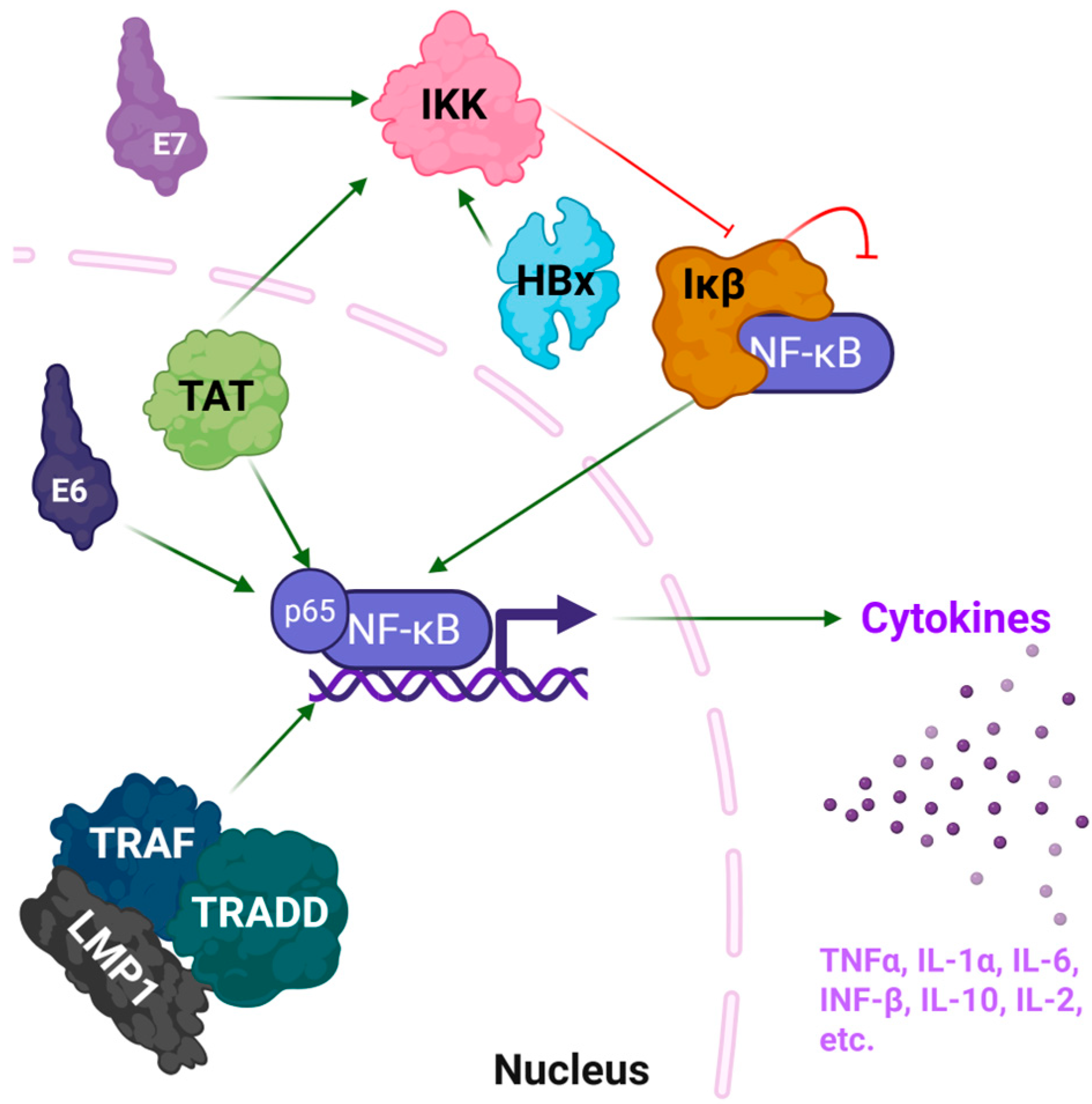
Figure 1. Summary of viral proteins and their mechanism of inflammation. The HIV-1 transactivator of transcription (TAT) and HPV E6 proteins interact with the p65 subunit of nuclear factor-kappa B (NF-κB), leading to direct activation. Similarly, the EBV latent membrane protein 1 (LMP1) mimics the tumor necrosis factor receptor, interacts with the adaptor proteins TRAF and TRADD, and leads to overstimulation of NF-κB signaling. HPV E7, HBV HBx, and Tat interact with the IκB kinase (IKK) complex, leading to degradation of Ikβ and the transport of NF-κB into the nucleus, where it activated target genes. NF-κB activation leads to production of pro-inflammatory cytokines, including TNFα, IL-6, and IL-10. Created in BioRender. Carabetta, V. (2025) https://BioRender.com/rrbgicu.
Pharmaceutics 17 01466 g001
Figure 2. Tat (residues 1–72) structure from subtype B (Red, PDB: 1JFW), with the C terminus represented in blue. This structure was visualized using PyMOL software, version 2.5.8. This structure shows a flexible, elongated conformation with transient α-helical regions.
Pharmaceutics 17 01466 g002
Figure 3. The predicted structure of HBx was generated using AlphaFold and visualized in PyMOL, version 2.5.8. The red coloration represents the C-terminal region, which has transactivation function and is likely responsible for transcriptional activation of both viral and host genes.
Pharmaceutics 17 01466 g003
Figure 4. (A) The HPV16 E6 structure (PDB: 4GIZ) forms a dimer, with each monomer bound to the ubiquitin ligase (blue) E6AP LxxLL motif (yellow spheres). To show the dimeric arrangement, rather than the N- and C-terminal domains of a single E6 molecule, the two E6 monomers are colored cyan and pale green. (B) The CxxC motif in HPV E7′s C-terminal zinc-binding domain is highlighted in red (PDB: 2EWL). The grey sphere represents a coordinated zinc ion. PyMOL, version 2.5.8 was used to visualize all structures.
Pharmaceutics 17 01466 g004
Figure 5. The predicted structure of LMP1 generated using AlphaFold and visualized in PyMOL, version 2.5.8.
Pharmaceutics 17 01466 g005
Figure 6. Summary of fungal proteins or constituents that lead to inflammation. The A. fumigatus protein AspF1 is recognized by toll-like receptors (TLRs), while the cell-wall component chitin is recognized by TLRs, Dectins and Nod-like receptors (NLRs). Fungal secreted chitin binding proteins (CBPs) protect the chitin from immune cell detection. The C. albicans protein Als3 is recognized by Dectins and TLRs. The C. albicans Saps lead to degradation of the extracellular matrix (ECM), which releases collagen, laminin, and fibronectin, all of which can serve as damage-associated molecular patterns (DAMPs) and activate TLRs and Dectin-1. Asp F1 and DAMPs also activate the NLRP3 inflammasome. Following activation of the pattern recognition receptors (PRRs), the downstream effects lead to activation of the MAPK and NF-κB signaling pathways, which leads to extensive cytokine production and inflammation. Created in BioRender. Carabetta, V. (2025) https://BioRender.com/nrz7c5k.
Pharmaceutics 17 01466 g006
Figure 7. Structural insights into Sap2. (A) The structure of Sap2 domains as a cartoon representation, showing the N-terminal domain (NTD) in blue and C-terminal domain (CTD) in cyan, with the A70450 inhibitor depicted in green and the conserved catalytic dyad (Asp32 and Asp218) in red (PDB: 1EAG), visualized in PyMOL, version 2.5.8. (B) The structure of Sap2 using spheres to illustrate how the NTD and CTD interact to form the active-site cleft. The colors are depicted as in panel A.
Pharmaceutics 17 01466 g007
Figure 8. Structural analysis of restrictocin. (A) The crystal structure of restrictocin bound to DNA (PDB: 1JBS) as a cartoon, with the conserved α/β ribotoxin fold indicated. (B) Restrictocin visualized as spheres. All structures were visualized using PyMOL, version 2.5.8.
Pharmaceutics 17 01466 g008
Figure 9. The predicted structure of Asp F1 using AlphaFold (green) aligned with the crystal structure of restrictocin (red, (PDB: 1JBS)), visualized in PyMOL, version 2.5.8.
Pharmaceutics 17 01466 g009
Figure 10. The crystal structure of the S. marcescens chitin-binding protein CBP21 (PDB: 2BEM), which is trimeric and contains conserved β -sandwich folds. Structures were visualized using PyMOL, version 2.5.8.
Pharmaceutics 17 01466 g010
Figure 11. The crystal structure of Als3 from C. albicans (PDB: 4LE8). This structure contains a β-barrel fold characterized by a distinctive hydrophobic peptide-binding cleft. Structure was visualized using PyMOL, version 2.5.8.
Pharmaceutics 17 01466 g011
Figure 12. Crystal structure of N39, a trimeric peptide that mimics the NHR of gp41 (green) with T20 (red). (A) Spheres and (B) cartoon models, both visualized using PyMOL software, version 2.5.8.
Pharmaceutics 17 01466 g012
Table 1. Summary of viral proteins that induce inflammation.
Viral ProteinVirusMechanism of InflammationStructural FeaturesKnown Motifs or Binding SitesPDBRefs.
TatHIV-1NF-κB activation; cytokine induction (IL-6, TNF-α, IL-1β) Intrinsically disordered with transient helicesInteracts with cyclin T1 via core region (aa 49–57)1JFW[22]
HBxHBVNF-κB activation via IKK complex; oxidative stress; cytokine upregulationMultiple helical segments and a flexible C-terminal regionInteracts with IKKγ through C-terminal motifsNone 1[23,24]
E6HPVDegrades p53, activates NF-κB and promotes cytokine expressionTwo zinc-binding domainsBinds E6AP via two LxxLL motifs
(aa 95–99)
4GIZ[25]
E7HPVInactivation of Rb; cell cycle dysregulation; NF-κB pathway stimulationZinc-binding C-terminal domainZinc-binding motif CxxC
(aa 49 to 52)
2EWL[26]
LMP1EBVMimics TNFR; constitutive NF-κB activation; cytokine storm (IL-6, IL-8)Predicted α-helical domains; partially disorderedC-terminal activation regions (CTAR1 and CTAR2) mediate TRAF bindingNone 1[27]
1 For structures with no PDB structure, predictions were made using AlphaFold. aa, amino acids.
Table 3. Summary table of biologics used to target common infection-induced inflammatory pathways.
Targeted MechanismShared inTherapeutic BiologicsMechanism of Action
NF-κB pathwayViruses, FungiBortezomib,
Anti-TNF mAbs
Inhibits IκB degradation, blocks TNF-driven activation
IL-6/IL-1 cytokine axisViruses, FungiTocilizumab, AnakinraIL-6R/IL-1R blockade to reduce cytokine storm
Table 4. The major hurdles and possible solutions for the use of therapeutic proteins.
Major ChallengeDescription Proposed Solutions & StrategiesRefs.
Targeted Delivery & BioavailabilityComplex biological barriers at inflamed sites, e.g., thick mucus or altered vasculatures, impede the penetration and retention of large protein conjugates.Development of advanced delivery systems:
  • Nanoparticles
  • Liposomes
  • Inhalable formulations
  • Ligands specific to targeted tissues
[110,111]
Immunogenicity & Rapid DegradationConjugates with non-human sequences or novel linkers can trigger immune responses that neutralize the drug. High protease activity in inflamed tissues leads to rapid degradation and reduced half-life.Protein Engineering Techniques:
  • Amino acid substitutions to reduce immunogenicity
  • Chemical modifications
  • Peptide cyclization
  • Incorporation of D-amino acids to resist proteolysis
[112,113]
Limitations of Computational DesignAI tools (e.g., AlphaFold) have constraints in predicting the behavior of flexible linkers, post-translational modifications, and complex physiological interactions.
  • Hybrid workflows that integrate computational design with rigorous experimental validation
  • Iterative design-test-learn cycles to refine models
[114,115]
Disclaimer/Publisher’s Note: The statements, opinions and data contained in all publications are solely those of the individual author(s) and contributor(s) and not of MDPI and/or the editor(s). MDPI and/or the editor(s) disclaim responsibility for any injury to people or property resulting from any ideas, methods, instructions or products referred to in the content.