Article

Synthesis, Molecular Docking, and Bioactivity Study of Novel Hybrid Benzimidazole Urea Derivatives: A Promising α-Amylase and α-Glucosidase Inhibitor Candidate with Antioxidant Activity

by
Lotfi M. Aroua
1,2,3,*,
Abdulelah H. Alosaimi
1,
Fahad M. Alminderej
1,
Sabri Messaoudi
1,3,
Hamdoon A. Mohammed
4,5,*,
Suliman A. Almahmoud
4,
Sridevi Chigurupati
4,6,
Abuzar E. A. E. Albadri
1 and
Nejib H. Mekni
2,7
1
Department of Chemistry, College of Science, Qassim University, Qassim Main Campus, King Abdulaziz Road, P.O. Box 6644, Al-Malida, Buraydah 51452, Saudi Arabia
2
Laboratory of Structural Organic Chemistry—Synthesis and Physicochemical Studies (LR99ES14), Department of Chemistry, Faculty of Sciences of Tunis, University of Tunis El Manar, Tunis 2092, Tunisia
3
Faculty of Sciences of Bizerte, Carthage University, Jarzouna, Bizerte 7021, Tunisia
4
Department of Medicinal Chemistry and Pharmacognosy, College of Pharmacy, Qassim University, Buraidah 51452, Saudi Arabia
5
Department of Pharmacognosy and Medicinal Plants, Faculty of Pharmacy, Al-Azhar University, Cairo 11371, Egypt
6
Department of Biotechnology, Saveetha School of Engineering, Saveetha Institute of Medical and Technical Sciences, Saveetha University, Saveetha Nagar, Thandalam, Chennai 602105, India
7
High Institute of Medical Technologies of Tunis, El Manar University, Tunis 1006, Tunisia
*
Authors to whom correspondence should be addressed.
Pharmaceutics 2023, 15(2), 457; https://doi.org/10.3390/pharmaceutics15020457
Submission received: 19 December 2022 / Revised: 24 January 2023 / Accepted: 26 January 2023 / Published: 30 January 2023

Abstract

:
A novel series of benzimidazole ureas 3a–h were elaborated using 2-(1H-benzoimidazol-2-yl) aniline 1 and the appropriate isocyanates 2a–h. The antioxidant and possible antidiabetic activities of the target benzimidazole-ureas 3a–h were evaluated. Almost all compounds 3a–h displayed strong to moderate antioxidant activities. When tested using the three antioxidant techniques, TAC, FRAP, and MCA, compounds 3b and 3c exhibited marked activity. The most active antioxidant compound in this family was compound 3g, which had excellent activity using four different methods: TAC, FRAP, DPPH-SA, and MCA. In vitro antidiabetic assays against α-amylase and α-glucosidase enzymes revealed that the majority of the compounds tested had good to moderate activity. The most favorable results were obtained with compounds 3c, 3e, and 3g, and analysis revealed that compounds 3c (IC50 = 18.65 ± 0.23 μM), 3e (IC50 = 20.7 ± 0.06 μM), and 3g (IC50 = 22.33 ± 0.12 μM) had good α-amylase inhibitory potential comparable to standard acarbose (IC50 = 14.21 ± 0.06 μM). Furthermore, the inhibitory effect of 3c (IC50 = 17.47 ± 0.03 μM), 3e (IC50 = 21.97 ± 0.19 μM), and 3g (IC50 = 23.01 ± 0.12 μM) on α-glucosidase was also comparable to acarbose (IC50 = 15.41 ± 0.32 μM). According to in silico molecular docking studies, compounds 3a–h had considerable affinity for the active sites of human lysosomal acid α-glucosidase (HLAG) and pancreatic α-amylase (HPA), indicating that the majority of the examined compounds had potential anti-hyperglycemic action.

1. Introduction

Diabetes mellitus (DM) is a broad and complex endocrine disease, which is a major global health issue. It is a disease characterized by marked multiple organ dysfunction and failure caused by hyperglycemia resulting from insulin deficiency or dysregulation of the insulin action pathway [1].
Type 2 diabetes is brought on by a confluence of genetic factors linked to impaired insulin secretion and insulin resistance, as well as environmental factors such as obesity, overeating, a lack of exercise, stress, and aging. However, type 1 diabetes is the result of an autoimmune reaction to proteins of the islets cells of the pancreas [2]. Since 2019, an estimated 463 million people (8.8% of the adult population) have been diagnosed with diabetes; type II diabetes represents about 85% to 95% of all diabetes cases in developed countries, with an even higher percentage in developing nations [3,4].
α-Amylase is a calcium-based metalloenzyme that degrades saccharides into maltose by cleaving α (1–4) glycosidic bonds. It is abundant in human pancreatic juice and saliva, as well as in the saliva of some other mammals [5]. The small intestine contains some α-glucosidases, such as the hexamer-based hydrolyze enzyme, which converts polysaccharides and disaccharides into glucose by processing the non-reducing terminal 1,4-linked α-glucose residues. In diabetes, the inhibition of these two enzymes becomes very important as it reduces the digestion rate of carbohydrates, resulting in less glucose absorption in the bloodstream [6]. Three drugs are commercially available for diabetes: naturally derived glycosidase inhibitor molecules (acarbose, miglitol, and voglibose), as well as many other types of drugs [7] (Figure 1).
There appears to be a strong direct correlation between antioxidant and anti-diabetic effects; this relationship may be attributed to the significant role of oxidative stress in hyperglycemia and overall diabetic complications [8]. The evaluated levels of blood glucose are combined with the overproduction of free radicals “reactive oxygen species” [8]. In addition, part of the immunogenic damage to beta cells of the pancreas is mediated by ROS [9]. Therefore, natural and synthetic antioxidant compounds, e.g., phenolics and flavonoids, are promising candidates for the protection, treatment, and management of diabetic complications [10,11].
Furthermore, free radical scavengers and chelators have been reported for their potential role in diabetes management, such as reducing the vascular relaxation and neurovascular impairments associated with hyperalgesia, improving endoneurial blood flow, and overall reducing the production of OH- and the initiation of lipid peroxidation associated with vascular reactivity impairment and nerve function in diabetes [12].
Benzimidazole species have an excellent structural range that covers large biological and therapeutic application fields. The research describing the importance of structures containing benzimidazole cores has been extensively developed and possesses a wide range of bioactive activities, including antifungal [13], antitubercular [14,15], antiulcer and antimicrobial [16], antileishmanial [17], antiglycation and antioxidant [18], antimycobacterial [19], anti-HIV [20], antitumor [21], antiproliferative [22], anti-inflammatory [23,24], antiviral [25], and antihelminthic activities [26]. Several candidates of this class were tested for anti-tumor properties and showed activities against several tumor cell lines, including breast cancer, human cells, and human lung cancer cells [27,28,29,30,31,32,33].
In addition, some benzimidazole derivatives are well recognized for their antidiabetic potential, especially as potent α-glucosidase and α-amylase inhibitors. A multitude of benzimidazole derivatives were reviewed as α-amylase inhibitors including benzimidazole bearing sulfonamide [34], 2-arylbenzimidazole [35], 2-mercaptobenzimidazole [36], 2-aminobenzimidazole derivatives [37], substituted benzimidazole analogs [38], 2-(2-methyl-5-nitro-1H-imidazol-1-yl)ethylaryl ether [39], and benzimidazole-based Cu (II)/Zn (II) complexes [40].
Moreover, some benzimidazole structures having 5-bromo-2-arylbenzimidazole [41], conjugated biphenylbenzimidazole [42], benzimidazole linked with morpholine or piperazine [43], oxadiazole bearing benzimidazole motif [44], substituted arylbenzimidazol at para-position bridged with thiazolidinone [45], benzimidazole linked with Schiff base [46], benzimidazole bearing triazole [47], hybrid benzimidazole quinolinyl oxadiazole squeleton [48], hybrid benzimidazole with thiourea [49], and fluoro-2-substitued-1H-benzimidazol substituted with morpholine [50] were reported as α-glucosidase inhibitors. In this context, benzimidazole derived from 5-oxo-pyrido triazepine and aminomethyloxo-pyrimido [51], benzoylaryl benzimidazole [52], benzimidazol with substituted benzylidene containing 2,4-thiazolidinedione, diethyl malonate, and methylacetoacetate [53], and benzimidazole bridged with triazole [54] also exhibited excellent α-amylase and α-glucosidase activities.
The urea structure involves one of the most significant intermediates in organic and medicinal chemistry. This edifice is useful to develop biological active compounds, including anticancer, antiviral, antibacterial, and antihypertensive agents, which are clinically approved therapeutics [55,56,57,58]. Due to its remarkable structure, urea can stably form hydrogen bonds with target proteins and receptors. Furthermore, a targeted urea scaffold was used to find antidiabetic agents covering hybrid-urea pyrimidine-triazole [59], thiadiazole-urea bearing quinazolinone [60], sulfonylamino pyridinylbenzothiazol-urea [61], pyrimidine hybrid phenylaminourea [62], diarylurea containing quinazoline [63], urea moiety with coumarin-chalcone derivatives [64], and diarylurea linked with benzothiophene [65].
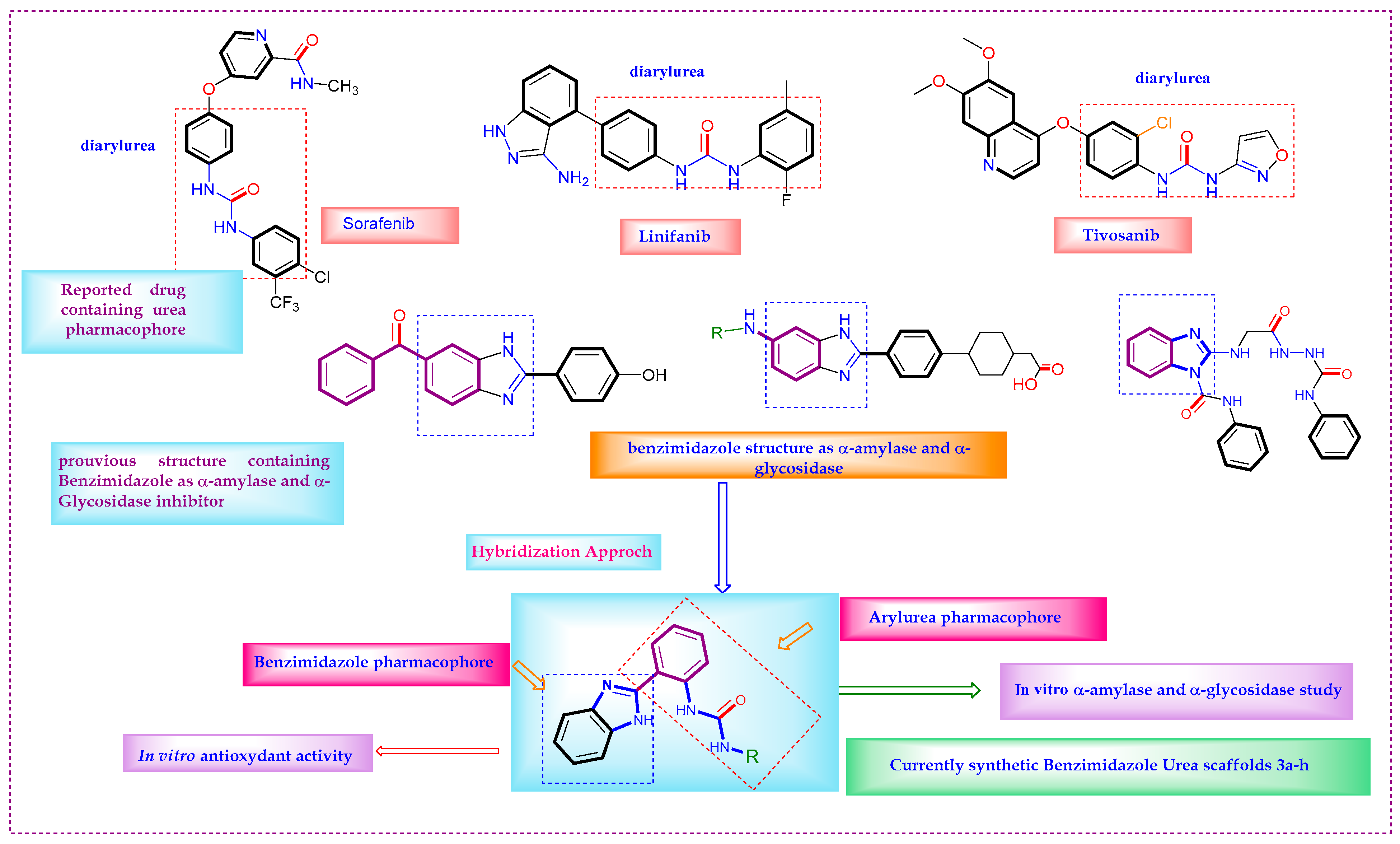
Following the previous scientific report, we elaborated herein a new target structure, containing both benzimidazole and diarylurea moieties in one component. Our purpose was to combine their properties, which represent a new route to improve the effectiveness of bioactive molecules (Figure 2).
There is a critical need to find better drugs to treat type I and type II diabetes. The discovery of new α-glucosidase and α-amylase inhibitor agents is an interesting method of obtaining such drugs. To contribute to the advancement of the design and the discovery of new bioactive species [66,67,68,69,70,71], we combined the two patterns, benzimidazole and urea, in the same molecule, allowing for potential antidiabetic agents, and having good anti-oxidant activity.
A simple route to novel hybrid benzimidazole urea compounds 3a-h was utilized from the reaction of 2-(1H-benzimizadol-2-yl) aniline 1 and appropriate isocyanates substrates 2a-h. The evaluation of antioxidant, α-amylase, and α-glucosidase activities was assessed. To estimate the interaction with α-amylase and α-glucosidase enzymes, an in silico molecular docking study of benzimidazole-urea was performed.

2. Results and Discussion

2.1. Chemistry

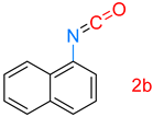
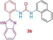
The synthesis of target benzimidazole-ureas 3a-f was realized from commercially available 2-(1H-benzoimidazol-2-yl)aniline 1 and six different isocyanates 2a-f. In a first attempt, the reaction of 2-(1H-benzoimidazol-2-yl)aniline 1 and naphthylisocyanate 2b in anhydrous acetone produced the expected benzimidazole-urea 3b with a moderate yield of 60% (Scheme 1).
To enhance the performance of this synthetic method, different reaction conditions were studied. The principal reaction parameters, including the solvent and number of equivalent reagents, were varied using the same substrates (1 and 2b). Among the investigated solvents: tetrahydrofuran, acetone, dichloromethane, and mixtures of dichloromethane/acetone, this latter mixture, in the respective proportions of 80/20, gave the best results with total substrate conversion and a 90% reaction yield.
As a result of optimizing the equivalent number of the substrate 2b suitable for the reaction process, the substrate was fully converted with 1.5 equivalents of the isocyanate 2b.
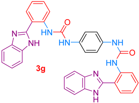
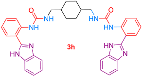
1,4-phenelenediisocyanate 2g and 1,4-bis(isocyanatomethyl)cyclohexane 2h reacted with two equivalents of substrate 1 to afford the corresponding products 3g and 3h in good yields, 93% and 85%, respectively (Scheme 2).
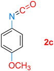
The reactions progressed easily, offering good yields of the proposed substrates 3a-h, and the results are collected in Table 1. It is worth noting that the reaction yields of the aryl substrates were higher than those of the alkyl and cycloalkyl ones. The low reaction yields as well as the rates of the reactions observed with alkyl and cycloalkyl isocyanate substrates may be due principally to the low reactivity of such radicals, which exert an inductive effect that decreases the isocyanate carbon electrophilicity.
The structure of novel prepared benzimidazole urea was determined by spectroscopic data from 1H and 13C NMR, FT-IR, and HRMS. The IR spectra of compound 3a showed the absorption at 3249 cm−1 related to the NH group of the benzimidazole ring. The spectra also displayed stretching at 1672, 1586, and 1536 cm−1 assigned to (C=O), (C=N), and (C=C), respectively.
The 1H NMR spectra of compound 3a showed a band at 13.08 ppm attributed to the NH group of benzimidazole. The amino proton of the urea group resonated at 12.02 ppm. Aromatic protons resonated in the range of 9.69–7.01 ppm. The compound 3a was identified with 13C NMR. The spectra exhibited a singlet at 153.2 ppm corresponding to the carbon atom of the urea carbonyl group. In addition, the carbon of the benzimidazole ring appeared at 151.45 ppm. The high-resolution mass spectra of compound 3a revealed the presence of a molecular peak with m/z: 328.12280 attributed to [M]+ compatible with the formula for C20H16N4O (see Supplementary Materials).

2.2. Biology

2.2.1. Antioxidant Activity

Four in vitro assays were used to determine the antioxidant activity of the compounds 3a-h. The methods used provide information about the compounds’ reducing capacity (TAC and FRAP methods), scavenging capability (DPPH-SA), and metal chelating activity (MCA). Table 2 gives the antioxidant levels for the compounds, measured in a comparable manner using three different in vitro mechanisms, namely, reducing, scavenging, and metal chelating activities.
In the series of benzimidazole ureas 3a-h, compound 3g demonstrated the highest reducing power for molybdenum (VI) with 10.06 ± 1.51 mM trolox equivalents in the total antioxidant capacity (TAC). The results of reducing activity also revealed that some compounds reduced molybdenum (VI) ions selectively in the TAC. For instance, compounds 3b, 3c, and 3f showed significant molybdenum (VI) reducing activity compared to other compounds at levels of 5.16 ± 0.69, 4.43 ± 0.53, and 8.50 ± 0.87 mM trolox equivalents.
Regarding Table 2, the results of the ferric reducing antioxidant power (FRAP) of compounds 3a-h indicated higher activities of derivatives 3a and 3c with 9.25 ± 0.20 and 8.65 ± 0.29 mM trolox equivalents, respectively. The most potent compound 3g yielded the best results, with 16.12 ± 0.29 mM trolox equivalents.
The results of DPPH-SA of hybrid benzimidazole urea revealed the potency of derivatives 3d and 3g as free radical scavengers. Compound 3d presented good activity with 2.89 ± 1.07 mM trolox equivalents. Compound 3g also gave an excellent result with 3.86 ± 0.04 mM trolox equivalents. The other compounds exerted moderate DPPH-SA activity at values ranging from 1.70 to 0.96 mM trolox equivalents.
The metal chelating activity (MCA) of compounds 3a-h manifested potential activity for the compound 3h at the MCA level of 2.95 ± 0.21 mM EDTA equivalents due to the presence of two urea groups. In addition, compounds 3a-c exhibited moderate MCA in the range of 1.36–1.62 mM EDTA equivalents.
The overall antioxidant activities of compounds 3a-h confirmed that the majority manifested excellent to moderate activities. Benzimidazole-urea 3b and 3c displayed good activity with the three-antioxidant methods TAC, FRAP, and MCA. Targeted compound 3g represented the most derivatives in this family, manifesting excellent activity in three methods: TAC, FRAP, and DPPH-SA. It is worth noting that compound 3g also had significant activity for the TAC method, with 10.06 ± 1.51 mM trolox equivalents. Compound 3g also gave higher activity with 16.12 ± 0.29 mM trolox equivalents for the FRAP method. Concerning the DPPH-SA method, compound 3g also gave the best results with 3.86 ± 0.04 mM trolox equivalents.

2.2.2. α-Amylase and α-Glucosidase Inhibition Activity

All the synthesized novel hybrid benzimidazole urea compounds 3a-h were subjected for the evaluation of their possible α-amylase and α-glucosidase inhibitory activities. It is worth noting that the majority of the analogs were found to have good to moderately active IC50 values ranging from 18.65 ± 0.23 to 28.33 ± 0.02 µM for α-amylase inhibition and with the IC50 values ranging from 17.47 ± 0.03 to 29.01 ± 0.12 µM for α-glucosidase inhibition except compounds 3a, 3b, and 3h. The inhibitory activities of α-amylase and α-glucosidase, according to Table 3, revealed that the tested compounds in the current study demonstrate higher activities than those reported in previous studies [47,72].
Consulting Table 3, the results indicate that compounds 3c (IC50 = 18.65 ± 0.23 µM), 3e (IC50 = 20.7 ± 0.06 µM), and 3g (IC50 = 22.33 ± 0.12 µM) manifested a good α-amylase inhibitory potential comparable to standard acarbose (IC50 = 14.21 ± 0.06 µM) (Table 3). The same observation was mentioned with compounds 3c (IC50 = 17.47 ± 0.03 µM), 3e (IC50 = 21.97 ± 0.19 µM), and 3g (IC50 = 23.01 ± 0.12 µM), which exerted an α-glucosidase inhibitory potential comparable also to standard drug acarbose (IC50 = 15.41 ± 0.32 µM).
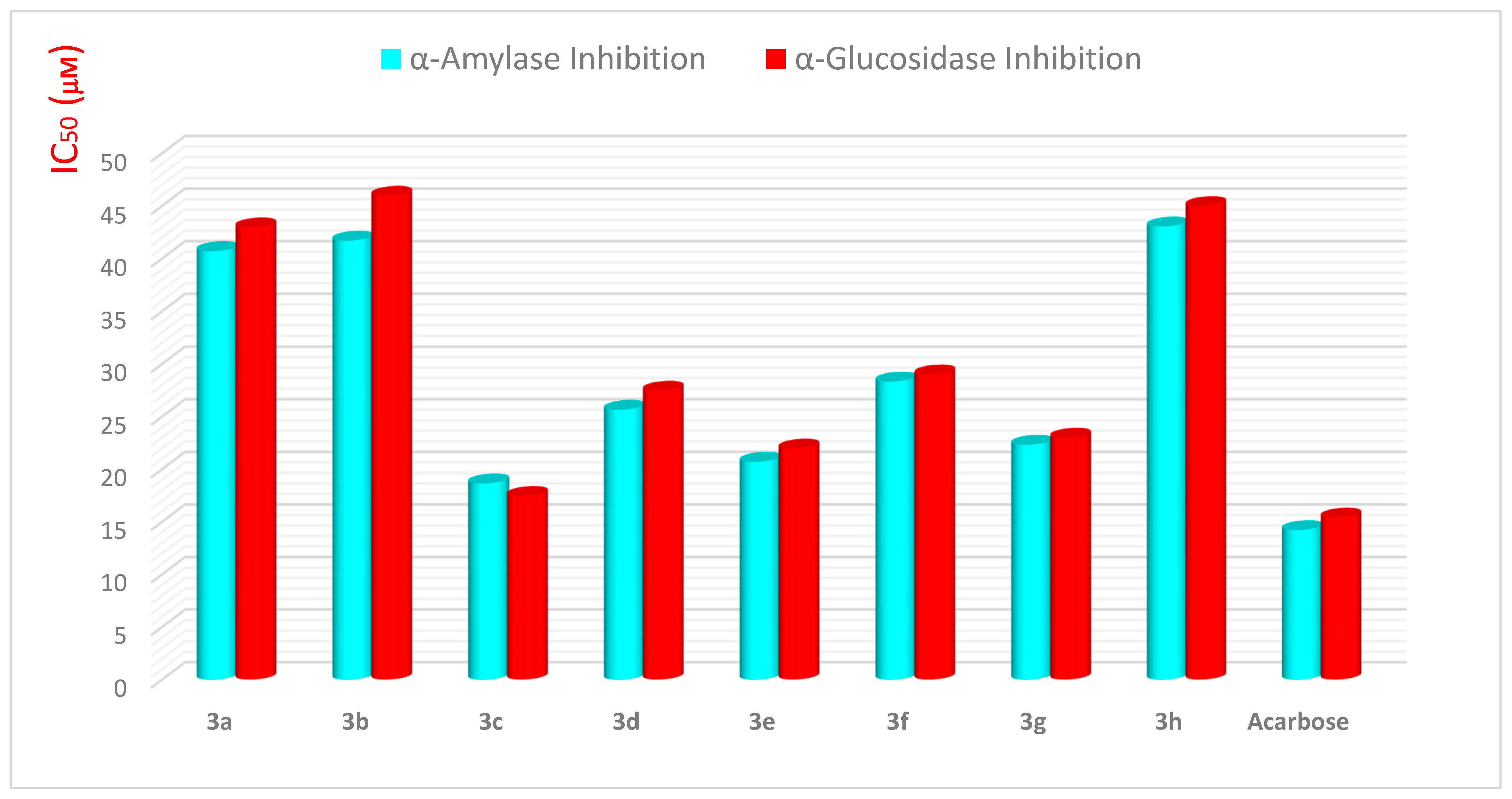
As illustrated in Figure 3, compounds 3d and 3f displayed a moderate α-amylase inhibitory potential with IC50 = 25.65 ± 0.03 and IC50 = 28.33 ± 0.02 µM, respectively. The same results were mentioned with 3d analogs with IC50 = 27.47 ± 0.13 and 3f with IC50 = 29.01 ± 0.12 µM for α-glycosidase inhibition.
Indeed, the whole molecule played an important role in the inhibitory potential; however, limited SAR was rationalized by emphasizing the effects of varying structural features such as different substitutions on nitrogen of the amide side chain of urea, on the inhibitory potential in terms of IC50 values.
Comparing the inhibitory potential of benzimidazole-urea derivatives with phenyl substitution 3a (IC50 = 40.7 ± 0.06 µM for α-amylase; IC50 = 42.97 ± 0.19 µM for α-glucosidase), naphthyl substitution 3b (IC50 = 41.7 ± 0.06 µM for α-amylase; IC50 = 45.97 ± 0.19 µM for α-glucosidase), and di-cyclohexyl urea substitution 3h (IC50 = 43.04 ± 0.02 µM for α-amylase inhibition; IC50 = 44.99 ± 0.09 µM for α-glucosidase) manifested weak activities for both α-amylase and α-glucosidase inhibition.
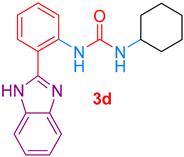
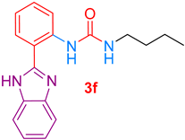
Amongst the other benzimidazole urea derivatives, compound 3d bearing a cyclohexyl side on nitrogen (IC50 = 25.65 ± 0.03 µM α-amylase inhibition; IC50 = 27.47 ± 0.13 µM α-glucosidase inhibition) and compound 3f containing a butyl chain on nitrogen (IC50 = 28.33 ± 0.02 µM α-amylase inhibition; IC50 = 29.01 ± 0.12 µM α-glucosidase inhibition) exerted moderate activities for both α-amylase and α- glucosidase inhibition.
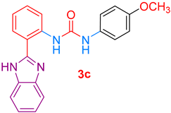
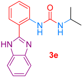
Derivative 3c having a methoxy phenyl group displayed a significant α-amylase and α-glucosidase inhibitory activity. Substrate 3e with an isopropyl group and 3g substituted with a di-phenyl urea group on nitrogen also exhibited good inhibitory potential compared to standard acarbose.
In summary, in the series of aromatic substituents, compounds containing phenyl group 3a and naphthyl group 3b exhibited a weak activity for inhibition of both α-glucosidase and α-amylase. The excellent results were shown with target analog 3c bearing 4-methoxyphenyl, showing remarkable inhibitory action against both α-amylase and α-glucosidase. The inclusion of a methoxy substituent in the phenyl group at the para-position significantly improved activity, which might be attributed to the favorable interaction for the inhibition activity of both α-amylase and α-glucosidase (Figure 4).
In the series of alkane and cycloalkane, it is clear that compounds 3d and 3f exhibited higher potential activity in comparison with aromatic compounds 3a and 3b. This observation is explained based on the donor effect of cyclohexyl and butyl groups when compared to phenyl or naphthyl groups. The same result was observed with compound 3e bearing an isopropyl group manifesting a good inhibition of enzyme α-amylase and α-glucosidase. The above observation was interpreted by the sufficient donor effect of the isopropyl group, which exerted a favorable interaction and demonstrated the highest activity. Compound 3g also indicated a strong inhibition of α-amylase and α-glucosidase compared with 3d and 3f. The presence of two phenylbenzimidazoles in the structure of compound 3g allowed favorable interactions involving a good inhibition activity of α-amylase and α-glucosidase (Figure 4).

2.3. In Silico Docking Study

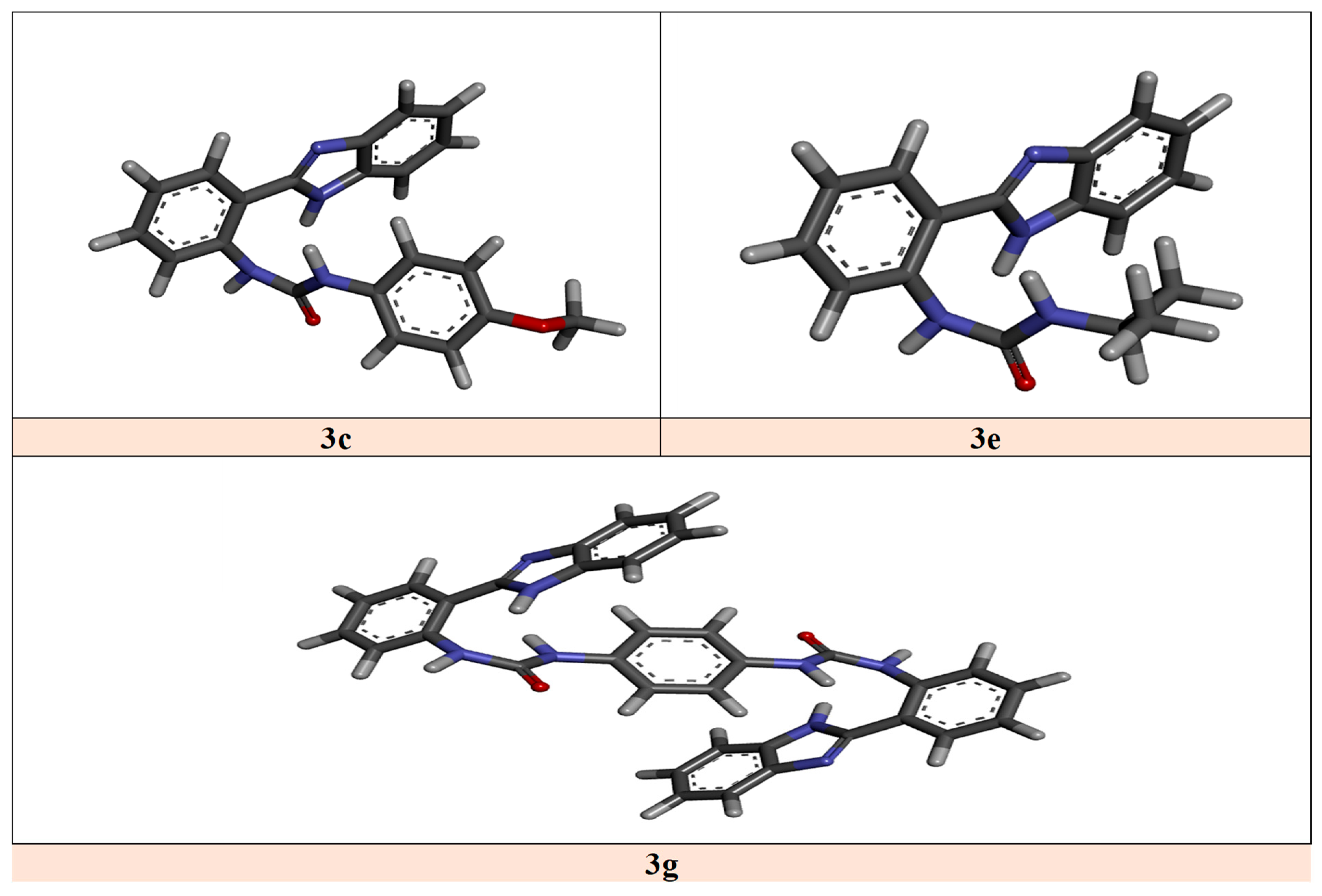
The objective of the in-silico study was to clarify and rationalize the type of interactions of the new benzimidazole urea 3a-h in the target catalytic site. Molecular docking is a useful tool in the design of structure-based drugs. The optimized structures of the most potent benzimidazole urea 3c, 3e, and 3g used in the docking study are presented in Figure 5. We notice that the most stable structures for 3c and 3e had a cis structure with intramolecular interactions. The most stable structure of 3g had almost a Ci symmetry also with intramolecular interactions.
Benzimidazole urea derivatives showed good values of binding energies with α-amylase (HPA) human pancreatic and also the human lysosomal acid-α-glucosidase (HLAG) (Table 4).
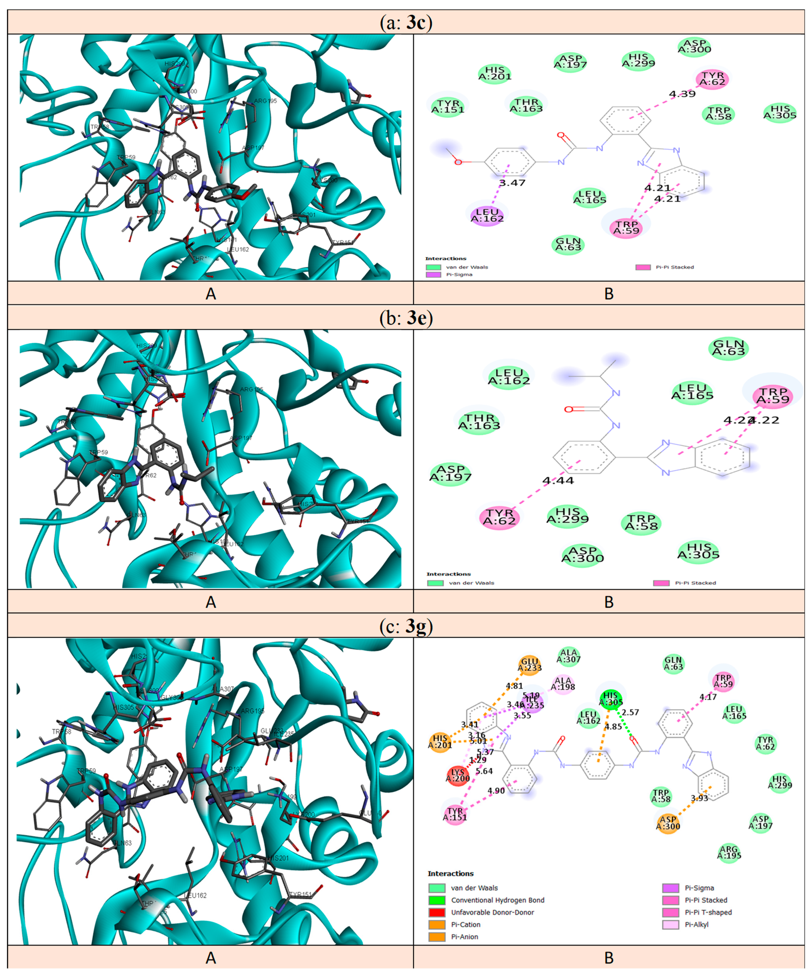
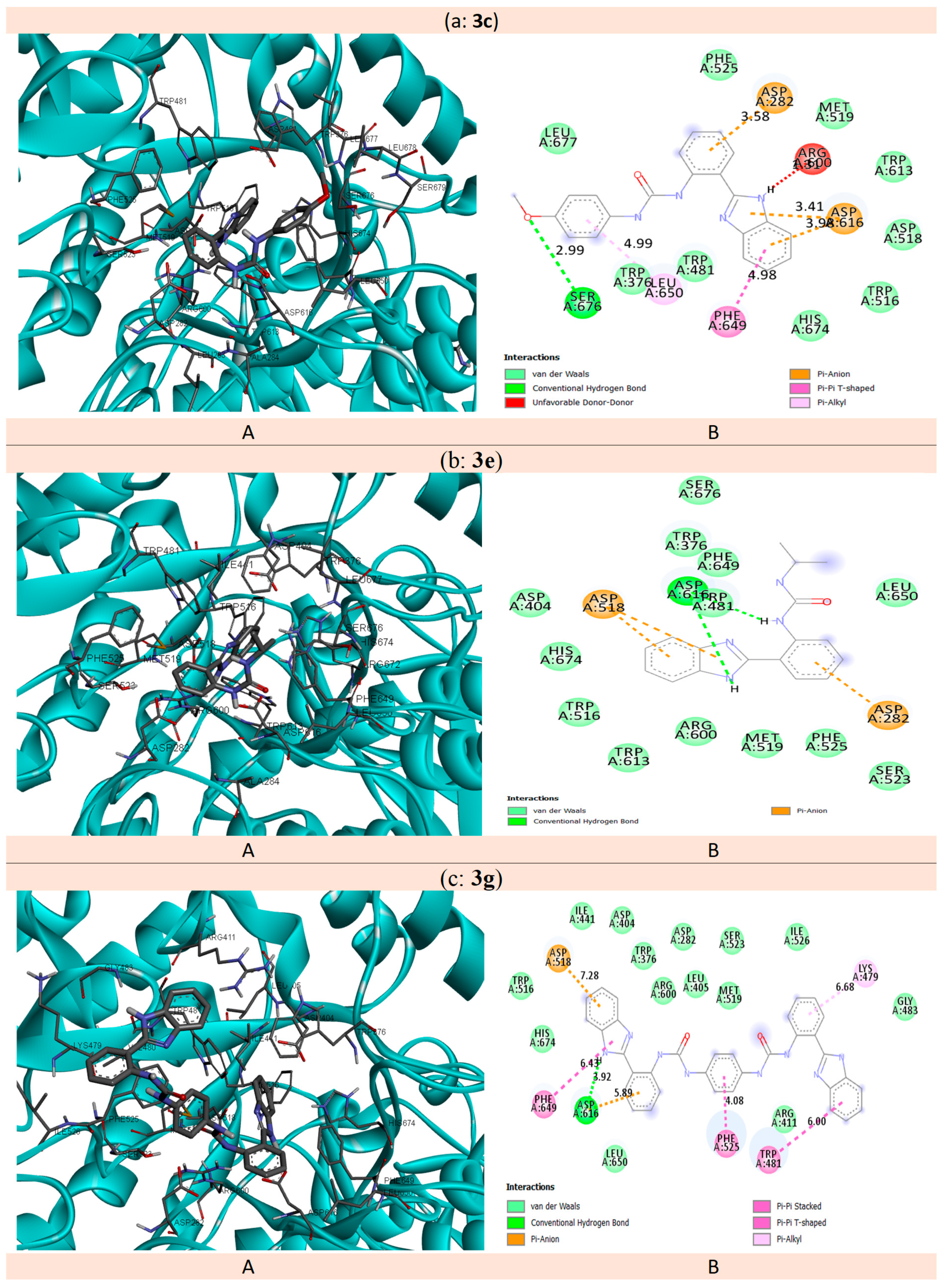
Our study was interested in explaining the interactions of the most effective analogs molecules 3c, 3e, and 3g manifesting appropriate interactions (2D) and (3D) with the different amino acids illustrated in Figure 6 and Figure 7. Due to their high inhibition activity amongst molecules, the structures 3c, 3e, and 3g were chosen for docking studies with 5E0F. Table 4 reveals that compounds 3c, 3e, and 3g with potent inhibitory activities occupied suitable positions at the binding center of HPA, with energies of −8.8, −8.0, and −11.2 kcal.mol−1, respectively.
Presented in Figure 6, the structure of 3c presented (π-σ) interaction with Leu162 (3.47 Å), two (π-π) interactions with Trp59, and one (π-π) interaction with Tyr62 (4.39 Å). The same type of interactions: two (π-π) interactions with Trp59 and one (π-π) interaction with Tyr62 (4.44 Å), existed in molecule 3e. Compound 3g presented many interactions with the protein: one hydrogen bond with His305 (2.57 Å); (π-ion) interactions with Glu233, His201, and Asp300; (π-π) interactions with Tyr151 and Trp59; (π-σ) interaction with Ile235; (π-Alkyl) interaction with Ala235.
The study of Ramasubbu et al. [73] showed that residues Asp 236, Glu233, Arg 61, Asp300, Lys 200, Leu165, and Asp197, and some of the aromatic and non-polar residues, Tyr 258, Ile235, His305, His299, Trp58, Ala307, Tyr62, Trp59, Ser163, and His101, defined the catalytic site of α-amylase. Williams and coworkers [74] reported that the inhibition property of myricetin with α-amylase has an appropriate position in the catalytic site of α-amylase through four hydrogen bonds with Gln63, His101, and Asp197, and possessing two hydrophobic interactions with Leu165 and Tyr62.
The data indicated that the structures of 3c, 3e, and 3g had interactions with the catalytic site through Trp59, Trp58, Leu165, Tyr62, and Gln63 remaining from the catalytic HPA site. This confirms that 3c, 3e, and 3g are good inhibitors for HPA.
The compounds 3c, 3e, and 3g showing high inhibitory effects with HLAG gave important values of binding energies with the catalytic site of 5NN8, as mentioned in Table 4. These values were −8.2, −7.6, and −10.0 kcal mol−1, respectively.
Illustrated in Figure 7, the structure 3c had one hydrogen bond with Ser676 (2.99 Å); (π-anion) interactions with Asp282 and Asp616; (π-π) interaction with Phe649; (π-Alkyl) interaction with Leu650. The molecule 3e had two hydrogen bonds with Asp616 (2.64 Å and 2.69 Å) and (π-anion) interactions with Asp518 and Asp282. Compound 3g had one hydrogen bond with Asp616; (π-anion) interaction with Asp518; three (π-π) interactions with Phe649, Phe525, and Trp481.
Roig-Zamboni et al. [75] observed that when 1-deoxynojirimycin is inserted into the catalytic site of HLAG, it has a high inhibitory activity. This molecule has interactions with Trp376, Asp404, Leu405, Ile441, Trp481, Trp516, Asp518, Met519, Arg600, Trp613, Asp616, Asp645, Arg672, His674, and Phe649. Our data indicate that our structures 3c, 3e, and 3g interacted with the HLAG catalytic site via many similar residues (Trp376, His674, Trp481, Trp376, Trp516, Trp481, Trp516, Asp518, Met519, Asp518, Arg600, Asp518, and Asp616). The different complexes of 3c, 3e, and 3g were stabilized by these interactions and suggests that this stabilization contributed to the inhibitory activity.

3. Conclusions

A prominent series of benzimidazole-urea analogs 3a-h was designed and synthesized from 2-(1H-benzoimidazol-2-yl) aniline 1 and the appropriate isocyanates 2a-h. The antioxidant and antidiabetic activities of the target benzimidazole-ureas 3a-h were evaluated. When measured using four different methods, the antioxidant data revealed that the most desired benzimidazole-urea exhibited high activity. The results indicated that among the series, compounds 3a and 3b displayed good inhibition with three methods: FRAP, MCA, and DPPH-SA. The most potent antioxidant compound was 3g, which demonstrated strong activity in all four methods. Furthermore, the ability of benzimidazole-urea scaffolds to inhibit α-glycosidase and α-amylase was investigated. According to our predictions, the majority of tested compounds exhibited good to moderate activities. The results indicated that compounds 3e and 3g exhibited good inhibition for both enzymes, α-amylase, and α-glucosidase. Compound 3c bearing the 4-methoxy moiety on the phenyl group at the para-position was found to be a more potent inhibitor of α-amylase, and α-glucosidase similar to the standard drug acarbose. The study confirms that compounds 3c, 3e, and 3g have strong in vitro antioxidant properties, as well as α-glucosidase and α-amylase inhibition activities comparable to those of acarbose, suggesting that they could be used as anti-diabetic medications.

4. Experimental

4.1. General Details

Without any further purification, all commercially available reagents were used. Unless otherwise specified, all reactions were carried out in a dry nitrogen-protected atmosphere using oven-dried glassware. FT-IR spectra were recorded on an Agilent (Agilent Technologies, Inc. Headquarters, address: 5301 Stevens Creek Blvdv, Santa Clara, CA 95051, USA) apparatus type Cary 600 in the range of 400–4000 cm−1. A Bruker (Bruker Daltonics Inc., Bremen, Germany) NMR spectrometer Avance III HD 400 MHz was used to analyze 1H and 13C NMR spectra at 400 and 100 MHz, respectively. All spectra were recorded and referenced to TMS using DMSO d6 as the solvent. According to an internal standard of residual DMSO d6 at 2.50 ppm, chemical shifts in 1H NMR spectra were mentioned in parts per million (ppm) on the scale. Spin multiplicities are denoted by the letters s (singlet), brs (broad singlet), d (doublet), t (triplet), q (quartet), and m (multiplet). Chemical shifts in 13C NMR spectra were also indicated in ppm from the peak position of DMSO d6 (39.52 ppm). The values of the coupling constant J are given in hertz (Hz). High-resolution mass spectra were recorded on an Agilent 6545 LC/Q-TOF MS (Agilent Technologies, Inc. Headquarters, address: 5301 Stevens Creek Blvd, Santa Clara, CA 95051, USA). Using the Stuart SMP30 apparatus (New Jersey 07740, États-Unis), the melting points in open capillary tubes were determined. For analytical TLC, Silica Gel 60 F254 plates (Sigma 40–60 m) were employed, and the chromatogram was developed using a UV lamp 254 nm. DMSO-d6 (99.9 atom % D; CAS no. 2206-27-1) were purchased from Sigma-Aldrich and used without further purification.

4.2. Chemistry: Benzimidazole-Ureas 3a–h Synthesis: General Procedure

The reaction was carried out under a nitrogen atmosphere. In a 100 mL round bottom flask, 0.52 g (2.5 mmol) of 2-(1H-benzoimidazol-2-yl) aniline was placed in 20 mL of dichloromethane and 5 mL of acetone, and the appropriate isocyanate (3.75 mmol) was added dropwise in 20 mL of dichloromethane and 5 mL of acetone. The reaction was stirred under reflux. TLC and diethyl ether/hexane (50/50) as the eluent were used to monitor the reaction progress (see Table 3). After filtering the reaction mixture, the raw solid was purified through recrystallization from ethanol (see Supplementary Materials).
1-(2-(1H-benzo[d]imidazol-2-yl)phenyl)-3-phenylurea 3a: White precipitate, yield: 84%; M.p. = 129–131 °C; IR (cm−1) ν: 3249 (NH), 1672 (C=O), 1586 (C=N), 1536 (C=C); 1H NMR (400 MHz, DMSO-d6) δ(ppm): 13.08 (s, 1H, NH), 12.02 (s, 1H, NH), 9.69 (s, 1H, NH), 8.40 (d, 1H, J = 8.44 Hz, Harom), 7.78 (m, 1H, Harom), 7.56 (d, 3H, J = 7.68 Hz, Harom), 7.45 (td, 1H, J = 8.44 Hz, Harom), 7.33–7.28 (m, 1H, Harom), 7.17 (td, 1H, J = 8.00 Hz, Harom); 7.01 (t, 1H, J = 7.36 Hz, Harom); 13C NMR (75 MHz, DMSO-d6) δ(ppm): 153.2 (C=O), 151.45 (C=N), 142.7, 140,3, 139.8, 134.1, 130,7, 129.1, 127.7, 123.6, 122.6, 122.4, 121.7, 120.9, 119.7, 119.3, 115.7, 111.8; HRMS: m/z for C20H16N4O calcd: 328.1324, found: 328.12280 [M]+.
1-(2-(1H-benzo[d]imidazol-2-yl)phenyl)-3-(naphthalen-1-yl)urea 3b: White precipitate, yield: 90%; M.p. = 203–205 °C; IR (cm−1) ν: 3394 (NH), 1647 (C=O), 1586 (C=N), 1518 (C=C); 1H NMR (400 MHz, DMSO-d6) δ(ppm): 13.01 (s, 1H, NH), 12.04 (s, 1H, NH), 9.45 (s, 1H, Harom), 8.50 (d, 1H, J= 8.48 Hz, Harom), 7.72–7.71 (dd, 1H, Harom), 8.03–7.95 (m, 2H, Harom), 7.85 (d, 1H, Harom), 7.56 (d, 1H, J = 7.44 Hz, Harom); 7.53–7.51 (m, 4H, Harom), 7.53 (td, 2H, J = 8.48 Hz, Harom), 7.22–7.13 (m, 3H, Harom); 13C NMR (75 MHz, DMSO-d6) δ(ppm): 154.6 (1C, C=O), 151.3 (C=N), 139.9, 134.6, 134.4, 130.7, 129.6, 128.5, 127.8, 126.5, 126.36, 126.34, 125.9, 123.5, 123.4, 122.22, 121.7, 120.7, 115.75, 111.6; HRMS: m/z for C24H18N4O calcd: 378.1481, found: 378.33484 [M]+.
1-(2-(1H-benzo[d]imidazol-2-yl)phenyl)-3-(4-methoxyphenyl)urea 3c: White precipitate, yield: 80%; M.p. = 197–199 °C; IR (cm−1) ν: 3405 (NH), 1646 (C=O), 1587 (C=N), 1533 (C=C); 1H NMR (400 MHz, DMSO-d6) δ(ppm): 13.11 (s, 1H, NH), 12.08 (s, 1H, NH), 9.49 (s, 1H, NH), 8.53 (d, 1H, J = 8.3 Hz, Harom), 8.11 (d, 1H, J = 7.16 Hz, Harom), 7.80–7.78 (m, 1H, Harom), 7.63–7.61 (m, 1H, Harom), 7.54–7.45 (m, 3H, Harom), 7.41–7.31 (m, 2H, Harom), 7.17 (t, 1H, J = 7.28 Hz, Harom), 6.95 (d, 1H, J = 8.96 Hz, Harom); 6.90 (d, 1H, J = 9.00 Hz, Harom), 3.76 (s, 3H, OCH3); 13C NMR (75 MHz, DMSO-d6) δ(ppm): 154.8 (C=O), 153.6 (C=N), 153.5, 151.5, 142.8, 140.1, 134.1, 130.7, 127.7, 123.6, 122.3, 121.5, 120.7, 119.3, 115.8, 115.2, 114.9, 114.7, 114.0, 111.8, 55.5; HRMS: m/z for C21H18N4O2 calcd: 358.1430, found: 358.33315 [M]+.
1-(2-(1H-benzo[d]imidazol-2-yl)phenyl)-3-cyclohexylurea 3d: White precipitate, yield: 75%; M.p. = 330–332 °C; IR (cm−1) ν: 3335 (NH), 1654 (C=O), 1587 (C=N), 1531 (C=C); 1H NMR (400 MHz, DMSO-d6) δ(ppm): 13.04 (s, 1H, NH), 11.67 (s, 1H, NH), 8.52 (d, 1H, J = 8.36 Hz, Harom), 8.02 (d, 1H, J = 7.36 Hz, Harom), 7.80–7.78 (m, 1H, Harom), 7.59–7.57 (m, 1H, Harom), 7.39 (t, 1H, J = 7.40 Hz, Harom); 7.28–7.27 (m, 2H, Harom), 7.08 (t, 2H, J = 7.24 Hz, Harom), 3.58 (m, 1H, CHN), 1.91–1.89 (m, 2H, CH2), 1.75–1.60 (m, 3H), 1.37–1.22 (m, 5H); 13C NMR (75 MHz, DMSO-d6) δ(ppm): 154.9 (C=O), 151.7 (C=N), 140.7, 134.1, 130.6, 127.7, 123.5, 122.3, 120.8, 128.0, 119.1, 116.5, 115.4, 114.8, 111.7; 55.3, 49.2, 33.5, 25.7, 25.4; HRMS: m/z for C20H22N4O calcd: 334.1794, found: 334.19339 [M]+.
1-(2-(1H-benzo[d]imidazol-2-yl)phenyl)-3-isopropylurea 3e: White precipitate, yield: 65%; M.p. = 196–198 °C; IR (cm−1) ν: 3355 (NH), 1654 (C=O), 1587 (C=N), 1532 (C=C); 1H NMR (400 MHz, DMSO-d6) δ(ppm): 13.07 (s, 1H, NH), 11.73 (s, 1H, NH), 8.53 (d, 1H, J = 8.40 Hz, Harom), 8.03 (d, 1H, J = 7.32 Hz, Harom), 7.78 (m, 1H, Harom), 7.58 (m, 1H, Harom), 7.39 (t, 1H, J = 7.40 Hz, Harom); 7.28–7.27 (m, 2H, Harom), 7.08 (t, 2H, J = 7.28 Hz, Harom), 3.95–3.90 (m, 1H, CHN), 1.19 (s, 6H, 2CH3); 13C NMR (75 MHz, DMSO-d6) δ(ppm): 154.9 (C=O), 153.0 (C=N), 151.7, 148.7, 142.7, 140.7, 134.0, 130.6, 127.7, 123.5, 122.7, 122.3, 121.8, 120.8, 119.9, 119.2, 118.6, 116.5, 115.4, 114.7, 111.7, 111,2, 110.5; HRMS: m/z for C17H30O2N2 calcd: 294.23073, found: 294.23016 [M]+.
1-(2-(1H-benzo[d]imidazol-2-yl)phenyl)-3-butylurea 3f: White precipitate, yield: 60%; M.p. = 196–198 °C; IR (cm−1) ν: 3407 (NH), 1651 (C=O), 1510 (C=N), 1512(C=C); 1H NMR (400 MHz, DMSO-d6) δ(ppm): 7.91 (dd, 1H, J = 7.64 Hz, Harom), 7.74–7.71 (m, 1H, Harom), 7.65–7.63 (m, 1H, Harom), 6.94 (m, 2H, NH2), 6.82–6.78 (m, 2H, Harom), 3.61 (m, 2H, CH2N), 1.19 (s, 6H, 3CH2); 13C NMR (75 MHz, DMSO-d6) δ(ppm): 148.7, 144.6, 143.4.0, 132.7, 132.0, 125.1, 122.5, 122.3, 121.8, 119.3, 118.4, 116.5, 115.4, 112.3, 111,9, 110.5, 72.3, 28.4; HRMS: m/z for C18H20N4O calcd: 308.1637, found: 308.18590 [M]+.
1,1’-(1,4-phenylene)bis(3-(2-(1H-benzo[d]imidazol-2-yl)phenyl)urea) 3g: White precipitate, yield: 93%; M.p. = 190–192 °C, IR (cm−1) ν: 3326 (NH), 1652 (C=O), 1588 (C=N), 1527 (C=C); 1H NMR (400 MHz, DMSO-d6) δ(ppm): 13.05 (s, 2H, 2NH), 11.97 (s, 2H, 2NH), 9.53 (s, 2H, NH), 8.45 (d, 2H, J = 6.35 Hz, Harom), 8.05 (d, 2H, J = 6.35 Hz, Harom), 7.78–7.69 (m, 2H, Harom), 7.58–7.57 (m, 2H, Harom), 7.50–7.41 (m, 12H, Harom); 13C NMR (75 MHz, DMSO-d6) δ(ppm): 153.45, (C=O), 151.47 (C=N), 142.7, 139.9, 142.7, 140.7, 134.9, 134.1, 130.7, 127.7, 123.5, 122.3, 121.61, 120.8, 120.7, 119.4, 119.1, 115.6, 114.5; HRMS: m/z for C34H26N8O2 calcd: 578.2179, found: 578.20871 [M]+.
1,1′-(cyclohexane-1,4-diylbis(methylene))bis(3-(2-(1H-benzo[d]imidazol-2-yl) phenyl) urea) 3h: White precipitate, yield: 85%; M.p. = 190–192 °C, IR (cm−1) ν: 3410 (NH), 1651 (C=O), 1587 (C=N), 1512 (C=C); 1H NMR (400 MHz, DMSO-d6) δ(ppm): 13.01 (s, 2H, 2NH), 11.8–11.5 (s, 2H, 2NH), 8.58–8.45 (m, 2H, Harom), 8.07–7.88 (m, 2H, Harom), 7.85–7.33 (m, 8H, Harom), 7.32–7.14 (m, 6H, Harom), 7.14–7.87 (m, 4H, Harom), 3.1–3.9 (m, 4H, 2CH2N), 0.5–2.1 (m, 10H); 13C NMR (75 MHz, DMSO-d6) δ(ppm): 155.1, 155.0 (C=O), 151.7, 151.6 (C=N), 142.8, 140.6, 140.5, 134.1, 130.8, 130.6, 127.8, 127.7, 123.4, 122.4, 120.9, 120.8, 120.3, 120.0, 119.1, 116.5, 115.4, 115.1, 114.8, 111,8, 49.7. 48.8; 47.2, 44.6, 34.0, 33.9, 33.3, 32.5, 31.7, 29.5, 28.4, 28,2; HRMS: m/z for C36H36N8O2 calcd: 612.2961, found: 612.34533 [M]+.

4.3. Biology

4.3.1. Antioxidant Study

Total Antioxidant Capacity (TAC)

An amount of 2 mL of freshly prepared 0.6 M solution of sulfuric acid, a solution of ammonium molybdate (4 mM), and a buffer solution of sodium phosphate (28 mM) were placed in small glass test tubes and thoroughly mixed with the compound solution (200 μL in DMSO with a concentration of 1 mM) [76]. An amount of 2 mL of molybdate reagent was mixed with 200 μL of distilled water to make a blank test. The cylinders were warmed to 85 °C for 1.5 h before cooling to ambient temperature. At 695 nm, the produced blue color was assessed with a UV spectrophotometer, with the blank reading having to serve as the auto-zero point. The TAC results were calculated using the Trolox standard calibration curve.

DPPH Scavenging Activity (DPPH-SA)

The ability of the compounds to scavenge free radicals was tested against DPPH-stable free radicals. The degree of reduction in the DPPH violet color is directly proportional to the scavenging efficacy of the compounds. According to the method of Aroua et al. [52], 1 mL of the DMSO solutions of the tested compounds (1 mM final concentration) was thoroughly mixed in test tubes with the DPPH solution prepared at the concentration of 300 µM in methanol. After 30 min at room temperature, the reduction in DPPH-violet color was measured at 517 nm. The DPPH-SA of the compounds was calculated using the DPPH-Trolox calibration curve.

Ferric Reducing Antioxidant Power (FRAP) Assay

The FRAP reagent is a 1:1:10 mixture of TPTZ (2,4,6-Tris(2-pyridyl)-S-triazine, 10 mM prepared in 40 mM HCl), FeCl3.6H2O (20 mM), and acetate buffer (300 mM, pH 3.6) [77]. Accurately, 0.1 mL of the samples (final concentration 1 mM in DMSO) was thoroughly mixed with 2 mL of the FRAP reagent and incubated at room temperature for 30 min. At 593 nm, the absorbance was measured. The compounds FRAP activity was calculated using the FRAP-trolox calibration curve in trolox equivalent.

Iron Chelating Activity Assay (MCA)

Using the method of Zengin et al. [78] with modification, the ability of compounds to chelate iron compared to the EDTA was estimated. To inchoate the color, a compound solution in DMSO (2 with a final concentration of 1 mM) plus ferrous chloride (25 μL, 2 mM, FeCl2) was added to 100 μL of ferrozine. At 562 nm, the absorbance of the mixture was measured in comparison to a blank (prepared similarly to a test without ferrozine). The chelating activity of the extract was calculated in equivalents to the EDTA standard calibration curve.

4.3.2. α-Amylase and α-Glucosidase Inhibition Assay

α-Amylase Inhibition Assay

The α-amylase inhibition activity was measured using the previously reported methodologies [52]. An amount of 500 μL of test sample (10–1000 M) and 500 μL of substrate α-amylase (source: Aspergillus oryzae) solution (0.5 mg/mL) in 0.2 mM phosphate buffer (pH 6.9) were incubated for 10 min at 25 °C. Following pre-incubation, 500 μL of 1% starch solution in 0.02 M sodium phosphate buffer (pH 6.9) was added and incubated for 10 min at 25 °C. An amount of 1 mL of dinitro salicylic acid color reagent was used to stop the reaction. The tubes were then placed in boiling water for 5 min before being cooled to room temperature. After diluting the solutions with 10 mL of distilled water, the absorbance at 540 nm was measured. The standard medication was acarbose [79]. The percentage of enzyme inhibition was calculated using the following equation:
%   of   Enzyme   Inhibition = ( Absorbance   of   control     Absorbance   of   compound ) × 100   Absorbance   of   control
The IC50 values, concentrations necessary to reduce α-amylase activity by 50%, were measured and used Graph Pad Prism Software and a non-linear correlation graph plotting percentage inhibition (x axis) versus concentrations (y axis) (Version 5).

α-Glucosidase Inhibition Assay

The inhibition of α-glucosidase activity was measured using a modified version of a previously published method [80]. In 100 mL of phosphate buffer (pH 6.8) containing 200 mg of bovine serum albumin, one milligram of α-glucosidase (Saccharomyces cerevisiae, Sigma-Aldrich, St. Louis, MO, USA) was dissolved (Merck, Darmstadt, Germany). The reaction mixture was made up of 10 μL of sample at various concentrations (10–1000 M), 490 μL of phosphate buffer pH 6.8, and 250 μL of 5 mM p-nitrophenyl-D-glucopyranoside (Sigma-Aldrich, Buchs, Switzerland). After a 5 min preincubation at 37 °C, 250 μL of α-glucosidase (0.15 unit/mL) was added and incubated at 37 °C for 15 min. The reaction was stopped by adding 2000 μL Na2CO3 200 mM. α-glucosidase activity was measured using a spectrophotometer UV-Vis (Shimadzu 265, Kyoto, Japan) at 400 nm by measuring the amount of p-nitrophenol released from p-NPG. As a positive control for α-glucosidase inhibitor, acarbose was used [81]. The IC50 value was defined as the concentration of the extract required to inhibit 50% of α-glucosidase activity under the assay conditions. Equation (1) was used to calculate the percentage of inhibition.

4.4. Statistical Analysis

The IC50 values, concentrations necessary to inhibit the α-amylase and α-glucosidase activities by 50%, were determined using Graph Pad Prism Software (Version 5) and a non-linear regression graph plotting percentage inhibition (x axis) versus concentrations (y axis).

4.5. Molecular Docking

The new benzimidazole-urea 3a-h and their interactions with HPA and HLAG were studied by molecular docking in order to clarify the appropriate position for the ligands in the catalytic site of the proteins. We imported HPA (PDB code: 5E0F) and HLAG (PDB code: 5NN8) from the Protein Data Bank. Molecules of water and co-crystallized ligand from the receptors were removed. AutoDockTools1.5.2 (ADT) was used to add Gasteiger charges and polar hydrogens to the files, which were then converted to PDBQT format [82,83]. A docking grid was chosen by ADT. The grid box in HPA was centered at x = −7.946 Å, y = 10.438 Å, and z = −21.863 Å using a grid of x = 80, y = 72, and z = 66 points. The grid box for HLAG was centered at x = −12.175 Å, y = −35.415 Å, and z = 88.753 Å using a grid of x = 74, y = 70, and z = 90 points. A spacing of 0.375 Å was chosen.
The optimization of the compounds was performed using a conjugate gradient AMMP implemented in VEGA ZZ software [84]. The file format was converted from PDB to PDBQT using ADT. The docking calculations were performed by AutoDock Vina software [85] with an exhaustiveness parameter of 32. ADT allowed the analysis of the conformations obtained from the docking calculations. The ligand–receptor interaction types were determined using Discovery Studio Visualizer [86].

Supplementary Materials

The following supporting information can be downloaded at: https://www.mdpi.com/article/10.3390/pharmaceutics15020457/s1, Figure S1: FTIR spectrum of compound 3a; Figure S2: 1H-NMR spectrum of compound 3a; Figure S3: 13C NMR spectrum of compound 3a, Figure S4: HRMS spectrum of compound 3a, Figure S5: FTIR spectrum of compound 3b; Figure S6: 1H-NMR spectrum of compound 3b; Figure S7: 13C NMR spectrum of compound 3b, Figure S8: HRMS spectrum of compound 3b, Figure S9: FTIR spectrum of compound 3c; Figure S10: 1H-NMR spectrum of compound 3c; Figure S11: 13C NMR spectrum of compound 3c, Figure S12: HRMS spectrum of compound 3c, Figure S13: FTIR spectrum of compound 3d; Figure S14: 1H-NMR spectrum of compound 3d; Figure S15: 13C NMR spectrum of compound 3d, Figure S16: HRMS spectrum of compound 3d, Figure S17: FTIR spectrum of compound 3e; Figure S18: 1H-NMR spectrum of compound 3e; Figure S19: 13C NMR spectrum of compound 3e, Figure S20: HRMS spectrum of compound 3e, Figure S21: FTIR spectrum of compound 3f; Figure S22: 1H-NMR spectrum of compound 3f; Figure S23: 13C NMR spectrum of compound 3f, Figure S24: HRMS spectrum of compound 3f, Figure S25: FTIR spectrum of compound 3g; Figure S26: 1H-NMR spectrum of compound 3g; Figure S27: 13C NMR spectrum of compound 3g, Figure S28: HRMS spectrum of compound 3g, Figure S29: FTIR spectrum of compound 3h; Figure S30: 1H-NMR spectrum of compound 3h; Figure S31: 13C NMR spectrum of compound 3h, Figure S32: HRMS spectrum of compound 3h.

Author Contributions

Conceptualization, L.M.A. and H.A.M.; methodology, L.M.A., A.H.A., F.M.A., S.M., S.A.A., S.C., A.E.A.E.A. and H.A.M.; software, S.M. and N.H.M.; validation, L.M.A., F.M.A. and H.A.M.; formal analysis, L.M.A., A.H.A., S.M., S.A.A., S.C. and H.A.M.; investigation, L.M.A., A.H.A., F.M.A., S.M., S.A.A., S.C., A.E.A.E.A. and H.A.M.; resources, L.M.A. and F.M.A.; data curation, S.M., S.A.A., S.C., A.E.A.E.A., N.H.M. and H.A.M.; writing—original draft preparation, L.M.A., A.H.A., F.M.A., S.M., S.A.A., S.C., A.E.A.E.A. and H.A.M.; writing—review and editing, L.M.A., S.M. and H.A.M. All authors have read and agreed to the published version of the manuscript.

Funding

The authors extend their appreciation to the Deputyship for Research & Innovation, Ministry of Education, Saudi Arabia for funding this research work through the project number (QU-IF-4-5-4-31680). The authors also thank Qassim University for technical support.

Institutional Review Board Statement

Not applicable.

Informed Consent Statement

Not applicable.

Data Availability Statement

All data are available in the manuscript and Supplementary File.

Acknowledgments

The authors extend their appreciation to the Deputyship for Research & Innovation, Ministry of Education, Saudi Arabia for funding this research work through the project number (QU-IF-4-5-4-31680). The authors also thank Qassim University for technical support.

Conflicts of Interest

The authors declare no conflict of interest.

References

  1. Shahidpour, S.; Panahi, F.; Yousefi, R.; Nourisefat, M.; Nabipoor, M.; Khalafi-Nezhad, A. Design and synthesis of new antidiabetic α-glucosidase and α-amylase inhibitors based on pyrimidine-fused heterocycles. Med. Chem. Res. 2015, 24, 3086–3096. [Google Scholar] [CrossRef]
  2. Cheng, H.C.; Chang, T.K.; Su, W.C.; Tsai, H.L.; Wang, J.Y. Narrative review of the influence of diabetes mellitus and hyperglycemia on colorectal cancer risk and oncological outcomes. Transl. Oncol. 2021, 14, 101089. [Google Scholar] [CrossRef]
  3. Ozougwu, J.; Obimba, K.; Belonwu, C.; Unakalamba, K. The pathogenesis and pathophysiology of type 1 and type 2 diabetes mellitus. J. Physiol. Pathophysiol. 2013, 4, 46–57. [Google Scholar] [CrossRef] [Green Version]
  4. Saeedi, P.; Petersohn, I.; Salpea, P.; Malanda, B.; Karuranga, S.; Unwin, N.; Colagiuri, S.; Guariguata, L.; Motala, A.A.; Ogurtsova, K.; et al. Global and regional diabetes prevalence estimates for 2019 and projections for 2030 and 2045: Results from the International Diabetes Federation Diabetes Atlas. Diabetes Res. Clin. Pract. 2019, 157, 107843. [Google Scholar] [CrossRef] [Green Version]
  5. Al-Omar, M.S.; Mohammed, H.A.; Mohammed, S.A.A.; Abd-Elmoniem, E.; Kandil, Y.I.; Eldeeb, H.M.; Chigurupati, S.; Sulaiman, G.M.; Al-Khurayyif, H.K.; Almansour, B.S. Anti-Microbial, Anti-Oxidant, and α-Amylase Inhibitory Activity of Traditionally-Used Medicinal Herbs: A Comparative Analyses of Pharmacology, and Phytoconstituents of Regional Halophytic Plants’ Diaspora. Molecules 2020, 25, 5457. [Google Scholar] [CrossRef] [PubMed]
  6. Mani, V.; Arfeen, M.; Mohammed, H.A.; Elsisi, H.A.; Sajid, S.; Almogbel, Y.; Aldubayan, M.; Dhanasekaran, M.; Alhowail, A. Sukkari Dates Seed Improves Type-2 Diabetes Mellitus-Induced Memory Impairment by Reducing Blood Glucose Levels and Enhancing Brain Cholinergic Transmission: In Vivo and Molecular Modeling Studies. Saudi Pharm. J. 2022, 30, 750–763. [Google Scholar] [CrossRef]
  7. Asgari, M.S.; Mohammadi-Khanaposhtani, M.; Kiani, M.; Ranjbar, P.R.; Zabihi, E.; Pourbagher, R.; Larijani, B. Biscoumarin-1,2,3-triazole hybrids as novel anti-diabetic agents: Design, synthesis, in vitro α-glucosidase inhibition, kinetic, and docking studies. Bioorg. Chem. 2019, 92, 103206. [Google Scholar] [CrossRef]
  8. Piconi, L.; Quagliaro, L.; Ceriello, A. Oxidative stress in diabetes. Clin. Chem. Lab. Med. 2003, 41, 1144–1149. [Google Scholar] [CrossRef] [PubMed]
  9. Oberley, L.W. Free radicals and diabetes. Free Radic. Biol. Med. 1988, 5, 113–124. [Google Scholar] [CrossRef] [PubMed]
  10. Sun, C.; Liu, Y.; Zhan, L.; Rayat, G.R.; Xiao, J.; Jiang, H.; Li, X.; Chen, K. Anti-diabetic effects of natural antioxidants from fruits. Trends Food Sci. Technol. 2021, 117, 3–14. [Google Scholar] [CrossRef]
  11. Amin, E.; Abdel-Bakky, M.S.; Darwish, M.A.; Mohammed, H.A.; Chigurupati, S.; Qureshi, K.A.; Hassan, M.H.A. The Glycemic Control Potential of Some Amaranthaceae Plants, with Particular Reference to In Vivo Antidiabetic Potential of Agathophora alopecuroides. Molecules 2022, 27, 973. [Google Scholar] [CrossRef] [PubMed]
  12. Yorek, M.A. The role of oxidative stress in diabetic vascular and neural disease. Free Radic. Res. 2003, 37, 471–480. [Google Scholar] [CrossRef]
  13. Morcoss, M.M.; El Shimaa, M.N.; Ibrahem, R.A.; Abdel-Rahman, H.M.; Abdel-Aziz, M.; Abou El-Ella, D.A. Design, Synthesis, Mechanistic Studies and In Silico ADME predictions of Benzimidazole derivatives as Novel Antifungal Agents. Bioorg. Chem. 2020, 22, 103956. [Google Scholar] [CrossRef] [PubMed]
  14. Patel, V.M.; Patel, N.B.; Chan-Bacab, M.J.; Rivera, G. N-Mannich bases of benzimidazole as a potent antitubercular and antiprotozoal agents: Their synthesis and computational studies. Synth. Commun. 2020, 50, 858–878. [Google Scholar] [CrossRef]
  15. Onajole, O.K.; Lun, S.; Yun, Y.J.; Langue, D.Y.; Jaskula-Dybka, M.; Flores, A.; Frazier, E.; Scurry, A.C.; Zavala, A.; Arreola, K.R.; et al. Design, Synthesis and biological evaluation of novel imidazo [1,2-a] pyridinecarboxamides as potent anti-tuberculosis agents. Chem. Biol. Drug Des. 2020, 96, 1362–1371. [Google Scholar] [CrossRef]
  16. Ganie, A.M.; Dar, A.M.; Khan, F.A.; Dar, B.A. Benzimidazole derivatives as potential antimicrobial and antiulcer agents: A mini review. Mini Rev. Med. Chem. 2019, 19, 1292–1297. [Google Scholar] [CrossRef]
  17. Tonelli, M.; Gabriele, E.; Piazza, F.; Basilico, N.; Parapini, S.; Tasso, B.; Loddo, R.; Sparatore, F.; Sparatore, A. Benzimidazole derivatives endowed with potent antileishmanial activity. J. Enzym. Inhib. Med. Chem. 2018, 33, 210–226. [Google Scholar] [CrossRef] [PubMed] [Green Version]
  18. Taha, M.; Mosaddik, A.; Rahim, F.; Ali, S.; Ibrahim, M.; Almandil, N.B. Synthesis, antiglycation and antioxidant potentials of benzimidazole derivatives. J. King Saud Univ. Sci. 2020, 32, 191–194. [Google Scholar] [CrossRef]
  19. Sirim, M.M.; Krishna, V.S.; Sriram, D.; Tan, O.U. Novel benzimidazole-acrylonitrile hybrids and their derivatives: Design, synthesis and antimycobacterial activity. Eur. J. Med. Chem. 2020, 188, 112010. [Google Scholar] [CrossRef]
  20. Pan, T.; He, X.; Chen, B.; Chen, H.; Geng, G.; Luo, H.; Zhang, H.; Bai, C. Development of benzimidazole derivatives to inhibit HIV-1 replication through protecting APOBEC3G protein. Eur. J. Med. Chem. 2015, 95, 500–513. [Google Scholar] [CrossRef]
  21. Zhang, X.; Zhang, C.; Tang, L.; Lu, K.; Zhao, H.; Wu, W.; Jiang, Y. Synthesis and biological evaluation of piperidyl benzimidazole carboxamide derivatives as potent PARP-1 inhibitors and antitumor agents. Chin. Chem. Lett. 2020, 31, 136–140. [Google Scholar] [CrossRef]
  22. Ashok, D.; Reddy, M.R.; Nagaraju, N.; Dharavath, R.; Ramakrishna, K.; Gundu, S.; Shravani, P.; Sarasija, M. Microwave-assisted synthesis and in vitro antiproliferative activity of some novel 1,2,3-triazole-based pyrazole aldehydes and their benzimidazole derivatives. Med. Chem. Res. 2020, 29, 699–706. [Google Scholar] [CrossRef]
  23. Sharma, S.; Kumar, D.; Singh, G.; Monga, V.; Kumar, B. Recent advancements in the development of heterocyclic anti-inflammatory agents. Eur. J. Med. Chem. 2020, 16, 112438. [Google Scholar] [CrossRef] [PubMed]
  24. Achar, K.C.; Hosamani, K.M.; Seetharamareddy, H.R. In-vivo analgesic and anti-inflammatory activities of newly synthesized benzimidazole derivatives. Eur. J. Med. Chem. 2010, 45, 2048–2054. [Google Scholar] [CrossRef] [PubMed]
  25. Francesconi, V.; Cichero, E.; Schenone, S.; Naesens, L.; Tonelli, M. Synthesis and Biological Evaluation of Novel (thio) semicarbazone-Based Benzimidazoles as Antiviral Agents against Human Respiratory Viruses. Molecules 2020, 25, 1487. [Google Scholar] [CrossRef] [PubMed] [Green Version]
  26. Mavrova, A.T.; Anichina, K.K.; Vuchev, D.I.; Tsenov, J.A.; Denkova, P.S.; Kondeva, M.S.; Micheva, M.K. Antihelminthic activity of some newly synthesized 5 (6)-(un)substituted-1H-benzimidazol-2-ylthioacetylpiperazine derivatives. Eur. J. Med. Chem. 2006, 41, 1412–1420. [Google Scholar] [CrossRef] [PubMed]
  27. Hori, A.; Imaeda, Y.; Kubo, K.; Kusaka, M. Novel benzimidazole derivatives selectively inhibit endothelial cell growth and suppress angiogenesis in vitro and in vivo. Cancer Lett. 2002, 183, 53. [Google Scholar] [CrossRef] [PubMed]
  28. Abdel-Mohsen, H.T.; Ragab, F.A.F.; Ramla, M.M.; El Diwani, H.I. Novel benzimidazole-pyrimidine conjugates as potent antitumor agents. Eur. J. Med. Chem. 2010, 45, 2336. [Google Scholar] [CrossRef]
  29. Velaparthi, U.; Liu, P.; Balasubramanian, B.; Carboni, J.; Attar, R.; Gottardis, M.; Li, A.; Greer, A.; Zoeckler, M.; Wittman, M.D.; et al. Imidazole moiety replacements in the 3-(1H-benzo[d]imidazol-2-yl)pyridin-2(1H)-one inhibitors of insulin-like growth factor receptor-1 (IGF-1R) to improve cytochrome P450 profile. Med. Chem. Lett. 2007, 17, 3072. [Google Scholar] [CrossRef] [PubMed]
  30. Pagano, M.A.; Andrzejewska, M.; Ruzzene, M.; Sarno, S.; Cesaro, L.; Bain, J.; Elliott, M.; Meggio, F.; Kazimierczuk, Z.; Pinna, L.A. Optimization of protein kinase CK2 inhibitors derived from 4,5,6,7-tetrabromobenzimidazole. J. Med. Chem. 2004, 47, 6239. [Google Scholar] [CrossRef]
  31. Neff, D.K.; LeeDutra, A.; Blevitt, J.M.; Axe, F.U.; Hack, M.D.; Buma, J.C.; Rynberg, R.; Brunmark, A.; Karlsson, L.; Breitenbucher, G. 2-Aryl benzimidazoles featuring alkyl-linked pendant alcohols and amines as inhibitors of checkpoint kinase Chk2. Bioorg. Med. Chem. Lett. 2007, 17, 6467. [Google Scholar] [CrossRef]
  32. Arienti, K.L.; Brunmark, A.; Axe, F.U.; McClure, K.; Lee, A.; Blevitt, J.; Neff, D.K.; Huang, L.; Crawford, S.; Pandit, C.R.; et al. Checkpoint kinase inhibitors: SAR and radioprotective properties of a series of 2-arylbenzimidazoles. J. Med. Chem. 2005, 48, 1873. [Google Scholar] [CrossRef] [PubMed]
  33. Hajduk, J.P.; Boyd, S.; Nettesheim, D.; Nienaber, V.; Severin, J.; Smith, R.; Davidson, D.; Rockway, T.; Fesik, S.W. Identification of novel inhibitors of urokinase via NMR-based screening. J. Med. Chem. 2000, 43, 3862–3866. [Google Scholar] [CrossRef] [PubMed]
  34. Hussain, S.; Taha, M.; Rahim, F.; Hayat, S.; Zaman, K.; Iqbal, N.; Selvaraj, M.; Sajid, M.; Bangesh, M.A.; Khan, F.; et al. Synthesis of benzimidazole derivatives as potent inhibitors for α-amylase and their molecular docking study in management of type-II diabetes. J. Mol. Struct. 2021, 1232, 130029. [Google Scholar] [CrossRef]
  35. Liu, Z.; Ma, S. Recent Advances in Synthetic α-Glucosidase Inhibitors. ChemMedChem 2017, 12, 819–829. [Google Scholar] [CrossRef]
  36. Ullah, H.; Ullah, H.; Taha, M.; Khan, F.; Rahim, F.; Uddin, I.; Sarfraz, M.; Ali Shah, S.A.; Aziz, A.; Mubeen, S. Synthesis, In Vitro α-Amylase Activity, and Molecular Docking Study of New Benzimidazole Derivatives. Russ. J. Org. Chem. 2021, 57, 968–975. [Google Scholar] [CrossRef]
  37. Cakmak, U.; Oz-Tuncay, F.; Basoglu-Ozdemir, S.; Ayazoglu-Demir, E.; Demir, I.; Colak, A.; Celik-Uzuner, S.; Erdem, S.S.; Yildirim, N. Synthesis of hydrazine containing piperazine or benzimidazole derivatives and their potential as α-amylase inhibitors by molecular docking, inhibition kinetics and in vitro cytotoxicity activity studies. Med. Chem. Res. 2021, 30, 1886–1904. [Google Scholar] [CrossRef]
  38. Khan, S.; Ullah, H.; Rahim, F.; Nawaz, M.; Hussain, R.; Rasheed, L. Synthesis, in vitro α-Amylase, α-Glucosidase Activities and Molecular Docking Study of New Benzimidazole Bearing Thiazolidinone Derivatives. J. Mol. Struct. 2022, 1269, 133812. [Google Scholar] [CrossRef]
  39. Taha, M.; Imran, S.; Ismail, N.H.; Selvaraj, M.; Rahim, F.; Chigurupati, S.; Ullah, H.; Khan, F.; Salar, U.; Javid, M.T.; et al. Biology-oriented drug synthesis (BIODS) of 2-(2-methyl-5-nitro-1H-imidazol-1-yl) ethyl aryl ether derivatives, in vitro α-amylase inhibitory activity and in silico studies. Bioorg. Chem. 2017, 74, 1–9. [Google Scholar] [CrossRef]
  40. Wang, X.; Ling, N.; Che, Q.T.; Zhang, Y.W.; Yang, H.X.; Ruan, Y.; Zhao, T.T. Synthesis, structure and biological properties of benzimidazole-based Cu (II)/Zn (II) complexes. Inorg. Chem. Commun. 2019, 105, 97–101. [Google Scholar] [CrossRef]
  41. Sivaramakarthikeyan, R.; Iniyaval, S.; Saravanan, V.; Lim, W.M.; Mai, C.W.; Ramalingan, C. Molecular Hybrids Integrated with Benzimidazole and Pyrazole Structural Motifs: Design, Synthesis, Biological Evaluation, and Molecular Docking Studies. ACS Omega 2020, 5, 10089–10098. [Google Scholar] [CrossRef]
  42. Arshad, T.; Khan, K.M.; Rasool, N.; Salar, U.; Hussain, S.; Asghar, H.; Ashraf, M.; Wadood, A.; Riaz, M.; Perveen, S.; et al. 5-Bromo-2-aryl benzimidazole derivatives as non-cytotoxic potential dual inhibitors of α-glucosidase and urease enzymes. Bioorg. Chem. 2017, 72, 21–31. [Google Scholar] [CrossRef]
  43. Ozil, M.; Parlak, C.; Baltas, N.A. Simple and efficient synthesis of benzimidazoles containing piperazine or morpholine skeleton at C-6 position as glucosidase inhibitors with antioxidant activity. Bioorg. Chem. 2018, 6, 468–477. [Google Scholar] [CrossRef] [PubMed]
  44. Taha, M.; Rahim, F.; Zaman, K.; Selvaraj, M.; Uddin, N.; Farooq, R.K.; Nawaz, M.; Sajid, M.; Nawaz, F.; Ibrahim, M.; et al. Synthesis, α-glycosidase inhibitory potential and molecular docking study of benzimidazole derivatives. Bioorg. Chem. 2020, 95, 103555. [Google Scholar] [CrossRef] [PubMed]
  45. Singh, G.; Singh, A.; Singh, V.; Verma, R.K.; Tomar, J.; Mall, R. Synthesis, molecular docking, α-glucosidase inhibition, and antioxidant activity studies of novel benzimidazole derivatives. Med. Chem. Res. 2020, 29, 1846–1866. [Google Scholar] [CrossRef]
  46. Rahim, F.; Zaman, K.; Taha, M.; Ullah, H.; Ghufran, M.; Wadood, A.; Rehman, W.; Uddin, N.; Shah, S.A.A.; Sajid, M.; et al. Synthesis, in vitro alphaglucosidase inhibitory potential of benzimidazole bearing bis-Schiff bases and their molecular docking study. Bioorg. Chem. 2020, 94, 103394. [Google Scholar] [CrossRef]
  47. Asemanipoor, N.; Mohammadi-Khanaposhtani, M.; Moradi, S.; Vahidi, M.; Asadi, M.; Faramarzi, M.A.; Mahdavi, M.; Biglar, M.; Larijani, B.; Hamedifar, H.; et al. Synthesis and biological evaluation of new benzimidazole-1,2,3-triazole hybrids as potential α-glucosidase inhibitors. Bioorg. Chem. 2020, 95, 103482. [Google Scholar] [CrossRef]
  48. Bharadwaj, S.S.; Poojary, B.; Nandish, S.K.M.; Kengaiah, J.; Kirana, M.P.; Shankar, M.K.; Das, A.J.; Kulal, A.; Sannaningaiah, D. Efficient synthesis and in silico studies of the benzimidazole hybrid scaffold with the quinolinyloxadiazole skeleton with potential α-glucosidase inhibitory, anticoagulant, and antiplatelet activities for type-II diabetes mellitus management and treating thrombotic disorders. ACS Omega 2018, 3, 12562–12574. [Google Scholar] [CrossRef]
  49. Zawawi, N.K.N.A.; Taha, M.; Ahmat, N.; Ismail, N.H.; Wadood, A.; Rahim, F. Synthesis, molecular docking studies of hybrid benzimidazole as α-glucosidase inhibitor. Bioorg. Chem. 2017, 70, 184–191. [Google Scholar] [CrossRef]
  50. Menteşe, E.; Baltas, N.; Emirik, M. Synthesis, α-Glucosidase Inhibition and in Silico Studies of Some 4-(5-Fluoro-2-substituted-1H-benzimidazol-6-yl) morpholine Derivatives. Bioorg. Chem. 2020, 101, 104002. [Google Scholar] [CrossRef]
  51. El Bakri, Y.; Anouar, E.H.; Marmouzi, I.; Sayah, K.; Ramli, Y.; El Abbes Faouzi, M.; Essassi, E.M.; Mague, J.T. Potential antidiabetic activity and molecular docking studies of novel synthesized benzimidazole and 10-amino-2-methyl-4-oxo pyrimido [1,2-a] benzimidazole derivatives. J. Mol. Model. 2018, 24, 1–10. [Google Scholar] [CrossRef]
  52. Aroua, L.M.; Almuhaylan, H.R.; Alminderej, F.M.; Messaoudi, S.; Chigurupati, S.; Al-Mahmoud, S.; Mohammed, H.A. A facile approach synthesis of benzoylaryl benzimidazole as potential α-amylase and α-glucosidase inhibitor with antioxidant activity. Bioorg. Chem. 2021, 114, 105073. [Google Scholar] [CrossRef] [PubMed]
  53. Singh, G.; Singh, A.; Verma, R.K.; Mall, R.; Azeem, U. Synthesis, biological evaluation and molecular docking studies of novel benzimidazole derivatives. Comput. Biol. Chem. 2018, 72, 45–52. [Google Scholar] [CrossRef] [PubMed]
  54. Deswal, L.; Verma, V.; Kumar, D.; Kaushik, C.P.; Kumar, A.; Deswal, Y.; Punia, S. Synthesis and antidiabetic evaluation of benzimidazole-tethered 1,2,3-triazoles. Arch. Pharm. 2020, 353, 2000090. [Google Scholar] [CrossRef] [PubMed]
  55. Gündüz, M.G.; Ugur, S.B.; Güney, F.; Özkul, C.; Krishna, V.S.; Kaya, S.; Sriramd, D.; Dogan, S.D. 1,3-Disubstituted urea derivatives: Synthesis, antimicrobial activity evaluation and in silico studies. Bioorg. Chem. 2020, 102, 104104. [Google Scholar] [CrossRef] [PubMed]
  56. Ghosh, A.K.; Brindisi, M. Urea derivatives in modern drug discovery and medicinal chemistry. J. Med. Chem. 2019, 63, 2751–2788. [Google Scholar] [CrossRef]
  57. Mishra, C.B.; Mongre, R.K.; Kumari, S.; Jeong, D.K.; Tiwari, M. Synthesis, in vitro and in vivo anticancer activity of novel 1-(4-imino-1-substituted-1 H-pyrazolo [3,4-d] pyrimidin-5-(4 H)-yl) urea derivatives. RSC Adv. 2016, 6, 24491–24500. [Google Scholar] [CrossRef]
  58. Kilic-Kurt, Z.; Ozmen, N.; Bakar-Ates, F. Synthesis and anticancer activity of some pyrimidine derivatives with aryl urea moieties as apoptosis-inducing agents. Bioorg. Chem. 2020, 101, 104028. [Google Scholar] [CrossRef]
  59. Ma, L.Y.; Wang, B.; Pang, L.P.; Zhang, M.; Wang, S.Q.; Zheng, Y.C.; Shao, K.P.; Xue, D.Q.; Liu, H.M. Design and synthesis of novel 1,2,3-triazole-pyrimidine-urea hybrids as potential anticancer agents. Bioorg. Med. Chem. Lett. 2015, 25, 1124–1128. [Google Scholar] [CrossRef]
  60. Faraji, A.; Motahari, R.; Hasanvand, Z.; Bakhshaiesh, T.O.; Toolabi, M.; Moghimi, S.; Firoozpour, L.; Boshagh, M.A.; Rahmani, R.; Ketabforoosh, S.H.M.E.; et al. Quinazolin-4(3H)-one based agents bearing thiadiazole-urea: Synthesis and evaluation of anti-proliferative and antiangiogenic activity. Bioorg. Chem. 2021, 108, 104553. [Google Scholar] [CrossRef]
  61. Xie, X.X.; Li, H.; Wang, J.; Mao, S.; Xin, M.H.; Lu, S.M.; Mei, Q.B.; Zhang, S.Q. Synthesis and anticancer effects evaluation of 1-alkyl-3-(6-(2-methoxy-3-sulfonylaminopyridin-5-yl) benzo[d]thiazol-2-yl) urea as anticancer agents with low toxicity. Bioorg. Med. Chem. 2015, 23, 6477–6485. [Google Scholar] [CrossRef]
  62. Ture, A.; Kahraman, D.C.; Cetin-Atalay, R.; Helvacioglu, S.; Charehsaz, M.; Küçükgüzel, I. Synthesis, anticancer activity, toxicity evaluation and molecular docking studies of novel phenylaminopyrimidine-(thio) urea hybrids as potential kinase inhibitors. Comput. Biol. Chem. 2019, 78, 227–241. [Google Scholar] [CrossRef] [PubMed]
  63. Chen, J.N.; Wang, X.F.; Li, T.; Wu, D.W.; Fu, X.B.; Zhang, G.J.; Shen, X.C.; Wang, H.S. Design, synthesis, and biological evaluation of novel quinazolinyl-diaryl urea derivatives as potential anticancer agents. Eur. J. Med. Chem. 2016, 107, 12–25. [Google Scholar] [CrossRef] [PubMed]
  64. Kurt, B.Z.; Kandas, N.O.; Dag, A.; Sonmez, F.; Kucukislamoglu, M. Synthesis and biological evaluation of novel coumarin-chalcone derivatives containing urea moiety as potential anticancer agents. Arab. J. Chem. 2020, 13, 1120–1129. [Google Scholar] [CrossRef]
  65. Zarei, O.; Azimian, F.; Hamzeh-Mivehroud, M.; Mojarrad, J.S.; Hemmati, S.; Dastmalchi, S. Design, synthesis, and biological evaluation of novel benzo[b]thiophene-diaryl urea derivatives as potential anticancer agents. Med. Chem. Res. 2020, 29, 1438–1448. [Google Scholar] [CrossRef]
  66. Aroua, L.M.; Al-Hakimi, A.N.; Abdulghani, M.A.M.; Alhag, S.K. Cytotoxic urea Schiff base complexes for multidrug discovery as anticancer activity and low in vivo oral assessing toxicity. Arab. J. Chem. 2022, 15, 103986. [Google Scholar] [CrossRef]
  67. Al-Hakimi, A.N.; Alminderej, F.; Aroua, L.; Alhag, S.K.; Alfaifi, M.Y.; Mahyoub, J.A.; Elbehairi, S.I.; Alnafisah, A.S. Design, synthesis, characterization of zirconium (IV), cadmium (II) and iron (III) complexes derived from Schiff base 2-aminomethylbenzimidazole, 2-hydroxynaphtadehyde and evaluation of their biological activity. Arab. J. Chem. 2020, 13, 7378–7389. [Google Scholar] [CrossRef]
  68. Alminderej, F.M.; Aroua, L. Design, Synthesis, Characterization and Anticancer Evaluation of Novel Mixed Complexes Derived from 2-(1H-Benzimizadol-2-yl) aniline Schiff base and 2-Mercaptobenzimidazole or 2-Aminobenzothiazole. Egypt. J. Chem. 2021, 64, 3351–3364. [Google Scholar] [CrossRef]
  69. Aroua, L.M.; Al-Hakimi, A.N.; Abdulghani, M.A.M.; Alhag, S.K. Elaboration of novel urea bearing schiff bases as potent in vitro anticancer candidates with low in vivo acute oral toxicity. Main Group Met. Chem. 2022, 21, 953–973. [Google Scholar]
  70. Ghrab, S.; Aroua, L.; Mekni, N.; Beji, M. Simple approach for the regioselective synthesis of a bis (β-aminoalcohol) derived from polyoxyethylene: First report of fast ring-opening of polyoxyethylene diglycidyl ethers with sodium amide. Res. Chem. Intermed. 2018, 44, 3537–3548. [Google Scholar] [CrossRef]
  71. Ghrab, S.; Aroua, L.; Beji, M. One-pot Three Component Synthesis of ω-(oxathiolan-2-thion-5-yl)-α-oxazolidin-2-ones. J. Heterocycl. Chem. 2017, 54, 2397–2404. [Google Scholar] [CrossRef]
  72. Khan, I.A.; Saddique, F.A.; Aslam, S.; Ashfaq, U.A.; Ahmad, M.; Al-Hussain, S.A.; Zaki, M.E.A. Synthesis of Novel N-Methylmorpholine-Substituted Benzimidazolium Salts as Potential α-Glucosidase Inhibitors. Molecules 2022, 27, 6012. [Google Scholar] [CrossRef] [PubMed]
  73. Ramasubbu, N.; Paloth, V.; Luo, Y.; Brayer, G.D.; Levine, M.J. Structure of human salivary α-amylase at 1.6 Å resolution: Implications for its role in the oral cavity. Acta Crystallogr. Sect. 1996, D52, 435–446. [Google Scholar] [CrossRef] [PubMed]
  74. Williams, L.K.; Li, C.; Withers, S.G.; Brayer, G.D. Order and Disorder: Differential structural impacts of myricetin and ethyl caffeate on human amylase, an antidiabetic target. J. Med. Chem. 2012, 55, 10177–10186. [Google Scholar] [PubMed]
  75. Roig-Zamboni, V.; Cobucci-Ponzano, B.; Iacono, R.; Ferrara, M.C.; Germany, S.; Bourne, Y.; Parenti, G.; Moracci, M.; Sulzenbacher, G. Structure of human lysosomal acid α-glucosidase–a guide for the treatment of Pompe disease. Nat. Commun. 2017, 8, 1111. [Google Scholar] [CrossRef] [PubMed] [Green Version]
  76. Mohammed, H.A.; Al-Omar, M.S.; Khan, R.A.; Mohammed, S.A.A.; Qureshi, K.A.; Abbas, M.M.; Al Rugaie, O.; Abd-Elmoniem, E.; Ahmad, A.M.; Kandil, Y.I. Chemical Profile, Antioxidant, Antimicrobial, and Anticancer Activities of the Water-Ethanol Extract of Pulicaria Undulata Growing in the Oasis of Central Saudi Arabian Desert. Plants 2021, 10, 1811. [Google Scholar] [CrossRef]
  77. Benzie, I.F.F.; Strain, J.J. The ferric reducing ability of plasma (FRAP) as a measure of “antioxidant power”: The FRAP assay. Anal. Biochem. 1996, 239, 70–76. [Google Scholar] [CrossRef] [Green Version]
  78. Mohammed, H.A. Phytochemical Analysis, Antioxidant Potential, and Cytotoxicity Evaluation of Traditionally Used Artemisia Absinthium L. (Wormwood) Growing in the Central Region of Saudi Arabia. Plants 2022, 11, 1028. [Google Scholar] [CrossRef]
  79. Martin, A.E.; Montgomery, P.A. Acarbose: An Alpha-Glucosidase Inhibitor. Am. J. Health-Syst. Pharm. 1996, 53, 2277–2290. [Google Scholar] [CrossRef]
  80. Yousuf, S.; Khan, K.M.; Salar, U.; Chigurupati, S.; Muhammad, M.T.; Wadood, A.; Aldubayan, M.; Vijayan, V.; Riaz, M.; Perveen, S. 2′-Aryl and 4′-arylidene substituted pyrazolones: As potential α-amylase inhibitors. Eur. J. Med. Chem. 2018, 159, 47–58. [Google Scholar] [CrossRef]
  81. Saleem, F.; Khan, K.M.; Chigurupati, S.; Solangi, M.; Nemala, A.R.; Mushtaq, M.; Ul-Haq, Z.; Taha, M.; Perveen, S. Synthesis of azachalcones, their α-amylase, α-glucosidase inhibitory activities, kinetics, and molecular docking studies. Bioorg. Chem. 2021, 106, 104489. [Google Scholar] [CrossRef]
  82. Morris, G.M.; Huey, R.; Olson, A.J. Using AutoDock for ligand-receptor docking. Curr. Protoc. Bioinform. 2008, 24, 8–14. [Google Scholar] [CrossRef] [PubMed]
  83. Mseddi, K.; Alimi, F.; Noumi, E.; Veettil, V.N.; Deshpande, S.; Adnan, M.; Hamdi, A.; Elkahoui, S.; Ahmed, A.; Kadri, A.; et al. Thymus musilii Velen. as a promising source of potent bioactive compounds with its pharmacological properties: In vitro and in silico analysis. Arab. J. Chem. 2020, 13, 6782–6801. [Google Scholar]
  84. Pedretti, A.; Villa, L.; Vistoli, G. VEGA-An open platform to develop chemo-bioinformatics applications, using plug-in architecture and script programming. J. Comput. Aided Mol. Des. 2004, 18, 167–173. [Google Scholar] [CrossRef] [PubMed]
  85. Trott, O.; Olson, A.J. AutoDock Vina: Improving the speed and accuracy of docking with a new scoring function, efficient optimization, and multithreading. J. Comput. Chem. 2010, 31, 455–461. [Google Scholar] [CrossRef]
  86. Dassault Systems BIOVIA. BIOVIA Discovery Studio Visualizer; v16.1.0.15350; Dassault Systems: San Diego, CA, USA, 2015. [Google Scholar]
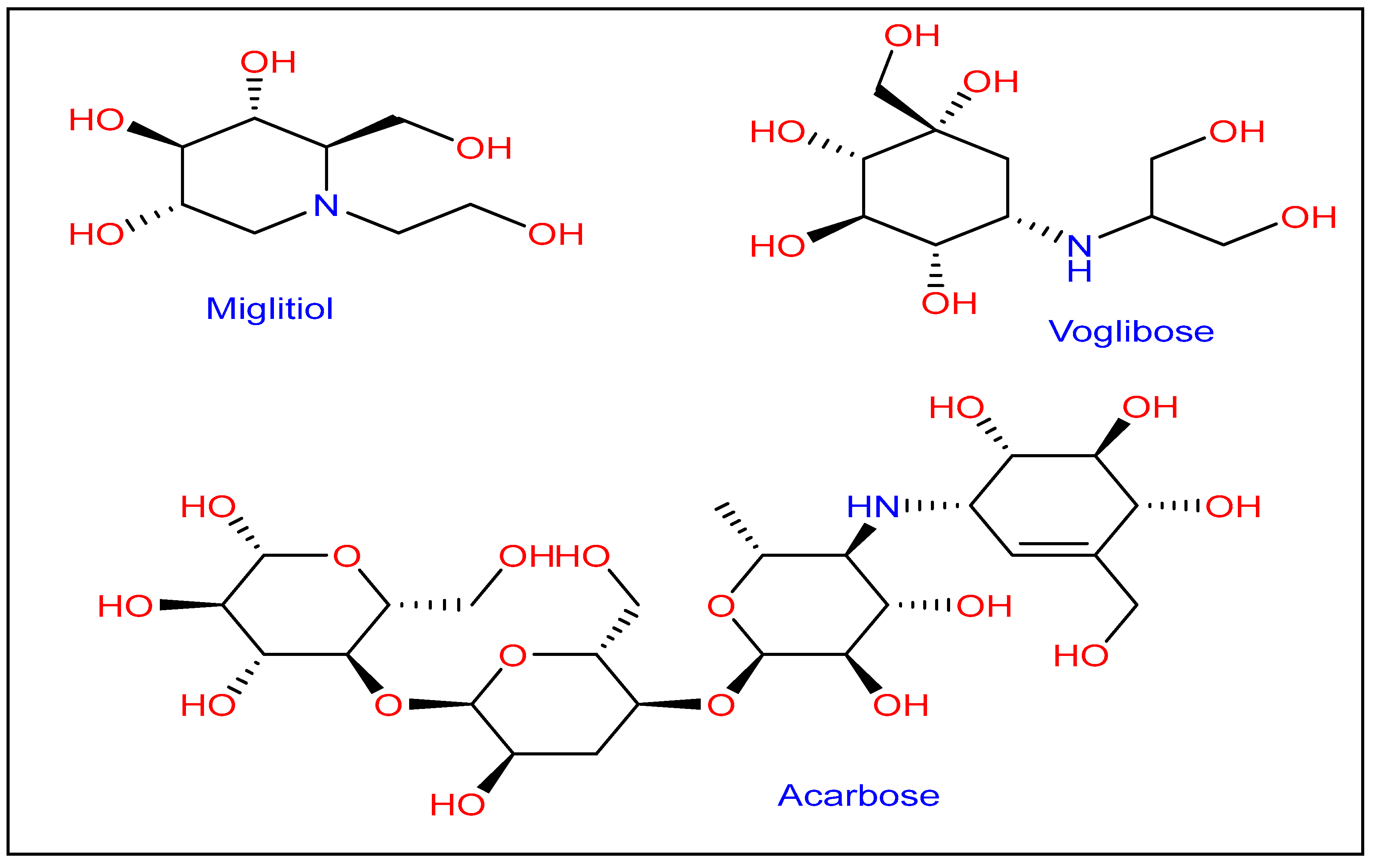
Figure 1. Type 2 diabetes mellitus drug on the market (T2DM).
Pharmaceutics 15 00457 g001
Figure 2. Design of novel hybrid benzimidazole connected with urea.
Pharmaceutics 15 00457 g002
Scheme 1. Synthesis of benzimidazole-urea 3a–f from 2-(1H-benzoimidazol-2-yl)aniline 1 and isocyanates 2a–f.
Pharmaceutics 15 00457 sch001
Scheme 2. Synthesis of compounds 3g and 3h from 2-(1H-benzoimidazol-2-yl)aniline 1.
Pharmaceutics 15 00457 sch002
Figure 3. Benzimidazole-ureas 3a-h inhibition activity against α-amylase and α-glucosidase.
Pharmaceutics 15 00457 g003
Figure 4. Structure-activity relationships related to benzimidazole urea derivatives 3a-h.
Pharmaceutics 15 00457 g004
Figure 5. Optimized structures of 3c, 3e, and 3g with Ammp calculations.
Pharmaceutics 15 00457 g005
Figure 6. The interaction types of (a) 3c, (b) 3e, and (c) 3g with surrounding amino acids of HPA are shown in 3D (A) and 2D (B).
Pharmaceutics 15 00457 g006
Figure 7. The interaction types of (a) 3c, (b) 3e, and (c) 3g with surrounding amino acids of HLAG are shown in 3D (A) and 2D (B).
Pharmaceutics 15 00457 g007
Table 1. Benzimidazole-ureas 3a-h synthesis from 2-(1H-benzoimidazol-2-yl)aniline 1 and isocyanates 2a-h a.
EntryIsocyanate 2Benzimidazole Urea 3Time b (h)Yield c (%)
1Pharmaceutics 15 00457 i001Pharmaceutics 15 00457 i002185
2Pharmaceutics 15 00457 i003Pharmaceutics 15 00457 i0041 90
3Pharmaceutics 15 00457 i005Pharmaceutics 15 00457 i006280
4Pharmaceutics 15 00457 i007Pharmaceutics 15 00457 i008875
5Pharmaceutics 15 00457 i009Pharmaceutics 15 00457 i010465
6Pharmaceutics 15 00457 i011Pharmaceutics 15 00457 i0121060
7Pharmaceutics 15 00457 i013Pharmaceutics 15 00457 i0142 93
8Pharmaceutics 15 00457 i015Pharmaceutics 15 00457 i0161085
a Reaction conditions: 1 (1 mmol, 1 equiv.); isocyanate 2 (1.5 mmol, 1.5 equiv.) in 40 mL of dichloromethane and 10 mL of acetone. b TLC (eluent: Ether/Hexane: 50/50) is used to determine reaction time. c Yields were measured after purification.
Table 2. Antioxidant activities of target benzimidazole-urea 3a-h.
Compounds 3TACDPPH-SAFRAPMCA
3a1.33 ± 0.461.17 ± 0.076.53 ± 0.031.62 ± 0.10
3b5.16 ± 0.690.96 ± 0.069.25 ± 0.201.55 ± 0.07
3c4.43 ± 0.531.61 ± 0.028.65 ± 0.291.55 ± 0.22
3d2.47 ± 0.422.89 ± 1.075.67 ± 0.691.46 ± 0.10
3e2.52 ± 0.801.70 ± 0.012.75 ± 0.151.47 ± 0.09
3f8.50 ± 0.871.55 ± 0.035.99 ± 0.021.40 ± 0.07
3g10.06 ± 1.513.86 ± 0.0416.12 ± 0.291.36 ± 0.09
3h2.25 ± 0.651.09 ± 0.036.79 ± 0.502.95 ± 0.21
TAC: total antioxidant capacity (trolox equivalent per 1 mM of the compounds), DPPH-SA: 2,2-diphenyl-1-picrylhydrazyl scavenging activity (trolox equivalent per 1 mM of the compounds), FRAP: ferric reducing antioxidant power (trolox equivalent per 1 mM of the compounds), MCA: metal chelating activity (EDTA equivalent per 1 mM of the compounds).
Table 3. α-amylase and α-glucosidase inhibition activity of benzimidazole-urea 3a-h.
Compounds 3α-Amylase Inhibition
IC50 ± SEM (µM)
α-Glucosidase Inhibition
IC50 ± SEM (µM)
3a40.7 ± 0.0642.97 ± 0.19
3b41.7 ± 0.0645.97 ± 0.19
3c18.65 ± 0.2317.47 ± 0.03
3d25.65 ± 0.0327.47 ± 0.13
3e20.7 ± 0.0621.97 ± 0.19
3f28.33 ± 0.0229.01 ± 0.12
3g22.33 ± 0.1223.01 ± 0.12
3h43.04 ± 0.0244.99 ± 0.09
Acarbose14.21 ± 0.0215.41 ± 0.32
SEM = Standard error mean for triplicate values.
Table 4. Complexes with HPA and HLAG docking binding energies (kcal.mol−1).
Compounds 3α-Amylase (E kcal/mol)α-Glucosidase (E kcal/mol)
3a−8.8−8.7
3b−10.3−9.9
3c−8.8−8.2
3d−8.6−8.6
3e−8.0−7.6
3f−7.8−7.6
3g−11.2−10.0
3h−10.4−10.0
Disclaimer/Publisher’s Note: The statements, opinions and data contained in all publications are solely those of the individual author(s) and contributor(s) and not of MDPI and/or the editor(s). MDPI and/or the editor(s) disclaim responsibility for any injury to people or property resulting from any ideas, methods, instructions or products referred to in the content.