Review

Targeting Protein Kinases and Epigenetic Control as Combinatorial Therapy Options for Advanced Prostate Cancer Treatment

1
Medical Biology Research Center, Health Technology Institute, Kermanshah University of Medical Sciences, Kermanshah 6714415185, Iran
2
Institute of Biochemistry and Biophysics, University of Tehran, Tehran 1417614335, Iran
3
Department of Biology, Jahrom Branch, Islamic Azad University, Jahrom 7414785318, Iran
4
Department of Biology, Faculty of Sciences, University of Mohaghegh Ardabili, Ardabil 5619911367, Iran
5
Department of Internal Medicine, Angiology and Physical Medicine, Faculty of Medical Sciences in Zabrze, Medical University of Silesia, Batorego 15 St, 41-902 Bytom, Poland
*
Authors to whom correspondence should be addressed.
Pharmaceutics 2022, 14(3), 515; https://doi.org/10.3390/pharmaceutics14030515
Submission received: 9 January 2022 / Revised: 15 February 2022 / Accepted: 21 February 2022 / Published: 25 February 2022
(This article belongs to the Special Issue Protein Kinase Inhibitors for Targeted Anticancer Therapies)

Abstract

:
Prostate cancer (PC), the fifth leading cause of cancer-related mortality worldwide, is known as metastatic bone cancer when it spreads to the bone. Although there is still no effective treatment for advanced/metastatic PC, awareness of the molecular events that contribute to PC progression has opened up opportunities and raised hopes for the development of new treatment strategies. Androgen deprivation and androgen-receptor-targeting therapies are two gold standard treatments for metastatic PC. However, acquired resistance to these treatments is a crucial challenge. Due to the role of protein kinases (PKs) in the growth, proliferation, and metastases of prostatic tumors, combinatorial therapy by PK inhibitors may help pave the way for metastatic PC treatment. Additionally, PC is known to have epigenetic involvement. Thus, understanding epigenetic pathways can help adopt another combinatorial treatment strategy. In this study, we reviewed the PKs that promote PC to advanced stages. We also summarized some PK inhibitors that may be used to treat advanced PC and we discussed the importance of epigenetic control in this cancer. We hope the information presented in this article will contribute to finding an effective treatment for the management of advanced PC.

1. Introduction

A considerable number of about 2.5% of the human coding genome belongs to the protein kinase (PKs) family, and the mutation and dysregulation of PKs play a critical role in several diseases, including cancers. Due to this, PKs have become one of the leading pharmacological drug targets in the 21st Century [1], and protein kinase inhibitors (PKIs) are a promising new class of therapeutic agents [2]. The availability of the potent inhibitors of understudied kinases could greatly aid the discovery of uncovering new targets for drug development [3]. Ferguson et al. provided an overview of the novel targets, biological processes, and disease areas that kinase-targeting small molecules are being developed against and evaluated the strategies and technologies that generate highly optimized kinase inhibitors [4]. Recently, Klaeger et al. performed a comprehensive analysis of 243 kinase inhibitors that are either approved for use or in clinical trials [5,6]. According to the latest update (21 January 2022) of the PK Inhibitor Database (PKIDB) [7], 72 FDA-approved medicinal products target different types of PKs. Eight of these PKIs were approved in 2021 and one in 2022. The PKIDB shows that although most of these drugs are indicated as various cancer therapeutics (solid and nonsolid tumors); however, information on the use of these drugs in prostate cancer (PC) is still insufficient.
PC is an endocrine-related disease [8], ranked as the most commonly diagnosed malignancy [9]. The male hormone androgens play a crucial role in PC progression through androgen receptor (AR) activation [10]. This issue has made AR an important therapeutic target for PC therapy [11]. The first-line treatments for metastatic PC are androgen-deprivation therapy (ADT) and AR-targeting therapy, but secondary resistance coupled with enhanced metastatic potential is a crucial challenge in these treatments [12]. Mounting evidence suggests that PKs play a crucial role in tumor growth, proliferation, and metastasis in PC. Furthermore, they may be responsible for resistance to standard treatments [13,14,15]. Additionally, recent discoveries indicated the complex crosstalk between the PKs and epigenetic events and critical biological pathways, including AR signaling pathways [12,16]. Therefore, a deep understanding of how genetic and epigenetic mechanisms regulate the progression of PC appears to be essential to design therapeutic agents for PC patients [16]. For these reasons, hopeful studies are now underway with epigenetic modulators [17] and kinase inhibitors [13,18] as combination therapy options to gain selectivity and overcome resistance. In this review article, we focused on the role of PKs and epigenetic processes in PC progression and discussed the new advancements for the management and treatment of this cancer by controlling epigenetics and targeting the PK family.

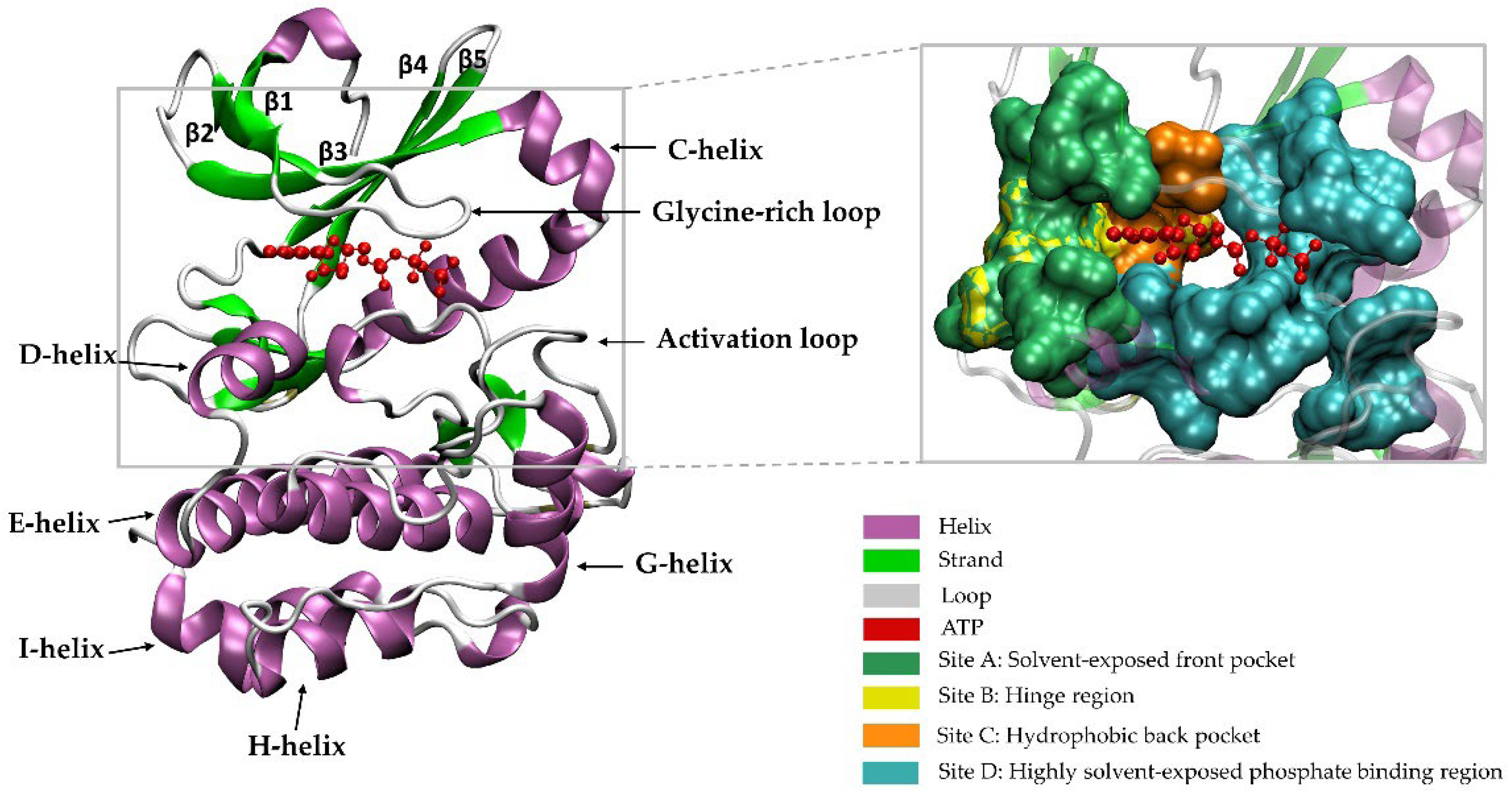
2. A Brief Introduction to the Family of PKs

PKs catalyze phosphorylation reactions to regulate the enzyme activity, protein functioning, and signal transduction pathways by transferring a phosphate group to an acceptor amino acid of the substrate protein [19]. They play a crucial role in cellular processes, such as metabolism, motility, and cell division [20,21,22]. The dysregulation of PK activity is associated with the pathogenesis of several diseases, including cardiovascular [23,24], autoimmune, and inflammatory [25] diseases, as well as cancers [26]. The first classification of PKs is related to the efforts of Tony Hunter and Steven Hanks [27,28]. This classification was extended by Manning et al. [21]. Based on the phosphate acceptor amino acid specificity, PKs can mainly be divided into two subdivisions: protein–serine/threonine kinases (Ser/Thr kinases) and protein–tyrosine kinases (Tyr kinases). Additionally, comprehensive sequence analysis of PKs led to a classification system consisting of nine main groups in phylogenetic trees: calcium/calmodulin-dependent PK (CAMK), AGC (containing PKA, PKG, and PKC families), Tyr kinase (TK), TK-like kinase (TKL), CMGC (containing cyclin-dependent kinase (CDK), mitogen-activated PK (MAPK), glycogen synthase kinase-3 (GSK3), and CDC2-Like PK (CLK) families), casein kinase 1 (CK1), STE (homologs of yeast Sterile 7, Sterile 11, and Sterile 20 kinases), receptor guanylate cyclases (RGC) group, and atypical PKs [21].
Currently, among the 497 typical kinase domains in the human genome, 284 kinase structures have been determined experimentally, either as apo or in complexes with inhibitors or ATP. Generally, the protein kinase fold consists of two domains: an N-terminal domain and a C-terminal domain. The N-terminal region consists of an alpha helix called C-helix, as well as five beta-sheet strands, and the C-terminal domain commonly contains five or six helices, namely the D, E, F, G, H, and I alpha helices. A deep cleft created by the N-terminal and C-terminal lobes in the middle region of the protein forms the active site for ATP-binding. In addition, the activation loop is one of the most important regions that helps ATP and the substrate bind in the enzyme’s active site. The activation loop includes the Asp–Phe–Gly motif called the “DFG motif,” which adopts an extended unique orientation in the active state conformation of the enzyme and several types of folded conformations in an inactive state [29].
Figure 1 shows the structure of a PK, namely, Aurora A kinase (AURKA) (PDB: 5DNR), and four key sites A–D on the surface binding groove (Figure 1). Site A is the solvent-exposed front pocket (composed of residues 137, 139, 157, 212–216, 220, 224, and 264–266) and site B is the hinge region (residues 210–216) that mainly focus on the hydrogen bonding network. Site C is the hydrophobic back pocket, which is not conserved and identified as the selectivity pocket, and is present in most of the kinases, created by residues 210, 211, 147, 160, and 194 in AURKA. Site D is a highly solvent-exposed phosphate-binding region (formed by amino acid residues 143, 144, 162, 164, 178, 181, 194, 208, 255, 258, 260, 261, 263, 271–275, and 277). Site D is relatively larger compared to site A [30]. Most drugs that bind to the ATP site are considerably hydrophobic and inhibit kinase catalytic activity [31]. However, structural analysis suggested that the solvent-exposed sites A and D, located outside the ATP binding site, could be used to improve the pharmacokinetic properties of lead compounds [30].
Recently, Modi and Dunbrack presented a web resource called Kincore (the Kinase Conformation Resource) that automatically organizes a collection of all PK structures and assigns conformational state and inhibitor type tags. They identified eight active and inactive functional states of the DFG motif. Additionally, they classified the inhibitor type bound to each kinase domain into five categories: type 1, 1.5, 2, 3, and allosteric. Type 1, 1.5, 2, and 3 inhibitors bind to the ATP site, the ATP binding site + a portion of the C-helix region, the ATP binding site + the C-helix region, and the C-helix region, respectively. Allosteric inhibitors bind elsewhere. By combining the classification of the DFG motif conformation and inhibitor types, over 200 inhibitors were found that bind to multiple states of kinases [29].

Catalytic and Non-Catalytic Activities of PKs

Despite the success of targeted kinase inhibitors in responsive patients, their tumors almost show resistance over time, leading to disease progression and a central challenge for clinical care [33]. Thus, multiple strategies are required to overcome this resistance. A critical approach is inducing and stabilizing inactive kinase forms allosterically [34]. Although PKs are known primarily for their ability to phosphorylate protein substrates, accumulated evidence has recently suggested that most human kinases have non-catalytic activity beyond catalysis through their scaffolds. The non-catalytic activity of PKs involves the allosteric regulation of other kinases or enzymes through protein–protein interactions, assembly of signaling complexes, or even transcriptional regulation via direct binding to DNA or interaction with a transcriptional factor [35]. These non-catalytic activities play a critical role in normal cellular activities and diseases, especially in mediating drug resistance to kinase inhibitors [35]. Most FDA-approved PK inhibitors inhibit kinase catalytic activity upon binding to the ATP binding site. Recently, accumulated evidence has suggested that small molecules modulating the non-catalytic functions of kinases can emerge as new promising therapeutic strategies for various diseases. To date, classes of agents have emerged that can regulate the non-catalytic function of kinases. Orthosteric and allosteric kinase inhibitors, protein degraders, and protein−protein interaction blockers are three categories of these modulators [31].

3. PK Targeting Tools

Irregular signaling pathways are a hallmark of cancer [36]. Therefore, it is not surprising that PKs are the most drug targets, after the G-protein-coupled receptors [35]. Currently, several tools are available for targeting PKs, each with their advantages and disadvantages. One of these methods is the use of small-molecule kinase inhibitors, which has been widely studied and has been successful in the treatment of various cancers [35]. Accordingly, the FDA has approved 70 small-molecule kinase inhibitors for application in oncology [37]. However, despite their advantages, such as the ability to target multiple cell survival pathways, ease of oral administration, and low production costs, the clinical use of these inhibitors faces a variety of challenges, including cytotoxicity, chemotherapy resistance, and off-target effects [35]. Another method that has been studied to inhibit kinases is the use of synthetic peptides that, despite their advantages, such as high specificity, they have some drawbacks, such as poor pharmacokinetic and biodistribution parameters [38,39]. Short interfering RNA (siRNA), also referred to as RNA interference (RNAi), is a well-known technology that has shown promising therapeutic results in cancer treatment [40,41]. Numerous studies showed that the concomitant use of siRNA and TK inhibitors (TKIs) could sensitize resistant cells to chemotherapy [42,43]. In addition, other evidence revealed that the use of siRNA against various kinases had anticancer effects and significantly reduced the chemotherapy resistance in different cancer cells. Some related studies in this field include studies on polo-like kinase (PLK) [41,44], focal adhesion kinase (FAK) [45], PKB/Akt [46], B-RAF [47], receptor tyrosine kinase-like orphan receptor-1 (ROR1) [48], AURKA [49], eukaryotic elongation factor 2 kinase (EF2K) [50], pyruvate kinase M2 (PKM2) [51], and CDK8 [52]. Nevertheless, despite its special advantages, such as its high degree of specificity [53,54], siRNA faces numerous challenges, such as systemic toxicity, obstacles with delivery to various tissues, and a high degradation rate in the presence of serum proteins and enzymes [40,42,43]. Another way to inhibit kinases is the use of kinase-targeted antibodies, which has been reported to be effective in various cancers [34], for instance, monoclonal antibody against epidermal growth factor receptor (EGFR) in colorectal cancer [55] and against HER2 in breast cancer [56,57]. The inhibition of kinases with monoclonal antibodies, despite having advantages, such as high specificity, presents adverse effects, such as allergic reactions and the development of various cytotoxicities [58]. Another tool for blocking kinases is the use of proteolysis-targeting chimera (PROTAC) technology. PROTACs bind to proteins of interest and use E3 ligase to degrade the entire target protein via the ubiquitin–proteasome pathway [59]. However, as with other methods, PROTAC technology faces challenges, such as acquired and intrinsic resistance to drugs in cancer cells [60]. The use of natural products and probiotics are other tools that have been considered as potential kinase inhibitors in recent years. Various natural products, including curcumin [61], green tea extracts [62], luteolin [63], quercetin [64,65], and resveratrol [66], have shown inhibitory activity against different kinases in various cancers, and several mechanisms have been proposed for the effect of these compounds on the reduction of kinase mutations. The use of natural products also has its own problems due to issues such as accessibility, sustainable supply, and intellectual property constraints [67]. Various probiotics and their metabolites have also been studied as kinase inhibitors, and research in this field is expanding [68,69].

4. PKs and PC Progression

In general, studies on kinases have suggested both anti-cancer and pro-cancer roles for them, and this dual role has been attributed to having different subunits, the localization of isozymes in different cell subunits, and the different contexts of their activity. In this article, the complexity of the role of kinases is neglected, and the focus is on reports that have shown the pro-cancer role of them. In addition, although the role of many kinases in PC progression has been reported, such as EGFR [70], EphA2 [71,72], Janus kinase 1 (JAK1) [73], JAK2 [74], c-Jun N-terminal kinase (JNK) [15], MAPK4 [75], protein tyrosine kinase 6 (PTK6) [76], ribosomal S6 kinases (RSKs) [77], vascular endothelial growth factor receptor 3 (VEGFR-3) [78], etc., only a few of them have been cited as examples, and the mechanism of their effect on PC progression is discussed in detail.

4.1. AMP-Activated PK (AMPK)

AMPK belongs to Ser/Thr PKs, which is activated by enhanced intracellular AMP concentrations [79]. AMPK is a main cellular energetic biosensor that regulates a large number of metabolic pathways activated by nutrient (glucose) deprivation, low oxygen gradients, mitochondrial dysfunction, oxidative stress, and cytokines [80,81]. Activated AMPK promotes energy-sparing mechanisms and induces anti-apoptotic functions. As a result, it allows cells to survive for a very long time in very hostile conditions [81]. Multiple studies showed a positive correlation of AMPK phosphorylation/activation with the Gleason score and disease progression in PC patients [82,83,84]. AMPK activity is regulated by androgen and upstream kinases, including the CAMK kinase 2 (CAMKK2) in PC [85,86]. Indeed, androgen enhances AMPK activation and autophagy, thereby promoting PC growth [87]. New findings on the association between AMPK and metabolic reprogramming showed that the AMPK/GSK3β/β-catenin cascade might upregulate the cell-migration-inducing protein (CEMIP), which could drive the migration and invasion of anoikis-resistant PC cells via increasing pyruvate dehydrogenase kinase isoform4 (PDK4)-associated metabolic reprogramming [88].
Macropinocytosis is a process of non-selective swallowing of extracellular material through the ruffling of the plasma membrane [89]. Ras-related C3 botulinum toxin substrate 1 (RAC1-GTP) and phosphatidylinositol (3,4,5)-trisphosphate (PIP3) are necessary for macropinosome formation [90,91]. In fact, by macropinocytosis, cancer cells with activating mutations in RAS use extracellular proteins as a fuel when amino acids are limiting [92]. PIP3 is produced by phosphoinositide 3-kinases (PI3K) and converted to (phosphatidylinositol (4,5)-bisphosphate (PIP2) by phosphatase and the tensin homolog (PTEN) [93]. PTEN is a tumor suppressor gene that is most frequently disrupted in PC [94,95] and is correlated with an increased risk of metastasis and resistance to castration [94,96]. Indeed, macropinocytosis is a cancer-related phenotype caused by a loss of PTEN function. As a regulator of PI3K signaling, the loss of PTEN leads to Akt overactivity, followed by reduced apoptosis, uncontrolled cell proliferation, and increased tumor angiogenesis [97]. Recent studies have revealed that the loss of PTEN is not sufficient to induce macropinocytosis in PC cells and that AMPK activation is essential. In fact, AMPK activates RAC1, which is essential for membrane ruffling in macropinocytosis [98].

4.2. Protein Kinase A (PKA)

PKA, also known as cAMP-dependent PK, is a member of Ser/Thr PKs that regulates the signal transduction of G-protein-coupled receptors through its binding to cAMP [99], and its role in the onset and progression of many tumors has been demonstrated [99,100]. cAMP is a second messenger that is involved in various cellular functions, including the ion channel activation, gene expression, cell growth and differentiation, and apoptosis [100].
Progress toward castration resistance is a crucial problem in the treatment of advanced PC. Numerous evidence suggests that PC cells develop castration resistance by activating multiple molecular pathways, including AR and PKA [101]. In the absence of androgen, increasing levels of cAMP/PKA pathways have been shown to increase the expression of AR and prostate-specific antigen (PSA) proteins in PC cells [102], which, in turn, can lead to increased androgen signaling, resulting in cell proliferation and subsequent castration-resistant PC (CRPC) [103]. The type II beta regulatory subunit of PKA, cAMP-dependent protein kinase type II–beta regulatory subunit (PRKAR2B), is highly expressed in CRPC and is involved in tumor proliferation and metastasis. A new study has revealed that PRKAR2B enhanced the expression level of hypoxia-inducible factor 1α (HIF-1α), a crucial moderator of the Warburg effect, thereby promoting tumor growth [104].
Recent studies have shown the phosphorylation of different proteins by PKA that suppresses apoptosis or stimulates invasion and metastasis. PKA activation is essential for the phosphorylation of heat-shock protein 90 (Hsp90), which binds to the ligand-free AR in the cytoplasm and restricts its entry into the nucleus. However, new findings suggest that the PKA-mediated phosphorylation of Thr89 residue of Hsp90 can lead to the release of AR from Hsp90, subsequently binding AR to Hsp27 and its migration to the nucleus [103]. Some studies have demonstrated that PKA phosphorylates caspase-9 induces the disassembly of the large and small subunits of caspase-9 and prevents its self-processing, thus inactivating caspase-9 and suppressing the progression of apoptosis [105]. Another study indicated that the calcitonin receptor, which enhances PC cell invasion, activated PKA that phosphorylated the tight junction proteins ZO-1 and claudin 3, destabilizing the tight junctions and increased PC cell invasion [106]. Other studies revealed other aspects of the association between PKA and PC progression, including the association between PKA and angiogenesis [107], the reduction of the Ca2+-store content in the endoplasmic reticulum (ER) [108], and the mediating of the tumor-associated macrophage polarization phenotype [109].

4.3. Protein Kinase B (PKB)

PKB, also known as Akt, is a member of the Ser/Thr kinases whose protein expression and activity have been shown to increase in many tumors and tumor cells [110,111]. Several kinases, including 3-Phosphoinositide-dependent kinase 1(PDK1) and mammalian target of rapamycin (mTOR) complex 2 (mTORC2), activate Akt. An increase in the PIP3 levels by PI3K causes the uptake of Akt into the plasma membrane and its activation [112]. The association of Akt activation with the PC progression from an androgen-dependent stage to an androgen-independent stage has been shown [113,114]. Akt enhances the androgen-independent survival of prostate tumor cells by regulating AR expression and activation [113]. Akt phosphorylates the residues Ser213 and Ser791 of AR, leading to AR signaling and cell survival [115]. The role of Akt in the PI3K/Akt/mTOR pathway has been demonstrated in many studies, so this pathway has been proposed as the main regulatory factor of pro-survival/anti-apoptotic pathways in the absence of AR signaling [116]. Studies on different human PC cell lines have shown that the inhibition of PI3K or expression of the dominant negative mutant of Akt inhibits invasion and decreases the expression of urokinase-type plasminogen activator (uPA) and matrix metalloproteinase-9 (MMP-9), which are markers of cell invasion [117].
Various studies have revealed different aspects of the PKB’s effect on cancer progression. For instance, the overexpression of fatty acid synthase (FAS) in PC tissues is associated with Akt phosphorylation and nuclear accumulation. FAS is an essential metabolic enzyme associated with the synthesis of membrane phospholipids in cancer cells, high levels of which are expressed in human epithelial cancers, especially those with poor prognosis [118]. The forkhead box transcription factor FoxO3a is known to be a tumor suppressor whose activity has been shown to be negatively regulated by Akt through post-translational modifications [119,120]. Indeed, phosphorylation at Ser253 increases the accumulation of FoxO3a and its binding chaperone protein 14-3-3 in the cytosol, thus reducing its level in the nucleus [119]. Subsequent studies disclosed that Par-4 is one of the crucial FoxO3a transcriptional targets, and par-4 activation is necessary to induce apoptosis in CRPC cells [120]. Furthermore, other reports indicated that the inhibition of FoxO3a accelerated PC progression in the transgenic adenocarcinoma of mouse prostate (TRAMP) mice, which was correlated with increased proliferation and survival markers [121].

4.4. Protein Kinase C (PKC)

PKC belongs to Ser/Thr PKs, in which its different isoforms play significant roles in the cell cycle and cell death, and changes in their expression or activity have been identified in human diseases [122,123]. The effect of PKC on the cell cycle is highly context-dependent and varies depending on the specific isoenzyme involved and other factors, such as the time and duration of enzyme activation [122]. Some studies suggested that PKC-α, PKC-ε [124], and the atypical PKCs (aPKCs), PKC-λ/ι [125] and PKC- ζ [124,126], preferably induce cell proliferation and survival, while PKC-δ regulates apoptosis [127].
It has been shown that AR phosphorylation at the Ser-578 residue, which is attributed to PKC [128], may cause PC progression [129]. Immunohistochemical studies of human PC tissue microarrays showed that the PKCε expression levels were associated with PC aggressiveness. Further studies on human PC, human PC cell lines, and PC developed in TRAMP mice illustrated those signal transducers and activators of transcription 3 (STAT3) that are primarily active in a wide range of human cancers, including PC, which interact with PKCε and is phosphorylated at Ser727. The inhibition of PKCε expression inhibited STAT3Ser727 phosphorylation, followed by decreased DNA binding and STAT3 transcriptional activity, as well as reduced cell invasion. These results suggest that PKCε activation is necessary for STAT3 activation and PC progression [130].
PKCε collaboration with PTEN loss for PC development has been demonstrated in a mouse model. The overexpression of PKCε and PTEN loss, individually and synergistically, positively regulates chemokine (C-X-C motif) ligand 13 (CXCL13) production. In addition, the disruption of CXCL13 or its receptor in PC cells affects its tumorigenic and migratory properties. The role of the chemokine CXCL13 and its receptor, C-X-C chemokine receptor type 5 (CXCR5), has been reported to be a major factor in the progression of many cancers, including PC [131]. Various studies have described various mechanisms by which PKCε is involved in the progression and metastasis of PC, including enhancing aerobic glycolysis [132], the activation of nuclear factor kappa-light-chain-enhancer of activated B cells (NF-κB)) [131], phosphorylation of Vimentin [133], and interaction with BCL2 associated x, apoptosis regulator (Bax) [134].
Available evidence suggests that both aPKCs, as with other PKC isoforms, play pleiotropic context-dependent roles, and some studies have reported pro-tumorigenic roles for them [124]. Vimentin overexpression is known as a hallmark of the epithelial–mesenchymal transition (EMT), and the molecular dynamics of Vimentin intermediate filaments (VIFs) play a significant role in metastasis [135]. New findings show that aPKCs activate Vimentin by phosphorylating Ser33, Ser39, and Ser56 residues in Vimentin, resulting in VIF disassembly, which contributes to PC cell metastasis [136]. In addition, both PKC-ι and PKC-ζ have been shown to induce cell survival through the NF-κB/PI3K/Akt pathways [125].

4.5. Protein Kinase D (PKD)

PKD is a family of Ser/Thr kinases belonging to the CAMK superfamily. The physiological functions and regulatory mechanisms of PKD, including the regulation of gene expression, protein/membrane trafficking, cell proliferation, survival, migration, and angiogenesis, are well documented [137]. Three PKD isoforms, PKD1, PKD2, and PKD3, are stimulated by various extracellular stimuli and transduce cellular signals that affect many aspects of primary cellular function [138]. Dependent on subcellular localization, PKD isoforms control various processes, including cell signaling, Golgi transport, and the oxidative stress response [139]. Further studies at the cellular level and in animal models have shown the vital role of PKD in numerous pathological conditions, including cancer [138]. An in-depth in vitro migration study on Panc1 pancreatic cancer cells to clarify the role of PKD in cancer cell migration indicated that the absence of each PKD isoform exerts a considerable effect on cell speed and migration persistence, and that the absence of PKD1 is associated with a significant increase in Panc1 cell deformability [140].
Current findings suggest a potential tumor-promoting function for selective PKD isoforms in PC [141]. It has been shown that PKD3 interacts with sterol regulatory element-binding protein 1 (SREBP-1) and consequently promotes cell proliferation via lipid metabolism in PC cells [142]. Some evidence has shown that PKD2 and PKD3 enhance NF-κB signaling and uPA expression/activation, that are critical for PC invasion [143]. EMT and cell migration play a key role in the onset and progression of diverse malignancies, including PC. Different PKD isoforms act differently in these processes, with PKD1 inhibiting EMT and cell migration, but PKD2 and PKD3 induce these processes [144]. Previous studies have indicated that a PKC/PKD pathway protects PC cells against phorbol ester-induced apoptosis via elevating the extracellular signal-related kinase1/2 (ERK1/2) and NF-κB transcriptional activities [105]. Furthermore, a more recent report revealed that PKC and PKD play a significant role in PC cell migration induced by CXCL12 chemokine [145].

4.6. DNA-Dependent Protein Kinase (DNA-PK)

DNA-PK is a nuclear Ser/Thr PK [146], which plays a significant role in the repair of double-strand breaks [147]. In addition to interfering with DNA repair, DNA-PK plays a regulatory role in transcription by phosphorylating transcription factors, thereby regulating their functions [148]. Recent studies have shown new, different roles beyond DNA repair for the DNA-PK catalytic subunit (DNA-PKcs) in cancer, including its involvement in cell cycle progression, metastasis, treatment resistance, metabolic dysregulation, and immune escape [147].
DNA-PK is well documented to control tumor metastasis and progression in PC by various mechanisms, such as interaction with AR or the phosphorylation of insulin-like growth factor (IGF)-binding protein 3 [149]. A recent study was performed to recognize the kinases that drive PC progression, and tumor samples were collected from 545 patients with high-risk diseases. The results of this study identified DNA-PK as the most important kinase related to metastatic progression in high-risk PC. It also showed that DNA-PK mainly drives PC by regulating the transcription of Wnt signaling members [150]. Some studies revealed that DNA-PK and mTOR, through localization in chromatin at specific regulatory sites, function as AR cofactors in PC cells. In fact, the nuclear localization of mTOR and DNA-PK expression, both of which increase in advanced PC, are associated with metastasis and reduced overall survival [151,152].

4.7. CDC2-Like Protein Kinase (CLKs)

The CLK family, known as signaling kinases [153], consists of four isoforms, including CLK1, CLK2, CLK3, and CLK4 [154], with specific and conserved ATP binding sites like other kinases. CLKs can phosphorylate serine, threonine, and tyrosine residues. Therefore, dual-specificity kinase activity is observed in the CLK family [155]. The structure of CLKs consists of domains N and C, which are connected by the hinge region, β-strands, and α-helices distributed between the N and C regions as a catalytic domain [156]. CLKs and then bind to pre-mRNA and stabilize the serin and arginine-rich splicing factors 1–12 (SRSF1–12) are phosphorylated by the interaction of spliceosome components and spliceosome assembly [157]. The expression levels of CLK isoforms are different between cell types and problems, such as prostate, testes, brain, leukocytes, muscle, liver, lung, kidney, and thyroid [158].
In PC, the expression level of prostate-associated gene 4 (PAGE4) increases. PAGE4 is an intrinsically disordered protein (IDP) with significant roles in the development and differentiation of PC. PAGE4 is not detectable in the normal adult gland [159]. Hence, it has the hallmarks of a proto-oncogene. Homeodomain-Interacting PK 1 (HIPK1) is an element of the cellular stress-response pathway and can phosphorylate the Ser9 and Thr51 of PAGE4, but phosphorylation in Thr51 is critical. In addition, hyperphosphorylation occurs at multiple Ser/Thr residues by CLK2. PAGE4 phosphorylation by HIPK1 increases c-Jun activity (a component of the stress–response pathway), whereas phosphorylation by CLK2 decreases this activity. Therefore, these two kinases have opposite functions [160]. Androgen receptor, a crucial therapeutic target in PC, is negatively regulated by activator protein-1 (AP-1). The formation of transcription factor Ap-1 is related to the heterodimerization of the proto-oncogene c-Jun with c-Fos [161]. Furthermore, CLK2-PAGE4 shows a low affinity for transcription factor Ap-1. Therefore, the conformational dynamics of PAGE4, which are induced by phosphorylation, may play a role in modulating alterations between PC cell phenotypes [159,160].

4.8. Serine-Argnine Protein Kinase 1 (SRPK1)

Ser-Arg PKs (SRPKs) can phosphorylate serine residues located in the rich region of Arg/Ser or Ser/Arg dipeptides motifs. Therefore, the SRPK family can regulate alternative splicing as SR splicing factor phosphorylation [162]. SRPK1 phosphorylates SR proteins, such as SRSF1 (splicing factor 1), and regulates RNA maturation, protein phosphorylation, cell cycle progression, the regulation of viral genome replication, chromatin reorganization, and immune response. Therefore, various functions of SRPK1 in cellular processes distinguish it from other kinases [163,164]. SRPK1, as with other Tyr kinases, consists of two conserved kinase domains. The large lobe of the C-terminal domain is the substrate-binding site and consists of α-helices, whereas the small lobe of the N-terminal domain comprises β-strands and is an ATP binding site [165].
Vascular endothelial growth factor-A (VEGF-A) induces angiogenesis, which is required for tumor growth. The level of VEGF increases in the urine and plasma of advanced stages of PC. The two families of VEGF isoforms of pro-angiogenic and anti-angiogenic are produced during the alternative splicing of VEGF-A pre-mRNA with the dominant isoform of VEGF165b. VEGF165b is anti-angiogenic. In PC, only the pro-angiogenic isoform is upregulated. SRSF1 phosphorylation by SRPK1 can control VEGF splice isoforms. The overexpression of SRPK1 is observed in PC progression [166,167,168]. Therefore, the inhibition of SRPK1 can switch to the expression of the anti-angiogenic isoform [164,166].

4.9. Pyruvate Kinase M2 (PKM2)

PKM2, a key glycolysis enzyme, is overexpressed in many tumor cells and plays a critical role as a regulator in tumor metabolism [169]. PKM2 has been demonstrated to be overexpressed in PC and promotes PC metastasis via ERK-cyclooxygenase (COX-2) [170]. Other studies have shown that there is a significant positive correlation between PKM2 nuclear localization and PC aggressiveness; also, the pharmacological targeting of PKM2 nuclear translocation disrupts the metastatic dissemination of PC cells in SCID mice [171]. In addition, the comparison of serum-derived exosomes from PC patients with healthy men showed that increased exosome PKM2 expression was associated with metastasis. A recent study identified the exosome-mediated transfer of PKM2 from PC cells to bone marrow stromal cells as a new mechanism by which exosomes derived from the primary tumor promote the formation of pre-metastatic niches [172].

4.10. T lAK Cell Originated PK (TOPK)

T lAK Cell-Originated PK (TOPK) plays a role in the mitotic progression and regulation of the cell cycle, and is expressed in both the nucleus and the cytoplasm. Due to its high homology to mitogen-activated protein kinase kinase 3 (MKK3), TOPK is a MAPK kinase (MAPKK) and is a dual-specificity Ser/Thr kinase [173]. It seems that TOPK plays a role in the activation of Akt, ERK, and JNK due to the dual-specificity family of kinases. Akt is activated when PTEN is phosphorylated and deactivated by TOPK [174]. The tissues with high levels of proliferation overexpress TOPK, while the expression of TOPK is minimal in differentiated cells. Therefore, the invasion, aggressiveness, and metastatic growth of tumors are linked to the overexpression of TOPK [175]. CDK1/cyclin B1 complex phosphorylates the TOPK in Thr9; hence, TOPK is functionally activated and can destabilize the tumor suppressor P53 and damage mitosis. Indeed, the overexpression of TOPK leads to aberrant entry into the mitotic phase by phosphorylating histone H3 at Ser10 via bypass of the G2/M checkpoint, downregulation of p53 (tumor suppressor), and upregulation of the CDK inhibitor p21 [174]. Conversely, the inhibition of TOPK activity leads to a reduction in the phosphorylation and activation of MAPK, reducing the inhibition of Akt activation and inhibiting the expression of mutant p53; therefore, the tumorigenic properties are impaired. Recently, Alhawas et al. reported the direct role of TOPK in the regulation of an alternatively spliced AR variant, ARv7, and the driving of androgen-independence in PC cells [176].

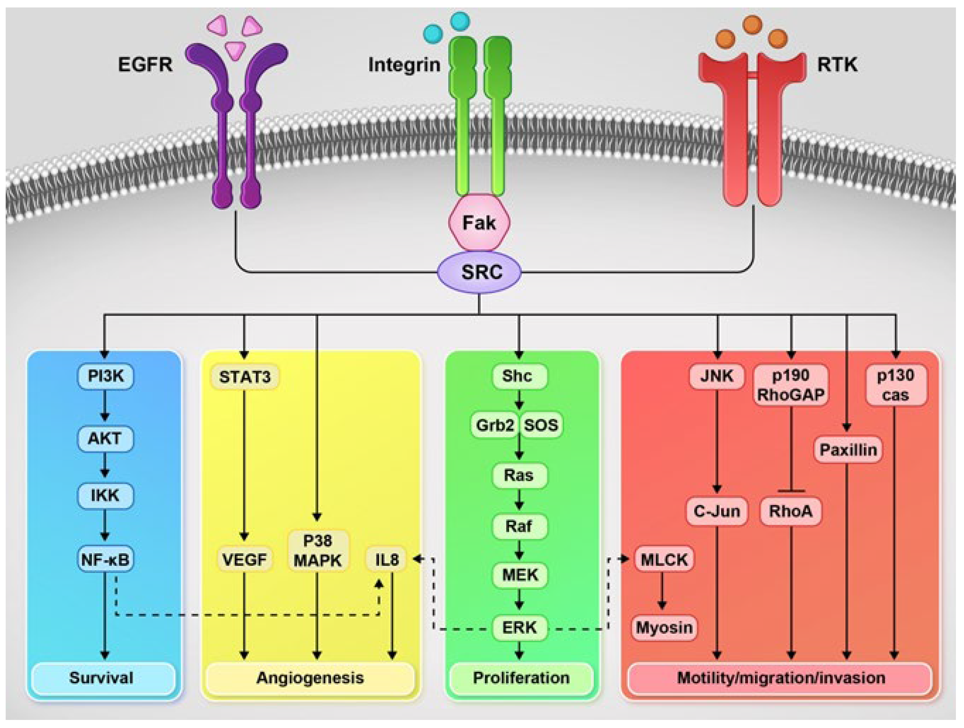
4.11. Src Family Kinases (SFKs)

Src family kinases (SFKs), the largest family of non-receptor Tyr kinases, are responsible for signal transduction during cell differentiation, adhesion, and migration during normal cellular processes. Due to these roles, SFK-activated signaling pathways are involved in angiogenesis, motility, invasion, and tumor adhesion (Figure 2). Recent evidence suggests that Src activity may play a prominent role in cancer progression, including PC. Drake et al. demonstrated a significant upregulation of Tyr phosphorylation in CRPC. Additionally, they found that the increased expression of Src and AR can synergistically drive the frank of prostate carcinoma [177].

4.12. Focal Adhesion Kinase (FAK)

FAK, a member of the non-receptor Tyr kinase located at the extracellular matrix cell adhesion site, is associated with the development and progression of cancer. FAK regulates downstream signaling pathways on the cell-extracellular matrix of integrins, growth factor receptors, cytokine receptors, and G-protein-coupled receptors. It has been found that the development of tumor malignancy is often associated with disturbance in these signaling cascades [178]. FAK, an essential mediator of integrin-associated signaling, is a well-established example of the non-catalytic function of PKs. Following integrin clustering, FAK acts as a scaffolding protein to assemble focal adhesion by interacting with the integrin-binding [31]. Studies by Marcellus et al. showed that the activation of FAK in the metastatic PC3 cell line is an essential factor for the colony formation in PC3 cells, thus affecting cell motility [179]. Additionally, Slak et al. investigated the role of FAK in cell migration and demonstrated that the metastatic potential of PC correlates with its intrinsic migratory capacity, and the metastatic potential correlates with the FAK expression and activation. Moreover, they reported that the autophosphorylation of FAK is adhesion-dependent in PC3, whereas Tyr861, as the second site of phosphorylation, an Src-specific site, is uncoupled from adhesion-dependent events. Significant inhibition of prostate cell migration is achieved by inhibiting the FAK/Src signaling pathway (Figure 2), demonstrating that cell migration depends on signals emanating from this pathway [180]. In a study on 100 patients with prostate adenocarcinoma, a strong functional interaction between FAK and MMP-9 has been shown and, consequently, enhanced the angiogenesis, invasion, and progression of prostate adenocarcinoma [181]. Taken together, these studies suggest that, for patients with prostate adenocarcinomas, FAK/Src may be considered as new therapeutic targets. Further investigations are needed to clarify their importance.

4.13. Cyclin G-Associated Kinase (GAK)

Cyclin G-associated kinase (GAK), also known as auxilin II, is a Ser/Thr kinase, which is homologous to auxilin I, except that there is a kinase domain at the N terminus of GAK [182,183]. GAK, which is localized in the cytoplasm (particularly at the trans-Golgi network) and nucleus [184], plays a significant role in membrane trafficking and the sorting of proteins [185,186]. GAK is localized principally in the nucleus in cancer cells and nuclear GAK overexpression was reported in surgical specimens from PC patients [187]. GAK overexpression was identified in over 90% of androgen independent (AI) tumor biopsies from PC patients [188]; a positive correlation between GAK expression and the Gleason score in surgical specimens from PC patients was reported [186]. GAK has been shown to be involved in the progression of cancer to AI [188], although this is not because GAK is a direct coregulator of AR. Recent studies have shown that the inhibition of the GAK kinase domain can inhibit the growth of PC cells [189,190].

5. The Role of PKs in Epigenetic Changes and Progression of PC

Epigenetic disorders have been identified as a major factor in escaping cell death during cancer treatment and radiotherapy [191]. Epigenetic changes involve inherited and reversible changes in gene expression and mRNA translation without any modification of DNA sequences, which is considered as a link between phenotype and genotype [192]. In the epigenetic process, chemical groups have been added (writers) or removed (erasers) and recognized (readers) to alter gene expression after cell division and determine cellular fate. Epigenetic markers include DNA methylation, histone modification, chromatin remodeling, and noncoding RNA (ncRNA), especially for microRNAs (miRNAs) [193].
Epigenetic disorders have been reported to play a key role in the onset and progression of PC [194]. As many of the signaling pathways in advanced PC, including those involved in cell–to–cell adhesion, epithelial–mesenchymal transition, and the maintenance and regulation of stem cells, are epigenetically impaired, PC is considered as a cancer of the epigenome [195,196,197]. In fact, several enzymes, such as kinases contribute to these epigenetic abnormalities [198]. While the mechanism of PKs as cytoplasmic signaling transducers has been extensively studied, their roles as chromatin regulators are not as well-studied. The first evidence of a signaling kinase involvement in the direct regulation of chromatin in yeast found that key signaling kinase Hog1 was physically associated with promoter regions due to osmotic stress conditions. Since then, more evidence has demonstrated the role of kinases as epigenetic regulators that can modify transcriptional regulatory factors, histones, as well as histone modifiers in the nucleus. For example, the PKC family in the nucleus directly phosphorylates histones and transcription factors or forms complexes that associate with chromatin [199]. Akt, CDKs, PLK1, PKA, ataxia telangiectasia and Rad3-related kinase (ATR), and DNA-PK are the established kinases responsible for the phosphorylation of various epigenetic regulators. Epigenetic regulators undergo extensive post-translational modifications, in particular, phosphorylation. The deregulation of PKs can be frequently observed through neoplastic transformation and tumor progression. Therefore, kinases are required to be regulated via different genetic and epigenetic processes [194].

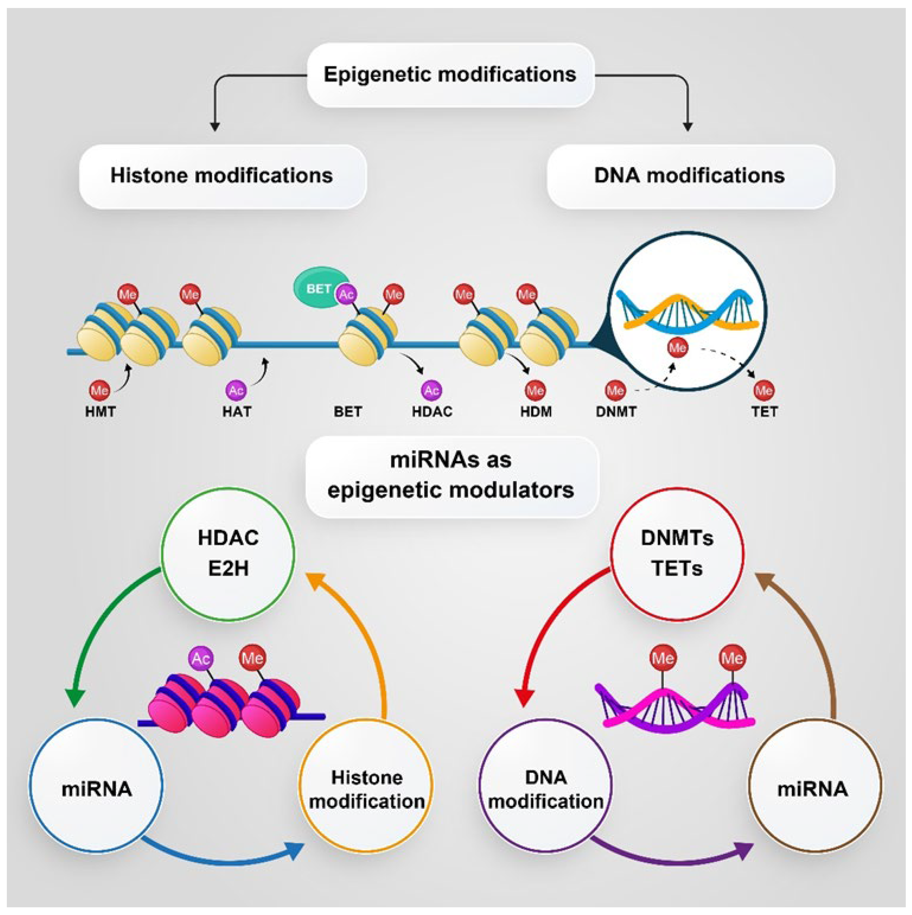
5.1. DNA Methylation and Histone Modification

DNA methylation occurring by the DNA methyltransferase (DNMT) family [200] has been regarded as the most important epigenetic alteration [201], which plays a critical role in some biological phenomena, such as X chromosomal inactivation, differentiation, and genome imprinting during development. This phenomenon occurs mainly in cytosine residues in the C-phosphodiester-G (CpG) islands and suppresses gene expression [191]. Aberrant de novo methylation of CpG islands is a typical sign of human cancers and can be detected in the early stages of carcinogenesis [202].
Histone modification is another epigenetic change where the N-terminal tails of histones, in which lysine and arginine residues are located, target several post-translational modifications, including acetylation, phosphorylation, and methylation. Histone acetylation and deacetylation occur by histone acetyltransferase (HATs) and histone deacetylases (HDACs), respectively [194]. Depending on the modification position, the target gene is activated or suppressed [203].
Studies have shown that several epigenetic modifiers in cancer cells, including DNA methyltransferases, histone acetyltransferases, deacetylases, histone methyltransferases, and histone demethylases, are abnormally hyperphosphorylated or hypophosphorylated. Phosphorylation modification may directly suppress or activate these enzymes, indirectly regulate the interaction between modifiers with RNAs or proteins, or tighten or loosen the chromatin structure [194]. Among different epigenetic alterations, changes in the methylation of DNA are best identified and characterized in PC [16]. According to reliable evidence, the hypomethylation and hypermethylation of DNA occur in PC, leading to alterations in the methylation pattern in the tissue, and there is also a significant relationship between hypomethylation and hypermethylation with the progression of benign prostatic hyperplasia to metastatic tumors [192]. uPA causes tumor invasion and metastasis in some malignancies, such as PC. In highly invasive PC3 cells, the uPA promoter is hypomethylated [204]. The Ras family plays an important role as tumor suppressor proteins by activating the apoptosis process. This gene is commonly silenced through a methylated promotor in PC and several other cancers. Recently, the hypermethylation of Ras families has been observed in more than 70% of primary PCs) [205]. PTEN is a tumor suppressor gene located on chromosome 10. According to studies, the lack of PTEN activity has a profound effect on several Tyr kinases, such as PTK6 and Akt, that promotes PC progression [206,207]. Some evidence suggests that the epigenetic pathway is responsible for PTEN regulation and PTEN silencing in PC, which occurs through hypermethylation. PTEN can regain its activity by treatment with a DNA demethylating agent, such as azacitidine [198,208]. Furthermore, alteration in the methylation pattern of DNA is believed to be a significant source of tumor heterogeneity in metastatic PC and can lead to the development of therapeutic resistance [209].
Histone modifications, including acetylation, methylation, and phosphorylation, are other epigenetic alterations [210] that play an important role in the onset and progression of PC. The role of histone modifications has been identified in PC. For example, the main histone methyltransferase, responsible for H3K27 (the lysine residue at N-terminal position 27 of histone 3) trimethylation and the aberrant silencing of multiple tumor suppressor genes, namely the enhancer of zeste homolog 2 (EZH2), has been shown to be overexpressed in PC cells and hyperphosphorylated by several kinases, such as Akt, and thus promotes the expression of several critical oncogenes and induces PC metastasis [16,211].
Histone phosphorylation that depends on amino acids in histone is a dynamic process. Histone phosphorylation occurs by altering many cellular processes, including the cell cycle, repair of DNA damage, and cell apoptosis, so impaired regulation often leads to tumor formation. Hence, the kinases that regulate the phosphorylation of histones are always overexpressed in cancers. For example, high levels of PRK1, which mediates the phosphorylation of histone H3 (at Thr 11) [212], are associated with the advanced stages of PC [213]. In another study, Mahajan et al. reported that activated cdc42-associated Tyr kinase (ACK1) phosphorylates histone H4 at Tyr88 upstream of the AR transcription start site, leading to a WDR5/MLL2 complex-mediated increase in AR transcription. AR plays a major role in the onset and progression of PC. Therefore, the interaction between AR and ACK1 drives the positive feedback epigenetic circuitry that is ultimately conducive to promoting AR transcription. The inhibition of ACK1 reverses the phosphorylated Tyr88 at histone 4 (pY88-H4) marks and reduces AR and AR-V7 splice variant levels to mitigate castration-resistant prostate tumor growth [214,215].

5.2. MicroRNAs (miRNAs), as Epigenetic Modulators

There are several studies that clearly demonstrate the ability of miRNA in the epigenetic and post-translational regulation of gene expression. In PC progression, miRNAs play crucial roles through the regulation of kinase expression. In this review, some studies on this issue will be highlighted. It has been reported that the miR-135-a level was significantly reduced in metastatic PC tumors, indicating a correlation between tumor progression and a higher Gleason score. In fact, miR-135-a suppressed PC cell proliferation via the targeting of several oncogenic pathways, such as EGFR [216]. Another tumor-suppressive miRNA, miR-34c, plays a key role in PC through the targeting of the MET proto-oncogene. The MET proto-oncogene is a Tyr kinase family receptor that plays an important role in the invasion and migration of tumor cells. The upregulation of MET has been reported in metastatic tumors [217,218].
Other studies show that miR-139 and miR-302a downregulate Akt in PC. Cell cycle arrest through the upregulation of the CDK inhibitor p21 and downregulation of Akt and cyclin D1 has been attributed to the overexpression of mir-139 [219]. miRNA-302a also binds directly to the 3′UTR mRNA of the Akt gene, leading to the induction of cell cycle arrest in the G1/S phase [220]. Figure 3 shows the epigenetic regulation of DNA and histone modifications that are discussed above.

6. PC Treatment and Management

As stated earlier, ADT and AR-targeted therapy are two gold-standard options for PC treatment [116]. Apart from AR blockade, immunotherapy, poly-ADP ribose polymerase inhibitors (PARPIs), and targeted therapies for prostate-specific membrane antigen (PSMA) are other options that have been developed for targeted therapies for PC, especially for the most aggressive, castration-resistant types. Nevertheless, after a while with these treatments, the tumor eventually develops resistance [221].
According to several studies, some pathways related to PKs, activated in the advanced stages of PC, are responsible for cases of resistance, and targeting these pathways may lead to overcoming the resistance to targeted AR treatment. For instance, mutations in the PTEN/PI3K/Akt signaling pathway are one of events responsible for resistance to PARPIs and PC progression [221]. Recent discoveries indicate that the crosstalk between this pathway and multiple signaling cascades can further promote PC progression and influence the sensitivity of PC cells to PI3K/Akt/mTOR-targeted approaches explored in the clinic, as well as standard treatments [112].
Although there has been a lot of progress in kinase drug discovery, many challenges remain in this field. As a challenge, tumors targeted by kinase inhibitors usually show resistance over time, leading to disease progression [33]. The search for targeted therapies of mCRPC has focused on developing new effective systemic treatments and identifying mechanisms of drug resistance [17]. Mounting evidence supports epigenetic events as potential mechanisms for PC transdifferentiation to an AR-indifferent state. Extensive studies have shown that DNA methylation plays a significant role in mediating these mechanisms in PC, among other cancers [12]. These are the key reasons why PK and epigenetic modulators have emerged as two combination therapy options to overcome acquired resistance to traditional therapies. However, understanding the mechanisms of synergy and resistance remains a crucial challenge.

6.1. PK Inhibitors in PC

PK inhibitor-based therapies exhibited a shift from conventional chemotherapy to targeted cancer therapy by overcoming a leading drawback of traditional cancer therapies. They effectively distinguish between normal cells and cancer cells [33]. Different PK inhibitors have been studied in various types of studies, including in vitro, in vivo, and clinical trials, in monotherapy or in combination with cytotoxic chemotherapy or radiation therapy, though with mixed results for mCRPC treatment [34]. About 20–25% of mCRPC subtypes that show somatic or germline alterations in DNA repair genes involved in homologous recombination are usually associated with more invasive disease. In the treatment of these subtypes, PARPI have shown significant effects. However, some epigenetic alterations or genetic mutations prevent PARP from binding to its inhibitors and consequently drug resistance. In PC, mutations in the PTEN/PI3K/Akt signaling pathway are one of the frequent events responsible for resistance to PARP inhibitors and disease progression [221]. As a result, several targeted PC therapies mainly affecting AR and the PI3K/Akt/mTOR pathway are in various stages of development [18]. Based on recent investigations, PTEN, PI3K, and PKB (Akt) inhibitors have offered promising results for mCRPC treatment with acquired resistance to PARP inhibitors, both in monotherapy and combined therapy with PARP inhibitors [221]. In a review article, Pungsrinont et al. summarized and discussed several inhibitors of the PI3K/Akt/mTOR pathway tested as monotherapy or in combination with other agents in preclinical and clinical trials for PC treatment [116]. Additionally, Shorning et al. presented new mechanical insights into the fundamental interaction between the PI3K/Akt/mTOR pathway and several oncogenic cascades (particularly the AR, MAPK, and WNT signaling cascades), which could facilitate PC growth and drug resistance [112]. Accordingly, Yadav et al. carried out a systematic study on the combined effect of therapies targeting the AR-signaling and the PI3K/AKT/mTOR pathways upon various PC cell lines. Their observation demonstrated that a combination of MDV3100 (AR-inhibitor) and BKM120 (PI3K-inhibitor) is highly synergistic. Furthermore, combining BKM120 with TKI258 (pan RTK inhibitor) has better synergy than BKM120+RAD001 (mTOR inhibitor) or RAD001+TKI258 in all of the lines, irrespective of androgen sensitivity. Finally, the PI3K inhibitor also displayed synergy when combined with the chemotherapy drug cabazitaxel [18].
Other potential targets, including ATR, CHK1, WEE1, AURK, and PlK1 have been successfully examined in preclinical studies for PC treatment [220]. Several preclinical studies showed that the association of PARP inhibitors with ATR inhibitors could resensitize PARP-resistant cells [222,223,224]. Neeb et al. characterized ATM-deficient lethal PC and studied ATR inhibition, PARP inhibition, and combined PARP and ATR inhibition as therapeutic strategies for this subset. They found variable sensitivity of this subtype to PARP inhibition, sensitivity to ATR inhibition, and the most sensitivity to combined inhibition, which now merits clinical evaluation [225].
Currently, some pK inhibitors investigated in mCRPC clinical trials include dasatinib [226,227], trametinib [228], masitinib [229], sunitinib [230,231], bevacizumab [232], cediranib [233], cabozantinib [234], erlotinib [235], and ipatasertib [236,237,238] (Table 1). Of all the agents presented, ipatasertib has shown excellent preliminary therapeutic results and a favorable safety profile (in both early-phase and late-phase testing) in patients who have lost PTEN, and it may be a good combination partner with multiple anticancer agents [236,237,238]. The upregulation of the RAS pathway following ipatasertib suggests that the coadministration of ipatasertib with the inhibitors of the RAS/MEK pathway may be more effective [236]. For all other agents, further definitive testing to clearly evaluate their clinical potential has been recommended.

6.2. Epigenetic Targeting as a Therapeutic Strategy for Advanced PC

As previously stated, pieces of evidence from several studies suggest a cross-link between kinase pathways and epigenetic reprogramming during the progression of PC. In our opinion, this evidence opens an opportunity to develop new strategies in PC treatment and management, particularly for patients with developed CRPC and AR-indifferent forms of the disease.
In a review article published by Angus et al. [33], they highlighted the epigenetic changes underlying resistant phenotypes and discussed phenotypic switching as an adaptive response to kinase inhibition. They mentioned that developed strategies are needed to block the dynamic changes in the chromatin landscape in response to kinase inhibitors, leading to adaptive resistance and durable responses. Thus, they suggested that the small-molecule inhibitors of these epigenetic regulators have the potential to attenuate the transcriptional rewiring that leads to drug resistance. Finally, they proposed potential therapeutic approaches by the combination of targeting key oncogenic kinases with drugs targeting the components of the transcriptional machinery and histone-modifying enzymes.
Currently, several epigenetic inhibitors are under preclinical and clinical trials for the management and treatment of PC. They include histone methyltransferase inhibitors, DNMT inhibitors, HDAC inhibitors, and many other numerous epigenetic therapies, which are currently under preclinical and clinical investigations for the management and treatment of PC. Some epigenetic drugs that are reported from active and completed clinical trials and used to treat PC include: tazemetostat as an EZH2 inhibitor; guadecitabine, disulfiram, and azacitidine as DNMT inhibitors; belinostat, entinostat, vorinostat, and panobinostat; HDAC inhibitor SB939, 5-fluorouracil, and bicalutamide as HDAC inhibitors; and nivolumab, INCB059872, all-trans retinoic acid, and azacytidine as lysine-specific histone demethylase 1 inhibitors [16]. These epigenetic inhibitors are depicted in Figure 4.

7. Conclusions

Due to acquired resistance to conventional treatments for PC, including radiotherapy, prostatectomy, and androgen deprivation, recently, novel treatments, including targeting several signaling pathways and epigenetic modifiers, are in development. As mentioned above, different PKs play a significant role in several pathways related to PC progression; hence, PK inhibitors may be suggested as potential therapeutic agents in PC. The promising clinical results of phases I, II, and III on ipatasertib show that this Akt inhibitor can be a good combination partner with several anti-cancer agents for mCRPC treatment. Given that many of the signaling pathways are epigenetically dysregulated in PC, epigenetic targeting may represent an alternative therapeutic strategy to treat advanced PC with genetic modulator inhibitors. Additionally, various studies show that there is an interplay between PKs and epigenetic changes in PC; thus, it seems that simultaneously targeting these pathways may be a suitable treatment option for advanced PC.

Author Contributions

Conceptualization, S.B. and S.Z.; investigation, writing—original draft preparation, S.B., M.R., F.E., A.B., F.B. and S.Z.; writing—review and editing, S.B., S.Z. and A.S; supervision, S.Z. and A.S. All authors have read and agreed to the published version of the manuscript.

Funding

This research received no external funding.

Institutional Review Board Statement

Not applicable.

Informed Consent Statement

Not applicable.

Data Availability Statement

No new data were created or analyzed in this study. Data sharing does not apply to this article.

Conflicts of Interest

The authors declare no conflict of interest.

References

  1. Roskoski, R., Jr. Properties of FDA-approved small molecule protein kinase inhibitors: A 2022 update. Pharmacol. Res. 2022, 175, 106037. [Google Scholar] [CrossRef]
  2. Fabian, M.A.; Biggs, W.H.; Treiber, D.K.; Atteridge, C.E.; Azimioara, M.D.; Benedetti, M.G.; Carter, T.A.; Ciceri, P.; Edeen, P.T.; Floyd, M.; et al. A small molecule–kinase interaction map for clinical kinase inhibitors. Nat. Biotechnol. 2005, 23, 329–336. [Google Scholar] [CrossRef]
  3. Wells, C.I.; Al-Ali, H.; Andrews, D.M.; Asquith, C.R.M.; Axtman, A.D.; Dikic, I.; Ebner, D.; Ettmayer, P.; Fischer, C.; Frederiksen, M.; et al. The Kinase Chemogenomic Set (KCGS): An Open Science Resource for Kinase Vulnerability Identification. Int. J. Mol. Sci. 2021, 22, 566. [Google Scholar] [CrossRef]
  4. Ferguson, F.M.; Gray, N.S. Kinase inhibitors: The road ahead. Nat. Rev. Drug Discov. 2018, 17, 353–377. [Google Scholar] [CrossRef]
  5. Klaeger, S.; Heinzlmeir, S.; Wilhelm, M.; Polzer, H.; Vick, B.; Koenig, P.-A.; Reinecke, M.; Ruprecht, B.; Petzoldt, S.; Meng, C.; et al. The target landscape of clinical kinase drugs. Science 2017, 358, eaan4368. [Google Scholar] [CrossRef] [Green Version]
  6. Anastassiadis, T.; Deacon, S.W.; Devarajan, K.; Ma, H.; Peterson, J.R. Comprehensive assay of kinase catalytic activity reveals features of kinase inhibitor selectivity. Nat. Biotechnol. 2011, 29, 1039–1045. [Google Scholar] [CrossRef] [Green Version]
  7. Carles, F.; Bourg, S.; Meyer, C.; Bonnet, P. PKIDB: A Curated, Annotated and Updated Database of Protein Kinase Inhibitors in Clinical Trials. Molecules 2018, 23, 908. [Google Scholar] [CrossRef] [Green Version]
  8. Corti, M.; Lorenzetti, S.; Ubaldi, A.; Zilli, R.; Marcoccia, D. Endocrine Disruptors and Prostate Cancer. Int. J. Mol. Sci. 2022, 23, 1216. [Google Scholar] [CrossRef]
  9. Wang, X.; Wei, L.; Xiao, J.; Shan, K.; He, Q.; Huang, F.; Ge, X.; Gao, X.; Feng, N.; Chen, Y.Q. Cholesterol and saturated fatty acids synergistically promote the malignant progression of prostate cancer. Neoplasia 2022, 24, 86–97. [Google Scholar] [CrossRef]
  10. Zhang, H.; Spencer, K.; Burley, S.K.; Zheng, X.S. Toward improving androgen receptor-targeted therapies in male-dominant hepatocellular carcinoma. Drug Discov. Today 2021, 26, 1539–1546. [Google Scholar] [CrossRef]
  11. Lv, S.; Pu, X.; Luo, M.; Wen, H.; Xu, Z.; Wei, Q.; Dang, Q. Long noncoding RNA GAS5 interacts and suppresses androgen receptor activity in prostate cancer cells. Prostate 2021, 81, 893–901. [Google Scholar] [CrossRef] [PubMed]
  12. Thienger, P.; Rubin, M.A. SETting Up for Epigenetic Regulation of Advanced Prostate Cancer. Cancer Cell 2020, 38, 309–311. [Google Scholar] [CrossRef] [PubMed]
  13. Chau, V.; Madan, R.A.; Aragon-Ching, J.B. Protein kinase inhibitors for the treatment of prostate cancer. Expert Opin. Pharmacother. 2021, 22, 1889–1899. [Google Scholar] [CrossRef]
  14. Gioeli, D.; Mandell, J.W.; Petroni, G.R.; Frierson, H.F.; Weber, M.J. Activation of mitogen-activated protein kinase associated with prostate cancer progression. Cancer Res. 1999, 59, 279–284. [Google Scholar]
  15. Xu, R.; Hu, J. The role of JNK in prostate cancer progression and therapeutic strategies. Biomed. Pharmacother. 2020, 121, 109679. [Google Scholar] [CrossRef]
  16. Ge, R.; Wang, Z.; Montironi, R.; Jiang, Z.; Cheng, M.; Santoni, M.; Huang, K.; Massari, F.; Lu, X.; Cimadamore, A. Epigenetic modulations and lineage plasticity in advanced prostate cancer. Ann. Oncol. 2020, 31, 470–479. [Google Scholar] [CrossRef] [PubMed] [Green Version]
  17. Conteduca, V.; Hess, J.; Yamada, Y.; Ku, S.-Y.; Beltran, H. Epigenetics in prostate cancer: Clinical implications. Transl. Androl. Urol. 2021, 10, 3104–3116. [Google Scholar] [CrossRef] [PubMed]
  18. Yadav, S.S.; Li, J.; Stockert, J.A.; O’Connor, J.; Herzog, B.; Elaiho, C.; Galsky, M.D.; Tewari, A.K.; Yadav, K.K. Combination effect of therapies targeting the PI3K- and AR-signaling pathways in prostate cancer. Oncotarget 2016, 7, 76181–76196. [Google Scholar] [CrossRef]
  19. Haubrich, B.A.; Swinney, D.C. Enzyme Activity Assays for Protein Kinases: Strategies to Identify Active Substrates. Curr. Drug Discov. Technol. 2016, 13, 2–15. [Google Scholar] [CrossRef] [Green Version]
  20. Han, K.-H.E.; McGonigal, T. Role of Focal Adhesion Kinase in Human Cancer: A Potential Target for Drug Discovery. Anti-Cancer Agents Med. Chem. 2007, 7, 681–684. [Google Scholar] [CrossRef]
  21. Manning, G.; Whyte, D.B.; Martinez, R.; Hunter, T.; Sudarsanam, S. The Protein Kinase Complement of the Human Genome. Science 2002, 298, 1912–1934. [Google Scholar] [CrossRef] [Green Version]
  22. Martin, J.; Anamika, K.; Srinivasan, N. Classification of Protein Kinases on the Basis of Both Kinase and Non-Kinase Regions. PLoS ONE 2010, 5, e12460. [Google Scholar] [CrossRef] [PubMed] [Green Version]
  23. Borgo, C.; D’Amore, C.; Sarno, S.; Salvi, M.; Ruzzene, M. Protein kinase CK2: A potential therapeutic target for diverse human diseases. Signal Transduct. Target. Ther. 2021, 6, 183. [Google Scholar] [CrossRef] [PubMed]
  24. Ijomone, O.M.; Iroegbu, J.D.; Aschner, M.; Bornhorst, J. Impact of environmental toxicants on p38-and ERK-MAPK signaling pathways in the central nervous system. Neurotoxicology 2021, 86, 166–171. [Google Scholar] [CrossRef] [PubMed]
  25. Patterson, H.; Nibbs, R.; McInnes, I.; Siebert, S. Protein kinase inhibitors in the treatment of inflammatory and autoimmune diseases. Clin. Exp. Immunol. 2014, 176, 1–10. [Google Scholar] [CrossRef]
  26. Abdellatif, K.R.A.; Bakr, R.B. Pyrimidine and fused pyrimidine derivatives as promising protein kinase inhibitors for cancer treatment. Med. Chem. Res. 2021, 30, 31–49. [Google Scholar] [CrossRef]
  27. Hanks, S.K.; Hunter, T. The eukaryotic protein kinase superfamily: Kinase (catalytic) domain structure and classification1. FASEB J. 1995, 9, 576–596. [Google Scholar] [CrossRef]
  28. Hunter, T. [1] Protein kinase classification. In Methods in Enzymology; Academic Press: Cambridge, MA, USA, 1991; Volume 200, pp. 3–37. [Google Scholar]
  29. Modi, V.; Dunbrack, R.L., Jr. Kincore: A web resource for structural classification of protein kinases and their inhibitors. Nucleic Acids Res. 2021, 50, D654–D664. [Google Scholar] [CrossRef]
  30. Talele, T.T.; McLaughlin, M.L. Molecular docking/dynamics studies of Aurora A kinase inhibitors. J. Mol. Graph. Model. 2008, 26, 1213–1222. [Google Scholar] [CrossRef]
  31. Wang, Z.; Huang, W.; Zhou, K.; Ren, X.; Ding, K. Targeting the Non-Catalytic Functions: A New Paradigm for Kinase Drug Discovery? J. Med. Chem. 2022, 65, 1735–1748. [Google Scholar] [CrossRef]
  32. Humphrey, W.; Dalke, A.; Schulten, K. VMD: Visual molecular dynamics. J. Mol. Graph. 1996, 14, 33–38. [Google Scholar] [CrossRef]
  33. Angus, S.P.; Zawistowski, J.S.; Johnson, G.L. Epigenetic Mechanisms Regulating Adaptive Responses to Targeted Kinase Inhibitors in Cancer. Annu. Rev. Pharmacol. Toxicol. 2018, 58, 209–229. [Google Scholar] [CrossRef] [PubMed]
  34. Bhullar, K.S.; Lagarón, N.O.; McGowan, E.M.; Parmar, I.; Jha, A.; Hubbard, B.P.; Rupasinghe, H.P.V. Kinase-targeted cancer therapies: Progress, challenges and future directions. Mol. Cancer 2018, 17, 48. [Google Scholar] [CrossRef] [PubMed]
  35. Kung, J.E.; Jura, N. Structural Basis for the Non-catalytic Functions of Protein Kinases. Structure 2016, 24, 7–24. [Google Scholar] [CrossRef] [PubMed] [Green Version]
  36. Knapp, S. New opportunities for kinase drug repurposing and target discovery. Br. J. Cancer 2018, 118, 936–937. [Google Scholar] [CrossRef] [PubMed] [Green Version]
  37. Imran, M.; Asdaq, S.M.B.; Khan, S.A.; Unnikrishnan Meenakshi, D.; Alamri, A.S.; Alsanie, W.F.; Alhomrani, M.; Mohzari, Y.; Alrashed, A.; AlMotairi, M.; et al. Innovations and Patent Trends in the Development of USFDA Approved Protein Kinase Inhibitors in the Last Two Decades. Pharmaceuticals 2021, 14, 710. [Google Scholar] [CrossRef]
  38. Bidwell, G.L., 3rd; Raucher, D. Therapeutic peptides for cancer therapy. Part I-peptide inhibitors of signal transduction cascades. Expert Opin. Drug Deliv. 2009, 6, 1033–1047. [Google Scholar] [CrossRef]
  39. Liu, C.; Ke, P.; Zhang, J.; Zhang, X.; Chen, X. Protein kinase inhibitor peptide as a tool to specifically inhibit protein kinase A. Front. Physiol. 2020, 11, 1532. [Google Scholar] [CrossRef]
  40. Srikar, R.; Suresh, D.; Zambre, A.; Taylor, K.; Chapman, S.; Leevy, M.; Upendran, A.; Kannan, R. Targeted nanoconjugate co-delivering siRNA and tyrosine kinase inhibitor to KRAS mutant NSCLC dissociates GAB1-SHP2 post oncogene knockdown. Sci. Rep. 2016, 6, 30245. [Google Scholar] [CrossRef]
  41. Jain, A.; Mahira, S.; Majoral, J.P.; Bryszewska, M.; Khan, W.; Ionov, M. Dendrimer mediated targeting of siRNA against polo-like kinase for the treatment of triple negative breast cancer. J. Biomed. Mater. Res. A 2019, 107, 1933–1944. [Google Scholar] [CrossRef]
  42. Moradpour, Z.; Barghi, L. Novel Approaches for Efficient Delivery of Tyrosine Kinase Inhibitors. J. Pharm. Pharm. Sci. 2019, 22, 37–48. [Google Scholar] [CrossRef] [PubMed] [Green Version]
  43. Smidova, V.; Michalek, P.; Goliasova, Z.; Eckschlager, T.; Hodek, P.; Adam, V.; Heger, Z. Nanomedicine of tyrosine kinase inhibitors. Theranostics 2021, 11, 1546–1567. [Google Scholar] [CrossRef] [PubMed]
  44. Reagan-Shaw, S.; Ahmad, N. Silencing of polo-like kinase (Plk) 1 via siRNA causes induction of apoptosis and impairment of mitosis machinery in human prostate cancer cells: Implications for the treatment of prostate cancer. FASEB J. 2005, 19, 611–613. [Google Scholar] [CrossRef]
  45. Tsutsumi, K.; Kasaoka, T.; Park, H.M.; Nishiyama, H.; Nakajima, M.; Honda, T. Tumor growth inhibition by synthetic and expressed siRNA targeting focal adhesion kinase. Int. J. Oncol. 2008, 33, 215–224. [Google Scholar] [CrossRef] [PubMed] [Green Version]
  46. Gagnon, V.; Mathieu, I.; Sexton, E.; Leblanc, K.; Asselin, E. AKT involvement in cisplatin chemoresistance of human uterine cancer cells. Gynecol. Oncol. 2004, 94, 785–795. [Google Scholar] [CrossRef] [PubMed]
  47. Karasarides, M.; Chiloeches, A.; Hayward, R.; Niculescu-Duvaz, D.; Scanlon, I.; Friedlos, F.; Ogilvie, L.; Hedley, D.; Martin, J.; Marshall, C.J. B-RAF is a therapeutic target in melanoma. Oncogene 2004, 23, 6292–6298. [Google Scholar] [CrossRef] [Green Version]
  48. Hojjat-Farsangi, M.; Ghaemimanesh, F.; Daneshmanesh, A.H.; Bayat, A.A.; Mahmoudian, J.; Jeddi-Tehrani, M.; Rabbani, H.; Mellstedt, H. Inhibition of the receptor tyrosine kinase ROR1 by anti-ROR1 monoclonal antibodies and siRNA induced apoptosis of melanoma cells. PLoS ONE 2013, 8, e61167. [Google Scholar] [CrossRef] [Green Version]
  49. Tao, Y.; Zhang, P.; Frascogna, V.; Lecluse, Y.; Auperin, A.; Bourhis, J.; Deutsch, E. Enhancement of radiation response by inhibition of Aurora-A kinase using siRNA or a selective Aurora kinase inhibitor PHA680632 in p53-deficient cancer cells. Br. J. Cancer 2007, 97, 1664–1672. [Google Scholar] [CrossRef] [Green Version]
  50. Asik, E.; Akpinar, Y.; Caner, A.; Kahraman, N.; Guray, T.; Volkan, M.; Albarracin, C.; Pataer, A.; Arun, B.; Ozpolat, B. EF2-kinase targeted cobalt-ferrite siRNA-nanotherapy suppresses BRCA1-mutated breast cancer. Nanomedicine 2019, 14, 2315–2338. [Google Scholar] [CrossRef]
  51. Goldberg, M.S.; Sharp, P.A. Pyruvate kinase M2-specific siRNA induces apoptosis and tumor regression. J. Exp. Med. 2012, 209, 217–224. [Google Scholar] [CrossRef] [Green Version]
  52. He, S.B.; Yuan, Y.; Wang, L.; Yu, M.J.; Zhu, Y.B.; Zhu, X.G. Effects of cyclin-dependent kinase 8 specific siRNA on the proliferation and apoptosis of colon cancer cells. J. Exp. Clin. Cancer Res. 2011, 30, 109. [Google Scholar] [CrossRef] [PubMed] [Green Version]
  53. Hu, B.; Zhong, L.; Weng, Y.; Peng, L.; Huang, Y.; Zhao, Y.; Liang, X.J. Therapeutic siRNA: State of the art. Signal Transduct. Target. 2020, 5, 101. [Google Scholar] [CrossRef] [PubMed]
  54. Ahmadzada, T.; Reid, G.; McKenzie, D.R. Fundamentals of siRNA and miRNA therapeutics and a review of targeted nanoparticle delivery systems in breast cancer. Biophys. Rev. 2018, 10, 69–86. [Google Scholar] [CrossRef]
  55. Jonker, D.J.; O’Callaghan, C.J.; Karapetis, C.S.; Zalcberg, J.R.; Tu, D.; Au, H.-J.; Berry, S.R.; Krahn, M.; Price, T.; Simes, R.J. Cetuximab for the treatment of colorectal cancer. N. Engl. J. Med. 2007, 357, 2040–2048. [Google Scholar] [CrossRef] [Green Version]
  56. von Minckwitz, G.; Procter, M.; de Azambuja, E.; Zardavas, D.; Benyunes, M.; Viale, G.; Suter, T.; Arahmani, A.; Rouchet, N.; Clark, E.; et al. Adjuvant Pertuzumab and Trastuzumab in Early HER2-Positive Breast Cancer. N. Engl. J. Med. 2017, 377, 122–131. [Google Scholar] [CrossRef] [PubMed]
  57. Hudis, C.A. Trastuzumab—mechanism of action and use in clinical practice. N. Engl. J. Med. 2007, 357, 39–51. [Google Scholar] [CrossRef] [Green Version]
  58. Gharwan, H.; Groninger, H. Kinase inhibitors and monoclonal antibodies in oncology: Clinical implications. Nat. Rev. Clin. Oncol. 2016, 13, 209–227. [Google Scholar] [CrossRef] [PubMed]
  59. Yu, F.; Cai, M.; Shao, L.; Zhang, J. Targeting Protein Kinases Degradation by PROTACs. Front. Chem. 2021, 9, 679120. [Google Scholar] [CrossRef]
  60. Kurimchak, A.M.; Herrera-Montávez, C.; Montserrat, S.; Araiza, D.; Hu, J.; Jin, J.; Duncan, J.S. MDR1 Drug Efflux Pump Promotes Intrinsic and Acquired Resistance to PROTACs in Cancer Cells. bioRxiv 2021. [Google Scholar] [CrossRef]
  61. Sakurai, R.; Villarreal, P.; Husain, S.; Liu, J.; Sakurai, T.; Tou, E.; Torday, J.S.; Rehan, V.K. Curcumin protects the developing lung against long-term hyperoxic injury. Am. J. Physiol.-Lung Cell. Mol. Physiol. 2013, 305, L301–L311. [Google Scholar] [CrossRef] [Green Version]
  62. Masuda, M.; Wakasaki, T.; Toh, S.; Shimizu, M.; Adachi, S. Chemoprevention of Head and Neck Cancer by Green Tea Extract: EGCG-The Role of EGFR Signaling and “Lipid Raft”. J. Oncol. 2011, 2011, 540148. [Google Scholar] [CrossRef] [PubMed] [Green Version]
  63. Byun, S.; Lee, K.W.; Jung, S.K.; Lee, E.J.; Hwang, M.K.; Lim, S.H.; Bode, A.M.; Lee, H.J.; Dong, Z. Luteolin inhibits protein kinase C(epsilon) and c-Src activities and UVB-induced skin cancer. Cancer Res. 2010, 70, 2415–2423. [Google Scholar] [CrossRef] [PubMed] [Green Version]
  64. Wang, G.; Wang, J.J.; Chen, X.L.; Du, S.M.; Li, D.S.; Pei, Z.J.; Lan, H.; Wu, L.B. The JAK2/STAT3 and mitochondrial pathways are essential for quercetin nanoliposome-induced C6 glioma cell death. Cell Death Dis. 2013, 4, e746. [Google Scholar] [CrossRef] [Green Version]
  65. Boly, R.; Gras, T.; Lamkami, T.; Guissou, P.; Serteyn, D.; Kiss, R.; Dubois, J. Quercetin inhibits a large panel of kinases implicated in cancer cell biology. Int. J. Oncol. 2011, 38, 833–842. [Google Scholar] [CrossRef] [PubMed] [Green Version]
  66. Khan, A.; Aljarbou, A.N.; Aldebasi, Y.H.; Faisal, S.M.; Khan, M.A. Resveratrol suppresses the proliferation of breast cancer cells by inhibiting fatty acid synthase signaling pathway. Cancer Epidemiol. 2014, 38, 765–772. [Google Scholar] [CrossRef] [PubMed]
  67. Atanasov, A.G.; Zotchev, S.B.; Dirsch, V.M.; International Natural Product Sciences Taskforce; Supuran, C.T. Natural products in drug discovery: Advances and opportunities. Nat. Rev. Drug Discov. 2021, 20, 200–216. [Google Scholar] [CrossRef]
  68. Sankarapandian, V.; Venmathi Maran, B.A.; Rajendran, R.L.; Jogalekar, M.P.; Gurunagarajan, S.; Krishnamoorthy, R.; Gangadaran, P.; Ahn, B.C. An Update on the Effectiveness of Probiotics in the Prevention and Treatment of Cancer. Life 2022, 12, 59. [Google Scholar] [CrossRef]
  69. Asoudeh-Fard, A.; Barzegari, A.; Dehnad, A.; Bastani, S.; Golchin, A.; Omidi, Y. Lactobacillus plantarum induces apoptosis in oral cancer KB cells through upregulation of PTEN and downregulation of MAPK signalling pathways. BioImpacts BI 2017, 7, 193. [Google Scholar] [CrossRef]
  70. Mandel, A.; Larsson, P.; Sarwar, M.; Semenas, J.; Syed Khaja, A.S.; Persson, J.L. The interplay between AR, EGF receptor and MMP-9 signaling pathways in invasive prostate cancer. Mol. Med. 2018, 24, 1–13. [Google Scholar] [CrossRef]
  71. Kurose, H.; Ueda, K.; Kondo, R.; Ogasawara, S.; Kusano, H.; Sanada, S.; Naito, Y.; Nakiri, M.; Nishihara, K.; Kakuma, T. Elevated expression of EPHA2 is associated with poor prognosis after radical prostatectomy in prostate Cancer. Anticancer Res. 2019, 39, 6249–6257. [Google Scholar] [CrossRef]
  72. Taddei, M.L.; Parri, M.; Angelucci, A.; Onnis, B.; Bianchini, F.; Giannoni, E.; Raugei, G.; Calorini, L.; Rucci, N.; Teti, A.; et al. Kinase-dependent and -independent roles of EphA2 in the regulation of prostate cancer invasion and metastasis. Am. J. Pathol. 2009, 174, 1492–1503. [Google Scholar] [CrossRef] [PubMed] [Green Version]
  73. Xu, L.J.; Ma, Q.; Zhu, J.; Li, J.; Xue, B.X.; Gao, J.; Sun, C.Y.; Zang, Y.C.; Zhou, Y.B.; Yang, D.R. Combined inhibition of JAK1, 2/Stat3-PD-L1 signaling pathway suppresses the immune escape of castration-resistant prostate cancer to NK cells in hypoxia. Mol. Med. Rep. 2018, 17, 8111–8120. [Google Scholar] [PubMed] [Green Version]
  74. Luo, Y.; Yang, X.; Basourakos, S.P.; Zuo, X.; Wei, D.; Zhao, J.; Li, M.; Li, Q.; Feng, T.; Guo, P. Enzalutamide-Resistant Progression of Castration-Resistant Prostate Cancer Is Driven via the JAK2/STAT1-Dependent Pathway. Front. Mol. Biosci. 2021, 8. [Google Scholar] [CrossRef] [PubMed]
  75. Shen, T.; Wang, W.; Zhou, W.; Coleman, I.; Cai, Q.; Dong, B.; Ittmann, M.M.; Creighton, C.J.; Bian, Y.; Meng, Y.; et al. MAPK4 promotes prostate cancer by concerted activation of androgen receptor and AKT. J. Clin. Investig. 2021, 131. [Google Scholar] [CrossRef] [PubMed]
  76. Alwanian, W.M.; Tyner, A.L. Protein tyrosine kinase 6 signaling in prostate cancer. Am. J. Clin. Exp. Urol. 2020, 8, 1–8. [Google Scholar]
  77. Cronin, R.; Brooke, G.N.; Prischi, F. The role of the p90 ribosomal S6 kinase family in prostate cancer progression and therapy resistance. Oncogene 2021, 40, 3775–3785. [Google Scholar] [CrossRef]
  78. Yamamura, A.; Nayeem, M.J.; Muramatsu, H.; Nakamura, K.; Sato, M. MAZ51 Blocks the Tumor Growth of Prostate Cancer by Inhibiting Vascular Endothelial Growth Factor Receptor 3. Front. Pharm. 2021, 12, 667474. [Google Scholar] [CrossRef]
  79. Suzuki, A.; Lu, J.; Kusakai, G.-i.; Kishimoto, A.; Ogura, T.; Esumi, H. ARK5 is a tumor invasion-associated factor downstream of Akt signaling. Mol. Cell. Biol. 2004, 24, 3526–3535. [Google Scholar] [CrossRef] [Green Version]
  80. Laderoute, K.R.; Amin, K.; Calaoagan, J.M.; Knapp, M.; Le, T.; Orduna, J.; Foretz, M.; Viollet, B. 5′-AMP-activated protein kinase (AMPK) is induced by low-oxygen and glucose deprivation conditions found in solid-tumor microenvironments. Mol. Cell. Biol. 2006, 26, 5336–5347. [Google Scholar] [CrossRef] [Green Version]
  81. Bonini, M.G.; Gantner, B.N. The multifaceted activities of AMPK in tumor progression—Why the “one size fits all” definition does not fit at all? IUBMB Life 2013, 65, 889–896. [Google Scholar] [CrossRef]
  82. Park, H.U.; Suy, S.; Danner, M.; Dailey, V.; Zhang, Y.; Li, H.; Hyduke, D.R.; Collins, B.T.; Gagnon, G.; Kallakury, B. AMP-activated protein kinase promotes human prostate cancer cell growth and survival. Mol. Cancer Ther. 2009, 8, 733–741. [Google Scholar] [CrossRef] [Green Version]
  83. Tennakoon, J.B.; Shi, Y.; Han, J.J.; Tsouko, E.; White, M.A.; Burns, A.R.; Zhang, A.; Xia, X.; Ilkayeva, O.R.; Xin, L. Androgens regulate prostate cancer cell growth via an AMPK-PGC-1α-mediated metabolic switch. Oncogene 2014, 33, 5251–5261. [Google Scholar] [CrossRef] [PubMed] [Green Version]
  84. Choudhury, Y.; Zichu Yang, I.A.; Nixon, C.; Salt, I.P.; Leung, H.Y. AMP-activated protein kinase (AMPK) as a potential therapeutic target independent of PI3K/Akt signaling in prostate cancer. Oncoscience 2014, 1, 446. [Google Scholar] [CrossRef] [PubMed] [Green Version]
  85. Khan, A.S.; Frigo, D.E. A spatiotemporal hypothesis for the regulation, role, and targeting of AMPK in prostate cancer. Nat. Rev. Urol. 2017, 14, 164–180. [Google Scholar] [CrossRef] [PubMed]
  86. Popovics, P.; Frigo, D.E.; Schally, A.V.; Rick, F.G. Targeting the 5′-AMP-activated protein kinase and related metabolic pathways for the treatment of prostate cancer. Expert Opin. Targets 2015, 19, 617–632. [Google Scholar] [CrossRef] [PubMed]
  87. Lee, Y.G.; Nam, Y.; Shin, K.J.; Yoon, S.; Park, W.S.; Joung, J.Y.; Seo, J.K.; Jang, J.; Lee, S.; Nam, D.; et al. Androgen-induced expression of DRP1 regulates mitochondrial metabolic reprogramming in prostate cancer. Cancer Lett. 2020, 471, 72–87. [Google Scholar] [CrossRef] [PubMed]
  88. Zhang, P.; Song, Y.; Sun, Y.; Li, X.; Chen, L.; Yang, L.; Xing, Y. AMPK/GSK3beta/beta-catenin cascade-triggered overexpression of CEMIP promotes migration an.nd invasion in anoikis-resistant prostate cancer cells by enhancing metabolic reprogramming. FASEB J. 2018, 32, 3924–3935. [Google Scholar] [CrossRef] [PubMed] [Green Version]
  89. Kerr, M.C.; Teasdale, R.D. Defining macropinocytosis. Traffic 2009, 10, 364–371. [Google Scholar] [CrossRef]
  90. Dharmawardhane, S.; Schurmann, A.; Sells, M.A.; Chernoff, J.; Schmid, S.L.; Bokoch, G.M. Regulation of macropinocytosis by p21-activated kinase-1. Mol. Biol. Cell 2000, 11, 3341–3352. [Google Scholar] [CrossRef] [Green Version]
  91. Ridley, A.J.; Paterson, H.F.; Johnston, C.L.; Diekmann, D.; Hall, A. The small GTP-binding protein rac regulates growth factor-induced membrane ruffling. Cell 1992, 70, 401–410. [Google Scholar] [CrossRef]
  92. Commisso, C.; Davidson, S.M.; Soydaner-Azeloglu, R.G.; Parker, S.J.; Kamphorst, J.J.; Hackett, S.; Grabocka, E.; Nofal, M.; Drebin, J.A.; Thompson, C.B. Macropinocytosis of protein is an amino acid supply route in Ras-transformed cells. Nature 2013, 497, 633–637. [Google Scholar] [CrossRef] [Green Version]
  93. Fruman, D.A.; Rommel, C. PI3K and cancer: Lessons, challenges and opportunities. Nat. Rev. Drug Discov. 2014, 13, 140–156. [Google Scholar] [CrossRef] [PubMed] [Green Version]
  94. Phin, S.; Moore, M.W.; Cotter, P.D. Genomic Rearrangements of PTEN in Prostate Cancer. Front. Oncol. 2013, 3, 240. [Google Scholar] [CrossRef] [PubMed] [Green Version]
  95. M Dillon, L.; W Miller, T. Therapeutic targeting of cancers with loss of PTEN function. Curr. Drug Targets 2014, 15, 65–79. [Google Scholar] [CrossRef] [PubMed] [Green Version]
  96. Shen, M.M.; Abate-Shen, C. Molecular genetics of prostate cancer: New prospects for old challenges. Genes Dev. 2010, 24, 1967–2000. [Google Scholar] [CrossRef] [Green Version]
  97. Araki, N.; Johnson, M.T.; Swanson, J.A. A role for phosphoinositide 3-kinase in the completion of macropinocytosis and phagocytosis by macrophages. J. Cell Biol. 1996, 135, 1249–1260. [Google Scholar] [CrossRef] [Green Version]
  98. Kim, S.M.; Nguyen, T.T.; Ravi, A.; Kubiniok, P.; Finicle, B.T.; Jayashankar, V.; Malacrida, L.; Hou, J.; Robertson, J.; Gao, D. PTEN deficiency and AMPK activation promote nutrient scavenging and anabolism in prostate cancer cells. Cancer Discov. 2018, 8, 866–883. [Google Scholar] [CrossRef] [Green Version]
  99. Sapio, L.; Di Maiolo, F.; Illiano, M.; Esposito, A.; Chiosi, E.; Spina, A.; Naviglio, S. Targeting protein kinase A in cancer therapy: An update. EXCLI J. 2014, 13, 843–855. [Google Scholar]
  100. Caretta, A.; Mucignat-Caretta, C. Protein kinase a in cancer. Cancers 2011, 3, 913–926. [Google Scholar] [CrossRef] [Green Version]
  101. Eder, I.E.; Egger, M.; Neuwirt, H.; Seifarth, C.; Maddalo, D.; Desiniotis, A.; Schäfer, G.; Puhr, M.; Bektic, J.; Cato, A.C. Enhanced inhibition of prostate tumor growth by dual targeting the androgen receptor and the regulatory subunit type Iα of protein kinase A in vivo. Int. J. Mol. Sci. 2013, 14, 11942–11962. [Google Scholar] [CrossRef] [Green Version]
  102. Sarwar, M.; Sandberg, S.; Abrahamsson, P.A.; Persson, J.L. Protein kinase A (PKA) pathway is functionally linked to androgen receptor (AR) in the progression of prostate cancer. Urol. Oncol. 2014, 32, 25.e1–25.e12. [Google Scholar] [CrossRef] [PubMed]
  103. Dagar, M.; Singh, J.P.; Dagar, G.; Tyagi, R.K.; Bagchi, G. Phosphorylation of HSP90 by protein kinase A is essential for the nuclear translocation of androgen receptor. Urol. Oncol. Semin. Orig. Investig. 2019, 294, 8699–8710. [Google Scholar] [CrossRef] [PubMed] [Green Version]
  104. Xia, L.; Sun, J.; Xie, S.; Chi, C.; Zhu, Y.; Pan, J.; Dong, B.; Huang, Y.; Xia, W.; Sha, J. PRKAR2B-HIF-1α loop promotes aerobic glycolysis and tumour growth in prostate cancer. Cell Prolif. 2020, 53, e12918. [Google Scholar] [CrossRef] [PubMed]
  105. Chen, J.; Giridhar, K.V.; Zhang, L.; Xu, S.; Wang, Q.J. A protein kinase C/protein kinase D pathway protects LNCaP prostate cancer cells from phorbol ester-induced apoptosis by promoting ERK1/2 and NF-{kappa}B activities. Carcinogenesis 2011, 32, 1198–1206. [Google Scholar] [CrossRef] [Green Version]
  106. Aljameeli, A.; Thakkar, A.; Shah, G. Calcitonin receptor increases invasion of prostate cancer cells by recruiting zonula occludens-1 and promoting PKA-mediated TJ disassembly. Cell. Signal. 2017, 36, 1–13. [Google Scholar] [CrossRef]
  107. Menon, J.; Doebele, R.C.; Gomes, S.; Bevilacqua, E.; Reindl, K.M.; Rosner, M.R. A novel interplay between Rap1 and PKA regulates induction of angiogenesis in prostate cancer. PLoS ONE 2012, 7, e49893. [Google Scholar] [CrossRef]
  108. Boutin, B.; Tajeddine, N.; Monaco, G.; Molgo, J.; Vertommen, D.; Rider, M.; Parys, J.B.; Bultynck, G.; Gailly, P. Endoplasmic reticulum Ca2+ content decrease by PKA-dependent hyperphosphorylation of type 1 IP3 receptor contributes to prostate cancer cell resistance to androgen deprivation. Cell Calcium 2015, 57, 312–320. [Google Scholar] [CrossRef]
  109. Shan, K.; Feng, N.; Cui, J.; Wang, S.; Qu, H.; Fu, G.; Li, J.; Chen, H.; Wang, X.; Wang, R.; et al. Resolvin D1 and D2 inhibit tumour growth and inflammation via modulating macrophage polarization. J. Cell Mol. Med. 2020, 24, 8045–8056. [Google Scholar] [CrossRef]
  110. Lindsley, C.W. The Akt/PKB family of protein kinases: A review of small molecule inhibitors and progress towards target validation: A 2009 update. Curr. Top. Med. Chem. 2010, 10, 458–477. [Google Scholar] [CrossRef]
  111. Yoeli-Lerner, M.; Toker, A. Akt/PKB signaling in cancer: A function in cell motility and invasion. Cell Cycle 2006, 5, 603–605. [Google Scholar] [CrossRef] [Green Version]
  112. Shorning, B.Y.; Dass, M.S.; Smalley, M.J.; Pearson, H.B. The PI3K-AKT-mTOR Pathway and Prostate Cancer: At the Crossroads of AR, MAPK, and WNT Signaling. Int. J. Mol. Sci. 2020, 21, 4507. [Google Scholar] [CrossRef]
  113. Ghosh, P.M.; Malik, S.; Bedolla, R.; Kreisberg, J.I. Akt in prostate cancer: Possible role in androgen-independence. Curr. Drug Metab. 2003, 4, 487–496. [Google Scholar] [CrossRef]
  114. Li, B.; Sun, A.; Youn, H.; Hong, Y.; Terranova, P.F.; Thrasher, J.B.; Xu, P.; Spencer, D. Conditional Akt activation promotes androgen-independent progression of prostate cancer. Carcinogenesis 2007, 28, 572–583. [Google Scholar] [CrossRef] [PubMed] [Green Version]
  115. Wen, Y.; Hu, M.C.; Makino, K.; Spohn, B.; Bartholomeusz, G.; Yan, D.-H.; Hung, M.-C. HER-2/neu promotes androgen-independent survival and growth of prostate cancer cells through the Akt pathway. Cancer Res. 2000, 60, 6841–6845. [Google Scholar] [PubMed]
  116. Pungsrinont, T.; Kallenbach, J.; Baniahmad, A. Role of PI3K-AKT-mTOR Pathway as a Pro-Survival Signaling and Resistance-Mediating Mechanism to Therapy of Prostate Cancer. Int. J. Mol. Sci. 2021, 22, 1088. [Google Scholar] [CrossRef]
  117. Shukla, S.; Maclennan, G.T.; Hartman, D.J.; Fu, P.; Resnick, M.I.; Gupta, S. Activation of PI3K-Akt signaling pathway promotes prostate cancer cell invasion. Int. J. Cancer 2007, 121, 1424–1432. [Google Scholar] [CrossRef] [PubMed]
  118. Van de Sande, T.; Roskams, T.; Lerut, E.; Joniau, S.; Van Poppel, H.; Verhoeven, G.; Swinnen, J.V. High-level expression of fatty acid synthase in human prostate cancer tissues is linked to activation and nuclear localization of Akt/PKB. J. Pathol. 2005, 206, 214–219. [Google Scholar] [CrossRef] [PubMed]
  119. Shukla, S.; Shukla, M.; Maclennan, G.T.; Fu, P.; Gupta, S. Deregulation of FOXO3A during prostate cancer progression. Int. J. Oncol. 2009, 34, 1613–1620. [Google Scholar] [CrossRef] [Green Version]
  120. Das, T.; Suman, S.; Alatassi, H.; Ankem, M.; Damodaran, C. Inhibition of AKT promotes FOXO3a-dependent apoptosis in prostate cancer. Cell Death Dis. 2016, 7, e2111. [Google Scholar] [CrossRef] [Green Version]
  121. Shukla, S.; Bhaskaran, N.; Maclennan, G.T.; Gupta, S. Deregulation of FoxO3a accelerates prostate cancer progression in TRAMP mice. Prostate 2013, 73, 1507–1517. [Google Scholar] [CrossRef] [Green Version]
  122. Black, J.D. PKC and control of the cell cycle. In Protein Kinase C in Cancer Signaling and Therapy; Springer: Berlin/Heidelberg, Germany, 2010; pp. 155–188. [Google Scholar]
  123. Reyland, M.E.; Bradford, A.P. PKC and the Control of Apoptosis. In Protein Kinase C in Cancer Signaling and Therapy; Springer: Berlin/Heidelberg, Germany, 2010; pp. 189–222. [Google Scholar]
  124. Reina-Campos, M.; Diaz-Meco, M.T.; Moscat, J. The Dual Roles of the Atypical Protein Kinase Cs in Cancer. Cancer Cell 2019, 36, 218–235. [Google Scholar] [CrossRef] [PubMed]
  125. Apostolatos, A.H.; Ratnayake, W.S.; Win-Piazza, H.; Apostolatos, C.A.; Smalley, T.; Kang, L.; Salup, R.; Hill, R.; Acevedo-Duncan, M. Inhibition of atypical protein kinase C-ι effectively reduces the malignancy of prostate cancer cells by downregulating the NF-κB signaling cascade. Int. J. Oncol. 2018, 53, 1836–1846. [Google Scholar] [CrossRef] [PubMed] [Green Version]
  126. Baek, H.-S.; Park, N.; Kwon, Y.-J.; Ye, D.-J.; Shin, S.; Chun, Y.-J. Annexin A5 suppresses cyclooxygenase-2 expression by downregulating the protein kinase C-ζ–nuclear factor-κB signaling pathway in prostate cancer cells. Oncotarget 2017, 8, 74263. [Google Scholar] [CrossRef] [Green Version]
  127. Reyland, M.E. Protein kinase C isoforms: Multi-functional regulators of cell life and death. Front. BioSci. (Landmark Ed.) 2009, 14, 2386–2399. [Google Scholar] [CrossRef] [PubMed] [Green Version]
  128. Ponguta, L.A.; Gregory, C.W.; French, F.S.; Wilson, E.M. Site-specific androgen receptor serine phosphorylation linked to epidermal growth factor-dependent growth of castration-recurrent prostate cancer. J. Biol. Chem. 2008, 283, 20989–21001. [Google Scholar] [CrossRef] [Green Version]
  129. Patek, S.; Willder, J.; Heng, J.; Taylor, B.; Horgan, P.; Leung, H.; Underwood, M.; Edwards, J. Androgen receptor phosphorylation status at serine 578 predicts poor outcome in prostate cancer patients. Oncotarget 2017, 8, 4875–4887. [Google Scholar] [CrossRef] [PubMed] [Green Version]
  130. Aziz, M.H.; Manoharan, H.T.; Church, D.R.; Dreckschmidt, N.E.; Zhong, W.; Oberley, T.D.; Wilding, G.; Verma, A.K. Protein kinase Cε interacts with signal transducers and activators of transcription 3 (Stat3), phosphorylates Stat3Ser727, and regulates its constitutive activation in prostate cancer. Cancer Res. 2007, 67, 8828–8838. [Google Scholar] [CrossRef] [PubMed] [Green Version]
  131. Garg, R.; Blando, J.M.; Perez, C.J.; Abba, M.C.; Benavides, F.; Kazanietz, M.G. Protein kinase C epsilon cooperates with PTEN loss for prostate tumorigenesis through the CXCL13-CXCR5 pathway. Cell Rep. 2017, 19, 375–388. [Google Scholar] [CrossRef] [PubMed]
  132. Xu, W.; Zeng, F.; Li, S.; Li, G.; Lai, X.; Wang, Q.J.; Deng, F. Crosstalk of protein kinase C epsilon with Smad2/3 promotes tumor cell proliferation in prostate cancer cells by enhancing aerobic glycolysis. Cell Mol. Life Sci. 2018, 75, 4583–4598. [Google Scholar] [CrossRef]
  133. Ivaska, J.; Vuoriluoto, K.; Huovinen, T.; Izawa, I.; Inagaki, M.; Parker, P.J. PKCε-mediated phosphorylation of vimentin controls integrin recycling and motility. EMBO J. 2005, 24, 3834–3845. [Google Scholar] [CrossRef]
  134. McJilton, M.A.; Van Sikes, C.; Wescott, G.G.; Wu, D.; Foreman, T.L.; Gregory, C.W.; Weidner, D.A.; Harris Ford, O.; Morgan Lasater, A.; Mohler, J.L.; et al. Protein kinase Cepsilon interacts with Bax and promotes survival of human prostate cancer cells. Oncogene 2003, 22, 7958–7968. [Google Scholar] [CrossRef] [Green Version]
  135. Usman, S.; Waseem, N.H.; Nguyen, T.K.N.; Mohsin, S.; Jamal, A.; Teh, M.T.; Waseem, A. Vimentin Is at the Heart of Epithelial Mesenchymal Transition (EMT) Mediated Metastasis. Cancers 2021, 13, 4985. [Google Scholar] [CrossRef]
  136. Ratnayake, W.S.; Apostolatos, C.A.; Breedy, S.; Dennison, C.L.; Hill, R.; Acevedo-Duncan, M. Atypical PKCs activate Vimentin to facilitate prostate cancer cell motility and invasion. Cell Adhes. Migr. 2021, 15, 37–57. [Google Scholar] [CrossRef]
  137. Roy, A.; Ye, J.; Deng, F.; Wang, Q.J. Protein kinase D signaling in cancer: A friend or foe? Biochim. Biophys. Acta Rev. Cancer 2017, 1868, 283–294. [Google Scholar] [CrossRef]
  138. Zhang, X.; Connelly, J.; Chao, Y.; Wang, Q.J. Multifaceted Functions of Protein Kinase D in Pathological Processes and Human Diseases. Biomolecules 2021, 11, 483. [Google Scholar] [CrossRef]
  139. Durand, N.; Bastea, L.I.; Döppler, H.; Eiseler, T.; Storz, P. Src-mediated tyrosine phosphorylation of protein kinase D2 at focal adhesions regulates cell adhesion. Sci. Rep. 2017, 7, 1–12. [Google Scholar] [CrossRef]
  140. Khachaturyan, G. The Role of Protein Kinase D Signaling and the Thermal Microenvironment on Single Cell Migration. Ph.D. Thesis, Ruperto-Carola University, Heidelberg, Germany, 2020. [Google Scholar]
  141. Roy, A.; Wang, Q.J. Protein Kinase D: A Potential Therapeutic Target in Prostate Cancer. Mol. Cell Pharm. 2017, 9, 1–4. [Google Scholar]
  142. Li, L.; Hua, L.; Fan, H.; He, Y.; Xu, W.; Zhang, L.; Yang, J.; Deng, F.; Zeng, F. Interplay of PKD3 with SREBP1 Promotes Cell Growth via Upregulating Lipogenesis in Prostate Cancer Cells. J. Cancer 2019, 10, 6395–6404. [Google Scholar] [CrossRef] [Green Version]
  143. Zou, Z.; Zeng, F.; Xu, W.; Wang, C.; Ke, Z.; Wang, Q.J.; Deng, F. PKD2 and PKD3 promote prostate cancer cell invasion by modulating NF-kappaB- and HDAC1-mediated expression and activation of uPA. J. Cell Sci. 2012, 125, 4800–4811. [Google Scholar] [CrossRef] [Green Version]
  144. Durand, N.; Borges, S.; Storz, P. Protein kinase D enzymes as regulators of EMT and cancer cell invasion. J. Clin. Med. 2016, 5, 20. [Google Scholar] [CrossRef] [Green Version]
  145. Hamshaw, I.; Ajdarirad, M.; Mueller, A. The role of PKC and PKD in CXCL12 directed prostate cancer migration. Biochem. Biophys. Res. Commun. 2019, 519, 86–92. [Google Scholar] [CrossRef]
  146. Anderson, C.; Lees-Miller, S. The nuclear serine/threonine protein kinase DNA-PK. Crit. Rev. Eukaryot. Gene Expr. 1992, 2, 283–314. [Google Scholar]
  147. Yang, H.; Yao, F.; Marti, T.M.; Schmid, R.A.; Peng, R.W. Beyond DNA Repair: DNA-PKcs in Tumor Metastasis, Metabolism and Immunity. Cancers 2020, 12, 3389. [Google Scholar] [CrossRef]
  148. Törmänen Persson, H.; Aksaas, A.K.; Kvissel, A.K.; Punga, T.; Engström, Å.; Skålhegg, B.S.; Akusjärvi, G. Two cellular protein kinases, DNA-PK and PKA, phosphorylate the adenoviral L4-33K protein and have opposite effects on L1 alternative RNA splicing. PLoS ONE 2012, 7, e31871. [Google Scholar] [CrossRef] [Green Version]
  149. Medova, M.; Medo, M.; Hovhannisyan, L.; Munoz-Maldonado, C.; Aebersold, D.M.; Zimmer, Y. DNA-PK in human malignant disorders: Mechanisms and implications for pharmacological interventions. Pharmacol. Ther. 2020, 215, 107617. [Google Scholar] [CrossRef]
  150. Kothari, V.; Goodwin, J.F.; Zhao, S.G.; Drake, J.M.; Yin, Y.; Chang, S.L.; Evans, J.R.; Wilder-Romans, K.; Gabbara, K.; Dylgjeri, E. DNA-dependent protein kinase drives prostate cancer progression through transcriptional regulation of the Wnt signaling pathway. Clin. Cancer Res. 2019, 25, 5608–5622. [Google Scholar] [CrossRef]
  151. Dylgjeri, E.; McNair, C.; Goodwin, J.F.; Raymon, H.K.; McCue, P.A.; Shafi, A.A.; Leiby, B.E.; De Leeuw, R.; Kothari, V.; McCann, J.J. Pleiotropic impact of DNA-PK in cancer and implications for therapeutic strategies. Clin. Cancer Res. 2019, 25, 5623–5637. [Google Scholar] [CrossRef]
  152. Giguère, V. DNA-PK, nuclear mTOR, and the androgen pathway in prostate cancer. Trends Cancer 2020, 6, 337–347. [Google Scholar] [CrossRef]
  153. Patel, R.S.; Rupani, R.; Impreso, S.; Lui, A.; Patel, N.A. Role of alternatively spliced, pro-survival Protein Kinase C delta VIII (PKCδVIII) in ovarian cancer. FASEB BioAdv. 2021, 1–19. [Google Scholar] [CrossRef]
  154. Jain, P.; Karthikeyan, C.; Moorthy, N.S.H.; Waiker, D.; Jain, A.K.; Trivedi, P. Human CDC2-like kinase 1 (CLK1): A novel target for Alzheimer’s disease. Curr. Drug Targets 2014, 15, 539–550. [Google Scholar] [CrossRef]
  155. Lindberg, M.F.; Meijer, L. Dual-Specificity, Tyrosine Phosphorylation-Regulated Kinases (DYRKs) and cdc2-Like Kinases (CLKs) in Human Disease, an Overview. Int. J. Mol. Sci. 2021, 22, 6047. [Google Scholar] [CrossRef] [PubMed]
  156. Bullock, A.N.; Das, S.; Debreczeni, J.É.; Rellos, P.; Fedorov, O.; Niesen, F.H.; Guo, K.; Papagrigoriou, E.; Amos, A.L.; Cho, S.; et al. Kinase Domain Insertions Define Distinct Roles of CLK Kinases in SR Protein Phosphorylation. Structure 2009, 17, 352–362. [Google Scholar] [CrossRef] [Green Version]
  157. Zhou, Z.; Fu, X.-D. Regulation of splicing by SR proteins and SR protein-specific kinases. Chromosoma 2013, 122, 191–207. [Google Scholar] [CrossRef] [PubMed]
  158. Martín Moyano, P.; Němec, V.; Paruch, K. Cdc-Like Kinases (CLKs): Biology, Chemical Probes, and Therapeutic Potential. Int. J. Mol. Sci. 2020, 21, 7549. [Google Scholar] [CrossRef]
  159. Kulkarni, P.; Uversky, V.N. Cancer/Testis Antigens: “Smart” Biomarkers for Diagnosis and Prognosis of Prostate and Other Cancers. Int. J. Mol. Sci. 2017, 18, 740. [Google Scholar] [CrossRef] [Green Version]
  160. Kulkarni, P.; Jolly, M.K.; Jia, D.; Mooney, S.M.; Bhargava, A.; Kagohara, L.T.; Chen, Y.; Hao, P.; He, Y.; Veltri, R.W.; et al. Phosphorylation-induced conformational dynamics in an intrinsically disordered protein and potential role in phenotypic heterogeneity. Proc. Natl. Acad. Sci. USA 2017, 114, E2644–E2653. [Google Scholar] [CrossRef] [Green Version]
  161. Lin, X.; Roy, S.; Jolly, M.K.; Bocci, F.; Schafer, N.P.; Tsai, M.-Y.; Chen, Y.; He, Y.; Grishaev, A.; Weninger, K.; et al. PAGE4 and Conformational Switching: Insights from Molecular Dynamics Simulations and Implications for Prostate Cancer. J. Mol. Biol. 2018, 430, 2422–2438. [Google Scholar] [CrossRef]
  162. Giannakouros, T.; Nikolakaki, E.; Mylonis, I.; Georgatsou, E. Serine-arginine protein kinases: A small protein kinase family with a large cellular presence. FEBS J. 2011, 278, 570–586. [Google Scholar] [CrossRef]
  163. Nikas, I.P.; Themistocleous, S.C.; Paschou, S.A.; Tsamis, K.I.; Ryu, H.S. Serine-Arginine Protein Kinase 1 (SRPK1) as a Prognostic Factor and Potential Therapeutic Target in Cancer: Current Evidence and Future Perspectives. Cells 2020, 9, 19. [Google Scholar] [CrossRef] [Green Version]
  164. Patel, M.; Sachidanandan, M.; Adnan, M. Serine arginine protein kinase 1 (SRPK1): A moonlighting protein with theranostic ability in cancer prevention. Mol. Biol. Rep. 2019, 46, 1487–1497. [Google Scholar] [CrossRef]
  165. Ngo, J.C.K.; Gullingsrud, J.; Giang, K.; Yeh, M.J.; Fu, X.-D.; Adams, J.A.; McCammon, J.A.; Ghosh, G. SR Protein Kinase 1 Is Resilient to Inactivation. Structure 2007, 15, 123–133. [Google Scholar] [CrossRef] [PubMed] [Green Version]
  166. Mavrou, A.; Brakspear, K.; Hamdollah-Zadeh, M.; Damodaran, G.; Babaei-Jadidi, R.; Oxley, J.; Gillatt, D.A.; Ladomery, M.R.; Harper, S.J.; Bates, D.O.; et al. Serine–arginine protein kinase 1 (SRPK1) inhibition as a potential novel targeted therapeutic strategy in prostate cancer. Oncogene 2015, 34, 4311–4319. [Google Scholar] [CrossRef] [PubMed] [Green Version]
  167. Bullock, N.; Potts, J.; Simpkin, A.J.; Koupparis, A.; Harper, S.J.; Oxley, J.; Oltean, S. Serine-arginine protein kinase 1 (SRPK1), a determinant of angiogenesis, is upregulated in prostate cancer and correlates with disease stage and invasion. J. Clin. Pathol. 2016, 69, 171–175. [Google Scholar] [CrossRef] [PubMed] [Green Version]
  168. Melegh, Z.; Oltean, S. Targeting Angiogenesis in Prostate Cancer. Int. J. Mol. Sci. 2019, 20, 2676. [Google Scholar] [CrossRef] [PubMed] [Green Version]
  169. Zhu, S.; Guo, Y.; Zhang, X.; Liu, H.; Yin, M.; Chen, X.; Peng, C. Pyruvate kinase M2 (PKM2) in cancer and cancer therapeutics. Cancer Lett. 2021, 503, 240–248. [Google Scholar] [CrossRef]
  170. Guo, W.; Zhang, Z.; Li, G.; Lai, X.; Gu, R.; Xu, W.; Chen, H.; Xing, Z.; Chen, L.; Qian, J. Pyruvate kinase M2 promotes prostate cancer metastasis through regulating ERK1/2-COX-2 signaling. Front. Oncol. 2020, 10, 2022. [Google Scholar] [CrossRef]
  171. Giannoni, E.; Taddei, M.L.; Morandi, A.; Comito, G.; Calvani, M.; Bianchini, F.; Richichi, B.; Raugei, G.; Wong, N.; Tang, D.; et al. Targeting stromal-induced pyruvate kinase M2 nuclear translocation impairs oxphos and prostate cancer metastatic spread. Oncotarget 2015, 6, 24061–24074. [Google Scholar] [CrossRef] [Green Version]
  172. Dai, J.; Escara-Wilke, J.; Keller, J.M.; Jung, Y.; Taichman, R.S.; Pienta, K.J.; Keller, E.T. Primary prostate cancer educates bone stroma through exosomal pyruvate kinase M2 to promote bone metastasis. J. Exp. Med. 2019, 216, 2883–2899. [Google Scholar] [CrossRef]
  173. Zhu, F.; Zykova, T.A.; Kang, B.S.; Wang, Z.; Ebeling, M.C.; Abe, Y.; Ma, W.Y.; Bode, A.M.; Dong, Z. Bidirectional Signals Transduced by TOPK-ERK Interaction Increase Tumorigenesis of HCT116 Colorectal Cancer Cells. Gastroenterology 2007, 133, 219–231. [Google Scholar] [CrossRef]
  174. Han, Z.; Li, L.; Huang, Y.; Zhao, H.; Luo, Y. PBK/TOPK: A Therapeutic Target Worthy of Attention. Cells 2021, 10, 371. [Google Scholar] [CrossRef]
  175. Herbert, K.J.; Ashton, T.M.; Prevo, R.; Pirovano, G.; Higgins, G.S. T-LAK cell-originated protein kinase (TOPK): An emerging target for cancer-specific therapeutics. Cell Death Dis. 2018, 9, 1089. [Google Scholar] [CrossRef] [PubMed]
  176. Alhawas, L.; Amin, K.S.; Salla, B.; Banerjee, P.P. T-LAK cell-originated protein kinase (TOPK) enhances androgen receptor splice variant (ARv7) and drives androgen-independent growth in prostate cancer. Carcinogenesis 2021, 42, 423–435. [Google Scholar] [CrossRef] [PubMed]
  177. Drake, J.M.; Graham, N.A.; Stoyanova, T.; Sedghi, A.; Goldstein, A.S.; Cai, H.; Smith, D.A.; Zhang, H.; Komisopoulou, E.; Huang, J.; et al. Oncogene-specific activation of tyrosine kinase networks during prostate cancer progression. Proc. Natl. Acad. Sci. USA 2012, 109, 1643–1648. [Google Scholar] [CrossRef] [Green Version]
  178. Chauhan, A.; Khan, T. Focal adhesion kinase—An emerging viable target in cancer and development of focal adhesion kinase inhibitors. Chem. Biol. Drug Des. 2021, 97, 774–794. [Google Scholar] [CrossRef]
  179. Marcellus, K.A.; Crawford Parks, T.E.; Almasi, S.; Jasmin, B.J. Distinct roles for the RNA-binding protein Staufen1 in prostate cancer. BMC Cancer 2021, 21, 120. [Google Scholar] [CrossRef] [PubMed]
  180. Slack, J.K.; Adams, R.B.; Rovin, J.D.; Bissonette, E.A.; Stoker, C.E.; Parsons, J.T. Alterations in the focal adhesion kinase/Src signal transduction pathway correlate with increased migratory capacity of prostate carcinoma cells. Oncogene 2001, 20, 1152–1163. [Google Scholar] [CrossRef] [Green Version]
  181. Ok Atılgan, A.; Özdemir, B.H.; Yılmaz Akçay, E.; Tepeoğlu, M.; Börcek, P.; Dirim, A. Association between focal adhesion kinase and matrix metalloproteinase-9 expression in prostate adenocarcinoma and their influence on the progression of prostatic adenocarcinoma. Ann. Diagn. Pathol. 2020, 45, 151480. [Google Scholar] [CrossRef]
  182. Kimura, S.H.; Tsuruga, H.; Yabuta, N.; Endo, Y.; Nojima, H. Structure, expression, and chromosomal localization of human GAK. Genomics 1997, 44, 179–187. [Google Scholar] [CrossRef]
  183. Kanaoka, Y.; Kimura, S.H.; Okazaki, I.; Ikeda, M.; Nojima, H. GAK: A cyclin G associated kinase contains a tensin/auxilin-like domain. FEBS Lett. 1997, 402, 73–80. [Google Scholar] [CrossRef] [Green Version]
  184. Sato, J.; Shimizu, H.; Kasama, T.; Yabuta, N.; Nojima, H. GAK, a regulator of clathrin-mediated membrane trafficking, localizes not only in the cytoplasm but also in the nucleus. Genes Cells 2009, 14, 627–641. [Google Scholar] [CrossRef]
  185. Zhang, C.X.; Engqvist-Goldstein, A.E.; Carreno, S.; Owen, D.J.; Smythe, E.; Drubin, D.G. Multiple roles for cyclin G-associated kinase in clathrin-mediated sorting events. Traffic 2005, 6, 1103–1113. [Google Scholar] [CrossRef]
  186. Eisenberg, E.; Greene, L.E. Multiple roles of auxilin and hsc70 in clathrin-mediated endocytosis. Traffic 2007, 8, 640–646. [Google Scholar] [CrossRef] [Green Version]
  187. Sakurai, M.A.; Ozaki, Y.; Okuzaki, D.; Naito, Y.; Sasakura, T.; Okamoto, A.; Tabara, H.; Inoue, T.; Hagiyama, M.; Ito, A.; et al. Gefitinib and luteolin cause growth arrest of human prostate cancer PC-3 cells via inhibition of cyclin G-associated kinase and induction of miR-630. PLoS ONE 2014, 9, e100124. [Google Scholar] [CrossRef] [Green Version]
  188. Ray, M.R.; Wafa, L.A.; Cheng, H.; Snoek, R.; Fazli, L.; Gleave, M.; Rennie, P.S. Cyclin G-associated kinase: A novel androgen receptor-interacting transcriptional coactivator that is overexpressed in hormone refractory prostate cancer. Int. J. Cancer 2006, 118, 1108–1119. [Google Scholar] [CrossRef]
  189. Asquith, C.R.M.; Bennett, J.M.; Su, L.; Laitinen, T.; Elkins, J.M.; Pickett, J.E.; Wells, C.I.; Li, Z.; Willson, T.M.; Zuercher, W.J. Towards the Development of an In vivo Chemical Probe for Cyclin G Associated Kinase (GAK). Molecules 2019, 24, 4016. [Google Scholar] [CrossRef] [Green Version]
  190. Asquith, C.R.M.; Berger, B.T.; Wan, J.; Bennett, J.M.; Capuzzi, S.J.; Crona, D.J.; Drewry, D.H.; East, M.P.; Elkins, J.M.; Fedorov, O.; et al. SGC-GAK-1: A Chemical Probe for Cyclin G Associated Kinase (GAK). J. Med. Chem 2019, 62, 2830–2836. [Google Scholar] [CrossRef]
  191. Macedo-Silva, C.; Benedetti, R.; Ciardiello, F.; Cappabianca, S.; Jerónimo, C.; Altucci, L. Epigenetic mechanisms underlying prostate cancer radioresistance. Clin. Epigenetics 2021, 13, 125. [Google Scholar] [CrossRef]
  192. Goldberg, A.D.; Allis, C.D.; Bernstein, E. Epigenetics: A Landscape Takes Shape. Cell 2007, 128, 635–638. [Google Scholar] [CrossRef] [Green Version]
  193. Upadhyay, N.; Tilekar, K.; Hess, J.D.; Pokrovsky, V.S.; Aguilera, R.J.; Ramaa, C.S. Benefits and pitfalls: Epigenetic modulators in prostate cancer intervention. Curr. Res. Chem. Biol. 2021, 1, 100006. [Google Scholar] [CrossRef]
  194. Wang, R.; Liu, X. Epigenetic regulation of prostate cancer. Genes Dis. 2020, 7, 606–613. [Google Scholar] [CrossRef]
  195. Cheng, L.; MacLennan, G.T.; Lopez-Beltran, A.; Montironi, R. Anatomic, morphologic and genetic heterogeneity of prostate cancer: Implications for clinical practice. Expert Rev. Anticancer Ther. 2012, 12, 1371–1374. [Google Scholar] [CrossRef] [Green Version]
  196. Robinson, D.; Van Allen, E.M.; Wu, Y.-M.; Schultz, N.; Lonigro, R.J.; Mosquera, J.-M.; Montgomery, B.; Taplin, M.-E.; Pritchard, C.C.; Attard, G. Integrative clinical genomics of advanced prostate cancer. Cell 2015, 161, 1215–1228. [Google Scholar] [CrossRef] [Green Version]
  197. Grasso, C.S.; Wu, Y.-M.; Robinson, D.R.; Cao, X.; Dhanasekaran, S.M.; Khan, A.P.; Quist, M.J.; Jing, X.; Lonigro, R.J.; Brenner, J.C. The mutational landscape of lethal castration-resistant prostate cancer. Nature 2012, 487, 239–243. [Google Scholar] [CrossRef] [Green Version]
  198. Spangle, J.M.; Roberts, T.M. Epigenetic regulation of RTK signaling. J. Mol. Med. 2017, 95, 791–798. [Google Scholar] [CrossRef]
  199. Lim, P.S.; Sutton, C.R.; Rao, S. Protein kinase C in the immune system: From signalling to chromatin regulation. Immunology 2015, 146, 508–522. [Google Scholar] [CrossRef] [Green Version]
  200. Jurkowska, R.Z.; Jurkowski, T.P.; Jeltsch, A. Structure and function of mammalian DNA methyltransferases. Chembiochem 2011, 12, 206–222. [Google Scholar] [CrossRef]
  201. Arzate-Mejía, R.G.; Valle-García, D.; Recillas-Targa, F. Signaling epigenetics: Novel insights on cell signaling and epigenetic regulation. IUBMB Life 2011, 63, 881–895. [Google Scholar] [CrossRef]
  202. Egger, G.; Liang, G.; Aparicio, A.; Jones, P.A. Epigenetics in human disease and prospects for epigenetic therapy. Nature 2004, 429, 457–463. [Google Scholar] [CrossRef]
  203. Biswas, S.; Rao, C.M. Epigenetic tools (The Writers, The Readers and The Erasers) and their implications in cancer therapy. Eur. J. Pharmacol. 2018, 837, 8–24. [Google Scholar] [CrossRef]
  204. Pakneshan, P.; Xing, R.H.; Rabbani, S.A. Methylation status of uPA promoter as a molecular mechanism regulating prostate cancer invasion and growth in vitro and in vivo. FASEB J. 2003, 17, 1081–1088. [Google Scholar] [CrossRef]
  205. Chin, S.P.; Dickinson, J.L.; Holloway, A.F. Epigenetic regulation of prostate cancer. Clin. Epigenet. 2011, 2, 151–169. [Google Scholar] [CrossRef] [Green Version]
  206. Panigrahi, A.R.; Pinder, S.E.; Chan, S.Y.; Paish, E.C.; Robertson, J.F.; Ellis, I.O. The role of PTEN and its signalling pathways, including AKT, in breast cancer; an assessment of relationships with other prognostic factors and with outcome. J. Pathol. 2004, 204, 93–100. [Google Scholar] [CrossRef]
  207. Wozniak, D.J.; Kajdacsy-Balla, A.; Macias, V.; Ball-Kell, S.; Zenner, M.L.; Bie, W.; Tyner, A.L. PTEN is a protein phosphatase that targets active PTK6 and inhibits PTK6 oncogenic signaling in prostate cancer. Nat. Commun. 2017, 8, 1508. [Google Scholar] [CrossRef]
  208. Kang, Y.-H.; Lee, H.S.; Kim, W.H. Promoter Methylation and Silencing of PTEN in Gastric Carcinoma. Lab. Investig. 2002, 82, 285–291. [Google Scholar] [CrossRef] [Green Version]
  209. Yegnasubramanian, S.; Haffner, M.C.; Zhang, Y.; Gurel, B.; Cornish, T.C.; Wu, Z.; Irizarry, R.A.; Morgan, J.; Hicks, J.; DeWeese, T.L.; et al. DNA hypomethylation arises later in prostate cancer progression than CpG island hypermethylation and contributes to metastatic tumor heterogeneity. Cancer Res. 2008, 68, 8954–8967. [Google Scholar] [CrossRef] [Green Version]
  210. Baxter, E.; Windloch, K.; Gannon, F.; Lee, J.S. Epigenetic regulation in cancer progression. Cell BioSci. 2014, 4, 1–11. [Google Scholar] [CrossRef] [Green Version]
  211. Valk-Lingbeek, M.E.; Bruggeman, S.W.; van Lohuizen, M. Stem cells and cancer: The polycomb connection. Cell 2004, 118, 409–418. [Google Scholar] [CrossRef] [Green Version]
  212. Metzger, E.; Yin, N.; Wissmann, M.; Kunowska, N.; Fischer, K.; Friedrichs, N.; Patnaik, D.; Higgins, J.M.; Potier, N.; Scheidtmann, K.-H. Phosphorylation of histone H3 at threonine 11 establishes a novel chromatin mark for transcriptional regulation. Nat. Cell Biol. 2008, 10, 53–60. [Google Scholar] [CrossRef]
  213. Kim, J.Y.; Banerjee, T.; Vinckevicius, A.; Luo, Q.; Parker, J.B.; Baker, M.R.; Radhakrishnan, I.; Wei, J.J.; Barish, G.D.; Chakravarti, D. A role for WDR5 in integrating threonine 11 phosphorylation to lysine 4 methylation on histone H3 during androgen signaling and in prostate cancer. Mol. Cell 2014, 54, 613–625. [Google Scholar] [CrossRef] [Green Version]
  214. Mahajan, K.; Malla, P.; Lawrence, H.R.; Chen, Z.; Kumar-Sinha, C.; Malik, R.; Shukla, S.; Kim, J.; Coppola, D.; Lawrence, N.J.; et al. ACK1/TNK2 Regulates Histone H4 Tyr88-phosphorylation and AR Gene Expression in Castration-Resistant Prostate Cancer. Cancer Cell 2017, 31, 790–803.e798. [Google Scholar] [CrossRef] [Green Version]
  215. Kim, E.H.; Cao, D.; Mahajan, N.P.; Andriole, G.L.; Mahajan, K. ACK1–AR and AR–HOXB13 signaling axes: Epigenetic regulation of lethal prostate cancers. NAR Cancer 2020, 2, zcaa018. [Google Scholar] [CrossRef]
  216. Xu, B.; Tao, T.; Wang, Y.; Fang, F.; Huang, Y.; Chen, S.; Zhu, W.; Chen, M. hsa-miR-135a-1 inhibits prostate cancer cell growth and migration by targeting EGFR. Tumor Biol. 2016, 37, 14141–14151. [Google Scholar] [CrossRef]
  217. Zangoue, M.; Zangouei, A.S.; Mojarrad, M.; Moghbeli, M. MicroRNAs as the critical regulators of protein kinases in prostate and bladder cancers. Egypt. J. Med. Hum. Genet. 2021, 22, 72. [Google Scholar] [CrossRef]
  218. Hagman, Z.; Haflidadottir, B.S.; Ansari, M.; Persson, M.; Bjartell, A.; Edsjö, A.; Ceder, Y. The tumour suppressor miR-34c targets MET in prostate cancer cells. Br. J. Cancer 2013, 109, 1271–1278. [Google Scholar] [CrossRef] [Green Version]
  219. Nam, R.K.; Benatar, T.; Wallis, C.J.D.; Kobylecky, E.; Amemiya, Y.; Sherman, C.; Seth, A. MicroRNA-139 is a predictor of prostate cancer recurrence and inhibits growth and migration of prostate cancer cells through cell cycle arrest and targeting IGF1R and AXL. Prostate 2019, 79, 1422–1438. [Google Scholar] [CrossRef]
  220. Zhang, G.M.; Bao, C.Y.; Wan, F.N.; Cao, D.L.; Qin, X.J.; Zhang, H.L.; Zhu, Y.; Dai, B.; Shi, G.H.; Ye, D.W. MicroRNA-302a Suppresses Tumor Cell Proliferation by Inhibiting AKT in Prostate Cancer. PLoS ONE 2015, 10, e0124410. [Google Scholar] [CrossRef] [Green Version]
  221. Hernando Polo, S.; Moreno Muñoz, D.; Rosero Rodríguez, A.C.; Silva Ruiz, J.; Rosero Rodríguez, D.I.; Couñago, F. Changing the History of Prostate Cancer with New Targeted Therapies. Biomedicines 2021, 9, 392. [Google Scholar] [CrossRef]
  222. Yazinski, S.A.; Comaills, V.; Buisson, R.; Genois, M.-M.; Nguyen, H.D.; Ho, C.K.; Kwan, T.T.; Morris, R.; Lauffer, S.; Nussenzweig, A. ATR inhibition disrupts rewired homologous recombination and fork protection pathways in PARP inhibitor-resistant BRCA-deficient cancer cells. Genes Dev. 2017, 31, 318–332. [Google Scholar] [CrossRef]
  223. Kim, H.; Xu, H.; George, E.; Hallberg, D.; Kumar, S.; Jagannathan, V.; Medvedev, S.; Kinose, Y.; Devins, K.; Verma, P. Combining PARP with ATR inhibition overcomes PARP inhibitor and platinum resistance in ovarian cancer models. Nat. Commun. 2020, 11, 1–16. [Google Scholar] [CrossRef]
  224. Teyssonneau, D.; Margot, H.; Cabart, M.; Anonnay, M.; Sargos, P.; Vuong, N.-S.; Soubeyran, I.; Sevenet, N.; Roubaud, G. Prostate cancer and PARP inhibitors: Progress and challenges. J. Hematol. Oncol. 2021, 14, 51. [Google Scholar] [CrossRef]
  225. Neeb, A.; Herranz, N.; Arce-Gallego, S.; Miranda, S.; Buroni, L.; Yuan, W.; Athie, A.; Casals, T.; Carmichael, J.; Rodrigues, D.N.; et al. Advanced Prostate Cancer with ATM Loss: PARP and ATR Inhibitors. Eur. Urol. 2021, 79, 200–211. [Google Scholar] [CrossRef]
  226. Twardowski, P.W.; Beumer, J.H.; Chen, C.; Kraft, A.S.; Chatta, G.S.; Mitsuhashi, M.; Ye, W.; Christner, S.M.; Lilly, M.B. A phase II trial of dasatinib in patients with metastatic castration-resistant prostate cancer treated previously with chemotherapy. Anti-Cancer Drugs 2013, 24, 743. [Google Scholar] [CrossRef] [Green Version]
  227. Araujo, J.C.; Trudel, G.C.; Saad, F.; Armstrong, A.J.; Evan, Y.Y.; Bellmunt, J.; Wilding, G.; McCaffrey, J.; Serrano, S.V.; Matveev, V.B. Docetaxel and dasatinib or placebo in men with metastatic castration-resistant prostate cancer (READY): A randomised, double-blind phase 3 trial. Lancet Oncol. 2013, 14, 1307–1316. [Google Scholar] [CrossRef] [Green Version]
  228. Nickols, N.G.; Nazarian, R.; Zhao, S.G.; Tan, V.; Uzunangelov, V.; Xia, Z.; Baertsch, R.; Neeman, E.; Gao, A.C.; Thomas, G.V. MEK-ERK signaling is a therapeutic target in metastatic castration resistant prostate cancer. Prostate Cancer Prostatic Dis. 2019, 22, 531–538. [Google Scholar] [CrossRef]
  229. Soria, J.; Massard, C.; Magne, N.; Bader, T.; Mansfield, C.; Blay, J.; Bui, B.; Moussy, A.; Hermine, O.; Armand, J. Phase 1 dose-escalation study of oral tyrosine kinase inhibitor masitinib in advanced and/or metastatic solid cancers. Eur. J. Cancer 2009, 45, 2333–2341. [Google Scholar] [CrossRef]
  230. Michaelson, M.D.; Regan, M.; Oh, W.; Kaufman, D.; Olivier, K.; Michaelson, S.; Spicer, B.; Gurski, C.; Kantoff, P.; Smith, M. Phase II study of sunitinib in men with advanced prostate cancer. Ann. Oncol. 2009, 20, 913–920. [Google Scholar] [CrossRef]
  231. Michaelson, M.D.; Oudard, S.; Ou, Y.-C.; Sengeløv, L.; Saad, F.; Houede, N.; Ostler, P.; Stenzl, A.; Daugaard, G.; Jones, R. Randomized, placebo-controlled, phase III trial of sunitinib plus prednisone versus prednisone alone in progressive, metastatic, castration-resistant prostate cancer. J. Clin. Oncol. 2014, 32, 76–82. [Google Scholar] [CrossRef]
  232. Kelly, W.K.; Halabi, S.; Carducci, M.; George, D.; Mahoney, J.F.; Stadler, W.M.; Morris, M.; Kantoff, P.; Monk, J.P.; Kaplan, E. Randomized, double-blind, placebo-controlled phase III trial comparing docetaxel and prednisone with or without bevacizumab in men with metastatic castration-resistant prostate cancer: CALGB 90401. J. Clin. Oncol. 2012, 30, 1534. [Google Scholar] [CrossRef]
  233. Dahut, W.L.; Madan, R.A.; Karakunnel, J.J.; Adelberg, D.; Gulley, J.L.; Turkbey, I.B.; Chau, C.H.; Spencer, S.D.; Mulquin, M.; Wright, J. Phase II clinical trial of cediranib in patients with metastatic castration-resistant prostate cancer. Br. J. Urol. 2013, 111, 1269. [Google Scholar] [CrossRef]
  234. Basch, E.M.; Scholz, M.; de Bono, J.S.; Vogelzang, N.; de Souza, P.; Marx, G.; Vaishampayan, U.; George, S.; Schwarz, J.K.; Antonarakis, E.S.; et al. Cabozantinib Versus Mitoxantrone-prednisone in Symptomatic Metastatic Castration-resistant Prostate Cancer: A Randomized Phase 3 Trial with a Primary Pain Endpoint. Eur. Urol. 2019, 75, 929–937. [Google Scholar] [CrossRef] [Green Version]
  235. Gravis, G.; Bladou, F.; Salem, N.; Gonçalves, A.; Esterni, B.; Walz, J.; Bagattini, S.; Marcy, M.; Brunelle, S.; Viens, P. Results from a monocentric phase II trial of erlotinib in patients with metastatic prostate cancer. Ann. Oncol. 2008, 19, 1624–1628. [Google Scholar] [CrossRef]
  236. Saura, C.; Roda, D.; Roselló, S.; Oliveira, M.; Macarulla, T.; Pérez-Fidalgo, J.A.; Morales-Barrera, R.; Sanchis-García, J.M.; Musib, L.; Budha, N.; et al. A First-in-Human Phase I Study of the ATP-Competitive AKT Inhibitor Ipatasertib Demonstrates Robust and Safe Targeting of AKT in Patients with Solid Tumors. Cancer Discov. 2017, 7, 102–113. [Google Scholar] [CrossRef] [Green Version]
  237. De Bono, J.S.; De Giorgi, U.; Rodrigues, D.N.; Massard, C.; Bracarda, S.; Font, A.; Arranz Arija, J.A.; Shih, K.C.; Radavoi, G.D.; Xu, N.; et al. Randomized Phase II Study Evaluating Akt Blockade with Ipatasertib, in Combination with Abiraterone, in Patients with Metastatic Prostate Cancer with and without PTEN Loss. Clin. Cancer Res. Off. J. Am. Assoc. Cancer Res. 2019, 25, 928–936. [Google Scholar] [CrossRef] [Green Version]
  238. De Bono, J.; Bracarda, S.; Sternberg, C.; Chi, K.; Olmos, D.; Sandhu, S.; Massard, C.; Matsubara, N.; Alekseev, B.; Gafanov, R. LBA4 IPATential150: Phase III study of ipatasertib (ipat) plus abiraterone (abi) vs placebo (pbo) plus abi in metastatic castration-resistant prostate cancer (mCRPC). Ann. Oncol. 2020, 31, S1153–S1154. [Google Scholar] [CrossRef]
Figure 1. The crystal structure of the Aurora A complex with ATP (PDB:5DNR). The protein’s N-terminus is composed of C-helix, β1 to β5 strands, and a glycine-rich loop, while the C-terminus is formed by helices D, E, F, G, H, and I, the catalytic loop, and the activation loop. Figure produced with visual molecular dynamics (VMD) software [32].
Pharmaceutics 14 00515 g001
Figure 2. Src signaling. FAK, focal adhesion kinase; RTK, receptor Tyr kinase; PI3K, phosphatidylinositol 3-kinase; Akt, PKB; IKK, IkappaB kinase; NF-κB, nuclear factor kappa light chain enhancer of activated B cells; STAT3, signal transducer and activator of transcription 3; VEGF, vascular endothelial growth factor; MAPK, mitogen-activated PK; IL-8, interleukin 8; Shc, Src homology 2 domain-containing; Grb2, Growth factor receptor-bound protein 2; SOS, son of sevenless; MEK, mitogen-activated protein kinase kinase; ERK, extracellular signal-regulated kinase; MLCK, myosin light chain kinase; JNK, c-Jun N-terminal kinase; RhoGAP, Rho GTPase-activating protein; and CAS, Crk-associated substrate).
Pharmaceutics 14 00515 g002
Figure 3. Epigenetic marks including DNA methylation, histone modification, and the relationship between miRNAs and epigenetics.
Pharmaceutics 14 00515 g003
Figure 4. Some epigenetic inhibitors that have shown activity in prostate cancer.
Pharmaceutics 14 00515 g004
Table 1. Summary of several active and completed clinical trials evaluating the efficacy of some kinase inhibitors in monotherapy and in combination with other treatments in mCRPC.
CompoundTargetResultType of StudyReference
DasatinibSRC Tyr kinase family
Poorly tolerated and limited activity in advanced mCRPC patients who were treated previously with chemotherapy
Considerable toxicity that limits its broad application
Observation of a case with a prolonged objective response and clinical benefit warrants molecular profiling to select the appropriate patient population
Phase II trial[226]
Addition of dasatinib to docetaxel did not improve median overall survival.
Phase III trial[227]
TrametinibMAPK
Observation of a case with biochemical and clinical response in a patient experiencing failure of several previous treatments for mCRPC
Phase II trial[228]
MasitinibFAK
Favorable and compatible safety profile of masitinib with a long-term regimen at 12 mg/kg/day
Tumor control rate in imatinib-resistant patients was encouraging
The maximum tolerated dose was not reached, and the acceptable dose was identified at 12 mg/kg/day.
Phase I trial[229]
SunitinibRTK
Common adverse effects included transaminase elevation, nausea, fatigue, diarrhea, and myelosuppression.
Only 1 of 17 patients showed a 50% decline in PSA and radiographic measurements of disease were discordant, indicating that alternate end points are important in future trials.
Phase II trial[230]
Addition of sunitinib to prednisone did not improve median overall survival.
Common adverse effects included fatigue, asthenia, and hand–foot syndrome.
Phase II trial[231]
BevacizumabVEGFR
Tyr kinase
Despite an improvement in median progression-free survival and objective response in men with mCRPC, the addition of bevacizumab to docetaxel and prednisone did not improve overall survival and was associated with greater toxicity.
Phase III trial[232]
CediranibRTK
Well tolerated with anti-tumour effect in mCRPC patients who had progressive disease after docetaxel-based therapy
Common adverse effects included hypertension, weight loss, anorexia, and fatigue; the addition of prednisone reduced the toxicity.
Phase II trial[233]
CabozantinibRTK
Cabozantinib did not significantly improve overall survival
Phase III trial[234]
ErlotinibVEGFR Tyr kinase
Moderate toxicity
No patient had a decrease in PSA and 14% had stabilization, less than the ≥20% expected.
Clinical benefit was achieved in 40% of patients.
Phase II trial[235]
IpatasertibAkt
Ipatasertib monotherapy demonstrated a favorable safety profile and preliminary antitumor activity (30%)
Phase I trial[236]
In the PTEN-loss group, patients demonstrated improved radiographic progression-free survival compared to those without PTEN loss.
Phase II trial[237]
Ipatasertib plus abiraterone/prednisone demonstrated that radiographic progression-free survival was improved in the PTEN-loss population.
Overall survival and other secondary endpoint data are awaited.
Phase III trial[238]
Publisher’s Note: MDPI stays neutral with regard to jurisdictional claims in published maps and institutional affiliations.

