Review

Inhibiting CK2 among Promising Therapeutic Strategies for Gliomas and Several Other Neoplasms

by
Emanuela B. Pucko
and
Robert P. Ostrowski
*
Department of Experimental and Clinical Neuropathology, Mossakowski Medical Research Institute, Polish Academy of Sciences, 5 Pawińskiego St., 02-106 Warsaw, Poland
*
Author to whom correspondence should be addressed.
Pharmaceutics 2022, 14(2), 331; https://doi.org/10.3390/pharmaceutics14020331
Submission received: 21 December 2021 / Revised: 24 January 2022 / Accepted: 28 January 2022 / Published: 30 January 2022
(This article belongs to the Special Issue Protein Kinase Inhibitors for Targeted Anticancer Therapies)

Abstract

:
In gliomas, casein kinase 2 (CK2) plays a dominant role in cell survival and tumour invasiveness and is upregulated in many brain tumours. Among CK2 inhibitors, benzimidazole and isothiourea derivatives hold a dominant position. While targeting glioma tumour cells, they show limited toxicity towards normal cells. Research in recent years has shown that these compounds can be suitable as components of combined therapies with hyperbaric oxygenation. Such a combination increases the susceptibility of glioma tumour cells to cell death via apoptosis. Moreover, researchers planning on using any other antiglioma investigational pharmaceutics may want to consider using these agents in combination with CK2 inhibitors. However, different compounds are not equally effective when in such combination. More research is needed to elucidate the mechanism of treatment and optimize the treatment regimen. In addition, the role of CK2 in gliomagenesis and maintenance seems to have been challenged recently, as some compounds structurally similar to CK2 inhibitors do not inhibit CK2 while still being effective at reducing glioma viability and invasion. Furthermore, some newly developed inhibitors specific for CK2 do not appear to have strong anticancer properties. Further experimental and clinical studies of these inhibitors and combined therapies are warranted.

Graphical Abstract

1. Introduction

For many decades, much attention has been given to the mechanisms of protein kinases in cellular processes. Protein phosphorylation is a recurring topic in medical science as the mechanism that regulates various cellular processes. Protein modification is catalysed by protein kinases that use ATP as the source of phosphate [1,2]. Protein kinases have become the aim of targeted therapy because they play a key role in proliferation, cell cycle, and cell death. Unfortunately, mutations and amplification of genes encoding kinases disrupt many signalling pathways, resulting in numerous diseases, including malignant neoplasms. Therefore, protein kinases have become a priority in research work of scientific centres and pharmaceutical companies around the world [3]. Chemical compounds that can regulate or inhibit kinases, and consequently prevent diseases, are being sought. Ideally, these would be specific inhibitors assigned to a particular kinase, in which case the inhibitor would serve as the target of personalized treatment. One of the milestones in cancer treatment turned out to be the chemical inhibition of protein kinases and the use of kinase inhibitors in the fight against cancer. Particularly successful was imatinib (Glivec; Gleevec in the USA), one of the first small molecule inhibitors of oncogenic tyrosine kinase, registered by the FDA in 2001. The discovery of imatinib changed the approach to treatment and at the same time started the expansion of kinase inhibitors as a new class of drugs [4].

2. Neoplasms and the Significant Role of CK2 in Tumour Biology

2.1. Glioblastoma

Numerous scientific studies have suggested that it is worth identifying new classes of protein kinase inhibitors with different mechanisms of action and different target points. One such kinase is the protein kinase CK2, which has been found to play a role in the viability and invasiveness of several neoplasms. Among those, glial neoplasms constitute the largest group of tumours in the central nervous system, and glioblastoma (per WHO G4, the fifth edition of the WHO Classification of Tumours of the Central Nervous System) is the representative of this group, carrying the highest degree of malignancy [5]. Glioblastomas comprise 16% of all primary brain tumours and is most often diagnosed in middle-aged patients [6]. American statistics have indicated that 3.19 people per 100,000 suffer from malignant gliomas every year [7]. The classification introduced by the World Health Organization (WHO) distinguishes four degrees of histological malignancy of astroglial tumours. In the first grade are benign tumours with a positive prognosis for patients. In contrast, histological high-grade tumours are characterized by rapid cell proliferation, infiltrating growth, and, unfortunately, poor prognosis [8,9].
In case of the glioblastoma, a surgical resection, radiotherapy and chemotherapy with temozolomide, lomustine, and numerous experimental methods are used [10]. For this reason, the priority is to identify effective therapies that are capable of eliciting sustained responses in patients. Unfortunately, most patients experience a relapse and die within a few months. The reasons for the ineffectiveness in the treatment of gliomas may rest with the blood–brain barrier (BBB), the tumour microenvironment, mutations in genes encoding kinases, overexpression of growth factors, and a subpopulation of glioblastoma stem cells (GSCs) [11,12,13,14,15]. Experimental studies have suggested that it is GSCs that may contribute to, and at the same time justify, tumour regrowth and metastasis [16]. In the diagnosis of tumours of the central nervous system (CNS), the WHO has introduced a classification in which genetic abnormalities are of utmost importance, although remaining wedded to clinical and pathological features of gliomas. Mutations within the genes encoding IDH1 or IDH2 dehydrogenase isocitrate and several other genes, including EGFR (epidermal growth factor receptor), pTERT (telomerase reverse transcriptase promoter), CDKN2A/B (cyclin-dependent kinase inhibitor 2A/B), and BRAFV600 (mutation of the B-Raf gene in which valine is substituted by glutamic acid at amino acid 600) are fundamental in this classification. In general, IDH-mutated gliomas belong to grades 2–4, while IDH wild type is mainly grade 4. The WHO classification of 2021 removed the term IDH-mutant glioblastoma, ascribing this mutation to astrocytic tumours grades 2–4 (G2–4) designated by Arabic numerals [5]. CK2 levels have been found elevated in glioblastoma biopsy samples. Moreover, CK2 regulates glioma cell viability and confers resistance to TNFα-induced apoptosis [17].

2.2. Medulloblastoma

Medulloblastoma (MB) is a malignant brain tumour in children and accounts for approximately 20% of all paediatric central nervous system (CNS). Treatment of medulloblastoma has led to a 70–90% five-year overall survival rate, but the prognosis for patients with tumour dissemination and recurrent neoplasm MB remains poor, and the majority of survivors exhibit long-term neurocognitive and neuroendocrine complications as a result of the cytotoxic drugs and radiation. CK2 contributes to MB tumorigenesis, and exogenous expression of CK2 enhances MB cell growth and tumour growth in vivo [18].

2.3. Acute Lymphoblastic Leukaemia

Acute lymphoblastic leukaemia (ALL) is derived from B- and T-lymphoid progenitors. Identification is based on morphologic, immunophenotypic, and genetic characteristics. Chemotherapy regimens have accomplished overall cure rates of 40 to 50% in adults and 85 to 90% in children [19]. Unfortunately, ALL is the most common child malignancy, and despite an eighty percent cure rate, relapsed disease remains the leading cause of mortality. Associated mutations in genes and transcriptional dysregulations may lead to chemotherapy resistance, higher-risk ALL, and poorer prognosis [20]. CK2 overexpression has been demonstrated in acute lymphoblastic leukaemia. In this context, CK2 is a prosurvival kinase contributing to resistance to chemotherapy, hence being a potential therapeutic target [21].

2.4. Chronic Lymphocytic Leukaemia

Chronic lymphocytic leukaemia (CLL) is a disease of aging adults. The disease results from the overgrowth of a single CD5+ B lymphocyte coexpressing low levels of surface membrane immunoglobulin (smIg); a single IG light (L) chain type; and CD79b, CD20, and CD23. The clinical consequences of this clonal overgrowth are highly variable: some patients die within 2 years of diagnosis, whereas others survive several years. This variability is due to factors intrinsic to the leukemic B cell, genetic and epigenetic changes in genes, and factors extrinsic to the leukemic cell (e.g., inputs delivered by various signalling pathways in the tissue microenvironment) [22]. CK2 has been found overexpressed and hyperactivated in primary CLL cells from untreated patients and has been postulated to play an important role in the biology of CLL [23].

2.5. Acute Myeloid Leukaemia

Acute myeloid leukaemia (AML) is the most common acute leukaemia in adults, and prognosis varies widely. AML is a highly heterogeneous disease caused by chromosomal translocations and mutations in the genes involved in hematopoietic proliferation and differentiation, which result in the accumulation of poorly differentiated myeloid cells. The backbone of therapy remains a combination of cytarabine- and anthracycline-based regimens with allogeneic stem cell transplantation for eligible candidates, but elderly patients are often unable to tolerate such regimens and carry a particularly poor prognosis [24]. Protein kinase CK2 has been found to play pivotal roles in AML biology, and targeting CK2 has emerged as viable therapeutic option [25].

2.6. Acute Promyelocytic Leukaemia

Acute promyelocytic leukaemia (APL) accounts for 10–15% of all acute myeloid leukaemias and is characterized by a block in differentiation during which leukemic cells are halted at a distinct stage in cellular maturation, specifically the promyelocyte stage. The molecular basis behind APL has been largely focused on the role of the PML-RARA fusion protein, which interferes with gene expression of hematopoietic progenitor self-renewal as well as myeloid differentiation [26]. CK2 is highly expressed and active in the cytoplasm of APL cells and relocalizes in perinuclear areas upon retinoic acid stimulation. In these cells, CK2 has been found responsible for G1 arrest and a significant amount of the major phosphorylation changes [27].

2.7. Adrenocortical Cancer

Adrenocortical cancer (ACC) is a rare endocrine tumour with a poor prognosis. Current nonsurgical treatment options include radiotherapy and cytotoxic chemotherapy, but margin-negative resection remains the only approach for a durable cure in most cases [28]. CK2 activity has been implicated in human ACC endocrine activity and growth, being an important constituent of a neoplastic milieu [29].

2.8. Colorectal Cancer

Colorectal cancer is the third most common cancer, and its incidence increases with increasing age. Most colorectal cancers are localized with lymph node metastases, and 20% of patients present with metastatic disease, most commonly to the liver. Surgery, radiation therapy, and chemotherapy are the key components of rectal cancer therapy. Studies have shown that patients with recurrent and metastatic disease can be salvaged with surgery and chemotherapy and that substantial progress has been observed in the treatment of metastatic colorectal cancer in recent years [30]. CK2 activity and expression levels are elevated in colorectal tumours, including adenomas and carcinomas [31,32].

2.9. Breast Cancer

Breast cancer is one of the most common cancers in women and can commonly transfer to distant organs such as the bone, liver, lung and brain, which mainly accounts for its incurability, although early diagnosis of the disease can lead to a good prognosis. There are numerous risk factors such as sex, aging, and oestrogen gene mutations. Breast tumours usually start from ductal hyperproliferation and then develop into benign tumours or even metastatic carcinomas [33]. High levels of CK2 activity have been detected in breast cancer, where they seem to be necessary to maintain the cancer phenotype [34].

2.10. Cholangiocarcinoma

Cholangiocarcinoma (CCA) is an epithelial cell malignancy arising from varying locations within the biliary tree showing markers of cholangiocyte differentiation. The classification based on anatomical location includes intrahepatic, perihilar, and distal cholangiocarcinoma. Surgery and curative liver transplantation are options for selected patients with perihilar cholangiocarcinoma. However, 5-year survival rates are very low. The chemotherapy regimen of gemcitabine and cisplatin is often used for inoperable disease [35]. In CCA, high CK2 expression is associated with higher tumour grade and impaired survival [36].

2.11. Human Cervical Cancer

Cervical cancer (CC) is the fourth most common cancer among women globally and the fourth most common cause of cancer-related deaths in women. The most important risk factor for the development of CC is cervical infection with human papilloma virus (HPV) [37]. Human cervical cancer invariably demonstrates CK2 transcript upregulation associated with poorer patient survival [38].

3. CK2 Structure and Function

CK2 was discovered in 1954 by Burnett and Kennedy. This kinase was isolated from an extract of rat liver, and it was identified with the use of a substrate protein—casein. Hence, it is called CK2 (casein kinase II) [39]. The CK2 holoenzyme is a tetramer comprising two catalytic α- or α’- and two noncatalytic β-subunits. In addition, the two CK2α subunits could be identical (i.e., two CK2α or two CK2α’) or nonidentical (i.e., one CK2α and one CK2α’) [40]. The α-subunits are encoded by two distinct homologous genes: CSNK2A1, which encodes CK2α, and CSNK2A2, which encodes CK2α’. The β-subunit is encoded by CSNK2B. CK2β is not a simple on–off regulator of the catalytic activity of CK2α. It regulates thermostability, substrate specificity, and the ability to attach and penetrate cell membranes [41]. CK2 is constitutively active, and the phosphate group donor is ATP and GTP. CK2 is a pleiotropic kinase that catalyses the phosphorylation of numerous cellular substrates. Many of these proteins are involved in apoptotic signalling pathways. Thus, CK2 is involved in a complex series of cellular functions, including maintaining cell viability. CK2 can exert an antiapoptotic role by protecting regulatory proteins from caspase-mediated degradation, and this antiapoptotic function of CK2 may contribute to its ability to participate in tumorigenesis [40]. High levels of CK2 have been reported in many neoplasms [42], including neoplasms of the central nervous system such as glioblastoma [43,44,45,46] and medulloblastoma [18,47]. Extensive research has shown that in glioblastoma, CK2 regulates many cell signalling pathways and processes including proliferation, rRNA and tRNA synthesis, apoptosis, the cell cycle, and DNA damage [11,17,48] (Figure 1). Additionally, it activates signalling pathways, e.g., JAK/STAT, NF-κB, PI3K/Akt, and regulates suppressor proteins PTEN and p53 and proto-oncogenes c-Myc and c-Myb [11]. It participates in the protection of antiapoptotic proteins [49] and shows proangiogenic activity [34].
Numerous studies show that the CK2α and CK2β subunits are subjected to different physical forces that lead to the reversible formation of different molecular forms, e.g., the tetrameric holoenzyme. This makes it possible to target the surface of each kinase subassembly by its small molecule inhibitors (Appendix A). Most inhibitors target the ATP binding pocket using hydrogen bonding and hydrophobic interactions [3]. In cells, CK2α and CK2α’ were identified as bona fide targets of TBB (4,5,6,7-tetrabromo-1H-benzotriazole), TBBz (4,5,6,7-tetrabromo-1H-benzimidazole), and DMAT (2-dimethylamino-4,5,6,7-1H-tetrabromobenzimidazole). The binding site for CK2 inhibitors in this hydrophobic pocket is located at the interface with CK2β [50,51]. Lowering the hyperactivity of CK2 by chemical or molecular methods induces apoptosis in cells and has a significant effect on the inhibition of tumorigenesis [52,53]. Research into compounds that are kinase inhibitors has been going on for several decades, including research into CK2 inhibitors [54]. The class of CK2 inhibitors (competitive inhibitors) directed to the active site with ATP includes, among others, compounds such as 4,5,6,7-tetrabromobenzimidazole (TBB) derivatives [55], polyphenol derivatives [56], and indolequinazoline derivatives [57]. These compounds show high specificity for CK2 and show high efficacy in the low micromolar range [49]. The benzimidazole derivative family began with a scaffold derived from the 5,6-dichloro-1- (β-d-ribofuranosyl) benzimidazole (DRB) molecule. Based on this, the structure of the inhibitors was optimized to better fit the ATP binding pocket. Therefore, the most effective inhibitors of CK2, which are also derivatives of benzimidazole, are TBB, TBI (4,5,6,7-tetrabromo-1H-benzimidazole), and DMAT compounds. In general, benzimidazole derivatives have effectively inhibited both native and recombinant CK2 activity in in vitro tests and shown proapoptotic properties on various tumour lines [58].

4. DRB/DRB Derivatives CX-4945 and ZKK

4.1. DRB

The CK2 protein kinase inhibitor 5,6-dichloro-1-β-D-ribofuranosyl-1H-benzimidazole (DRB) was described by Zandomeni et al. in 1986 [70]. It is still employed as a CK2 inhibitor despite its low efficacy (IC50 around 15 μM) [71]. DRB reduced glioma cell viability in vitro, inhibited TNFα (tumour necrosis factor α)-mediated NF-κB activation, and sensitized cells to TNFα-induced apoptosis [17]. Because of its weak inhibitory properties, attempts were made to modify it. To this end, the sugar part was removed from the compound molecule, and the chlorine derivatives of benzimidazole were replaced with bromine derivatives. Four added bromine atoms were critical to encapsulate the inhibitor in a relatively small hydrophobic cavity. Therefore, selectivity toward CK2 was strengthened. In addition, the negative charge that is present on the triazole ring of TBB, and not present in DMAT or TBI, makes TBB less effective on kinases other than CK2 [72]. Through various modifications of the 5,6-dichloro-1-β-D-ribofuranosyl-1H-benzimidazole (DRB) molecule, new tetrabromo-benzimidazole and benzotriazole inhibitors were developed [71]. The summary of the chemical structure of DRB and its derivatives, CX-4945 and ZKK, is presented in the Figure 2.

4.2. TBI

An example of tetrabromo-benzimidazoles would be 4,5,6,7-tetrabromo-1H-benzimidazole TBI (TBBz or TBBi) (TBI IC50 = 0.5 μM) [50,72]. It has been demonstrated that the compounds TBI and DMAT (the latter also made from the modification of DRB) are potent inhibitors of PIM (proviral insertion site in Moloney murine leukaemia virus) family kinases, including PIM2 and PIM3, and of other kinases, for example, PKD1 (protein kinase D1), HIPK2 (homeodomain-interacting protein kinase 2), and DYRK1a (dual-specificity tyrosine phosphorylated and regulated kinase 1a) [72].
In a study by Pucko et al., it was observed that T98G cells, after 24 and 48 h of incubation with 4,5,6,7-tetrabromo-1H-benzimidazol (TBI) at concentrations of 25–100 µM, showed statistically significant changes in cell viability and proliferation, with T98G control without compounds as reference. After 48 h of incubation, TBI decreased the number of SEGA (subependymal giant cell astrocytoma) cells (SEGA is a benign brain tumour of childhood) at concentrations of 25–100 µM [73]. The cytotoxic effect of TBI was also documented in other studies, where after 24 h incubation, TBI at 50 µM resulted in a reduction in cell viability in rat glioma C6 cells. TBI at higher concentration was very effective in the induction of cell death in T98G cells [74]. Most tumours contain mutations in the genes encoding kinases, including kinases that are parts of important signalling pathways such as the PI3K/Akt/mTOR pathway. Studies have shown that various CK2 inhibitors, including TBI, may also participate in the modulation and transduction of many signalling pathways, including mTOR kinase-related pathways, which play a dominant role in the formation of SEGA tumours [73]. However, further studies are needed to elucidate the exact mechanism of treatment.

4.3. DMAT

The compound 4,5,6,7-tetrabromo-1H-benzimidazole-2-N, N-dimethylamine (DMAT) (another record: 2-dimethylamino-4,5,6,7-1H-tetrabromobenzimidazole) was also made from the modification of DRB [55]. DMAT, with IC50 = 0.14 μM (as determined in one study), has a remarkable affinity and selectivity for CK2 [50]. This compound easily penetrates into cells. Additionally, it has the lowest kinetic Ki value among CK2 inhibitors (Ki = 0.040 µM). Studies have shown that DMAT exerts a better antitumour effect than TBB. This compound is also more effective at inhibiting CK2 [75]. Studies on malignant glioma cells of the T98G lineage showed that DMAT reduced viability and proliferation [73]. It also caused a decrease in cell viability in the range of 10–50 µM after 24 h of incubation with the LN229 glioma cell line, while in T98G cells, it induced the activation of caspases 3, 7, and 8; increased the expression of FasL and Fas; and weakened the membrane potential and mitochondrial function [74]. Other reports have also indicated high effectiveness of this compound. For example, in malignant lymphoblastic leukaemia cells, it inhibited growth with better efficiency than imatinib [76,77]. It shows cytotoxic activity in the range of 20–40 µM in in vitro cells of colorectal cancer and breast cancer [78]. It induced apoptosis of human MCF-7 breast cancer cells at a concentration of 10 µM, as well as death by apoptosis in human acute leukaemia myeloid cells (KG-1), and when in combination with pentabromobenzylisothiouronium bromides (ZKK-13), it showed a synergistic proapoptotic effect [79]. On the other hand, a significant inhibitory effect on the viability of human adrenocortical cancer cell line (H295R) has been documented after 72 h incubation with DMAT at concentrations 4–10 µM [80].

4.4. TBB

TBB (or TBBt), 4,5,6,7-tetrabromo-1H-benzotriazole is a benzotriazole derivative IC50 0.5 µM [50]. In 1995, the compound was reported as a potent and selective inhibitor of CK2 [81]. The structure of this compound was based on the backbone of a known DRB inhibitor [50]. The newly formed TBB compound is characterized by a low Ki value (Ki = 0.4 µM) and an ATP site-directed protein kinase inhibitor, and it perfectly fits and fills the hydrophobic CK2 pocket [82]. Research by E. Pucko et al. has documented that TBB, at 75 μM and 100 µM concentrations after 24 h and 48 h of incubation, reduced the proliferation of T98G malignant glioma cells. Studies have shown that TBB has a lower cytotoxic efficacy against glioblastoma cells than TBI and DMAT [73,74]. TBB inhibits PIM family kinases including PIM1 and PIM3 as well; however, the highest selectivity is for CK2 [72]. In addition, studies in human stromal cells in chronic lymphocytic leukaemia (CLL) showed that TBB inhibited CK2 and induced time- and dose-dependent cell death by apoptosis [57,82], which was accompanied by a reduction in PTEN and Akt phosphorylation [83].

4.5. TDB

The TBI inhibitor was the basis for the development of another CK2 inhibitor compound. By modifying the TBI molecule by adding deoxyribose, 1-β-D-2′-deoxyribofuranosyl-4,5,6,7-tetrabromo-1H-benzimidazole K164 (also known as TDB) was formed (IC50 for CK2 of TDB = 32 nM) [84]. TDB benzimidazole belongs to the group of ATP competitors and inhibitors of kinases CK2, PIM1, CLK2, and DYRK1A. One study showed that TDB readily permeated cells and induced apoptosis of neoplastic cells [85]. CK2 inhibitors have been proven to pass the BBB; however, the exact mechanism of passage through the BBB is far from being understood and thus requires further studies. Even more importantly, CK2 can regulate the activity of multidrug resistance pumps. It has been revealed that CK2 phosphorylates and upregulates the P-glycoprotein (P-gp, also known as ABCB1), a product of the multidrug resistance 1 (MDR1) [86]. Therefore, inhibitors can be used as “boosters” to overcome the MDR phenomenon and increase the uptake of chemotherapeutics. The cell permeability of CK2 inhibitors has been experimentally confirmed by demonstrating an inhibited endogenous CK2 in cell lysates and a depletion of phosphosites directly generated by CK2 [85]. In vitro studies have shown that TDB reduces proliferation of glioblastoma cells and that coadministering hyperbaric oxygen (HBO) potentiates the action of this compound (Pucko et al., unpublished data). A study by G. Cozza on the CEM (human T-lymphoblastoid cells) and HeLa (human cervical cancer cells) cell lines showed a significant decrease in cell viability. The cytotoxic/antiproliferative effect of TDB on CEM cells was almost entirely due to apoptosis, whereas percent necrosis was very small [85]. Nevertheless, importantly, TBB, DMAT, and TBI as glioblastoma treatments tend to relatively selectively target glioblastoma cells, while normal cellular components of the brain are moderately resistant to their action [73] (Pucko 2021, unpublished observation). However, in order to further substantiate this notion, indexes of selectivity should be evaluated in further studies.

4.6. CX-4945

Other research has shown that the inhibitor CX-4945 is also very effective in inducing apoptosis and cell death. It needs to be highlighted that among the compounds reviewed here, CX-4945 is the only molecule of which the chemical structure is not derived from that of DRB. It has been determined that the binding pocket of CK2α is composed of hydrophobic regions, a positive area, and a hinge region. CX-4945, an inhibitor with high inhibitory activity (IC50 = 0.3 nM), establishes interactions with the hinge and positive regions via its pyridine and carboxylate groups, respectively. The tricyclic skeleton of CX-4945 assures strong contacts with residues in the hydrophobic regions, thus stabilizing binding to CK2 [87]. The first oral small-molecule CK2 inhibitor is 5- (3-chlorophenylamino) benzo [c] [2,6] naphthyridine-8-carboxylic acid (CX-4945), the activity of which has been assessed in vitro and in vivo [32,88]. To date, many inhibitors of CK2 have been described in the literature, but CX-4945 (silmitasertib) was the first compound to enter clinical trials (NCT00891280, NCT02128282) and to be effective in both human haematological and solid tumours. In addition, CX-4945 has the ability to synergistically work with various classes of anticancer agents, which can establish multidirectional approaches to cancer [89]. It has been proven that CX-4945 may act synergistically with several anticancer drugs such as gemcitabine, cisplatin, and bortezomib against cholangiocarcinoma and acute lymphoblastic leukaemia [90,91]. Studies have shown that CK2 is involved in inducing medulloblastoma tumorigenesis. However, CX-4945 inhibited the proliferation of different medulloblastoma cell lines, while CX-4945 treatment in association with temozolomide strongly delayed cell growth and promoted apoptosis in vitro, thus showing a strong synergy between both drugs [18]. CX-4945, when administered with gefitinib, an epidermal growth factor receptor (EGFR) inhibitor, exerted a strong antiproliferative effect on glioblastoma in vitro [45]. Genetic EGFR alterations have been found in about 60% of glioblastoma patients, leading to uncontrolled activation of signalling pathways (MAPK, PI3K/AKT, JAK/STAT, NF-κB, AKT, and others), which in turn promote cell and tumour growth, apoptosis resistance, and angiogenesis. However, EGFR-targeted therapies brought poor results in patients with glioblastoma [92,93].
For quite some time, CX-4945 was referred to as a compound with a relatively high selectivity towards CK2. CX-4945 was found to be selective for CK2 when evaluated in a 235-kinase biochemical panel [72,94]. However, a newly developed compound, SGC-CK2-1, which belongs to the pyrazolo-pyrimidines, was synthesized via acylation of the aniline followed by reduction of the nitro group and coupling with the pyrazolo-pyrimidine core [95]. SGC-CK2-1 showed stronger inhibition of both CK2 catalytic subunits (IC50 = 36 nM and 16 nM for CK2α and CK2α’ HEK-293 cells, respectively) than CX-4945 (IC50 of 45 nM for CK2α’). In a panel of 403 kinases, CX-4945 inhibited 28 kinases, while SGC-CK2-1 inhibited 3 kinases (including CK2α and CK2α’), by >90% at 1 µM. This pointed towards much higher selectivity of SGC-CK2-1 regarding CK2 inhibition. However, SGC-CK2-1 showed neither antiproliferative activity against U-87 MG cells nor caspase 3/7 activation. This gave rise to the notion that the antiproliferative activity exhibited by less selective CK2 inhibitors was due to off-target effects. Nevertheless, it seems that any reliable conclusions should be withdrawn at this point, considering that the investigation is still ongoing. In addition, recent reports seem to have indicated that other inhibitors under development that are more specific than CX-4945 showed anticancer effect [96].
Modification of a biologically active molecule by introducing various substituents may result in an unexpected effect. For example, the addition of chlorine in the structure of a compound may result in its low or no cytotoxic activity, as in the case of BEN compound, which showed very little or no cytotoxicity towards low- (G1) and high-grade (G4) glioma cells [97]. TBB, TBI, and DMAT compounds show strong CK2 inhibitory properties, while S-pentabromobenzylisothiourea derivatives, which are structurally similar to polybrominated compounds (TBB, TBI, DMAT), show a distinct protein kinase inhibition profile. Isothioureas are a class of amphiphilic compounds with very basic functions of isothiourea, with pKa ≈ 10. Under physiological pH conditions, these compounds exist in a proton form, which may be of importance for their specific effects in the cell. The synthesis of these compounds is not demanding, because they show poor solubility in the reaction medium. In the solid form, they form salts, usually with better solubility in water, which makes them particularly attractive compounds for scientific research [79,98].
Isothioureas are also blockers of CXCR4 receptors, which, when combined with the CXCL12 chemokine (a.k.a. stromal cell-derived factor 1, SDF-1), activate various signalling pathways, including phosphatidylinositol kinase-3 (PI3K)/AKT. Subsequently, other signal transduction pathways are triggered, including the MEK/MAP pathway, which is associated with the proliferation and survival of tumour cells [99,100]. It has been shown that glioblastomas exhibit the highest levels of expression of the SDF-1 chemokine and the CXCR4 receptor, which makes these tumours even more virulent [101].

4.7. ZKKs

One of the pentabromobenzylisothiourea compounds, N, N’-dimethyl-S-2,3,4,5,6-pentabromobenzylisothiourea (ZKK-3), at a concentration of 10 µM, inhibited the activity of protein kinases to a different level, expressed as the residual activity (i.e., percentage of the control activity without inhibitor), but did not inhibit CK2, as determined with kinase profiling assay methods at the Division of Signal Transduction Therapy, University of Dundee [98].
The isothiourea derivatives ZKK-1, ZKK-2, ZKK-3, ZKK-4 and ZKK-5 (ZKKs) showed cytotoxic and proapoptotic activity in the HL-60 line (human promyelocytic leukaemia) and in the K-562 line (human chronic erythromyeloblastic leukaemia) [79]. It has also been shown that ZKKs (ZKK1–8, IC50, 7–50 μM) have a cytotoxic effect on glioblastoma cells, including the isothiourea derivative ZKK-1 showing an inhibitory effect on the survival of C6 rat glioma cells and human glioblastoma lines (LN229 and T98G) in vitro [74]. Extended studies on isothiourea derivatives showed that these compounds, including the S-pentabromobenzylisothioureas derivatives S-(2,3,4,5,6-pentabromobenzyl) -isothiouronium bromide (ZKK-1), N-methyl- S-(2,3,4,5,6-pentabromobenzyl) -isothiouronium bromide (ZKK-2), N,N’-dimethyl- S-(2,3,4,5,6-pentabromobenzyl) -isothiouronium bromide (ZKK-3), N,N’-diisopropyl- S-(2,3,4,5,6-pentabromobenzyl) -isothiouronium bromide (ZKK-13), and N,N,N’-trimethyl- S-(2,3,4,5,6-pentabromobenzyl)-isothiouronium bromide (TRIM), had various cytotoxic and proapoptotic effects on glioblastoma cells [97].
Additionally, studies have shown that the combination of HBO with the ZKK3 isothiourea derivative increases cytotoxicity and makes T98G cells more sensitive to antitumour effect [102]. Thus, HBO may promote sensitivity to molecular targeted therapy in glioblastoma cells. The question of whether the mechanism relies on potentiating kinase inhibitory effects or suppression of hypoxia inducible factor 1α (HIF-1α)- and HIF-2α-dependent mechanisms requires further studies [103]. HBO therapy is a treatment that delivers 100% oxygen at a pressure greater than atmospheric pressure at sea level. Research in recent years has shown that these compounds can be suitable as components of combined therapies with HBO. Such a combination increases the susceptibility of glioma tumour cells to cell death via apoptosis. The possible synergy of CK2 inhibitors and HBO could be based on HBO targeting hypoxic signalling and therefore diminishing hypoxia-driven CK2 intratumoural expression alongside the suppression of CK2 activity by inhibitors [104]. HBO the improved efficacy of anticancer drugs and may help improve oxygen tension within the hypoxic regions of the neoplastic tissue [105]. Thus, patients undergoing treatment with these compounds might receive oxygen therapy in a hyperbaric chamber. However, further investigations are needed to establish HBO as an adjuvant treatment to potentiate radio- and chemotherapy treatment of gliomas.
Extended studies of ZKK3’s properties showed that it inhibits about 70 percent of the activity of seven kinases, ERK8, PKD1, NEK2a (never in mitosis (NIMA)-related kinase 2a), PIM1, PIM3, IGF-1R (insulin-like growth factor-1 receptor), and IR (insulin receptor), that play an important role in the invasiveness of gliomas [98]. These kinases inhibited by ZKK3 include, but are not limited to, PIM kinases. PIM kinases also play a crucial role in glioma cell signalling pathways [106] and are involved in the regulation of cancer stem cells (e.g., PIM-3 kinase is overexpressed in glioblastoma stem cells) [107]. Various studies have shown that the inhibition of PIM3 kinase activity induces apoptosis and a suppression of glioblastoma cell proliferation [108]. Concordantly, overexpression of PIM kinases correlates with a poor prognosis in the treatment of neoplasms, including glioblastoma. ZKK-3 also inhibits insulin-like growth factor-1 receptor (IGF-1R) and insulin receptor (IR) kinases overexpressed by glioblastoma [79,109]. The insulin receptor (IR) belongs to the receptor tyrosine kinases and has two isoforms, IR-A and IR-B, which differ in the structure of the α-subunit and in ligand-binding capacity. IR binds insulin and regulates cellular metabolism by activating the PI3K/AKT pathway [110]. The inhibition of IR reduces proliferation and increases the sensitivity of cancer cells to anti-IGF-1R therapies [111]. Another kinase inhibited by ZKK3 is PKD1, which belongs to the PKD family of kinases [97]. The kinases of this family play various roles in biological processes including cell metabolism. The state of knowledge on the expression and function of PKD in gliomas is limited, although recent studies have shown that ZKK3 inhibits the activity of PKD1 in glial cell lines [97] as well as that of PKD isoforms under various tumour oxygen conditions [102].

5. Other Inhibitors of Kinases

5.1. EGFR Inhibitors

Inhibitors of other kinases, potentially combined with CK2 inhibitors, may prove instrumental in developing clinically successful therapies for patients with gliomas. The family of RTK catalytic receptors, which regulates various biological processes, is responsible for the activation of many signalling pathways in the cell [112]. As a result of genetic changes in the cell, RTK is deregulated [113]. Epidermal growth factor receptor (EGFR) signalling leads to the activation of the MAPK pathway, as well as the PI3K pathway and other pathways intracellularly. Overexpressed EGFR, which was seen in 22–89% of glioblastomas [114,115], disrupts downstream signalling pathways, including PI3K, Akt, and MAPK. Attempts to inhibit EGFR, or the mutant form EGFRvIII, using the biological drugs cetuximab, panitumumab, and nimotuzumab have not been successful [116,117]. Likewise, though gefitinib, erlotinib, and afatinib inhibited EGFR in vitro, reduced proliferation and angiogenesis in glioblastoma cells, these results were not confirmed in the clinic [118]. However, the third-generation EGFR inhibitor osimertinib (AZD9291), which crosses the BBB and inhibits the proliferation of glioblastoma cells, is very promising [119].

5.2. PI3K/Akt/mTOR Inhibitors

Binding to receptor tyrosine kinases activates the PI3K/Akt/mTOR pathway. Its first member, which is phosphatidylinositol 3-kinase (PI3K), belongs to the lipid kinase family and is often hyperactive in glioblastomas. PI3K catalyses the conversion of phospatidylinositol-4,5-diphosphate (PIP2) to phosphatidylinositol-3,4,5-triphosphate (PIP3), which is regulated by PTEN (phosphatase and tensin homolog). However, PTEN mutation is a genetic feature in 50% of patients with glioblastoma. The loss of PTEN function results in a great activation of AKT kinases and mTOR kinases. The permanently activated RTK/PI3K/Akt signalling path in neoplastic cells promotes the development of neoplasms, including gliomas. Therefore, it is important to develop inhibitors to counteract these molecular events. Among many AKT kinase inhibitors is buparlisib (BKM120), which induces apoptosis and G2/M cell cycle arrest in glioblastoma cells, prevents the growth of intracerebral U87 MG glioblastoma xenografts, and prolongs survival, as determined in preclinical studies [120]. Interestingly, BKM120 passes the BBB and has been used in combination with other drugs or radiotherapy and in clinical trials for glioblastoma [121].
mTOR kinases include mTORC1 and mTORC2, which are made of, among others, p70S6K1 kinase, playing an important role in the formation of malignant gliomas. TORC2, on the other hand, activates Akt and PKCα. All kinases play important roles in proliferation, survival, and procellular regulation. In contrast, breaking their action may provide an anticancer effect. mTORC1 inhibitors include rapamycin (sirolimus) and its analogues, such as RAD001 (everolimus), CCL-779 (temsirolimus), and AP23573 (ridaforolimus) [122]. The AZD8055 inhibitor reduces the growth and activity of S6 and AKT kinases in vivo [123]. On the other hand, the Torin1 inhibitor inhibits the activity of the mTORC1 and mTORC2 complex and is well compatible with TMZ in glioblastoma cells [124]. Synergy between Torin1 and rapamycin has also been shown to inhibit cell migration and interfere with the Wnt/β-catenin pathway in glioblastoma cells [125]. Additionally, the combination of Torin1 and AZD8055 mediates the internalization of EGFR [124]. Research has shown that PI3K/Akt affects Bcl-2 proteins and, more specifically, the expression levels of pro- and antiapoptotic proteins. The level of Bcl-2 proteins can determine the fate of cancer cells—its reduction may increase susceptibility to chemotherapy. To this end, it has been shown that double inhibitors, e.g., dactolisib (NVP-BEZ235), which inhibits PI3K/mTORC1/2 and reduces the activation of AKT kinase, give improved results. It has been proven that dactolisib participates in the induction of apoptosis by increasing the expression of the proapoptotic proteins Bax and caspase-3 and sensitizes glioblastoma cells to radiotherapy in vivo [126]. This compound (NVP-BEZ235) was included in a Phase IIB study (NCT02430363), in combination with pembrolizumab (MK-3475, monoclonal antibodies), on the treatment of glioblastoma patients [121,127]. GDC-0084 (RG7666) is a PI3K/mTOR inhibitor capable of crossing the BBB. Based upon the results of in vitro studies, it inhibited the proliferation and growth of U87 MG glioblastomas, possibly by reducing the phosphorylation of Akt kinase [128].

5.3. Therapies Combined with CK2 Inhibitors

As mentioned before, these therapeutics agents targeting other kinases can be potentially combined with CK2 inhibitors, as demonstrated by several authors [89,90,91]. Several authors have so far provided interesting insights on how CK2 inhibitors behave with these compounds in combination therapy. In a study by Bliesath et al., EGFR and CK2 were inhibited with a combination of CX-4945 and erlotinib (i.e., EGFR tyrosine kinase inhibitor) in in vitro models of cancer (non-small cell lung carcinoma, squamous cell carcinoma cells). This combination enhanced attenuation of the PI3K/Akt/mTOR pathway more than EGFR inhibitor alone, including enhancing the tumour cell killing effect, which is unsurprising given the positive role of CK2 in phosphorylating prosurvival Akt [129]. The CK2 inhibitor CX-4945 was also combined with selumetinib, an inhibitor of mitogen-activated protein kinase 1/2 (MEK 1/2), to treat non-small cell lung cancers, synergizing in targeting cancer cell survival, proliferation, differentiation, and migration [130]. Other classes of kinase inhibitors that CX-4945 cooperates with are GS-1101 (idelalisib), a phosphoinositide 3-kinase p110δ (PI3Kδ) inhibitor; ibrutinib, a potent and irreversible inhibitor of Burton’s tyrosine kinase (BTK); imatinib, for treatment of haematological malignancies; dasatinib (an inhibitor of Src family tyrosine kinases), for treatment of ovarian cancer; and LY2157299 (a TGF-β receptor I kinase inhibitor), for treatment of human cholangiocarcinoma [89,131].
Combined treatments have been also proposed for glioma. As mentioned before, CX-4945, combined with gefitinib (an EGFR inhibitor), exerted a strong antiviability effect on glioblastoma cells in vitro [45]. Therefore, further studies utilizing this approach are warranted, possibly including BBB modulators in order to increase levels of kinase inhibitors in glioma tissues in vivo [132].
Several compounds other than kinase inhibitors have also shown suitability to be combined with CX-4945, including gemcitabine and cisplatin, for treatment of cholangiocarcinoma cells and grafts [91]. The proteasome inhibitor bortezomib was combined with CX-4945 to experimentally treat acute lymphoblastic leukaemia. A synergistic apoptotic effect was observed, as BIP/Grp78—ER chaperone, as well as the antiapoptotic genes BCL-XL and XIAP, were profoundly repressed under the combined treatment [90]. Importantly, CX-4945 potentiated the antiglioma effect of temozolomide by reducing the function of CK2-dependent O-6-methylguanine-DNA methyltransferase (MGMT) [133].
Among frequently reported effects of combined therapies are the significant enhancement of signalling pathway interference, oftentimes leading to apoptosis, and the impairment of neoplastic cell growth. However, data have also shown that certain molecular events working towards killing neoplastic cells are obtainable only when combination treatment with inhibitors is applied. CK2 inhibition may synergize with other kinase inhibitors and sensitize to pharmacological (e.g., TMZ) and nonpharmacological (e.g., thermal stress or HBO) treatments [102,134].

6. Effects of Silencing CK2 on Glioma Development

The inhibition of CK2 is not limited to the described inhibitors, as other methods targeting CK2 have been used in researching the role of CK2 in major pathways of glioma development, including cell proliferation, adhesion and migration, survivability, and stemness maintenance, thus carrying therapeutic potential. Small interfering RNA for CK2 suppressed activation of the JAK/STAT, NF-κB, and AKT pathways and downstream gene expression in human glioblastoma xenografts as well as decreasing U251-MG cell growth [46]. CK2 siRNA reduced glioma cell viability, inhibited TNFα-mediated NF-κB activation, and sensitized cells to TNFα-induced apoptosis [17]. In vivo study further verified the validity of targeting CK2 with siRNA, which reduced cell growth, decreased tumour size, and increased survival rates in GBM xenograft mouse models [66]. In this study, inducible short hairpin RNAs (shRNAs) specific to CK2α resulted in reductions in markers of stemness and the sphere-forming capacity of brain tumour-initiating cells, thus confirming the importance of CK2α in glioblastoma stem cell maintenance. This was also confirmed by siRNA knockdown of the CK2 catalytic subunits, which reduced neurosphere formation in glioblastoma xenolines [45]. Reducing the expression of CK2 subunits with siRNA resulted in a decreased proliferation, survival, migration, and invasiveness in malignant glioma cells and a variety of other cancer cells. Knockout of CK2 with the use of CRISPR technology further confirmed reduced cell proliferation, motility, and invasiveness as a result of CK2 targeting [96].

7. Conclusions

Among the CK2 inhibitors reviewed here, CX-4945 appears particularly interesting for further research, judging from the preclinical data. This inhibitor exerted an antiproliferative effect verified both in vitro and in vivo (human glioblastoma xenografts), which cannot be said for all CK2 inhibitors. In addition, orally administered CX-4945 showed high bioavailability, over 70%, and was well tolerated. CX-4945 could trigger antiangiogenic and anti-inflammatory responses and CK2-dependent HIF-1α transcriptions. In addition, it has been proven as a valuable component of combined therapies, e.g., with gefitinib [32,45,46,135]. The described concentrations of CK2 inhibitors are achievable in vivo. Hence, CK2 inhibitors were administered in several in vivo studies with measurable therapeutic concentrations [42,135]. Research into anticancer therapies is still a huge challenge, and therefore, new methods and chemicals must be sought to combat this disease. The development of CK2 inhibitors resulted in a variety of agents with broadened kinase inhibitory profiles, and the search for compounds with improved selectivity continues. On the other hand, a high selectivity towards CK2 may not result in antitumour effectiveness, which has been associated with earlier inhibitors, possibly because of off-target effects. There is still a paucity of studies investigating the molecular mechanisms of cell penetration and distribution of CK2 inhibitors as well as antiglioma synergic effects with other kinase inhibitors and treatment modalities. The antiglioma effectiveness of novel CK2 inhibitors needs to be further verified in clinical trials. Combined therapies for glioma, with inhibitors of kinases and HBO, have brought promising results in recent years. In this respect, clinical studies are still awaited.

Author Contributions

Conceptualization, E.B.P.; formal analysis, E.B.P. and R.P.O.; writing—original draft preparation, E.B.P.; writing—review and editing, R.P.O.; visualization, E.B.P. and R.P.O., supervision, R.P.O.; project administration, E.B.P.; funding acquisition, E.B.P. All authors have read and agreed to the published version of the manuscript.

Funding

This work was supported by the Foundation for the Development of Diagnostics and Therapy, Warsaw. Award #11/2020.

Institutional Review Board Statement

Not applicable.

Informed Consent Statement

Not applicable.

Data Availability Statement

Not applicable.

Conflicts of Interest

The authors declare no conflict of interest.

List of Abbreviations

ABCB1P-glycoprotein, also known as ABCB1
AKTprotein kinase B
AP23573ridaforolimus
ATPadenosine 5′-triphosphate
AZD8055mTOR inhibitor
BaxBcl-2-associated protein X
BBBblood–brain -barrier
BCL-XLB-cell lymphoma-extra large
Bcl-2B-cell CLL/lymphoma 2
BENS-benzylisothiourea hydrochloride
BRAFV600mutation of the B-Raf gene in which valine is substituted by glutamic acid at amino acid 600
BKM120buparlisib
CCL-779temsirolimus
CDC34cell division cycle 34
CDKN2A/Bcyclin-dependent kinase inhibitor 2A/B
CEMhuman T-lymphoblastoid cells
CK2casein kinase 2
CK2αcasein kinase 2 alpha
CK2βcasein kinase 2 beta
CLK2CDC-like kinase 2
c-Mycmyelocytomatosis viral oncogene homolog
c-MybV-myb avian myeloblastosis viral oncogene homolog
CNScentral nervous system
CSNK2A1gene encoding casein kinase 2 alpha 1
CSNK2A2gene encoding casein kinase 2 alpha 2
CSNK2Bgene encoding casein kinase 2 beta
CXCL12/SDF-1stromal derived factor-1
CX-4945(silmitasertib) 5-((3-Chlorophenyl)amino)benzo[c][2,6]naphthyridine-8-carboxylic acid)
NVP-BEZ235dactolisib
DMAT2-dimethylamino-4,5,6,7-1H-tetrabromobenzimidazole
DNAdeoxyribonucleic acid
DRB5,6-dichloro-1-beta-D-ribofuranosylbenzimidazole
DYRK1adual-specificity tyrosine phosphorylated and-regulated kinase 1a
EGFRvIIIepidermal growth factor receptor variant III
EGFRepidermal growth factor receptor
ERKextracellular signal-regulated kinase
FDAFood and Drug Administration
FasLFas ligand
GSCglioblastoma stem cells
GTPguanosine-5’-triphosphate
HBOhyperbaric oxygen
HeLahuman cervical cancer cells
HIF-1αhypoxia-inducible factor 1α
HIF-2αhypoxia- inducible factor 2α
HIPK2homeodomain-interacting protein kinase 2
HL-60cell line human promyelocytic leukaemia
H295Rhuman adrenocortical cancer cell line
IDH1/IDH2isocitrate dehydrogenase 1/2
IGF-1Rinsulin-like growth factor-1 receptor
IRinsulin receptor
IR-Ainsulin receptor isoform A
IR-Binsulin receptor isoform B
JAKJanus kinase
K-562cell line human chronic erythromyeloblastoid leukaemia
KG-1cell line human acute myelogenous leukaemia
LN229glioma cell line
MAPKmitogen-activated protein kinases
MCF-7breast cancer cells
MDR1multidrug resistance 1
mTORmammalian target of rapamycin
mTORC1mammalian target of rapamycin complex 1
mTORC2mammalian target of rapamycin complex 2
NANOGNANOG homeobox
NEK2anever in mitosis (NIMA)-related kinase 2a
NF-κBnuclear factor kappa-light-chain-enhancer of activated B cells
OCT4octamer-binding transcription factor 4
OLIG2oligodendrocyte transcription factor 2
AZD9291osimertinib
MK-3475pembrolizumab, monoclonal antibodies
PI3Kphosphatidyl-inositol 3-kinase
PIMproviral insertion site in Moloney murine leukaemia virus
PIM2kinase PIM2
PIM3kinase PIM3
PIP2phosptidyl-inositol-4,5-diphosphate
PIP3phosphatidyl-inositol-3,4,5-triphosphate
PKCαprotein kinase C alpha
PKDprotein kinase D
PKD1protein kinase D1
PTENphosphatase and tensin homolog deleted on chromosome 10
pTERTtelomerase reverse transcriptase promoter
p53tumour protein
p70S6K1ribosomal S6 kinase p70
RAD001everolimus
RNAribonucleic acid
rRNAribosomal RNA
SDF-1chemokine and the CXCR4 receptor
SEGAsubependymal giant cell astrocytoma
SGC-CK2-1N-(5-(3-Cyano-7-(cyclopropylamino)pyrazolo[1,5-a]pyrimidin-5-ylamino)-2-methylphenyl) propionamide
SHHSonic hedgehog
shRNAsshort hairpin RNAs
siRNAsmall interfering RNA
STAT3signal transducer and activator of transcription 3
S6KsS6 kinases

Appendix A

Table A1. Activity and cytotoxicity of selected kinase inhibitors in different cell models.
CompoundActivity and Cytotoxicity on Different Cell ModelsPossible Off-Target InteractionsReference
5,6-dichloro-1-β-D-ribofuranosyl-1H-benzimidazole
(DRB)
IC50 (15 μM)
-glioma cells
-inhibition of TNFα-mediated NF-κB activation and sensitizing of cells to TNFα-induced apoptosisCozza et al., 2013
Dixit et al., 2012
4,5,6,7-tetrabromo-1H-benzimidazole
(TBI/TBBz/TBBi)
IC50 (0.50 μM)
-C6 (rat glioma cells)
-T98G (glioblastoma cells)
-SEGA (subependymal giant cell astrocytoma)
-modulation and transduction of many signalling pathways, including mTOR kinase-related pathways
-inhibition of PIM, PKD1, HIPK2, DYRK1a kinase
Duncan et al., 2008
Kaminska et al., 2009
Pucko et al., 2019
Pagano et al., 2008
4,5,6,7-tetrabromo-1H-benzimidazole-2-N, N-dimethylamine
(DMAT)
IC50 (0.14 μM)
-T98G (glioblastoma cells)
-LN229 glioblastoma cells
-MCF-7 (human breast cancer cells)
-KG-1 (human acute leukaemia myeloid cells)
-H295R (human adrenocortical cancer cell line)
-colorectal cancer
-activation of caspases 3, 7, and 8; increased expression of FasL and Fas; weakened membrane potential and mitochondrial function
-induction of cell apoptosis
Duncan et al., 2008
Pucko et al., 2019
Kaminska et al., 2009
Koronkiewicz et al., 2013
Lawnicka et al., 2010
Tapia et al., 2006
4,5,6,7-tetrabromo-1H-benzotriazole
(TBB/TBBt)
IC50 (0.50 μM)
-T98G (glioblastoma cells)
-CLL (chronic lymphocytic leukaemia cells)
-induction of cell apopotosis
-inhibition of PIM family kinases including PIM1 and PIM3
-reduction in PTEN and phosphorylation kinase Akt
Duncan et al., 2008
Kaminska et al., 2009; Pucko et al., 2019
Andrzejewska et al., 2003
Pagano et al., 2008
Shehata et al., 2010
1-β-D-2′-deoxyribofuranosyl-4,5,6,7-tetrabromo-1H-benzimidazole
TDB/K164
IC50 (32 nM)
-CEM (human T-lymphoblastoid cells)
-HeLa (human cervical cancer cells)
-inhibition of PIM1, CLK2, DYRK1A kinases
-induced apoptosis
Girardi et al., 2015
G. Cozza at al., 2014
5- (3-chlorophenylamino) benzo [c] [2,6] naphthyridine-8-carboxylic acid
CX-4945
IC50 (0.3 nM)
-human hematological malignancies
-solid tumours
-cholangiocarcinoma
-medulloblastoma cell lines
-acute lymphoblastic leukaemia
glioblastoma
-may act synergistically with several anticancer drugs such as gemcitabine, cisplatin, and bortezomib against cholangiocarcinoma and acute lymphoblastic leukaemia
-induction of apoptosis
-synergistic action with gefitinib exerted a strong antiproliferative effect on glioblastoma
Zhou et al., 2017
D’Amore et al., 2020
Buontempo et al., 2016
Nitta et al., 2019
Zakharia et al., 2019
Rowse et al., 2017
N-(5-(3-Cyano-7-(cyclopropylamino)pyrazolo[1,5-a]pyrimidin-5-ylamino)-2-methylphenyl)propionamide
SGC-CK2-1
IC50 (36 nM)
U-87MG (glioblastoma cells)
showed no antiproliferative activity Salvi et al., 2021
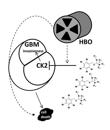
Isothiourea derivative (pentabromobenzylisothioureas)
ZKK
IC50 (7–50 μM)
-LN229 (glioblastoma cells)
-C6 (rat glioma cells)
-T98G (glioblastoma cells)
-HL-60 (human promyelocytic leukaemia)
-K-562l (human chronic erythromyeloblastic leukaemia)
-induction of apoptosis
-combination of HBO with the ZKK3 increased cytotoxicity and made T98G cells more sensitive to antitumour effect
-ZKK3 inhibited PIM, IGF-1R, IR, PKD1 kinases
Kaminska et al., 2009
Pucko et al., 2018
Koronkiewicz et al., 2013
Zembrzuska et al., 2019
Koronkiewicz et al., 2013

References

  1. Avendaño, C.; Menéndez, J.C. Chapter 9-Drugs That Inhibit Signalling Pathways for Tumor Cell Growth and Proliferation. In Medicinal Chemistry of Anticancer Drugs; Avendaño, C., Menéndez, J.C., Eds.; Elsevier: Amsterdam, The Netherlands, 2008; pp. 251–305. [Google Scholar]
  2. Cohen, P. The origins of protein phosphorylation. Nat. Cell Biol. 2002, 4, E127–E130. [Google Scholar] [CrossRef] [PubMed]
  3. Prudent, R.; Cochet, C. New Protein Kinase CK2 Inhibitors: Jumping out of the Catalytic Box. Chem. Biol. 2009, 16, 112–120. [Google Scholar] [CrossRef] [PubMed]
  4. Reichardt, P. The Story of Imatinib in GIST-A Journey through the Development of a Targeted Therapy. Oncol. Res. Treat. 2018, 41, 472–477. [Google Scholar] [CrossRef] [PubMed]
  5. Louis, D.N.; Perry, A.; Wesseling, P.; Brat, D.J.; Cree, I.A.; Figarella-Branger, D.; Hawkins, C.; Ng, H.K.; Pfister, S.M.; Reifenberger, G.; et al. The 2021 WHO Classification of Tumors of the Central Nervous System: A summary. Neuro-Oncology 2021, 23, 1231–1251. [Google Scholar] [CrossRef] [PubMed]
  6. Thakkar, J.P.; Dolecek, T.A.; Horbinski, C.; Ostrom, Q.; Lightner, D.D.; Barnholtz-Sloan, J.; Villano, J.L. Epidemiologic and Molecular Prognostic Review of Glioblastoma. Cancer Epidemiol. Biomark. Prev. 2014, 23, 1985–1996. [Google Scholar] [CrossRef] [PubMed] [Green Version]
  7. Rodriguez, F.J.; Orr, B.A.; Ligon, K.L.; Eberhart, C.G. Neoplastic cells are a rare component in human glioblastoma microvasculature. Oncotarget 2012, 3, 98–106. [Google Scholar] [CrossRef] [PubMed] [Green Version]
  8. Louis, D.N.; Ohgaki, H.; Wiestler, O.D.; Cavenee, W.K.; Burger, P.C.; Jouvet, A.; Scheithauer, B.W.; Kleihues, P. The 2007 WHO Classification of Tumours of the Central Nervous System. Acta Neuropathol. 2007, 114, 97–109. [Google Scholar] [CrossRef] [PubMed] [Green Version]
  9. Malzkorn, B.; Reifenberger, G. Integrated diagnostics of diffuse astrocytic and oligodendroglial tumors. Der Pathol. 2019, 40, 9–17. [Google Scholar] [CrossRef]
  10. Herrlinger, U.; Tzaridis, T.; Mack, F.; Steinbach, J.P.; Schlegel, U.; Sabel, M.; Hau, P.; Kortmann, R.-D.; Krex, D.; Grauer, O.; et al. Lomustine-temozolomide combination therapy versus standard temozolomide therapy in patients with newly diagnosed glioblastoma with methylated MGMT promoter (CeTeG/NOA–09): A randomised, open-label, phase 3 trial. Lancet 2019, 393, 678–688. [Google Scholar] [CrossRef]
  11. Agarwal, M.; Nitta, R.T.; Li, G. Casein Kinase 2: A Novel Player in Glioblastoma Therapy and Cancer Stem Cells. J. Mol. Genet. Med. 2013, 8, 1000094. [Google Scholar]
  12. Oliver, L.; Lalier, L.; Salaud, C.; Heymann, D.; Cartron, P.F.; Vallette, F. Drug Resistance in Glioblastoma: Are Persisters the Key to Therapy? Cancer Drug Resist. 2020, 3, 287–301. [Google Scholar] [CrossRef]
  13. Pardridge, W.M. Csf, Blood-Brain Barrier, and Brain Drug Delivery. Expert Opin. Drug Deliv. 2016, 13, 963–975. [Google Scholar] [CrossRef] [PubMed]
  14. Purow, B.; Schiff, D. Advances in the Genetics of Glioblastoma: Are We Reaching Critical Mass? Nat. Rev. Neurol. 2009, 5, 419–426. [Google Scholar] [CrossRef] [PubMed]
  15. Shergalis, A.; Bankhead, A.; Luesakul, U.; Muangsin, N.; Neamati, N. Current Challenges and Opportunities in Treating Glioblastoma. Pharmacol. Rev. 2018, 70, 412–445. [Google Scholar] [CrossRef] [Green Version]
  16. Sharifzad, F.; Ghavami, S.; Verdi, J.; Mardpour, S.; Sisakht, M.M.; Azizi, Z.; Taghikhani, A.; Łos, M.J.; Fakharian, E.; Ebrahimi, M.; et al. Glioblastoma cancer stem cell biology: Potential theranostic targets. Drug Resist. Updat. 2019, 42, 35–45. [Google Scholar] [CrossRef]
  17. Dixit, D.; Sharma, V.; Ghosh, S.; Mehta, V.S.; Sen, E. Inhibition of Casein kinase-2 induces p53-dependent cell cycle arrest and sensitizes glioblastoma cells to tumor necrosis factor (TNFα)-induced apoptosis through SIRT1 inhibition. Cell Death Dis. 2012, 3, e271. [Google Scholar] [CrossRef] [Green Version]
  18. Nitta, R.T.; Bolin, S.; Luo, E.; Solow-Codero, D.E.; Samghabadi, P.; Purzner, T.; Aujla, P.S.; Nwagbo, G.; Cho, Y.-J.; Li, G. Casein kinase 2 inhibition sensitizes medulloblastoma to temozolomide. Oncogene 2019, 38, 6867–6879. [Google Scholar] [CrossRef] [Green Version]
  19. Onciu, M. Acute Lymphoblastic Leukemia. Hematol. Oncol. Clin. N. Am. 2009, 23, 655–674. [Google Scholar] [CrossRef] [Green Version]
  20. Gowda, C.; Song, C.; Kapadia, M.; Payne, J.; Hu, T.; Ding, Y.; Dovat, S. Regulation of cellular proliferation in acute lymphoblastic leukemia by Casein Kinase II (CK2) and Ikaros. Adv. Biol. Regul. 2016, 63, 71–80. [Google Scholar] [CrossRef]
  21. Chon, H.J.; Bae, K.J.; Lee, Y.; Kim, J. The Casein Kinase 2 Inhibitor, Cx-4945, as an Anti-Cancer Drug in Treatment of Human Hematological Malignancies. Front. Pharmacol. 2015, 6, 70. [Google Scholar] [CrossRef]
  22. Chiorazzi, N.; Rai, K.R.; Ferrarini, M. Chronic Lymphocytic Leukemia. N. Engl. J. Med. 2005, 352, 804–815. [Google Scholar] [CrossRef] [PubMed] [Green Version]
  23. Martins, L.R.; Lúcio, P.; Silva, M.C.; Gameiro, P.; Silva, M.G.; Barata, J.T. On CK2 regulation of chronic lymphocytic leukemia cell viability. Mol. Cell. Biochem. 2011, 356, 51–55. [Google Scholar] [CrossRef] [PubMed]
  24. De Kouchkovsky, I.; Abdul-Hay, M. Acute Myeloid Leukemia: A Comprehensive Review and 2016 Update. Blood Cancer J. 2016, 6, e441. [Google Scholar] [CrossRef]
  25. Rosales, M.; Pérez, G.V.; Ramón, A.C.; Cruz, Y.; Rodriguez-Ulloa, A.; Besada, V.; Ramos, Y.; Vazquez-Blomquist, D.; Caballero, E.; Aguilar, D.; et al. Targeting of Protein Kinase Ck2 in Acute Myeloid Leukemia Cells Using the Clinical-Grade Synthetic-Peptide Cigb-300. Biomedicines 2021, 9, 766. [Google Scholar] [CrossRef] [PubMed]
  26. Jimenez, J.J.; Chale, R.S.; Abad, A.C.; Schally, A.V. Acute promyelocytic leukemia (APL): A review of the literature. Oncotarget 2020, 11, 992–1003. [Google Scholar] [CrossRef] [Green Version]
  27. Gurrieri, C.; Piazza, F.A.; Ruzzene, M.; Tubi, L.Q.; Tosoni, K.; Gnoato, M.; Cabrelle, A.; Bonanni, L.; Trentin, L.; Zambello, R.; et al. Role of Protein Kinase CK2 in the Retinoic Acid-Induced Differentiation of Acute Promyelocytic Leukemia Cells. Blood 2007, 110, 879. [Google Scholar] [CrossRef]
  28. Nicolson, N.G.; Korah, R.; Carling, T. Adrenocortical cancer cell line mutational profile reveals aggressive genetic background. J. Mol. Endocrinol. 2019, 62, 179–186. [Google Scholar] [CrossRef]
  29. Tanaka, T.; Ohe, H.; Nomiyama, T.; Yanase, T. OR02-2 CX-4945 as a Potential Drug for Adrenorcortical Carcinoma That induces Multiple Exon-Skipping and Circular RNA of NR5A1. J. Endocr. Soc. 2019, 3 (Suppl. 1), OR02-2. [Google Scholar] [CrossRef]
  30. Haraldsdottir, S.; Einarsdottir, H.M.; Smaradottir, A.; Gunnlaugsson, A.; Halfdanarson, T.R. Colorectal Cancer-Review. Laeknabladid 2014, 100, 75–82. [Google Scholar]
  31. Pistorius, K.; Seitz, G.; Remberger, K.; Issinger, O.G. Differential Ckii Activities in Human Colorectal Mucosa, Adenomas and Carcinomas. Oncol. Res. Treat 1991, 14, 256–260. [Google Scholar] [CrossRef]
  32. Siddiqui-Jain, A.; Drygin, D.; Streiner, N.; Chua, P.; Pierre, F.; O’Brien, S.E.; Bliesath, J.; Omori, M.; Huser, N.; Ho, C.; et al. CX-4945, an Orally Bioavailable Selective Inhibitor of Protein Kinase CK2, Inhibits Prosurvival and Angiogenic Signaling and Exhibits Antitumor Efficacy. Cancer Res. 2010, 70, 10288–10298. [Google Scholar] [CrossRef] [PubMed] [Green Version]
  33. Sun, Y.-S.; Zhao, Z.; Yang, Z.-N.; Xu, F.; Lu, H.-J.; Zhu, Z.-Y.; Shi, W.; Jiang, J.; Yao, P.-P.; Zhu, H.-P. Risk Factors and Preventions of Breast Cancer. Int. J. Biol. Sci. 2017, 13, 1387–1397. [Google Scholar] [CrossRef] [PubMed] [Green Version]
  34. Ruzzene, M.; Pinna, L.A. Addiction to Protein Kinase Ck2: A Common Denominator of Diverse Cancer Cells? Biochim. Biophys. Acta 2010, 1804, 499–504. [Google Scholar] [CrossRef] [PubMed]
  35. Razumilava, N.; Gores, G.J. Cholangiocarcinoma. Lancet 2014, 383, 2168–2179. [Google Scholar] [CrossRef] [Green Version]
  36. Zhou, F.; Xu, J.; Ding, G.; Cao, L. Overexpressions of CK2β and XIAP are Associated with Poor Prognosis of Patients with Cholangiocarcinoma. Pathol. Oncol. Res. 2013, 20, 73–79. [Google Scholar] [CrossRef]
  37. Nahand, J.S.; Moghoofei, M.; Salmaninejad, A.; Bahmanpour, Z.; Karimzadeh, M.; Nasiri, M.; Mirzaei, H.R.; Pourhanifeh, M.H.; Bokharaei-Salim, F.; Mirzaei, H.; et al. Pathogenic role of exosomes and microRNAs in HPV-mediated inflammation and cervical cancer: A review. Int. J. Cancer 2019, 146, 305–320. [Google Scholar] [CrossRef]
  38. Chua, M.M.J.; Lee, M.; Dominguez, I. Cancer-type dependent expression of CK2 transcripts. PLoS ONE 2017, 12, e0188854. [Google Scholar] [CrossRef] [Green Version]
  39. APinna, L. A historical view of protein kinase CK2. Cell. Mol. Biol. Res. 1994, 40, 383–390. [Google Scholar]
  40. Litchfield, D.W. Protein kinase CK2: Structure, regulation and role in cellular decisions of life and death. Biochem. J. 2003, 369, 1–15. [Google Scholar] [CrossRef]
  41. Montenarh, M.; Götz, C. Protein kinase CK2 and ion channels (Review). Biomed. Rep. 2020, 13, 55. [Google Scholar] [CrossRef]
  42. Borgo, C.; D’Amore, C.; Sarno, S.; Salvi, M.; Ruzzene, M. Protein kinase CK2: A potential therapeutic target for diverse human diseases. Signal Transduct. Target. Ther. 2021, 6, 183. [Google Scholar] [CrossRef] [PubMed]
  43. Dubois, N.; Willems, M.; Nguyen-Khac, M.-T.; Kroonen, J.; Goffart, N.; Deprez, M.; Bours, V.; Robe, P.A. Constitutive activation of casein kinase 2 in glioblastomas: Absence of class restriction and broad therapeutic potential. Int. J. Oncol. 2016, 48, 2445–2452. [Google Scholar] [CrossRef] [PubMed] [Green Version]
  44. Ferrer-Font, L.; Villamañan, L.; Arias-Ramos, N.; Vilardell, J.; Plana, M.; Ruzzene, M.; Pinna, L.A.; Itarte, E.; Arús, C.; Candiota, A.P. Targeting Protein Kinase CK2: Evaluating CX-4945 Potential for GL261 Glioblastoma Therapy in Immunocompetent Mice. Pharmaceuticals 2017, 10, 24. [Google Scholar] [CrossRef] [PubMed] [Green Version]
  45. Rowse, A.L.; Gibson, S.A.; Meares, G.P.; Rajbhandari, R.; Nozell, S.E.; Dees, K.J.; Hjelmeland, A.B.; McFarland, B.C.; Benveniste, E.N. Protein kinase CK2 is important for the function of glioblastoma brain tumor initiating cells. J. Neuro-Oncol. 2017, 132, 219–229. [Google Scholar] [CrossRef] [PubMed] [Green Version]
  46. Zheng, Y.; McFarland, B.C.; Drygin, D.; Yu, H.; Bellis, S.L.; Kim, H.; Bredel, M.; Benveniste, E.N. Targeting Protein Kinase CK2 Suppresses Prosurvival Signaling Pathways and Growth of Glioblastoma. Clin. Cancer Res. 2013, 19, 6484–6494. [Google Scholar] [CrossRef] [PubMed] [Green Version]
  47. Purzner, T.; Purzner, J.; Buckstaff, T.; Cozza, G.; Gholamin, S.; Rusert, J.M.; Hartl, T.A.; Sanders, J.; Conley, N.; Ge, X.; et al. Developmental phosphoproteomics identifies the kinase CK2 as a driver of Hedgehog signaling and a therapeutic target in medulloblastoma. Sci. Signal. 2018, 11, eaau5147. [Google Scholar] [CrossRef] [Green Version]
  48. Kroonen, J.; Artesi, M.; Capraro, V.; Nguyen-Khac, M.-T.; Willems, M.; Chakravarti, A.; Bours, V.; Robe, P.A. Casein kinase 2 inhibition modulates the DNA damage response but fails to radiosensitize malignant glioma cells. Int. J. Oncol. 2012, 41, 776–782. [Google Scholar] [CrossRef] [Green Version]
  49. Duncan, J.S.; Litchfield, D.W. Too much of a good thing: The role of protein kinase CK2 in tumorigenesis and prospects for therapeutic inhibition of CK2. Biochim. Biophys. Acta (BBA) Proteins Proteom. 2008, 1784, 33–47. [Google Scholar] [CrossRef]
  50. Duncan, J.S.; Gyenis, L.; Lenehan, J.; Bretner, M.; Graves, L.M.; Haystead, T.A.; Litchfield, D.W. An Unbiased Evaluation of Ck2 Inhibitors by Chemoproteomics: Characterization of Inhibitor Effects on Ck2 and Identification of Novel Inhibitor Targets. Mol. Cell Proteom. 2008, 7, 1077–1088. [Google Scholar] [CrossRef] [PubMed] [Green Version]
  51. Iegre, J.; Atkinson, E.L.; Brear, P.D.; Cooper, B.M.; Hyvönen, M.; Spring, D.R. Chemical probes targeting the kinase CK2: A journey outside the catalytic box. Org. Biomol. Chem. 2021, 19, 4380–4396. [Google Scholar] [CrossRef]
  52. Faust, M.; Montenarh, M. Subcellular localization of protein kinase CK2. Cell Tissue Res. 2000, 301, 329–340. [Google Scholar] [CrossRef] [PubMed]
  53. Tawfic, S.; Yu, S.; Wang, H.; Faust, R.; Davis, A.; Ahmed, K. Protein kinase CK2 signal in neoplasia. Histol. Histopathol. 2001, 16, 573–582. [Google Scholar] [PubMed]
  54. Cozza, G. The Development of CK2 Inhibitors: From Traditional Pharmacology to in Silico Rational Drug Design. Pharmaceuticals 2017, 10, 26. [Google Scholar] [CrossRef] [PubMed] [Green Version]
  55. Pagano, M.A.; Andrzejewska, M.; Ruzzene, M.; Sarno, S.; Cesaro, L.; Bain, J.; Elliott, M.; Meggio, F.; Kazimierczuk, Z.; Pinna, L.A. Optimization of Protein Kinase Ck2 Inhibitors Derived from 4,5,6,7-Tetrabromobenzimidazole. J. Med. Chem. 2004, 47, 6239–6247. [Google Scholar] [CrossRef]
  56. Meggio, F.; Pagano, M.A.; Moro, S.; Zagotto, G.; Ruzzene, M.; Sarno, S.; Cozza, G.; Bain, J.; Elliott, M.; Deana, A.D.; et al. Pinna. Inhibition of Protein Kinase Ck2 by Condensed Polyphenolic Derivatives. An in Vitro and in Vivo Study. Biochemistry 2004, 43, 12931–12936. [Google Scholar] [CrossRef] [Green Version]
  57. Sarno, S.; de Moliner, E.; Ruzzene, M.; Pagano, M.A.; Battistutta, R.; Bain, J.; Fabbro, D.; Schoepfer, J.; Elliott, M.; Furet, P.; et al. Biochemical and Three-Dimensional-Structural Study of the Specific Inhibition of Protein Kinase Ck2 by [5-Oxo-5,6-Dihydroindolo-(1,2-a)Quinazolin-7-Yl]Acetic Acid (Iqa). Biochem. J. 2003, 374, 639–646. [Google Scholar] [CrossRef]
  58. Kubiński, K.; Masłyk, M.; Orzeszko, A. Benzimidazole inhibitors of protein kinase CK2 potently inhibit the activity of atypical protein kinase Rio1. Mol. Cell. Biochem. 2016, 426, 195–203. [Google Scholar] [CrossRef] [Green Version]
  59. Schaefer, S.; Svenstrup, T.H.; Fischer, M.; Guerra, B. D11-Mediated Inhibition of Protein Kinase CK2 Impairs HIF-1α-Mediated Signaling in Human Glioblastoma Cells. Pharmaceuticals 2017, 10, 5. [Google Scholar] [CrossRef] [Green Version]
  60. Malric, L.; Monferran, S.; Gilhodes, J.; Boyrie, S.; Dahan, P.; Skuli, N.; Sesen, J.; Filleron, T.; Kowalski-Chauvel, A.; Moyal, E.C.-J.; et al. Interest of integrins targeting in glioblastoma according to tumor heterogeneity and cancer stem cell paradigm: An update. Oncotarget 2017, 8, 86947–86968. [Google Scholar] [CrossRef] [Green Version]
  61. Di Maira, G.; Salvi, M.; Arrigoni, G.; Marin, O.; Sarno, S.; Brustolon, F.; Pinna, L.A.; Ruzzene, M. Protein kinase CK2 phosphorylates and upregulates Akt/PKB. Cell Death Differ. 2005, 12, 668–677. [Google Scholar] [CrossRef]
  62. Ponce, D.P.; Yefi, R.; Cabello, P.; Maturana, J.L.; Niechi, I.; Silva, E.; Galindo, M.; Antonelli, M.; Marcelain, K.; Armisen, R.; et al. CK2 functionally interacts with AKT/PKB to promote the β-catenin-dependent expression of survivin and enhance cell survival. Mol. Cell. Biochem. 2011, 356, 127–132. [Google Scholar] [CrossRef] [PubMed]
  63. Song, C.; Ge, Z.; Ding, Y.; Tan, B.-H.; Desai, D.; Gowda, K.; Amin, S.G.; Gowda, R.; Robertson, G.P.; Yue, F.; et al. IKAROS and CK2 regulate expression of BCL-XL and chemosensitivity in high-risk B-cell acute lymphoblastic leukemia. Blood 2020, 136, 1520–1534. [Google Scholar] [CrossRef] [PubMed]
  64. Mehta, A.; Awah, C.U.; Sonabend, A.M. Topoisomerase II Poisons for Glioblastoma; Existing Challenges and Opportunities to Personalize Therapy. Front. Neurol. 2018, 9, 459. [Google Scholar] [CrossRef] [PubMed]
  65. Nager, M.; Bhardwaj, D.; Cantí, C.; Medina, L.; Nogués, P.; Herreros, J. Β-Catenin Signalling in Glioblastoma Multiforme and Glioma-Initiating Cells. Chemother Res. Pract. 2012, 2012, 192362. [Google Scholar] [CrossRef] [Green Version]
  66. Nitta, R.T.; Gholamin, S.; Feroze, A.H.; Agarwal, M.; Cheshier, S.H.; Mitra, S.S.; Li, G. Casein kinase 2α regulates glioblastoma brain tumor-initiating cell growth through the β-catenin pathway. Oncogene 2014, 34, 3688–3699. [Google Scholar] [CrossRef] [PubMed] [Green Version]
  67. Alves, A.L.V.; Gomes, I.N.F.; Carloni, A.C.; Rosa, M.N.; da Silva, L.S.; Evangelista, A.F.; Reis, R.M.; Silva, V.A.O. Role of glioblastoma stem cells in cancer therapeutic resistance: A perspective on antineoplastic agents from natural sources and chemical derivatives. Stem Cell Res. Ther. 2021, 12, 206. [Google Scholar] [CrossRef]
  68. Yuan, Y.; Zhang, M.; Yan, G.; Ma, Q.; Yan, Z.; Wang, L.; Yang, K.; Guo, D. Nanog Promotes Stem-Like Traits of Glioblastoma Cells. Front. Biosci. 2021, 26, 552–565. [Google Scholar]
  69. Zhou, J.; Tien, A.-C.; Alberta, J.A.; Ficarro, S.B.; Griveau, A.; Sun, Y.; Deshpande, J.S.; Card, J.D.; Morgan-Smith, M.; Michowski, W.; et al. A Sequentially Priming Phosphorylation Cascade Activates the Gliomagenic Transcription Factor Olig2. Cell Rep. 2017, 18, 3167–3177. [Google Scholar] [CrossRef]
  70. Zandomeni, R.; Zandomeni, M.C.; Shugar, D.; Weinmann, R. Casein kinase type II is involved in the inhibition by 5,6-dichloro-1-beta-D-ribofuranosylbenzimidazole of specific RNA polymerase II transcription. J. Biol. Chem. 1986, 261, 3414–3419. [Google Scholar] [CrossRef]
  71. Cozza, G.; Sarno, S.; Ruzzene, M.; Girardi, C.; Orzeszko, A.; Kazimierczuk, Z.; Zagotto, G.; Bonaiuto, E.; Di Paolo, M.L.; Pinna, L.A. Exploiting the repertoire of CK2 inhibitors to target DYRK and PIM kinases. Biochim. Et Biophys. Acta (BBA) Proteins Proteom. 2013, 1834, 1402–1409. [Google Scholar] [CrossRef]
  72. Pagano, M.A.; Bain, J.; Kazimierczuk, Z.; Sarno, S.; Ruzzene, M.; Di Maira, G.; Elliott, M.; Orzeszko, A.; Cozza, G.; Meggio, F.; et al. The selectivity of inhibitors of protein kinase CK2: An update. Biochem. J. 2008, 415, 353–365. [Google Scholar] [CrossRef] [PubMed] [Green Version]
  73. Pucko, E.; Ostrowski, R.; Matyja, E. Novel small molecule protein kinase CK2 inhibitors exert potent antitumor effects on T98G and SEGA cells in vitro. Folia Neuropathol. 2019, 57, 239–248. [Google Scholar] [CrossRef] [PubMed]
  74. Kaminska, B.; Ellert-Miklaszewska, A.; Oberbek, A.; Wisniewski, P.; Kaza, B.; Makowska, M.; Bretner, M.; Kazimierczuk, Z. Efficacy and mechanism of anti-tumor action of new potential CK2 inhibitors toward glioblastoma cells. Int. J. Oncol. 2009, 35, 1091–1100. [Google Scholar] [CrossRef] [PubMed] [Green Version]
  75. Pagano, M.A.; Meggio, F.; Ruzzene, M.; Andrzejewska, M.; Kazimierczuk, Z.; Pinna, L.A. 2-Dimethylamino-4,5,6,7-tetrabromo-1H-benzimidazole: A novel powerful and selective inhibitor of protein kinase CK2. Biochem. Biophys. Res. Commun. 2004, 321, 1040–1044. [Google Scholar] [CrossRef] [PubMed]
  76. Mishra, S.; Pertz, V.; Zhang, B.; Kaur, P.; Shimada, H.; Groffen, J.; Kazimierczuk, Z.; Pinna, L.A.; Heisterkamp, N. Treatment of P190 Bcr/Abl lymphoblastic leukemia cells with inhibitors of the serine/threonine kinase CK2. Leukemia 2006, 21, 178–180. [Google Scholar] [CrossRef] [PubMed]
  77. Mishra, S.; Reichert, A.; Cunnick, J.; Senadheera, D.; Hemmeryckx, B.; Heisterkamp, N.; Groffen, J. Protein Kinase Ckiialpha Interacts with the Bcr Moiety of Bcr/Abl and Mediates Proliferation of Bcr/Abl-Expressing Cells. Oncogene 2003, 22, 8255–8262. [Google Scholar] [CrossRef] [PubMed] [Green Version]
  78. Tapia, J.C.; Torres, V.A.; Rodriguez, D.; Leyton, L.; Quest, A.F.G. Casein kinase 2 (CK2) increases survivin expression via enhanced beta-catenin-T cell factor/lymphoid enhancer binding factor-dependent transcription. Proc. Natl. Acad. Sci. USA 2006, 103, 15079–15084. [Google Scholar] [CrossRef] [Green Version]
  79. Koronkiewicz, M.; Chilmonczyk, Z.; Kazimierczuk, Z. Synergistic anti-leukemic effects of CK2 inhibitors and pentabromobenzylisothioureas in vitro. Anticancer Res. 2013, 33, 4891–4899. [Google Scholar]
  80. Lawnicka, H.; Kowalewicz-Kulbat, M.; Sicinska, P.; Kazimierczuk, Z.; Grieb, P.; Stepien, H. Anti-neoplastic effect of protein kinase CK2 inhibitor, 2-dimethylamino-4,5,6,7-tetrabromobenzimidazole (DMAT), on growth and hormonal activity of human adrenocortical carcinoma cell line (H295R) in vitro. Cell Tissue Res. 2010, 340, 371–379. [Google Scholar] [CrossRef]
  81. Szyszka, R.; Grankowski, N.; Felczak, K.; Shugar, D. Halogenated Benzimidazoles and Benzotriazoles as Selective Inhibitors of Protein Kinases CK-I and CK-II from Saccharomyces Cerevisiae and Other Sources. Biochem. Biophys. Res. Commun. 1995, 208, 418–424. [Google Scholar] [CrossRef]
  82. Andrzejewska, M.; Pagano, M.A.; Meggio, F.; Brunati, A.M.; Kazimierczuk, Z. Polyhalogenobenzimidazoles: Synthesis and Their inhibitory activity against casein kinases. Bioorg. Med. Chem. 2003, 11, 3997–4002. [Google Scholar] [CrossRef]
  83. Shehata, M.; Schnabl, S.; Demirtas, D.; Hilgarth, M.; Hubmann, R.; Ponath, E.; Badrnya, S.; Lehner, C.; Hoelbl, A.; Duechler, M.; et al. Reconstitution of PTEN activity by CK2 inhibitors and interference with the PI3-K/Akt cascade counteract the antiapoptotic effect of human stromal cells in chronic lymphocytic leukemia. Blood 2010, 116, 2513–2521. [Google Scholar] [CrossRef] [PubMed]
  84. Girardi, C.; Ottaviani, D.; Pinna, L.A.; Ruzzene, M. Different Persistence of the Cellular Effects Promoted by Protein Kinase CK2 Inhibitors CX-4945 and TDB. BioMed Res. Int. 2015, 2015, 185736. [Google Scholar] [CrossRef] [PubMed] [Green Version]
  85. Cozza, G.; Girardi, C.; Ranchio, A.; Lolli, G.; Sarno, S.; Orzeszko, A.; Kazimierczuk, Z.; Battistutta, R.; Ruzzene, M.; Pinna, L.A. Cell-permeable dual inhibitors of protein kinases CK2 and PIM-1: Structural features and pharmacological potential. Cell. Mol. Life Sci. 2014, 71, 3173–3185. [Google Scholar] [CrossRef]
  86. Borgo, C.; Ruzzene, M. Role of protein kinase CK2 in antitumor drug resistance. J. Exp. Clin. Cancer Res. 2019, 38, 287. [Google Scholar] [CrossRef]
  87. Zhou, Y.; Zhang, N.; Tang, S.; Qi, X.; Zhao, L.; Zhong, R.; Peng, Y. Exploring the Pivotal Role of the CK2 Hinge Region Sub-Pocket in Binding with Tricyclic Quinolone Analogues by Computational Analysis. Molecules 2017, 22, 840. [Google Scholar] [CrossRef] [Green Version]
  88. Ku, M.J.; Park, J.W.; Ryu, B.J.; Son, Y.-J.; Kim, S.H.; Lee, S.Y. CK2 inhibitor CX4945 induces sequential inactivation of proteins in the signaling pathways related with cell migration and suppresses metastasis of A549 human lung cancer cells. Bioorg. Med. Chem. Lett. 2013, 23, 5609–5613. [Google Scholar] [CrossRef]
  89. D’Amore, C.; Borgo, C.; Sarno, S.; Salvi, M. Role of CK2 inhibitor CX-4945 in anti-cancer combination therapy–potential clinical relevance. Cell. Oncol. 2020, 43, 1003–1016. [Google Scholar] [CrossRef]
  90. Buontempo, F.; Orsini, E.; Lonetti, A.; Cappellini, A.; Chiarini, F.; Evangelisti, C.; Evangelisti, C.; Melchionda, F.; Pession, A.; Bertaina, A.; et al. Synergistic Cytotoxic Effects of Bortezomib and Ck2 Inhibitor Cx-4945 in Acute Lymphoblastic Leukemia: Turning Off the Prosurvival Er Chaperone Bip/Grp78 and Turning on the Pro-Apoptotic Nf-Κb. Oncotarget 2016, 7, 1323–1340. [Google Scholar] [CrossRef] [Green Version]
  91. Zakharia, K.; Miyabe, K.; Wang, Y.; Wu, D.; Moser, C.D.; Borad, M.J.; Roberts, L.R. Preclinical in Vitro and in Vivo Evidence of an Antitumor Effect of Cx-4945, a Casein Kinase Ii Inhibitor, in Cholangiocarcinoma. Transl. Oncol. 2019, 12, 143–153. [Google Scholar] [CrossRef]
  92. Eskilsson, E.; Røsland, G.V.; Solecki, G.; Wang, Q.; Harter, P.N.; Graziani, G.; Verhaak, R.G.; Winkler, F.; Bjerkvig, R.; Miletic, H. Egfr Heterogeneity and Implications for Therapeutic Intervention in Glioblastoma. Neuro Oncol. 2018, 20, 743–752. [Google Scholar] [CrossRef] [PubMed] [Green Version]
  93. Furnari, F.; Cloughesy, T.F.; Cavenee, W.K.; Mischel, P.S. Heterogeneity of epidermal growth factor receptor signalling networks in glioblastoma. Nat. Cancer 2015, 15, 302–310. [Google Scholar] [CrossRef] [PubMed] [Green Version]
  94. Pierre, F.; Chua, P.C.; O’Brien, S.E.; Siddiqui-Jain, A.; Bourbon, P.; Haddach, M.; Michaux, J.; Nagasawa, J.; Schwaebe, M.K.; Stefan, E.; et al. Pre-clinical characterization of CX-4945, a potent and selective small molecule inhibitor of CK2 for the treatment of cancer. Mol. Cell. Biochem. 2011, 356, 37–43. [Google Scholar] [CrossRef] [PubMed]
  95. Atkinson, E.L.; Iegre, J.; Brear, P.D.; Zhabina, E.A.; Hyvönen, M.; Spring, D.R. Downfalls of Chemical Probes Acting at the Kinase Atp-Site: Ck2 as a Case Study. Molecules 2021, 26, 1977. [Google Scholar] [CrossRef] [PubMed]
  96. Salvi, M.; Borgo, C.; Pinna, L.A.; Ruzzene, M. Targeting Ck2 in Cancer: A Valuable Strategy or a Waste of Time? Cell Death Discov. 2021, 7, 325. [Google Scholar] [CrossRef]
  97. Pucko, E.; Matyja, E.; Koronkiewicz, M.; Ostrowski, R.P.; Kazimierczuk, Z. Potent Antitumour Effects of Novel Pentabromobenzylisothioureas Studied on Human Glial-derived Tumour Cell Lines. Anticancer Res. 2018, 38, 2691–2705. [Google Scholar] [CrossRef] [Green Version]
  98. Koronkiewicz, M.; Kazimierczuk, Z.; Szarpak, K.; Chilmonczyk, Z. Proapoptotic effects of new pentabromobenzylisothiouronium salts in a human prostate adenocarcinoma cell line. Acta Pol. Pharm. Drug Res. 2013, 69, 1325–1333. [Google Scholar]
  99. Ehtesham, M.; Winston, J.A.; Kabos, P.; Thompson, R.C. CXCR4 expression mediates glioma cell invasiveness. Oncogene 2006, 25, 2801–2806. [Google Scholar] [CrossRef] [Green Version]
  100. Ganju, R.K.; Brubaker, S.A.; Meyer, J.; Dutt, P.; Yang, Y.; Qin, S.; Newman, W.; Groopman, J.E. The α-Chemokine, Stromal Cell-derived Factor-1α, Binds to the Transmembrane G-protein-coupled CXCR-4 Receptor and Activates Multiple Signal Transduction Pathways. J. Biol. Chem. 1998, 273, 23169–23175. [Google Scholar] [CrossRef] [Green Version]
  101. Bian, X.-W.; Yang, S.-X.; Chen, J.-H.; Ping, Y.-F.; Zhou, X.-D.; Wang, Q.-L.; Jiang, X.-F.; Gong, W.; Xiao, H.-L.; Du, L.-L.; et al. Preferential Expression of Chemokine Receptor Cxcr4 By Highly Malignant Human Gliomas And Its Association With Poor Patient Survival. Neurosurgery 2007, 61, 570–579. [Google Scholar] [CrossRef]
  102. Zembrzuska, K.; Ostrowski, R.P.; Matyja, E. Hyperbaric oxygen increases glioma cell sensitivity to antitumor treatment with a novel isothiourea derivative in vitro. Oncol. Rep. 2019, 41, 2703–2716. [Google Scholar] [CrossRef] [PubMed] [Green Version]
  103. Wang, P.; Gong, S.; Pan, J.; Wang, J.; Zou, D.; Xiong, S.; Zhao, L.; Yan, Q.; Deng, Y.; Wu, N.; et al. Hyperbaric oxygen promotes not only glioblastoma proliferation but also chemosensitization by inhibiting HIF1α/HIF2α-Sox2. Cell Death Discov. 2021, 7, 103. [Google Scholar] [CrossRef] [PubMed]
  104. Mottet, D.; Ruys, S.P.D.; Demazy, C.; Raes, M.; Michiels, C. Role for casein kinase 2 in the regulation of HIF-1 activity. Int. J. Cancer 2005, 117, 764–774. [Google Scholar] [CrossRef] [PubMed]
  105. Huang, L.; Boling, W.; Zhang, J.H. Hyperbaric oxygen therapy as adjunctive strategy in treatment of glioblastoma multiforme. Med. Gas Res. 2018, 8, 24–28. [Google Scholar] [CrossRef]
  106. Asati, V.; Mahapatra, D.K.; Bharti, S.K. PIM kinase inhibitors: Structural and pharmacological perspectives. Eur. J. Med. Chem. 2019, 172, 95–108. [Google Scholar] [CrossRef]
  107. Herzog, S.; Fink, M.A.; Weitmann, K.; Friedel, C.; Hadlich, S.; Langner, S.; Kindermann, K.; Holm, T.; Böhm, A.; Eskilsson, E.; et al. Pim1 kinase is upregulated in glioblastoma multiforme and mediates tumor cell survival. Neuro-Oncology 2014, 17, 223–242. [Google Scholar] [CrossRef] [Green Version]
  108. Quan, J.; Zhou, L.; Qu, J. Knockdown of Pim-3 suppresses the tumorigenicity of glioblastoma by regulating cell cycle and apoptosis. Cell. Mol. Biol. 2015, 61, 42–50. [Google Scholar]
  109. Zamykal, M.; Martens, T.; Matschke, J.; Günther, H.S.; Kathagen, A.; Schulte, A.; Peters, R.; Westphal, M.; Lamszus, K. Inhibition of intracerebral glioblastoma growth by targeting the insulin-like growth factor 1 receptor involves different context-dependent mechanisms. Neuro-Oncology 2014, 17, 1076–1085. [Google Scholar] [CrossRef]
  110. Vigneri, R.; Goldfine, I.D.; Frittitta, L. Insulin, Insulin Receptors, and Cancer. J. Endocrinol. Investig. 2016, 39, 1365–1376. [Google Scholar] [CrossRef]
  111. Ulanet, D.B.; Ludwig, D.L.; Kahn, C.R.; Hanahan, D. Insulin receptor functionally enhances multistage tumor progression and conveys intrinsic resistance to IGF-1R targeted therapy. Proc. Natl. Acad. Sci. USA 2010, 107, 10791–10798. [Google Scholar] [CrossRef] [Green Version]
  112. Brognard, J.; Hunter, T. Protein kinase signaling networks in cancer. Curr. Opin. Genet. Dev. 2011, 21, 4–11. [Google Scholar] [CrossRef] [PubMed] [Green Version]
  113. Du, Z.; Lovly, C.M. Mechanisms of receptor tyrosine kinase activation in cancer. Mol. Cancer 2018, 17, 58. [Google Scholar] [CrossRef] [PubMed]
  114. Saadeh, F.S.; Mahfouz, R.; Assi, H.I. EGFR as a clinical marker in glioblastomas and other gliomas. Int. J. Biol. Markers 2017, 33, 22–32. [Google Scholar] [CrossRef] [PubMed] [Green Version]
  115. Zhang, J.; Antonyak, M.A.; Singh, G.; Cerione, R.A. A Mechanism for the Upregulation of EGF Receptor Levels in Glioblastomas. Cell Rep. 2013, 3, 2008–2020. [Google Scholar] [CrossRef] [Green Version]
  116. Bode, U.; Massimino, M.; Bach, F.; Zimmermann, M.; Khuhlaeva, E.; Westphal, M.; Fleischhack, G. Nimotuzumab treatment of malignant gliomas. Expert Opin. Biol. Ther. 2012, 12, 1649–1659. [Google Scholar] [CrossRef]
  117. Liang, W.; Wu, X.; Fang, W.; Zhao, Y.; Yang, Y.; Wenhua, L.; Xue, C.; Zhang, J.; Zhang, J.; Ma, Y.; et al. Network Meta-Analysis of Erlotinib, Gefitinib, Afatinib and Icotinib in Patients with Advanced Non-Small-Cell Lung Cancer Harboring EGFR Mutations. PLoS ONE 2014, 9, e85245. [Google Scholar] [CrossRef]
  118. Kinsella, P.; Howley, R.; Doolan, P.; Clarke, C.; Madden, S.F.; Clynes, M.; Farrell, M.; Amberger-Murphy, V. Characterization and Response of Newly Developed High-Grade Glioma Cultures to the Tyrosine Kinase Inhibitors, Erlotinib, Gefitinib and Imatinib. Exp. Cell Res. 2012, 318, 641–652. [Google Scholar] [CrossRef]
  119. Liu, X.; Chen, X.; Shi, L.; Shan, Q.; Cao, Q.; Yue, C.; Li, H.; Li, S.; Wang, J.; Gao, S.; et al. The third-generation EGFR inhibitor AZD9291 overcomes primary resistance by continuously blocking ERK signaling in glioblastoma. J. Exp. Clin. Cancer Res. 2019, 38, 219. [Google Scholar] [CrossRef]
  120. Koul, D.; Fu, J.; Shen, R.; LaFortune, T.A.; Wang, S.; Tiao, N.; Kim, Y.-W.; Liu, J.-L.; Ramnarian, D.; Yuan, Y.; et al. Antitumor Activity of NVP-BKM120—A Selective Pan Class I PI3 Kinase Inhibitor Showed Differential Forms of Cell Death Based on p53 Status of Glioma Cells. Clin. Cancer Res. 2011, 18, 184–195. [Google Scholar] [CrossRef] [Green Version]
  121. Zhao, H.F.; Wang, J.; Shao, W.; Wu, C.P.; Chen, Z.P.; To, S.S.T.; Li, W.P. Recent Advances in the Use of Pi3k Inhibitors for Glioblastoma Multiforme: Current Preclinical and Clinical Development. Mol. Cancer 2017, 16, 100. [Google Scholar] [CrossRef] [Green Version]
  122. Gao, Q.; Lei, T.; Ye, F. Therapeutic targeting of EGFR-activated metabolic pathways in glioblastoma. Expert Opin. Investig. Drugs 2013, 22, 1023–1040. [Google Scholar] [CrossRef] [PubMed]
  123. Chresta, C.M.; Davies, B.R.; Hickson, I.; Harding, T.; Cosulich, S.; Critchlow, S.E.; Vincent, J.P.; Ellston, R.; Jones, D.; Sini, P.; et al. AZD8055 Is a Potent, Selective, and Orally Bioavailable ATP-Competitive Mammalian Target of Rapamycin Kinase Inhibitor with In vitro and In vivo Antitumor Activity. Cancer Res. 2009, 70, 288–298. [Google Scholar] [CrossRef] [PubMed] [Green Version]
  124. Colella, B.; Colardo, M.; Iannone, G.; Contadini, C.; Saiz-Ladera, C.; Fuoco, C.; Barilà, D.; Velasco, G.; Segatto, M.; Di Bartolomeo, S. mTOR Inhibition Leads to Src-Mediated EGFR Internalisation and Degradation in Glioma Cells. Cancers 2020, 12, 2266. [Google Scholar] [CrossRef] [PubMed]
  125. Catalano, M.; D’Alessandro, G.; Lepore, F.; Corazzari, M.; Caldarola, S.; Valacca, C.; Faienza, F.; Esposito, V.; Limatola, C.; Cecconi, F.; et al. Autophagy induction impairs migration and invasion by reversing EMT in glioblastoma cells. Mol. Oncol. 2015, 9, 1612–1625. [Google Scholar] [CrossRef] [Green Version]
  126. Del Alcazar, C.R.G.; Hardebeck, M.C.; Mukherjee, B.; Tomimatsu, N.; Gao, X.; Yan, J.; Xie, X.J.; Bachoo, R.; Li, L.; Burma, S.; et al. Inhibition of DNA Double-Strand Break Repair by the Dual Pi3k/Mtor Inhibitor Nvp-Bez235 as a Strategy for Radiosensitization of Glioblastoma. Clin. Cancer Res. 2014, 20, 1235–1248. [Google Scholar] [CrossRef] [PubMed] [Green Version]
  127. Colardo, M.; Segatto, M.; Di Bartolomeo, S. Targeting Rtk-Pi3k-Mtor Axis in Gliomas: An Update. Int. J. Mol. Sci. 2021, 22, 4899. [Google Scholar] [CrossRef]
  128. Heffron, T.P.; Ndubaku, C.O.; Salphati, L.; Alicke, B.; Cheong, J.; Drobnick, J.; Edgar, K.; Gould, S.E.; Lee, L.B.; Lesnick, J.D.; et al. Discovery of Clinical Development Candidate Gdc-0084, a Brain Penetrant Inhibitor of Pi3k and Mtor. ACS Med. Chem. Lett. 2016, 7, 351–356. [Google Scholar] [CrossRef] [Green Version]
  129. Bliesath, J.; Huser, N.; Omori, M.; Bunag, D.; Proffitt, C.; Streiner, N.; Ho, C.; Siddiqui-Jain, A.; O’Brien, S.E.; Lim, J.K.; et al. Combined Inhibition of Egfr and Ck2 Augments the Attenuation of Pi3k-Akt-Mtor Signaling and the Killing of Cancer Cells. Cancer Lett. 2012, 322, 113–118. [Google Scholar] [CrossRef]
  130. Gober, M.K.; Flight, R.M.; Lambert, J.; Moseley, H.; Stromberg, A.; Black, E.P. Deregulation of a Network of mRNA and miRNA Genes Reveals That CK2 and MEK Inhibitors May Synergize to Induce Apoptosis KRAS-Active NSCLC. Cancer Inform. 2019, 18, 1176935119843507. [Google Scholar] [CrossRef]
  131. Lustri, A.M.; Di Matteo, S.; Fraveto, A.; Costantini, D.; Cantafora, A.; Napoletano, C.; Bragazzi, M.C.; Giuliante, F.; De Rose, A.M.; Berloco, P.B.; et al. TGF-β signaling is an effective target to impair survival and induce apoptosis of human cholangiocarcinoma cells: A study on human primary cell cultures. PLoS ONE 2017, 12, e0183932. [Google Scholar] [CrossRef] [Green Version]
  132. Whelan, R.; Hargaden, G.C.; Knox, A.J. Modulating the Blood & Ndash; Brain Barrier: A Comprehensive Review. Pharmaceutics 2021, 13, 1980. [Google Scholar] [PubMed]
  133. Liu, X.; Chen, J.; Li, W.; Hang, C.; Dai, Y. Inhibition of Casein Kinase II by CX-4945, But Not Yes-associated protein (YAP) by Verteporfin, Enhances the Antitumor Efficacy of Temozolomide in Glioblastoma. Transl. Oncol. 2019, 13, 70–78. [Google Scholar] [CrossRef] [PubMed]
  134. Borgo, C.; Vilardell, J.; Bosello-Travain, V.; Pinna, L.A.; Venerando, A.; Salvi, M. Dependence of HSP27 cellular level on protein kinase CK2 discloses novel therapeutic strategies. Biochim. Biophys. Acta (BBA) Gen. Subj. 2018, 1862, 2902–2910. [Google Scholar] [CrossRef] [PubMed]
  135. Son, Y.H.; Song, J.S.; Kim, S.H.; Kim, J. Pharmacokinetic characterization of CK2 inhibitor CX-4945. Arch. Pharmacal. Res. 2013, 36, 840–845. [Google Scholar] [CrossRef]
Figure 1. CK2 implications in glioblastoma development. CK2 activity can be stimulated indirectly by growth factors, kinase ERK, and cytokines. Downstream from growth factor signalling, kinase ERK phosphorylates and activates CK2. Activated CK2 regulates AKT and STAT3, HIF-1α, and GSC maintenance to promote glioblastoma cell adhesion, migration, proliferation, and survival. The action of CK2 may lead, through HIF-1α and its targets genes VEGF and MMP2 activation, to angiogenic responses and increased invasiveness [59]. Integrin α4 and Integrin β1 are responsible for glial tumour cell adhesion [60], while CK2-phosphorylated AKT underlies cell protection, as does survivin, the expression of which can be enhanced by CK2 [61,62]. In order to reach this goal, the activation of the antiapoptotic protein BCL-XL is also controlled by CK2 [63]. CDC34 and topoisomerase II are involved in functioning of the cell cycle in glioma cells. CK2 was shown to phosphorylate these cell cycle regulators [51,64]. Several proteins controlled by CK2 have been established to be involved in the maintenance of glioma stem cells, including Wnt/β-catenin, NANOG, OCT4, OLIG2, SHH, and Notch. CK2 is responsible for the phosphorylation of α-catenin and transactivation of β-catenin [65]. The β-catenin-regulated genes OCT4 and NANOG showed significant reductions in expression upon CK2 silencing or pharmacological inhibition in glioma cells [66]. CK2 may also activate SHH and Notch, which are involved not only in stemness maintenance but in mediating chemoresistance (e.g., to TMZ) [67,68]. CK2 regulates gliomagenic functions of Olig2 by participating in phosphorylation of triple serine motif in the amino terminus [69]. Recently, it came under scrutiny whether the use of novel CK2 inhibitors with improved selectivity (e.g., SGC-CK2-1) translated into anticancer effect. DMAT (dimethylamino-4,5,6,7-1H-tetrabromobenzimidazole), TBI (4,5,6,7-tetrabromo-1H-benzimidazol), TBB (4,5,6,7-tetrabromo-1H-benzotriazole), TDB (1-β-D-2′-deoxyribofuranosyl-4,5,6,7-tetrabromo-1H-benzimidazole), CX-4945 (silmitasertib; 5-((3-Chlorophenyl)amino)benzo[c][2,6]naphthyridine-8-carboxylic acid), SGC-CK2-1 (N-(5-(3-Cyano-7-(cyclopropylamino)pyrazolo [1,5-a]pyrimidin-5-ylamino) -2-methylphenyl)propionamide), BCL-XL (B-cell lymphoma-extra large); CDC34 (cell division cycle 34), ERK (extracellular signal-regulated kinase), GSC (glioblastoma stem cells), NANOG (NANOG homeobox), HIF-1α (hypoxia-inducible factor 1α), NF-κB (nuclear factor kappa-light-chain-enhancer of activated B cells), OCT4 (octamer-binding transcription factor 4), OLIG2 (oligodendrocyte transcription factor 2), SHH (Sonic hedgehog), STAT3 (signal transducer and activator of transcription 3), VEGFA (vascular endothelial growth factor A).
Pharmaceutics 14 00331 g001
Figure 2. Chemical structure of DRB and its derivatives, CX-4945 and ZKK, with encircled modifications of DRB structure. Modifications that nucleoside DRB underwent included the deletion its sugar moiety and the replacement of the chlorine and hydrogen atoms of its benzene ring. Encircled among others are bromine atoms that are critical for encapsulating inhibitors in the hydrophobic cavity of CK2. The bottom panel shows the chemical synthesis of ZKK.
Pharmaceutics 14 00331 g002
Publisher’s Note: MDPI stays neutral with regard to jurisdictional claims in published maps and institutional affiliations.

