Novel Nanocarriers for Targeted Topical Skin Delivery of the Antioxidant Resveratrol


 ,
, 

Abstract
1. Introduction
2. Materials and Methods
2.1. Materials
2.2. Nanocarrier Development Strategy
2.3. Formulation of Nanoemulsions
2.4. Physical Characterization and Stability Evaluation
2.5. In Vitro Skin Penetration/Permeation Study
2.5.1. Experimental Set Up
2.5.2. Skin Distribution Study
2.6. HPLC Assay of Resveratrol
2.7. Data Analysis
2.8. Statistical Analysis
3. Results
3.1. Physical Characteristics of RSV Nanoformulations
3.2. Effect of Modifying the Oil Phase and Surfactant Composition
3.3. Incorporation of Eugenol: Effect on Physical Characteristics and Skin Delivery
3.4. Incorporation of Limonene and Eucalyptol: Effect on Physical Characteristics and Skin Delivery
3.5. Stability of Nanoformulations
4. Discussion
Author Contributions
Funding
Acknowledgments
Conflicts of Interest
References
- Amri, A.; Chaumeil, J.C.; Sfar, S.; Charrueau, C. Administration of resveratrol: What formulation solutions to bioavailability limitations? J. Control. Release 2012, 158, 182–193. [Google Scholar] [CrossRef]
- Renaud, S.; de Lorgeril, M. Wine, alcohol, platelets, and the French paradox for coronary heart disease. The Lancet 1992, 339, 1523–1526. [Google Scholar] [CrossRef]
- Dyck, G.J.B.; Raj, P.; Zieroth, S.; Dyck, J.R.B.; Ezekowitz, J.A. The Effects of Resveratrol in Patients with Cardiovascular Disease and Heart Failure: A Narrative Review. Int. J. Mol. Sci. 2019, 20, 904. [Google Scholar] [CrossRef]
- Renaud, J.; Martinoli, M.G. Resveratrol as a protective molecule for neuroinflammation: A review of mechanisms. Curr. Pharm. Biotechnol. 2014, 15, 318–329. [Google Scholar] [CrossRef]
- Rege, S.D.; Geetha, T.; Griffin, G.D.; Broderick, T.L.; Babu, J.R. Neuroprotective effects of resveratrol in Alzheimer disease pathology. Front. Aging Neurosci. 2014, 6. [Google Scholar] [CrossRef]
- Szkudelski, T.; Szkudelska, K. Resveratrol and diabetes: From animal to human studies. Biochim. Biophys. Acta, Mol. Basis Dis. 2015, 1852, 1145–1154. [Google Scholar] [CrossRef] [PubMed]
- Xiao, Q.; Zhu, W.; Feng, W.; Lee, S.S.; Leung, A.W.; Shen, J.; Gao, L.; Xu, C. A Review of Resveratrol as a Potent Chemoprotective and Synergistic Agent in Cancer Chemotherapy. Front. Pharmacol. 2019, 9, 1534. [Google Scholar] [CrossRef] [PubMed]
- Farris, P.; Krutmann, J.; Li, Y.-H.; McDaniel, D.; Krol, Y. Resveratrol: A Unique Antioxidant Offering a Multi-Mechanistic Approach for Treating Aging Skin. J Drugs Dermatol 2013, 12, 1389–1394. [Google Scholar] [PubMed]
- Ratz-Łyko, A.; Arct, J. Resveratrol as an active ingredient for cosmetic and dermatological applications: A review. J. Cosmet. Laser Ther. 2019, 21, 84–90. [Google Scholar] [CrossRef] [PubMed]
- Walle, T. High Absorption but Very Low Bioavailability of Oral Resveratrol in Humans. Drug Metab. Dispos. 2004, 32, 1377–1382. [Google Scholar] [CrossRef]
- Murakami, I.; Chaleckis, R.; Pluskal, T.; Ito, K.; Hori, K.; Ebe, M.; Yanagida, M.; Kondoh, H. Metabolism of skin-absorbed resveratrol into its glucuronized form in mouse skin. PLoS ONE 2014, 9. [Google Scholar] [CrossRef] [PubMed]
- Zupančič, S.; Lavrič, Z.; Kristl, J. Stability and solubility of trans-resveratrol are strongly influenced by pH and temperature. Eur. J. Pharm. Biopharm. 2015, 93, 196–204. [Google Scholar] [CrossRef] [PubMed]
- Hung, C.-F.; Lin, Y.-K.; Huang, Z.-R.; Fang, J.-Y. Delivery of Resveratrol, a Red Wine Polyphenol, from Solutions and Hydrogels via the Skin. Biol. Pharm. Bull. 2008, 31, 955–962. [Google Scholar] [CrossRef] [PubMed]
- Yutani, R.; Komori, Y.; Takeuchi, A.; Teraoka, R.; Kitagawa, S. Prominent efficiency in skin delivery of resveratrol by novel sucrose oleate microemulsion. J. Pharm. Pharmacol. 2016, 68, 46–55. [Google Scholar] [CrossRef]
- Pando, D.; Caddeo, C.; Manconi, M.; Fadda, A.M.; Pazos, C. Nanodesign of olein vesicles for the topical delivery of the antioxidant resveratrol. J. Pharm. Pharmacol. 2013, 65, 1158–1167. [Google Scholar] [CrossRef]
- Arora, D.; Nanda, S. Quality by design driven development of resveratrol loaded ethosomal hydrogel for improved dermatological benefits via enhanced skin permeation and retention. Int. J. Pharm. 2019, 567, 118448. [Google Scholar] [CrossRef]
- Ansari, K.A.; Vavia, P.R.; Trotta, F.; Cavalli, R. Cyclodextrin-Based Nanosponges for Delivery of Resveratrol: In Vitro Characterisation, Stability, Cytotoxicity and Permeation Study. AAPS PharmSciTech 2011, 12, 279–286. [Google Scholar] [CrossRef]
- Scalia, S.; Trotta, V.; Iannuccelli, V.; Bianchi, A. Enhancement of in vivo human skin penetration of resveratrol by chitosan-coated lipid microparticles. Colloid. Surf. B: Biointerfaces 2015, 135, 42–49. [Google Scholar] [CrossRef]
- Rigon, R.; Fachinetti, N.; Severino, P.; Santana, M.; Chorilli, M. Skin Delivery and in Vitro Biological Evaluation of Trans-Resveratrol-Loaded Solid Lipid Nanoparticles for Skin Disorder Therapies. Molecules 2016, 21, 116. [Google Scholar] [CrossRef]
- Nastiti, C.M.R.R.; Ponto, T.; Abd, E.; Grice, J.E.; Benson, H.A.E.; Roberts, M.S. Topical nano and microemulsions for skin delivery. Pharmaceutics 2017, 9, 37. [Google Scholar] [CrossRef]
- Abolmaali, S.S.; Tamaddon, A.M.; Farvadi, F.S.; Daneshamuz, S.; Moghimi, H. Pharmaceutical Nanoemulsions and Their Potential Topical and Transdermal Applications. Iranian J. Pharm. Sci. 2011, 7, 139–150. [Google Scholar]
- Juškaitė, V.; Ramanauskienė, K.; Briedis, V. Design and Formulation of Optimized Microemulsions for Dermal Delivery of Resveratrol. Evid. Based Complement. Alternat. Med. 2015, 2015, 10. [Google Scholar] [CrossRef]
- Kreilgaard, M. Influence of microemulsions on cutaneous drug delivery. Adv. Drug Deliv. Rev. 2002, 54, S77–S98. [Google Scholar] [CrossRef]
- Pund, S.; Thakur, R.; More, U.; Joshi, A. Lipid based nanoemulsifying resveratrol for improved physicochemical characteristics, in vitro cytotoxicity and in vivo antiangiogenic efficacy. Colloid. Surf. B: Biointerfaces 2014, 120, 110–117. [Google Scholar] [CrossRef]
- Ali, S.M.; Yosipovitch, G. Skin pH: From Basic Science to Basic Skin Care. Acta Derm. Venereol. 2013, 93, 261–269. [Google Scholar] [CrossRef]
- Davies, D.J.; Heylings, J.R.; McCarthy, T.J.; Correa, C.M. Development of an in vitro model for studying the penetration of chemicals through compromised skin. Toxicol. In Vitro 2015, 29, 176–181. [Google Scholar] [CrossRef]
- Pawar, K.R.; Babu, R.J. Lipid Materials for Topical and Transdermal Delivery of Nanoemulsions. Crit. Rev. Ther. Drug Carrier. Syst. 2014, 31, 429–458. [Google Scholar] [CrossRef]
- Osborne, D.W.; Musakhanian, J. Skin Penetration and Permeation Properties of Transcutol®—Neat or Diluted Mixtures. AAPS PharmSciTech 2018, 19, 3512–3533. [Google Scholar] [CrossRef]
- Sobhani, H.; Tarighi, P.; Ostad, S.N.; Shafaati, A.; Nafissi-Varcheh, N.; Aboofazeli, R. Formulation Development and Toxicity Assessment of Triacetin Mediated Nanoemulsions as Novel Delivery Systems for Rapamycin. Iranian J. Pharm. Res. IJPR 2015, 14, 3–21. [Google Scholar]
- Flume, M.; Cosmetic Ingredients Review Expert Panel. Final report on the safety assessment of triacetin. Int. J. Toxicol. 2003, 22. [Google Scholar]
- Quan, D.; Deshpanday, N.A.; Venkateshwaran, S.; Ebert, C.D. Triacetin as a penetration enhancer for transdermal delivery of a basic drug. U.S. Patent 5,601,839, 11 February 1997. [Google Scholar]
- Berthelsen, R.; Holm, R.; Jacobsen, J.; Kristensen, J.; Abrahamsson, B.; Müllertz, A. Kolliphor Surfactants Affect Solubilization and Bioavailability of Fenofibrate. Studies of in Vitro Digestion and Absorption in Rats. Mol. Pharm. 2015, 12, 1062–1071. [Google Scholar] [CrossRef] [PubMed]
- Technical Information. Kolliphor RH 40; BASF: Florham Park, NJ, USA, 2019. [Google Scholar]
- Tran, T.; Rades, T.; Müllertz, A. Formulation of self-nanoemulsifying drug delivery systems containing monoacyl phosphatidylcholine and Kolliphor® RH40 using experimental design. Asian J. Pharm. Sci. 2018, 13, 536–545. [Google Scholar] [CrossRef]
- Csizmazia, E.; Erős, G.; Berkesi, O.; Berkó, S.; Szabó-Révész, P.; Csányi, E. Pénétration enhancer effect of sucrose laurate and Transcutol on ibuprofen. J. Drug Deliv. Sci. Technol. 2011, 21, 411–415. [Google Scholar] [CrossRef]
- Harrison, J.E.; Watkinson, A.C.; Green, D.M.; Hadgraft, J.; Brain, K. The Relative Effect of Azone® and Transcutol® on Permeant Diffusivity and Solubility in Human Stratum Corneum. Pharm. Res. 1996, 13, 542–546. [Google Scholar] [CrossRef]
- Javadzadeh, Y.; Adibkia, K.; Hamishekar, H. Transcutol® (Diethylene Glycol Monoethyl Ether): A Potential Penetration Enhancer. In Percutaneous Penetration Enhancers Chemical Methods in Penetration Enhancement: Modification of the Stratum Corneum; Dragicevic, N., Maibach, H.I., Eds.; Springer: Berlin/Heidelberg, Germany, 2015; pp. 195–205. [Google Scholar]
- Yousef, S.; Mohammed, Y.; Namjoshi, S.; Grice, J.; Benson, H.; Sakran, W.; Roberts, M. Mechanistic evaluation of enhanced curcumin delivery through human skin in vitro from optimised nanoemulsion formulations fabricated with different penetration enhancers. Pharmaceutics 2019, 11, 639. [Google Scholar] [CrossRef]
- Chessa, M.; Caddeo, C.; Valenti, D.; Manconi, M.; Sinico, C.; Fadda, A.M. Effect of penetration enhancer containing vesicles on the percutaneous delivery of quercetin through new born pig skin. Pharmaceutics 2011, 3, 497. [Google Scholar] [CrossRef]
- Cilurzo, F.; Minghetti, P.; Sinico, C. Newborn pig skin as model membrane in in vitro drug permeation studies: A technical note. AAPS PharmSciTech 2007, 8, 97–100. [Google Scholar] [CrossRef]
- Gerstel, D.; Jacques-Jamin, C.; Schepky, A.; Cubberley, R.; Eilstein, J.; Grégoire, S.; Hewitt, N.; Klaric, M.; Rothe, H.; Duplan, H. Comparison of protocols for measuring cosmetic ingredient distribution in human and pig skin. Toxicol. In Vitro 2016, 34, 153–160. [Google Scholar] [CrossRef]
- Nastiti, C.M.R.R.; Mohammed, Y.; Telaprolu, K.C.; Liang, X.; Grice, J.E.; Roberts, M.S.; Benson, H.A.E. Evaluation of quantum dot skin penetration in porcine skin: Effect of age and anatomical site of topical application. Skin Pharmacol. Physiol. 2019. [Google Scholar] [CrossRef]
- Songkro, S.; Purwo, Y.; Becket, G.; Rades, T. Investigation of newborn pig skin as an in vitro animal model for transdermal drug delivery. S.T.P. Pharm. Sci. 2003, 13, 133–139. [Google Scholar]
- Higuchi, T. Physical Chemical analysis of Percutaneous Absorption Process from Creams and Ointments. J. Soc. Cosmet. Chem 1960, 11, 85–97. [Google Scholar]
- Otto, A.; Du Plessis, J.; Wiechers, J.W. Formulation effects of topical emulsions on transdermal and dermal delivery. Int. J. Cosmet. Sci. 2009, 31, 1–19. [Google Scholar] [CrossRef]
- Herman, A.; Herman, A.P. Essential oils and their constituents as skin penetration enhancer for transdermal drug delivery: A review. J. Pharm. Pharmacol. 2015, 67, 473–485. [Google Scholar] [CrossRef] [PubMed]
- Williams, A.C.; Barry, B.W. The enhancement index concept applied to terpene penetration enhancers for human skin and model lipophilic (oestradiol) and hydrophilic (5-fluorouracil) drugs. Int. J. Pharm. 1991, 74, 157–168. [Google Scholar] [CrossRef]
- Williams, A.C.; Barry, B.W. Terpenes and the Lipid–Protein–Partitioning Theory of Skin Penetration Enhancement. Pharm. Res. 1991, 8, 17–24. [Google Scholar] [CrossRef] [PubMed]
- Cornwell, P.; Barry, B. The routes of penetration of ions and 5-fluorouracil across human skin and the mechanisms of action of terpene skin penetration enhancers. Int. J. Pharm. 1993, 94, 189–194. [Google Scholar] [CrossRef]
- El Khayat, N.W.; Donia, A.A.; Mady, O.Y.; El Maghraby, G.M. Optimization of eugenol microemulsion for transdermal delivery of indomethacin. J. Drug Deliv. Sci. Technol. 2018, 48, 311–318. [Google Scholar] [CrossRef]
- Shakeel, F.; Baboota, S.; Ahuja, A.; Ali, J.; Shafiq, S. Celecoxib nanoemulsion: Skin permeation mechanism and bioavailability assessment. J. Drug Target. 2008, 16, 733–740. [Google Scholar] [CrossRef]
- Shakeel, F.; Baboota, S.; Ahuja, A.; Ali, J.; Shafiq, S. Skin permeation mechanism and bioavailability enhancement of celecoxib from transdermally applied nanoemulsion. J. Nanobiotechnol. 2008, 6, 8. [Google Scholar] [CrossRef]
- National Center for Biotechnology Information. PubChem Database. Eugenol, CID=3314; NCBI: Bethesda, MD, USA, 2019. [Google Scholar]
- National Center for Biotechnology Information. PubChem Database. Eucalyptol, CID=2758; NCBI: Bethesda, MD, USA, 2019. [Google Scholar]
- National Center for Biotechnology Information. PubChem Database. D-Limonene, CID=440917; NCBI: Bethesda, MD, USA, 2019. [Google Scholar]
- El-Kattan, A.F.; Asbill, C.S.; Kim, N.; Michniak, B.B. The effects of terpene enhancers on the percutaneous permeation of drugs with different lipophilicities. Int. J. Pharm. 2001, 215, 229–240. [Google Scholar] [CrossRef]
- El-Kattan, A.F.; Asbill, C.S.; Michniak, B.B. The effect of terpene enhancer lipophilicity on the percutaneous permeation of hydrocortisone formulated in HPMC gel systems. Int. J. Pharm. 2000, 198, 179–189. [Google Scholar] [CrossRef]
- Wang, X.; Wang, Y.-W.; Huang, Q. Enhancing Stability and Oral Bioavailability of Polyphenols Using Nanoemulsions. In Micro/Nanoencapsulation of Active Food Ingredients; Huang, Q., Given, P., Qian, M., Eds.; American Chemical Society: Washington, DC, USA, 2009; Volume 1007, pp. 198–212. [Google Scholar]


| Composition | Formula | ||||||
|---|---|---|---|---|---|---|---|
| TKLT2P | TKTP | ETKTP | E5K30TP | E1K20TP | LKTP | EuKTP | |
| Triacetin | 25.7 | 5 | 5 | - | - | - | - |
| Kolliphor® RH 40 | 25.7 | 20 | 30 | 30 | 20 | 20 | 20 |
| Labrasol® | 12.8 | - | - | - | - | - | - |
| Transcutol® | 12.8 | 10 | 10 | 10 | 10 | 10 | 10 |
| Eugenol | - | - | 5 | 5 | 1 | - | - |
| D-limonene | - | - | - | - | - | 1 | - |
| Eucalyptol | - | - | - | - | - | - | 1 |
| PBS pH 6 | 23 | 65 | 50 | 55 | 69 | 69 | 69 |
| Formula | Appearance | RSV Solubility (mg/mL) | Viscosity (dPas) * | Refractive Index * | ||
|---|---|---|---|---|---|---|
| Clarity | Single Phase | Colour | ||||
| TKLT2P | translucent | ✓ | Light brown | 177.16 ± 25.95 | 0.790 ± 0.070 | 1.4253 ± 0.0007 |
| TKTP | transparent | ✓ | Light brown | 44.77 ± 4.16 | 0.107 ± 0.021 | 1.3769 ± 0.0005 |
| ETKTP | transparent | ✓ | Light brown | n.a | 1.627 ± 0.136 | 1.4021 ± 0.0002 |
| E5K30TP | transparent | ✓ | Light brown | n.a | 1.280 ± 0.053 | 1.3850 ± 0.0033 |
| E1K20TP | transparent | ✓ | Light brown | 34.09 ± 1.13 | 0.097 ± 0.006 | 1.3747 ± 0.0003 |
| LKTP | transparent | ✓ | Light brown | 35.46 ± 1.60 | 0.083 ± 0.015 | 1.3732 ± 0.0011 |
| EuKTP | transparent | ✓ | Light brown | 37.25 ± 3.68 | 0.093 ± 0.015 | 1.3918 ± 0.0329 |
| Formula | Globule Size (nm) | PDI |
|---|---|---|
| TKLT2P | 14.30 ± 0.05 | 0.229 ± 0.010 |
| TKTP | 13.72 ± 0.40 | 0.106 ± 0.072 |
| ETKTP | 13.97 ± 0.18 | 0.055 ± 0.007 |
| E5K30TP | 13.60 ± 0.07 | 0.046 ± 0.008 |
| E1K20TP | 13.84 ± 0.01 | 0.071 ± 0.010 |
| LKTP | 15.73 ± 0.07 | 0.117 ± 0.003 |
| EuKTP | 14.54 ± 0.04 | 0.091 ± 0.044 |
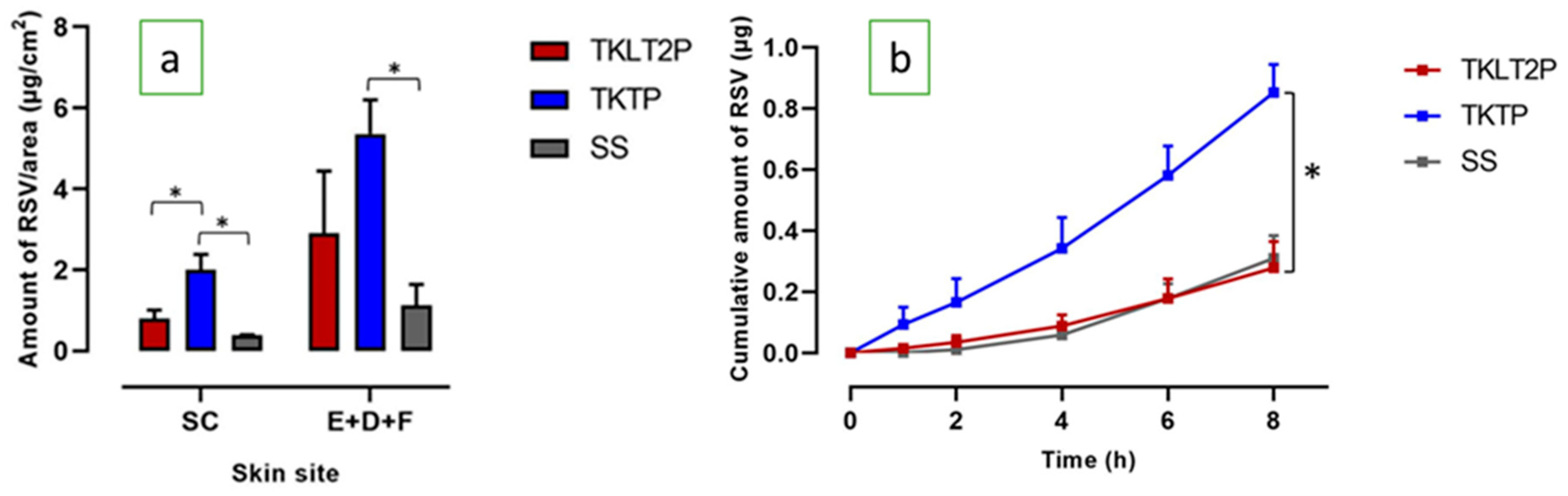
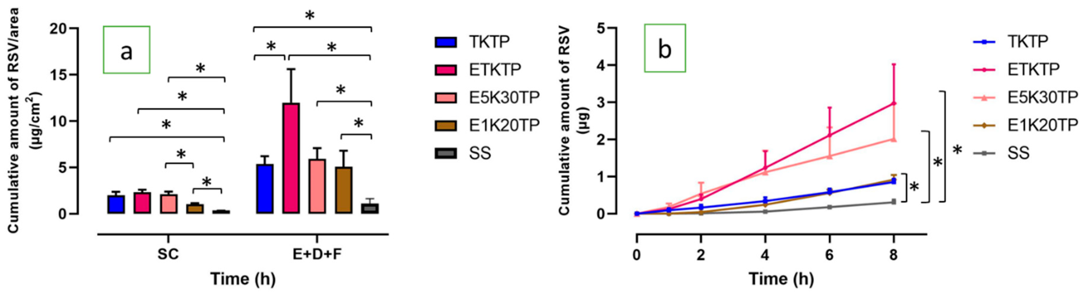
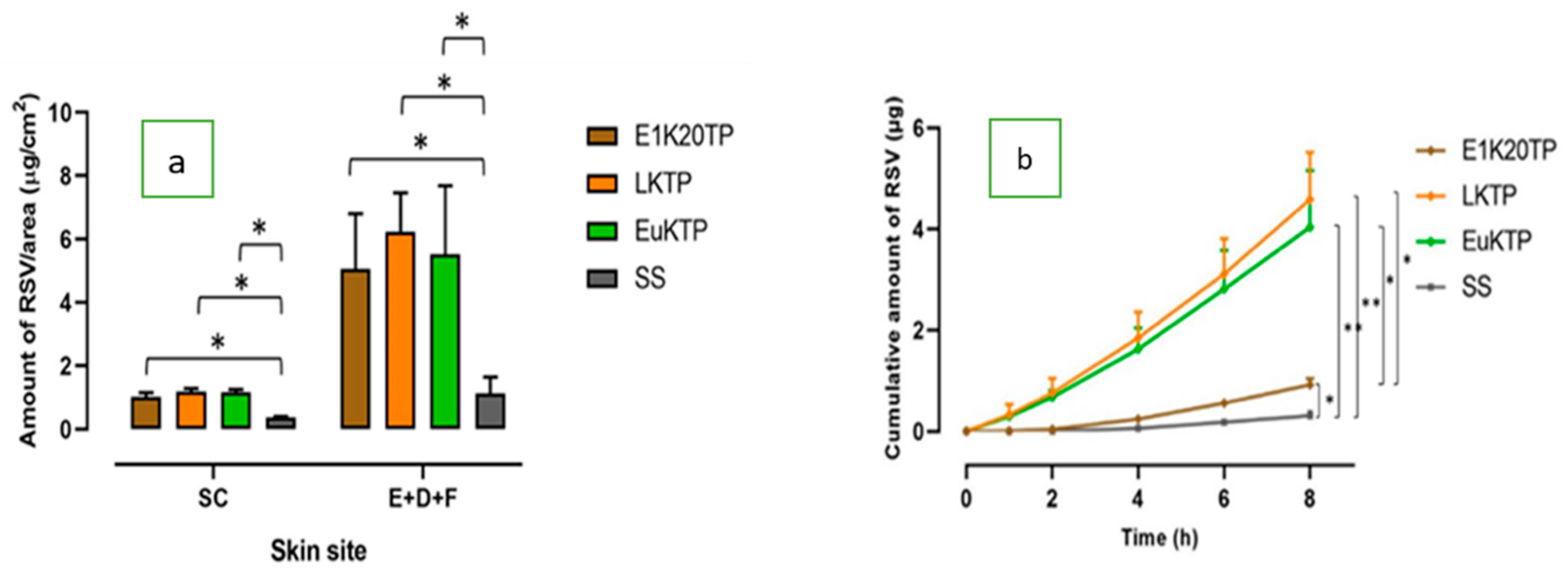
| Formula | RSV Distribution in the Skin | ERSD | |
|---|---|---|---|
| SC | E+D+F | ||
| TKLT2P | 0.805 ± 0.208 | 2.915 ± 1.523 | 2.48 |
| TKTP | 1.998 ± 0.383 | 5.359 ± 0.845 | 4.90 |
| ETKTP | 2.342 ± 0.269 | 12.000 ± 3.598 | 9.55 |
| E5K30TP | 2.104 ± 0.297 | 5.914 ± 1.169 | 5.34 |
| E1K20TP | 1.022 ± 0.129 | 5.059 ± 1.744 | 4.05 |
| LKTP | 1.190 ± 0.092 | 6.234 ± 1.231 | 4.94 |
| EuKTP | 1.172 ± 0.085 | 5.526 ± 2.160 | 4.46 |
| SS | 0.378 ± 0.025 | 1.124 ± 0.519 | 1.00 |
| Formula | Cumulative Amount (µg) | Flux (µg/cm2/h) | Lag Time (h) | ERFLX | |
|---|---|---|---|---|---|
| Steady State Flux (Jss) | Maximum Flux (Jmax) | ||||
| TKLT2P | 0.278 ± 0.086 | 0.038 ± 0.010 | 0.339 ± 0.091 | 2.330 ± 0.248 | 0.75 |
| TKTP | 0.853 ± 0.091 | 0.103 ± 0.006 | 0.227 ± 0.013 | 1.711 ± 0.605 | 2.01 |
| ETKTP | 2.973 ±1.051 | 0.358 ± 0.125 | n.a | 1.195 ± 0.280 | 6.98 |
| E5K30TP | 2.017 ± 0.954 | 0.116 ± 0.059 | n.a | 0.636 ± 0.188 | 2.27 |
| E1K20TP | 0.918 ± 0.126 | 0.142 ± 0.017 | 0.258 ± 0.029 | 2.689 ± 0.224 | 2.76 |
| LKTP | 4.585 ± 0.936 | 0.647 ± 0.103 | 1.191 ± 0.209 | 1.252 ± 0.715 | 12.61 |
| EuKTP | 4.036 ± 1.125 | 0.510 ± 0.153 | 0.920 ± 0.277 | 1.143 ± 0.164 | 9.95 |
| SS | 0.309 ± 0.074 | 0.051 ± 0.009 | 0.051 ± 0.009 | 3.185 ± 0.176 | 1.00 |
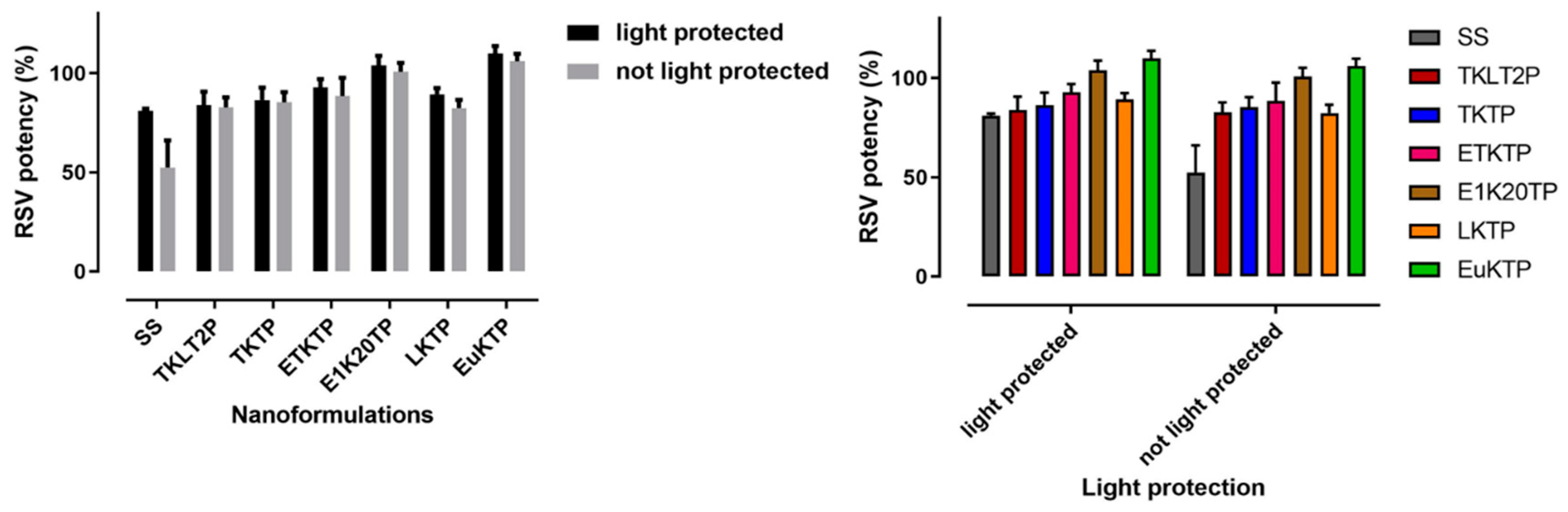
| Formula | Duration of Storage (month) | Physical Stability | Chemical Stability (%) | ||
|---|---|---|---|---|---|
| Clarity | Single Phase | Tendency of Darker Appearance | |||
| TKTP | 8 | transparent | ✓ | + | 98.73 ± 4.00 |
| ETKTP | 5 | transparent | ✓ | ++ | 89.25 ± 1.70 |
| E1K20TP | 6 | transparent | ✓ | ++ | 93.39 ± 8.17 |
| LKTP | 6 | transparent | ✓ | + | 107.02 ± 8.73 |
| EuKTP | 6 | transparent | ✓ | + | 108.41 ± 4.62 |
© 2020 by the authors. Licensee MDPI, Basel, Switzerland. This article is an open access article distributed under the terms and conditions of the Creative Commons Attribution (CC BY) license (http://creativecommons.org/licenses/by/4.0/).
Share and Cite
Nastiti, C.M.R.R.; Ponto, T.; Mohammed, Y.; Roberts, M.S.; Benson, H.A.E. Novel Nanocarriers for Targeted Topical Skin Delivery of the Antioxidant Resveratrol. Pharmaceutics 2020, 12, 108. https://doi.org/10.3390/pharmaceutics12020108
Nastiti CMRR, Ponto T, Mohammed Y, Roberts MS, Benson HAE. Novel Nanocarriers for Targeted Topical Skin Delivery of the Antioxidant Resveratrol. Pharmaceutics. 2020; 12(2):108. https://doi.org/10.3390/pharmaceutics12020108
Chicago/Turabian StyleNastiti, Christofori M. R. R., Thellie Ponto, Yousuf Mohammed, Michael S. Roberts, and Heather A. E. Benson. 2020. "Novel Nanocarriers for Targeted Topical Skin Delivery of the Antioxidant Resveratrol" Pharmaceutics 12, no. 2: 108. https://doi.org/10.3390/pharmaceutics12020108
APA StyleNastiti, C. M. R. R., Ponto, T., Mohammed, Y., Roberts, M. S., & Benson, H. A. E. (2020). Novel Nanocarriers for Targeted Topical Skin Delivery of the Antioxidant Resveratrol. Pharmaceutics, 12(2), 108. https://doi.org/10.3390/pharmaceutics12020108