1. Introduction
Powassan virus (POWV) is an emerging tick-borne orthoflavivirus endemic to the northeastern United States, Canada, and Far East Russia [
1,
2]. Disease associated with POWV infection in people initially presents as fever, headache, and vomiting, and severe disease can result from neuroinvasive infection leading to encephalitis and/or meningitis [
2]. Neuroinvasive POWV infection has a mortality rate of approximately 10%, and patients who recover from acute disease struggle with neurological sequelae in around 50% of cases [
3,
4]. POWV disease currently has no cure or treatment, and no approved vaccine exists for prevention. POWV remains a rarely diagnosed disease; however, 2025 marked the fourth consecutive year with the highest reported POWV cases at 63 reported infections worldwide [
5]. POWV is organized into two viral lineages that are serologically indistinguishable but differ in overall vector ecology, POWV lineage I (prototypic POWV) and POWV lineage II, commonly referred to as deer tick virus (DTV) [
6].
Although POWV typically presents with non-specific or neurological symptoms, a lethal case of POWV lineage II described in 2016 initially reported testicular pain [
7]. Testicular ultrasonography revealed orchiepididymitis, and postmortem analysis detected POWV antigen in the patient’s testis. The patient had been previously diagnosed with follicular lymphoma and was receiving rituximab. Due to the paucity of POWV infections and autopsy reports following death, the incidence of even one confirmed case of testicular infection is noteworthy and merits further investigation.
The potential for POWV to establish testicular infection is not without precedence; viral infection of the testes has been well documented for a variety of pathogens across different viral families [
8]. Mumps and human immunodeficiency viruses are classical examples, and filoviruses like Ebola and Marburg viruses are now recognized as testicular tropic, especially after mounting epidemiological evidence emerged from the 2014 to 2016 West African epidemic [
9,
10,
11,
12,
13]. Zika virus (ZIKV), another arthropod-borne orthoflavivirus, has been shown to infect the male genital tract, including testes [
14]. Epidemiological evidence as early as 2008 suggested the possibility of sexual transmission and, therefore, testicular infection; however, these observations remained largely unexplored until the American ZIKV epidemic almost a decade later [
15,
16]. Recently, the Oropouche virus was detected in a traveler’s semen in 2024, and the severe fever with thrombocytopenia syndrome virus has been found to infect the male reproductive tract, causing orchitis, impaired spermatogenesis, and seminal viral shedding [
17,
18]. These cases of testicular tropic viruses underscore the need for more dedicated research into viral testicular infection, particularly when incidental evidence emerges.
Based upon evidence from arboviral infections and the aforementioned clinical POWV case, we sought to investigate the testicular tropism of POWV infection. Previous literature documenting testicular infection of POWV in mice has been incidental and inconsistent. One 2020 study found in 65% of tested male mice between 11 and 55 focus forming units (FFUs) equivalents/μg RNA in animals that had succumbed to infection, and a report on POWV tick transmission in mice in 2022 reported one male mouse with between 10
5 and 10
6 FFU equivalents/ug RNA at 28 days post-infection [
19,
20]. Furthermore, testicular tropism was supported only by detection of viral RNA and limited to study endpoints. Our study seeks to fill these gaps by systematically quantifying the dynamics and potential mechanisms underlying POWV testicular tropism.
Using the previously established BALB/c murine model for POWV, we aimed to infect mice and collect testes and epididymides to quantify infectious viruses and testicular mass and observe pathologic changes. Furthermore, we sought to evaluate the role macrophages play in controlling POWV at early stages of infection as a cell type susceptible to POWV infection [
21]. As macrophages have been implicated in delivering ZIKV to the testes following a “Trojan horse”-like mechanism, we were interested in determining if POWV may follow a similar mechanism should it display a tropism for the testes [
22]. Lastly, we used a gene expression analysis to characterize the transcriptional response to infection in testes of POWV-infected mice. Collectively, these studies represent the first dedicated investigation into POWV infection of the male reproductive tract in mice, evaluate a potential mechanism of testicular infection, and characterize the testicular pro-inflammatory response during infection.
2. Materials and Methods
2.1. Cells and Viruses
Vero CCL-81 cells (American Type Culture Collection, Manassas, VA, USA) were grown in Dulbecco’s minimal essential media (DMEM, Gibco, Thermo Fisher Scientific, Waltham, MA, USA) with 10% fetal bovine serum (FBS), 200 U/mL penicillin and 200 mg/mL streptomycin (Gibco, Thermo Fisher Scientific). Human primary Sertoli cells (HSerCs) (IX Cells Biotechnologies, San Diego, CA, USA) were maintained in Sertoli cell basal media (IX Cells Biotechnologies) supplemented with 10% FBS and supplemented with Sertoli cell antibiotics and growth supplements according to the manufacturer’s recommendations (IX Cells Biotechnologies). BHKsa cells were grown in Minimal Essential Medium Alpha (1X) + GlutaMAX (Gibco, Thermo Fisher Scientific) with 10% FBS, 200 U/mL penicillin and 200 mg/mL streptomycin (Gibco, Thermo Fisher Scientific). BKHsa cells were obtained from an existing laboratory stock at the University of Texas Medical Branch (UTMB, Galveston, TX, USA), originally from Dr. Ilya Frolov. HSerCs were used within 3–5 passages after acquisition. BHKsa cells and Vero CCL-81 cells were used between 5 and 15 passages after acquisition.
ZIKV PRVABC-59, POWV Lineage I (LB), and POWV Lineage II (DTV, IPS-001 strain) were received from the World Reference Center for Emerging Viruses and Arboviruses (WRCEVA; UTMB, Galveston, TX, USA). The passage history of all stocks is as follows: ZIKV PRVABC-59 was passaged five times in Vero CCL-81 cells, POWV LB was passaged twice in LLC-MK-2 cells and once in Vero CCL-81 cells, and DTV was passaged thrice in suckling mouse brains. The LB strain was selected as the primary virus for these studies, as it is a well-characterized prototypic POWV common in murine models for POWV infection. All infectious work was conducted at BSL-3 at UTMB.
2.2. Ethics for Studies with Infectious Agents
Animal experiments involving infectious POWV and DTV were conducted at animal biosafety level 3 (ABSL3) at UTMB under IACUC protocol 2507034 and performed in accredited facilities, ensuring compliance with the guidelines of the Association for Assessment and Accreditation of Laboratory Animal Care International (AAALAC).
2.3. Virus Titration by Plaque Assays
Plaque forming units (PFUs) were determined by plaque assays on monolayers of BHKsa cells for POWV and Vero CCL-81 cells for ZIKV and DTV in 12-well plates [
23]. Briefly, samples were serially diluted and used to infect Vero CCL-81 or BHKsa monolayers. After 1 h of adsorption with gentle rocking at 37 °C, cells were overlayed with 1XMEM (Gibco, Thermo Fisher Scientific) containing 0.8% tragacanth (Sigma Aldrich, St. Louis, MO, USA). Plates were incubated at 37 °C with 5% CO
2 for approximately 4 days for ZIKV, 3 days for POWV, or 6 days for DTV. Monolayers were fixed using a 10% formaldehyde solution. The cells were treated with a solution of 0.2% crystal violet in 30% methanol to visualize the plaques. Data are shown as log
10 PFU/mL, with the limit of detection (LOD) for each assay indicated on each graph by a dotted line.
2.4. Viral Growth Curve
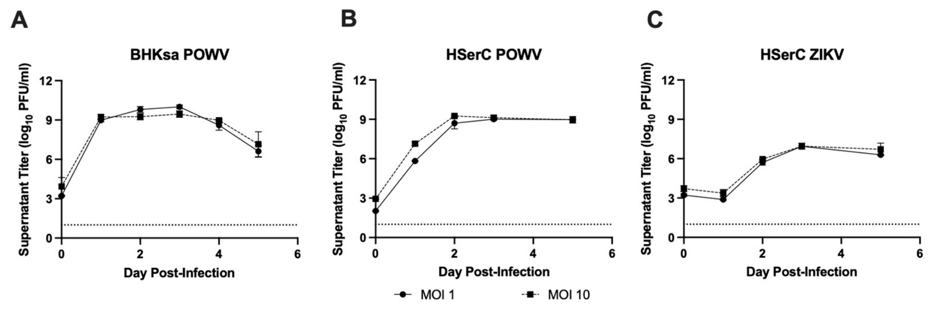
Vero CCL-81 and BKHsa cells were seeded in 12-well plates and allowed to reach 80–90% confluency prior to infecting. Cells were infected with ZIKV or POWV at various multiplicities of infection (MOI) in a total infection volume of 100 μL and incubated at 37 °C with 5% CO2 for 60 min. Cells were rocked every 15 min to prevent cells from drying, ensuring the entire monolayer was exposed to the inoculum. After infection, viral inoculum was removed, and each well was washed with 1 mL of 1X PBS three times before 1 mL of cell growth medium was added to each well. At a consistent time each day post-infection (dpi), 100 μL of supernatant from each well was removed for freezing at −80 °C for later titration and replaced with 100 μL of cell growth media. Cell monolayers were visually assessed for signs of cytopathic effect such as cell rounding, cell detachment, formation of syncytia, and cell lysis using a phase-contrast light microscope.
2.5. Animal Manipulations
Male 9–10-week-old BALB/c mice were obtained from Charles River (Charles River Laboratories, Bar Harbor, ME, USA). Groups of 5 mice were infected by the intraperitoneal (IP) or intradermal (ID) route with a target dose of 2 log10 PFU of POWV or DTV. Viral stocks were diluted 1:500 in 0.9% sodium chloride (Hospira, Lake Forest, IL, USA) to target inoculum. Uninfected control mice (n = 3 per group per study) were injected with sterile 0.9% sodium chloride via the IP or ID route. After infection, mice were monitored and weighed at least once daily. Blood was collected at 1-, 2-, and 3-days post-infection by the retroorbital route. Animals were anesthetized with isoflurane for each manipulation.
Organs were collected in pre-weighed tubes to determine organ mass. Tubes were homogenized at a frequency of 30 Hz for 2 min prior to centrifugation at 6000× g for 5 min. Supernatants were collected, and titers were determined as described above. Data are shown as log10 PFU/gram of tissue.
For macrophage depletion studies, mice received IP injections containing clodronate-containing or control liposomes (Liposoma, Amsterdam, The Netherlands) at 100 μL reagent/10 g mouse weight 48 and 24 h prior to infection, according to the manufacturer’s recommendations. When required, perfusion was performed with PBS following euthanasia. Perfusions were performed for mice infected with DTV and in clodronate-treated and control liposome-treated mice. These mice were perfused since we anticipated viremia before 5 dpi as well as during phagocytic cell depletion. POWV-infected mice at 3 dpi were also perfused. POWV-infected mice at 5 dpi were nonperfused.
2.6. Flow Cytometry
Macrophage populations in mouse spleen and testes were identified and quantified by flow cytometry. Splenic macrophage populations were assessed as an indicator of systemic phagocytic-cell depletion at the time of viral challenge. Individual organs were macerated through 70 µm cell strainers and digested with 0.05% collagenase type IV (Thermo Fisher Scientific) in Roswell Park Memorial Institute medium 1640 (RPMI 1640) for 30 min at 37 °C and again passed through 70 µm cell strainers to prepare a single-cell suspension. Red blood cells were lysed through a 3 s wash in pure molecular grade, followed by the addition of 10X PBS to restore isotonic conditions. Live/Dead antibody (Zombie NIR Dye, Biolegend, San Diego, CA, USA) was incubated at 1:5000 at room temperature for 15 min. Cells were then washed in buffer (containing 2% FBS in sterile 1X PBS). The fluorochrome-labeled antibodies used were purchased from Thermo Fisher Scientific, Tonbo Biosciences (San Diego, CA, USA), or Biolegend as described below: violetFluor 450 anti-CD8 (Clone 53-6.7), PerCP-Cy5.5 anti-CD4 (Clone RM4-5), FITC anti-CD4 (Clone GK1.5), PerCP-Cy5.5 anti-CD11b (Clone M1/70), BV421 anti-F4/80 (Clone T45-2342), and APC anti-Ly6G (Clone 1A8). Cells were fixed in 2% paraformaldehyde for 16 h at 4 °C. Cell samples were processed on a BD LSR Fortessa (BD Biosciences, San Jose, CA, USA) and data were analyzed using FlowJo 10.
2.7. Histology
Tissue samples were fixed in a solution of 10% neutral buffered formalin (Thermo Fisher Scientific, Waltham, MA, USA) for 24 h, then exchanged with new formalin and fixed for 31 days. Formalin solution was replaced with 95% ethanol and sent to UTMB’s Anatomic Pathology Laboratory core facility, where organ samples were embedded in paraffin and cut into 5 µm sections. Representative slides from each animal were stained with hematoxylin and eosin (H&E) or left unstained for use in immunofluorescence analysis. A blinded board-certified pathologist interpreted the H&E slides. Images were captured using the brightfield channel of an inverted fluorescence microscope (Olympus-IX73; Olympus corporation, Tokyo, Japan) and Olympus cellSense Standard 2.3 software.
2.8. Immunofluorescence Assay
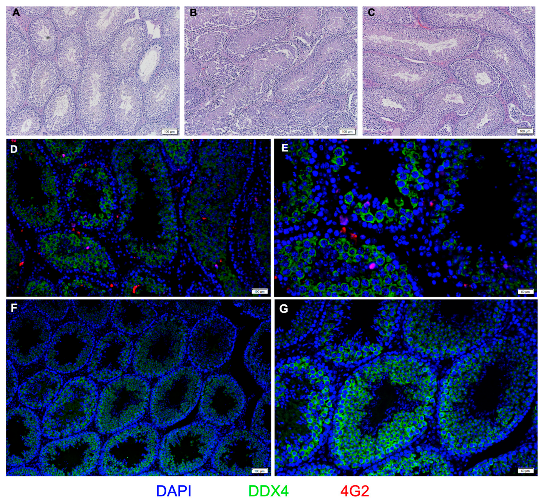
Slides were prepared for immunofluorescence by heating 5 µm sections of paraffin-embedded mouse testes at 65 °C overnight for dewaxing, followed by incubations in multiple xylene baths and a series of graded ethanol (Millipore, Sigma, Burmington, MA, USA). Antigen retrieval was performed through a 30 min incubation in 90 °C citrate buffer pH 6 (Abcam, Cambridge, UK) diluted in PBS (Gibco, ThermoFisher Scientific). Sections were washed in deionized water and incubated in a blocking buffer (5% normal goat serum in PBS) for 15 min at 37 °C in a humidified chamber. The primary antibody was incubated for 16 h in a humidified chamber at 4 °C with the 4G2 antibody targeting the flavivirus envelope protein (Abcam) at a 1:200 dilution and the DDX4 antibody targeting migratory germ cells (Abcam) at a 1:500 dilution in blocking buffer. Samples were washed in PBS and incubated with 0.2% Sudan black B (SigmaAldrich) diluted in 70% ethanol for 12 min at 37 °C in a humidified chamber. Samples were then stained for 45 min at 37 °C with a secondary antibody in a humidified chamber at a dilution of 1:1000, AlexaFluor 594 anti-mouse IgG (Thermo Fisher Scientific) or AlexaFluor 488 anti-rabbit IgG (ThermoFisher Scientific). The slides were incubated with DAPI for 5 min at room temperature, followed by five PBS washes prior to mounting with Vectashield (Vector Labs, Newark, CA, USA). Images were captured using an inverted fluorescence microscope (Olympus-IX73) and Olympus cellSense Standard 2.3 software.
2.9. Gene Expression Analysis
RNA from mouse testes was extracted using phenol–chloroform extraction. Clarified supernatant from organ homogenate inactivated in TRIzol (Thermo Fisher Scientific) was incubated with 1 part chloroform (SigmaAldrich) to 4 parts sample and incubated for 3 min. Tubes were centrifuged at 12,000× g for 15 min, and the upper aqueous phase was removed to 0.5 mL isopropanol (SigmaAldrich) for 10 min incubation. Samples were centrifuged for 20 min at 20,000× g, and the supernatant was replaced with 75% ethanol (SigmaAldrich). Samples were shaken and re-centrifuged. The supernatant was discarded, and RNA was eluted in RNase-free molecular-grade water (Invitrogen, Waltham, MA, USA). All samples were diluted to 30 ng/µL prior to hybridization. RNA quality and concentration were confirmed using a Nanodrop spectrophotometer (DeNovix DS-11+ Spectrophotometer; DeNovix Inc., Wilmington, DE, USA). Gene expression analysis was performed using a NanoString nCounter SPRINT profiler using NCounter Mouse Myeloid V2 and NCounter Mouse Immunology V2 panels (Bruker Spatial Biology, Bothell, WA, USA). Analysis was performed using the Mouse Myeloid panel supplemented with non-overlapping genes from the Mouse Immunology panel. Data analysis was performed using nSolver (nSolver Analysis Software version 4.0) and Prism 11 (GraphPad Prism version 11.0.0) software. Data normalization was performed on raw gene counts by the nSolver software using housekeeping genes and positive controls.
2.10. Quantification and Statistical Analysis
For viral organ load and viremia, data were compared using a one-way ANOVA when comparing multiple groups or an unpaired Student’s
t-test when only comparing two groups. Viral titers were log
10 transformed before statistical tests were applied. Volcano plots were generated using multiple unpaired Welch’s
t-tests with log
2 transformed gene counts. Left and right testicular masses were averaged before statistical analysis. Statistical significance for gene fold changes in
Table 1 was calculated using multiple unpaired t-tests on normalized values on Graphpad Prism. Statistical analyses were done using R version 4.6.0 and Prism 11 software (Graphpad Prism version 11.0.0); a
p-value lower than 0.05 was considered significant.
4. Discussion
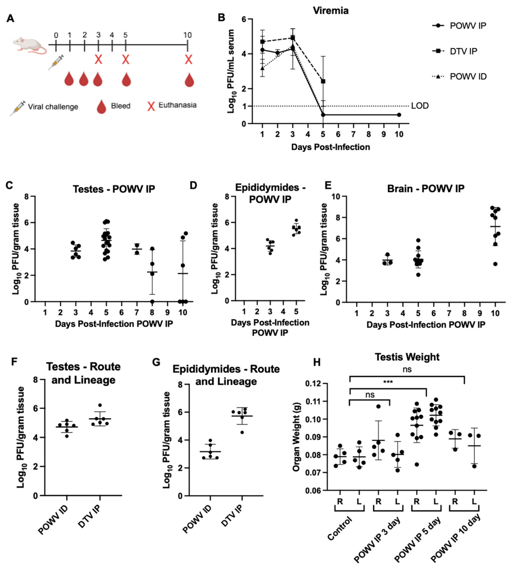
In this study, we report that POWV consistently infects the male reproductive tract of mice during the acute phase of infection. Infectious viruses were recovered from each analyzed testis and epididymis collected at 3 and 5 dpi regardless of viral lineage or challenge route. Immunofluorescence imaging revealed POWV envelope protein localized within the seminiferous tubules, implying that POWV may be capable of breaching the blood–testis barrier (BTB). Notably, despite the high testicular titer and presence of viral antigen, there was a lack of observed acute histopathological events. In some mouse models, especially at post-acute timepoints, ZIKV infection of mice can be associated with severe pathological damage, including tubular necrosis and impaired spermatogenesis [
14,
24]. The absence of overt pathology and lack of disrupted testicular architecture may be a result of the relatively early timepoints of our study, and further analysis is required to determine if testicular damage may be an outcome beyond our study’s scope of testicular POWV infection.
POWV is primarily recognized as a neuroinvasive virus, and its tropism for the central nervous system has been well supported by clinical and animal model data. A 2016 clinical case of a 63-year-old man who succumbed to POWV disease challenged the current understanding of disease progression by initially presenting as testicular pain [
7]. Postmortem analysis found extensive neuropathological findings; however, the postmortem presence of POWV antigen in the patient’s seminiferous tubules provided the first evidence of potential testicular infection by this virus. The patient was taking rituximab antibody therapy, which obscures whether the presence of testicular viral antigen was evidence of a previously unknown tropism or a rare disease outcome as a consequence of antibody therapy. Rituximab depletes B cell populations, which can impair humoral immunity response to viral infections, leading to atypical outcomes of viral disease [
26]. Impaired humoral immunity can alter viral disease severity and result in altered viral tropism, including prolonged enteroviral meningoencephalitis, severe neuroinvasive West Nile virus, and prolonged SARS-CoV-2 infection [
26]. Despite this, the confirmation of even one case of testicular infection, albeit suspect, warrants further study given the paucity of human cases.
Our study reports POWV testicular infection in mice between 3 and 10 dpi. A noteworthy feature of viral testicular infections is the potential to establish a persistent infection, similar to what is reported in ZIKV and EBOV infections [
14,
27,
28]. Our study reported 3/6 testes in 2/3 mice positive for POWV infection at 10 dpi, without insight into the viral dynamics that may have resulted at later timepoints. Raney et al. reported high testicular titer at 28 dpi in 1/5 mice infected with POWV lineage II, suggesting that long-term testicular POWV infection in mice is a potential outcome [
19].
Consistent with the clinical outcome of orchiepididymitis in the previously discussed patient, POWV-infected mice in our study were associated with increased testicular mass at 5 dpi. This finding is contrasted with ZIKV infection, which causes testicular atrophy and reduced testicular mass in murine models prior to 10 dpi [
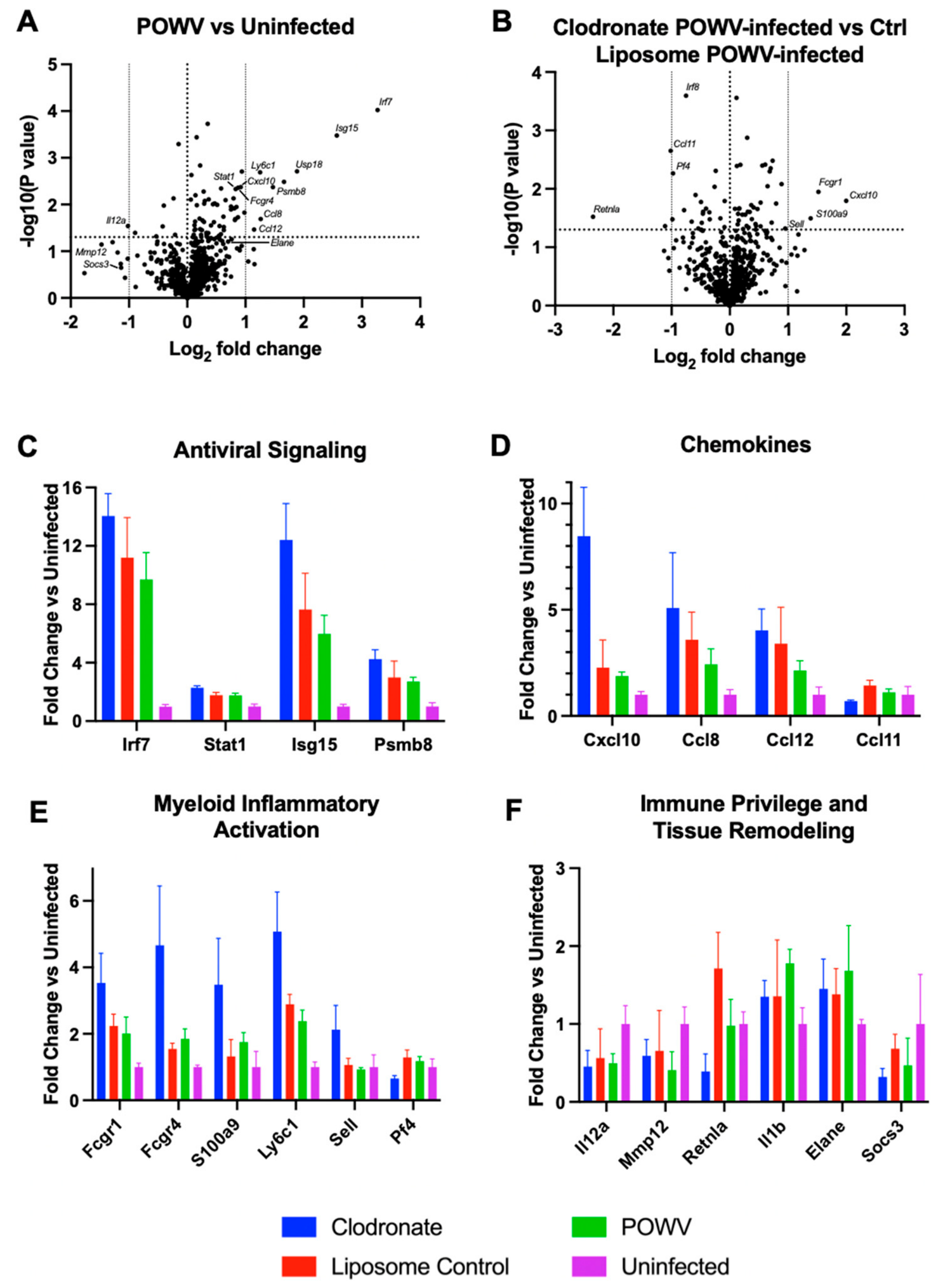
29]. ZIKV mouse models are typically interferon signaling impaired, which can exacerbate disease outcomes, preventing a direct comparison from our immunocompetent mouse model. These findings suggest infected mice are experiencing testicular edema and swelling due to a pro-inflammatory response at the testes during POWV infection. This phenotype is consistent with our gene expression analysis results that confirm a robust antiviral and pro-inflammatory host response at the testes.
Due to macrophages implicated in driving ZIKV testicular infection in mice through a “Trojan horse”-like mechanism, we sought to determine if POWV uses a similar mechanism [
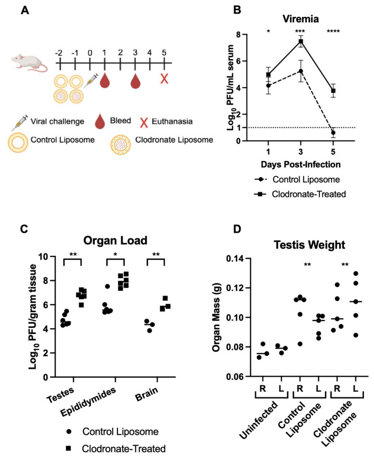
22]. Interestingly, we found that after phagocytic-cell depletion, POWV viral load in the testes was significantly increased, indicating that POWV most likely reaches the testes independent of infected macrophages. In addition, depletion of phagocytic cells was associated with significantly higher and sustained POWV viremia. Phagocytic cells play key roles in mediating both pro- and anti-inflammatory pathways, especially at the testis, where their anti-inflammatory phenotype contributes to maintaining immune privilege [
30]. Our study reflects this dynamic with shifting transcriptional levels of M1 and M2-associated genes present in the experimental conditions evaluated (
Table 1). Notably, when phagocytic cells were depleted in the testes, the anti-inflammatory M2-associated gene
Retnla was significantly downregulated. The loss of this M2-associated gene and the upregulation of M1-associated genes during phagocytic-cell depletion reflect the baseline anti-inflammatory environment of the testes. Upregulation of M1-associated genes during infection reflects the induction of a pro-inflammatory response at the testis. Our data suggest that phagocytic cells play a protective role in clearing viremia and controlling testicular POWV infection. This finding challenged expectations, as monocyte-derived cells are often implicated in driving arbovirus dissemination.
Gene expression analysis of perfused testes harvested at 5 dpi indicated a strong antiviral and inflammatory response associated with POWV infection. Interferon-stimulated genes, antigen presentation pathways, chemokine signaling, and myeloid activation markers were consistently upregulated transcriptionally. Among the highest upregulated genes,
Irf7 and
Cxcl10 represent pro-inflammatory transcriptional signals in the testes, and
Fcgr1,
Fcgr4,
Ctss, and
Ccl8 transcriptional upregulations imply recruitment and activation of innate immune cell populations. These indicate a strong pro-inflammatory response induced in the testes, an immune-privileged space, likely contributing to the observed increase in testicular mass despite overt signs of inflammation in the H&E-stained histology slides. In addition to transcripts associated with classical pro-inflammatory pathways, genes responsible for epithelial barrier integrity and immune privilege regulation are of particular interest in the testes. Downregulation of
Socs3, an anti-inflammatory gene linked to immunoregulatory signaling in the testes, could indicate potentially impaired immune privilege maintenance [
31]. These factors could contribute to disruption of the delicate immune homeostasis within the immune-privileged environment of the testes. Upregulation of
Il1b may imply BTB involvement, as
Il1b is associated with epithelial tissue disruption, and interleukin-1 family proteins regulate BTB restructuring [
32,
33]. Furthermore,
Elane, another upregulated cytokine, is associated with disruption of epithelial barriers; relationships between the BTB and this cytokine have yet to be directly evaluated, but
Elane-mediated disruption of other epithelial barriers is through cleavage of the same tight-junction proteins that contribute to BTB strength [
34,
35]. The observed changes in
Socs3,
Il1b, and
Elane expression suggest that pathways associated with testicular immune homeostasis and epithelial barrier regulation may be altered during POWV infection; however, the present data do not directly demonstrate disruption of the BTB.
As splenic phagocytic-cell depletion was confirmed in a separate control cohort (
Supplemental Figure S2), the increased and prolonged viremia observed was most likely due to an impaired innate immune response. The high testicular organ viral load and transcriptional response are most likely a downstream response to the robust early viral replication due to the loss of a typical antiviral response, similar to what has been observed with West Nile virus infection of macrophage-depleted mice [
36]. Infected testes at 5 dpi in clodronate-treated mice therefore show potential compensatory induction of
Cxcl10,
Ccl2,
Ccl5, and
Fcgr4 upregulated compared to liposome control-treated mice. These reflect the host response to an increased viral burden. Due to macrophage depletion only being confirmed at day 0, our data do not evaluate the level of macrophage depletion in the testes at 5 dpi, and transcriptomic data show myeloid-associated genes in testes at 5 dpi. Previous depletion studies show that recovery of testicular macrophages can take over 30 days after depletion [
37]. These transcriptional findings are consistent with enhanced immune activation in the testes following phagocytic-cell depletion and parallel the increased viremia and higher testicular viral burden observed in clodronate-treated animals.
Though infection of testes and epididymides was consistently observed in mice, the translatability of our findings to clinical cases remains unclear. Currently, there have been no reported cases of POWV resulting from human-to-human transmission, and sexual transmission would represent a major departure from the current understanding of POWV epidemiology. We found consistently high testicular POWV viral load in testes at early timepoints prior to presentation of clinical disease manifestations in mice.
We report consistent testicular infection in mice by POWV; however, the potential for POWV to spread sexually requires many unexplored biological events such as shedding in the seminal fluid and a susceptible recipient host. In many cases, viral infection of testes does not correspond to substantial shedding in seminal fluid. For ZIKV, testicular infection is associated with viral shedding in semen, resulting in potential sexual transmission. Spondweni virus, an orthoflavivirus closely related to ZIKV, reaches similar viral organ loads in the male reproductive tract to ZIKV. Unlike ZIKV, Spondweni virus shedding in semen is a significantly rarer event [
38]. Japanese encephalitis virus (JEV) is detected in testes and semen of pigs and boars; however, this does not translate to humans, as sexual transmission is not considered a viable route of transmission for JEV [
39]. Ultimately, whether POWV sheds in the semen, is capable of transmitting sexually, or if testicular infection is a regular outcome of human disease is unknown and requires further analysis to confirm.
Several limitations should be considered when interpreting these findings. Here, we measured the effects of two strains and two injection sites; further studies are required for a more robust analysis of the effects of virus strain and injection method, including by tick bite, if any. Current studies noting testicular infection were conducted by tick-bite-delivered DTV, and future studies will be focused on analyzing DTV in our model [
19,
20]. Although clodronate treatment resulted in robust systemic phagocytic-cell depletion, depletion of resident testicular macrophages was not demonstrated, limiting conclusions regarding their specific role during infection. Furthermore, the increased viral burden in clodronate-treated animals prevents transcriptional differences between treatment groups from being specifically attributed to phagocytic-cell depletion. Finally, the relatively acute timepoints examined and the absence of seminal-fluid analysis preclude conclusions regarding persistence or potential sexual transmission.
In conclusion, this study establishes the male reproductive tract as a site of acute POWV infection in an immunocompetent mouse model and demonstrates productive infection of the testes and epididymides during acute infection. Our findings further indicate that systemic phagocytic cells contribute to early control of POWV infection rather than being required for viral dissemination to the testes, as their depletion resulted in increased systemic and testicular viral burden. Testicular infection was accompanied by a robust antiviral and inflammatory transcriptional response, which was further enhanced under conditions of phagocytic-cell depletion and increased viral burden. Together, these findings provide a foundation for understanding POWV infection of the male reproductive tract and highlight the need to determine the potential of persistence, pathological consequences, and clinical relevance of testicular POWV infection.




 and
and