Trends in Tick-Borne Virus Ecology: Vector Incrimination, Focal Distribution, and Reservoir Associations

Abstract
1. Role of Ticks as Vectors for Viral Disease
1.1. Tick Natural Histories
1.2. Virus Transmission in Ticks
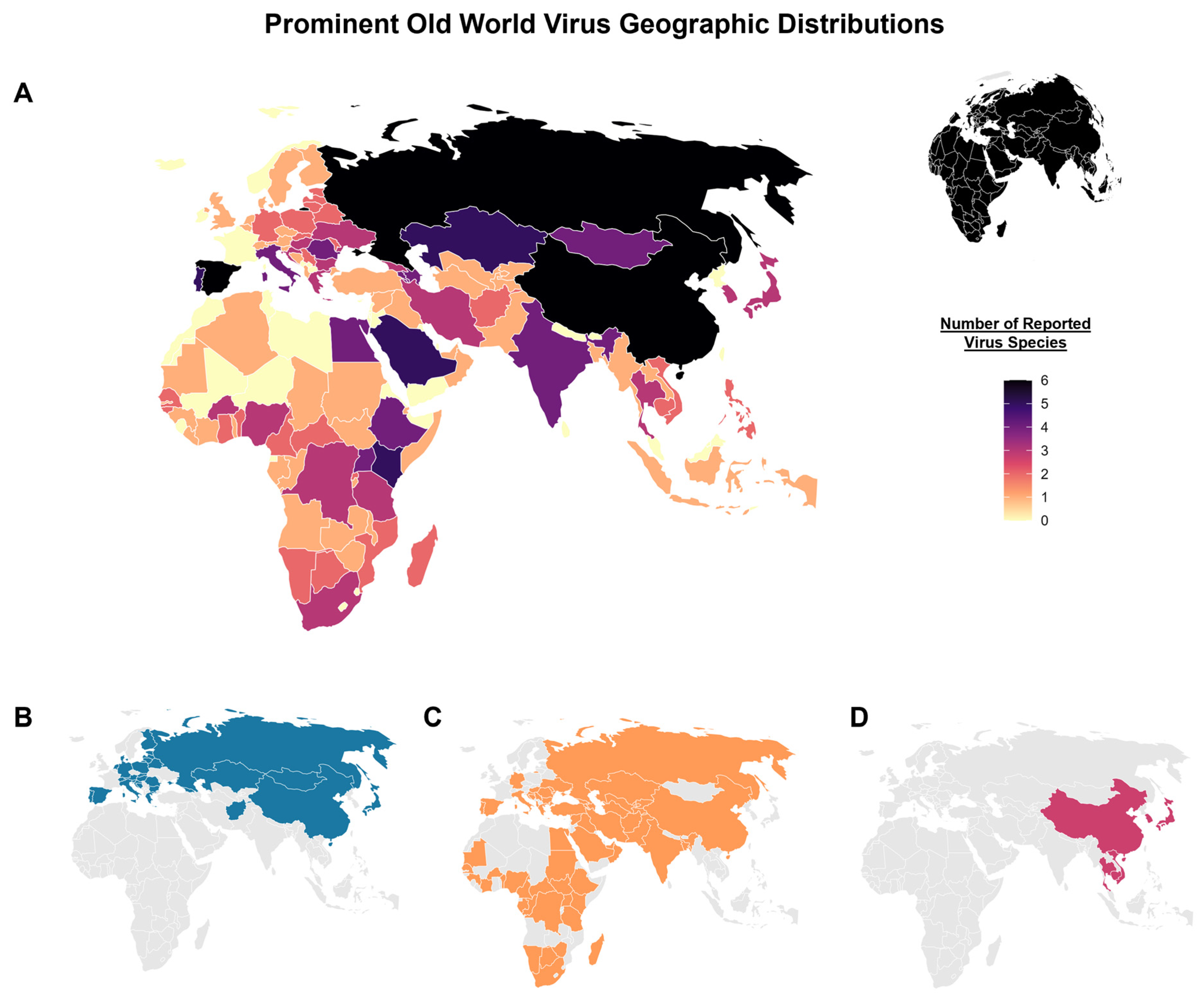
2. Tick-Borne Viruses of the Old World
2.1. Tick-Borne Encephalitis Virus
2.2. Crimean–Congo Hemorrhagic Fever Virus
2.3. Severe Fever with Thrombocytopenia Syndrome Virus
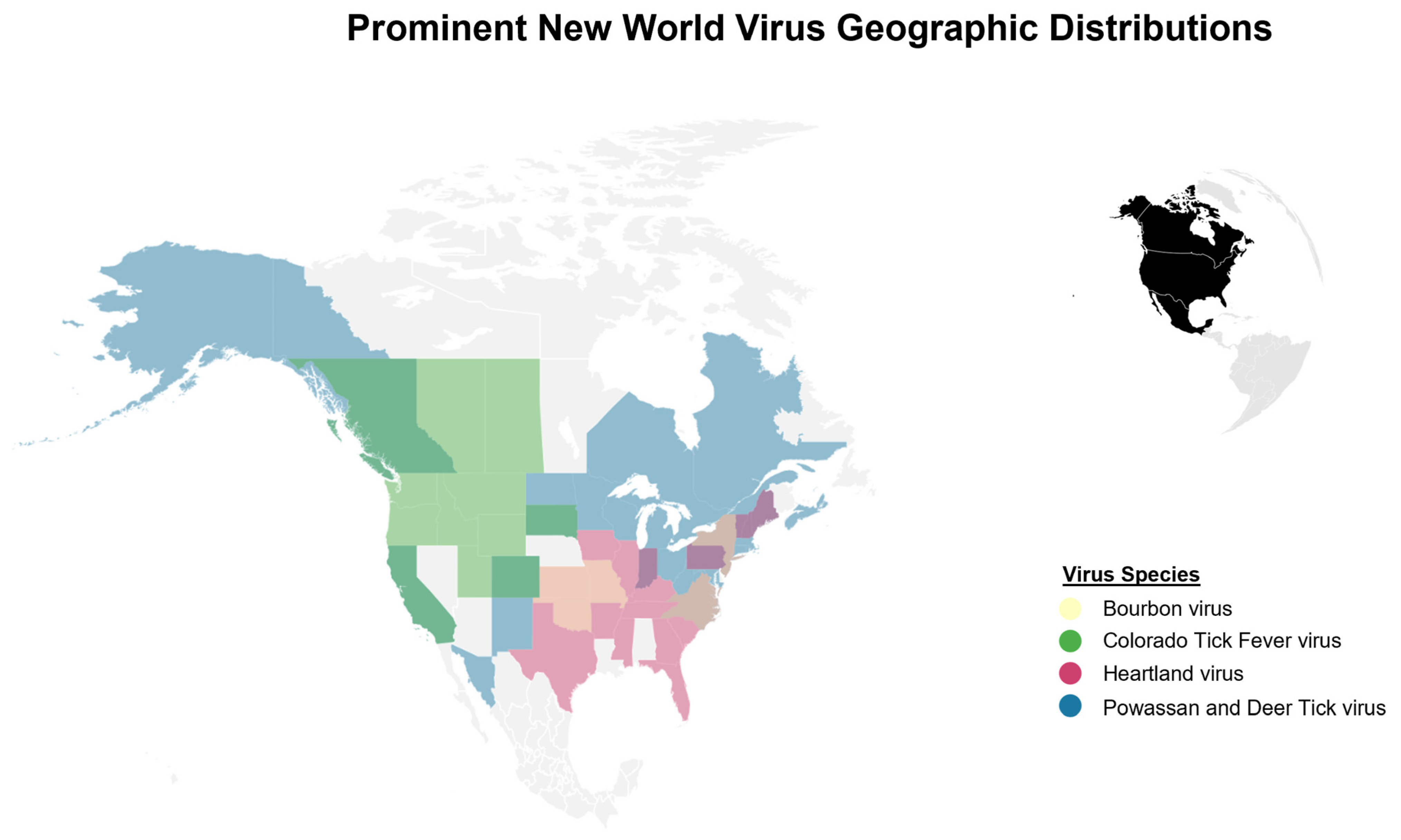
3. Tick-Borne Viruses of the New World
3.1. Colorado Tick Fever Virus
3.2. Powassan and Deer Tick Virus
3.3. Heartland and Bourbon Viruses
4. Tick-Borne Viruses of Unknown Pathogenic Potential in the New World
5. Conclusions and Future Priorities
Author Contributions
Funding
Data Availability Statement
Acknowledgments
Conflicts of Interest
Abbreviations
| BRBV | Bourbon virus |
| CCHFV | Crimean–Congo Hemorrhagic Fever virus |
| CTFV | Colorado Tick Fever virus |
| DTV | Deer Tick virus |
| HRTV | Heartland virus |
| POWV | Powassan virus |
| SFTSV | Severe Fever with Thrombocytopenia Syndrome virus |
| TBEV | Tick-borne Encephalitis virus |
References
- Theiler, M.; Downs, W.G. The Arthropod Borne Viruses of Vertebrates. An Account of the Rockefeller Foundation Virus Program, 1951–1970; Yale University Press: New Haven, CT, USA, 1973; 578p. [Google Scholar]
- Andersen, S.R. The Eye and Its Diseases in Ancient Egypt. Acta Ophthalmol. Scand. 1997, 75, 338–344. [Google Scholar] [CrossRef] [Scilit] [PubMed]
- Calisher, C.H. A Very Brief History of Arbovirology, Focusing on Contributions by Workers of the Rockefeller Foundation. Vector-Borne Zoonotic Dis. 2005, 5, 202–211. [Google Scholar] [CrossRef] [Scilit] [PubMed]
- Smith, T.; Kilbourne, F.L. Investigations into the Nature Causation and Prevention of Texas or Southern Cattle Fever. In Ninth Annual Report of the Bureau of Animal Industry for the Year 1892; Oxford University Press: Oxford, UK, 1893. [Google Scholar]
- Dutton, J.E.; Todd, J.L. The Nature of Tick Fever in the Eastern Part of The Congo Free State. With Notes on the Distribution and Bionomics of the Tick. Br. Med. J. 1905, 1259–1260.
- Theiler, A.; Stockman, S. Some Observations and Experiments in Connection with Tropical Bovine Piroplasmosis (East Coast Fever or Rhodesian Redwater). J. Comp. Pathol. Ther. 1904, 17, 193–203. [Google Scholar] [CrossRef] [Scilit]
- Ricketts, H.T. The Transmission of Rocky Mountain Spotted Fever by the Bite of the Wood-Tick (Dermacentor Occidentalis). J. Am. Med. Assoc. 1906, 47, 358. [Google Scholar] [CrossRef] [Scilit]
- Steere, A.C.; Malawista, S.E. Cases of Lyme Disease in the United States: Locations Correlated with Distribution of Ixodes Dammini. Ann. Intern. Med. 1979, 91, 730–733. [Google Scholar] [CrossRef] [Scilit] [PubMed]
- Anderson, B.E.; Dawson, J.E.; Jones, D.C.; Wilson, K.H. Ehrlichia Chaffeensis, a New Species Associated with Human Ehrlichiosis. J. Clin. Microbiol. 1991, 29, 2838–2842. [Google Scholar] [CrossRef] [Scilit] [PubMed]
- Zlobin, V.I.; Pogodina, V.V.; Kahl, O. A Brief History of the Discovery of Tick-Borne Encephalitis Virus in the Late 1930s (Based on Reminiscences of Members of the Expeditions, Their Colleagues, and Relatives). Ticks Tick Borne Dis. 2017, 8, 813–820. [Google Scholar] [CrossRef] [Scilit] [PubMed]
- Chumakov, M.P.; Naidenova, G.A. Search for Ultra-Viral Encephalitis. Report V. Tick Ixodes Ricinus as Vector for Tick-Borne (Spring-Summer) Encephalitis. Meditsinskaya Parazitol. I Parazit. Bolezn. 1944, 89–93. [Google Scholar]
- Florio, L.; Miller, M.S.; Mugrage, E.R. Colorado Tick Fever—Isolation of the Virus from Dermacentor-Andersoni in Nature and a Laboratory Study of the Transmission of the Virus in the Tick. J. Immunol. 1950, 64, 257–263. [Google Scholar] [PubMed]
- Mclean, D.M.; Larke, R.P. Powassan and Silverwater Viruses: Ecology of Two Ontario Arboviruses. Can. Med. Assoc. J. 1963, 88, 182. [Google Scholar] [PubMed]
- McLean, D.M.; Best, J.M.; Mahalingam, S.; Chernesky, M.A. Powassan Virus: Summer Infection Cycle, 1964. Can. Med. Assoc. J. 1964, 91, 1360–1362. [Google Scholar] [PubMed]
- Sonenshine, D.E. The Biology of Tick Vectors of Human Disease. In Tick-Borne Diseases of Humans; ASM Press: Washington, DC, USA, 2005; pp. 12–36. [Google Scholar]
- Estrada-Peña, A.; Mihalca, A.D.; Petney, T.N. Ticks of Europe and North Africa: A Guide to Species Identification; Springer: Berlin/Heidelberg, Germany, 2018. [Google Scholar]
- Giribet, G.; Edgecombe, G.D. The Phylogeny and Evolutionary History of Arthropods. Curr. Biol. 2019, 29, 592–602. [Google Scholar] [CrossRef] [Scilit] [PubMed]
- Reeves, W.C. Ecology of Mosquitoes in Relation to Arboviruses. Annu. Rev. Entomol. 1965, 10, 25–46. [Google Scholar] [CrossRef] [Scilit]
- Apanaskevich, D.; Oliver, J. Biology of Ticks. Infect. Dis. Clin. N. Am. 2008, 22, 195–215. [Google Scholar] [CrossRef] [Scilit] [PubMed]
- Dantas-Torres, F.; Mans, B.J. Ticks (Ixodida: Argasidae, Ixodidae, Khimairidae, and Nuttalliellidae) of the World. Parasit. Vectors 2026, 19, 256. [Google Scholar] [CrossRef] [Scilit] [PubMed]
- Apanskevich, D.A.; Oliver, J.H., Jr. Life Cycles and Natural History of Ticks. In Biology of Ticks; Oxford University Press: Oxford, UK, 2014; Volume 1. [Google Scholar]
- Mather, T.N.; Ginsberg, H.S. Vector-Host-Pathogen Relationships: Transmission Dynamics of Tick-Borne Infections. In Ecological Dynamics of Tick-Borne Zoonoses; Oxford University Press: Oxford, UK, 2023. [Google Scholar]
- Gray, J.S.; Estrada-Peña, A.; Vial, L. Ecology of Nidicolous Ticks. Biol. Ticks 2013, 2, 39–60. [Google Scholar]
- Randolph, S.; Sonenshine, D.; Roe, R. Ecology of Non-Nidicolous Ticks. In Biology of Ticks, 2nd ed.; Oxford University Press: Oxford, UK, 2014; Volume 2. [Google Scholar]
- Nuttall, P. Mechanisms of Tick-Borne Virus Transmission. In History of Arbovirology: Memories from the Field; Springer: Berlin/Heidelberg, Germany, 2023; pp. 43–65. [Google Scholar]
- Mlera, L.; Bloom, M.E. The Role of Mammalian Reservoir Hosts in Tick-Borne Flavivirus Biology. Front. Cell. Infect. Microbiol. 2018, 8, 298. [Google Scholar] [CrossRef] [Scilit] [PubMed]
- Wilson, M. Population Ecology of the Tick Vectors: Interaction, Measurement, and Analysis. In Ecological Dynamics of Tick-Borne Zoonoses; Oxford University Press: New York, NY, USA, 2023; pp. 20–38. [Google Scholar]
- Jones, L.D.; Davies, C.R.; Green, B.M.; Nuttall, P.A. Reassortment of Thogoto Virus (a Tick-Borne Influenza-like Virus) in a Vertebrate Host. J. Gen. Virol. 1987, 68, 1299–1306. [Google Scholar] [CrossRef] [Scilit] [PubMed]
- Nuttall, P.A.; Labuda, M. Dynamics of Infection in Tick Vectors and at the Tick-Host Interface. Adv. Virus Res. 2003, 60, 233–272. [Google Scholar] [CrossRef] [Scilit] [PubMed]
- Randolph, S.E.; Gern, L.; Nuttall, P.A. Co-Feeding Ticks: Epidemiological Significance for Tick-Borne Pathogen Transmission. Parasitol. Today 1996, 12, 472–479. [Google Scholar] [CrossRef] [Scilit] [PubMed]
- Panov, A.G. Seasonal Sommer Encephalitis. Vladivostok 1940, 52, 1981–1997. [Google Scholar] [CrossRef] [Scilit]
- Schneider, H. Die Epidemische Akute “Meningitis Serosa”; Wilheim Maudrich: Vienna, Austria, 1932. [Google Scholar]
- Schneider, H. Über Epidemische Akute “Meningitis Serosa”. Wien. Klin. Wochenschr. 1931, 44, 350–352. [Google Scholar]
- Dobler, G.; Gniel, D. A History of Tick-Borne Encephalitis and Its Virus. In History of Arbovirology: Memories from the Field; Springer: Berlin/Heidelberg, Germany, 2023; pp. 453–467. [Google Scholar]
- Labuda, M.; Kozuch, O.; Zuffová, E.; Elecková, E.; Hails, R.S.; Nuttall, P.A. Tick-Borne Encephalitis Virus Transmission between Ticks Cofeeding on Specific Immune Natural Rodent Hosts. Virology 1997, 235, 138–143. [Google Scholar] [CrossRef] [Scilit] [PubMed]
- Pavlovsky, E.N.; Plous, F.K.; Levine, N.D. Natural Nidality of Transmissible Diseases. Am. J. Med. Sci. 1966, 252, 913. [Google Scholar] [CrossRef] [Scilit]
- Deviatkin, A.A.; Karganova, G.G.; Vakulenko, Y.A.; Lukashev, A.N. Tbev Subtyping in Terms of Genetic Distance. Viruses 2020, 12, 1240. [Google Scholar] [CrossRef] [Scilit] [PubMed]
- Ruzek, D.; Avšič Županc, T.; Borde, J.; Chrdle, A.; Eyer, L.; Karganova, G.; Kholodilov, I.; Knap, N.; Kozlovskaya, L.; Matveev, A.; et al. Tick-Borne Encephalitis in Europe and Russia: Review of Pathogenesis, Clinical Features, Therapy, and Vaccines. Antivir. Res. 2019, 164, 23–51. [Google Scholar] [CrossRef] [Scilit] [PubMed]
- Kovalev, S.Y.; Mukhacheva, T.A. Tick-Borne Encephalitis Virus Subtypes Emerged through Rapid Vector Switches Rather than Gradual Evolution. Ecol. Evol. 2014, 4, 4307–4316. [Google Scholar] [CrossRef] [Scilit] [PubMed]
- Bakhvalova, V.N.; Dobrotvorsky, A.K.; Panov, V.V.; Matveeva, V.A.; Tkachev, S.E.; Morozova, O.V. Natural Tick-Borne Encephalitis Virus Infection among Wild Small Mammals in the Southeastern Part of Western Siberia, Russia. Vector-Borne Zoonotic Dis. 2006, 6, 32–41. [Google Scholar] [CrossRef] [Scilit] [PubMed]
- Bakhvalova, V.N.; Chicherina, G.S.; Potapova, O.F.; Panov, V.V.; Glupov, V.V.; Potapov, M.A.; Seligman, S.J.; Morozova, O.V. Tick-Borne Encephalitis Virus Diversity in Ixodid Ticks and Small Mammals in South-Western Siberia, Russia. Vector-Borne Zoonotic Dis. 2016, 16, 541–549. [Google Scholar] [CrossRef] [Scilit] [PubMed]
- Yoshii, K.; Mottate, K.; Omori-Urabe, Y.; Chiba, Y.; Seto, T.; Sanada, T.; Maeda, J.; Obara, M.; Ando, S.; Ito, N.; et al. Epizootiological Study of Tick-Borne Encephalitis Virus Infection in Japan. J. Vet. Med. Sci. 2011, 73, 409–412. [Google Scholar] [CrossRef] [Scilit] [PubMed]
- Biernat, B.; Karbowiak, G.; Werszko, J.; Stańczak, J. Prevalence of Tick-Borne Encephalitis Virus (TBEV) RNA in Dermacentor Reticulatus Ticks from Natural and Urban Environment, Poland. Exp. Appl. Acarol. 2014, 64, 543–551. [Google Scholar] [CrossRef] [Scilit] [PubMed]
- Rubel, F.; Brugger, K.; Walter, M.; Vogelgesang, J.R.; Didyk, Y.M.; Fu, S.; Kahl, O. Geographical Distribution, Climate Adaptation and Vector Competence of the Eurasian Hard Tick Haemaphysalis Concinna. Ticks Tick Borne Dis. 2018, 9, 1080–1089. [Google Scholar] [CrossRef] [Scilit] [PubMed]
- Grashchenkov, N. Investigations of Etiology, Pathogenesis, and Clinical Symptomatology of Crimean Hemorrhagic Fever; Scientific Research Institute of Neurology, Academy of Medical Sciences of the USSR: Moscow, Russian, 1944; pp. 100–107. (In Russian) [Google Scholar]
- Simpson, D.I.; Knight, E.M.; Courtois, G.; Williams, M.C.; Weinbren, M.P.; Kibukamusoke, J.W. Congo Virus: A Hitherto Undescribed Virus Occurring in Africa. I. Human Isolations—Clinical Notes. East Afr. Med. J. 1967, 44, 87–92. [Google Scholar]
- Nurmakhanov, T.; Sansyzbaev, Y.; Atshabar, B.; Deryabin, P.; Kazakov, S.; Zholshorinov, A.; Matzhanova, A.; Sadvakassova, A.; Saylaubekuly, R.; Kyraubaev, K.; et al. Crimean-Congo Haemorrhagic Fever Virus in Kazakhstan (1948–2013). Int. J. Infect. Dis. 2015, 38, 19–23. [Google Scholar] [CrossRef] [Scilit] [PubMed]
- Casel, M.A.; Park, S.J.; Choi, Y.K. Severe Fever with Thrombocytopenia Syndrome Virus: Emerging Novel Phlebovirus and Their Control Strategy. Exp. Mol. Med. 2021, 53, 713–722. [Google Scholar] [CrossRef] [Scilit] [PubMed]
- Bente, D.A.; Forrester, N.L.; Watts, D.M.; McAuley, A.J.; Whitehouse, C.A.; Bray, M. Crimean-Congo Hemorrhagic Fever: History, Epidemiology, Pathogenesis, Clinical Syndrome and Genetic Diversity. Antivir. Res. 2013, 100, 159–189. [Google Scholar] [CrossRef] [Scilit] [PubMed]
- Al-Abri, S.S.; Al Abaidani, I.; Fazlalipour, M.; Mostafavi, E.; Leblebicioglu, H.; Pshenichnaya, N.; Memish, Z.A.; Hewson, R.; Petersen, E.; Mala, P.; et al. Current Status of Crimean-Congo Haemorrhagic Fever in the World Health Organization Eastern Mediterranean Region: Issues, Challenges, and Future Directions. Int. J. Infect. Dis. 2017, 58, 82–89. [Google Scholar] [CrossRef] [Scilit] [PubMed]
- Mackenzie, J.S.; Lam, S.-K. Human Arboviruses in Eastern, South-Eastern and Southern Asia: A Brief History of Their Isolation and Characteristics. In History of Arbovirology: Memories from the Field; Springer: Berlin/Heidelberg, Germany, 2023; pp. 313–378. [Google Scholar]
- Hoogstraal, H. The Epidemiology of Tick-Borne Crimean-Congo Hemorrhagic Fever in Asia, Europe, and Africa. J. Med. Entomol. 1979, 15, 307–417. [Google Scholar] [CrossRef] [Scilit] [PubMed]
- Nalca, A.; Whitehouse, C.A. Crimean-Congo Hemorrhagic Fever Virus Infection among Animals. In Crimean-Congo Hemorrhagic Fever: A Global Perspective; Springer: Berlin/Heidelberg, Germany, 2007; pp. 155–165. [Google Scholar]
- Zhang, X.S.; Liu, Y.; Zhao, L.; Li, B.; Yu, H.; Wen, H.L.; Yu, X.J. An Emerging Hemorrhagic Fever in China Caused by a Novel Bunyavirus SFTSV. Sci. China Life Sci. 2013, 56, 697–700. [Google Scholar] [CrossRef] [Scilit] [PubMed]
- Luo, L.M.; Zhao, L.; Wen, H.L.; Zhang, Z.T.; Liu, J.W.; Fang, L.Z.; Xue, Z.F.; Ma, D.Q.; Zhang, X.S.; Ding, S.J.; et al. Haemaphysalis Longicornis Ticks as Reservoir and Vector of Severe Fever with Thrombocytopenia Syndrome Virus in China. Emerg. Infect. Dis. 2015, 21, 1770. [Google Scholar] [CrossRef] [Scilit] [PubMed]
- Wang, J.N.; Li, T.Q.; Liu, Q.M.; Wu, Y.Y.; Luo, M.Y.; Gong, Z.Y. Vectors, Hosts, and the Possible Risk Factors Associated with Severe Fever with Thrombocytopenia Syndrome. Can. J. Infect. Dis. Med. Microbiol. 2021, 1, 8518189. [Google Scholar]
- Yun, S.M.; Lee, W.G.; Ryou, J.; Yang, S.C.; Park, S.W.; Roh, J.Y.; Lee, Y.J.; Park, C.; Han, M.G. Severe Fever with Thrombocytopenia Syndrome Virus in Ticks Collected from Humans, South Korea, 2013. Emerg. Infect. Dis. 2014, 20, 1358. [Google Scholar] [CrossRef] [Scilit] [PubMed]
- Wu, X.; Li, M.; Zhang, Y.; Liang, B.; Shi, J.; Fang, Y.; Su, Z.; Li, M.; Zhang, W.; Xu, L.; et al. Novel SFTSV Phylogeny Reveals New Reassortment Events and Migration Routes. Virol. Sin. 2021, 36, 300. [Google Scholar] [CrossRef] [Scilit] [PubMed]
- Perez, L.J.; Suputtamongkol, Y.; Hadji, A.; Phoompoung, P.; Weiss, S.; Yamaguchi, J.; Forberg, K.; Hodges, A.; Olivo, A.; Cloherty, G.A.; et al. Retrospective Detection of Severe Fever with Thrombocytopenia Syndrome Virus (SFTSV) Revises Timeline of Emergence in Thailand. Virus Evol. 2025, 11, veaf078. [Google Scholar] [CrossRef] [Scilit] [PubMed]
- Tran, X.C.; Yun, Y.; Van An, L.; Kim, S.H.; Thao, N.T.P.; Man, P.K.C.; Yoo, J.R.; Heo, S.T.; Cho, N.H.; Lee, K.H. Endemic Severe Fever with Thrombocytopenia Syndrome, Vietnam. Emerg. Infect. Dis. 2019, 25, 1029. [Google Scholar] [CrossRef] [Scilit] [PubMed]
- Liu, J.W.; Zhao, L.; Luo, L.M.; Liu, M.M.; Sun, Y.; Su, X.; Yu, X.J. Molecular Evolution and Spatial Transmission of Severe Fever with Thrombocytopenia Syndrome Virus Based on Complete Genome Sequences. PLoS ONE 2016, 11, e0151677. [Google Scholar] [CrossRef] [Scilit] [PubMed]
- Li, H.; Lu, Q.B.; Xing, B.; Zhang, S.F.; Liu, K.; Du, J.; Li, X.K.; Cui, N.; Yang, Z.D.; Wang, L.Y.; et al. Epidemiological and Clinical Features of Laboratory-Diagnosed Severe Fever with Thrombocytopenia Syndrome in China, 2011–2017: A Prospective Observational Study. Lancet Infect. Dis. 2018, 18, 1127–1137. [Google Scholar] [CrossRef] [Scilit] [PubMed]
- Kobayashi, Y.; Kato, H.; Yamagishi, T.; Shimada, T.; Matsui, T.; Yoshikawa, T.; Kurosu, T.; Shimojima, M.; Morikawa, S.; Hasegawa, H.; et al. Severe Fever with Thrombocytopenia Syndrome, Japan, 2013–2017. Emerg. Infect. Dis. 2020, 26, 692. [Google Scholar] [CrossRef] [Scilit] [PubMed]
- Wu, H.; Liu, T.; Mu, Y.; Zhang, R.; Yuan, J.; Xu, Y.; Niu, W.; Sun, Y.; Lin, W.; Song, C.; et al. The Pathogen of SFTS: Bandavirus Dabieense Phylogenetic Analysis and Development of an Online Tool for Genotyping. Acta Trop. 2025, 266, 107625. [Google Scholar] [CrossRef] [Scilit] [PubMed]
- Yun, Y.; Heo, S.T.; Kim, G.; Hewson, R.; Kim, H.; Park, D.; Cho, N.H.; Oh, W.S.; Ryu, S.Y.; Kwon, K.T.; et al. Phylogenetic Analysis of Severe Fever with Thrombocytopenia Syndrome Virus in South Korea and Migratory Bird Routes between China, South Korea, and Japan. Am. J. Trop. Med. Hyg. 2015, 93, 468. [Google Scholar] [CrossRef] [Scilit] [PubMed]
- Shi, J.; Hu, S.; Liu, X.; Yang, J.; Liu, D.; Wu, L.; Wang, H.; Hu, Z.; Deng, F.; Shen, S. Migration, Recombination, and Reassortment Are Involved in the Evolution of Severe Fever with Thrombocytopenia Syndrome Bunyavirus. Infect. Genet. Evol. 2017, 47, 109–117. [Google Scholar] [CrossRef] [Scilit] [PubMed]
- Zhao, L.; Zhai, S.; Wen, H.; Cui, F.; Chi, Y.; Wang, L.; Xue, F.; Wang, Q.; Wang, Z.; Zhang, S.; et al. Severe Fever with Thrombocytopenia Syndrome Virus, Shandong Province, China. Emerg. Infect. Dis. 2012, 18, 963. [Google Scholar] [CrossRef] [Scilit] [PubMed]
- Zhao, C.; Zhang, X.; Si, X.; Ye, L.; Lawrence, K.; Lu, Y.; Du, C.; Xu, H.; Yang, Q.; Xia, Q.; et al. Hedgehogs as Amplifying Hosts of Severe Fever with Thrombocytopenia Syndrome Virus, China. Emerg. Infect. Dis. 2022, 28, 2491. [Google Scholar] [CrossRef] [Scilit] [PubMed]
- Hwang, J.; Kang, J.G.; Oh, S.S.; Chae, J.B.; Cho, Y.K.; Cho, Y.S.; Lee, H.; Chae, J.S. Molecular Detection of Severe Fever with Thrombocytopenia Syndrome Virus (SFTSV) in Feral Cats from Seoul, Korea. Ticks Tick Borne Dis. 2017, 8, 9–12. [Google Scholar] [CrossRef] [Scilit] [PubMed]
- Li, Z.; Hu, J.; Bao, C.; Li, P.; Qi, X.; Qin, Y.; Wang, S.; Tan, Z.; Zhu, Y.; Tang, F.; et al. Seroprevalence of Antibodies against SFTS Virus Infection in Farmers and Animals, Jiangsu, China. J. Clin. Virol. 2014, 60, 185–189. [Google Scholar] [CrossRef] [Scilit] [PubMed]
- Jiao, Y.; Qi, X.; Liu, D.; Zeng, X.; Han, Y.; Guo, X.; Shi, Z.; Wang, H.; Zhou, M. Experimental and Natural Infections of Goats with Severe Fever with Thrombocytopenia Syndrome Virus: Evidence for Ticks as Viral Vector. PLoS Negl. Trop. Dis. 2015, 9, e0004092. [Google Scholar] [CrossRef] [Scilit] [PubMed]
- Florio, L.; Stewart, M.O.; Mugrage, E.R. The Etiology of Colorado Tick Fever. J. Exp. Med. 1946, 83, 1. [Google Scholar] [CrossRef] [Scilit]
- Florio, L.; Stewart, M.O.; Mugrage, E.R. The Experimental Transmission of Colorado Tick Fever. J. Exp. Med. 1944, 80, 165. [Google Scholar] [CrossRef] [Scilit] [PubMed]
- Harris, E.K.; Foy, B.D.; Ebel, G.D. Colorado Tick Fever Virus: A Review of Historical Literature and Research Emphasis for a Modern Era. J. Med. Entomol. 2023, 60, 1214–1220. [Google Scholar] [CrossRef] [Scilit] [PubMed]
- Relich, R.F.; Grabowski, J.M. Tick-Borne Viruses of North America. Clin. Microbiol. Newsl. 2020, 42, 79–86. [Google Scholar] [CrossRef] [Scilit]
- Yendell, S.J.; Fischer, M.; Staples, J.E. Colorado Tick Fever in the United States, 2002–2012. Vector-Borne Zoonotic Dis. 2015, 15, 311–316. [Google Scholar] [CrossRef] [Scilit] [PubMed]
- Burgdorfer, W.; Eklund, C.M. Studies on the Ecology of Colorado Tick Fever Virus in Western Montana. Am. J. Epidemiol. 1959, 69, 127–137. [Google Scholar] [CrossRef] [Scilit] [PubMed]
- Burgdorfer, W. Tick-Borne Diseases in the United States: Rocky Mountain Spotted Fever and Colorado Tick Fever. A Review. Acta Trop. 1977, 34, 103–126. [Google Scholar] [PubMed]
- McLean, R.G.; Carey, A.B.; Kirk, L.J.; Francy, D.B. Ecology of Porcupines (Erethizon Dorsatum) and Colorado Tick Fever Virus in Rocky Mountain National Park, 1975–1977. J. Med. Entomol. 1993, 30, 236–238. [Google Scholar] [CrossRef] [Scilit] [PubMed]
- Williamson, B.N.; Fischer, R.J.; Lopez, J.E.; Ebihara, H.; Schwan, T.G. Prevalence and Strains of Colorado Tick Fever Virus in Rocky Mountain Wood Ticks in the Bitterroot Valley, Montana. Vector-Borne Zoonotic Dis. 2019, 19, 694–702. [Google Scholar] [CrossRef] [Scilit] [PubMed]
- Eklund, C.M.; Kohls, G.M.; Brennan, J.M. Distribution of Colorado Tick Fever and Virus-Carrying Ticks. J. Am. Med. Assoc. 1955, 157, 335–337. [Google Scholar] [CrossRef] [Scilit] [PubMed]
- Emmons, R.W. Ecology of Colorado Tick Fever. Annu. Rev. Microbiol. 1988, 42, 49–64. [Google Scholar] [CrossRef] [Scilit]
- Padgett, K.A.; Kjemtrup, A.; Novak, M.; Velez, J.O.; Panella, N. Colorado Tick Fever Virus in the Far West: Forgotten, but Not Gone. Vector-Borne Zoonotic Dis. 2022, 22, 443–448. [Google Scholar] [CrossRef] [Scilit] [PubMed]
- Emmons, R.W. Experimental Colorado Tick Fever Infection in Wild Rodents: Viremia and Antibody Response in Active and Hibernating Animals. Doctoral Dissertation, University of California, Berkeley, CA, USA, 1965. [Google Scholar]
- Mclean, D.M.; Donohue, W.L. Powassan Virus: Isolation of Virus from a Fatal Case of Encephalitis. Can. Med. Assoc. J. 1959, 80, 708–711. [Google Scholar] [PubMed]
- Piantadosi, A.; Solomon, I.H. Powassan virus encephalitis. Infect. Dis. Clin. N. Am. 2022, 36, 671–688. [Google Scholar] [CrossRef] [Scilit] [PubMed]
- Centers for Disease Control and Prevention. Powassan Virus—Data & Maps—Historic Data (2004–2022); Centers for Disease Control and Prevention: Atlanta, GA, USA, 2022. [Google Scholar]
- Hinten, S.R.; Beckett, G.A.; Gensheimer, K.F.; Pritchard, E.; Courtney, T.M.; Sears, S.D.; Woytowicz, J.M.; Preston, D.G.; Smith, R.P.; Rand, P.W.; et al. Increased Recognition of Powassan Encephalitis in the United States, 1999–2005. Vector-Borne Zoonotic Dis. 2008, 8, 733–740. [Google Scholar] [CrossRef] [Scilit] [PubMed]
- Johnson, D.K.H.; Staples, J.E.; Sotir, M.J.; Warshauer, D.M.; Davis, J.P. Tickborne Powassan Virus Infections among Wisconsin Residents. Wis. Med. J. 2010, 109, 91–97. [Google Scholar]
- Neitzel, D.F.; Lynfield, R.; Smith, K. Powassan Virus Encephalitis, Minnesota, USA. Emerg. Infect. Dis. 2013, 19, 1669. [Google Scholar] [CrossRef] [Scilit] [PubMed]
- Eisen, L.; Eisen, R.J. Changes in the Geographic Distribution of the Blacklegged Tick, Ixodes Scapularis, in the United States. Ticks Tick Borne Dis. 2023, 14, 102233. [Google Scholar] [CrossRef] [Scilit] [PubMed]
- Telford, S.R.; Armstrong, P.M.; Katavolos, P.; Foppa, I.; Garcia, A.S.O.; Wilson, M.L.; Spielman, A. A New Tick-Borne Encephalitis-like Virus Infecting New England Deer Ticks, Ixodes Dammini. Emerg. Infect. Dis. 1997, 3, 165. [Google Scholar] [CrossRef] [Scilit] [PubMed]
- Thomas, L.A.; Kennedy, R.C.; Eklund, C.M. Isolation of a Virus Closely Related to Powassan Virus from Dermacentor Andersoni Collected along North Cache La Poudre River, Colo. Proc. Soc. Exp. Biol. Med. 1960, 104, 355–359. [Google Scholar] [CrossRef] [Scilit] [PubMed]
- Hart, C.; Hassett, E.; Vogels, C.B.F.; Shapley, D.; Grubaugh, N.D.; Thangamani, S. Powassan Virus Lineage I in Field-Collected Dermacentor Variabilis Ticks, New York, USA. Emerg. Infect. Dis. 2023, 29, 415. [Google Scholar] [CrossRef] [Scilit] [PubMed]
- Lvov, D.K.; Leonova, G.N.; Gromashevsky, V.L. Isolation of Powassan Virus from Haemaphysalis Neumanni Donitz, 1905 Ticks in Primorye (Russian). Vopr. Virusol. 1974, 19, 538–541. [Google Scholar]
- Artsob, H. Powassan encephalitis. In The Arboviruses; CRC Press: Boca Raton, FL, USA, 2019; pp. 29–50. [Google Scholar] [CrossRef] [Scilit]
- Hassett, E.M.; Thangamani, S. Ecology of Powassan Virus in the United States. Microorganisms 2021, 9, 2317. [Google Scholar] [CrossRef] [Scilit] [PubMed]
- Dupuis, A.P.; Lange, R.E.; Ciota, A.T. Emerging Tickborne Viruses Vectored by Amblyomma Americanum (Ixodida: Ixodidae): Heartland and Bourbon Viruses. J. Med. Entomol. 2023, 60, 1183–1196. [Google Scholar] [CrossRef] [Scilit] [PubMed]
- McMullan, L.K.; Folk, S.M.; Kelly, A.J.; MacNeil, A.; Goldsmith, C.S.; Metcalfe, M.G.; Batten, B.C.; Albariño, C.G.; Zaki, S.R.; Rollin, P.E.; et al. A New Phlebovirus Associated with Severe Febrile Illness in Missouri. N. Engl. J. Med. 2012, 367, 834–841. [Google Scholar] [CrossRef] [Scilit] [PubMed]
- Kosoy, O.I.; Lambert, A.J.; Hawkinson, D.J.; Pastula, D.M.; Goldsmith, C.S.; Charles Hunt, D.; Staples, J.E. Novel Thogotovirus Associated with Febrile Illness and Death, United States, 2014. Emerg. Infect. Dis. 2015, 21, 760. [Google Scholar] [CrossRef] [Scilit] [PubMed]
- Centers for Disease Control and Prevention. Heartland Virus Disease Cases by State (as of November 2022); Centers for Disease Control and Prevention: Atlanta, GA, USA, 2022. [Google Scholar]
- Miller, M. Bourbon Virus Linked to Death of Park Official—CDC Testing Tick Samples. The Missourian, 2018.
- Bricker, T.L.; Shafiuddin, M.; Gounder, A.P.; Janowski, A.B.; Zhao, G.; Williams, G.D.; Jagger, B.W.; Diamondid, M.S.; Bailey, T.; Kwon, J.H.; et al. Therapeutic Efficacy of Favipiravir against Bourbon Virus in Mice. PLoS Pathog. 2019, 15, e1007790. [Google Scholar] [CrossRef] [Scilit] [PubMed]
- Bateman, V.A.; Zamor, R.G.; Maczaj, B.; Blyufer, A.; Vorkas, C.K.; Ciota, A.T.; Dupuis, A.P.; Marcos, L.A. Evidence of Heartland and Bourbon Viruses Transmitted by Ticks in Suffolk County, New York. Am. J. Trop. Med. Hyg. 2026, 115, 140–144. [Google Scholar] [CrossRef] [Scilit] [PubMed]
- Egizi, A.; Wagner, N.E.; Jordan, R.A.; Price, D.C. Lone Star Ticks (Acari: Ixodidae) Infected with Bourbon Virus in New Jersey, USA. J. Med. Entomol. 2023, 60, 842–846. [Google Scholar] [CrossRef] [Scilit] [PubMed]
- Newman, B.C.; Sutton, W.B.; Moncayo, A.C.; Hughes, H.R.; Taheri, A.; Moore, T.C.; Schweitzer, C.J.; Wang, Y. Heartland Virus in Lone Star Ticks, Alabama, USA. Emerg. Infect. Dis. 2020, 26, 1954. [Google Scholar] [CrossRef] [Scilit] [PubMed]
- Tuten, H.C.; Burkhalter, K.L.; Noel, K.R.; Hernandez, E.J.; Yates, S.; Wojnowski, K.; Hartleb, J.; Debosik, S.; Holmes, A.; Stone, C.M. Heartland Virus in Humans and Ticks, Illinois, USA, 2018–2019. Emerg. Infect. Dis. 2020, 26, 1548. [Google Scholar] [CrossRef] [Scilit] [PubMed]
- Cumbie, A.N.; Trimble, R.N.; Eastwood, G. Pathogen Spillover to an Invasive Tick Species: First Detection of Bourbon Virus in Haemaphysalis Longicornis in the United States. Pathogens 2022, 11, 454. [Google Scholar] [CrossRef] [Scilit] [PubMed]
- Dupuis, A.P.; Prusinski, M.A.; O’Connor, C.; Maffei, J.G.; Ngo, K.A.; Koetzner, C.A.; Santoriello, M.P.; Romano, C.L.; Xu, G.; Ribbe, F.; et al. Heartland Virus Transmission, Suffolk County, New York, USA. Emerg. Infect. Dis. 2021, 27, 3128. [Google Scholar] [CrossRef] [Scilit] [PubMed]
- Rochlin, I.; Egizi, A.; Ginsberg, H.S. Modeling of Historical and Current Distributions of Lone Star Tick, Amblyomma Americanum (Acari: Ixodidae), Is Consistent with Ancestral Range Recovery. Exp. Appl. Acarol. 2023, 89, 85–103. [Google Scholar] [CrossRef] [Scilit] [PubMed]
- Kollars, J.; Oliver, J.; Durden, L.A.; Kollars, P.G. Host Associations and Seasonal Activity of Amblyomma Americanum (Acari: Ixodidae) in Missouri. J. Parasitol. 2000, 86, 1156–1159. [Google Scholar] [CrossRef] [Scilit]
- Jackson, K.C.; Gidlewski, T.; Root, J.J.; Bosco-Lauth, A.M.; Lash, R.R.; Harmon, J.R.; Brault, A.C.; Panella, N.A.; Nicholson, W.L.; Komar, N. Bourbon Virus in Wild and Domestic Animals, Missouri, USA, 2012–2013. Emerg. Infect. Dis. 2019, 25, 1752. [Google Scholar] [CrossRef] [Scilit] [PubMed]
- Main, A.J.; Carey, A.B. Connecticut Virus: A New Sawgrass Group Virus from Ixodes Dentatus (Acari: Ixodidae)1. J. Med. Entomol. 1980, 17, 473–476. [Google Scholar] [CrossRef] [Scilit]
- Ritter, D.G.; Calisher, C.H.; Muth, D.J.; Shope, R.E.; Murphy, F.A.; Whitfield, S.G. New Minto Virus: A New Rhabdovirus from Ticks in Alaska. Can. J. Microbiol. 1978, 24, 422–426. [Google Scholar] [CrossRef] [Scilit] [PubMed]
- Main, A.J.; Downs, W.G.; Shope, R.E.; Wallis, R.C. Great Island and Bauline Two New Kemerovo Group Orbiviruses from Ixodes Uriae in Eastern Canada. J. Med. Entomol. 1973, 10, 229–235. [Google Scholar] [CrossRef] [Scilit] [PubMed]
- Nuttall, P.A.; Carey, D.; Moss, S.R.; Green, B.M.; Spence, R.P. Hughes Group Viruses (Bunyaviridae) from the Seabird Tick Ixodes (Ceratixodes) Uriae (Acari: Ixodidae). J. Med. Entomol. 1986, 23, 437–440. [Google Scholar] [CrossRef] [Scilit]
- Vial, L. Biological and Ecological Characteristics of Soft Ticks (Ixodida: Argasidae) and Their Impact for Predicting Tick and Associated Disease Distriblftion. Parasite 2009, 16, 191–202. [Google Scholar] [CrossRef] [Scilit] [PubMed]
- Yunker, C.E.; Clifford, C.M.; Keirans, J.E. Aransas Bay Virus, a New Arbovirus of the Upolu Serogroup from Ornithodoros Capensis (Acari: Argasidae) in Coastal Texas. J. Med. Entomol. 1979, 16, 453–460. [Google Scholar] [CrossRef] [Scilit]
- Yunker, C.E.; Clifford, C.M.; Thomas, L.A.; Cory, J.; George, J.E. Isolation of Viruses from Swallowticks, Argas Cooleyi, in the Southwestern United States. Acta Virol. 1972, 16, 415–421. [Google Scholar] [PubMed]
- Chiu, C.Y.; Rama Godasi, R.; Hughes, H.R.; Servellita, V.; Foresythe, K.; Tubati, A.; Zorn, K.; Sidhu, S.; Wilson, M.R.; Varun Bethina, S.; et al. Two Human Cases of Fatal Meningoencephalitis Associated with Potosi and Lone Star Virus Infections, United States, 2020–2023. Emerg. Infect. Dis. 2025, 31, 215. [Google Scholar] [CrossRef] [Scilit] [PubMed]



| Virus Family | Geographic Distribution | Vector Species (Primary) | Potential Reservoir or Intermediate Hosts |
|---|---|---|---|
| African Swine Fever Virus (Asfarviridae) | Africa Eurasia Southeast Asia | Ornithodoros species (moubata, erraticus, parkeri) | Suids |
| Alkhumra Hemorrhagic Fever Virus (Flaviviridae) | Africa Middle East | Amblyomma lepidum Hyalomma species (dromedarii, rufipes) Ornithodoros savignyi | Camelids |
| Bhanja Virus (Phenuviridae) | Africa Asia Europe | Amblyomma variegatum Dermacentor marginatus Haemaphysalis species Hyalomma species Rhipicephalus species | Ruminants Hedgehogs Squirrels |
| Crimean–Congo Hemorrhagic Fever Virus (Nairoviridae) | Africa Asia Europe (East) Middle East | Amblyomma variegatum Dermacentor species Haemaphysalis species Hyalomma species Ixodes species Ornithodoros species Rhipicephalus species | Ruminants |
| Dhori Virus (Orthomyxoviridae) | Asia Europe (South) Middle East | Dermacentor marginatus Hyalomma species (dromedarii, marginatum, schulzei, etc.) | Camelids |
| Kemerovo Virus (Reoviridae) | Asia Europe (East) | Dermacentor reticulatus Ixodes species (persulcatus, ricinus, etc.) | Unknown |
| Kyasanur Forest Disease Virus (Flaviviridae) | Asia | Dermacentor auratus Haemaphysalis species (kyasanurensis, spinigera, wellingtoni, etc.) Hyalomma marginatum Ixodes petauristae Rhipicephalus haemaphysaloides | Rodents Primates |
| Langat Virus (Flaviviridae) | Asia | Ixodes granulatus Haemaphysalis species (papuana, longicornus) | Unknown |
| Louping Ill Virus (Flaviviridae) | Europe (West) Japan Russia | Ixodes species (ricinus, persulcatus, etc.) Rhipicephalus appendiculatus | Ruminants Lagomorphs Rodents |
| Nairobi Sheep Disease Virus [Dugbe/Ganjam Virus] (Nairoviridae) | Africa (Central) Asia | Amblyomma species (variegatum, gemma, lepidum) Haemaphysalis species (intermedia, longicornis, wellingtoni) Rhipicephalus species (appendiculatis, pulchellus, haemaphysaloides, etc.) | Ruminants |
| Omsk Hemorrhagic Fever Virus (Flaviviridae) | Asia (primarily Russia) | Dermacentor Ixodes | Rodents |
| Dabie bandavirus [Severe Fever with Thrombocytopenia Syndrome Virus] (Phenuviridae) | Asia (East) | Amblyomma testudinarium Haemaphysalis species (concinna, longicornus, hystricis, etc.) Ixodes nipponensis Rhipicephalus microplus | Ruminants Hedgehogs |
| Tamdy Virus (Nairoviridae) | Asia | Haemaphysalis concinna Hyalomma species (asisticum, anatolicum, marginatum, etc.) Rhipicephalus turanicus | Camelids |
| Tick-borne Encephalitis Virus (Flaviviridae) | Asia Europe Middle East | Dermacentor species (reticulatus, marginatus, silvarum, etc.) Haemaphysalis species (concinna, longicornis, punctata, etc.) Ixodes species (ricinus, persulcatus, ovatus, etc.) | Rodents |
| Thogoto Virus (Orthomyxoviridae) | Africa Europe (South) | Amblyomma variegatum Haemaphysalis species (longicornis, spinulosa) Rhipicephalus species (appendiculatus, bursa, sanguineus, etc.) | Unknown |
| Virus (Family) | Geographic Distribution | Vector Species (Primary) | Potential Reservoir Hosts |
|---|---|---|---|
| Colorado Tick Fever Virus (Spinareoviridae) | USA (West) Canada (Southwest) | Dermacentor andersoni | Golden-mantled ground squirrel |
| Powassan and Deer Tick Viruses (Flaviviridae) | USA (Northeast, Midwest) Canada (Southeast) Russia (Far East) | Ixodes species (cookei, marxi, scapularis); Dermacentor species (andersoni, variabilis) | Squirrels, shrews, various mustelids |
| Heartland Virus (Phenuviridae) | USA (Northeast, Central) | Amblyomma americanum | Unknown |
| Bourbon Virus (Nairoviridae) | USA (Northeast, Central) | Amblyomma americanum | Unknown |
| Virus (Family) | Geographic Distribution | Vector Species (Primary) | Potential Reservoir Hosts |
|---|---|---|---|
| Silverwater Virus (Phenuiviridae) | USA (Northeast) Canada | Haemaphysalis leporis-palustris | Lagomorphs (Snowshoe Hares) |
| Lone Star Virus (Phenuiviridae) | USA | Amblyomma americanum | Unknown |
| Connecticut Virus (Rhabdoviridae) | USA (Northeast) | Ixodes dentatus | Unknown (Potentially Lagomorphs—i.e., eastern cottontails) |
| Long Island tick rhabdovirus (Rhabdoviridae) | USA (Northeast) | Amblyomma americanum | Unknown |
| Great Island Virus (Sedoreoviridae) | Canada (Northeast) | Ixodes uriae | Unknown (Potentially seabirds—i.e., puffins) |
| Bauline Virus (Sedoreoviridae) | Canada (Northeast) | Ixodes uriae | Unknown (Potentially seabirds—i.e., puffins) |
| Sawgrass Virus (Rhabdoviridae) | USA (Southeast) | Dermacentor variabilis Haemaphysalis leporis-palustris | Unknown |
| Hughes Virus (Nairoviridae) | USA Caribbean Islands | Ornithodoros species (capensis, denmarki) Ixodes uriae | Seabirds (terns) |
| Estero Real Virus (Nairoviridae) | Cuba | Ornithodoros tadaridae | Unknown (Potentially bats) |
| Sixgun City Virus (Sedoreoviridae) | USA (Southwest) | Argas cooleyi | Unknown (Potentially birds—i.e., swallows) |
| Mono Lake Virus (Sedoreoviridae) | USA (West) | Argas cooleyi | Unknown (Potentially birds—i.e., swallows, gulls) |
| Sierra Nevada Virus (Rhabdoviridae) | USA (West) | Ornithodoros coriaeceus | Unknown |
| Pacific Coast Tick Virus (Nairoviridae) | USA (West) | Dermacentor occidentalis | Unknown |
| Farallon Virus (Nairoviridae) | USA (West) | Ornithodoros species (capensis, denmarki) | Unknown (Potentially seabirds—i.e., gulls) |
| New Minto Virus (Rhabdoviridae) | USA (Northwest) | Haemaphysalis leporis-palustris | Lagomorphs (Snowshoe hares) |
| South Bay Virus (Nairoviridae) | USA | Ixodes scapularis | Unknown |
| Aransas Bay Virus (Orthomyxoviridae) | USA | Ornithodoros capensis | Unknown |
Disclaimer/Publisher’s Note: The statements, opinions and data contained in all publications are solely those of the individual author(s) and contributor(s) and not of MDPI and/or the editor(s). MDPI and/or the editor(s) disclaim responsibility for any injury to people or property resulting from any ideas, methods, instructions or products referred to in the content. |
© 2026 by the authors. Licensee MDPI, Basel, Switzerland. This article is an open access article distributed under the terms and conditions of the Creative Commons Attribution (CC BY) license.
Share and Cite
Lange, R.E.; Dupuis, A.P., II; Ciota, A.T. Trends in Tick-Borne Virus Ecology: Vector Incrimination, Focal Distribution, and Reservoir Associations. Viruses 2026, 18, 921. https://doi.org/10.3390/v18080921
Lange RE, Dupuis AP II, Ciota AT. Trends in Tick-Borne Virus Ecology: Vector Incrimination, Focal Distribution, and Reservoir Associations. Viruses. 2026; 18(8):921. https://doi.org/10.3390/v18080921
Chicago/Turabian StyleLange, Rachel E., Alan P. Dupuis, II, and Alexander T. Ciota. 2026. "Trends in Tick-Borne Virus Ecology: Vector Incrimination, Focal Distribution, and Reservoir Associations" Viruses 18, no. 8: 921. https://doi.org/10.3390/v18080921
APA StyleLange, R. E., Dupuis, A. P., II, & Ciota, A. T. (2026). Trends in Tick-Borne Virus Ecology: Vector Incrimination, Focal Distribution, and Reservoir Associations. Viruses, 18(8), 921. https://doi.org/10.3390/v18080921