Labeling and Localization Strategies for In Situ Cryo-Electron Tomography Across the Viral Life Cycle

Abstract
1. Introduction
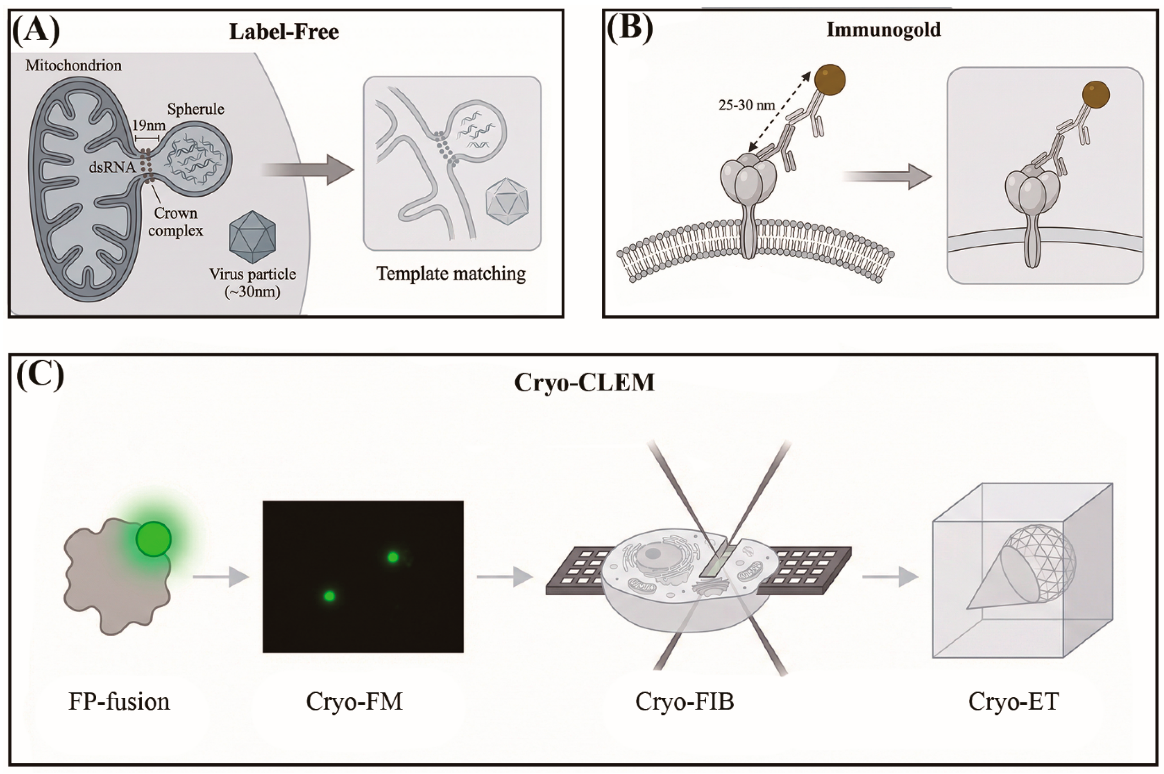
2. Label-Free Cryo-ET: When Morphology Is the Label
3. Immunogold Labeling: Molecular Identity at the Cost of Access
4. Genetic and Synthetic Tags: Promise Constrained by Viral Assembly Logic
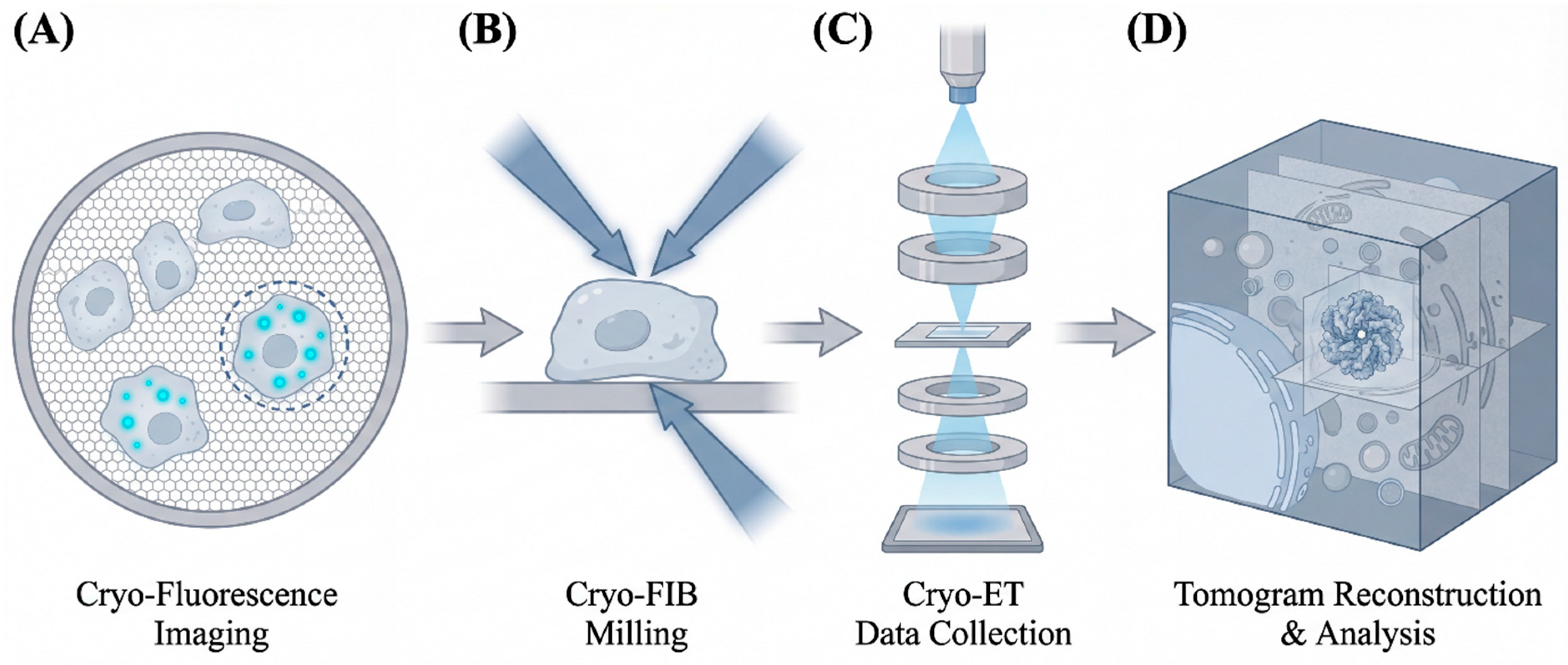
5. Cryo-CLEM and Cryo-FIB-ET: Finding the Needle by Its Glow
6. Conclusions and Future Perspectives
Author Contributions
Funding
Data Availability Statement
Conflicts of Interest
References
- Graham, M.; Zhang, P. Cryo-electron tomography to study viral infection. Biochem. Soc. Trans. 2023, 51, 1701–1711. [Google Scholar] [CrossRef] [PubMed]
- Hong, Y.; Song, Y.; Zhang, Z.; Li, S. Cryo-Electron Tomography: The Resolution Revolution and a Surge of In Situ Virological Discoveries. Annu. Rev. Biophys. 2023, 52, 339–360. [Google Scholar] [CrossRef] [PubMed]
- Schur, F.K.; Obr, M.; Hagen, W.J.; Wan, W.; Jakobi, A.J.; Kirkpatrick, J.M.; Sachse, C.; Kräusslich, H.G.; Briggs, J.A. An atomic model of HIV-1 capsid-SP1 reveals structures regulating assembly and maturation. Science 2016, 353, 506–508. [Google Scholar] [CrossRef] [PubMed]
- Tegunov, D.; Xue, L.; Dienemann, C.; Cramer, P.; Mahamid, J. Multi-particle cryo-EM refinement with M visualizes ribosome-antibiotic complex at 3.5 Å in cells. Nat. Methods 2021, 18, 186–193. [Google Scholar] [CrossRef] [PubMed]
- Zivanov, J.; Otón, J.; Ke, Z.L.; von Kügelgen, A.; Pyle, E.; Qu, K.; Morado, D.; Castaño-Díez, D.; Zanetti, G.; Bharat, T.A.M.; et al. A Bayesian approach to single-particle electron cryo-tomography in RELION-4.0. eLife 2022, 11, e83724. [Google Scholar] [CrossRef] [PubMed]
- Tegunov, D.; Cramer, P. Real-time cryo-electron microscopy data preprocessing with Warp. Nat. Methods 2019, 16, 1146–1152. [Google Scholar] [CrossRef] [PubMed]
- Castaño-Díez, D.; Kudryashev, M.; Arheit, M.; Stahlberg, H. A flexible, user-friendly development tool for subtomogram averaging of cryo-EM data in high-performance computing environments. J. Struct. Biol. 2012, 178, 139–151. [Google Scholar] [CrossRef] [PubMed]
- Ni, T.; Frosio, T.; Mendonça, L.; Sheng, Y.; Clare, D.; Himes, B.A.; Zhang, P. High-resolution in situ structure determination by cryo-electron tomography and subtomogram averaging using emClarity. Nat. Protoc. 2022, 17, 421–444. [Google Scholar] [CrossRef] [PubMed]
- Mamede, J.I.; Griffin, J.; Gambut, S.; Hope, T.J. A New Generation of Functional Tagged Proteins for HIV Fluorescence Imaging. Viruses 2021, 13, 386. [Google Scholar] [CrossRef] [PubMed]
- Kopek, B.G.; Perkins, G.; Miller, D.J.; Ellisman, M.H.; Ahlquist, P. Three-dimensional analysis of a viral RNA replication complex reveals a virus-induced mini-organelle. PLoS Biol. 2007, 5, e220. [Google Scholar] [CrossRef] [PubMed]
- Ertel, K.J.; Benefield, D.; Castaño-Diez, D.; Pennington, J.G.; Horswill, M.; den Boon, J.A.; Otegui, M.S.; Ahlquist, P. Cryo-electron tomography reveals novel features of a viral RNA replication compartment. eLife 2017, 6, e25940. [Google Scholar] [CrossRef] [PubMed]
- Unchwaniwala, N.; Zhan, H.; Pennington, J.; Horswill, M.; den Boon, J.A.; Ahlquist, P. Subdomain cryo-EM structure of nodaviral replication protein A crown complex provides mechanistic insights into RNA genome replication. Proc. Natl. Acad. Sci. USA 2020, 117, 18680–18691. [Google Scholar] [CrossRef] [PubMed]
- Wolff, G.; Limpens, R.; Zevenhoven-Dobbe, J.C.; Laugks, U.; Zheng, S.; de Jong, A.W.M.; Koning, R.I.; Agard, D.A.; Grünewald, K.; Koster, A.J.; et al. A molecular pore spans the double membrane of the coronavirus replication organelle. Science 2020, 369, 1395–1398. [Google Scholar] [CrossRef] [PubMed]
- Huang, Y.; Wang, T.; Zhong, L.; Zhang, W.; Zhang, Y.; Yu, X.; Yuan, S.; Ni, T. Molecular architecture of coronavirus double-membrane vesicle pore complex. Nature 2024, 633, 224–231. [Google Scholar] [CrossRef] [PubMed]
- Turoňová, B.; Sikora, M.; Schürmann, C.; Hagen, W.J.H.; Welsch, S.; Blanc, F.E.C.; von Bülow, S.; Gecht, M.; Bagola, K.; Hörner, C.; et al. In situ structural analysis of SARS-CoV-2 spike reveals flexibility mediated by three hinges. Science 2020, 370, 203–208. [Google Scholar] [CrossRef] [PubMed]
- Ke, Z.; Oton, J.; Qu, K.; Cortese, M.; Zila, V.; McKeane, L.; Nakane, T.; Zivanov, J.; Neufeldt, C.J.; Cerikan, B.; et al. Structures and distributions of SARS-CoV-2 spike proteins on intact virions. Nature 2020, 588, 498–502. [Google Scholar] [CrossRef] [PubMed]
- Laurent, T.; Kumar, P.; Liese, S.; Zare, F.; Jonasson, M.; Carlson, A.; Carlson, L.A. Architecture of the chikungunya virus replication organelle. eLife 2022, 11, e83042. [Google Scholar] [CrossRef] [PubMed]
- Jones, R.; Bragagnolo, G.; Arranz, R.; Reguera, J. Capping pores of alphavirus nsP1 gate membranous viral replication factories. Nature 2021, 589, 615–619. [Google Scholar] [CrossRef] [PubMed]
- Watanabe, R.; Zyla, D.; Parekh, D.; Hong, C.; Jones, Y.; Schendel, S.L.; Wan, W.; Castillon, G.; Saphire, E.O. Intracellular Ebola virus nucleocapsid assembly revealed by in situ cryo-electron tomography. Cell 2024, 187, 5587–5603.e5519. [Google Scholar] [CrossRef] [PubMed]
- Vallbracht, M.; Bodmer, B.S.; Fischer, K.; Makroczyova, J.; Winter, S.L.; Wendt, L.; Wachsmuth-Melm, M.; Hoenen, T.; Chlanda, P. Nucleocapsid assembly drives Ebola viral factory maturation and dispersion. Cell 2025, 188, 704–720.e717. [Google Scholar] [CrossRef] [PubMed]
- Wachsmuth-Melm, M.; Peterl, S.; O’Riain, A.; Makroczyová, J.; Fischer, K.; Krischuns, T.; Vale-Costa, S.; Amorim, M.J.; Chlanda, P. Visualizing influenza A virus assembly by in situ cryo-electron tomography. Nat. Commun. 2025, 16, 9394. [Google Scholar] [CrossRef] [PubMed]
- Sutanto, R.; Rana, J.; Tsai, B.; Mosalaganti, S. In situ cryo-ET redefines HPV disassembly and transport paradigms. bioRxiv 2025. [Google Scholar] [CrossRef] [PubMed]
- Mattei, S.; Glass, B.; Hagen, W.J.; Kräusslich, H.G.; Briggs, J.A. The structure and flexibility of conical HIV-1 capsids determined within intact virions. Science 2016, 354, 1434–1437. [Google Scholar] [CrossRef] [PubMed]
- Zila, V.; Margiotta, E.; Turoňová, B.; Müller, T.G.; Zimmerli, C.E.; Mattei, S.; Allegretti, M.; Börner, K.; Rada, J.; Müller, B.; et al. Cone-shaped HIV-1 capsids are transported through intact nuclear pores. Cell 2021, 184, 1032–1046.e1018. [Google Scholar] [CrossRef] [PubMed]
- Hou, Z.; Shen, Y.; Fronik, S.; Shen, J.; Shi, J.; Xu, J.; Chen, L.; Hardenbrook, N.; Engelman, A.N.; Aiken, C.; et al. HIV-1 nuclear import is selective and depends on both capsid elasticity and nuclear pore adaptability. Nat. Microbiol. 2025, 10, 1868–1885. [Google Scholar] [CrossRef] [PubMed]
- Klumpe, S.; Senti, K.A.; Beck, F.; Sachweh, J.; Hampoelz, B.; Ronchi, P.; Oorschot, V.; Brandstetter, M.; Yeroslaviz, A.; Briggs, J.A.G.; et al. In-cell structure and snapshots of copia retrotransposons in intact tissue by cryo-ET. Cell 2025, 188, 2094–2110.e2018. [Google Scholar] [CrossRef] [PubMed]
- Zhou, Q.; Lok, S.M. Visualizing the virus world inside the cell by cryo-electron tomography. J. Virol. 2024, 98, e0108523. [Google Scholar] [CrossRef] [PubMed]
- Ahlquist, P. Parallels among positive-strand RNA viruses, reverse-transcribing viruses and double-stranded RNA viruses. Nat. Rev. Microbiol. 2006, 4, 371–382. [Google Scholar] [CrossRef] [PubMed]
- Unchwaniwala, N.; Zhan, H.; den Boon, J.A.; Ahlquist, P. Cryo-electron microscopy of nodavirus RNA replication organelles illuminates positive-strand RNA virus genome replication. Curr. Opin. Virol. 2021, 51, 74–79. [Google Scholar] [CrossRef] [PubMed]
- Klein, S.; Cortese, M.; Winter, S.L.; Wachsmuth-Melm, M.; Neufeldt, C.J.; Cerikan, B.; Stanifer, M.L.; Boulant, S.; Bartenschlager, R.; Chlanda, P. SARS-CoV-2 structure and replication characterized by in situ cryo-electron tomography. Nat. Commun. 2020, 11, 5885. [Google Scholar] [CrossRef] [PubMed]
- Mangala Prasad, V.; Leaman, D.P.; Lovendahl, K.N.; Croft, J.T.; Benhaim, M.A.; Hodge, E.A.; Zwick, M.B.; Lee, K.K. Cryo-ET of Env on intact HIV virions reveals structural variation and positioning on the Gag lattice. Cell 2022, 185, 641–653.e617. [Google Scholar] [CrossRef] [PubMed]
- Melia, C.E.; van der Schaar, H.M.; de Jong, A.W.M.; Lyoo, H.R.; Snijder, E.J.; Koster, A.J.; van Kuppeveld, F.J.M.; Bárcena, M. The Origin, Dynamic Morphology, and PI4P-Independent Formation of Encephalomyocarditis Virus Replication Organelles. mBio 2018, 9, e00420-18. [Google Scholar] [CrossRef] [PubMed]
- Romero-Brey, I.; Bartenschlager, R. Membranous Replication Factories Induced by Plus-Strand RNA Viruses. Viruses 2014, 6, 2826–2857. [Google Scholar] [CrossRef] [PubMed]
- Vijayakrishnan, S.; McElwee, M.; Loney, C.; Rixon, F.; Bhella, D. In situ structure of virus capsids within cell nuclei by correlative light and cryo-electron tomography. Sci. Rep. 2020, 10, 17596. [Google Scholar] [CrossRef] [PubMed]
- Cruz-León, S.; Majtner, T.; Hoffmann, P.C.; Kreysing, J.P.; Kehl, S.; Tuijtel, M.W.; Schaefer, S.L.; Geissler, K.; Beck, M.; Turonová, B.; et al. High-confidence 3D template matching for cryo-electron tomography. Nat. Commun. 2024, 15, 3992. [Google Scholar] [CrossRef] [PubMed]
- Berger, C.; Premaraj, N.; Ravelli, R.B.G.; Knoops, K.; López-Iglesias, C.; Peters, P.J. Cryo-electron tomography on focused ion beam lamellae transforms structural cell biology. Nat. Methods 2023, 20, 499–511. [Google Scholar] [CrossRef] [PubMed]
- Wagner, F.R.; Watanabe, R.; Schampers, R.; Singh, D.; Persoon, H.; Schaffer, M.; Fruhstorfer, P.; Plitzko, J.; Villa, E. Preparing samples from whole cells using focused-ion-beam milling for cryo-electron tomography. Nat. Protoc. 2020, 15, 2041–2070. [Google Scholar] [CrossRef] [PubMed]
- Hagen, W.J.H.; Wan, W.; Briggs, J.A.G. Implementation of a cryo-electron tomography tilt-scheme optimized for high resolution subtomogram averaging. J. Struct. Biol. 2017, 197, 191–198. [Google Scholar] [CrossRef] [PubMed]
- Bos, E.; Hussaarts, L.; van Weering, J.R.; Ellisman, M.H.; de Wit, H.; Koster, A.J. Vitrification of Tokuyasu-style immuno-labelled sections for correlative cryo light microscopy and cryo electron tomography. J. Struct. Biol. 2014, 186, 273–282. [Google Scholar] [CrossRef] [PubMed]
- Yi, H.; Strauss, J.D.; Ke, Z.; Alonas, E.; Dillard, R.S.; Hampton, C.M.; Lamb, K.M.; Hammonds, J.E.; Santangelo, P.J.; Spearman, P.W.; et al. Native immunogold labeling of cell surface proteins and viral glycoproteins for cryo-electron microscopy and cryo-electron tomography applications. J. Histochem. Cytochem. 2015, 63, 780–792. [Google Scholar] [CrossRef] [PubMed]
- Griffiths, G.; Lucocq, J.M. Antibodies for immunolabeling by light and electron microscopy: Not for the faint hearted. Histochem. Cell Biol. 2014, 142, 347–360. [Google Scholar] [CrossRef] [PubMed]
- Hainfeld, J.F.; Powell, R.D. New frontiers in gold labeling. J. Histochem. Cytochem. 2000, 48, 471–480. [Google Scholar] [CrossRef] [PubMed]
- Silvester, E.; Baker, L.A. Molecular tags for electron cryo-tomography. Emerg. Top. Life Sci. 2025, 8, ETLS20240006. [Google Scholar] [CrossRef] [PubMed]
- Wang, Q.; Mercogliano, C.P.; Löwe, J. A ferritin-based label for cellular electron cryotomography. Structure 2011, 19, 147–154. [Google Scholar] [CrossRef] [PubMed]
- Clarke, N.I.; Royle, S.J. FerriTag is a new genetically-encoded inducible tag for correlative light-electron microscopy. Nat. Commun. 2018, 9, 2604. [Google Scholar] [CrossRef] [PubMed]
- Wang, C.; Khosrozadeh, A.; Iacovache, I.; Zuber, B. Genetically encoded FerriTag as a specific label for cryo-electron tomography. Structure 2025, 33, 2146–2156.e2144. [Google Scholar] [CrossRef] [PubMed]
- Le Sage, V.; Cinti, A.; Amorim, R.; Mouland, A.J. Adapting the Stress Response: Viral Subversion of the mTOR Signaling Pathway. Viruses 2016, 8, 152. [Google Scholar] [CrossRef] [PubMed]
- Fung, H.K.H.; Hayashi, Y.; Salo, V.T.; Babenko, A.; Zagoriy, I.; Brunner, A.; Ellenberg, J.; Müller, C.W.; Cuylen-Haering, S.; Mahamid, J. Genetically encoded multimeric tags for subcellular protein localization in cryo-EM. Nat. Methods 2023, 20, 1900–1908. [Google Scholar] [CrossRef] [PubMed]
- Peukes, J.; Xiong, X.; Erlendsson, S.; Qu, K.; Wan, W.; Calder, L.J.; Schraidt, O.; Kummer, S.; Freund, S.M.V.; Kräusslich, H.G.; et al. The native structure of the assembled matrix protein 1 of influenza A virus. Nature 2020, 587, 495–498. [Google Scholar] [CrossRef] [PubMed]
- Pornillos, O.; Ganser-Pornillos, B.K.; Kelly, B.N.; Hua, Y.; Whitby, F.G.; Stout, C.D.; Sundquist, W.I.; Hill, C.P.; Yeager, M. X-ray structures of the hexameric building block of the HIV capsid. Cell 2009, 137, 1282–1292. [Google Scholar] [CrossRef] [PubMed]
- Jacques, D.A.; McEwan, W.A.; Hilditch, L.; Price, A.J.; Towers, G.J.; James, L.C. HIV-1 uses dynamic capsid pores to import nucleotides and fuel encapsidated DNA synthesis. Nature 2016, 536, 349–353. [Google Scholar] [CrossRef] [PubMed]
- Müller, B.; Daecke, J.; Fackler, O.T.; Dittmar, M.T.; Zentgraf, H.; Kräusslich, H.G. Construction and characterization of a fluorescently labeled infectious human immunodeficiency virus type 1 derivative. J. Virol. 2004, 78, 10803–10813. [Google Scholar] [CrossRef] [PubMed]
- Silvester, E.; Vollmer, B.; Pražák, V.; Vasishtan, D.; Machala, E.A.; Whittle, C.; Black, S.; Bath, J.; Turberfield, A.J.; Grünewald, K.; et al. DNA origami signposts for identifying proteins on cell membranes by electron cryotomography. Cell 2021, 184, 1110–1121.e1116. [Google Scholar] [CrossRef] [PubMed]
- Lam, S.S.; Martell, J.D.; Kamer, K.J.; Deerinck, T.J.; Ellisman, M.H.; Mootha, V.K.; Ting, A.Y. Directed evolution of APEX2 for electron microscopy and proximity labeling. Nat. Methods 2015, 12, 51–54. [Google Scholar] [CrossRef] [PubMed]
- Mercogliano, C.P.; DeRosier, D.J. Concatenated metallothionein as a clonable gold label for electron microscopy. J. Struct. Biol. 2007, 160, 70–82. [Google Scholar] [CrossRef] [PubMed]
- Morphew, M.K.; O’Toole, E.T.; Page, C.L.; Pagratis, M.; Meehl, J.; Giddings, T.; Gardner, J.M.; Ackerson, C.; Jaspersen, S.L.; Winey, M.; et al. Metallothionein as a clonable tag for protein localization by electron microscopy of cells. J. Microsc. 2015, 260, 20–29. [Google Scholar] [CrossRef] [PubMed]
- Yang, J.E.; Larson, M.R.; Sibert, B.S.; Shrum, S.; Wright, E.R. CorRelator: Interactive software for real-time high precision cryo-correlative light and electron microscopy. J. Struct. Biol. 2021, 213, 107709. [Google Scholar] [CrossRef] [PubMed]
- Pierson, J.A.; Yang, J.E.; Wright, E.R. Recent advances in correlative cryo-light and electron microscopy. Curr. Opin. Struct. Biol. 2024, 89, 102934. [Google Scholar] [CrossRef] [PubMed]
- Jun, S.; Ke, D.; Debiec, K.; Zhao, G.; Meng, X.; Ambrose, Z.; Gibson, G.A.; Watkins, S.C.; Zhang, P. Direct visualization of HIV-1 with correlative live-cell microscopy and cryo-electron tomography. Structure 2011, 19, 1573–1581. [Google Scholar] [CrossRef] [PubMed]
- Jun, S.; Ro, H.J.; Bharda, A.; Kim, S.I.; Jeoung, D.; Jung, H.S. Advances in Cryo-Correlative Light and Electron Microscopy: Applications for Studying Molecular and Cellular Events. Protein J. 2019, 38, 609–615. [Google Scholar] [CrossRef] [PubMed]
- Yang, J.E.; Larson, M.R.; Sibert, B.S.; Kim, J.Y.; Parrell, D.; Sanchez, J.C.; Pappas, V.; Kumar, A.; Cai, K.; Thompson, K.; et al. Correlative montage parallel array cryo-tomography for in situ structural cell biology. Nat. Methods 2023, 20, 1537–1543. [Google Scholar] [CrossRef] [PubMed]
- Rodriguez, Z.K.; Andino-Moncada, J.R.; Buth, S.A.; Mehrani, A.; Ranaweera, A.; Shi, J.; Andrade, L.R.; Singh, S.P.; Strutzenberg, T.S.; Marin, M.; et al. Time-Resolved Fluorescence Imaging and Correlative Cryo-Electron Tomography to Study Structural Changes of the HIV-1 Capsid. ACS Nano 2025, 19, 30902–30918. [Google Scholar] [CrossRef] [PubMed]



| Virus/System | Structure Revealed In Situ | Labeling Strategy |
|---|---|---|
| Flock House virus (FHV) | Mitochondrial spherule interior; 12-fold symmetric protein A crown at the spherule neck (8.5 Å) [11,12] | Label-free; native Cryo-immunogold confirmation of replicase identity |
| SARS-CoV-2 | 12 + 12 nsp3–nsp4 pore in four stacked hexameric rings (~3 MDa, pseudo-12-fold) spanning the double-membrane vesicle [13,14] | Label-free (lumenal target; antibody-inaccessible) |
| SARS-CoV-2 | Spike conformational states and three-hinge stalk flexibility on intact virions [15,16] | Label-free |
| Chikungunya virus | nsP1 dodecameric ring capping plasma-membrane spherules [17,18] | Label-free |
| Ebola virus | Progressive nucleocapsid condensation intermediates in infected cells [19,20] | Label-free (Cryo-FIB-ET) |
| Influenza A virus | vRNP clustering at Rab11a-positive recycling endosomes [21] | Label-free (Cryo-FIB-ET) |
| Human papillomavirus 16 | Endolysosomal trafficking intermediates challenging the lysosomal disassembly model [22] | Label-free |
| HIV-1 | Cone-shaped capsid traversing an intact nuclear pore complex [23,24] | Label-free |
| HIV-1 | 1489 cores across four nuclear-import stages; capsid elasticity and NPC adaptability [25] | Cryo-CLEM (mNG-IN) + Cryo-FIB-ET |
| Copia LTR retrotransposon | Nuclear VLP capsid at 7.7 Å in intact Drosophila egg chambers [26] | Label-free + Cryo-lamella lift-out |
| Strategy | Size (nm) | Target Accessibility | Primary Limitation |
|---|---|---|---|
| Label-Free [1,27] | N/A | All compartments | Requires repetitive symmetry Monomeric proteins unidentifiable |
| Native Cryo-Immunogold [11,40] | 25–30 † | Surface or mild permeabilization | Spatial uncertainty ~25–30 nm Intracellular targets largely inaccessible |
| FerriTag [46] | 10–12 | Intracellular | Requires rapalog induction; rapamycin inhibits mTOR, confounding viral processes |
| GEM/Encapsulin [48] | ~25 | Intracellular | Too large for capsid subunit fusion; steric clash with assembly interfaces |
| DNA-Origami SPOT [53] | 30–100 | Extracellular only | Cannot access intracellular viral components |
| Fluorescent Protein (Cryo-CLEM) [25,59] | 3–4 | Intracellular | No direct Cryo-ET contrast Requires correlative workflow |
Disclaimer/Publisher’s Note: The statements, opinions and data contained in all publications are solely those of the individual author(s) and contributor(s) and not of MDPI and/or the editor(s). MDPI and/or the editor(s) disclaim responsibility for any injury to people or property resulting from any ideas, methods, instructions or products referred to in the content. |
© 2026 by the authors. Licensee MDPI, Basel, Switzerland. This article is an open access article distributed under the terms and conditions of the Creative Commons Attribution (CC BY) license.
Share and Cite
Park, Y.H.; Kim, R.; Song, K.-H.; Jung, H.S. Labeling and Localization Strategies for In Situ Cryo-Electron Tomography Across the Viral Life Cycle. Viruses 2026, 18, 790. https://doi.org/10.3390/v18070790
Park YH, Kim R, Song K-H, Jung HS. Labeling and Localization Strategies for In Situ Cryo-Electron Tomography Across the Viral Life Cycle. Viruses. 2026; 18(7):790. https://doi.org/10.3390/v18070790
Chicago/Turabian StylePark, Yoon Ho, Rana Kim, Kun-Ho Song, and Hyun Suk Jung. 2026. "Labeling and Localization Strategies for In Situ Cryo-Electron Tomography Across the Viral Life Cycle" Viruses 18, no. 7: 790. https://doi.org/10.3390/v18070790
APA StylePark, Y. H., Kim, R., Song, K.-H., & Jung, H. S. (2026). Labeling and Localization Strategies for In Situ Cryo-Electron Tomography Across the Viral Life Cycle. Viruses, 18(7), 790. https://doi.org/10.3390/v18070790