Review

Avian Metapneumovirus: Current Knowledge, Critical Gaps, and Future Directions in Transmission, Pathogenesis, and Control Across Poultry Systems

1
Department of Animal Sciences, College of Food, Agricultural and Environmental Sciences, The Ohio State University, Wooster, OH 44691, USA
2
Research and Analytical Service Core, College of Food, Agricultural and Environmental Sciences, The Ohio State University, Wooster, OH 44691, USA
*
Author to whom correspondence should be addressed.
Viruses 2026, 18(7), 781; https://doi.org/10.3390/v18070781
Submission received: 18 June 2026 / Revised: 13 July 2026 / Accepted: 13 July 2026 / Published: 16 July 2026

Abstract

Avian metapneumovirus (aMPV) is an economically significant respiratory virus of poultry that causes major losses due to reduced egg production and increased susceptibility to secondary infections. It has global significance in poultry production systems and mainly affects turkeys, chickens, and ducks. It was first detected in South Africa in 1978 and is classified into four distinct subtypes (A–D) based upon sequence divergence of the glycoprotein (G) gene. Subtype C has historically been the dominant subtype in the US, first detected in the late 1990s, causing approximately $15 million economic loss per year in Minnesota alone. However, after a decade, aMPV re-emerged in the fall of 2023. Subtype A in California and B in North Carolina was detected for the very first time in the US, impacting multiple states, with an estimated $112 million loss in Minnesota alone. The most recent US Animal Health Association (USAHA) annual survey has now ranked aMPV as the most frequently reported issue in turkey production. Even after four decades of virus identification, areas such as transmission routes, host–virus interactions and tissue tropism within the host are not studied in detail. Addressing these gaps via current modernized research tools is crucial in minimizing the impact of aMPV on the poultry industry. Therefore, this review article summarizes current knowledge on aMPV while identifying critical research gaps and emphasizing future research directions for combating aMPV and supporting long-term sustainability of the turkey industry.

1. Introduction

Avian metapneumovirus (aMPV) is a significant respiratory infectious agent of birds with a wide geographical distribution and substantial economic impacts. It primarily affects the respiratory and reproductive tracts of turkeys, chickens and ducks, leading to severe respiratory distress, nasal discharge, poor eggshell quality, and a drop in egg production [1,2,3]. The virus causes turkey rhinotracheitis (TRT) in turkeys and swollen head syndrome (SHS) in chickens, terms that reflect the characteristic clinical signs and lesions observed in each species [4]. Interestingly, aMPV has been detected in almost all poultry-producing countries except Australia, likely due to the country’s strict biosecurity measures and geographical isolation [4,5]. This highlights the extensive global distribution of aMPV that demands the detailed analysis of its transmission biology and pathogenesis.
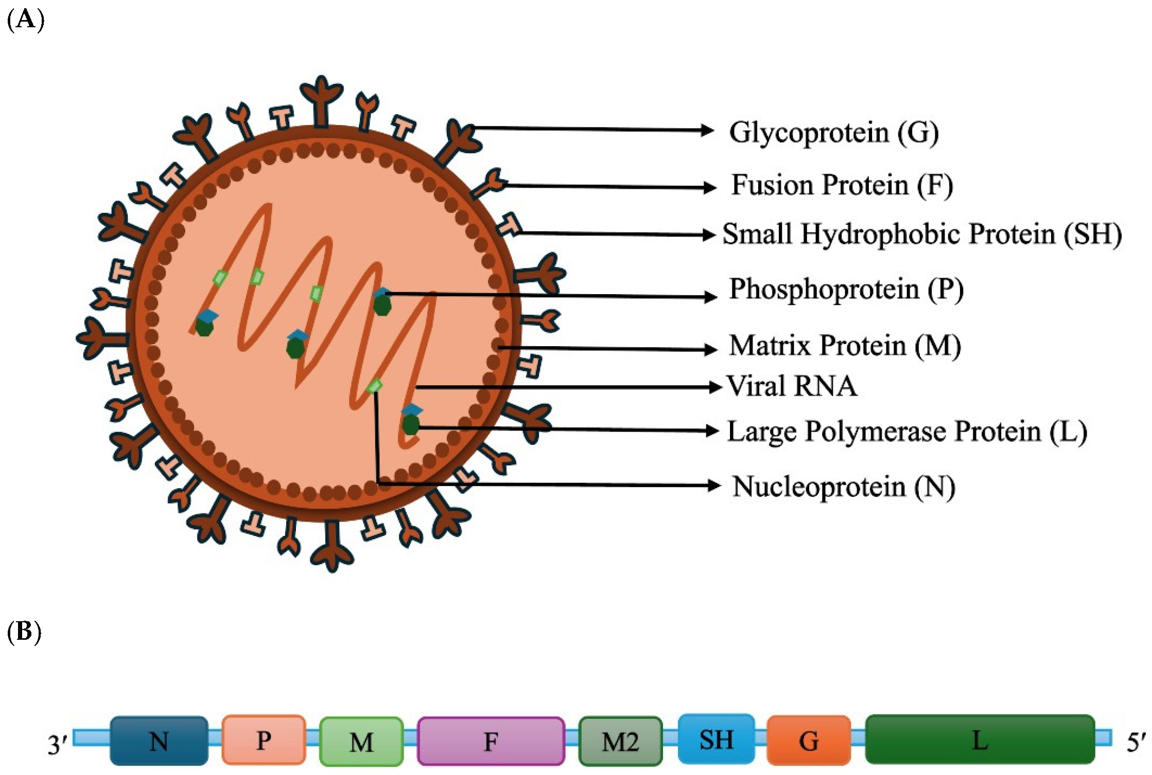
aMPV was initially reported in 1978 in turkeys in South Africa and was subsequently detected across multiple continents [6]. In the US, the virus was first documented in 1997 [4,7,8]. aMPV is classified under order Mononegavirales, family Pneumoviridae and genus Metapneumovirus. Metapneumovirus species are characterized by the absence of the NS1 and NS2 genes, which encode non-structural proteins and are defining features of other members of the genus Orthopneumovirus within the same family. The genus Metapneumovirus comprises human metapneumovirus (hMPV) and aMPV [9,10]. aMPV viral particles show pleomorphic, fringed, generally spherical forms measuring 80–200 nm in diameter and filamentous forms with surface projections measuring 80–100 nm in diameter (Figure 1A) [4,11]. The viral RNA is single-stranded, non-segmented, and negative-sense, with a genome length of approximately 13.3–14 kb [9,10]. aMPV has a genome consisting of eight genes that encode the fusion protein (F), phosphoprotein (P), matrix protein (M), nucleoprotein (N), second matrix protein (M2), attachment glycoprotein (G), small hydrophobic protein (SH), and large polymerase protein (L) (Figure 1B) [4,10]. Among the aMPV genes, the G gene, which encodes the attachment glycoprotein, is the most genetically heterogeneous gene in the genome, showing extensive sequence variability. Based on the sequence divergence of the G gene, aMPV is classified into four subtypes: A, B, C and D [12,13,14].
Subtype A and B are widely distributed globally, whereas in the US, subtype C was historically the most prevalent aMPV strain. The country remained free of aMPV for more than ten years, largely due to strict biosecurity practices and the use of live attenuated vaccines [3,8]. However, in 2023, subtypes A and B were identified in the US and caused disease outbreaks in commercial poultry flocks, marking their first detection in the country [3,8]. These outbreaks have created substantial economic losses in the poultry industry nationwide. A study by the Minnesota Turkey Growers Association reported that the turkey industry in Minnesota experienced approximately $112 million in lost sales in 2024 due to aMPV [15]. Although mortality is typically low, it may rise substantially when secondary bacterial infections occur, such as Mycoplasma gallisepticum, Escherichia coli and Ornithobacterium rhinotracheale [4,16,17]. Transmission occurs primarily through direct contact with infected birds, particularly via infectious aerosols and respiratory exudates [18,19]. Indirect transmission is also possible through contaminated equipment, litter, personnel and vehicles, all of which can serve as important sources of viral spread [4].
Diagnosing aMPV based on clinical and pathological findings is challenging, as its clinical features are generalized and closely resemble those of other respiratory diseases such as Newcastle disease and avian influenza. Therefore, molecular tools such as reverse transcription polymerase chain reaction (RT-PCR) and sequencing are crucial for reliable identification and characterization of the virus [4,14]. At present, there is no targeted therapeutic intervention for aMPV. Preventative and control strategies depend on strict biosecurity practices and vaccination programs. The US currently lacks USDA-approved vaccines for aMPV subtypes A and B, forcing reliance on European imports and highlighting the importance of developing domestic vaccine options [2]. The 2024–2025 outbreak in the US demanded the reliance on European vaccine manufacturers like Vaxxinova, Boehringer-Ingelheim, and HIPRA, which supply live attenuated or killed vaccines primarily derived from European aMPV-A and B strains. This reliance further contributes to the economic impact due to the costs associated with vaccine purchase and importation.
In this review, we discuss the current epidemiology, global distribution, viral evolution, host range, transmission routes, environmental persistence, pathogenesis, immune modulation, diagnostics, and surveillance of aMPV. We also explore available vaccines, industry-level impacts, existing knowledge gaps and future research directions for combating aMPV and supporting long-term sustainability of the turkey industry.

2. Epidemiology and Global Distribution

The first documentation of aMPV occurred in South Africa in the late 1970s, and the virus has since been detected in almost every country worldwide (Table 1). Subtypes A (aMPV-A) and B (aMPV-B) are the predominant aMPV subtypes reported across Europe, South America, Africa and Asia. Subtype C (aMPV-C) has been documented in the US, Canada, France and multiple countries across Asia [2,14]. The absence of standardized epidemiological tools and structured diagnostic field studies limits the ability to accurately assess aMPV global distribution and its impact on overall poultry production [20,21].

2.1. Europe

Turkey rhinotracheitis had been reported in France since 1981, yet none of the early viral isolates appeared to represent the causative agent. In 1985, an outbreak on a well-managed farm in France facilitated successful virus isolation, and the agent was subsequently confirmed to be aMPV. During the same period, the disease had also been identified in the United Kingdom [32,61,62]. The virus was later detected in several other European regions. The early aMPV isolates from Europe included the CVL14/1 strains from the United Kingdom, the 1556 strain from France, the 657/4 strain from Hungary, the 872S strain from Spain, and the 2119 strain from Italy [13,31]. Centered on nucleotide and amino acid sequence comparisons of the G gene, the early isolates from the United Kingdom were classified as subtype A, whereas those from France, Italy, Hungary, and Spain were classified as subtype B [8,13,28,63].
Field serological investigations conducted in Italy have demonstrated the presence of aMPV-A and B in both turkey and broiler farms [20,27]. In Northern Italy, field studies conducted in turkey and broiler farms between 2011 and 2013 indicated that subtype B was the most prevalent strain [2]. Screening for aMPV in broilers, layers, and backyard flocks led to the first molecular identification of the virus in Greece in 2016, which was classified as subtype B [21]. In Belgium, both aMPV-A and aMPV-B were isolated from turkey farms in 1998 [49]. aMPV-B was reported for the first time in a broiler flock with respiratory signs in Romania in 2016, showing a close relationship to Italian field strains of aMPV [59]. In Russia, aMPV subtypes A and B have been identified in chickens and turkeys [20]. During the period of 1987 to 1988, aMPV was isolated in Germany and identified as subtype A in turkeys and subtype B in broiler breeders [34]. In the United Kingdom, early aMPV isolates from turkeys during the late 1980s were mainly subtype A, whereas by the mid-1990s subtype B had become predominant [33]. Current epidemiological studies from Europe highlight aMPV-B as the dominant subtype [3,20,28].
aMPV-C is classified into two lineages: Eurasian aMPV and North American strains. The presence of Eurasian aMPV-C strains was detected in Muscovy ducks in France, confirming the circulation of this virus in the country [26,64]. Between 2017 and 2019, an aMPV subtype C-specific RT-qPCR assay was used to screen wild mallards in the Netherlands, which led to the identification of aMPV-C of Eurasian lineage [65]. Similarly, a subgroup-specific indirect ELISA conducted on blood samples from mallard flocks in Northern Italy showed 100% seroconversion to aMPV subtype C, marking the first report of this finding [29]. The Eurasian lineage of aMPV-C appears to exhibit greater tropism for anatids, as most detected cases have occurred in these species [66]. Two aMPV isolates recovered from turkey flocks in France in 1985 showed no similarity to subtypes A, B, or C and were classified as subtype D, which has not been reported after the initial report [23,63].

2.2. North America

The US was initially considered free of aMPV. However, a 1996 respiratory outbreak in Colorado turkeys led to testing in the United Kingdom, where ELISA results first suggested similarity to subtype A. The official isolation of aMPV from the Colorado outbreak was achieved in February 1997 by the National Veterinary Services Laboratories. The combined evidence from matrix and fusion protein phylogeny, along with the isolate’s distinct serological profile compared with European strains, confirmed it to be subtype C [7,67,68]. In March 1997, a similar illness was observed in Minnesota, and aMPV-C was detected in turkeys in the state for the first time using cell culture methods and RT-PCR (Table 2) [46]. Although the initial outbreak occurred in Colorado, the greatest economic impact from aMPV was observed in Minnesota, which ranks first for turkey production in the US [4]. Following its emergence in Minnesota, aMPV was subsequently detected in domestic turkeys in central North American states such as Iowa, Wisconsin, North Dakota and South Dakota using RT-PCR or ELISA [45]. Serum samples obtained from wild birds in Ohio, South Carolina, Georgia and Arkansas showed an 18% seroconversion rate to aMPV antibodies, which on sequencing confirmed as subtype C, underscoring the important role wild birds play in the virus’s epidemiology [69].
Although subtype C historically predominated in US poultry, recent detections in late 2023 and early 2024 indicate that subtypes A and B have entered commercial flocks, with subtype A more common in western states and subtype B more common in eastern states [3,8,47,72]. Evidence suggests that US aMPV-B strains derived from Eastern Asian lineages are connected to European variants, while aMPV-A introductions seem to trace back to Mexico, with genetic links to Asian strains [66]. Interestingly, the route of introduction of these subtypes to the US is still unknown. The National Centre for Foreign Animal Disease (NCFAD) reported Canada’s first detection of aMPV in May 2024, identifying both aMPV-A and aMPV-B in domestic birds [2]. A longitudinal study in breeder hens and pullets from highly productive poultry regions in Mexico identified aMPV-A, confirming its circulation in the country [54].

2.3. South America

RT-PCR testing and G-gene sequence analysis of vaccinated and non-vaccinated broiler, breeder, and turkey farms in Brazil revealed the presence of both aMPV-A and aMPV-B, demonstrating the cocirculation of these subtypes in Brazilian commercial flocks [44]. The presence of aMPV was demonstrated in turkey and chicken flocks in Chile in 1997 through ELISA [48]. In Colombia, G-gene targeted RT-PCR detected aMPV-B from laying hens, broilers, breeders, and wild birds [73].

2.4. Asia

Subtypes A, B and C were identified in commercial chickens with variable respiratory signs in China, highlighting the presence of the virus in the country [51,52,74]. A novel aMPV-C variant from Sheldrake ducks exhibiting hydrosalpinx fluid syndrome (HFS) was recently identified in China [50]. The presence of aMPV in Japan has been recognized since 1989, with aMPV antibodies detected in chickens exhibiting swollen head syndrome. Both subtypes A and B have been detected in Japan [36]. In Thailand, the first molecular detection of aMPV-B in turkeys was reported in 2022 [75]. Investigations in Malaysia and Vietnam have identified subtype B as the most prevalent aMPV strain [40,76]. Serological analysis of samples using commercial ELISAs capable of detecting aMPV-A, B, and C antibodies in both broiler breeder and layer flocks in India identified seropositive farms, indicating the need for further surveillance of the virus in the country [57,58].
In Turkey, aMPV-B was detected by RT-PCR in broiler flocks sampled between 2017 and 2018, and another study similarly identified aMPV-B in turkeys [55,56]. Serum samples collected from chickens of various age groups in Saudi Arabia between 2007 and 2008 showed a 50% seroconversion rate to aMPV antibodies in birds aged 11–18 weeks [77]. Antibodies to aMPV were also detected in backyard chickens in Saudi Arabia in 2019 [78]. In Jordan, aMPV-B was detected in broilers, layers, and breeder chickens through RT-PCR and ELISA [53]. In Israel, aMPV subtypes A and B were identified in vaccinated chicken as well as in turkey flocks, highlighting the need for more effective vaccination measures [79]. Studies from Iran indicate that subtype B is the most prevalent aMPV strain in chickens and turkeys [80,81].

2.5. Africa

The spread of aMPV in most African nations remains poorly characterized, pointing to how much more systematic surveillance is still needed. Initially, the prevalent aMPV strain in Egypt was subtype B, detected in turkeys, but in 2013, subtype A was also identified in turkeys [2,82]. RT-PCR testing in Moroccan broiler flocks identified circulation of aMPV-A and B, with subtype B emerging as the dominant strain [60]. Commercial chicken flocks in Nigeria tested positive for aMPV-B using RT-PCR, with Sanger sequencing validating the subtype [83].

3. Phylogenetic Analysis of Avian Metapneumovirus

Comparison of Virus Clustering Based on Whole-Genome and Full-Length G Sequence

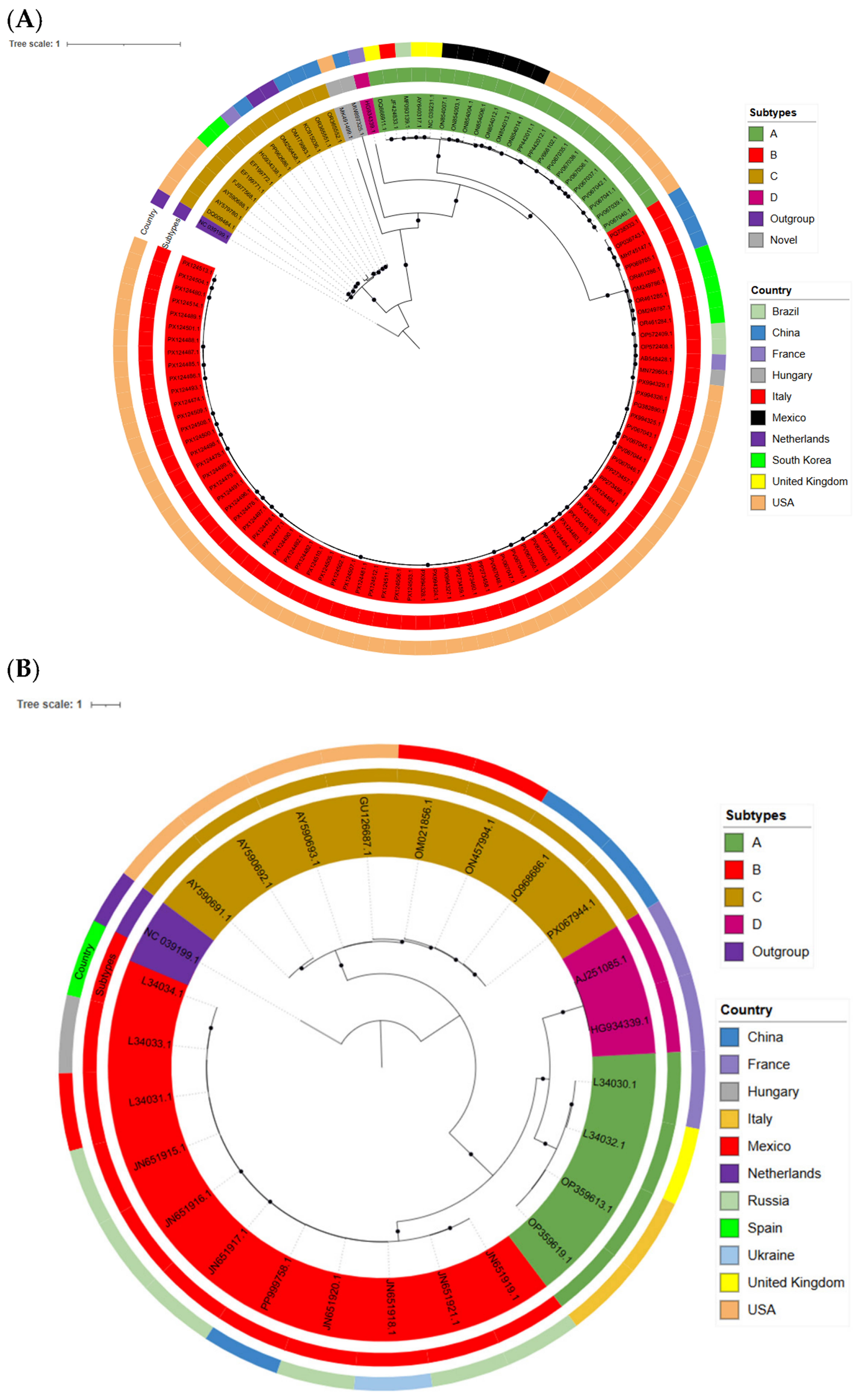
Phylogenetic analysis of 117 complete aMPV genomes, and human metapneumovirus as outgroup, demonstrated clear clustering based on the subtypes A, B, C, and D (Figure 2A). The phylogenetic tree further showed that the two major lineages, subtypes A and B, clustered together, whereas subtypes C and D formed separate sister lineages consistent with previous findings [84]. Among the sequences analyzed, subtype B was the most frequently detected subtype isolated predominantly from the USA, whereas subtype D was less frequently isolated. Furthermore, a second phylogenetic analysis was performed using 25 full-length G-gene sequences of aMPV (Figure 2B). For this tree, the K3Pu + F + R2 substitution model was chosen as the best-fit model in reference to BIC. Consistent with the whole-genome analysis, the G-gene phylogeny tree showed subtype-specific clustering and supported the overall evolutionary relationships observed in the complete genome analysis.

4. Viral Evolution and Host Range

4.1. Worldwide Evolutionary Patterns and Genetic Diversification of aMPV

The emergence and worldwide dissemination of aMPV subtypes illustrate the virus’s evolutionary expansion, shaped by mutation, vaccine-driven selection and host-specific pressures. The genome of aMPV is highly conserved, with stable polymerase and nucleocapsid functions, but the G gene shows extensive diversity and is the major contributor to evolutionary differences among strains [5]. Studies have shown that the time to the most recent common ancestor (TMRCA) for aMPV is estimated to be approximately 200 years ago. Further evolutionary analysis using the Bayesian serial coalescent approach in the Bayesian evolutionary analysis by sampling trees (BEAST) program (incorporates sampling dates and genetic data to estimate evolutionary rates and divergence times) estimated substitution rates for the F and N genes of aMPV-C at 9.3 × 10⁻⁴ and 1.1 × 10⁻³ substitutions per site per year, respectively [88]. In a similar study, evolutionary rates were estimated for the N, P, F, and M2 genes of aMPV-C. These rates ranged from 1.3 × 10⁻³ to 7 × 10⁻³ substitutions per site per year, indicating the fast-evolving nature of aMPV-C [89]. These estimates underscore how quickly RNA viruses can evolve, driving short generation times, high genetic diversity and the continual emergence of new subtypes. In the same study, phylogenetic analysis of the above genes revealed two distinct aMPV-C lineages, designated C1 and C2. The TMRCA at which these two lineages coalesce is estimated to be between 9 and 14 years [89]. Overall, this evidence points to aMPV-C as a virus that continues to evolve, with its rapid mutation rates and ongoing lineage formation helping drive its continued spread.
For in-depth analysis of the evolutionary dynamics of aMPV-B, researchers analyzed G-gene sequences collected between 1985 and 2019 from nine European countries: Greece, Romania, France, Russia, Italy, Spain, Ukraine, the Netherlands and the United Kingdom. The TMRCA estimates indicated that the first introduction of aMPV-B in Europe occurred in 1981 in France, followed by its introduction into Italy in 1984, from where it subsequently spread to other European countries. The evolutionary rate of 1.21 × 10⁻³ substitutions per site per year falls well within the range typical of RNA viruses, reflecting their high propensity to mutate [28,90]. In Italy, the detection of aMPV-B in vaccinated flocks showed that the circulating field strains were genetically unrelated to both the 1987 Italian isolate and the commonly used vaccine strain [91]. Complete nucleotide and amino acid sequences of the field strain were analyzed and showed the greatest variation in the SH and G proteins. The G mutations were non-synonymous, leading to modifications in viral characteristics as a response to selection pressure. A subtype B virus carrying these changes allowed the virus to infect birds and induce disease even after successful vaccination with an established subtype B vaccine. These changes in the G and SH proteins may have arisen from long-term vaccine-driven selection pressure, leading to the appearance of mutant strains [91]. In the US, the vaccination schedule for aMPV-A and B was generally not followed due to the historical absence of these viruses in the country, which may also have been a predisposing factor for the recent outbreak. Comparative analysis of aMPV-B isolates in Northern Italy from 1987 to 2007 and those circulating in Western Europe before 1994, using the F and G genes, identified clear genetic distinctions. Fusion protein gene sequences showed marked conservation, whereas G protein gene sequences exhibited more variation. The presence of a serine residue at position 382 in the G protein appeared to be unique to the Italian aMPV-B isolates. These changes in the Italian viruses also suggest that the mutations may have arisen from immune pressure, either due to vaccination or exposure to an undetected environmental field strain [92].
In a study conducted at the end of 2024, comparative phylogenomics of samples collected from aMPV-A prevalent areas in the US, aimed at understanding the evolution of circulating aMPV-A, revealed that these strains shared 99.5% identity with US strains in GenBank at both the whole-genome level (WGS) and across all eight genes, including the G gene [93]. The amino acid alignment of the entire G protein also revealed only a few substitutions, suggesting that the currently circulating aMPV-A strain has undergone limited evolution across turkey and chicken. Similarly, for subtype B, whole-genome and G-gene analysis showed that the current isolates were genetically distinct from previously reported strains worldwide but exhibit very high sequence identity among the US aMPV-B strains [93]. The amino acid analysis of aMPV-A and aMPV-B showed only minimal substitutions with the US strains, reflecting their high sequence identity. This high sequence identity in both subtypes potentially indicates limited vaccine-driven selection pressure and a recent introduction of the virus into the country. However, the number of amino acid mutations increased when these US strains were compared with other groups, such as European and Brazilian isolates [93]. Both aMPV-A and aMPV-B in the US show broader genetic connections and clear divergence when placed in a global context, highlighting how these viruses continue to evolve differently across regions worldwide.

4.2. Host Range and Susceptibility of aMPV

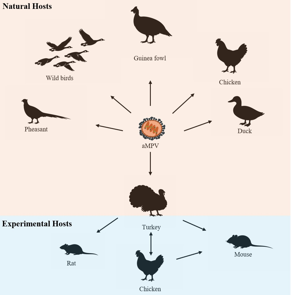
The common susceptible hosts for aMPV infection are turkeys and chickens, while birds of all age groups are vulnerable to infection with the aMPV. However, the disease is more severe in younger birds [4]. aMPV subtypes are adapted to cause infection in members of the order Galliformes (Table 3). Subtypes A, B, and C infect both turkeys and chickens, and subtype C of Eurasian lineage is well known to cause disease in ducks [24,26,32,36,74,94]. A recent study utilizing the aMPV-B/chicken/USA/SD-24/P6 demonstrated high pathogenicity in both chickens and turkeys with no age or host specificity [95]. This suggests the virulent nature of the virus, indicating the capability to adapt to multiple host ranges. aMPV-C was first isolated from pheasants in South Korea, where complete genome sequencing was also performed [39]. The existence of aMPV antibodies was detected in both reared and free-living pheasants in Italy, too, highlighting pheasants as a susceptible host [96]. Additionally, aMPV was identified in guinea fowl with respiratory signs, and the detected strain was classified as subtype B [97]. aMPV-C is known to cause disease in ducks, and its presence has been detected in Muscovy ducks in China and France [26,98], as well as in Sheldrake and Cherry Valley ducks in China [50,99]. aMPV-C was detected in wild birds (American crows, Cattle egrets, American coots, Canada geese and Rock pigeons) in Ohio, Arkansas, South Carolina and Georgia, highlighting the potential involvement of wild birds in introducing infection [69]. In addition, RT-PCR confirmation of aMPV RNA in several other wild species in the US, including Canada geese, blue-winged teal, sparrows, swallows, and starlings, further supports the contribution of wild birds in virus spread [100,101]. This implies the need for strict biosecurity control and identifies wild birds as the potential carriers of the novel US strain seen in 2023–2024. Moreover, aMPV subtypes A and B have also been reported in wild birds in Brazil, reinforcing that multiple subtypes can circulate across diverse wild avian populations [102,103].
In an experimental study, it was demonstrated that aMPV subtypes A, B, C and D were all capable of infecting turkeys and chickens, with infection most severe in turkeys. And the duck origin aMPV-C strain was able to be isolated and induced seroconversion in both turkeys and chickens [94]. A novel subtype of aMPV was detected in gulls (American herring and Great black-backed), and these viruses appear to be intermediate between aMPV-C and the subgroup of other aMPV strains [104]. Similarly, another novel strain of aMPV was isolated from monk parakeets, and its sequence divergence from known strains also points to a new subgroup of aMPV [105]. Future studies aimed at understanding the pathogenic potential of these strains in domestic birds are essential for determining their impact on poultry production. A serosurvey in Zimbabwe detected aMPV antibodies in ostriches, demonstrating a 100% seroprevalence within the sampled population [106]. Experimental host range studies demonstrated that aMPV-C could be detected in mice for up to 14 days and in rats mainly at 4 days after inoculation [107]. Similarly, in an experimental study, an aMPV-C strain isolated from chickens in China was replication competent and persisted in the lungs of BALB/c mice for 21 days, showing lung lesions, fever, and upregulation of pulmonary inflammatory cytokines and chemokines. The aMPV-C isolate used in this study shared 78.5% similarity with hMPV, and although replication was observed in mice, no information is available regarding the common or potential receptors utilized by the virus [108]. These findings indicate that mice and rats may serve as potential carriers of aMPV and contribute to its transmission. aMPV has a broad and expanding host range, spanning domestic poultry, wild birds, and even non-avian species (Figure 3). This highlights the adaptable nature of aMPV and reinforces the importance of continued surveillance to prevent spillover events and protect poultry health.

5. Transmission Routes and Environmental Persistence

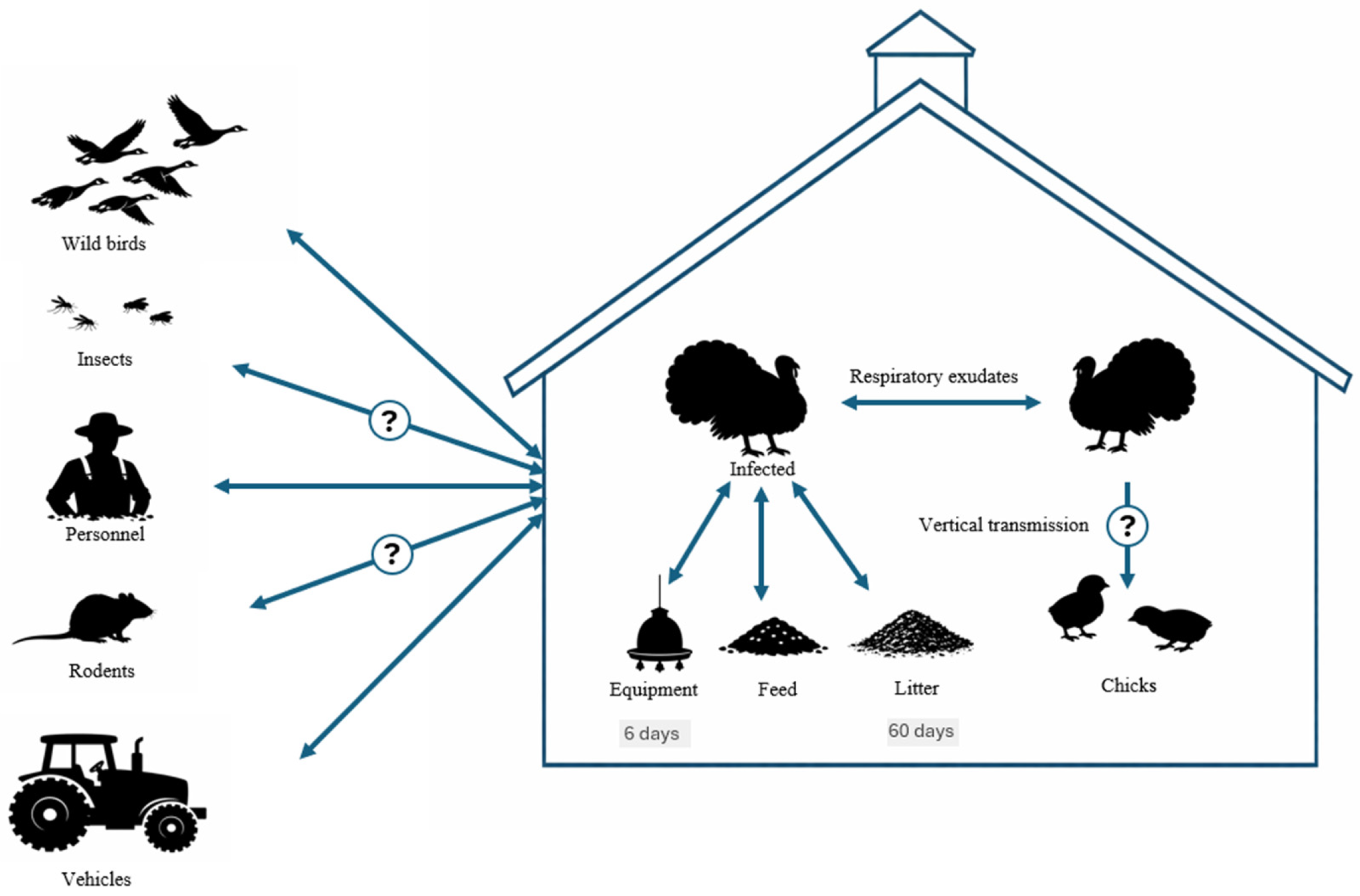
aMPV is primarily transmitted horizontally through infectious aerosols released by infected birds (Figure 4). Direct contact with respiratory secretions from infected birds through the oculonasal route represents the most common pathway of infection [4,18,19]. Studies utilizing subtype A and C have demonstrated direct contact transmission between birds; however, virus spread to birds housed in separate pens within the same room could not be established [18,19]. Even though the inoculated birds in these experiments became infected and transmitted the virus to birds in direct contact, birds in indirect contact did not develop disease, which highlights the difficulty of reproducing field-like transmission scenarios under laboratory conditions. Under experimental conditions, aMPV could be identified in the upper respiratory tract of birds for only a few days (6 or 7 days at most), indicating a short shedding period, yet infection still spreads rapidly within flocks and between farms under field conditions [14,18,19]. Aside from direct contact with infected respiratory secretions, other routes of infection remain unclear.
In addition to direct exposure to infected respiratory secretions, several indirect routes of infection, such as contaminated feed, water, litter, personnel, equipment, and vehicles, may also contribute to virus spread. Virus shed through respiratory secretions can contaminate these sources, allowing them to act as fomites for disease transmission [4,109]. Experimental studies investigating aMPV survivability in litter indicate that the virus can remain viable for up to 60 days under low temperature conditions [109]. Similarly, the survivability of aMPV on commonly encountered farm surfaces has been evaluated, and the virus was found to persist for up to six days [110]. Together, these findings highlight the potential for indirect transmission through contaminated materials and equipment.
Wild migratory birds also represent a major source of viral introduction and transmission. Several studies have reported the existence of aMPV in wild birds, highlighting their probable role in virus transmission [69,100,102]. Notably, aMPV RNA isolated from wild birds has been documented to share 90–95% nucleotide sequence identity with viruses from domestic turkeys [101]. Similarly, aMPV-C isolates recovered from wild birds have been demonstrated to cause disease in domestic turkeys [111]. Phylodynamic and phylogeographic analyses tracing the origins of the aMPV-A and aMPV-B outbreaks beginning in 2023 indicate introductions from Mexican and Eastern Asian strains respectively, further supporting the role of wild bird migration in viral spread [66]. The contribution of wild birds to the epidemiology and transmission of the disease in commercial turkey flocks is still not fully understood. Experimental studies have demonstrated the existence of the virus in the reproductive tract of layers, but there are still no findings consistent with vertical transmission to offspring [112,113]. aMPV-C isolated from turkeys and subsequently inoculated into ducks remained detectable in tissue samples for up to 21 days without causing any clinical signs, indicating that ducks can act as potential sub-clinical carriers capable of transmitting the virus [114]. The probable routes of introduction of aMPV-A and aMPV-B into US poultry remain poorly defined, and there is a need to expand investigations to include the potential roles of rodents and insects as mechanical vectors.
Generally, the virus is sensitive to external agents; however, lower temperature conditions favor the persistence of aMPV in the environment [115]. Experimental studies have demonstrated that aMPV is viable for more than 26 weeks at −70 °C and −20 °C, for less than 26 weeks at 4 °C, for less than 4 weeks at 20 °C, for 2 days at 37 °C, and for less than 6 h at 50 °C. aMPV was unaffected by variable pH for 1 h between pH 5 and 9, indicating that low pH environments could pose challenges for disposing of infected litter, with contaminated lakes potentially serving as sources of infection due to viral survival at lower pH levels. Common disinfectants such as quaternary ammonium compounds, ethanol, iodophors, phenolic derivatives, and sodium hypochlorite have been shown to reduce viral viability [115].

6. Pathogenesis and Immune Modulation

6.1. Clinical Presentation of aMPV Infection in Turkeys and Chickens

aMPV primarily targets the upper respiratory tract of susceptible birds following exposure to infected respiratory secretions, resulting in an acute respiratory infection. Clinical signs develop rapidly, with an incubation period ranging from 3 to 7 days, causing more severe infection in turkeys than in chickens, with the greatest severity observed in turkeys of 3 to 12 weeks of age [1,14,116]. The greater susceptibility of turkeys to the disease may be due to differences in viral replication rates and innate immune regulation between the two species [117]. The disease is referred to as turkey rhinotracheitis (TRT) in turkeys, and the symptoms include sneezing, coughing, tracheal crackles, nasal and ocular secretions, swollen infraorbital sinuses, and submandibular edema [1,4,116]. In chickens, in addition to lethargy, coughing, nasal mucus accumulation, and serous ocular exudates, swollen head syndrome (SHS), characterized by the edema of periorbital and infraorbital sinuses, has been associated with aMPV, which is often followed by cerebral disorientation, torticollis, and opisthotonos due to secondary E. coli infection [1,4,118]. In both turkeys and chickens, secondary infections caused by pathogens including Ornithobacterium rhinotracheale, Bordetella spp., Pasteurella multocida, Staphylococcus spp., Mycoplasma gallisepticum, Newcastle disease virus, infectious bronchitis virus (IBV) and infectious laryngotracheitis virus can exacerbate disease severity [4,16,17]. aMPV can also infect the reproductive tract (oviduct) of both laying hens and turkeys, resulting in poor shell quality, egg peritonitis, and reduced egg production, which in turn contributes to economic losses [112,113].

6.2. Pathogenesis and Tissue Tropism of aMPV

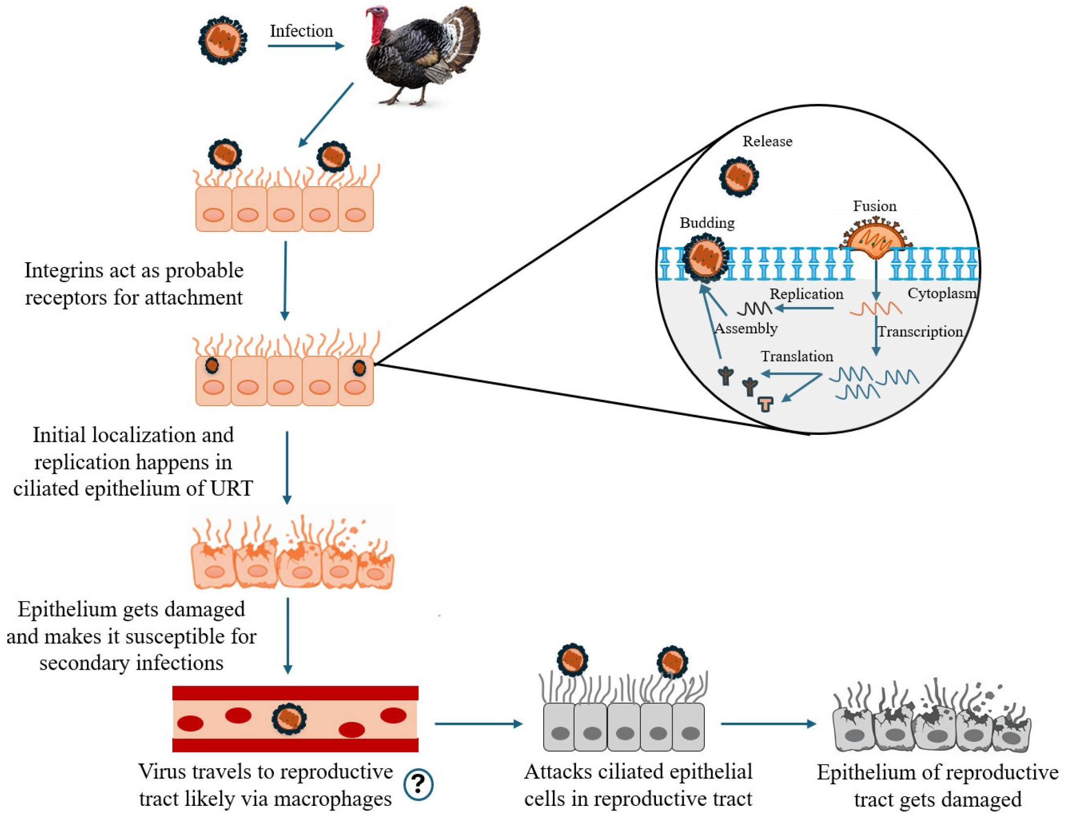
Following exposure to infected respiratory secretions, aMPV infects the ciliated epithelial cells of the upper respiratory tract, including the lining of cranial pneumatic bone air spaces and the nasolacrimal duct, causing ciliostasis and potentially resulting in total ciliary loss [119,120]. aMPV employs three proteins for cell entry and membrane fusion: G, SH and F protein. The G protein is accountable for virus binding to epithelial cells, whereas the SH protein contributes to cell-to-cell fusion. However, the F protein is capable of binding to cellular receptors and facilitating fusion of the viral and host cell membranes without requiring the involvement of the G or SH proteins, initiating virus infection [63,121,122]. For aMPV, the F protein exhibits dual functionality by mediating both receptor binding and membrane fusion, suggesting that fusion can occur independently of the attachment proteins [123,124]. The receptors for the aMPV F protein are not yet fully understood. Recent studies have indicated that integrins, a family of transmembrane cell adhesion molecules, can act as functional receptors for aMPV. In aMPV-C, integrin β1 has been identified as the receptor, whereas in aMPV-B, integrin αvβ1 has been shown to function as the receptor [121,124]. Viral replication occurs within the cytoplasm of the ciliated epithelium in both the respiratory and reproductive tracts [5,125].
After infection, viral replication occurs within the ciliated epithelial cells of the upper respiratory tract. Experimental studies in turkey poults inoculated by aMPV-A through the ocular route have shown that high levels of virus can be recovered from the upper respiratory tract (nose and trachea) for up to 5 days post-infection, indicating an early viral response [19]. Histopathologic changes, including epithelial exfoliation, enhanced glandular secretion, epithelial deciliation, and mild infiltration by mononuclear cells, were observed in the nasal turbinates of both 3-week-old turkeys and chickens on days 1 and 2 post-infection with aMPV-B in an experimental study [120]. In another study using chicken and turkey tracheal organ cultures (TOCs), loss of ciliary activity was witnessed at 48 h post-infection (hpi) with aMPV-A in turkey TOCs and at 96 hpi in chicken TOCs [117]. Immunohistochemistry frequently reveals viral antigen along the apical membrane and ciliary border of infected epithelial cells, highlighting aMPV’s strong affinity for ciliated epithelium [117,125]. These findings highlight that aMPV rapidly damages the ciliated epithelium of the upper respiratory tract and compromises mucociliary clearance, facilitating further complications by secondary infections (Figure 5).
After a respiratory infection, the virus can disseminate systemically to the reproductive tract, leading to infection of those tissues. In a study on turkey poults with aMPV-C, viral antigen was identified in a small number of macrophages within the nasal turbinates and sinuses. This finding suggests that macrophage-mediated leukocyte trafficking could facilitate the virus’s movement to the reproductive tract [119,126]. The role of macrophages in aMPV infection needs to be studied in detail, as studies of other avian viruses, such as IBV, which targets avian macrophages, suggest that macrophage involvement may facilitate viral dissemination beyond the respiratory tract [127,128]. aMPV infection damages the ciliated epithelium of the reproductive tract in laying hens and turkeys, contributing to decreased egg production [112,113,119]. In an experimental study utilizing oviduct organ cultures (OOCs) infected with aMPV-A and aMPV-B, ciliary loss was observed beginning at 48 hpi, along with epithelial cell flattening and submucosal edema. These lesions highlight that the decline in egg output and formation of thin-shelled eggs might be attributed to aMPV infection instead of secondary infections [125].

6.3. Immune Responses to aMPV Infection

aMPV infection elicits strong innate and adaptive immune responses in susceptible hosts, involving both humoral and cell-mediated responses [129]. Turkey poults inoculated via the oculonasal route at 4 weeks of age developed a robust humoral response, with aMPV-C specific IgG antibodies first detected in serum samples at 7 days post-infection and persisting for up to 14 weeks [126]. Infiltration of IgA+ B cells into the upper respiratory tract after challenge with aMPV-A and aMPV-C has been confirmed, and similar infiltration by IgM+ and IgG+ B cells has also been documented [14,130,131]. However, studies have shown that humoral immunity alone does not provide effective shielding against aMPV infection and does not reliably reflect actual protective immunity against aMPV. Infection of turkey poults with aMPV-A after chemical treatment with cyclophosphamide, a B-cell suppressor, resulted in a lack of clinical signs, indicating the implication of cell-mediated immunity [132]. Similarly, reduction in T-lymphocytes using cyclosporin A, followed by infection with aMPV-A in turkeys, resulted in prolonged resolution of clinical signs and lesions, as well as positive choanal swabs, demonstrating the importance of cell-mediated immunity [133].
Experiments in turkeys have demonstrated an increase in CD4+ T cells induced by both virulent and vaccine strains of aMPV-A and aMPV-B at 7 and 14 days post-infection in the Harderian gland and spleen. In chickens, CD4+ T cells increased fourfold at 6 days post-inoculation (dpi) and a concurrent rise in CD8α+ cells was observed in the Harderian gland at 6 dpi [129,134]. CD8+ T cells are known to contribute to viral clearance in respiratory infections through contact-dependent effector mechanisms (perforin and CD95L) as well as the production of IFN-γ and TNF-α. Therefore, the stronger CD8α+ (CD8+ T-cell and natural killer cell) response in the Harderian gland of broilers likely contributes to their faster viral clearance and recovery compared with turkeys [134,135]. In these same studies, IFN-γ and IL-6 levels were upregulated in turkeys in the spleen cells and Harderian gland following infection with aMPV-A and aMPV-B, beginning at 3 and 5 days post-infection, respectively. In chickens, IFN-γ upregulation was observed in the Harderian gland after aMPV-A infection, with increased expression also detected in the nasal turbinates at 3 dpi. Even though IFN-γ levels were upregulated in the nasal turbinates of chickens infected with aMPV-B, the levels were downregulated in the Harderian gland at 3 dpi [129,134]. In studies utilizing turkey and chicken TOCs, induction of apoptosis was evaluated following aMPV infection. Apoptotic cells were not discovered in turkey TOCs, whereas modest counts were observed in chicken TOCs at 96 and 168 hpi, correlating with the onset of epithelial cell loss. Histological examination of turkey TOCs revealed necrosis and degeneration of epithelial cells, suggesting that epithelial cell loss in turkey TOCs may be attributable to necrosis [117]. IFN-α mRNA expression was downregulated in both TOCs, with a non-significant upregulation observed only in chicken TOCs at 192 hpi. This may explain why aMPV-infected turkey TOCs did not exhibit increased apoptosis, despite IFN-α being a known inducer of apoptotic pathways [117]. There was also an upregulation of iNOS in both TOCs, which is known to be expressed in macrophages and epithelial cells. The sustained upregulation up to 120 hpi in chicken TOCs may also indicate a correlation with the apoptosis observed in chicken TOCs [117]. These findings help describe the greater vulnerability and more serious clinical signs observed in turkeys, both in TOC studies and under experimental trials and field observations. In a study on aMPV-C, the P protein was shown to reduce IFN-β production and the interferon stimulated genes (ISGs) expressions. Interferon regulatory factor 3 (IRF3) was identified as the target of the P protein, which inhibits IRF3 translocation from the cytoplasm to the nucleus, thereby suppressing IFN-β induction [136]. Similarly, aMPV-C was shown to induce reduction in mitochondrial antiviral signaling (MAVS) protein in Vero cells, which is crucial for linking upstream viral RNA recognition to downstream signaling pathways [137]. These studies highlight immune evasion mechanism employed by aMPV, underscoring its ability to modulate host antiviral signaling.

7. Diagnostics and Surveillance

Various diagnostic tools have been established over time to enable the reliable detection of aMPV. Diagnosis centered solely on clinical signs and gross lesions is challenging, as many respiratory diseases present similar clinical observations. Thus, differential diagnosis is needed as paramyxoviruses such as Newcastle disease virus and avian paramyxovirus-3, as well as IBV, influenza viruses, and multiple bacterial and Mycoplasma species, can produce similar clinical signs [4,119]. Therefore, confirmatory laboratory testing is required to establish a definitive diagnosis of aMPV. RT-PCR serves as the gold standard for aMPV diagnosis because it enables rapid and sensitive molecular detection, whereas virus isolation also serves as a gold standard by providing confirmation through recovery of live, infectious virus. However, due to the difficulty and low success rate of isolating aMPV, RT-PCR is the method most commonly used in routine diagnostic practice [47,138,139]. aMPV is shed for only a short period, with the highest likelihood of virus isolation occurring around 5–7 days post-infection, coinciding with the appearance of clinical signs. Beyond this point, isolation and detection become difficult, underscoring the critical importance of sampling time [18,19,113]. The most suitable samples for aMPV detection are obtained from ocular or nasal secretions, or from turbinate, sinus and tracheal tissues [4,138,140]. The commonly used methods for the definitive diagnosis of aMPV include:

7.1. Virus Isolation

Virus isolation can be performed using embryonated eggs, TOCs and cell culture systems. The initial successful isolation of aMPV in both South Africa and the United States was carried out in embryonated eggs using the yolk sac inoculation route [6,138]. Six to eight-day-old embryonated turkey and chicken eggs are typically used for inoculation via the yolk sac route. Hemorrhages and some embryo mortality may be observed as lesions. Serial passaging is needed to induce embryo death, making the procedure longer; however, highly reliable [6,138].
TOCs is the commonly performed system in today’s date for aMPV isolation. Early studies in South Africa achieved isolation using tracheal ring organ cultures from 27-day-old turkey embryos, subjected to sinus exudate collected from turkeys exhibiting rhinitis and sinusitis [6]. TOCs can be maintained for several weeks, and ciliostasis (cessation of ciliary beating) is the primary characteristic typically appearing around 5–7 days post-inoculation [6,32,138]. Cell cultures are used for aMPV multiplication, although they are often unreliable for primary isolation. Following initial isolation in embryonated eggs or TOCs, the virus can adapt to replicate in several cell culture systems, involving chicken embryo fibroblasts (CEF), chicken embryo liver cells (CEL) and Vero cells (Table 4) [141,142]. Vero cells (derived from African green monkey kidney epithelial cells) lack type I interferons [143]. A characteristic cytopathic effect, typically featuring syncytial formation, becomes evident within seven days after the virus adapts to cell culture [4,116,138]. In addition, an experimental study demonstrated that chicken embryo related (CER) cells can support the primary isolation of aMPV subtypes A and B, and that CER, Vero and BHK-21(baby hamster kidney) cells are capable of propagating these subtypes to high viral titers [144]. Compared with these cell lines, Vero cells exhibit more pronounced syncytia formation, likely due to the absence of type I interferon response [54,116].

7.2. Molecular Detection

Molecular identification methods, particularly RT-PCR and RT-qPCR, are the most widely used for aMPV detection because of their speed, high sensitivity, and specificity. Turbinates, choanal, and tracheal swabs are appropriate sample types for RT-PCR-based detection of the viral genome [14]. Due to the presence of distinct aMPV subtypes, most RT-PCR assays are designed to be subgroup-specific and therefore do not detect all subtypes in a single reaction. RT-PCR was first utilized to detect the F protein gene in six-week-old turkey poults inoculated with the virulent UK/3B/85 strain [63,151]. In a subsequent study comparing F-, G-, and N-based RT-PCR protocols, the results demonstrated that RT-PCR using N-gene primers exhibited broader specificity than the F- or G-based assays for diagnostic purposes, and the G-gene-specific RT-PCR primers enabled subtype identification. These findings highlight that N- and G-based RT-PCR assays are useful for rapid detection and early subtyping of aMPV in field samples, respectively [12]. The use of subtype-specific RT-PCR assays at the outset can result in other circulating subtypes being overlooked. Therefore, using an N-gene-based RT-PCR first for broad detection of aMPV, followed by subtype-specific PCRs or sequencing for accurate typing would be more appropriate [4,12,138]. In a study conducted in China, a quadruplex real-time RT-PCR assay was developed using four pairs of specific primers and four corresponding probes targeting the G or M genes of all aMPV subtypes, providing highly specific and highly sensitive detection of all aMPV subgroups [152]. Similarly, a five-plex digital droplet RT-PCR assay that targets the drift-resistant viral polymerase (a conserved gene of aMPV) was developed and successfully identified all four aMPV subgroups, whose principle relies on changing probe concentrations [153]. Next-generation sequencing of samples that tested positive by RT-qPCR was used to differentiate aMPV strains during the recent aMPV subtype A and B outbreak in the US [8,119]. The absence of a universal and extensively validated primer set targeting a truly conserved region of the aMPV genome across different molecular platforms represents a significant gap in current diagnostic capabilities. Developing such primers would be essential for reliable molecular detection and for accurately assessing viral distribution across regions.

7.3. Serological Tests

Serological methods are widely used to verify aMPV infection in commercial poultry and additional avian species because virus isolation and direct detection are often difficult. Serological techniques such as ELISA, virus neutralization tests, and indirect immunofluorescence are utilized, among which ELISA is the most widely applied. These assays are better suited for flock-level screening and surveillance than for definitive diagnosis [4,14].
Various commercial ELISA kits are available for the serological detection of aMPV. Most ELISA assays use the indirect format, while some employ a blocking (competitive) format. In the indirect ELISA format, microtiter plates coated with aMPV antigen are incubated with test serum and subsequently with an enzyme-labeled anti-turkey or anti-chicken conjugate. The measured enzyme activity correlates with the concentration of specific antibodies in the serum, and aMPV-specific antibodies are indicated by the development of color. Conversely, the blocking (competitive) ELISA functions by allowing antibodies in the test serum to compete with a labeled antibody for available antigen-binding sites [138]. In one study, a competitive ELISA based on the ability of serum antibodies to compete with a monoclonal antibody for binding to recombinant aMPV N protein, which was used as antigen, was developed. This assay demonstrated a specificity of 100% and a sensitivity of 98% when compared with the virus neutralization test. Notably, while the commercial ELISA kit detected aMPV antibodies only after 10 days post-infection (PI), the competitive ELISA was able to sense antibodies as early as 5 days PI [154]. For early diagnosis of aMPV infection to be successful, the choice of ELISA antigen is critical, as the sensitivity of the assay depends heavily on the antigen used [155]. In one study, ELISAs that used a subgroup A or B as antigen were able to detect antibodies to aMPV subtypes A and B but failed to detect antibodies against aMPV-C, demonstrating that antigen choice can significantly influence assay performance [7,138].
Virus neutralization tests are used less frequently than ELISA for diagnosing aMPV infections. They can be performed in several systems, including TOCs, tissue culture systems such as Vero cell monolayers, and CEFs [138]. Although virus neutralization is costly and not easily scalable for surveillance of large flock populations, it can be employed to corroborate and validate ELISA findings [119]. Indirect immunofluorescence and immunodiffusion techniques may likewise be performed on tissue sections to identify antibodies against aMPV [156].

8. Vaccines

aMPV infection cannot be controlled with medication; therefore, disease prevention relies primarily on strict biosecurity measures and vaccination. Inactivated and live attenuated vaccines are present for both turkeys and chickens. In earlier periods, the development of suitable vaccines was hindered by the lack of consistent challenge models needed to conduct vaccine-efficacy studies. However, recent studies have demonstrated effective attenuation of aMPV strains in various cell culture systems and their beneficial use as vaccine candidates [4,157,158]. Effective aMPV vaccines must induce robust cell-mediated and mucosal immunity, since humoral responses by themselves do not provide full protection and are poor predictors of vaccine performance [159].

8.1. Live Attenuated Vaccines

Live attenuated vaccines constituted the main approach for outbreak control in the early years of aMPV management in Europe. These attenuated vaccines used in Europe were developed from European aMPV strains that had been attenuated through serial passage in embryonated eggs, Vero cells, or TOCs [160,161]. In Europe, subtypes A and B were used to produce live attenuated vaccines, whereas in the United States, subtype C was used. Live attenuated vaccines may be delivered via coarse spray, aerosolization, drinking water, or oculonasal application, all of which aim to stimulate strong mucosal and cell-mediated immune responses in the respiratory mucosa. Protection against aMPV clinical signs and lesions equivalent to that induced by the oculonasal route was achieved when the vaccine was delivered by spray or through drinking water, methods that are more practical for large-scale field use [5,156,162].
In turkeys, vaccination with either subtype A or subtype B strains provides substantial cross-protection against both subtypes. Similarly, studies in day-old broiler chickens have demonstrated that vaccination with aMPV subtype B can induce protection not only against subtype B but also against subtype A [155,163,164]. A study reported that subtype A and B vaccines conferred protection in turkeys challenged with aMPV-C, while an aMPV-C-derived vaccine conferred protection in turkeys and somewhat worked in chickens against aMPV-A but failed to protect against aMPV-B [7]. These results indicate that although cross-protection can occur among certain aMPV subtypes, its inconsistency reinforces the need for subtype-specific vaccines to ensure dependable protection in the field.
Even with effective live attenuated vaccines in place, disease outbreaks persist in vaccinated flocks. This can result from reversion to virulence, whereby the vaccine virus acquires mutations during replication in birds and regains pathogenic characteristics. A live attenuated subtype A vaccine was shown to revert to virulence after only two coding mutations and was capable of infecting day-old turkey poults [165]. Farm-level spray administration can lead to uneven vaccine delivery, leaving some birds with suboptimal doses and others completely unvaccinated. Because vaccinated birds can shed aMPV, these unvaccinated flock mates may become infected by the shed vaccine virus, creating conditions that increase the likelihood of reversion to virulence [165]. Similar findings were reported for a subtype B live attenuated vaccine, which also reverted to virulence and caused disease in day-old poults comparable to that produced by field strains [166]. A turkey farm in Italy that had been vaccinated against aMPV-B became infected with aMPV-A, and sequencing showed that the virus originated from a vaccine strain that had not been used on the farm for at least six months, nor in any farms within a 5 km radius [167]. These outcomes indicate that reversion to virulence of live vaccines is a major concern and demonstrate how vaccine-derived virus can even introduce disease into farms where the pathogen was previously not present. Another major factor influencing vaccine efficacy is the presence of maternally derived antibodies (MDA). The higher levels of MDA can result in a lower protective effect from the live attenuated vaccine. In an experimental study in turkeys, vaccination with an attenuated aMPV-A strain induced increased CD8+ T-lymphocyte responses in the Harderian gland and tracheal mucosa of MDA-negative birds, whereas in MDA-positive birds the response was dominated by CD4+ T cells in these tissues. Similarly, vaccination elicited increased anti-aMPV IgY antibodies in MDA-negative birds, while in MDA-positive birds, the decline in serum MDA antibody levels occurred more rapidly, likely due to immunophagocytosis [168]. Together, these observations underscore the need to consider maternal antibody status when designing effective vaccination programs in young birds.

8.2. Inactivated Vaccines

Inactivated oil emulsion or water-based adjuvanted vaccines incorporating subtypes A and B are commonly employed as part of infection control strategies. In Europe, a commercially available inactivated oil-emulsion vaccine is widely used in both turkeys and chickens. Inactivated vaccines are generally administered as booster doses following live attenuated vaccines [14,156]. It was shown that administering an inactivated subtype A aMPV vaccine at 30 weeks of age, after priming with a live attenuated subtype A vaccine at 1 week of age, provided strong protection of laying performance when birds were challenged at 38 weeks of age [169]. Two-week-old turkeys inoculated oculonasally with an inactivated aMPV-C vaccine adjuvanted with the synthetic double-stranded RNA polyriboinosinic polyribocytidylic acid showed elevated mucosal IgA+ cells in the upper respiratory tract, along with enhanced virus-specific IgG and IgA in the lachrymal fluid and the serum IgG. Vaccinated birds were protected against challenge and exhibited reduced respiratory lesions when challenged at either 7 or 21 days post-vaccination [170]. In Egypt, intramuscular administration of an inactivated aMPV vaccine prepared from the GIZA TRT-4 strain (subtype B) and adjuvanted with Nigella sativa oil in 21-day-old turkey poults elicited superior humoral and cell-mediated immunity relative to commercially available inactivated or live vaccines [171]. Similarly, in China, an inactivated aMPV-B vaccine combined with a new adjuvant comprising immune-stimulating complexes (ISCOMs) administered intramuscularly to 21-day-old SPF chickens induced high levels of virus-specific and virus-neutralizing antibodies. It also stimulated B- and T-lymphocyte responses and upregulated the IL-4 and IFN-γ levels [172]. Together, these results highlight the importance of inactivated vaccines, as it reduces the chances of shedding the live virus into the environment, and also decrease the likelihood of reversion to virulence pathogen.

8.3. Other Types of Vaccines

The fusion protein of aMPV-A was utilized to develop the recombinant fowl pox virus vaccine when administered to one-week-old turkey poults intramuscularly and by wing-web at a two-week interval, resulting in milder clinical signs and a 1000-fold reduction in challenge virus recovered from the nose and trachea, demonstrating that the F protein plays a major role in protection against aMPV [173]. Similarly, DNA vaccination using plasmids encoding the F gene (pCMV-F) from aMPV-C produced comparable results, as intramuscular administration to one-week-old turkey poults induced significant protection [174]. Additionally, a DNA vaccine using the plasmid pGEM-T-Easy vector to express the F gene from aMPV-B, administered intramuscularly to 21-day-old turkey poults, provided 100% protection in the vaccinated birds [175].
Through a reverse-genetics approach, a bivalent recombinant vaccine was formulated using the glycoprotein of aMPV-C in a LaSota strain as a backbone. SPF turkey poults (sixty-one-week-old), inoculated intranasally and intraocularly, developed incomplete protection against pathogenic aMPV-C and complete protection against velogenic NDV, underscoring that a single aMPV-C G protein is inadequate for comprehensive protection against aMPV-C [176]. Similarly, NDV-derived recombinant vector vaccine expressing the F and G genes of aMPV-C worked substantially better, providing protection against pathogenic aMPV-C challenge, while complete protection was reported against velogenic NDV [177].
As of April 2026, the US poultry industry is actively using emergency import licenses and experimental autogenous vaccines to combat aMPV subtypes A and B. While no fully licensed, domestically manufactured vaccines were traditionally available for the recent outbreaks, the USDA now permits the import and use of specific products like RESPIVAC® aMPV (modified live vaccine, aMPV-B), HIPRAVIAR® TRT (inactivated vaccine, aMPV-B), Vaxxon® SHS (live vaccine, aMPV-B), Poulvac® TRT (modified live vaccine, aMPV-A) and Boehringer-Ingelheim products: NEMOVAC® (modified live vaccine, aMPV-B), AVIFFA RTI (modified live vaccine, aMPV-B), TUR-3 (inactivated vaccine, aMPV-B) to mitigate severe economic losses (Table 5) [47,71,178]. To support outbreak control within the US, Merck Animal Health, Cambridge Technologies and Ceva have begun producing experimental autogenous vaccines using subtype B isolates sourced from domestic poultry flocks. In addition, Vaxxinova USA is producing an experimental autogenous vaccine based on a US-origin subtype A isolate [47,71].
The USDA issued the first-ever permit for the importation of a modified live vaccine produced outside the US on 20 December 2024, and the vaccine approved under this authorization was Vaxxon® SHS [71,179]. The importation permit of this live vaccine into the US for the first time highlights the critical importance of controlling aMPV in the country, given that US regulatory requirements for vaccine importation are typically stringent and only done when no other options are available. Despite the permit for vaccine importation in 2024, aMPV cases continued to rise, with 13.3 million cases reported in 2025. This may indicate setbacks in the implementation of these vaccines across the US, or it may reflect limitations in the effectiveness of the imported vaccine under field conditions. Reversion to virulence is a risk associated with the use of live aMPV vaccines and has been reported in other countries [165,166]. Relying on live vaccines derived from European strains may also increase the risk of introducing vaccine-derived viruses into the US through reversion to virulence, and these vaccines could also be less effective against US-origin strains [167]. Therefore, the use of killed or inactivated vaccines may be more advantageous, and evaluation of vaccine performance is critical to prevent the circulation of potential vaccine-derived strains.

9. Industry Relevance

In the US poultry industry, aMPV has recently emerged as an economically important priority pathogen, causing major losses in turkeys, broilers, and commercial operations. aMPV infections result in substantial economic losses, reduced production efficiency, and broader market-level disruptions. Turkey breeders experience egg production losses ranging from 20% to 80%, lasting 2 to 4 weeks, resulting in a shortage of poults. Similarly, broiler breeders exhibit a moderate decline in egg production, typically ranging from 5% to 10%. Secondary infections with pathogens such as E. coli, Ornithobacterium rhinotracheale, and Mycoplasma gallisepticum further complicate the clinical disease, increasing mortality and adding to overall economic losses [17,71,119]. Together, these losses highlight the growing economic threat posed by aMPV and its significance as a priority pathogen for the US poultry sector.
In the recent US Animal Health Association (USAHA) annual turkey health survey, aMPV rose dramatically from its previous position at #38 in 2023 to the #1 reported issue in 2024 with 2355 reported cases [71]. This rapid change in ranking underscores the high transmissibility of aMPV, its capacity to cause substantial production losses across multiple poultry species, and the current absence of effective therapeutic options.
Historically, even in areas where aMPV vaccination is regularly practiced, the virus has been considered the most important respiratory pathogen of turkeys aside from avian influenza. Even during the initial aMPV-C outbreak in the US, the Minnesota turkey industry faced serious economic losses, estimated at approximately $15 million per year from 1997 to 2002 [4,180]. The 2024 report by the Minnesota Turkey Growers Association reported that the Minnesota turkey industry experienced $112 million in lost sales, a $17 million reduction in labor income, $31 million in lost value added to the state’s economy and nearly $8 million in reduced tax revenue. An estimated 2,201,903 turkeys were lost to aMPV in 2024, representing approximately 6.57% of Minnesota’s annual production [15]. These early 2024 outbreaks have led to increased diagnostic testing, biosecurity changes, production delays and heightened supply chain demands, further contributing to the economic burden.
The economic relevance of aMPV goes beyond farms into animal health and vaccines. The global aMPV treatment market, composed primarily of vaccines, is valued at $311.6 million in 2025 and is projected to reach $579.5 million by 2035 [181]. Within this market, the vaccine segment dominates with a 68.4% share, reflecting the industry’s strong reliance on immunization for disease control. Overall, aMPV accounts for approximately 3–5% of the global poultry healthcare market [181]. These patterns show that aMPV is not only a production-level threat but also a major contributor to shaping the global poultry health and vaccine markets.

Production, Economic Value and aMPV Disease Patterns

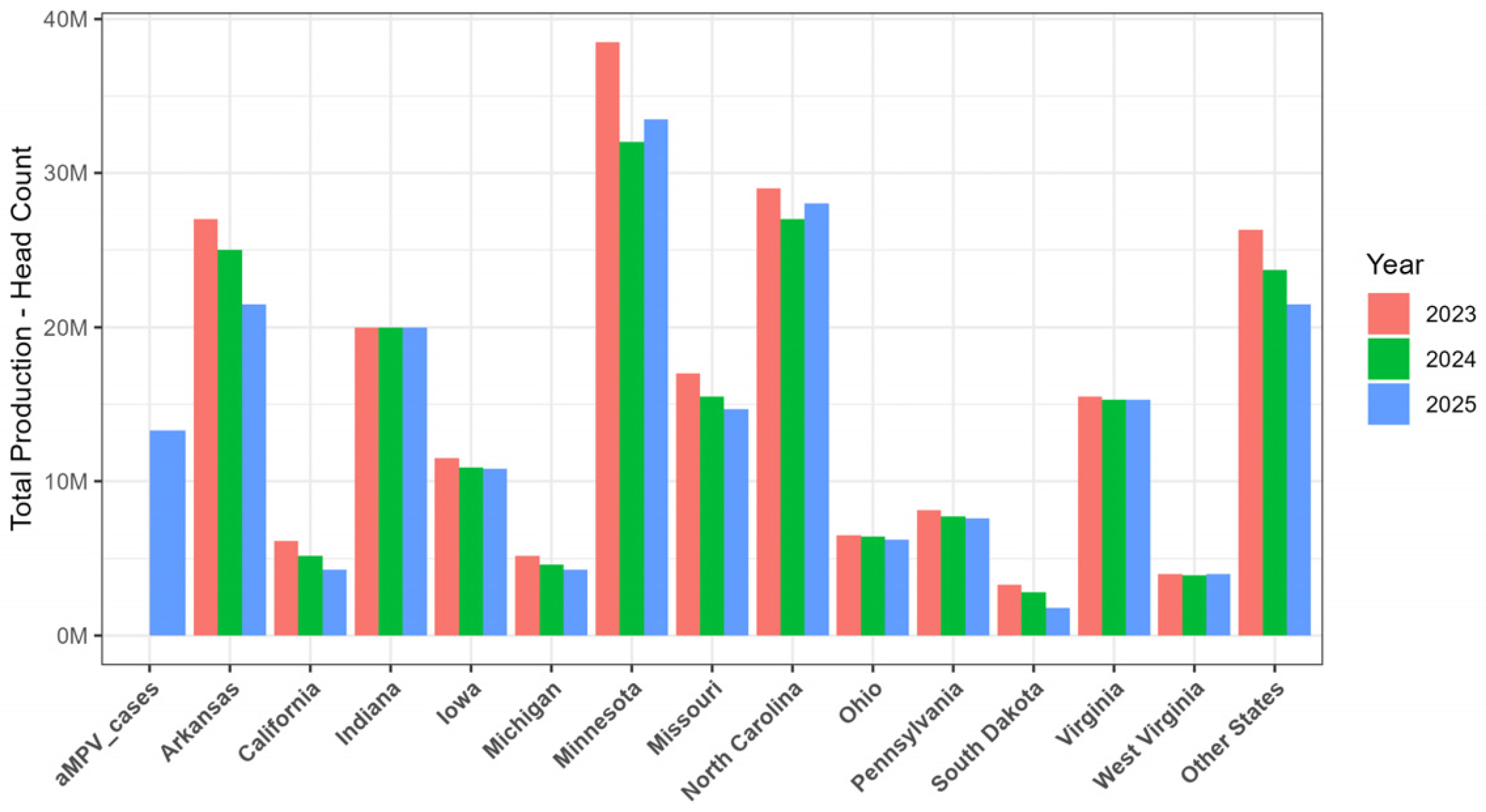
The decline in turkey production between 2023 and 2025 is majorly associated with the aMPV outbreaks in major turkey-producing states. The escalation of turkey losses during this period coincides with the first US detections of aMPV-A and aMPV-B. US states such as Arkansas, California, Minnesota, Missouri and North Carolina clearly demonstrate a decrease in turkey production from 2023 to 2025. Importantly, the largest turkey-producing state in the US, Minnesota, declined from approximately 38.5 million birds in 2023 to about 32 million birds in 2024, followed by 33.5 million birds in 2025 (Figure 6). There were approximately 13 million birds impacted by aMPV in 2025, an increase from 2355 reported cases in 2024, which is interestingly higher than the total production in head count for states such as California, Iowa, Michigan, Ohio, Pennsylvania, South Dakota, and West Virginia. This refers to the potential capability of aMPV to eradicate the turkey production, emphasizing the need to invest in understanding aMPV transmission, pathogenesis and prevention. The slight recovery documented in the year 2025 may be attributed to the first import of the live aMPV vaccines for emergency use in turkeys and the enhanced biosecurity measures, which would have reduced the viral pressure. In the poultry industry, viral pressure is directly proportional to vaccination, biosecurity, movement control, flock immunity and reduction in the viral shedding from the infected birds. The reduction in the quantity of infectious virus present on farm surfaces, equipment, litter and other fomites, which can serve as sources of aMPV infection, helps lower the overall viral pressure in the environment. As the environmental viral load decreases, the likelihood of susceptible birds becoming infected also diminishes, thereby interrupting transmission within and between poultry flocks.
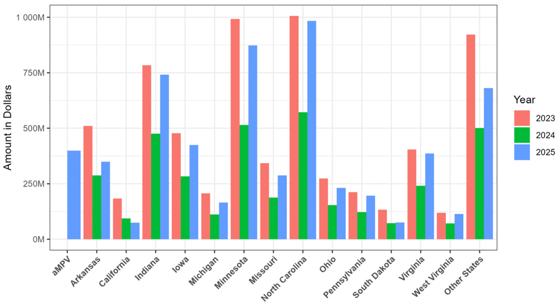
The aMPV disease impact was devastating for the turkey industry, leading to a substantial decrease in the value of production in several states, including Arkansas, California, Indiana, Iowa, Michigan, Minnesota, Missouri, North Carolina, Ohio, Pennsylvania, South Dakota, Virginia, West Virginia and several other states (Figure 7). For instance, Minnesota’s production value in 2023 was nearly $1 billion, which dropped to $0.88 billion in 2025, suggesting a drop of 0.12 billion. An overall estimated economic loss of approximately $398 million is speculated for the year 2025 in the US, with $0.898 per pound for turkey meat and considering the average weight of live turkeys processed under federal inspection to be 33.33 pounds. Surprisingly, the overall aMPV-related loss is higher than the value of production in Arkansas, California, Michigan, Missouri, Ohio, Pennsylvania, South Dakota and West Virginia. This emphasizes the significant economic impact aMPV can have on the overall agricultural production system in the US.
There were a few other reported cases in turkeys during 2025, including Histomoniasis, Mycoplasma synoviae, Turkey coronavirus, Turkey reovirus, and Mycoplasma gallisepticum, all of which were documented only in the low hundreds [71].

10. Knowledge Gaps and Future Directions

Despite being recognized since 1978, a lack of study regarding aMPV limits our ability to fully understand and control the virus. The transmission routes of aMPV remain unclear, as direct contact is the only fully established mode of transmission. However, additional transmission routes appear to exist, but these remain unidentified and poorly characterized. Under experimental conditions, aMPV has been shown to be shed through respiratory secretions for only very short periods by infected birds, and as an enveloped virus, it is not highly resistant outside the host. Given these characteristics, the rapid spread of aMPV over considerable distances remains difficult to explain [183].
Another significant knowledge gap concerns the mechanisms underlying host–virus interactions. Although aMPV and hMPV share conserved polymerase and nucleocapsid functions, the host-specific adaptations that shape pathogenicity and immune evasion strategies are less understood for aMPV than for hMPV [5]. Despite clear evidence that aMPV can infect the reproductive tract, the mechanisms behind its dissemination from the respiratory mucosa to the reproductive tissues have not been characterized, representing a significant gap in our understanding of aMPV pathogenesis. Addressing these gaps will be essential for expanding ground-level surveillance systems, refining intervention approaches and ultimately reducing the global impact of aMPV.
Lack of specific therapeutics and antivirals against aMPV signifies the disease management as an strategical approach via enhanced biosecurity, supportive care and control of secondary bacterial infections using broad-spectrum antibiotics like enrofloxacin [184]. In an experimental study, subcutaneous administration of gamithromycin to turkeys was shown to exert beneficial effects against secondary Ornithobacterium rhinotracheale infection in turkeys previously infected with aMPV-A [185]. Currently, there is no specific antiviral drug that has been approved to be used against aMPV infection. In vitro studies utilizing plant-derived compounds have shown promising antiviral activity against aMPV. Extracts obtained from four plant species, Gaylussacia brasiliensis, Arrabidaea chica, Virola sebifera and Aspidosperma tomentosum were able to inhibit approximately 99% of aMPV-A in vitro [186]. Although further studies are needed to identify the specific active compounds responsible for the observed antiviral effects, such findings provide promising evidence for plant-derived interventions. RNA interference using short interfering RNA (siRNA) targeting the nucleoprotein (N) mRNA has been demonstrated to reduce viral titers in vitro, indicating a unique approach that could be further explored as a potential antiviral therapy [187]. Overall, the limited availability of antivirals against aMPV highlights a significant knowledge gap, making vaccination and strict biosecurity the only control measures that are practically applicable in the poultry industry.
Future directions in aMPV research should emphasize epidemiology, pathogenesis, host–virus interactions and control strategies. Routine surveillance to track viral evolution and transmission networks is crucial for advancing our understanding of aMPV epidemiology. Studies on viral entry, replication, immune evasion, receptor usage and host-specific adaptations are all critical for increasing our understanding of aMPV pathogenesis and for improving strategies for prevention and control. Diagnostic screening should be expanded to include all aMPV subtypes (A, B, C and D), particularly given evidence that subtype D is well adapted to infect both turkeys and chickens, making it surprising that only a single case of aMPV-D has been reported since the 1980s [94]. Understanding of the virulence genes of subtypes A, B, and C via comparison with D would allow us to formulate well-targeted inhibitory antivirals against the aMPV. The re-emergence and rapid detection of aMPV-A and aMPV-B in the US after a decade of absence underscores significant weaknesses in existing biosecurity measures, highlighting the need for strengthened and more consistently implemented biosecurity practices.

11. Conclusions

aMPV is a highly contagious, devastating and economically important pathogen of turkeys. The introduction of aMPV subtypes A and B in the US resulted in significant economic loss in the turkey and overall poultry industry, leading to the first-ever import and utilization of the live attenuated vaccine in the US agriculture. Research encompassing the host range, transmission routes, pathogenesis, vaccine and effective control measures is urgently needed to combat the aMPV disease in the poultry system. In addition, the impact of field strains in the turkey production systems still needs to be investigated for the formulation of broad treatment and prevention strategies against aMPV. Summarizing the aMPV research gives us a somewhat clear picture of the knowledge gaps in the aMPV literature. Addressing these gaps while emphasizing diagnostic surveillance and biosecurity needs will be crucial for mitigating future outbreaks and lowering the overall impact of aMPV on poultry health and production.

Author Contributions

Conceptualization, K.K.Y. and M.B.; software, M.B.; validation, A.A., B.G., M.B. and K.K.Y.; formal analysis, K.K.Y. and M.B.; investigation, A.A., M.B. and K.K.Y.; resources, K.K.Y.; writing—original draft preparation, A.A., M.B. and K.K.Y.; writing—review and editing, B.G., M.B. and K.K.Y.; supervision, M.B. and K.K.Y.; project administration, K.K.Y.; funding acquisition, K.K.Y. All authors have read and agreed to the published version of the manuscript.

Funding

Salaries and research support were provided by the startup funds from The Ohio State University, Wooster, OH 44691, USA.

Institutional Review Board Statement

Not applicable.

Informed Consent Statement

Not applicable.

Data Availability Statement

No new data were created or analyzed in this study. Data sharing is not applicable to this article.

Acknowledgments

We would like to thank College of Food, Agricultural and Environmental Sciences, The Ohio State University, Wooster, OH 44691, USA.

Conflicts of Interest

The authors declare no conflicts of interest.

Abbreviations

The following abbreviations are used in this manuscript:
aMPVAvian metapneumovirus
TRTTurkey rhinotracheitis
SHSSwollen head syndrome
hMPVHuman metapneumovirus
FFusion protein
PPhosphoprotein
MMatrix protein
NNucleoprotein
M2Second matrix protein
GGlycoprotein
SHSmall hydrophobic protein
LLarge polymerase protein
NCFADNational Centre for Foreign Animal Disease
HFSHydrosalpinx fluid syndrome
BICBayesian Information Criterion
TMRCAThe most recent common ancestor
BEASTBayesian evolutionary analysis by sampling trees
TOCTracheal organ culture
OOCOviduct organ culture
ISGInterferon-stimulated gene
IRF3Interferon regulatory factor 3
CEFChicken embryo fibroblast
CELChicken embryo liver cell
BHKBaby hamster kidney
PIPost-infection
MDAMaternally derived antibodies
ISCOMImmune-stimulating complexes
USAHAUS Animal Health Association
RT-PCRReverse transcription polymerase chain reaction

References

  1. Cook, J.K.A. Avian Pneumovirus Infections of Turkeys and Chickens. Vet. J. 2000, 160, 118–125. [Google Scholar] [CrossRef] [Scilit] [PubMed]
  2. Enyetornye, B.; Naikare, H.K.; Velayudhan, B.T. Avian Metapneumovirus. In Veterinary Virology of Domestic and Pet Animals; Wang, L., Ed.; Springer Nature: Cham, Switzerland, 2025; pp. 1–13. [Google Scholar]
  3. Luqman, M.; Duhan, N.; Temeeyasen, G.; Selim, M.; Jangra, S.; Mor, S.K. Geographical Expansion of Avian Metapneumovirus Subtype B: First Detection and Molecular Characterization of Avian Metapneumovirus Subtype B in US Poultry. Viruses 2024, 16, 508. [Google Scholar] [CrossRef] [Scilit] [PubMed]
  4. Gough, R.E.; Jones, R.C. Avian Metapneumovirus. In Diseases of Poultry; Saif, Y.M., Fadly, A.M., Glisson, J.R., McDougald, L.R., Nolan, L.K., Swayne, D.E., Eds.; Blackwell Publishing: Ames, IA, USA, 2008; pp. 100–110. [Google Scholar]
  5. Hatfield, J.S.; Thielen, B.K.; Goyal, S.M. Avian Metapneumovirus: Virology, Epidemiology, and Insights from a Comparative Analysis with Human Metapneumovirus—A Review. Biomolecules 2026, 16, 351. [Google Scholar] [CrossRef] [Scilit] [PubMed]
  6. Buys, S.B.; du Preez, J.H.; Els, H.J. The Isolation and Attenuation of a Virus Causing Rhinotracheitis in Turkeys in South Africa. Onderstepoort J. Vet. Res. 1989, 56, 87–98. [Google Scholar] [PubMed]
  7. Cook, J.K.A.; Huggins, M.B.; Orbell, S.J.; Senne, D.A. Preliminary Antigenic Characterization of an Avian Pneumovirus Isolated from Commercial Turkeys in Colorado, USA. Avian Pathol. 1999, 28, 607–617. [Google Scholar] [CrossRef] [Scilit] [PubMed]
  8. Goraichuk, I.V.; Torchetti, M.K.; Killian, M.L.; Kapczynski, D.R.; Sary, K.; Kulkarni, A.; Suarez, D.L. Introduction of Avian Metapneumovirus Subtype A to the United States: Molecular Insights and Implications. Front. Microbiol. 2024, 15, 1428248. [Google Scholar] [CrossRef] [Scilit] [PubMed]
  9. Amarasinghe, G.K.; Ayllón, M.A.; Bào, Y.; Basler, C.F.; Bavari, S.; Blasdell, K.R.; Briese, T.; Brown, P.A.; Bukreyev, A.; Balkema-Buschmann, A.; et al. Taxonomy of the Order Mononegavirales: Update 2019. Arch. Virol. 2019, 164, 1967–1980. [Google Scholar] [CrossRef] [Scilit] [PubMed]
  10. Rima, B.; Collins, P.; Easton, A.; Fouchier, R.; Kurath, G.; Lamb, R.A.; Lee, B.; Maisner, A.; Rota, P.; Wang, L.; et al. ICTV Virus Taxonomy Profile: Pneumoviridae. J. Gen. Virol. 2017, 98, 2912–2913. [Google Scholar] [CrossRef] [Scilit] [PubMed]
  11. Collins, M.S.; Gough, R.E. Characterization of a Virus Associated with Turkey Rhinotracheitis. J. Gen. Virol. 1988, 69, 909–916. [Google Scholar] [CrossRef] [Scilit] [PubMed]
  12. Bäyon-Auboyer, M.H.; Jestin, V.; Toquin, D.; Cherbonnel, M.; Eterradossi, N. Comparison of F-, G- and N-Based RT-PCR Protocols with Conventional Virological Procedures for the Detection and Typing of Turkey Rhinotracheitis Virus. Arch. Virol. 1999, 144, 1091–1109. [Google Scholar] [CrossRef] [Scilit] [PubMed]
  13. Juhasz, K.; Easton, A.J. Extensive Sequence Variation in the Attachment (G) Protein Gene of Avian Pneumovirus: Evidence for Two Distinct Subgroups. J. Gen. Virol. 1994, 75, 2873–2880. [Google Scholar] [CrossRef] [Scilit] [PubMed]
  14. Kaboudi, K.; Lachheb, J. Avian Metapneumovirus Infection in Turkeys: A Review on Turkey Rhinotracheitis. J. Appl. Poult. Res. 2021, 30, 100211. [Google Scholar] [CrossRef] [Scilit]
  15. Decision Information Solutions. The Economic Impact of aMPV on the Minnesota Turkey Industry; Decision Innovation Solutions: Urbandale, IA, USA, 2025. [Google Scholar]
  16. Jirjis, F.F.; Noll, S.L.; Halvorson, D.A.; Nagaraja, K.V.; Martin, F.; Shaw, D.P. Effects of Bacterial Coinfection on the Pathogenesis of Avian Pneumovirus Infection in Turkeys. Avian Dis. 2004, 48, 34–49. [Google Scholar] [CrossRef] [Scilit] [PubMed]
  17. Naylor, C.J.; Al-Ankari, A.R.; Al-Afaleq, A.I.; Bradbury, J.M.; Jones, R.C. Exacerbation of Mycoplasma Gallisepticum Infection in Turkeys by Rhinotracheitis Virus. Avian Pathol. 1992, 21, 295–305. [Google Scholar] [CrossRef] [Scilit] [PubMed]
  18. Alkhalaf, A.N.; Ward, L.A.; Dearth, R.N.; Saif, Y.M. Pathogenicity, Transmissibility, and Tissue Distribution of Avian Pneumovirus in Turkey Poults. Avian Dis. 2002, 46, 650–659. [Google Scholar] [CrossRef] [Scilit]
  19. Cook, J.K.A.; Ellis, M.M.; Huggins, M.B. The Pathogenesis of Turkey Rhinotracheitis Virus in Turkey Poults Inoculated with the Virus Alone or Together with Two Strains of Bacteria. Avian Pathol. 1991, 20, 155–166. [Google Scholar] [CrossRef] [Scilit] [PubMed]
  20. Baston, R.; Tucciarone, C.M.; Franzo, G.; Legnardi, M.; Cecchinato, M. A History of Neglect: Avian Metapneumovirus (aMPV) Epidemiology in Europe, the Middle East, and North Africa. Avian Dis. 2026, 69, 483–495. [Google Scholar] [CrossRef] [Scilit] [PubMed]
  21. Tucciarone, C.M.; Andreopoulou, M.; Franzo, G.; Prentza, Z.; Chaligiannis, I.; Cecchinato, M. First Identification and Molecular Characterization of Avian Metapneumovirus Subtype B from Chickens in Greece. Avian Dis. 2017, 61, 409–413. [Google Scholar] [CrossRef] [Scilit] [PubMed]
  22. Cook, J.K.; Jones, B.V.; Ellis, M.M.; Jing, L.; Cavanagh, D. Antigenic Differentiation of Strains of Turkey Rhinotracheitis Virus Using Monoclonal Antibodies. Avian Pathol. 1993, 22, 257–273. [Google Scholar] [CrossRef] [Scilit] [PubMed]
  23. Bäyon-Auboyer, M.-H.; Arnauld, C.; Toquin, D.; Eterradossi, N. Nucleotide Sequences of the F, L and G Protein Genes of Two Non-A/Non-B Avian Pneumoviruses (APV) Reveal a Novel APV Subgroup. J. Gen. Virol. 2000, 81, 2723–2733. [Google Scholar] [CrossRef] [Scilit] [PubMed]
  24. Giraud, P.; Bennejean, G.; Guittet, M.; Toquin, D. Turkey Rhinotracheitis in France: Preliminary Investigations on a Ciliostatic Virus. Vet. Rec. 1986, 119, 606–607. [Google Scholar] [PubMed]
  25. Lupini, C.; Tucciarone, C.M.; Mescolini, G.; Quaglia, G.; Graziosi, G.; Turblin, V.; Brown, P.; Cecchinato, M.; Legnardi, M.; Delquigny, T.; et al. Longitudinal Survey on aMPV Circulation in French Broiler Flocks Following Different Vaccination Strategies. Animals 2022, 13, 57. [Google Scholar] [CrossRef] [Scilit] [PubMed]
  26. Toquin, D.; Bäyon-Auboyer, M.H.; Eterradossi, N.; Jestin, V.; Morin, H. Isolation of a Pneumovirus from a Muscovy Duck. Vet. Rec. 1999, 145, 680. [Google Scholar] [PubMed]
  27. Catelli, E.; Cecchinato, M.; Delogu, M.; De Matteo, P.; Ortali, G.; Franciosi, C.; De Marco, M.A.; John Naylor, C. Avian Pneumovirus Infection in Turkey and Broiler Farms in Italy: A Virological, Molecular and Serological Field Survey. Ital. J. Anim. Sci. 2004, 3, 287–292. [Google Scholar] [CrossRef] [Scilit]
  28. Franzo, G.; Legnardi, M.; Mescolini, G.; Tucciarone, C.M.; Lupini, C.; Quaglia, G.; Catelli, E.; Cecchinato, M. Avian Metapneumovirus Subtype B around Europe: A Phylodynamic Reconstruction. Vet. Res. 2020, 51, 88. [Google Scholar] [CrossRef] [Scilit] [PubMed]
  29. Legnardi, M.; Allée, C.; Franzo, G.; Cecchinato, M.; Brown, P. Research Note: Detection of Avian Metapneumovirus Subgroup C Specific Antibodies in a Mallard Flock in Italy. Poult. Sci. 2021, 100, 101186. [Google Scholar] [CrossRef] [Scilit] [PubMed]
  30. Cavanagh, D.; Mawditt, K.; Britton, P.; Naylor, C.J. Longitudinal Field Studies of Infectious Bronchitis Virus and Avian Pneumovirus in Broilers Using Type-Specific Polymerase Chain Reactions. Avian Pathol. 1999, 28, 593–605. [Google Scholar] [CrossRef] [Scilit] [PubMed]
  31. Collins, M.S.; Gough, R.E.; Alexander, D.J. Antigenic Differentiation of Avian Pneumovirus Isolates Using Polyclonal Antisera and Mouse Monoclonal Antibodies. Avian Pathol. 1993, 22, 469–479. [Google Scholar] [CrossRef] [Scilit] [PubMed]
  32. McDougall, J.S.; Cook, J.K. Turkey Rhinotracheitis: Preliminary Investigations. Vet. Rec. 1986, 118, 206–207. [Google Scholar] [CrossRef] [Scilit] [PubMed]
  33. Naylor, C.; Shaw, K.; Britton, P.; Cavanagh, D. Appearance of type B avian Pneumovirus in Great Britain. Avian Pathol. 1997, 26, 327–338. [Google Scholar] [CrossRef] [Scilit] [PubMed]
  34. Hafez, H.M.; Hess, M.; Prusas, C.; Naylor, C.J.; Cavanagh, D. Presence of Avian Pneumovirus Type A in Continental Europe During the 1980s. J. Vet. Med. Ser. B 2000, 47, 629–633. [Google Scholar] [CrossRef] [Scilit] [PubMed]
  35. Goraichuk, I.V.; Kapczynski, D.R.; Seal, B.S.; Suarez, D.L. Complete Genome Sequence of an Avian Metapneumovirus Subtype B Strain from Hungary. Microbiol. Resour. Announc. 2020, 9, e00177-20. [Google Scholar] [CrossRef] [Scilit] [PubMed]
  36. Mase, M.; Yamaguchi, S.; Tsukamoto, K.; Imada, T.; Imai, K.; Nakamura, K. Presence of Avian Pneumovirus Subtypes A and B in Japan. Avian Dis. 2003, 47, 481–484. [Google Scholar] [CrossRef] [Scilit]
  37. Kim, J.; Song, C.; Seong, H.; Mo, I.; Kwon, J.; Kim, K.; Kim, S. The Investigation of Sero-Prevalence and Occurrence of SHS from Broiler Breeder in Korea. Korean J. Vet. Res. 1992, 32, 25. [Google Scholar]
  38. Kwon, J.-S.; Lee, H.-J.; Jeong, S.-H.; Park, J.-Y.; Hong, Y.-H.; Lee, Y.-J.; Youn, H.-S.; Lee, D.-W.; Do, S.-H.; Park, S.-Y.; et al. Isolation and Characterization of Avian Metapneumovirus from Chickens in Korea. J. Vet. Sci. 2010, 11, 59. [Google Scholar] [CrossRef] [Scilit] [PubMed]
  39. Lee, E.h.; Song, M.-S.; Shin, J.-Y.; Lee, Y.-M.; Kim, C.-J.; Lee, Y.S.; Kim, H.; Choi, Y.K. Genetic Characterization of Avian Metapneumovirus Subtype C Isolated from Pheasants in a Live Bird Market. Virus Res. 2007, 128, 18–25. [Google Scholar] [CrossRef] [Scilit] [PubMed]
  40. Lim, A.A.S.; Abu, J.; Yoon, C.; Goh, Y. Retrospective survey (1996 and 2007) of avian metapneumoviurs (AMPV) sero-prevalence in local poultry farms in Malaysia. In Proceedings of the 20th Veterinary Association Malaysia Congress, Bangi, Malaysia, 27–29 August 2008. [Google Scholar]
  41. Lim, A.A.S.; Abu, J.; Yoon, C.; Jaganathan King, S.; Goh, Y. Detection of Avian Metapneumovirus Field Infection via Reverse Transcriptase Polymerase Chain Reaction (RT-PCR) and ELISA in Two-Layer Farms in Johor. J. Vet. Malays. 2009, 21, 9–13. [Google Scholar]
  42. Lim, K.T.; Wan Kamil, W.N.; Lim, S.S.; Cheah, N.Y.; Ku, B.D. Laboratory Investigation on Respiratory Disease Problems Involving Mixed Infections in a Poultry Breeder Farm. In Proceedings of the International Veterinary Congress on Quality Veterinary Services, Bangi, Malaysia, 3–6 October 1994; pp. 203–204. [Google Scholar]
  43. Chacon, J.L.; Brandão, P.E.; Buim, M.; Villarreal, L.; Piantino Ferreira, A.J. Detection by Reverse Transcriptase-Polymerase Chain Reaction and Molecular Characterization of Subtype B Avian Metapneumovirus Isolated in Brazil. Avian Pathol. 2007, 36, 383–387. [Google Scholar] [CrossRef] [Scilit] [PubMed]
  44. Chacón, J.L.; Mizuma, M.; Vejarano, M.P.; Toquín, D.; Eterradossi, N.; Patnayak, D.P.; Goyal, S.M.; Ferreira, A.J.P. Avian Metapneumovirus Subtypes Circulating in Brazilian Vaccinated and Nonvaccinated Chicken and Turkey Farms. Avian Dis. 2011, 55, 82–89. [Google Scholar] [CrossRef] [Scilit] [PubMed]
  45. Bennett, R.S.; Nezworski, J.; Velayudhan, B.T.; Nagaraja, K.V.; Zeman, D.H.; Dyer, N.; Graham, T.; Lauer, D.C.; Njenga, M.K.; Halvorson, D.A. Evidence of Avian Pneumovirus Spread Beyond Minnesota Among Wild and Domestic Birds in Central North America. Avian Dis. 2004, 48, 902–908. [Google Scholar] [CrossRef] [Scilit] [PubMed]
  46. Goyal, S.M.; Chiang, S.-J.; Dar, A.M.; Nagaraja, K.V.; Shaw, D.P.; Halvorson, D.A.; Kapur, V. Isolation of Avian Pneumovirus from an Outbreak of Respiratory Illness in Minnesota Turkeys. J. VET Diagn. Investig. 2000, 12, 166–168. [Google Scholar] [CrossRef] [Scilit] [PubMed]
  47. Zhang, J.; Tian, L.; Dittman, J.; Guo, B.; Kimpston-Burkgren, K.; Kalkwarf, E.; Gadu, E.; Gauger, P.; El-Gazzar, M.; Sato, Y. Isolation and Characterization of Avian Metapneumovirus Subtypes A and B Associated with the 2024 Disease Outbreaks among Poultry in the USA. J. Clin. Microbiol. 2025, 63, e00333-25. [Google Scholar] [CrossRef] [Scilit] [PubMed]
  48. Toro, H.; Hidalgo, H.; Ibanez, M.; Hafez, H.M. Serologic Evidence of Pneumovirus in Chile. Avian Dis. 1998, 42, 815. [Google Scholar] [CrossRef] [Scilit]
  49. Van De Zande, S.; Nauwynck, H.; Cavanagh, D.; Pensaert, M. Infections and Reinfections with Avian Pneumovirus Subtype A and B on Belgian Turkey Farms and Relation to Respiratory Problems. J. Vet. Med. Ser. B 1998, 45, 621–626. [Google Scholar] [CrossRef] [Scilit] [PubMed]
  50. Fu, Q.-L.; Wang, W.-W.; Zeng, T.; Cao, S.; Jiao, W.-L.; Lin, L.; Jiang, N.-S.; Liang, Q.-Z.; Cheng, L.-F.; Chen, H.-M.; et al. Novel Avian Metapneumovirus Subtype C Variant Is a Newly Emerged Pathogen Causing Hydrosalpinx Fluid Syndrome in Sheldrake Ducks in China. Virulence 2025, 16, 2590254. [Google Scholar] [CrossRef] [Scilit] [PubMed]
  51. Owoade, A.A.; Ducatez, M.F.; Hübschen, J.M.; Sausy, A.; Chen, H.; Guan, Y.; Muller, C.P. Avian Metapneumovirus Subtype A in China and Subtypes A and B in Nigeria. Avian Dis. 2008, 52, 502–506. [Google Scholar] [CrossRef] [Scilit] [PubMed]
  52. Yu, M.; Xing, L.; Chang, F.; Bao, Y.; Wang, S.; He, X.; Wang, J.; Wang, S.; Liu, Y.; Farooque, M.; et al. Genomic Sequence and Pathogenicity of the First Avian Metapneumovirus Subtype B Isolated from Chicken in China. Vet. Microbiol. 2019, 228, 32–38. [Google Scholar] [CrossRef] [Scilit] [PubMed]
  53. Gharaibeh, S.M.; Algharaibeh, G.R. Serological and Molecular Detection of Avian Pneumovirus in Chickens with Respiratory Disease in Jordan. Poult. Sci. 2007, 86, 1677–1681. [Google Scholar] [CrossRef] [Scilit] [PubMed]
  54. Rivera-Benitez, J.F.; Martínez-Bautista, R.; Ríos-Cambre, F.; Ramírez-Mendoza, H. Molecular Detection and Isolation of Avian Metapneumovirus in Mexico. Avian Pathol. 2014, 43, 217–223. [Google Scholar] [CrossRef] [Scilit] [PubMed]
  55. Bayraktar, E.; Umar, S.; Yilmaz, A.; Turan, N.; Franzo, G.; Tucciarone, C.M.; Cecchinato, M.; Cakan, B.; Iqbal, M.; Yilmaz, H. First Molecular Characterization of Avian Metapneumovirus (aMPV) in Turkish Broiler Flocks. Avian Dis. 2018, 62, 425. [Google Scholar] [CrossRef] [Scilit] [PubMed]
  56. Ongor, H.; Karahan, M.; Kalin, R.; Bulut, H.; Cetinkaya, B. Detection of Avian Metapneumovirus Subtypes in Turkeys Using RT-PCR. Vet. Rec. 2010, 166, 363–366. [Google Scholar] [CrossRef] [Scilit] [PubMed]
  57. Eswaran, M.A.; Sukumar, K.; Rajeswar, J.J.; Balasubramaniam, G.A.; Anna, T. Serological Survey of Avian Metapneumovirus Infection in Broiler Breeder Chicken Farms in Tamil Nadu. Int. J. Adv. Vet. Sci. Technol. 2014, 3, 84–87. [Google Scholar] [CrossRef] [Scilit]
  58. Kumar, M.; Tosh, C.; Nagarajan, S.; Murugkar, H.V.; Kumar, N.; Tripathi, S.K.; Namdeo, P.K.; Singh, R.; Mishra, S.; Singh, V.P. Serological Evidence of Avian Metapneumovirus Infection in Layer Chicken Farms of Madhya Pradesh, India. Indian J. Anim. Res. 2021. [Google Scholar] [CrossRef] [Scilit]
  59. Franzo, G.; Tucciarone, C.M.; Enache, M.; Bejan, V.; Ramon, G.; Koutoulis, K.C.; Cecchinato, M. First Report of Avian Metapneumovirus Subtype B Field Strain in a Romanian Broiler Flock During an Outbreak of Respiratory Disease. Avian Dis. 2017, 61, 250. [Google Scholar] [CrossRef] [Scilit] [PubMed]
  60. Mernizi, A.; Kadiri, O.; Criado Rius, J.L.; Bouslikhane, M.; Ghram, A.; Mohamed, M.; Catelli, E.; Nassik, S. Detection of Avian Metapneumovirus Subtypes A and B in Moroccan Broiler Farms. Iran. J. Veter-Med. 2024, 18, 479–488. [Google Scholar] [CrossRef] [Scilit]
  61. Andral, B.; Louzis, C.; Edlinger, E.; Newman, J.A.; Toquin, D.; Bennejean, G. Respiratory Disease (Rhinotracheitis) in Turkeys in Brittany, France, 1981-1982. II. Laboratory Findings. Avian Dis. 1985, 29, 35. [Google Scholar] [CrossRef] [Scilit]
  62. Giraud, P.; Bennejean, G.; Guittet, M.; Toquin, D. A Possible Viral Candidate for the Aetiology of Turkey Rhinotracheitis. Vet. Rec. 1986, 118, 81. [Google Scholar] [CrossRef] [Scilit]
  63. Rautenschlein, S.; Munir, M.; Seal, B.S. Avian metapneumoviruses. In Mononegaviruses of Veterinary Importance, Volume 1: Pathobiology and Molecular Diagnosis; Munir, M., Ed.; CABI: Wallingford, UK, 2013. [Google Scholar]
  64. Toquin, D.; Guionie, O.; Jestin, V.; Zwingelstein, F.; Allee, C.; Eterradossi, N. European and American Subgroup C Isolates of Avian Metapneumovirus Belong to Different Genetic Lineages. Virus Genes 2006, 32, 97–103. [Google Scholar] [CrossRef] [Scilit] [PubMed]
  65. Jesse, S.T.; Ribó-Molina, P.; Jo, W.K.; Rautenschlein, S.; Vuong, O.; Fouchier, R.A.M.; Ludlow, M.; Osterhaus, A.D.M.E. Molecular Characterization of Avian Metapneumovirus Subtype C Detected in Wild Mallards (Anas platyrhynchos) in The Netherlands. Transbounding Emerg. Dis. 2022, 69, 3360–3370. [Google Scholar] [CrossRef] [Scilit] [PubMed]
  66. Franzo, G.; Legnardi, M.; Faustini, G.; Baston, R.; Poletto, F.; Cecchinato, M.; Tucciarone, C.M. Tracing the Flight: Investigating the Introduction of Avian Metapneumovirus (aMPV) A and B. Animals 2024, 14, 1786. [Google Scholar] [CrossRef] [Scilit] [PubMed]
  67. Seal, B.S. Avian Pneumoviruses and Emergence of a New Type in the United States of America. Anim. Health Res. Rev. 2000, 1, 67–72. [Google Scholar] [CrossRef] [Scilit] [PubMed]
  68. Senne, D.A.; Edson, R.K.; Pedersen, J.C.; Panigrahy, B. Avian Pneumovirus Update. In Proceedings of the American Veterinary Medical Association, 134th Annual Congress, Reno, NV, USA, 19–23 July 1997; p. 190. [Google Scholar]
  69. Turpin, E.A.; Stallknecht, D.E.; Slemons, R.D.; Zsak, L.; Swayne, D.E. Evidence of avian metapneumovirus subtype C infection of wild birds in Georgia, South Carolina, Arkansas and Ohio, USA. Avian Pathol. 2008, 37, 343–351. [Google Scholar] [CrossRef] [Scilit] [PubMed]
  70. Clark, S. AMPV Working Group: Avian Metapneumovirus Outbreak in US Poultry Flocks 2023–2024. Huvepharma, 2024. Available online: https://www.poultryimprovement.org/documents/2024-GCC-Day-1-Clark-aMPV-Working-Group.pdf (accessed on 4 March 2026).
  71. Clark, S.; Froebel, L.; Cremers, C. 2025 Turkey Industry Annual Report-Current Health and Industry Issues Facing the US Turkey Industry. In Proceedings 129th Annual Meeting of the USAHA, Virtual, 20 October 2025; Committee on Poultry and Other Avian Species: Aurora, CO, USA, 2025. [Google Scholar]
  72. Frye, E.; Niel, K.; Davison, S.; Lighty, M. Overview of Avian Metapneumovirus Outbreak in Pennsylvania. Avian Dis. 2025, 69, 590–602. [Google Scholar] [CrossRef] [Scilit] [PubMed]
  73. Escobar-Alfonso, S.; Alvarez-Mira, D.M.; Beltran-Leon, M.; Ramirez-Nieto, G.; Gomez, A.P. Avian Metapneumovirus Subtype B Circulation in Poultry and Wild Birds of Colombia. Pathogens 2024, 13, 882. [Google Scholar] [CrossRef] [Scilit] [PubMed]
  74. Wei, L.; Zhu, S.; Yan, X.; Wang, J.; Zhang, C.; Liu, S.; She, R.; Hu, F.; Quan, R.; Liu, J. Avian Metapneumovirus Subgroup C Infection in Chickens, China. Emerg. Infect. Dis. 2013, 19, 1092–1094. [Google Scholar] [CrossRef] [Scilit] [PubMed]
  75. Wanarat, S.; Sukmak, M.; Soda, N.; Suwan, P.; Satayaphongpan, N.; Klinsawat, W.; Chumsing, W.; Janmeethat, C.; Songserm, T.; Sinwat, N.; et al. Avian Metapneumovirus in Thailand: Molecular Detection, Genetic Diversity, and Its Potential Threat to Poultry. Viruses 2025, 17, 965. [Google Scholar] [CrossRef] [Scilit] [PubMed]
  76. Nguyen, V.-G.; Chung, H.-C.; Do, H.-Q.; Nguyen, T.-T.; Cao, T.-B.-P.; Truong, H.-T.; Mai, T.-N.; Le, T.-T.; Nguyen, T.-H.; Le, T.-L.; et al. Serological and Molecular Characterization of Avian Metapneumovirus in Chickens in Northern Vietnam. Vet. Sci. 2021, 8, 206. [Google Scholar] [CrossRef] [Scilit] [PubMed]
  77. Alkhalaf, A.N. Serological Survey on Avian Pneumovirus Infection in Commercial Poultry Farms in Saudi Arabia. Pak. Vet. J. 2010, 30, 124–125. [Google Scholar]
  78. Al-Mubarak, A.I.A.; Hussen, J.; Kandeel, M.; Al-Kubati, A.A.G.A.-K.; Falemban, B.; Hemida, M.G. Evidence of the Circulation of Avian Metapneumovirus in Domestic Backyard Chickens in Eastern Saudi Arabia in 2019. Vet. World 2023, 16, 1246–1251. [Google Scholar] [CrossRef] [Scilit] [PubMed]
  79. Banet-Noach, C.; Simanov, L.; Laham-Karam, N.; Perk, S.; Bacharach, E. Longitudinal Survey of Avian Metapneumoviruses in Poultry in Israel: Infiltration of Field Strains into Vaccinated Flocks. Avian Dis. 2009, 53, 184–189. [Google Scholar] [CrossRef] [Scilit] [PubMed]
  80. Mayahi, M.; Momtaz, H.; Jafari, R.A.; Zamani, P. Detection and Subtyping Avian Metapneumovirus from Turkeys in Iran. Vet. Res. Forum 2017, 8, 105–108. [Google Scholar] [PubMed]
  81. Motamed Chaboki, P.; Ghalyanchilangeroudi, A.; Karimi, V.; Abdollahi, H.; Maghsoudloo, H.; Hosseini, H.; Khaltababdi Farahahni, R.; Ghafouri, S.A.; Falah, M.H.; Rezaee, H.; et al. Prevalence of Avian Metapneumovirus Subtype B in Live Bird Market in Gilan Province, Iran. Vet. Res. Forum 2018, 9, 93–97. [Google Scholar] [PubMed]
  82. Abdel-Azeem, A.-A.S.; Franzo, G.; Dalle Zotte, A.; Drigo, M.; Catelli, E.; Lupini, C.; Martini, M.; Cecchinato, M. First Evidence of Avian Metapneumovirus Subtype A Infection in Turkeys in Egypt. Trop. Anim. Health Prod. 2014, 46, 1093–1097. [Google Scholar] [CrossRef] [Scilit] [PubMed]
  83. Bakre, A.A.; Oladele, O.A.; Oluwayelu, D.O.; Meseko, C.; Shittu, I.; Inuwa, B.; Adesola, R.O.; Ishola, D.O.; Mungadi, H.U. Detection and Molecular Characterisation of Subtype B Avian Metapneumovirus in Commercial Chickens and Co-Infection with Bacteria Pathogens in Nigeria. Trop. Anim. Health Prod. 2025, 57, 243. [Google Scholar] [CrossRef] [Scilit] [PubMed]
  84. Álvarez-Narváez, S.; Sary, K.; Goraichuk, I.V.; Kulkarni, A.; Kapczynski, D.R.; Suarez, D.L. Genetic Diversity of Recent U.S. Avian Metapneumovirus Subtype B Viruses Suggests Separate Incursions into Commercial Poultry Flocks. Avian Dis. 2026, 69, 532–541. [Google Scholar] [CrossRef] [Scilit] [PubMed]
  85. Katoh, K.; Standley, D.M. MAFFT Multiple Sequence Alignment Software Version 7: Improvements in Performance and Usability. Mol. Biol. Evol. 2013, 30, 772–780. [Google Scholar] [CrossRef] [Scilit] [PubMed]
  86. Nguyen, L.-T.; Schmidt, H.A.; von Haeseler, A.; Minh, B.Q. IQ-TREE: A Fast and Effective Stochastic Algorithm for Estimating Maximum-Likelihood Phylogenies. Mol. Biol. Evol. 2015, 32, 268–274. [Google Scholar] [CrossRef] [Scilit] [PubMed]
  87. Letunic, I.; Bork, P. Interactive Tree of Life (iTOL) v5: An Online Tool for Phylogenetic Tree Display and Annotation. Nucleic Acids Res. 2021, 49, W293–W296. [Google Scholar] [CrossRef] [Scilit] [PubMed]
  88. De Graaf, M.; Osterhaus, A.D.M.E.; Fouchier, R.A.M.; Holmes, E.C. Evolutionary Dynamics of Human and Avian Metapneumoviruses. J. Gen. Virol. 2008, 89, 2933–2942. [Google Scholar] [CrossRef] [Scilit] [PubMed]
  89. Padhi, A.; Poss, M. Population Dynamics and Rates of Molecular Evolution of a Recently Emerged Paramyxovirus, Avian Metapneumovirus Subtype C. J. Virol. 2009, 83, 2015–2019. [Google Scholar] [CrossRef] [Scilit] [PubMed]
  90. Duffy, S.; Shackelton, L.A.; Holmes, E.C. Rates of Evolutionary Change in Viruses: Patterns and Determinants. Nat. Rev. Genet. 2008, 9, 267–276. [Google Scholar] [CrossRef] [Scilit] [PubMed]
  91. Catelli, E.; Lupini, C.; Cecchinato, M.; Ricchizzi, E.; Brown, P.; Naylor, C.J. Field Avian Metapneumovirus Evolution Avoiding Vaccine Induced Immunity. Vaccine 2010, 28, 916–921. [Google Scholar] [CrossRef] [Scilit] [PubMed]
  92. Cecchinato, M.; Catelli, E.; Lupini, C.; Ricchizzi, E.; Clubbe, J.; Battilani, M.; Naylor, C.J. Avian Metapneumovirus (AMPV) Attachment Protein Involvement in Probable Virus Evolution Concurrent with Mass Live Vaccine Introduction. Vet. Microbiol. 2010, 146, 24–34. [Google Scholar] [CrossRef] [Scilit] [PubMed]
  93. Selim, M.; Jangra, S.; Luqman, M.; Clement, T.; Duhan, N.; Temeeyasen, G.; Sharafeldin, T.; Mor, S. Tracking the Virus Spread and Evolution of Emerging Avian Metapneumovirus Subtypes A and B in U.S. Poultry. Avian Dis. 2026, 69, 548–561. [Google Scholar] [CrossRef] [Scilit] [PubMed]
  94. Brown, P.A.; Allée, C.; Courtillon, C.; Szerman, N.; Lemaitre, E.; Toquin, D.; Mangart, J.-M.; Amelot, M.; Eterradossi, N. Host Specificity of Avian Metapneumoviruses. Avian Pathol. 2019, 48, 311–318. [Google Scholar] [CrossRef] [Scilit] [PubMed]
  95. Selim, M.H.; Sharafeldin, T.A.; Luqman, M.; Gupta, R.; Florent, P.K.; Mor, S.K. Experimental Pathogenicity of the Newly Emerging North American Avian Metapneumovirus Subgroup B in Chickens and Turkeys. Avian Dis. 2026, 69, 574–583. [Google Scholar] [CrossRef] [Scilit] [PubMed]
  96. Catelli, E.; De Marco, M.A.; Delogu, M.; Terregino, C.; Guberti, V. Serological Evidence of Avian Pneumovirus Infection in Reared and Free-Living Pheasants. Vet. Rec. 2001, 149, 56–58. [Google Scholar] [CrossRef] [Scilit] [PubMed]
  97. Cecchinato, M. Molecular Characterization of Avian Metapneumovirus from Guinea Fowls (Numida meleagridis). Pak. Vet.-J. 2018, 38, 419–423. [Google Scholar] [CrossRef] [Scilit]
  98. Sun, S.; Chen, F.; Cao, S.; Liu, J.; Lei, W.; Li, G.; Song, Y.; Lu, J.; Liu, C.; Qin, J.; et al. Isolation and Characterization of a Subtype C Avian Metapneumovirus Circulating in Muscovy Ducks in China. Vet. Res. 2014, 45, 74. [Google Scholar] [CrossRef] [PubMed]
  99. Xu, G.; Wang, B.; Qiao, Z.; Fan, L.; Jiang, S.; Zhang, R. Isolation and Characterization of an Avian Metapneumovirus Subtype C Circulating in Cherry Valley Ducks. Poult. Sci. 2025, 104, 104650. [Google Scholar] [CrossRef] [Scilit] [PubMed]
  100. Bennett, R.S.; McComb, B.; Shin, H.-J.; Njenga, M.K.; Nagaraja, K.V.; Halvorson, D.A. Detection of Avian Pneumovirus in Wild Canada Geese (Branta canadensis) and Blue-Winged Teal (Anas discors). Avian Dis. 2002, 46, 1025–1029. [Google Scholar] [CrossRef] [Scilit]
  101. Shin, H.-J.; Njenga, M.K.; McComb, B.; Halvorson, D.A.; Nagaraja, K.V. Avian Pneumovirus (APV) RNA from Wild and Sentinel Birds in the United States Has Genetic Homology with RNA from APV Isolates from Domestic Turkeys. J. Clin. Microbiol. 2000, 38, 4282–4284. [Google Scholar] [CrossRef] [Scilit] [PubMed]
  102. Felippe, P.A.; Silva, L.H.A.d.; Santos, M.B.D.; Sakata, S.T.; Arns, C.W. Detection of and Phylogenetic Studies with Avian Metapneumovirus Recovered from Feral Pigeons and Wild Birds in Brazil. Avian Pathol. 2011, 40, 445–452. [Google Scholar] [CrossRef] [Scilit] [PubMed]
  103. Rizotto, L.S.; Simão, R.M.; Scagion, G.P.; Simasaki, A.A.; Caserta, L.C.; Benassi, J.C.; Arns, C.W.; Ferreira, H.L. Detection of Avian Metapneumovirus Subtype A from Wild Birds in the State of São Paulo, Brazil. Pesq. Vet. Bras. 2019, 39, 209–213. [Google Scholar] [CrossRef] [Scilit]
  104. Canuti, M.; Kroyer, A.N.K.; Ojkic, D.; Whitney, H.G.; Robertson, G.J.; Lang, A.S. Discovery and Characterization of Novel RNA Viruses in Aquatic North American Wild Birds. Viruses 2019, 11, 768. [Google Scholar] [CrossRef] [Scilit] [PubMed]
  105. Retallack, H.; Clubb, S.; DeRisi, J.L. Genome Sequence of a Divergent Avian Metapneumovirus from a Monk Parakeet (Myiopsitta monachus). Microbiol. Resour. Announc. 2019, 8, e00284-19. [Google Scholar] [CrossRef] [Scilit] [PubMed]
  106. Cadman, H.F.; Kelly, P.J.; Zhou, R.; Davelaar, F.; Mason, P.R. A Serosurvey Using Enzyme-Linked Immunosorbent Assay for Antibodies against Poultry Pathogens in Ostriches (Struthio camelus) from Zimbabwe. Avian Dis. 1994, 38, 621. [Google Scholar] [CrossRef] [Scilit]
  107. Nagaraja, K.V.; Shin, H.-J.; Halvorson, D.A. Avian pneumovirus of turkeys and its host range. In Proceedings of the 3rd International Symposium on Turkey Diseases, Berlin, Germany, 14–17 June 2000; pp. 208–213. [Google Scholar]
  108. Wei, L.; Zhu, S.; She, R.; Hu, F.; Wang, J.; Yan, X.; Zhang, C.; Liu, S.; Quan, R.; Li, Z.; et al. Viral Replication and Lung Lesions in BALB/c Mice Experimentally Inoculated with Avian Metapneumovirus Subgroup C Isolated from Chickens. PLoS ONE 2014, 9, e92136. [Google Scholar] [CrossRef] [Scilit] [PubMed]
  109. Velayudhan, B.T.; Lopes, V.C.; Noll, S.L.; Halvorson, D.A.; Nagaraja, K.V. Avian Pneumovirus and Its Survival in Poultry Litter. Avian Dis. 2003, 47, 764–768. [Google Scholar] [CrossRef] [Scilit] [PubMed]
  110. Tiwari, A.; Patnayak, D.P.; Chander, Y.; Parsad, M.; Goyal, S.M. Survival of Two Avian Respiratory Viruses on Porous and Nonporous Surfaces. Avian Dis. 2006, 50, 284–287. [Google Scholar] [CrossRef] [Scilit] [PubMed]
  111. Cha, R.M.; Yu, Q.; Zsak, L. The Pathogenicity of Avian Metapneumovirus Subtype C Wild Bird Isolates in Domestic Turkeys. Virol. J. 2013, 10, 38. [Google Scholar] [CrossRef] [Scilit] [PubMed]
  112. Cook, J.K.A.; Orthel, F.; Woods, M.A.; Orbell, S.J.; Baxendale, W.; Huggins, M.B. Avian Pneumovirus Infection of Laying Hens: Experimental Studies. Avian Pathol. 2000, 29, 545–556. [Google Scholar] [CrossRef] [Scilit] [PubMed]
  113. Jones, R.C.; Williams, R.A.; Baxter-Jones, C.; Savage, C.E.; Wilding, G.P. Experimental Infection of Laying Turkeys with Rhinotracheitis Virus: Distribution of Virus in the Tissues and Serological Response. Avian Pathol. 1988, 17, 841–850. [Google Scholar] [CrossRef] [Scilit] [PubMed]
  114. Shin, H.-J.; Njenga, M.K.; Halvorson, D.A.; Shaw, D.P.; Nagaraja, K.V. Susceptibility of Ducks to Avian Pneumovirus of Turkey Origin. Am. J. Vet.-Res. 2001, 62, 991–994. [Google Scholar] [CrossRef] [Scilit] [PubMed]
  115. Townsend, E.; Halvorson, D.A.; Nagaraja, K.V.; Shaw, D.P. Susceptibility of an Avian Pneumovirus Isolated from Minnesota Turkeys to Physical and Chemical Agents. Avian Dis. 2000, 44, 336. [Google Scholar] [CrossRef] [Scilit]
  116. Jirjis, F.F.; Noll, S.L.; Halvorson, D.A.; Nagaraja, K.V.; Townsend, E.L.; Sheikh, A.M.; Shaw, D.P. Avian Pneumovirus Infection in Minnesota Turkeys: Experimental Reproduction of the Disease. Avian Dis. 2000, 44, 222. [Google Scholar] [CrossRef] [Scilit]
  117. Hartmann, S.; Sid, H.; Rautenschlein, S. Avian Metapneumovirus Infection of Chicken and Turkey Tracheal Organ Cultures: Comparison of Virus–Host Interactions. Avian Pathol. 2015, 44, 480–489. [Google Scholar] [CrossRef] [Scilit] [PubMed]
  118. Htut Aung, Y.; Liman, M.; Neumann, U.; Rautenschlein, S. Reproducibility of Swollen Sinuses in Broilers by Experimental Infection with Avian Metapneumovirus Subtypes A and B of Turkey Origin and Their Comparative Pathogenesis. Avian Pathol. 2008, 37, 65–74. [Google Scholar] [CrossRef] [Scilit] [PubMed]
  119. Clontz, D.M.; Hoerr, F.J. Avian Metapneumovirus: A Review of Pathogenesis and Its Effect on Diagnosis. Avian Dis. 2026, 69, 505–515. [Google Scholar] [CrossRef] [Scilit] [PubMed]
  120. Majo, N.; Allan, G.M.; O’Loan, C.J.; Pages, A.; Ramis, A.J. A Sequential Histopathologic and Immunocytochemical Study of Chickens, Turkey Poults, and Broiler Breeders Experimentally Infected with Turkey Rhinotracheitis Virus. Avian Dis. 1995, 39, 887. [Google Scholar] [CrossRef] [Scilit] [PubMed]
  121. Cui, Y.; Li, S.; Xu, W.; Li, Y.; Xie, J.; Wang, D.; Guo, J.; Zhou, J.; Feng, X.; Hou, L.; et al. A Receptor Integrin Β1 Promotes Infection of Avian Metapneumovirus Subgroup C by Recognizing a Viral Fusion Protein RSD Motif. Int. J. Mol. Sci. 2024, 25, 829. [Google Scholar] [CrossRef] [Scilit] [PubMed]
  122. Naylor, C.J.; Brown, P.A.; Edworthy, N.; Ling, R.; Jones, R.C.; Savage, C.E.; Easton, A.J. Development of a Reverse-Genetics System for Avian Pneumovirus Demonstrates That the Small Hydrophobic (SH) and Attachment (G) Genes Are Not Essential for Virus Viability. J. Gen. Virol. 2004, 85, 3219–3227. [Google Scholar] [CrossRef] [Scilit] [PubMed]
  123. Wei, Y.; Feng, K.; Yao, X.; Cai, H.; Li, J.; Mirza, A.M.; Iorio, R.M.; Li, J. Localization of a Region in the Fusion Protein of Avian Metapneumovirus That Modulates Cell-Cell Fusion. J. Virol. 2012, 86, 11800–11814. [Google Scholar] [CrossRef] [Scilit] [PubMed]
  124. Yun, B.-L.; Guan, X.-L.; Liu, Y.-Z.; Zhang, Y.; Wang, Y.-Q.; Qi, X.-L.; Cui, H.-Y.; Liu, C.-J.; Zhang, Y.-P.; Gao, H.-L.; et al. Integrin Avβ1 Modulation Affects Subtype B Avian Metapneumovirus Fusion Protein-Mediated Cell-Cell Fusion and Virus Infection. J. Biol. Chem. 2016, 291, 14815–14825. [Google Scholar] [CrossRef] [Scilit] [PubMed]
  125. Bexter, F.; Rüger, N.; Sid, H.; Herbst, A.; Gabriel, G.; Osterhaus, A.; Rautenschlein, S. In Vitro Investigation of the Interaction of Avian Metapneumovirus and Newcastle Disease Virus with Turkey Respiratory and Reproductive Tissue. Viruses 2023, 15, 907. [Google Scholar] [CrossRef] [Scilit] [PubMed]
  126. Jirjis, F.F.; Noll, S.L.; Halvorson, D.A.; Nagaraja, K.V.; Shaw, D.P. Pathogenesis of Avian Pneumovirus Infection in Turkeys. Vet. Pathol. 2002, 39, 300–310. [Google Scholar] [CrossRef] [Scilit] [PubMed]
  127. Ali, A.; Farooq, M.; Altakrouni, D.; Najimudeen, S.M.; Hassan, M.S.H.; Isham, I.M.; Shalaby, A.A.; Gallardo, R.A.; Abdul-Careem, M.F. Comparative Pathogenicity of CA1737/04 and Mass Infectious Bronchitis Virus Genotypes in Laying Chickens. Front. Vet. Sci. 2024, 11, 1338563. [Google Scholar] [CrossRef] [Scilit] [PubMed]
  128. Amarasinghe, A.; Abdul-Cader, M.S.; Nazir, S.; De Silva Senapathi, U.; Van Der Meer, F.; Cork, S.C.; Gomis, S.; Abdul-Careem, M.F. Infectious Bronchitis Corona Virus Establishes Productive Infection in Avian Macrophages Interfering with Selected Antimicrobial Functions. PLoS ONE 2017, 12, e0181801. [Google Scholar] [CrossRef] [Scilit] [PubMed]
  129. Liman, M.; Rautenschlein, S. Induction of Local and Systemic Immune Reactions Following Infection of Turkeys with Avian Metapneumovirus (aMPV) Subtypes A and B. Vet. Immunol. Immunopathol. 2007, 115, 273–285. [Google Scholar] [CrossRef] [Scilit] [PubMed]
  130. Cha, R.M.; Khatri, M.; Sharma, J.M. B-Cell Infiltration in the Respiratory Mucosa of Turkeys Exposed to Subtype C Avian Metapneumovirus. Avian Dis. 2007, 51, 764–770. [Google Scholar] [CrossRef] [Scilit]
  131. Khehra, R.S. Avian Pneumovirus Infection in Chickens and Turkeys: Studies on Some Aspects of Immunity and Pathogenesis. Ph.D. Thesis, Liverpool University, Liverpool, UK, 1998; p. 216. [Google Scholar]
  132. Jones, R.C.; Naylor, C.J.; Al-Afaleq, A.; Worthington, K.J.; Jones, R. Effect of Cyclophosphamide Immunosuppression on the Immunity of Turkeys to Viral Rhinotracheitis. Res. Vet. Sci. 1992, 53, 38–41. [Google Scholar] [CrossRef] [Scilit] [PubMed]
  133. Rubbenstroth, D.; Dalgaard, T.S.; Kothlow, S.; Juul-Madsen, H.R.; Rautenschlein, S. Effects of Cyclosporin a Induced T-Lymphocyte Depletion on the Course of Avian Metapneumovirus (aMPV) Infection in Turkeys. Dev. Comp. Immunol. 2010, 34, 518–529. [Google Scholar] [CrossRef] [Scilit] [PubMed]
  134. Rautenschlein, S.; Aung, Y.H.; Haase, C. Local and Systemic Immune Responses Following Infection of Broiler-Type Chickens with Avian Metapneumovirus Subtypes A and B. Vet. Immunol. Immunopathol. 2011, 140, 10–22. [Google Scholar] [CrossRef] [Scilit] [PubMed]
  135. Bruder, D.; Srikiatkhachorn, A.; Enelow, R.I. Cellular Immunity and Lung Injury in Respiratory Virus Infection. Viral Immunol. 2006, 19, 147–155. [Google Scholar] [CrossRef] [Scilit] [PubMed]
  136. Hou, L.; Shi, Y.; Guo, J.; Sun, T.; Wang, D.; Yang, X.; Liu, C.; Cui, Y.; Zhu, N.; Tong, X.; et al. Avian Metapneumovirus Subgroup C Phosphoprotein Suppresses Type I Interferon Production by Blocking Interferon Regulatory Factor 3 Nuclear Translocation. Microbiol. Spectr. 2023, 11, e03413-22. [Google Scholar] [CrossRef] [Scilit] [PubMed]
  137. Hou, L.; Hu, X.; Guo, J.; Quan, R.; Wei, L.; Wang, J.; Song, J.; Liu, J. Avian Metapneumovirus Subgroup C Induces Mitochondrial Antiviral Signaling Protein Degradation through the Ubiquitin-Proteasome Pathway. Viruses 2021, 13, 1990. [Google Scholar] [CrossRef] [Scilit] [PubMed]
  138. Cook, J.K.A.; Cavanagh, D. Detection and Differentiation of Avian Pneumoviruses (Metapneumoviruses). Avian Pathol. 2002, 31, 117–132. [Google Scholar] [CrossRef] [Scilit] [PubMed]
  139. Eterradossi, N.; Brown, P. Turkey rhinotracheitis (avian metapneumovirus infections). In WOAH Terrestrial Manual; Chapter 3.3.11; WOAH: Paris, France, 2022. [Google Scholar]
  140. Pedersen, J.C.; Senne, D.A.; Panigrahy, B.; Reynolds, D.L. Detection of Avian Pneumovirus in Tissues and Swab Specimens from Infected Turkeys. Avian Dis. 2001, 45, 581–592. [Google Scholar] [CrossRef] [Scilit] [PubMed]
  141. Panigrahy, B.; Senne, D.A.; Pedersen, J.C.; Gidlewski, T.; Edson, R.K. Experimental and Serologic Observations on Avian Pneumovirus (APV/Turkey/Colorado/97) Infection in Turkeys. Avian Dis. 2000, 44, 17. [Google Scholar] [CrossRef] [Scilit]
  142. Williams, R.A.; Savage, C.E.; Jones, R.C. Development of a Live Attenuated Vaccine against Turkey Rhinotracheitis. Avian Pathol. 1991, 20, 45–55. [Google Scholar] [CrossRef] [Scilit]
  143. Desmyter, J.; Melnick, J.L.; Rawls, W.E. Defectiveness of Interferon Production and of Rubella Virus Interference in a Line of African Green Monkey Kidney Cells (Vero). J. Virol. 1968, 2, 955–961. [Google Scholar] [CrossRef] [Scilit] [PubMed]
  144. Coswig, L.T.; Santos, M.B.D.; Hafez, H.M.; Ferreira, H.L.; Arns, C.W. Propagation of Avian Metapneumovirus Subtypes A and B Using Chicken Embryo Related and Other Cell Systems. J. Virol. Methods 2010, 167, 1–4. [Google Scholar] [CrossRef] [Scilit] [PubMed]
  145. Hafez, H.M.; Weiland, F. Isolation of Turkey Rhinotracheitis Virus from Turkeys. Tierarztl. Umsch. 1990, 45, 103–111. [Google Scholar]
  146. Sabara, M.I.; Larence, J.E. Evaluation of a Japanese Quail Fibrosarcoma Cell Line (QT-35) for Use in the Propagation and Detection of Metapneumovirus. J. Virol. Methods 2002, 102, 73–81. [Google Scholar] [CrossRef] [Scilit] [PubMed]
  147. Sabara, M.I.; Larence, J.E. Plaque Assay for Avian Metapneumovirus Using a Japanese Quail Fibrosarcoma Cell Line (QT-35). J. Virol. Methods 2003, 107, 9–14. [Google Scholar] [CrossRef] [Scilit] [PubMed]
  148. Selim, M.; Jangra, S.; Luqman, M.; Duhan, N.; Temeeyasen, G.; Serrao, T.; Kaushik, R.S.; Sharafeldin, T.; Mor, S. Isolation and Molecular Characterization of Avian Metapneumovirus Subgroup B Virus Isolated from a Chicken Farm in the United States. Avian Dis. 2026, 69, 562–573. [Google Scholar] [CrossRef] [Scilit] [PubMed]
  149. Tiwari, A.; Patnayak, D.P.; Chander, Y.; Goyal, S.M. Permissibility of Different Cell Types for the Growth of Avian Metapneumovirus. J. Virol. Methods 2006, 138, 80–84. [Google Scholar] [CrossRef] [Scilit] [PubMed]
  150. Patnayak, D.P.; Tiwari, A.; Goyal, S.M. Growth of Vaccine Strains of Avian Pneumovirus in Different Cell Lines. Avian Pathol. 2005, 34, 123–126. [Google Scholar] [CrossRef] [Scilit] [PubMed]
  151. Jing, L.; Cook, J.K.; David, T.; Brown, K.; Shaw, K.; Cavanagh, D. Detection of Turkey Rhinotracheitis Virus in Turkeys Using the Polymerase Chain Reaction. Avian Pathol. 1993, 22, 771–783. [Google Scholar] [CrossRef] [Scilit] [PubMed]
  152. Wang, S.; Jiang, N.; Jiang, L.; Zhuang, Q.; Chen, Q.; Hou, G.; Xiao, Z.; Zhao, R.; Li, Y.; Zhao, C.; et al. Establishment and Application of a Quadruple Real-Time RT-PCR for Detecting Avian Metapneumovirus. PLoS ONE 2022, 17, e0270708. [Google Scholar] [CrossRef] [Scilit] [PubMed]
  153. Lemaitre, E.; Bougeard, S.; Allée, C.; Eterradossi, N.; Courtillon, C.; Brown, P.A. Avian Metapneumovirus: A Five-Plex Digital Droplet RT-PCR Method for Identification of Subgroups A, B, C, and D. Front. Vet. Sci. 2022, 9, 1058294. [Google Scholar] [CrossRef] [Scilit] [PubMed]
  154. Choi, K.-S.; Kim, J.-W.; Lee, E.-K.; Jeon, W.-J.; Park, M.-J.; Lyoo, Y.-N.; Kwon, J.-H. Development of Competitive ELISA for Detection of Avian Metapneumovirus Antibodies in Chicken. J. Bacteriol. Virol. 2010, 40, 131–143. [Google Scholar] [CrossRef] [Scilit]
  155. Eterradossi, N.; Toquin, D.; Guittet, M.; Bennejean, G. Evaluation of Different Turkey Rhinotracheitis Viruses Used as Antigens for Serological Testing Following Live Vaccination and Challenge. Zentralbl Vet. B 1995, 42, 175–186. [Google Scholar] [CrossRef] [Scilit] [PubMed]
  156. Abd El-Ghany, W.A. Avian Metapneumovirus Infection in Poultry Flocks: A Review of Current Knowledge. Pertanika J. Trop. Agric. Sci. 2023, 46, 971–1002. [Google Scholar] [CrossRef] [Scilit]
  157. Cook, J.K.A.; Holmes, H.C.; Finney, P.M.; Dolby, C.A.; Ellis, M.M.; Huggins, M.B. A Live Attenuated Turkey Rhinotracheitis Virus Vaccine. 2. The Use of the Attenuated Strain as an Experimental Vaccine. Avian Pathol. 1989, 18, 523–534. [Google Scholar] [CrossRef] [Scilit] [PubMed]
  158. Tiwari, A.; Patnayak, D.P.; Goyal, S.M. Attempts to Improve on a Challenge Model for Subtype C Avian Pneumovirus. Avian Pathol. 2006, 35, 117–121. [Google Scholar] [CrossRef] [Scilit] [PubMed]
  159. Rubbenstroth, D.; Rautenschlein, S. Investigations on the Protective Role of Passively Transferred Antibodies against Avian Metapneumovirus Infection in Turkeys. Avian Pathol. 2009, 38, 427–436. [Google Scholar] [CrossRef] [Scilit] [PubMed]
  160. Cook, J.K.A.; Ellis, M.M. Attenuation of Turkey Rhinotracheitis Virus by Alternative Passage in Embryonated Chicken Eggs and Tracheal Organ Cultures. Avian Pathol. 1990, 19, 181–185. [Google Scholar] [CrossRef] [Scilit] [PubMed]
  161. Williams, R.A.; Savage, C.E.; Worthington, K.J.; Jones, R.C. Further Studies on the Development of a Live Attenuated Vaccine against Turkey Rhinotracheitis. Avian Pathol. 1991, 20, 585–596. [Google Scholar] [CrossRef] [Scilit] [PubMed]
  162. Ganapathy, K.; Bufton, A.; Pearson, A.; Lemiere, S.; Jones, R.C. Vaccination of Commercial Broiler Chicks against Avian Metapneumovirus Infection: A Comparison of Drinking-Water, Spray and Oculo-Oral Delivery Methods. Vaccine 2010, 28, 3944–3948. [Google Scholar] [CrossRef] [Scilit] [PubMed]
  163. Ball, C.; Manswr, B.; Herrmann, A.; Lemiere, S.; Ganapathy, K. Avian Metapneumovirus Subtype B Vaccination in Commercial Broiler Chicks: Heterologous Protection and Selected Host Transcription Responses to Subtype A or B Challenge. Avian Pathol. 2022, 51, 181–196. [Google Scholar] [CrossRef] [Scilit] [PubMed]
  164. Cook, J.K.; Huggins, M.B.; Woods, M.A.; Orbell, S.J.; Mockett, A.P. Protection Provided by a Commercially Available Vaccine against Different Strains of Turkey Rhinotracheitis Virus. Vet. Rec. 1995, 136, 392–393. [Google Scholar] [CrossRef] [Scilit] [PubMed]
  165. Catelli, E.; Cecchinato, M.; Savage, C.E.; Jones, R.C.; Naylor, C.J. Demonstration of Loss of Attenuation and Extended Field Persistence of a Live Avian Metapneumovirus Vaccine. Vaccine 2006, 24, 6476–6482. [Google Scholar] [CrossRef] [Scilit] [PubMed]
  166. Cecchinato, M.; Catelli, E.; Lupini, C.; Ricchizzi, E.; Prosperi, S.; Naylor, C.J. Reversion to Virulence of a Subtype B Avian Metapneumovirus Vaccine: Is It Time for Regulators to Require Availability of Vaccine Progenitors? Vaccine 2014, 32, 4660–4664. [Google Scholar] [CrossRef] [Scilit] [PubMed]
  167. Lupini, C.; Cecchinato, M.; Ricchizzi, E.; Naylor, C.J.; Catelli, E. A Turkey Rhinotracheitis Outbreak Caused by the Environmental Spread of a Vaccine-Derived Avian Metapneumovirus. Avian Pathol. 2011, 40, 525–530. [Google Scholar] [CrossRef] [Scilit] [PubMed]
  168. Smialek, M.; Pestka, D.; Tykalowski, B.; Stenzel, T.; Koncicki, A. Development of Vaccine-Induced Immunity against TRT in Turkeys Depends Remarkably on the Level of Maternal Antibodies and the Age of Birds on the Day of Vaccination. BMC Vet. Res. 2015, 11, 28. [Google Scholar] [CrossRef] [Scilit] [PubMed]
  169. Cook, J.K.A.; Orthel, F.; Orbell, S.; Woods, M.A.; Huggins, M.B. An Experimental Turkey Rhinotracheitis (TRT) Infection in Breeding Turkeys and the Prevention of Its Clinical Effects Using Live-attenuated and Inactivated TRT Vaccines. Avian Pathol. 1996, 25, 231–243. [Google Scholar] [CrossRef] [Scilit] [PubMed]
  170. Cha, R.M.; Khatri, M.; Sharma, J.M. Protection against Avian Metapneumovirus Subtype C in Turkeys Immunized via the Respiratory Tract with Inactivated Virus. Vaccine 2011, 29, 459–465. [Google Scholar] [CrossRef] [Scilit] [PubMed]
  171. Tamam, S.M.; Hussein, A.S.; Arafa, A.M.; Madbouly, H.M. Preparation and Evaluation of Inactivated Avian Metapneumovirus Vaccine from Recently Isolated Egyptian Strain. J. Appl. Poult. Res. 2015, 24, 168–176. [Google Scholar] [CrossRef] [Scilit]
  172. Bao, Y.; Yu, M.; Liu, P.; Hou, F.; Muhammad, F.; Wang, Z.; Li, X.; Zhang, Z.; Wang, S.; Chen, Y.; et al. Novel Inactivated Subtype B Avian Metapneumovirus Vaccine Induced Humoral and Cellular Immune Responses. Vaccines 2020, 8, 762. [Google Scholar] [CrossRef] [Scilit] [PubMed]
  173. Qingzhong, Y.; Barrett, T.; Brown, T.; Cook, J.; Green, P.; Skinner, M.; Cavanagh, D. Protection against Turkey Rhinotracheitis Pneumovirus (TRTV) Induced by a Fowlpox Virus Recombinant Expressing the TRTV Fusion Glycoprotein (F). Vaccine 1994, 12, 569–573. [Google Scholar] [CrossRef] [Scilit] [PubMed]
  174. Kapczynski, D.R.; Sellers, H.S. Immunization of Turkeys with a DNA Vaccine Expressing Either the F or N Gene of Avian Metapneumovirus. Avian Dis. 2003, 47, 1376–1383. [Google Scholar] [CrossRef] [Scilit] [PubMed]
  175. Madbouly, H.M.; Tamam, S.M.; Hussein, A.S.; Mady, W.; Arafa, A.-S. Immunomodular Effect of Fusion Gene DNA Vaccine of Avian Metapneumoviruses. J. Appl. Poult. Res. 2014, 23, 478–485. [Google Scholar] [CrossRef] [Scilit]
  176. Hu, H.; Roth, J.P.; Estevez, C.N.; Zsak, L.; Liu, B.; Yu, Q. Generation and Evaluation of a Recombinant Newcastle Disease Virus Expressing the Glycoprotein (G) of Avian Metapneumovirus Subgroup C as a Bivalent Vaccine in Turkeys. Vaccine 2011, 29, 8624–8633. [Google Scholar] [CrossRef] [Scilit] [PubMed]
  177. Hu, H.; Roth, J.P.; Zsak, L.; Yu, Q. Engineered Newcastle Disease Virus Expressing the F and G Proteins of AMPV-C Confers Protection against Challenges in Turkeys. Sci. Rep. 2017, 7, 4025. [Google Scholar] [CrossRef] [Scilit] [PubMed]
  178. Infectious Diseases USA: Zoetis and BI Receives Import Permit for TRT Vaccine. Available online: https://www.poultrymed.com/news_37281 (accessed on 30 January 2026).
  179. USDA Approves First-Ever Import of Live Vaccine into the United States. Available online: https://vaxxinova.us.com/wp-content/uploads/2024/12/Vaxxon-SHS-Release.pdf (accessed on 30 January 2026).
  180. Lwamba, H.C.M.; Bennett, R.S.; Lauer, D.C.; Halvorson, D.A.; Njenga, M.K. Characterization of Avian Metapneumoviruses Isolated in the USA. Anim. Health Res. Rev. 2002, 3, 107–117. [Google Scholar] [CrossRef] [Scilit] [PubMed]
  181. Future Market Insights I Avian Metapneumovirus Treatment Market. Available online: https://www.futuremarketinsights.com/reports/avian-metapneumovirus-treatment-market (accessed on 5 May 2026).
  182. USDA-National Agricultural Statistics Service-Quick Stats. Available online: https://www.nass.usda.gov/Quick_Stats/ (accessed on 10 June 2026).
  183. Jones, R.C. Avian Pneumovirus Infection: Questions Still Unanswered. Avian Pathol. 1996, 25, 639–648. [Google Scholar] [CrossRef] [Scilit] [PubMed]
  184. Hatfield, J.S.; Lauer, D.C.; Goyal, S.M. History of Avian Metapneumovirus Subtype C in the United States. Avian Dis. 2026, 69, 496–504. [Google Scholar] [CrossRef] [Scilit] [PubMed]
  185. Watteyn, A.; Devreese, M.; Plessers, E.; Wyns, H.; Garmyn, A.; Reddy, V.R.A.P.; Pasmans, F.; Martel, A.; Haesebrouck, F.; De Backer, P.; et al. Efficacy of Gamithromycin against Ornithobacterium rhinotracheale in Turkey Poults Pre-Infected with Avian Metapneumovirus. Avian Pathol. 2016, 45, 545–551. [Google Scholar] [CrossRef] [Scilit] [PubMed]
  186. Kohn, L.; Foglio, M.; Rodrigues, R.; Sousa, I.D.O.; Martini, M.; Padilla, M.; Lima Neto, D.D.; Arns, C. In-Vitro Antiviral Activities of Extracts of Plants of The Brazilian Cerrado against the Avian Metapneumovirus (aMPV). Rev. Bras. Cienc. Avic. 2015, 17, 275–280. [Google Scholar] [CrossRef] [Scilit]
  187. Ferreira, H.L.; Spilki, F.R.; de Almeida, R.S.; Santos, M.M.A.B.; Arns, C.W. Inhibition of Avian Metapneumovirus (AMPV) Replication by RNA Interference Targeting Nucleoprotein Gene (N) in Cultured Cells. Antivir. Res. 2007, 74, 77–81. [Google Scholar] [CrossRef] [Scilit] [PubMed]
Figure 1. Schematic demonstration of aMPV. (A) aMPV viral structure. (B) Genomic organization of aMPV. N: nucleoprotein, P: phosphoprotein, M: matrix protein, F: fusion protein, M2: second matrix protein, SH: small hydrophobic protein, G: glycoprotein, L: large polymerase protein.
Viruses 18 00781 g001
Figure 2. Phylogenetic tree of the whole-genome sequence (n = 117) (A) and the full-length glycoprotein sequence (n = 25) (B) of aMPV. The rings of the phylogenetic trees indicate the subtypes and country of origin. The tips of the tree are color-coded based on the subtype. To create the phylogenetic tree, both the complete and partial aMPV sequences were obtained from the NCBI GenBank database using the Entrez Direct (search) command-line utility, with the query “[avian metapneumovirus] [Organism]” to filter sequences corresponding to the aMPV. In total, 1146 complete and partial sequences of aMPV were downloaded from NCBI on 08-06-2026. Two phylogenetic trees were constructed using 142 different sequences: whole-genome aMPV sequences (n = 117), and full-length glycoprotein (G) sequences (n = 25). For both trees, human metapneumovirus (NC_039199) was included as an outgroup. The multiple sequence alignment of nucleotide sequences was performed in MAFFT v7.110 [85], and the maximum likelihood phylogenetic tree was composed using IQ-TREE v3.0.1 [86]. The tree was constructed using models with 1000 bootstraps (only bootstrap values more than 70% are shown on the tree), and visualized in Interactive Tree of Life [87]. The tree was re-rooted using human metapneumovirus as outgroups. To understand the evolutionary relationships and global subtype distribution of aMPV, phylogenetic analyses were performed using publicly available whole-genome sequences and full-length G-gene sequences. Maximum likelihood phylogenies were inferred in IQ-TREE. For the whole-genome dataset, the GTR + F + I + R3 substitution model was selected as the best-fit model according to the Bayesian Information Criterion (BIC), and branch support was assessed using 1000 ultrafast bootstrap replicates.
Viruses 18 00781 g002
Figure 3. The host range for all aMPV subtypes (A, B, C and D). aMPV has been isolated from multiple species, including pheasant, wild birds, guinea fowl, chicken, and ducks. Experimental infection has demonstrated the ability of turkey-isolated aMPV to spill over in mice, rats, and bidirectionally transmit in chickens.
Viruses 18 00781 g003
Figure 4. Transmission routes of aMPV. Survival studies on inanimate farm objects have shown that aMPV can remain viable on these materials for up to six days and can persist in litter for up to sixty days at −12 °C, highlighting the potential for indirect transmission via contaminated fomites [109,110]. The symbol “?” indicates that the transmission route has been suspected but has not been proven yet.
Viruses 18 00781 g004
Figure 5. Schematic representation of the pathogenesis of aMPV. The symbol “?” indicates that this pathway has been suspected but has not been proven yet.
Viruses 18 00781 g005
Figure 6. Turkey production across states from 2023 to 2025 based on head count. aMPV cases per head count in production turkeys are reported with 2355 cases in 2024 and 13.3 million cases in 2025. Raw data were collected from the US Department of Agriculture, National Agriculture Statistics Service [182].
Viruses 18 00781 g006
Figure 7. Turkey production across states from 2023 to 2025, based on the amount in dollars. aMPV led to approximately $44,000 loss in 2024 and $398 million loss in 2025. Raw data were collected from the US Department of Agriculture, National Agriculture Statistics Service [182].
Viruses 18 00781 g007
Table 1. Countries with the presence of aMPV (“-” indicates unavailable data).
CountryFirst Detection (Year)Domestic Host SpeciesSubtypes DetectedSample TypesDetection MethodsReferences
South
Africa
1978TurkeysASinus exudateEmbryonated SPF eggs, tracheal-ring organ culture, electron microscopy[6,22]
France1981Turkeys, chickens and ducksB, C and DTrachea, rhino-pharyngeal swabs Embryonated SPF eggs, RT-PCR[23,24,25,26]
Italy1984Turkeys, chickens and ducksA, B and CBlood samples, choanal cleft swabIndirect ELISA, RT-nested PCR[27,28,29]
United Kingdom1985Turkeys and chickensA and BSwabs from the esophagus, trachea, nasal turbinate, pharynxVirus neutralization tests with mouse monoclonal antibodies and polyclonal antisera, RT-PCR[30,31,32,33]
Germany1987Turkeys and chickensA and BNasal exudate, tracheaNested RT-PCR[34]
Hungary1989TurkeysB-Virus neutralization tests with mouse monoclonal antibodies and polyclonal antisera, RT-PCR[31,35]
Japan1989ChickensA and BTracheaRT-PCR[36]
South Korea1992Chickens and pheasantsA, B and COropharyngeal and tracheal swab, nasal turbinateMultiplex RT-PCR, RT-PCR[37,38,39]
Malayasia1994Chickens and quailsA and BOropharyngeal, cloacal and tracheal swab, turbinate and lung tissuesRT-PCR, ELISA[40,41,42]
Brazil1995Turkeys and chickensA and BNasal turbinate, sinus, lung and tracheal tissues, oviduct and testis tissues (in cases with reproductive issues)RT-PCR[43,44]
United States1996Turkeys and chickensA, B and CNasal turbinate and tracheal tissues, tracheal swabs, serum samplesVirus isolation in Vero cells, chicken embryo fibroblast cells, immunofluorescence staining, RT-PCR, ELISA[7,45,46,47]
Chile1997Turkeys and chickensBSerum samplesELISA[48]
Belgium1998TurkeysA and BSerum samples, tracheal swabsDirect immunofluorescence technique, serum neutralization test[49]
China1999Turkeys, chickens and ducksA, B and CNasal turbinate, oviducts, follicles and air sacs, tracheal swabsElectron microscopy, RT-PCR, and indirect immunofluorescent antibody (IFA) analysis[50,51,52]
Jordan2004ChickensBNasal turbinateCompetitive ELISA, RT-PCR[53]
Mexico2007Turkeys and chickensASerum samples, tracheal tissueIndirect ELISA, nested RT-PCR, indirect immunofluorescence[54]
Turkey2008Turkeys and chickensBTrachea swab and tissueRT-PCR[55,56]
India2014Chickens-Serum samples, choanal cleft swabsCommercial ELISA, RT-PCR[57,58]
Greece2016ChickensBRhino-pharyngeal and tracheal swabRT-PCR[21]
Romania2016ChickensBTracheal swabRT-PCR[59]
Morocco2020ChickensA and BTracheal and turbinate swab, serum samplesRT-PCR, indirect ELISA[20,60]
Table 2. Timeline of aMPV outbreaks in the United States [70,71].
YearStateSubtype
1997–2007Colorado, North Dakota, South Dakota, Minnesota, Wisconsin, IndianaC
IndianaC
2014 (limited outbreak)WisconsinC
MinnesotaC
2023–2024 (limited outbreak)IndianaC
December 2023North CarolinaB
CaliforniaA
January 2024VirginiaB
South CarolinaB
7 February 2024West VirginiaB
PennsylvaniaB
Delaware B
29 February 2024GeorgiaB
MichiganB
TexasA
MarylandB
26 March 2024IndianaB
ArkansasA
IllinoisB
9 April 2024MissouriA
IllinoisA
OhioB
KentuckyB
ArkansasB
17 April 2024LouisianaA
IndianaA
OhioA
WisconsinA
TennesseeB
26 April 2024OklahomaA
MinnesotaA
MissouriB
10 May 2024MinnesotaB
IowaA
16 May 2024AlabamaB
7 June 2024IowaB
South DakotaA
1 July 2024South DakotaB
1 July 2024North DakotaA
UtahA
2 August 2024UtahB
MichiganA
29 August 2024KansasA
PennsylvaniaA
KentuckyA
10 March 2025TennesseeA
AlabamaA
MississippiA
FloridaB
16 May 2025GeorgiaA
North CarolinaA
Table 3. Natural and experimental hosts of aMPV subtypes.
aMPV SubtypeNatural Hosts (Identified)Experimental Hosts (Tested)
ATurkey, Chicken, Wild BirdsTurkey, Chicken
BTurkey, Chicken, Guinea Fowl, Wild BirdsTurkey, Chicken
CTurkey, Chicken, Ducks (only Eurasian strain), Pheasants, Wild BirdsTurkey and Chicken (Eurasian strain), Mice, Rats
DTurkeyChicken
Table 4. Cell lines that have been reported for aMPV isolation and propagation.
Cell Line/SystemSpecies/Tissue OriginUse in aMPV WorkSubtypesReferences
Tracheal Organ CultureTurkey or chicken tracheal ringsGold standard for primary isolation and propagationMainly A, B. No ciliostasis is observed with aMPV-C.[6,32,138]
Embryonated EggsEmbryonated turkey or chicken eggsPrimary isolation and propagationA, B, C[6,24,141]
Chicken Embryo Related CellsChicken/hamsterPrimary isolation and propagationA, B[144,145]
Chicken Embryo Fibroblasts CellsChicken embryoPrimary isolation and propagationA, B, C[47,116,141,144]
Chicken Embryo Lung CellsChicken embryoPrimary isolation and propagationA, B[47,54]
QT-35 CellsJapanese quail fibrosarcomaPrimary isolation and propagationA, B, C[46,146,147]
Turkey Tracheal CellsPrimary cell line from the trachea of 1-day-old turkey poultsPrimary isolation and propagationB[148]
Vero CellsAfrican green monkey kidneyPrimary isolation and propagationA, B, C, D[6,12,94,116,142]
DF-1 CellsImmortalized chicken fibroblastsPropagationC[149]
Chicken Embryo Liver CellsChicken embryoPropagationA[142]
Vero E6 CellsAfrican green monkey kidney derivativePropagationB[148]
BHK-21 CellsBaby hamster kidneyPropagationA, B, C[144,149,150]
BGM-70 CellsBaby grivet monkey kidneyPropagationC[149,150]
MA-104 CellsAfrican green monkey kidneyPropagationC[149,150]
Turkey Embryo FibroblastsTurkey embryoPropagationC[149,150]
Table 5. List of imported aMPV vaccines currently authorized for use in the US [71].
Vaccine NameManufacturerTypeSubtype UsedTarget Species
Vaxxon® SHSVaxxinova, ItalyModified liveBTurkeys and chickens
Poulvac® TRTZoetis, SpainModified liveATurkeys and chickens
AVIFFA RTIBoehringer-Ingelheim, FranceModified liveBTurkeys and chickens
NEMOVAC®Boehringer-Ingelheim, FranceModified liveBChickens
RESPIVAC® aMPVHipra, Spain Modified liveBChickens
TUR-3Boehringer-Ingelheim, FranceKilled/inactivated BTurkeys and chickens
HIPRAVIAR® TRTHipra, SpainKilled/inactivatedBTurkeys and chickens
Disclaimer/Publisher’s Note: The statements, opinions and data contained in all publications are solely those of the individual author(s) and contributor(s) and not of MDPI and/or the editor(s). MDPI and/or the editor(s) disclaim responsibility for any injury to people or property resulting from any ideas, methods, instructions or products referred to in the content.