Article

Development of IgY-Based Passive Immunization Against Tilapia Lake Virus: Development and In Vitro Neutralization Assays

by
Piyathip Setthawong
1,
Jidapa Yamkasem
2,
Matepiya Khemthong
2,
Puntanat Tattiyapong
2,
Pornphimon Metheenukul
3,
Noppadol Prasertsincharoen
3,
Tuchakorn Lertwanakarn
1,
Naris Thengchaisri
4 and
Win Surachetpong
2,5,*
1
Department of Physiology, Faculty of Veterinary Medicine, Kasetsart University, Bangkok 10900, Thailand
2
Department of Veterinary Microbiology and Immunology, Faculty of Veterinary Medicine, Kasetsart University, Bangkok 10900, Thailand
3
Department of Veterinary Technology, Faculty of Veterinary Technology, Kasetsart University, Bangkok 10900, Thailand
4
Department of Companion Animal, Faculty of Veterinary Medicine, Kasetsart University, Bangkok 10900, Thailand
5
Laboratory of Biotechnology, Chulabhorn Research Institute, Bangkok 10210, Thailand
*
Author to whom correspondence should be addressed.
Viruses 2025, 17(3), 448; https://doi.org/10.3390/v17030448
Submission received: 1 March 2025 / Revised: 14 March 2025 / Accepted: 19 March 2025 / Published: 20 March 2025
(This article belongs to the Special Issue Aquatic Animal Viruses and Antiviral Immunity)

Abstract

:
Tilapia lake virus (TiLV) poses a major threat to global tilapia aquaculture and contributes to significant economic losses due to the absence of effective vaccines and treatments. Given the high mortality rates and severe pathological effects of TiLV on tilapia, alternative strategies, such as immunoglobulin-based therapies, are being considered for disease control. In this study, we developed specific immunoglobulin Y (IgY) antibodies against TiLV and evaluated their neutralization activity. Laying hens were immunized via intramuscular injections of recombinant TiLV segment 4 protein, and IgY antibodies were extracted and purified from their egg yolks using polyethylene glycol precipitation. Western blot analysis confirmed the specificity of the IgY, which demonstrated no cross-reactivity with nontarget proteins. Neutralization assays revealed a dose-dependent reduction in TiLV infectivity, which declined from 5.01 × 10⁶ TCID50/mL to 5.01 × 10⁴–1.26 × 10⁵ TCID50/mL, with the highest efficacy observed at a 1:2 dilution. Despite the variability in neutralization infectivity among the different hens, IgY effectively inhibited TiLV-induced cytopathic effects. Immunofluorescence assays further confirmed a significant reduction in the TiLV antigen levels in IgY-treated RHTiB cells. Our findings highlight IgY as a promising strategy for TiLV control and suggest its potential application in the prevention of emerging viruses.

1. Introduction

Since its emergence in 2014, tilapia lake virus (TiLV) has posed a significant threat to global tilapia aquaculture. TiLV causes disease across various tilapia species, including wild Sarotherodon galilaeus, farmed Oreochromis niloticus, and commercially hybrid tilapia (O. niloticus × O. aureus), with markedly adverse economic and ecological impacts [1,2]. TiLV, a segmented RNA virus with 10 genome segments [3], was recently classified under the family Amnoonviridae [4] and recognized as a notifiable disease by the World Organization for Animal Health [5]. Infected tilapia typically show clinical signs, such as abnormal swimming, exophthalmia, skin congestion, scale protrusion, and abdominal swelling, with mortality rates exceeding 30% within a week of the initial infection [1]. TiLV spreads through direct fish-to-fish contact [6,7], vertical transmission from infected broodstock to offspring [8,9], and environmental pathways. The virus has been detected in water where infected fish reside, but the infectivity of the virus outside the host remains to be determined [10,11]. Notably, TiLV genomic material can persist in frozen tilapia fillets for up to 28 days at −20 °C, although the likelihood of this material causing wider infection in such fillets is extremely low [12]. With its presence now reported in 18 countries, TiLV continues to be of significant concern to the global tilapia aquaculture industry [2,13].
To mitigate the impact of TiLV, researchers have explored different strategies, among them, rapid diagnostics, biosecurity measures, disinfectants, antiviral agents, the selection of fish that are genetically resistant to the virus, and vaccine development [14,15,16,17]. However, despite these efforts, no commercial vaccine is yet available, and existing interventions have failed to fully contain the spread of the virus [5,18,19]. Novel approaches, such as passive immunization using antibodies, are being explored as potential strategies to manage disease in fish farms [20] and offer promising potential for reducing the impact of TiLV. Passive immunization, which involves the administration of preformed antibodies to confer immediate protection, holds promise for preventing infections and mitigating disease severity [21,22]. However, conventional antibody production often relies on animals such as rabbits and horses and requires invasive blood collection. This practice raises ethical concerns related to animal welfare, induces stress in the animals, and presents challenges in terms of the cost effectiveness for large-scale production [23,24]. These limitations highlight the need for alternative antibody production methods that are more sustainable and ethically responsible [25,26].
Immunoglobulin Y (IgY), an immunoglobulin isotype found in birds, reptiles, and amphibians, is considered the functional equivalent of mammalian IgG [27]. IgY antibodies can be produced in large quantities by immunizing laying hens with specific antigens, followed by the extraction of the antibody from their egg yolks [28]. This approach offers several advantages, including its noninvasive nature, which minimizes animal stress, and its cost effectiveness for large-scale antibody production [22,23]. Moreover, the accumulation of IgY in egg yolks enables its easy transfer to target organisms through ingestion, thereby facilitating its use in various applications. The efficacy of IgY antibodies in preventing the replication and spread of bacterial and viral pathogens is well documented for both terrestrial and aquatic animals [29,30]. In aquaculture, passive immunization with IgY has been successfully applied to combat infectious diseases caused by pathogens, such as Edwardsiella tarda in Japanese eels (Anguilla japonica) [31], Yersinia ruckeri and Vibrio anguillarum in rainbow trout (Oncorhynchus mykiss) [32,33], Pseudomonas fluorescens and Vibrio fluvialis in goldfish (Carassius auratus) [34,35], and Aeromonas salmonicida in koi carp (Cyprinus carpio koi) [36]. Despite its proven effectiveness against bacterial pathogens, the application of IgY to address viral infections in aquaculture has been relatively limited and has not been applied to emerging viral pathogens. Previous examples include research on the use of oral IgY against cyprinid herpesvirus 3 in common carp (Cyprinus carpio), which reduced mortality from 50% to 85% when fish were challenged with a lethal viral dose of 40 TCID50/fish [37]. Furthermore, IgY has been shown to inhibit the replication of red-spotted grouper nervous necrosis virus in cell cultures and demonstrated a protective effect in vivo [38,39]. In this study, we aimed to develop and produce TiLV-specific IgY antibodies by immunizing laying hens with TiLV antigens and to evaluate their efficacy in inhibiting viral activity. The findings of this study could offer a novel and sustainable strategy for managing TiLV infections.

2. Materials and Methods

2.1. Preparation of a Recombinant TiLV-S4 Antigen

The TiLV antigen was prepared from the tissue of moribund red hybrid tilapia infected with TiLV in Ayutthaya Province, Thailand, in 2021. To confirm the infection, the total RNA was extracted from the livers of the red hybrid tilapia using GENEzolTM reagent (Geneaid Biotech, New Taipei City, Taiwan), and in line with a previous protocol [40], the TiLV RNA was detected using reverse transcription quantitative polymerase chain reaction (RT-qPCR) primers targeting TiLV segment 3. The extracted RNA subsequently served as the template for cDNA synthesis and the amplification of TiLV segment 4 (TiLV-S4) via PCR using specific primer pairs (forward primer 5′-GGATCCATATGGTGAGAACTACAAAGAC-3′ and reverse primer 5′-GTCGACTCGAGCTATCTTCCAACAGCCCC-3′). The primers were designed based on the sequence with GenBank accession number MK425013.1. The PCR product was cloned into the pET28a expression vector (Novagen, Tokyo, Japan). The pET28a–TiLV-S4 construct was subjected to DNA sequencing (Macrogen, Seoul, Republic of Korea). The recombinant TiLV segment 4 protein (rTiLV-S4) was expressed in the Escherichia coli strain BL21 (DE3) by induction with 1 mM isopropyl β-D-thiogalactopyranoside (Fermentas, Waltham, MA, USA) at 18 °C for 6 h while shaking at 225 rpm. The cell pellets were collected by centrifugation at 3000× g at 4 °C for 10 min and resuspended in a phosphate buffer (NaH2PO4 and Na2HPO4, pH 7.4) that contained 1 mM phenylmethylsulfonyl fluoride. The cell suspension was then sonicated using an XL2020 Sonicator Ultrasonic Processor XL (Misonix, Farmingdale, NY, USA). The crude protein containing the rTiLV-S4, as previously described [41], was collected from the supernatant after centrifugation at 12,000× g at 4 °C for 20 min. The concentration of the crude protein was determined using a bicinchoninic acid assay (Fermentas, Waltham, MA, USA).

2.2. Chicken Immunization and Egg Collection

To promote IgY production against TiLV, two hens were immunized with rTiLV-S4. The first antigen immunization contained 2 mg/mL of rTiLV-S4 in Freund’s complete adjuvant (Sigma-Aldrich, St. Louis, MO, USA) and was followed by the second and third boosters, which contained rTiLV mixed with Freund’s incomplete adjuvant (Sigma-Aldrich, St. Louis, MO, USA). The injections were administered at one-week intervals, with 1 mL of the mixture injected intramuscularly into three different sites in the pectoral musculature. Eggs were collected from the hens prior to immunization (T0) and one (T1) or two weeks (T2) after the last immunization and stored at 4 °C until use (Supplementary Figure S1). The procedures were approved by the Institutional Animal Care and Use Committee at Kasetsart University under protocol number ACKU65-VET-088. The principles of replacement, reduction, and refinement were followed to ensure the ethical and humane treatment of the chickens involved in this study.

2.3. Total IgY Extraction

The total IgY was extracted from the egg yolks using gradients of the polyethylene glycol (PEG) precipitation techniques described elsewhere [42,43,44]. The eggshell was carefully cracked, and the yolk was separated from the egg white. After removing the remaining egg white with filter paper, the yolk membrane was punctured with a pipette tip, and the yolk was transferred to a 50 mL tube. The yolk was mixed with phosphate buffered saline (PBS) at twice the volume of the yolk. Subsequently, 3.5% PEG 6000 (w/v) (Sigma-Aldrich, St. Louis, MO, USA) was added, and the mixture was vortexed for 10 min. This step separated the suspension into two fractions: one containing the yolk and fatty substances and the other a liquid phase with IgY and other proteins. The tubes were centrifuged at 13,000× g for 20 min at 4 °C. The supernatant was filtered through paper and transferred to a new 50 mL tube. Next, 8.5% PEG 6000 (w/v) was added to the tube, mixed by a brief vortexing, and centrifuged at 13,000× g for 20 min at 4 °C. The supernatant was discarded, and the pellet was dissolved in 10 mL PBS and 12% PEG 6000 (w/v). The IgY extract was dialyzed overnight in 0.1% NaCl, followed by an additional 3 h in PBS at 4 °C, before being stored at −20 °C for further analysis.

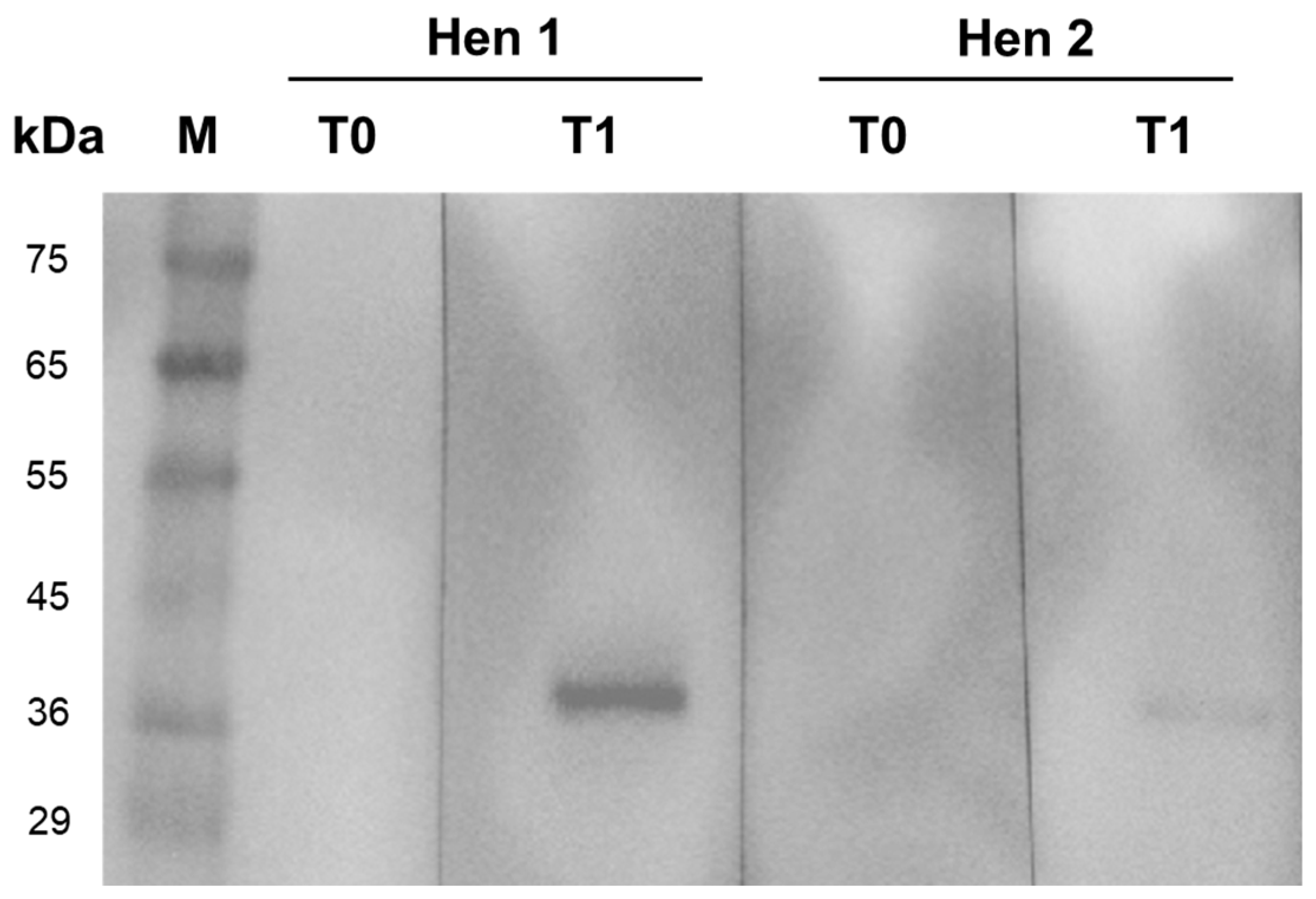
2.4. IgY Characterization by SDS-PAGE and Western Blot Analysis

We initially confirmed the presence of IgY in the egg yolks using sodium dodecyl sulfate-polyacrylamide gel electrophoresis (SDS-PAGE) with a Protean II electrophoresis system (Bio-Rad, Hercules, CA, USA) by following the discontinuous buffer system method [45]. The collected egg yolks were electrophoresed in a 15% resolving SDS-PAGE gel under reducing conditions at 120 V for 70 min, and the protein bands were visualized using a Coomassie blue staining solution (Bio-Rad, Hercules, CA, USA). The protein band sizes were determined using protein molecular marker standards (AccuProtein Chroma range 16–250 kDa, Enzmart Biotech, Bangkok, Thailand).
Western blot analysis was subsequently conducted to confirm the immunogenic property of anti-TiLV-S4 IgY against the purified virus. One microgram of TiLV, which had been purified using a sucrose gradient [46], was electrophoresed in 12% SDS-PAGE at 120 V for 80 min and transferred to a polyvinylidene fluoride membrane (Bio-Rad, Hercules, CA, USA) using a mini blot transfer system. The membrane was washed three times with 0.1% Tween-20 in PBS (PBS-T) and then incubated overnight at 4 °C with a blocking buffer comprising 3% bovine serum albumin (BSA) in PBS-T. Following three washes with PBS-T, the membrane was incubated with anti-TiLV-S4 IgY at a dilution of 1:100 for 1 h at room temperature. The membrane was washed again with PBS-T and incubated with a horseradish peroxidase-labelled goat anti-chicken IgY antibody (Abcam, Carlsbad, CA, USA) at a dilution of 1:2000 for 1 h at room temperature. After three final washes with PBS-T, the enhanced chemiluminescence substrate (LumiflashTM, Visual Protein, Taipei, Taiwan) was applied and visualized using the ChemiDoc MPTM Imaging System (Bio-Rad, Hercules, CA, USA).

2.5. In Vitro TiLV Neutralization Using Anti-TiLV-S4 IgY Antibodies

The anti-TiLV-S4 IgY antibody was serially diluted two-fold with Leibovitz’s L-15 medium (Sigma-Aldrich, St. Louis, MO, USA) to obtain dilutions of 1:2, 1:5, and 1:10. The prepared antibody was mixed with 103 TCID50/mL of TiLV strain VET-KUTV08 at a ratio of 1:1. The mixture was incubated in an Eppendorf® ThermoMixer C (Eppendorf, Hamburg, Germany) at 25 °C for 2 h with continuous shaking at 400 rpm. The positive control consisted of 103 TCID50/mL TiLV mixed with an L-15 medium at a ratio of 1:1, and the negative control was the L-15 medium only. One hundred microliters of these mixtures and controls were added to a 96-well plate containing confluent E-11 cells, with four replicates per dilution. The cells were incubated at 25 °C for 1 h, then the mixtures were discarded, and a new L-15 medium supplemented with 2% FBS was added to each well. The cells were grown continuously for 7–12 days at 25 °C and monitored daily for the cytopathic effect (CPE) under an inverted microscope (CKX53, Olympus, Tokyo, Japan).

2.6. Virus Titration

To quantify the viral concentration following inhibition by the anti-TiLV IgY antibodies, the cells were initially lysed using a freeze–thaw process. The media from each replicate well of each dilution were pooled and transferred to 1.5 mL microcentrifuge tubes. The samples were centrifuged at 3000× g for 10 min at 4 °C. The supernatant was collected and serially diluted 10-fold to obtain dilutions ranging from 10−1 to 10−8. The diluted samples were added to 96-well plates that contained confluent E-11 cells with five replicate wells per sample. The plates were incubated at 25 °C for 1 h. Following incubation, the media in each well were replaced with an L-15 media supplemented with 2% FBS, and the plates were incubated at 25 °C for 7–12 days. The plates were observed daily for CPEs. The 50% tissue culture infectious dose (TCID50) was calculated using the Reed and Muench method [47].

2.7. Immunofluorescence Assay to Detect TiLV in the RHTiB Cell Line

An immunofluorescence assay (IFA) targeting TiLV was used to confirm the inhibition of viral entry into RHTiB cells (a cell line from the brain tissue of red hybrid tilapia) [48]. Briefly, the RHTiB cells were cultured in Leibovitz’s L-15 medium supplemented with 10% FBS, 100 U/mL penicillin, 0.1 mg/mL streptomycin, and 0.25 µg/mL amphotericin B at a pH of 7.4 at 25 °C without CO2. When the cells achieved 70% confluence, they were trypsinized, counted, and diluted to a concentration of 2 × 10⁵ cells/mL. Subsequently, 500 µL of the cell suspension was seeded onto cell culture slides (SPL Life Science, Pocheon-si, Gyeonggi-do, Republic of Korea) in an L-15 medium containing 10% FBS and incubated until 80–90% confluence was achieved. The cells were infected using different methods: 100 µL TiLV mixed with 100 µL L-15 medium (positive control), TiLV mixed with a 1:2 or 1:10 IgY solution, TiLV mixed with IgY at T0, control egg, and control L-15 medium (negative control). These mixtures were incubated at 25 °C with continuous shaking at 400 rpm for 2 h (Eppendorf® ThermoMixer® C, AG, Darmstadt, Germany). The cells were washed twice with an L-15 medium without FBS, incubated with the virus mixtures for 1 h, and subsequently replaced with an L-15 medium containing 2% FBS, followed by incubation at 25 °C for 24 h.
The cells were fixed with ice-cold 100% methanol for 10 min and washed twice with PBS. They were then treated with 0.3% Triton X-100 in PBS for 10 min and washed again with PBS. The membrane was blocked with 2% BSA in PBS for 30 min to prevent nonspecific binding, followed by overnight incubation at 4 °C with the primary antibody (IgG of TiLV) in a blocking solution at a 1:100 dilution. The cells were then incubated with the secondary antibody (goat anti-rabbit IgG H&L Alexa FluorTM 488; Abcam, Carlsbad, CA, USA) in PBS at a dilution of 1:500 for 1 h at room temperature and washed with PBS. The cell nuclei were stained with 4′,6-diamidino-2-phenylindole (DAPI; Sigma-Aldrich, St. Louis, MO, USA) at a 1:1000 dilution for 15 min, washed with PBS, mounted with ProLongTM Gold Antifade reagent (Invitrogen, Thermo Fisher Scientific, Waltham, MA, USA) on glass slides, and then a cover glass was placed over them. All images were captured using a confocal microscope (Fluoview 3000, Olympus, Tokyo, Japan), which confirmed the specific binding of IgY to the TiLV-infected cells through the colocalization of the DAPI and Alexa Fluor signals. The fluorescence intensity values were quantified using cellSens Dimension software version 2.3 (Olympus, Tokyo, Japan). Five areas were randomly selected, and the fluorescence intensity of the green signal, which indicated positive TiLV-infected cells, was measured. The green signal intensity was analyzed as mean ± standard deviation (SD) and compared with the positive control, egg control, and TiLV mixed with the 1:2 or 1:10 IgY solutions.

2.8. Statistical Analysis

The viral titer and fluorescence intensity were presented as mean ± SD. The mean fluorescence intensity data between different IgY concentrations were assessed using one-way analysis of variance (ANOVA), and the TCID50 of the virus between different treatment and time points of infection was analyzed using two-way ANOVA. GraphPad Prism software version 8.0 was used in the analysis, and a p-value less than 0.05 was considered statistically significant.

3. Results

3.1. Immunization and Preparation of Chicken IgY

All the hens immunized with crude protein containing rTiLV-S4 via intramuscular injections remained healthy with no observed abnormalities or mortality throughout the study period. Furthermore, no signs of inflammation, adverse reactions, or pathological changes were detected at the injection sites. The IgY antibody purification process revealed protein bands at approximately 20 kDa and 60 kDa, which corresponded to light (LC) and heavy chains (HC) of immunoglobulin, respectively (Supplementary Figure S2). Additionally, an impurity band at approximately 35 kDa, identified as the C-terminal fragment of the vitellogenin II precursor, was also observed [49].
The specific binding of the purified IgY antibodies against rTiLV-S4 extracted from the eggs of two hens was evaluated using Western blot analysis at T0 and T1 (Figure 1). The analysis confirmed the presence of specific binding in the post-immunization samples, which was indicated by a distinct 38 kDa band corresponding to TiLV. Notably, the binding signal was stronger in the samples from the first hen (Hen 1) compared to the second hen (Hen 2). Importantly, no nonspecific binding was observed in any of the samples, which suggested the specificity of the purified IgY antibodies.

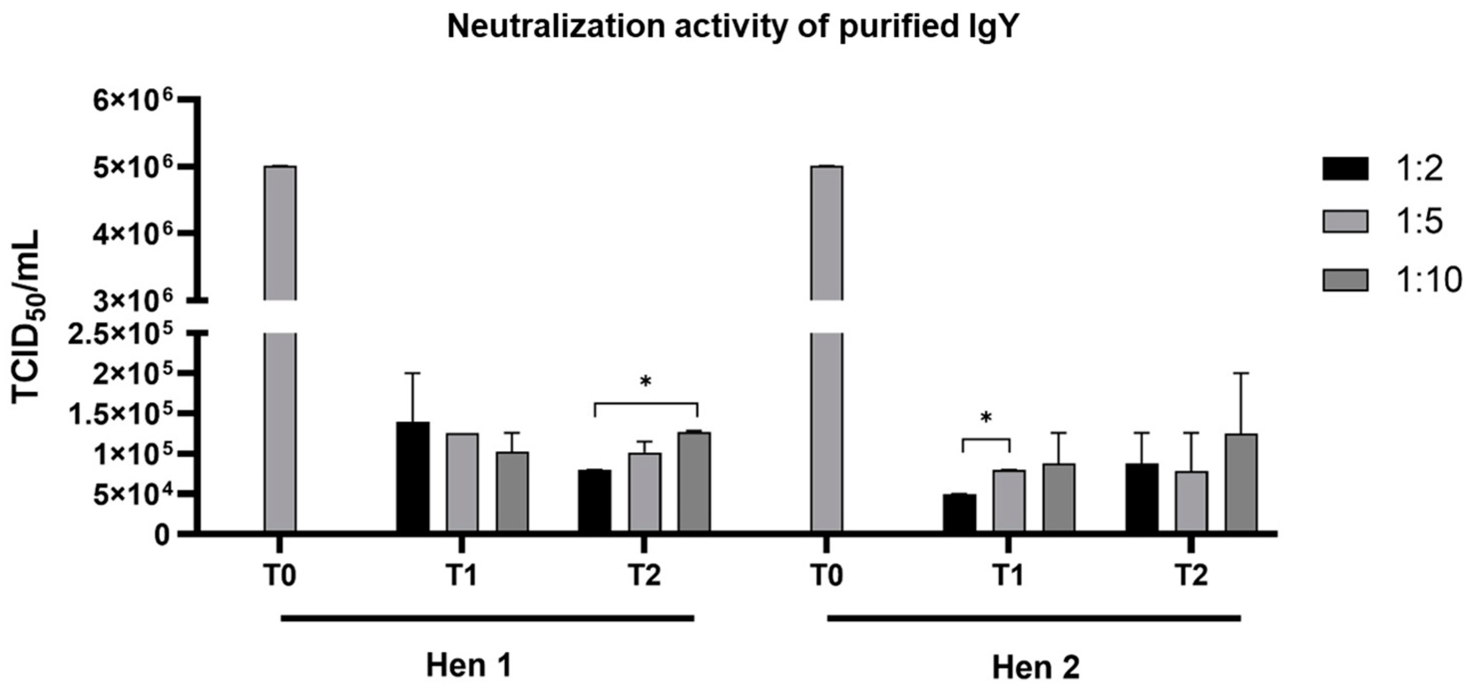
3.2. Neutralization of IgY Against TiLV

The neutralization activity of the purified IgY dilutions obtained from the eggs of the two hens at T1 and T2 post-immunization was evaluated at various dilutions (1:2, 1:5, and 1:10) against TiLV infectivity in E-11 cells (Figure 2). All the E-11 cells pretreated with anti-TiLV-S4 IgY, irrespective of the dilution, exhibited significantly reduced TiLV infection levels compared to the positive control, which had an infectivity of 5.01 × 10⁶ TCID50/mL. Of note, no statistical differences in TiLV infectivity were apparent between the cells neutralized with IgY from the T1 and T2 groups of either hen. For the IgY purified from Hen 1 at T2, the 1:2 dilution resulted in a significantly lower infectivity level of 7.94 × 10⁴ TCID50/mL compared to the 1:10 dilution, which yielded 1.26 × 10⁵ TCID50/mL. Similarly, for the IgY purified from Hen 2 at T1, the 1:2 dilution achieved a TiLV infectivity level of 5.01 × 10⁴ TCID50/mL, which was significantly lower than the 7.94 × 10⁴ TCID50/mL observed with the 1:5 dilution.
The neutralization activity of the purified IgY against TiLV was further assessed based on the presence of the CPE in the E-11 cells (Figure 3). In comparison to the uninfected E-11 cells, which exhibited no signs of the CPE (Figure 3A,B), the cells inoculated with TiLV as a positive control displayed a noticeable CPE at 7 days postinfection (dpi) (Figure 3C,D). When TiLV was incubated with IgY at different dilutions, a dose-dependent neutralization effect was observed. Specifically, the lowest level of CPE was detected in the group treated with IgY diluted at a ratio of 1:2, which demonstrated the highest neutralization efficiency (Figure 3E,F). In contrast, the cells treated with IgY diluted at a ratio of 1:5 showed an increased level of the CPE (Figure 3G,H), while those treated with IgY diluted at a ratio of 1:10 had CPE levels comparable to those of the positive control (Figure 3I,J). Based on these findings, the IgY purified from Hen 2 at T1 with a 1:2 dilution demonstrated the lowest TiLV infectivity in the E-11 cells and was selected for further study.

3.3. Immunofluorescence Assay

The neutralizing activities of the purified IgY in inhibiting TiLV entry into the RHTiB cells were further evaluated using an immunofluorescence assay (Figure 4). In comparison to the uninfected control cells, which showed no green fluorescence signals (Figure 4A–C), the TiLV-infected cells without IgY treatment (positive control) showed strong green fluorescence within the RHTiB cells at 1 dpi (Figure 4D–F). The infected cells incubated with the IgY samples from the pre-immunization period (egg control) exhibited similar levels of the green fluorescence signal (Figure 4G–I). Interestingly, treatment with the purified IgY remarkably reduced the presence of TiLV in the RHTiB cells. The cells treated with IgY at a dilution of 1:2 demonstrated a substantial reduction in green fluorescence intensity compared to the positive control (Figure 4J–L). However, although the cells treated with IgY at a 1:10 dilution showed higher fluorescence intensity relative to the 1:2 dilution, the intensity remained considerably lower than that of the positive control (Figure 4M–O).
When comparing the mean fluorescence intensity (MFI) values among the positive control, egg control, and IgY-treated groups (dilutions of 1:2 and 1:10), the positive control showed the highest MFI (1681 ± 171.5), followed by the egg control (1586 ± 19.78). The group treated with IgY at a 1:2 dilution showed an MFI of 1497 ± 17.18, while the group treated with IgY at a 1:10 dilution had an MFI of 1576 ± 13.59. Further statistical analysis revealed that the cells treated with IgY at a 1:2 dilution had significantly lower MFI values compared to the egg control and cells treated with IgY at 1:10 dilution groups (* p < 0.05 and *** p < 0.001, respectively) (Figure 4P).

4. Discussion

Infection with TiLV causes serious disease in tilapia and poses a major challenge to global tilapia aquaculture. Various strategies are being explored to prevent the spread of infection and reduce the impacts of TiLV disease. Current approaches to TiLV mitigation include vaccine development [41], the application of probiotics [50], and the use of feed additives [51]. However, these strategies either require prolonged development and regulatory approval or may not provide immediate protection during outbreaks. Passive immunization using IgY offers a complementary approach characterized by noninvasive production, high specificity, cost effectiveness, and minimal ethical concerns. These attributes position IgY as a valuable tool for enhancing biosecurity and sustainability in aquaculture [28]. In this study, we successfully generated TiLV-specific IgY antibodies by immunizing laying hens with the rTiLV-S4 protein expressed in E. coli [41]. The immunization protocol involved the intramuscular administration of rTiLV-S4 formulated with Freund’s complete adjuvant for the initial dose, followed by booster doses with Freund’s incomplete adjuvant [44]. A significant increase in IgY levels was detected in the eggs of the hens immunized with the antigen, thereby confirming that this immunization strategy effectively stimulated robust IgY production, which is consistent with the findings of previous studies [52]. The specificity of the purified IgY was confirmed by Western blot analysis, which demonstrated strong binding to the TiLV-S4 without nonspecific interactions. Upon antigen exposure, the immune systems of the hens produced specific antibodies, which were then transferred to the egg yolks [29]. To purify these IgY antibodies, the egg yolks were subjected to PEG precipitation, a widely used and cost-effective method for large-scale antibody extraction [53]. This process effectively removed lipids and enriched the IgY concentration, which ensured antibody stability and solubility [54]. Optimized PEG precipitation techniques have been shown to improve IgY purity and yield, which supports the feasibility of IgY for broader aquaculture applications [55]. In our study, both HCs and LCs were detected, and a specific band demonstrating the interaction between the antibody and rTiLV-S4 was confirmed. After PEG precipitation, the neutralizing activity of the purified IgY against TiLV was proved in a continuous cell line. Interestingly, neutralization assays using different concentrations of IgY revealed the dose-dependent inhibition of TiLV, with the highest antiviral efficacy at a 1:2 dilution. These findings are consistent with previous studies demonstrating the dose-dependent effects of IgY-mediated inhibition on viral and bacterial pathogens in fish [21,39]. In particular, our study showed that IgY antibodies effectively reduced the viral load and mitigated CPE, which highlights their potential as a therapeutic approach against this emerging virus. Although variations in infectivity were detected when tissue was treated with IgY from different hens, its ability to reduce the CPE in TiLV-infected E-11 cells remained consistent, which highlights the antiviral potential of IgY. It should be considered that the variation in neutralization efficiency among the IgY samples may have been influenced by individual differences in immune responses to the same antigenic stimulation. Factors such as genetic background, age, breed, and egg-laying capacity have been reported to influence antibody production in laying hens [23,29]. These findings emphasize the need for further optimization of immunization protocols to ensure the consistency and potency of IgY antibodies against TiLV.
The neutralization activity of IgY was further confirmed using an immunofluorescence assay, which demonstrated a significant reduction in TiLV antigens in RHTiB cells, with the highest inhibition observed at a 1:2 dilution of purified IgY. Mechanistically, IgY may inhibit viral replication by blocking viral entry into host cells through specific binding to viral proteins, thereby preventing their attachment to the host cell receptors [38,56]. This evidence supports the potential of IgY as an effective antiviral strategy for controlling TiLV infection. Despite these promising findings, however, challenges remain in optimizing IgY deployment for field applications. Effective implementation in aquaculture settings requires key factors, such as optimal fish size, dosage, and delivery methods, to be addressed [23]. Such factors must be thoroughly researched and validated before IgY can be adopted for use in fish farms. For example, while the administration methods of intraperitoneal and intramuscular injections ensure systemic immunity, they are labor-intensive and impractical for large-scale aquaculture operations [57]. As a noninvasive alternative, oral administration may be suitable for targeting gastrointestinal and systemic pathogens; however, its efficacy may be limited by enzymatic degradation in the digestive tract [58]. Recent advancements in encapsulation technologies, such as chitosan–alginate microcapsules, have improved IgY stability and bioavailability, which suggests the future feasibility of oral delivery for large-scale aquaculture applications [59]. Therefore, encapsulating IgY in coated feed pellets or specialized formulations to enhance its stability and protect against enzymatic degradation presents a promising approach for effective oral delivery in fish farming. Nevertheless, further research is needed to establish standardized protocols for TiLV-specific IgY production and delivery to ensure consistency, efficacy, and practical implementation in the management of TiLV infection using IgY.

5. Conclusions

Passive immunization with IgY presents a sustainable and ethical approach to disease control in aquaculture. In our study, we demonstrated the potential application of IgY antibodies to mitigate TiLV infections in tilapia. The rTiLV-S4-specific IgY antibodies derived from laying hens had a dose-dependent neutralization effect and significantly reduced viral infectivity and the CPE in E-11 cells. Immunofluorescence assays further confirmed the inhibitory properties of IgY and revealed its ability to prevent TiLV infection in RHTiB cells. Despite these advantages, challenges remain in optimizing IgY deployment, particularly in determining the most effective administration routes and ensuring long-term stability. Further studies should focus on developing scalable application strategies to maximize the impact of IgY immunization on disease prevention in commercial fish farming.

Supplementary Materials

The following supporting information can be downloaded at https://www.mdpi.com/article/10.3390/v17030448/s1, Figure S1: The schematic illustrates the immunization protocol and egg collection schedule for the production of chicken egg polyclonal IgY against tilapia lake virus (TiLV); Figure S2: Gel electrophoresis of the IgY samples purified using polyethylene glycol (PEG) precipitation at different stages.

Author Contributions

Conceptualization, P.M., N.P., P.S., T.L., N.T. and W.S.; methodology, P.S., J.Y., P.T., M.K., P.M., N.P. and T.L.; validation, P.S., P.T., J.Y., M.K. and T.L.; formal analysis, P.S. and T.L.; investigation, P.S., P.T., J.Y., M.K. and T.L.; resources, W.S.; data curation, P.S., T.L. and W.S.; writing—original draft preparation, P.S., J.Y., T.L., N.T. and W.S.; writing—review and editing, P.S., J.Y., P.M., N.P., T.L., N.T. and W.S.; visualization, P.S., T.L. and W.S.; supervision, N.T. and W.S.; project administration, N.T. and W.S.; funding acquisition, W.S. All authors have read and agreed to the published version of the manuscript.

Funding

This project received financial support from the Faculty of Veterinary Medicine, Kasetsart University. The research was partly supported by the Kasetsart University Research and Development Institute under project number FF(KU)51.67.

Institutional Review Board Statement

The animal study protocol was approved by the Institutional Animal Care and Use Committee of Kasetsart University (protocol number ACKU65-VET-088) for animal handling and care.

Informed Consent Statement

Not applicable.

Data Availability Statement

The data presented in this study are available on request from the corresponding author. The data are not publicly available due to the anonymity granted to all participating parties.

Acknowledgments

We would like to thank the Central Laboratory (CTL), Center for Veterinary Research and Innovation, Faculty of Veterinary Medicine, Kasetsart University.

Conflicts of Interest

The authors declare no conflicts of interest.

References

  1. Surachetpong, W.; Roy, S.R.K.; Nicholson, P. Tilapia lake virus: The story so far. J. Fish Dis. 2020, 43, 1115–1132. [Google Scholar] [CrossRef]
  2. Kembou-Ringert, J.E.; Steinhagen, D.; Readman, J.; Daly, J.M.; Adamek, M. Tilapia Lake Virus Vaccine Development: A Review on the Recent Advances. Vaccines 2023, 11, 251. [Google Scholar] [CrossRef] [PubMed]
  3. Bacharach, E.; Mishra, N.; Briese, T.; Zody, M.C.; Tsofack, J.E.K.; Zamostiano, R.; Berkowitz, A.; Ng, J.; Nitido, A.; Corvelo, A.; et al. Characterization of a Novel Orthomyxo-like Virus Causing Mass Die-Offs of Tilapia. mBio 2016, 7, e00431-16. [Google Scholar] [CrossRef] [PubMed]
  4. Koonin, E.V.; Krupovic, M.; Surachetpong, W.; Wolf, Y.I.; Kuhn, J.H. ICTV Virus Taxonomy Profile: Amnoonviridae 2023. J. Gen. Virol. 2023, 104, 001903. [Google Scholar] [CrossRef] [PubMed]
  5. Clyde, C.W.; Tan, J.P.; Yeap, S.K.; Yong, C.Y. Current updates on viral infections affecting tilapia. In Aquaculture and Fisheries; Elsevier: Amsterdam, The Netherlands, 2024. [Google Scholar] [CrossRef]
  6. Liamnimitr, P.; Thammatorn, W.; U-thoomporn, S.; Tattiyapong, P.; Surachetpong, W. Non-lethal sampling for Tilapia Lake Virus detection by RT-qPCR and cell culture. Aquaculture 2018, 486, 75–80. [Google Scholar] [CrossRef]
  7. Kembou-Ringert, J.E.; Hotio, F.N.; Steinhagen, D.; Thompson, K.D.; Surachetpong, W.; Rakus, K.; Daly, J.M.; Goonawardane, N.; Adamek, M. Knowns and unknowns of TiLV-associated neuronal disease. Virulence 2024, 15, 2329568. [Google Scholar] [CrossRef]
  8. Yamkasem, J.; Tattiyapong, P.; Kamlangdee, A.; Surachetpong, W. Evidence of potential vertical transmission of tilapia lake virus. J. Fish Dis. 2019, 42, 1293–1300. [Google Scholar] [CrossRef]
  9. Dong, H.T.; Senapin, S.; Gangnonngiw, W.; Nguyen, V.V.; Rodkhum, C.; Debnath, P.P.; Delamare-Deboutteville, J.; Mohan, C.V. Experimental infection reveals transmission of tilapia lake virus (TiLV) from tilapia broodstock to their reproductive organs and fertilized eggs. Aquaculture 2020, 515, 734541. [Google Scholar] [CrossRef]
  10. Taengphu, S.; Kayansamruaj, P.; Kawato, Y.; Delamare-Deboutteville, J.; Mohan, C.V.; Dong, H.T.; Senapin, S. Concentration and quantification of Tilapia tilapinevirus from water using a simple iron flocculation coupled with probe-based RT-qPCR. PeerJ 2022, 10, e13157. [Google Scholar] [CrossRef]
  11. Prasartset, T.; Surachetpong, W. Simultaneous detection of three important viruses affecting tilapia using a multiplex PCR assay. J. Fish Dis. 2023, 46, 459–464. [Google Scholar] [CrossRef]
  12. Thammatorn, W.; Rawiwan, P.; Surachetpong, W. Minimal risk of tilapia lake virus transmission via frozen tilapia fillets. J. Fish Dis. 2019, 42, 3–9. [Google Scholar] [CrossRef] [PubMed]
  13. Aich, N.; Paul, A.; Choudhury, T.G.; Saha, H. Tilapia Lake Virus (TiLV) disease: Current status of understanding. Aquac. Fish. 2022, 7, 7–17. [Google Scholar] [CrossRef]
  14. Jaemwimol, P.; Sirikanchana, K.; Tattiyapong, P.; Mongkolsuk, S.; Surachetpong, W. Virucidal effects of common disinfectants against tilapia lake virus. J. Fish Dis. 2019, 42, 1383–1389. [Google Scholar] [CrossRef] [PubMed]
  15. Barría, A.; Trinh, T.Q.; Mahmuddin, M.; Benzie, J.A.H.; Chadag, V.M.; Houston, R.D. Genetic parameters for resistance to Tilapia Lake Virus (TiLV) in Nile tilapia (Oreochromis niloticus). Aquaculture 2020, 522, 735126. [Google Scholar] [CrossRef]
  16. Lertwanakarn, T.; Trongwongsa, P.; Yingsakmongkol, S.; Khemthong, M.; Tattiyapong, P.; Surachetpong, W. Antiviral Activity of Ribavirin against Tilapia tilapinevirus in Fish Cells. Pathogens 2021, 10, 1616. [Google Scholar] [CrossRef] [PubMed]
  17. Tattiyapong, P.; Kitiyodom, S.; Yata, T.; Jantharadej, K.; Adamek, M.; Surachetpong, W. Chitosan nanoparticle immersion vaccine offers protection against tilapia lake virus in laboratory and field studies. Fish Shellfish Immunol. 2022, 131, 972–979. [Google Scholar] [CrossRef]
  18. He, T.; Zhang, Y.-Z.; Gao, L.-H.; Miao, B.; Zheng, J.-S.; Pu, D.-C.; Zhang, Q.-Q.; Zeng, W.-W.; Wang, D.-S.; Su, S.-Q.; et al. Identification and pathogenetic study of tilapia lake virus (TiLV) isolated from naturally diseased tilapia. Aquaculture 2023, 565, 739166. [Google Scholar] [CrossRef]
  19. Wang, B.; Thompson, K.D.; Wangkahart, E.; Yamkasem, J.; Bondad-Reantaso, M.G.; Tattiyapong, P.; Jian, J.; Surachetpong, W. Strategies to enhance tilapia immunity to improve their health in aquaculture. Rev. Aquac. 2023, 15, 41–56. [Google Scholar] [CrossRef]
  20. Sparrow, E.; Friede, M.; Sheikh, M.; Torvaldsen, S. Therapeutic antibodies for infectious diseases. Bull. World Health Organ. 2017, 95, 235–237. [Google Scholar] [CrossRef]
  21. Zhang, M.; Geng, H.; Tariq Javed, M.; Xu, L.; Li, X.; Wang, L.; Li, S.; Xu, Y. Passive protection of Japanese pufferfish (Takifugu rubripes) against Vibrio harveyi infection using chicken egg yolk immunoglobulins (IgY). Aquaculture 2021, 532, 736009. [Google Scholar] [CrossRef]
  22. Wang, H.; Zhong, Q.; Lin, J. Egg Yolk Antibody for Passive Immunization: Status, Challenges, and Prospects. J. Agric. Food Chem. 2023, 71, 5053–5061. [Google Scholar] [CrossRef] [PubMed]
  23. Kovacs-Nolan, J.; Mine, Y. Egg Yolk Antibodies for Passive Immunity. Annu. Rev. Food Sci. Technol. 2012, 3, 163–182. [Google Scholar] [CrossRef] [PubMed]
  24. Pantaleo, G.; Correia, B.; Fenwick, C.; Joo, V.S.; Perez, L. Antibodies to combat viral infections: Development strategies and progress. Nat. Rev. Drug Discov. 2022, 21, 676–696. [Google Scholar] [CrossRef] [PubMed]
  25. Hanly, W.C.; Artwohl, J.E.; Bennett, B.T. Review of Polyclonal Antibody Production Procedures in Mammals and Poultry. ILAR J. 1995, 37, 93–118. [Google Scholar] [CrossRef]
  26. El-Kafrawy, S.A.; Abbas, A.T.; Oelkrug, C.; Tahoon, M.; Ezzat, S.; Zumla, A.; Azhar, E.I. IgY antibodies: The promising potential to overcome antibiotic resistance. Front. Immunol. 2023, 14, 1065353. [Google Scholar] [CrossRef]
  27. Härtle, S.; Magor, K.E.; Göbel, T.W.; Davison, F.; Kaspers, B. Chapter 6—Structure and Evolution of Avian Immunoglobulins. In Avian Immunology, 2nd ed.; Schat, K.A., Kaspers, B., Kaiser, P., Eds.; Academic Press: Boston, MA, USA, 2014; pp. 103–120. [Google Scholar]
  28. Pereira, E.P.V.; van Tilburg, M.F.; Florean, E.O.P.T.; Guedes, M.I.F. Egg yolk antibodies (IgY) and their applications in human and veterinary health: A review. Int. Immunopharmacol. 2019, 73, 293–303. [Google Scholar] [CrossRef]
  29. Schade, R.; Calzado, E.G.; Sarmiento, R.; Chacana, P.A.; Porankiewicz-Asplund, J.; Terzolo, H.R. Chicken Egg Yolk Antibodies (IgY-technology): A Review of Progress in Production and Use in Research and Human and Veterinary Medicine. Altern. Lab. Anim. 2005, 33, 129–154. [Google Scholar] [CrossRef]
  30. Xu, Y.; Li, X.; Jin, L.; Zhen, Y.; Lu, Y.; Li, S.; You, J.; Wang, L. Application of chicken egg yolk immunoglobulins in the control of terrestrial and aquatic animal diseases: A review. Biotechnol. Adv. 2011, 29, 860–868. [Google Scholar] [CrossRef]
  31. Gutierrez, M.A.; Miyazaki, T.; Hatta, H.; Kim, M. Protective properties of egg yolk IgY containing anti-Edwardsiella tarda antibody against paracolo disease in the Japanese eel, Anguilla japonica Temminck & Schlegel. J. Fish Dis. 1993, 16, 113–122. [Google Scholar] [CrossRef]
  32. Norqvist, A.; Hagström, A.; Wolf-Watz, H. Protection of rainbow trout against vibriosis and furunculosis by the use of attenuated strains of Vibrio anguillarum. Appl. Environ. Microbiol. 1989, 55, 1400–1405. [Google Scholar] [CrossRef]
  33. Lee, S.B.; Mine, Y.; Stevenson, R.M.W. Effects of Hen Egg Yolk Immunoglobulin in Passive Protection of Rainbow Trout against Yersinia ruckeri. J. Agric. Food Chem. 2000, 48, 110–115. [Google Scholar] [CrossRef] [PubMed]
  34. Liu, X.; Xiao, H.; Chao, J.; Jian, S.; Wu, X.; Lu, J.; Wang, J.; Chen, C.; Liu, Y. Polyvalent passive vaccine candidates from egg yolk antibodies (IgY) of important outer membrane proteins (PF1380 and ExbB) of Pseudomonas fluorescens in fish. Fish Shellfish Immunol. 2023, 143, 109211. [Google Scholar] [CrossRef] [PubMed]
  35. Liu, X.; Xiao, H.; Cui, P.; Chen, J.; Chao, J.; Wu, X.; Lu, J.; Zhang, X.; Xu, G.; Liu, Y. Differential polyvalent passive immune protection of egg yolk antibodies (IgY) against live and inactivated Vibrio fluvialis in fish. Fish Shellfish Immunol. 2024, 151, 109751. [Google Scholar] [CrossRef] [PubMed]
  36. Gan, H.; He, H.; Sato, A.; Hatta, H.; Nakao, M.; Somamoto, T. Ulcer disease prophylaxis in koi carp by bath immersion with chicken egg yolk containing anti-Aeromonas salmonicida IgY. Res. Vet. Sci. 2015, 99, 82–86. [Google Scholar] [CrossRef]
  37. Zhenxing, L.; Ke, H.; Yanping, M.; Le, H.T.H.; Feng, G.-q.; Jiangyao, M.; Zhiling, L.; Li, Y. Oral Passive Immunization of Carp Cyprinus carpio with Anti-CyHV-3 Chicken Egg Yolk Immunoglobulin (IgY). Fish Pathol. 2014, 49, 113–120. [Google Scholar]
  38. Yi, L.; Qin, Z.; Lin, H.; Zhou, Y.; Li, J.; Xu, Z.; Babu, V.S.; Lin, L. Features of chicken egg yolk immunoglobulin (IgY) against the infection of red-spotted grouper nervous necrosis virus. Fish Shellfish Immunol. 2018, 80, 534–539. [Google Scholar] [CrossRef]
  39. Liu, J.; Qin, Y.; Yan, L.; Liu, W.; Shi, H.; Lu, Y.; Liu, X. Protective effects of egg yolk immunoglobulins (IgY) on juvenile groupers (Epinephelus fuscoguttatus × Epinephelus lanceolatus) with red-spotted grouper nervous necrosis virus infection. Aquaculture 2021, 545, 737218. [Google Scholar] [CrossRef]
  40. Tattiyapong, P.; Sirikanchana, K.; Surachetpong, W. Development and validation of a reverse transcription quantitative polymerase chain reaction for tilapia lake virus detection in clinical samples and experimentally challenged fish. J. Fish Dis. 2018, 41, 255–261. [Google Scholar] [CrossRef]
  41. Sanyalukruechai, C.; Watthanasakphuban, N.; Khemthong, M.; Surachetpong, W.; Rattanaporn, K. Expression and purification of recombinant tilapia lake virus segment 4 protein and its in-vitro biological activity for potential use in vaccine development. Sci. Rep. 2024, 14, 31529. [Google Scholar] [CrossRef]
  42. Polson, A.; von Wechmar, M.B.; van Regenmortel, M.H.V. Isolation of Viral IgY Antibodies from Yolks of Immunized Hens. Immunol. Commun. 1980, 9, 475–493. [Google Scholar] [CrossRef]
  43. Pauly, D.; Chacana, P.; Gutierrez Calzado, E.J.; Brembs, B.; Schade, R. IgY Technology: Extraction of Chicken Antibodies from Egg Yolk by Polyethylene Glycol (PEG) Precipitation. J. Vis. Exp. 2011, 51, e3084. [Google Scholar] [CrossRef]
  44. Metheenukul, P.; Surachetpong, W.; Prasertsincharoen, N.; Arreesrisom, P.; Thengchaisri, N. Comparison of immunoglobulin Y antibody production in new and spent laying hens. Vet. World 2024, 17, 2177–2184. [Google Scholar] [CrossRef] [PubMed]
  45. Laemmli, U.K. Cleavage of Structural Proteins during the Assembly of the Head of Bacteriophage T4. Nature 1970, 227, 680–685. [Google Scholar] [CrossRef]
  46. Tattiyapong, P.; Dechavichitlead, W.; Waltzek, T.B.; Surachetpong, W. Tilapia develop protective immunity including a humoral response following exposure to tilapia lake virus. Fish Shellfish Immunol. 2020, 106, 666–674. [Google Scholar] [CrossRef] [PubMed]
  47. Reed, L.J.; Muench, H. A simple method of estimating fifty per cent endpoints. Am. J. Epidemiol. 1938, 27, 493–497. [Google Scholar] [CrossRef]
  48. Mohamad, A.; Khemthong, M.; Trongwongsa, P.; Lertwanakarn, T.; Setthawong, P.; Surachetpong, W. A New Cell Line from the Brain of Red Hybrid Tilapia (Oreochromis spp.) for Tilapia Lake Virus Propagation. Animals 2024, 14, 1522. [Google Scholar] [CrossRef]
  49. Klimentzou, P.; Paravatou-Petsotas, M.; Zikos, C.; Beck, A.; Skopeliti, M.; Czarnecki, J.; Tsitsilonis, O.; Voelter, W.; Livaniou, E.; Evangelatos, G.P. Development and immunochemical evaluation of antibodies Y for the poorly immunogenic polypeptide prothymosin alpha. Peptides 2006, 27, 183–193. [Google Scholar] [CrossRef]
  50. Yang, Y.-F.; Yamkasem, J.; Surachetpong, W.; Lin, Y.-J.; You, S.-H.; Lu, T.-H.; Chen, C.-Y.; Wang, W.-M.; Liao, C.-M. Assessing the effect of probiotics on tilapia lake virus-infected tilapia: Transmission and immune response. J. Fish Dis. 2022, 45, 1117–1132. [Google Scholar] [CrossRef]
  51. Mohamad, A.; Yamkasem, J.; Paimeeka, S.; Khemthong, M.; Lertwanakarn, T.; Setthawong, P.; Nuez-Ortin, W.G.; Isern Subich, M.M.; Surachetpong, W. Efficacy of Feed Additives on Immune Modulation and Disease Resistance in Tilapia in Coinfection Model with Tilapia Lake Virus and Aeromonas hydrophila. Biology 2024, 13, 938. [Google Scholar] [CrossRef]
  52. Tabll, A.A.; Shahein, Y.E.; Omran, M.M.; Hussein, N.A.; El-Shershaby, A.; Petrovic, A.; Glasnovic, M.; Smolic, R.; Smolic, M. Monoclonal IgY antibodies: Advancements and limitations for immunodiagnosis and immunotherapy applications. Ther. Adv. Vaccines Immunother. 2024, 12, 25151355241264520. [Google Scholar] [CrossRef]
  53. Sugino, H.; Nitoda, T.; Juneja, L.R. General Chemical Composition of Hen Eggs. In Hen Eggs; CRC: Boca Raton, FL, USA, 2018; pp. 13–24. [Google Scholar]
  54. Zhang, L.; Lin, L.; Qin, Z. A review on the application of chicken immunoglobulin Y in aquaculture. Rev. Aquac. 2024, 16, 536–551. [Google Scholar] [CrossRef]
  55. Madera-Contreras, A.M.; Solano-Texta, R.; Cisneros-Sarabia, A.; Bautista-Santos, I.; Vences-Velázquez, G.; Vences-Velázquez, A.; Cortés-Sarabia, K. Optimized method for the extraction of contaminant-free IgY antibodies from egg yolk using PEG 6000. MethodsX 2022, 9, 101874. [Google Scholar] [CrossRef] [PubMed]
  56. Yang, Y.-e.; Wen, J.; Zhao, S.; Zhang, K.; Zhou, Y. Prophylaxis and therapy of pandemic H1N1 virus infection using egg yolk antibody. J. Virol. Methods 2014, 206, 19–26. [Google Scholar] [CrossRef] [PubMed]
  57. Arasteh, N.; Aminirissehei, A.H.; Yousif, A.N.; Albright, L.J.; Durance, T.D. Passive immunization of rainbow trout (Oncorhynchus mykiss) with chicken egg yolk immunoglobulins (IgY). Aquaculture 2004, 231, 23–36. [Google Scholar] [CrossRef]
  58. Wang, H.; Zeng, X.; Lin, J. Ex Vivo Evaluation of Egg Yolk IgY Degradation in Chicken Gastrointestinal Tract. Front. Immunol. 2021, 12, 746831. [Google Scholar] [CrossRef]
  59. Li, X.-Y.; Jin, L.-J.; McAllister, T.A.; Stanford, K.; Xu, J.-Y.; Lu, Y.-N.; Zhen, Y.-H.; Sun, Y.-X.; Xu, Y.-P. Chitosan−Alginate Microcapsules for Oral Delivery of Egg Yolk Immunoglobulin (IgY). J. Agric. Food Chem. 2007, 55, 2911–2917. [Google Scholar] [CrossRef]
Figure 1. Western blot analysis showing specific binding of purified IgY from two individual chickens (Hen 1 and Hen 2) to the recombinant TiLV segment 4 protein (rTiLV-S4). No binding was observed at the pre-immunization stage (T0). However, distinct bands corresponding to rTiLV-S4 at 38 kDa were detected one-week post-immunization (T1) in the samples from both hens. Lane M represents the protein marker.
Viruses 17 00448 g001
Figure 2. Neutralization activity of purified IgY against rTiLV-S4, assessed by the 50% tissue culture infective dose (TCID50)/mL. The E-11 cells treated with purified IgY from the eggs of two hens (Hen 1 and Hen 2) collected after one week (T1) and two weeks (T2) post-immunization and infected with TiLV showed significantly lower infectivity compared to the control group (pre-immunization). Bars represent the mean ± standard deviation (SD) of the measured neutralization activity. * p < 0.05.
Viruses 17 00448 g002
Figure 3. Evaluation of the neutralization activity of purified IgY from Hen 1 and Hen 2 against TiLV infectivity in E-11 cells at 7 days postinfection. The cellular morphologies are shown. (A,B) Negative control cells; (C,D) TiLV-infected cells treated with pre-immunized IgY (egg control); (E,F) infected cells treated with purified IgY at a 1:2 dilution; (G,H) 1:5 dilution; (I,J) 1:10 dilution. Asterisks (*) indicate the presence of cytopathic effects (CPE). Scale bar: 200 µm (10× magnification).
Viruses 17 00448 g003
Figure 4. Neutralization activity of purified IgY in reducing TiLV antigen levels in RHTiB cells assessed using an immunofluorescence assay (IFA). (AC) Negative control cells demonstrate only blue 4′,6-diamidino-2-phenylindole (DAPI) staining. (DF) TiLV-infected cells (positive control) show colocalization of the TiLV antigen (green fluorescence) within the cytoplasms of the RHTiB cells. (GI) Egg control group, namely RHTiB cells treated with purified IgY extracted from egg yolk during the pre-immunization period. (JL) RHTiB cells treated with purified IgY at a 1:2 dilution and infected with TiLV. (MO) RHTiB cells treated with purified IgY at a 1:10 dilution and infected with TiLV. (P) Graph showing the mean fluorescence intensity (MFI) values as mean ± SD for the positive control, egg control, and IgY-treated groups (1:2 and 1:10 dilutions). Statistically significant differences are indicated as * p < 0.05 and *** p < 0.001.
Viruses 17 00448 g004
Disclaimer/Publisher’s Note: The statements, opinions and data contained in all publications are solely those of the individual author(s) and contributor(s) and not of MDPI and/or the editor(s). MDPI and/or the editor(s) disclaim responsibility for any injury to people or property resulting from any ideas, methods, instructions or products referred to in the content.

