Abstract
Replicating RNA, including self-amplifying RNA (saRNA) and trans-amplifying RNA (taRNA), holds great potential for advancing the next generation of RNA-based vaccines. Unlike in vitro transcribed mRNA found in most current RNA vaccines, saRNA or taRNA can be massively replicated within cells in the presence of RNA-amplifying enzymes known as replicases. We recently demonstrated that this property could enhance immune responses with minimal injected RNA amounts. In saRNA-based vaccines, replicase and antigens are encoded on the same mRNA molecule, resulting in very long RNA sequences, which poses significant challenges in production, delivery, and stability. In taRNA-based vaccines, these challenges can be overcome by splitting the replication system into two parts: one that encodes replicase and the other that encodes a short antigen-encoding RNA called transreplicon. Here, we review the identification and use of transreplicon RNA in alphavirus research, with a focus on the development of novel taRNA technology as a state-of-the art vaccine platform. Additionally, we discuss remaining challenges essential to the clinical application and highlight the potential benefits related to the unique properties of this future vaccine platform.
1. Introduction
The recent success of SARS-CoV-2 mRNA vaccines exposed the profound potential of RNA therapeutics and created a broad awareness for the technology. However, challenges persist including side effects associated with innate immunity stimulation of the relatively high administration doses of 30 to 100 µg [1,2,3], and the need for repeated application to achieve durable immune responses. Efforts to reduce mRNA doses undertaken by vaccine developers often involve the usage of so called self-amplifying RNA (saRNA), an RNA format promising to minimize dosing mediated by autonomous intracellular multiplication of saRNA after transfer. Indeed, a newly licensed first saRNA-based vaccine appears to be highly effective upon injection of only 5 µg [4,5]. Additionally, it has been shown that intracellular saRNA amplification results in prolonged mRNA expression in animal studies [6], supporting the hope that saRNA may also reduce the need for repeated dosing.
Upon cellular uptake, both mRNA and saRNA-based therapeutics remain exclusively within the cytoplasm where they are translated into protein. saRNA bares the important additional feature of transient replication. Essentially, therapeutic RNA does not enter the nucleus, is not reverse transcribed and therefore avoids potential insertional mutagenesis [7]. In terms of structure, mRNA and saRNA share common features such as a 5′ cap, as well as 5′ and 3′ untranslated regions (UTRs) and a 3′ poly-A tail. Furthermore, both encode an open reading frame (ORF) of a therapeutic transgene. The therapeutic gene is transiently expressed and limited by the natural degradation of the in vitro transcribed (IVT) RNA [8]. saRNA features a second ORF encoding the enzyme complex needed for RNA replication called replicase. In addition, saRNA incorporates a short internal subgenomic promoter (SGP) that essentially enables generation of a transcript of the therapeutic transgene that is translated separately (Figure 1a,b) [9]. Advancing this technology, our group recently introduced trans-amplifying RNA (taRNA) to the vaccine field. taRNA is a derivative of saRNA that is composed of two RNAs: a non-replicating mRNA (nrRNA) encoding the replicase and a so called transreplicon (TR) encoding the therapeutic transgene that is multiplied by the replicase [10]. In taRNA, the replicase nrRNA contains the structural features of mRNA, and the TR the structural features of saRNA except for a functional replicase ORF (Figure 1c). For taRNA to function effectively, the nrRNA that encodes replicase currently needs to be supplied in similar amounts of what is administered in mRNA vaccines. Nonetheless, a distinguishing feature of taRNA is that exceptionally low amounts of transgene coding TR are sufficient to elicit effective immune responses.
Figure 1.
RNA vector platforms. (a) Non-replicating mRNA (nrRNA) consists of a transgene sequence flanked by 5′ and 3′ untranslated regions (UTR), a 5′ cap and a poly(A) tail. (b) Self-amplifying RNA (saRNA) contains the replicase gene composed of four non-structural proteins (nsPs) in the upstream position and a transgene downstream of the subgenomic promoter. It also has a 5′ cap and a 3′ poly(A) tail. saRNA possesses major conserved sequence elements (CSE), including stem loops (SL) that act as replicase recognition signals (adapted from [11]). (c) Trans-amplifying RNA (taRNA) comprises two separate RNA molecules: an nrRNA encoding the replicase and a transreplicon that encodes the transgene and contains the CSEs, ensuring its amplification by the replicase.
In this paper, we draw upon the history of transreplicating RNA discovery in alphavirus-infected cells and how understanding this RNA species continues to advance our knowledge on alphavirus biology. We review pioneering work unveiling so-called defective interfering particles (DIPs) and their exploitation, enabling generation of recombinant alphaviral particles. We highlight studies elucidating replicase functions and mechanisms of alphaviral replication before summarizing our work about how taRNA could be used as a vaccine. Finally, we provide an outlook on further work needed for optimization of taRNA and its clinical translation.
2. Natural Trans-Replicating RNA
Interestingly, trans-replicating RNA species can arise spontaneously in infected cells as a result of viral genome instability. RNA viruses are particularly prone to high mutation rates because viral RNA replicases do not contain proof-reading activity [20]. However, for viruses this genomic instability is not an evolutionary burden. It rather allows viruses to be remarkably adaptable helping them to persist and replicate within the complex physiological and immunological habitat of their host organisms, and to respond to changes in their environment. Besides point mutations, larger rearrangements and deletions of the genome can occur, giving rise to more or less largely truncated defective viral genomes (DVGs). These DVGs are unable to complete a replication cycle independently and therefore require the presence of a complementary helper virus with a full-length genome to compensate for compromised functions. Upon replication in the presence of a helper virus, DVGs can be packaged into viral particles that lack infectivity and are unable to establish a viral replication cycle. Because DVGs can potentially interfere with the formation of fully functional particles throughout the course of virus replication they have been coined “defective interfering particles” (DIPs) [21].
2.1. Investigation of Genome Sequence Requirements for Replication Using Defective Interfering Particles
In 1970, Huang and Baltimore proposed that the frequency of DIPs formation is significantly higher than what had previously been believed leading them to hypothesize that they might influence the course of viral diseases [21]. Subsequently, further studies began to provide evidence for the accumulation of DIPs through serial passaging of alphaviruses. These investigations not only shed light on the sequence requirements of the RNA genome essential for efficient replication by the replicase, but also offered insights into the molecular mechanisms underlying DIPs formation.
DIP accumulation was then observed during replication of Sindbis virus (SINV) in BHK-21 cells. Starting from a single plaque from the virus grown in chick embryo fibroblasts, the serial dilution of the virus on BHK-21 cells suggested the presence of DIPs by a significant drop of the hemagglutination titre and plaque-forming units (PFU) after several passages [22]. The idea was raised that DIPs could interfere with alphavirus replication during RNA synthesis simply because the DI RNA reduced in size would be more efficiently replicated compared to longer, unaltered virus RNA. In support of this idea, subsequent studies in various cell types, wherein DIP generation and propagation of Semliki Forest virus (SFV) was investigated, revealed that the formation of DIPs occurs at early passages. It was shown that the generation and replication of short DI RNA occurred at the expense of genomic RNA of SFV, as DI RNAs competed for the limited supply of virus RNA polymerase [23]. In most cell types, SFV DIPs evolved over time by a stepwise sequential deletion of internal regions of the standard virus genome while keeping 5′ and 3′ termini. These findings were important because they also suggested that the 5′ and 3′ terminal sequences of the alphavirus genome play a critical role for DI RNA and viral RNA replication. Subsequently, it was described that SFV DI RNA contains 106 conserved noncoding nucleotides important for replication or encapsidation located at its 3′ end that precede the poly(A) tail [24]. Sequencing of the 3′ end of genomic and subgenomic RNA of SINV, SFV and Middelburg virus confirmed that they all contained a highly conserved region of 20 nucleotides adjacent to the poly(A) tail [19]. More detailed insights into 5′ and 3′ end requirements were gathered by using a series of deletions spanning the entire SINV DI genome. For example, transfection of cells with these DI RNAs in the presence of helper SINV indicated that only sequences in the 5′-terminal 162 nucleotides and the 19 3′-terminal nucleotides are required for replication and packaging of these genomes [25]. The presence of highly conserved regions within the 5′ and 3′ termini suggested their potential recognition also by heterologous alphaviruses. Indeed, later it was shown that DIPs derived from wild-type SINV interfere with the replication of SFV and vice versa [26].
2.2. Engineered Defective Interfering RNAs to Introduce Foreign Genes into Cells
The alphavirus genome can serve as a stand-alone vector platform for gene expression. Along this line, the SINV RNA genome was successfully engineered by Xiong and colleagues to express the chloramphenicol acetyltransferase (CAT) gene downstream of the SINV subgenomic promoter. The resulting vector was shown to be self-replicating and enabled the expression of bacterial CAT in cultured insect, avian and mammalian cells. By supplying SINV structural genes in trans, vector genomic RNAs could be packaged into infectious particles that facilitate CAT expression, reflected by the production of recombinant alphaviral particles [27].
Later, it was demonstrated that SINV and its DIPs can be used as a bipartite vector to introduce foreign genes into cells [28]. The authors of this study replaced 75% of an SINV DI genome with foreign sequences, retaining a crucial 51-nucleotide segment at the 5′ end that is highly conserved in alphaviruses. Corresponding DI RNAs replicated in infected cells and were predominantly present at early passages. Notably, when the CAT gene was inserted into a DI RNA lacking this conserved region, DI RNAs’ amplification could not be detected, indicating the functional relevance of the 51-nucleotide sequence for DI RNA amplification. The findings suggested that DI RNAs could serve as vectors for introducing heterologous genes into cells.
3. Recombinant Alphavirus Production by Exploiting Defective Interfering RNA
3.1. Helper RNA
The efficient amplification and packaging of alphavirus DI genomes by a helper virus, coupled with their capacity to express foreign proteins, has paved the way for exploring and refining alphaviral vector systems. In the early 1990s, Peter Liljeström and Henrik Garoff pioneered this field by engineering SFV replicons to create a novel expression vector for mammalian cells [29]. They developed an in vivo packaging system for introducing recombinant RNA into cells through single round infectious viral particles (SRIPs). The recombinant RNA packaged into SRIPs contained transgenes downstream of the SGP, replacing the viral structural proteins while retaining the non-structural region of the SFV genome and the conserved 5′ and 3′ regions required for replication 3 cis. This recombinant RNA design equals the saRNA that is widely used today. For SRIP formation, they constructed defective ‘helper RNA’ based on DI genomes expressing the structural proteins, complementing the saRNA in trans to enable particle release. The helper RNA preserves essential 5′ and 3′ CSEs for helper replication and the entire structural region including the SGP. The SFV packaging signal located within nsP1 was conserved on the saRNA but deleted from the helper. Thereby, co-transfection of saRNA and helper RNA resulted in efficient SRIP production containing only saRNA but not helper RNA. Harvested and purified SRIPs efficiently transferred the saRNA to target cells leading to high transgene expression, while transfer and expression of helper-encoded structural proteins was avoided. The SFV system thus appeared to be a useful and safe method enabling the rapid production of recombinant virus stocks without recombination events leading to wild type SFV formation. Shortly thereafter, a comparable vector system was engineered for SINV [30]. Again, co-transfection of saRNA mediated replication and expression of structural proteins encoded by the defective helper RNAs. However, an encapsidation of defective helper RNAs was also observed despite the absence of a packaging signal. Overall, SINV saRNA transfer by SRIPs achieved comparable levels of transgene expression to the analogous SFV vector developed by Liljeström and Garoff [29].
3.2. Development of Advanced Helper RNA Systems
A few years later, Pushko et al. followed a similar approach to create an saRNA/helper RNA system from attenuated Venezuelan Equine Encephalitis virus (VEEV) [31], as with SFV and SINV expression vectors. However, their study noted the re-formation of live plaque-forming virus due to recombination events between the saRNA and the helper RNA. Since shortening of homologous sequence regions between both RNAs could not completely prevent recombination, the VEEV SRIPs system raised safety concerns. This problem could be solved by creating a bipartite helper system with one helper RNA molecule encoding the capsid and the other RNA molecule encoding the envelope proteins. Thereby, the required number of recombination events to reconstitute the genome of an infectious virus was increased, and upon co-transfer of VEEV saRNA with the two helper RNAs, the infectious virus was no longer detectable in vitro or in vivo. Additionally, immunization of mice with VEEV replicon particles elicited antibody responses against the expressed proteins. These developments essentially made the VEEV replicon system with the bipartite helper a compelling vaccine vector. A parallel study using SINV saRNA demonstrated that the co-transfection with two defective-helper RNAs facilitated high-titre SRIP production [32]. This system exhibited a high degree of safety, with no recombination events detected even after several passages. Later, Smerdou and Liljeström developed a comparable two-helper system based on the SFV RNA genome [33]. Similar to SINV, the SFV capsid integrated a translational enhancer at the 5′ end. The second helper RNA was constructed using the minimal enhancing sequence of the capsid followed by the 2A autoprotease from foot and mouth disease virus and the glycoprotein genes to express high levels of viral spike proteins. To further reduce the risk and probability of the formation of replication-competent particles, the helper RNA was engineered to carry a capsid gene with a mutation that abolishes self-cleavage capacity. This split system resulted in significantly improved safety and efficiency of packaging recombinant RNA expressing heterologous proteins.
The SRIP vector systems, comprising saRNA and helper RNAs, broadened the range of recombinant alphavirus vector applications, as potential vaccine vectors in particular (reviewed by [34]). Even though the safety of the SRIP platform increased considerably with the development of 2-helper RNA systems, concerns remained that the generation of replication-competent particles upon recombination was still occasionally detected. It was then found that SGP is required for either transgene or structural gene expression in both helper RNAs as well as the saRNA, which occasionally acted as a recombination site. A breakthrough to eliminate the concerns was thus achieved by generating helper RNAs devoid of SGPs [35]. These promoterless helper RNAs lack an SGP but still contain the viral 5′ and 3′ CSEs. Therefore, because the replicase generates only one type of transcript that is equivalent to the genomic RNA, a direct cap-dependent translation of the structural proteins is still possible. Remarkably, SRIP titres matched those produced with the previous helper RNA design. Moreover, promoterless helpers enhanced safety by introducing additional constraints on functional recombinants. In cell culture experiments, recombinants were not observed, affirming its safety. Finally, immunization of mice or non-human primates with VEEV SRIPs generated using promoterless helper RNAs encoding four vaccinia genes resulted in 100% survival upon challenge with the vaccinia virus and monkeypox virus. Essentially, this outcome solidified the SRIP vaccine platform as safe and effective.
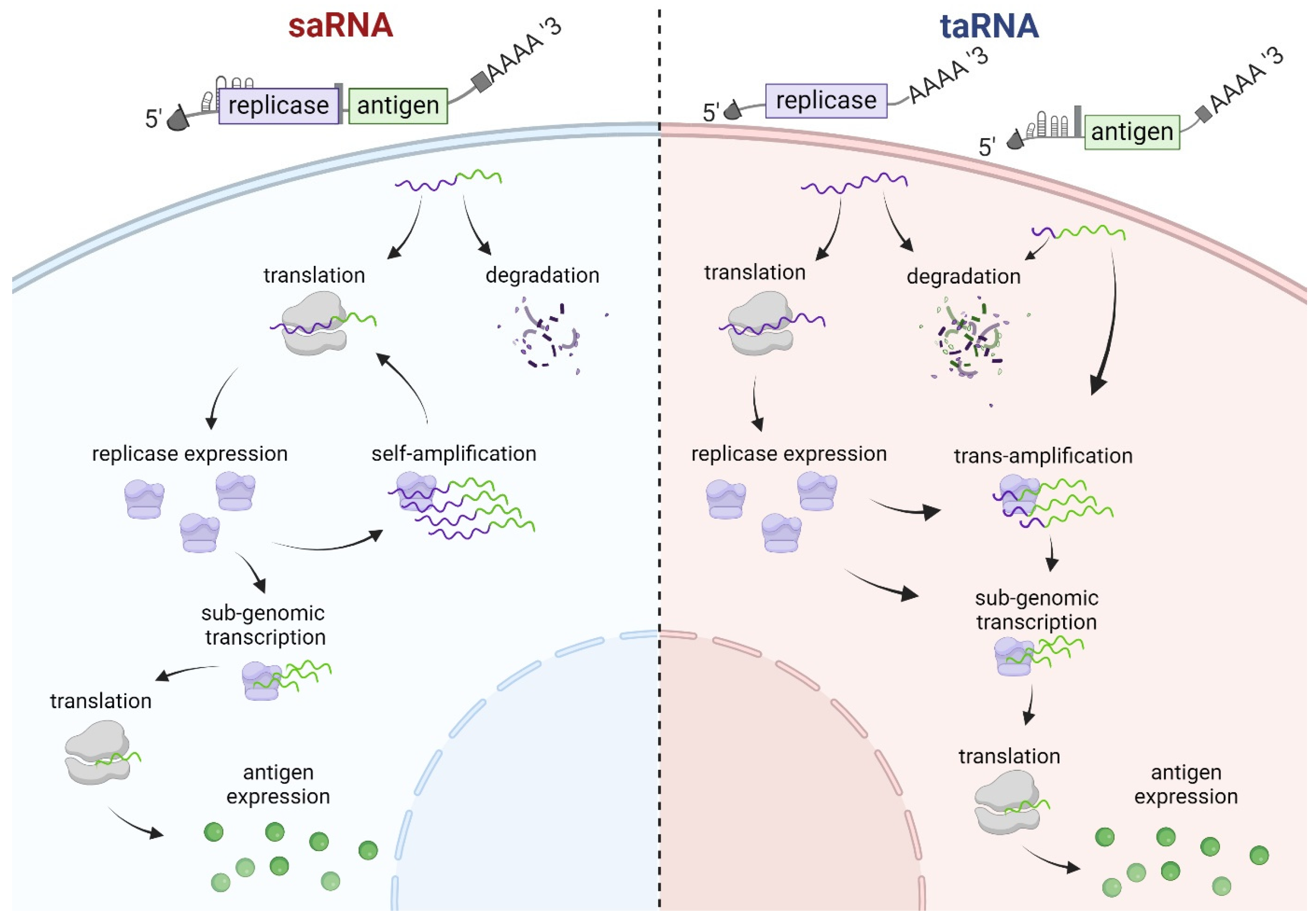
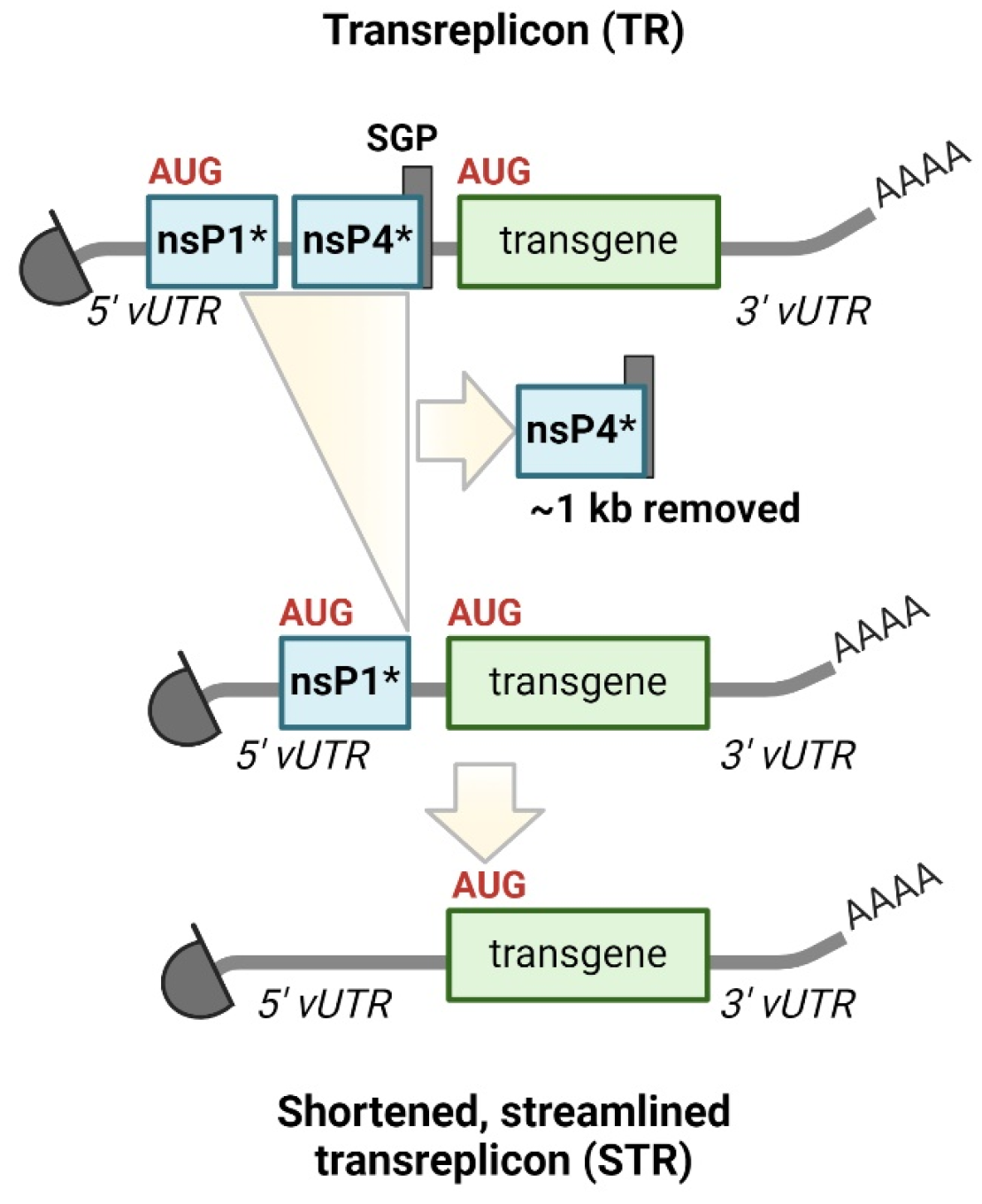
5. Trans-Amplifying RNA Vaccines
In several of the aforementioned studies, reporter genes were encoded by the transreplicons. Even though the expression levels of the transreplicons were not the primary focus of these studies, their expression levels were often very high [55,56,73]. Furthermore, the production of recombinant viral particles using helper RNA-encoding alphaviral structural genes was efficient, which indicated robust expression levels of the sPs from the helper RNA. Essentially, these observations, when combined, raised the idea that TRs could bear the potential to be used as an RNA-based vaccine.
A prerequisite for an RNA-based vaccine is that all required proteins are encoded on IVT RNA and delivered simultaneously. This approach was undertaken in a study that revisited the essential sequence elements required for alphaviral replication using a large set of TRs (named splitzicons) [73]. This study discussed a potential utilization of TRs as a vaccine. In a proof-of-concept study, we were then the first to explore antigen-coding TRs together with replicase-coding RNA provided in trans as a vaccine. We found that TR expression using replicase encoded on nrRNA was more efficient than replicase encoded on saRNA [10]. This all-RNA platform was named trans-amplifying RNA (taRNA), distinct from saRNA or the experimental use of trans-replicating RNA systems. When immunizing mice, a combination of 20 µg replicase nrRNA and 50 ng transreplicon-encoding influenza HA mounted HA-specific immune responses comparable to 20 µg conventional mRNA-HA or 1.25 µg saRNA-HA. The required dose of antigen-coding TR was strikingly 400x less TR than replicase nrRNA, highlighting the advantage of this strategy. Overall, this meant that the amount of TR was practically negligible compared to the total RNA dose. Moreover, the finding that nrRNA is a more suitable option for delivering replicase compared to saRNA simultaneously implied that deleterious RNA recombination would be unlikely. Indeed, a follow up study using a CHIKV taRNA revealed that nrRNA replicase and TRs do not recombine [74]. In this study, which introduced taRNA as a vaccine candidate against CHIKV, the researchers demonstrated the high amplification of TR and antigen expression in vitro, without recombining to replication-competent CHIKV. Importantly, the taRNA-based vaccine induced humoral and cellular immune responses in a mouse model and provided protection against CHIKV challenge infection. Further observations made in this study confirmed previous studies that longer TR-RNA templates amplified slower and reached lower total RNA levels compared to shorter ones [55,56]. In the same year, the researchers investigated the feasibility of a bivalent vaccine candidate [75]. Here, two TR-RNAs encoding CHIKV and RRV envelope proteins were used, demonstrating efficient co-amplification and high antigen co-expression. Immunization of mice induced specific humoral and cellular immune responses against both CHIKV and RRV. However, antibody titres and the neutralization capacity were higher after immunization when using a single TR-RNA, suggesting potential immune interferences or dosing effects in the bivalent vaccine candidate. Interestingly, alphavirus-specific T cell responses remained equally potent after bivalent vaccination. This seminal study provided important insights into the design of multivalent taRNA-based vaccines, expanding their potential applications.
Because the first generation of taRNA proved efficient in the PoC study, we recently generated strategies to optimize the taRNA platform by simplifying the structure of the TR [76]. The TR simplification involved removing remnants of the replicase gene, the SGP and subgenomic transcript formation, resulting in a shortened TR (STR) closely resembling conventional mRNA. Notably, the STR retained alphaviral RNA promoters within the UTRs (Figure 3). STRs resemble promoterless helper-RNA [35], albeit with additional 5′ CSE modifications. By eliminating the original start codon and further AUG triplets, the start codon of the desired antigens became the most 5′-AUG, resulting in bypassing the translation of peptides corresponding to the replicase nsP1 N-terminus. Compensating mutations were introduced to preserve the structure and function of the 5′ CSE, while the SGP was eliminated without replacement. Transgenes encoded on the STR became accessible for translation from the 5′ cap. Unlike previous TRs, and similar to promoterless helper-RNAs, STRs no longer form subgenomic RNA species upon replication, producing only genomic replication products. To enhance STR amplification, a directed evolution strategy was employed, resulting in the accumulation of faster replicating mutant templates characterized by an extended 5′ end. Moreover, the flexibility of the taRNA platform was highlighted by the successful replication of various heterologous STRs by the SFV replicase, including Forth Morgan virus, Aura virus, Highlands J virus, Madariaga virus and CHIKV, thereby confirming findings in the literature [45,57].
Figure 3.
Structure and simplification of transreplicon. Shortened and streamlined transreplicon (STR) was created by deleting the original as well as all putative start codons within 5‘ CSE/nsP1 of the transreplicon (TR) and by removing the nsP4-subgenomic promoter (SGP). The AUG-codon mutations in the STR construct prevent a putative nsP1-peptide translation, thus, STR replication no longer supports subgenomic transcription. Instead, transgenes encoded on the STR are translated directly from original in vitro transcribed STR RNA if capped, and from positive-sensed STR copies resulting from intracellular replication. nsP1*: 5′ end of nsP1 containing the CSE; nsP4*: 3′ end of nsP4 including the SGP [76].
Together these studies demonstrated the profound adaptability of the taRNA platform and its potential use for vaccinations against infectious diseases.
6. Future Perspective of taRNA Vaccines, Challenges and Open Questions
The hallmark of taRNA, the combination of two RNAs with different functions, opens up opportunities and poses particular challenges. The replicase-encoding nrRNA is the engine of the system and drives the expression of the antigen-encoding STR. As described above, replicase only requires CSEs at the ends of the STR, while it is indifferent to the sequences of the antigen ORF. The replicase is also indifferent to whether one, two or more antigens are encoded by different CSE-flanked STRs. Overall, the results of previous taRNA vaccine publications indicate that a much greater mass of replicase nrRNA than STR is required. Whether a taRNA vaccine will eventually require a lower total mass of both RNAs compared to the mass of modified non-replicating mRNA or saRNA still requires testing in larger animal models and humans. Probably further molecular optimization of the STR and the replicase is required to achieve this goal. Irrespective of the outcome of these studies, the bipartite nature of taRNA can be exploited to develop a replicating RNA vaccine with low immunogenicity, to assemble multivalent vaccines, or to prepare for future pandemics.
6.1. Towards a Non-Immunogenic taRNA Vaccine
Vaccines often cause immediate symptoms related to the innate immune response to vaccination, commonly referred to as reactogenicity, which ultimately limits the dose that can be administered. mRNA vaccines are more reactogenic when prepared from unmodified nucleotides, which was observed in clinical trials of SARS-CoV-2 vaccines [77,78]. The approved and successfully administered mRNA vaccines were modified with the nucleotide N1-methylpseudouridine (N1mΨ) instead of uridine. This greatly reduced the ability of the mRNA to activate innate immunity, subsequently reactogenicity, and the vaccines were well tolerated by the majority of vaccinated individuals [79,80]. Given this great effect of RNA modification, it was naturally considered for saRNA as well. Unfortunately, it was found that saRNA loses its function when modified by N1mΨ [81]. Accordingly, the recently approved saRNA vaccine does not contain modified nucleotides. However, its low dose of only 5 µg probably reduced reactogenicity to such an extent that it became tolerable [4,5]. Encouragingly, a recent comprehensive test of a variety of modified nucleotides has shown that 5-methyl cytidine (5 mC) reduces the immunogenicity of saRNA without affecting its function [82,83], raising hopes for even less reactogenic saRNA in the future.
The taRNA offers a unique advantage for the incorporation of modified nucleotides. The nrRNA encoding the replicase can accept all modifications that reduce immunogenicity without affecting RNA translation. Modified nucleotides, similar to saRNA, impede STR replication. Notably, N1mΨ exerts a potent inhibitory influence, while 5 mC allows replication (own unpublished observation). Since nrRNA replicase is used in excess, most of the RNA mass of taRNA may benefit from the clinically tested N1mΨ modification. The STR RNA could remain unmodified, with the total amount of unmodified RNA likely to remain below the threshold of unacceptable reactogenicity. Alternatively, the STR could be provided with 5 mC or another modification that may be developed for saRNA in the future. The production of a low-immunogenic taRNA is therefore already easily possible today, and the remaining immunogenicity could be further reduced in the near future.
6.2. Realizing Multivalent Vaccines Using taRNA
The CSEs located at the 5′ and 3′ ends of an RNA determine whether or not an RNA is a template for amplification by replicase, while the coding sequence inserted between these ends should not affect replication. Nevertheless, if in rare cases the coding sequences negatively affect replication, e.g., by intramolecular RNA interaction over larger distances, the use of synonymous codons should solve the problem. Overall, the indifference of the replicase to the coding sequence of the STR should allow the amplification of STR mixtures, which is the prerequisite for the construction of multivalent vaccines. The replication rates of STRs, however, could vary with the length or individual complexity of RNA folding of the different coding sequences. Therefore, further studies are needed to discover whether there are universal rules for determining the TR stoichiometry of a multivalent taRNA vaccine or whether each multivalent vaccine requires individual optimization of TR stoichiometry. Regardless of the ratio of the different TRs to each other, the total RNA amount of a multivalent taRNA vaccine will most likely largely depend on the amount of nrRNA replicase, similar to mono- and bivalent taRNA vaccines tested in mice [74,75]. This is in marked contrast to multivalent mRNA vaccines. Here, for each antigen encoded on a single mRNA molecule, the total amount is increased by the amount of mRNA required for a robust immune response against each of these antigens [84]. At an overall lower dose level, this is also likely to be the case for multivalent saRNA vaccines. Therefore, multivalent RNA vaccines run the risk of quickly exceeding the maximum tolerated dose, unless each antigen-encoding RNA only slightly increases the dose—as is the case with taRNA.
6.3. taRNA to Accelerate Seasonal Vaccine Production and Improve Pandemic Preparedness
As mentioned above, the two-part design of taRNA is the main difference from other nucleic acid vaccines. Replicase nrRNA as the main component of the vaccine not only enables low-immunogenic replicating RNA vaccines or facilitates multivalent vaccines, but can also accelerate the production of vaccines. Let us assume that a future vaccine will contain 9 times more replicase nrRNA than TR-RNA. This would mean that 90% of the RNA vaccine mass does not code for the antigen. The replicase nrRNA could therefore be produced independently of the antigen-coding STRs and stored until use. We believe that this will be invaluable in two particular situations. First, in the production of seasonally adapted tetravalent influenza vaccines. 90% of the vaccine—i.e., the mass that would correspond to the replicase nrRNA—would be available from stock before it is known which strains of influenza viruses need to be selected for the vaccine. The STRs that code for the hemagglutinin of these strains make up the remaining 10% of the vaccine, and only these would need to be produced in time for the start of the vaccination campaign. The cost of producing large batches of the flu vaccine each year would be much lower. Seasonally updated vaccines against SARS-CoV-2 would benefit in a similar way.
In addition, the replicase nrRNA in stock would also be immediately available should an emerging virus trigger a pandemic. The time needed to develop viral antigens and test them for their suitability as a vaccine could be used to produce even more replicase nrRNA. Apart from possible difficulties in finding suitable antigens for a vaccine, most of the required mRNA mass for a first large batch of a vaccine would already be available at the time of approval. The speed with which a completely new vaccine could be made available would benefit enormously from this approach.
6.4. taRNA Formulation
Therapeutic RNA must be formulated for effective delivery to humans. COVID-19 mRNA vaccines, for example, use LNPs for optimal delivery. The efficacy of taRNA will likely heavily depend on optimization of formulations, as both RNA species of taRNA must be delivered together to the same cell to be functional. A cell transfected only with replicase nrRNA cannot contribute to antigen-specific immune responses, and a cell receiving only STR will at best express basic levels of the antigen. Even with a monovalent taRNA vaccine, the question already arises whether it is preferable to mix the RNA before formulation (co-formulation) or to formulate both RNAs separately and mix the encapsulated RNAs shortly before administration. In vitro experiments with commercial liposomes indicate that both methods lead to transreplication, but in our experience, co-formulation with liposomes leads to higher transfection rates (own unpublished data). On the other hand, a separate formulation may offer more flexibility, especially for a vaccine that requires unanticipated adaptation due to the rapid emergence of a new strain. As long as the two RNAs are not mixed, the STR could be changed in the short term. Therefore, both strategies should be carefully evaluated and compared in terms of their potency and efficacy, as well as their feasibility in an industrial setting. Ultimately, the decision on which approach to pursue for a particular indication will involve both scientific and economic considerations.
7. Concluding Remarks
In light of the literature summarized in this manuscript, it is fascinating to observe the significant contributions made by naturally occurring defective interfering RNA and engineered transreplicons to the advancement of alphaviral research over the past 50 years. They have not only been pivotal in shaping our understanding but have also served as gene transfer tools for over three decades. With their recent adaptation as vectors for antigen delivery within taRNA, transreplicons have entered the field of nucleic acid-based vaccines, holding promise as potent tools in the fight against future infectious threats. While writing this review, the first saRNA vaccine was licenced in Japan [4], following the successful path of COVID-19 mRNA vaccines. Although taRNA is yet to undergo clinical translation, our optimism is grounded in sound reasons, anticipating its eventual success.
Author Contributions
Conceptualization, A.Y., C.R. and T.B.; literature research, A.Y., C.R. and T.B.; writing—original draft preparation, A.Y. and C.R.; writing—review and editing, A.Y., C.R. and T.B.; visualization, A.Y. and C.R.; supervision, T.B. All authors have read and agreed to the published version of the manuscript.
Funding
This research received no external funding.
Acknowledgments
We cordially thank Christin Schmidt, Mario Perkovic and Andreas Kuhn for critically reviewing the manuscript, and Johnny Kim for reviewing the English language usage and copy editing.
Conflicts of Interest
Tim Beissert and Aysegül Yildiz are inventors on patents covering taRNA optimization and application. Cristian Raileanu declares no conflicts of interest.
References
- Baden, L.R.; El Sahly, H.M.; Essink, B.; Kotloff, K.; Frey, S.; Novak, R.; Diemert, D.; Spector, S.A.; Rouphael, N.; Creech, C.B.; et al. Efficacy and Safety of the mRNA-1273 SARS-CoV-2 Vaccine. N. Engl. J. Med. 2021, 384, 403–416. [Google Scholar] [CrossRef]
- Polack, F.P.; Thomas, S.J.; Kitchin, N.; Absalon, J.; Gurtman, A.; Lockhart, S.; Perez, J.L.; Pérez Marc, G.; Moreira, E.D.; Zerbini, C.; et al. Safety and Efficacy of the BNT162b2 mRNA COVID-19 Vaccine. N. Engl. J. Med. 2020, 383, 2603–2615. [Google Scholar] [CrossRef]
- Lee, J.; Woodruff, M.C.; Kim, E.H.; Nam, J.-H. Knife’s edge: Balancing immunogenicity and reactogenicity in mRNA vaccines. Exp. Mol. Med. 2023, 55, 1305–1313. [Google Scholar] [CrossRef] [PubMed]
- Dolgin, E. Self-copying RNA vaccine wins first full approval: What’s next? Nature 2023, 624, 236–237. [Google Scholar] [CrossRef] [PubMed]
- Oda, Y.; Kumagai, Y.; Kanai, M.; Iwama, Y.; Okura, I.; Minamida, T.; Yagi, Y.; Kurosawa, T.; Greener, B.; Zhang, Y.; et al. Immunogenicity and safety of a booster dose of a self-amplifying RNA COVID-19 vaccine (ARCT-154) versus BNT162b2 mRNA COVID-19 vaccine: A double-blind, multicentre, randomised, controlled, phase 3, non-inferiority trial. Lancet Infect. Dis. 2024, 24, 351–360. [Google Scholar] [CrossRef]
- Vogel, A.B.; Lambert, L.; Kinnear, E.; Busse, D.; Erbar, S.; Reuter, K.C.; Wicke, L.; Perkovic, M.; Beissert, T.; Haas, H.; et al. Self-Amplifying RNA Vaccines Give Equivalent Protection against Influenza to mRNA Vaccines but at Much Lower Doses. Mol. Ther. J. Am. Soc. Gene Ther. 2018, 26, 446–455. [Google Scholar] [CrossRef]
- Sahin, U.; Karikó, K.; Türeci, Ö. mRNA-based therapeutics—Developing a new class of drugs. Nat. Rev. Drug Discov. 2014, 13, 759–780. [Google Scholar] [CrossRef]
- Houseley, J.; Tollervey, D. The many pathways of RNA degradation. Cell 2009, 136, 763–776. [Google Scholar] [CrossRef] [PubMed]
- Pourseif, M.M.; Masoudi-Sobhanzadeh, Y.; Azari, E.; Parvizpour, S.; Barar, J.; Ansari, R.; Omidi, Y. Self-amplifying mRNA vaccines: Mode of action, design, development and optimization. Drug Discov. Today 2022, 27, 103341. [Google Scholar] [CrossRef] [PubMed]
- Beissert, T.; Perkovic, M.; Vogel, A.; Erbar, S.; Walzer, K.C.; Hempel, T.; Brill, S.; Haefner, E.; Becker, R.; Türeci, Ö.; et al. A Trans-amplifying RNA Vaccine Strategy for Induction of Potent Protective Immunity. Mol. Ther. J. Am. Soc. Gene Ther. 2020, 28, 119–128. [Google Scholar] [CrossRef]
- Hyde, J.L.; Chen, R.; Trobaugh, D.W.; Diamond, M.S.; Weaver, S.C.; Klimstra, W.B.; Wilusz, J. The 5′ and 3′ ends of alphavirus RNAs--Non-coding is not non-functional. Virus Res. 2015, 206, 99–107. [Google Scholar] [CrossRef] [PubMed]
- Pietilä, M.K.; Hellström, K.; Ahola, T. Alphavirus polymerase and RNA replication. Virus Res. 2017, 234, 44–57. [Google Scholar] [CrossRef] [PubMed]
- Lundstrom, K. Alphaviruses in gene therapy. Viruses 2015, 7, 2321–2333. [Google Scholar] [CrossRef] [PubMed]
- Meshram, C.D.; Phillips, A.T.; Lukash, T.; Shiliaev, N.; Frolova, E.I.; Frolov, I. Mutations in Hypervariable Domain of Venezuelan Equine Encephalitis Virus nsP3 Protein Differentially Affect Viral Replication. J. Virol. 2020, 94, e01841-19. [Google Scholar] [CrossRef] [PubMed]
- Götte, B.; Liu, L.; McInerney, G.M. The Enigmatic Alphavirus Non-Structural Protein 3 (nsP3) Revealing Its Secrets at Last. Viruses 2018, 10, 105. [Google Scholar] [CrossRef]
- Ou, J.H.; Strauss, E.G.; Strauss, J.H. The 5′-terminal sequences of the genomic RNAs of several alphaviruses. J. Mol. Biol. 1983, 168, 1–15. [Google Scholar] [CrossRef]
- Ou, J.H.; Rice, C.M.; Dalgarno, L.; Strauss, E.G.; Strauss, J.H. Sequence studies of several alphavirus genomic RNAs in the region containing the start of the subgenomic RNA. Proc. Natl. Acad. Sci. USA 1982, 79, 5235–5239. [Google Scholar] [CrossRef]
- Levis, R.; Schlesinger, S.; Huang, H.V. Promoter for Sindbis virus RNA-dependent subgenomic RNA transcription. J. Virol. 1990, 64, 1726–1733. [Google Scholar] [CrossRef]
- Ou, J.H.; Strauss, E.G.; Strauss, J.H. Comparative studies of the 3′-terminal sequences of several alpha virus RNAs. Virology 1981, 109, 281–289. [Google Scholar] [CrossRef]
- Denison, M.R.; Graham, R.L.; Donaldson, E.F.; Eckerle, L.D.; Baric, R.S. Coronaviruses: An RNA proofreading machine regulates replication fidelity and diversity. RNA Biol. 2011, 8, 270–279. [Google Scholar] [CrossRef]
- Huang, A.S.; Baltimore, D. Defective viral particles and viral disease processes. Nature 1970, 226, 325–327. [Google Scholar] [CrossRef] [PubMed]
- Schlesinger, S.; Schlesinger, M.; Burge, B.W. Defective virus particles from Sindbis virus. Virology 1972, 48, 615–617. [Google Scholar] [CrossRef]
- Stark, C.; Kennedy, S.I. The generation and propagation of defective-interfering particles of Semliki Forest virus in different cell types. Virology 1978, 89, 285–299. [Google Scholar] [CrossRef] [PubMed]
- Lehtovaara, P.; Söderlund, H.; Keränen, S.; Pettersson, R.F.; Kääriäinen, L. 18S defective interfering RNA of Semliki Forest virus contains a triplicated linear repeat. Proc. Natl. Acad. Sci. USA 1981, 78, 5353–5357. [Google Scholar] [CrossRef] [PubMed]
- Levis, R.; Weiss, B.G.; Tsiang, M.; Huang, H.; Schlesinger, S. Deletion mapping of Sindbis virus DI RNAs derived from cDNAs defines the sequences essential for replication and packaging. Cell 1986, 44, 137–145. [Google Scholar] [CrossRef] [PubMed]
- Weiss, B.; Schlesinger, S. Defective interfering particles of Sindbis virus do not interfere with the homologous virus obtained from persistently infected BHK cells but do interfere with Semliki Forest virus. J. Virol. 1981, 37, 840–844. [Google Scholar] [CrossRef] [PubMed]
- Xiong, C.; Levis, R.; Shen, P.; Schlesinger, S.; Rice, C.M.; Huang, H.V. Sindbis virus: An efficient, broad host range vector for gene expression in animal cells. Science 1989, 243, 1188–1191. [Google Scholar] [CrossRef] [PubMed]
- Levis, R.; Huang, H.; Schlesinger, S. Engineered defective interfering RNAs of Sindbis virus express bacterial chloramphenicol acetyltransferase in avian cells. Proc. Natl. Acad. Sci. USA 1987, 84, 4811–4815. [Google Scholar] [CrossRef] [PubMed]
- Liljeström, P.; Garoff, H. A new generation of animal cell expression vectors based on the Semliki Forest virus replicon. Bio/Technol. 1991, 9, 1356–1361. [Google Scholar] [CrossRef]
- Bredenbeek, P.J.; Frolov, I.; Rice, C.M.; Schlesinger, S. Sindbis virus expression vectors: Packaging of RNA replicons by using defective helper RNAs. J. Virol. 1993, 67, 6439–6446. [Google Scholar] [CrossRef]
- Pushko, P.; Parker, M.; Ludwig, G.V.; Davis, N.L.; Johnston, R.E.; Smith, J.F. Replicon-helper systems from attenuated Venezuelan equine encephalitis virus: Expression of heterologous genes in vitro and immunization against heterologous pathogens in vivo. Virology 1997, 239, 389–401. [Google Scholar] [CrossRef] [PubMed]
- Frolov, I.; Frolova, E.; Schlesinger, S. Sindbis virus replicons and Sindbis virus: Assembly of chimeras and of particles deficient in virus RNA. J. Virol. 1997, 71, 2819–2829. [Google Scholar] [CrossRef]
- Smerdou, C.; Liljeström, P. Two-helper RNA system for production of recombinant Semliki forest virus particles. J. Virol. 1999, 73, 1092–1098. [Google Scholar] [CrossRef] [PubMed]
- Lundstrom, K. Replicon RNA Viral Vectors as Vaccines. Vaccines 2016, 4, 39. [Google Scholar] [CrossRef] [PubMed]
- Kamrud, K.I.; Alterson, K.; Custer, M.; Dudek, J.; Goodman, C.; Owens, G.; Smith, J.F. Development and characterization of promoterless helper RNAs for the production of alphavirus replicon particle. J. Gen. Virol. 2010, 91, 1723–1727. [Google Scholar] [CrossRef] [PubMed]
- Monroe, S.S.; Schlesinger, S. Common and distinct regions of defective-interfering RNAs of Sindbis virus. J. Virol. 1984, 49, 865–872. [Google Scholar] [CrossRef] [PubMed]
- Frolov, I.; Hardy, R.; Rice, C.M. Cis-acting RNA elements at the 5′ end of Sindbis virus genome RNA regulate minus- and plus-strand RNA synthesis. RNA 2001, 7, 1638–1651. [Google Scholar] [CrossRef] [PubMed]
- Monroe, S.S.; Schlesinger, S. RNAs from two independently isolated defective interfering particles of Sindbis virus contain a cellular tRNA sequence at their 5′ ends. Proc. Natl. Acad. Sci. USA 1983, 80, 3279–3283. [Google Scholar] [CrossRef]
- Gorchakov, R.; Hardy, R.; Rice, C.M.; Frolov, I. Selection of functional 5′ cis-acting elements promoting efficient sindbis virus genome replication. J. Virol. 2004, 78, 61–75. [Google Scholar] [CrossRef]
- Kulasegaran-Shylini, R.; Atasheva, S.; Gorenstein, D.G.; Frolov, I. Structural and functional elements of the promoter encoded by the 5′ untranslated region of the Venezuelan equine encephalitis virus genome. J. Virol. 2009, 83, 8327–8339. [Google Scholar] [CrossRef]
- Kulasegaran-Shylini, R.; Thiviyanathan, V.; Gorenstein, D.G.; Frolov, I. The 5′UTR-specific mutation in VEEV TC-83 genome has a strong effect on RNA replication and subgenomic RNA synthesis, but not on translation of the encoded proteins. Virology 2009, 387, 211–221. [Google Scholar] [CrossRef] [PubMed]
- Shirako, Y.; Strauss, E.G.; Strauss, J.H. Modification of the 5′ terminus of Sindbis virus genomic RNA allows nsP4 RNA polymerases with nonaromatic amino acids at the N terminus to function in RNA replication. J. Virol. 2003, 77, 2301–2309. [Google Scholar] [CrossRef]
- Hardy, R.W.; Rice, C.M. Requirements at the 3′ end of the sindbis virus genome for efficient synthesis of minus-strand RNA. J. Virol. 2005, 79, 4630–4639. [Google Scholar] [CrossRef] [PubMed]
- Hardy, R.W. The role of the 3′ terminus of the Sindbis virus genome in minus-strand initiation site selection. Virology 2006, 345, 520–531. [Google Scholar] [CrossRef]
- Lello, L.S.; Utt, A.; Bartholomeeusen, K.; Wang, S.; Rausalu, K.; Kendall, C.; Coppens, S.; Fragkoudis, R.; Tuplin, A.; Alphey, L.; et al. Cross-utilisation of template RNAs by alphavirus replicases. PLoS Pathog. 2020, 16, e1008825. [Google Scholar] [CrossRef]
- Götte, B.; Utt, A.; Fragkoudis, R.; Merits, A.; McInerney, G.M. Sensitivity of Alphaviruses to G3BP Deletion Correlates with Efficiency of Replicase Polyprotein Processing. J. Virol. 2020, 94, e01681-19. [Google Scholar] [CrossRef]
- Merits, A.; Vasiljeva, L.; Ahola, T.; Kääriäinen, L.; Auvinen, P. Proteolytic processing of Semliki Forest virus-specific non-structural polyprotein by nsP2 protease. J. Gen. Virol. 2001, 82, 765–773. [Google Scholar] [CrossRef] [PubMed]
- Vasiljeva, L.; Valmu, L.; Kääriäinen, L.; Merits, A. Site-specific protease activity of the carboxyl-terminal domain of Semliki Forest virus replicase protein nsP2. J. Biol. Chem. 2001, 276, 30786–30793. [Google Scholar] [CrossRef]
- Vasiljeva, L.; Merits, A.; Golubtsov, A.; Sizemskaja, V.; Kääriäinen, L.; Ahola, T. Regulation of the sequential processing of Semliki Forest virus replicase polyprotein. J. Biol. Chem. 2003, 278, 41636–41645. [Google Scholar] [CrossRef]
- Lemm, J.A.; Rice, C.M. Assembly of functional Sindbis virus RNA replication complexes: Requirement for coexpression of P123 and P34. J. Virol. 1993, 67, 1905–1915. [Google Scholar] [CrossRef]
- Lemm, J.A.; Rice, C.M. Roles of nonstructural polyproteins and cleavage products in regulating Sindbis virus RNA replication and transcription. J. Virol. 1993, 67, 1916–1926. [Google Scholar] [CrossRef]
- Lemm, J.A.; Bergqvist, A.; Read, C.M.; Rice, C.M. Template-dependent initiation of Sindbis virus RNA replication in vitro. J. Virol. 1998, 72, 6546–6553. [Google Scholar] [CrossRef]
- Li, G.P.; Prágai, B.M.; Rice, C.M. Rescue of Sindbis virus-specific RNA replication and transcription by using a vaccinia virus recombinant. J. Virol. 1991, 65, 6714–6723. [Google Scholar] [CrossRef]
- Thal, M.A.; Wasik, B.R.; Posto, J.; Hardy, R.W. Template requirements for recognition and copying by Sindbis virus RNA-dependent RNA polymerase. Virology 2007, 358, 221–232. [Google Scholar] [CrossRef]
- Kallio, K.; Hellström, K.; Balistreri, G.; Spuul, P.; Jokitalo, E.; Ahola, T. Template RNA length determines the size of replication complex spherules for Semliki Forest virus. J. Virol. 2013, 87, 9125–9134. [Google Scholar] [CrossRef]
- Spuul, P.; Balistreri, G.; Hellström, K.; Golubtsov, A.V.; Jokitalo, E.; Ahola, T. Assembly of alphavirus replication complexes from RNA and protein components in a novel trans-replication system in mammalian cells. J. Virol. 2011, 85, 4739–4751. [Google Scholar] [CrossRef]
- Lello, L.S.; Bartholomeeusen, K.; Wang, S.; Coppens, S.; Fragkoudis, R.; Alphey, L.; Ariën, K.K.; Merits, A.; Utt, A. nsP4 Is a Major Determinant of Alphavirus Replicase Activity and Template Selectivity. J. Virol. 2021, 95, e0035521. [Google Scholar] [CrossRef]
- Scholte, F.E.M.; Tas, A.; Albulescu, I.C.; Žusinaite, E.; Merits, A.; Snijder, E.J.; van Hemert, M.J. Stress granule components G3BP1 and G3BP2 play a proviral role early in Chikungunya virus replication. J. Virol. 2015, 89, 4457–4469. [Google Scholar] [CrossRef]
- Utt, A.; Quirin, T.; Saul, S.; Hellström, K.; Ahola, T.; Merits, A. Versatile Trans-Replication Systems for Chikungunya Virus Allow Functional Analysis and Tagging of Every Replicase Protein. PLoS ONE 2016, 11, e0151616. [Google Scholar] [CrossRef] [PubMed]
- Rausalu, K.; Utt, A.; Quirin, T.; Varghese, F.S.; Žusinaite, E.; Das, P.K.; Ahola, T.; Merits, A. Chikungunya virus infectivity, RNA replication and non-structural polyprotein processing depend on the nsP2 protease’s active site cysteine residue. Sci. Rep. 2016, 6, 37124. [Google Scholar] [CrossRef] [PubMed]
- Cherkashchenko, L.; Rausalu, K.; Basu, S.; Alphey, L.; Merits, A. Expression of Alphavirus Nonstructural Protein 2 (nsP2) in Mosquito Cells Inhibits Viral RNA Replication in Both a Protease Activity-Dependent and -Independent Manner. Viruses 2022, 14, 1327. [Google Scholar] [CrossRef]
- Schütz, S.; Sarnow, P. Interaction of viruses with the mammalian RNA interference pathway. Virology 2006, 344, 151–157. [Google Scholar] [CrossRef]
- Kash, J.C.; Goodman, A.G.; Korth, M.J.; Katze, M.G. Hijacking of the host-cell response and translational control during influenza virus infection. Virus Res. 2006, 119, 111–120. [Google Scholar] [CrossRef]
- Froshauer, S.; Kartenbeck, J.; Helenius, A. Alphavirus RNA replicase is located on the cytoplasmic surface of endosomes and lysosomes. J. Cell Biol. 1988, 107, 2075–2086. [Google Scholar] [CrossRef]
- Friedman, R.M.; Levin, J.G.; Grimley, P.M.; Berezesky, I.K. Membrane-associated replication complex in arbovirus infection. J. Virol. 1972, 10, 504–515. [Google Scholar] [CrossRef]
- Grimley, P.M.; Levin, J.G.; Berezesky, I.K.; Friedman, R.M. Specific membranous structures associated with the replication of group A arboviruses. J. Virol. 1972, 10, 492–503. [Google Scholar] [CrossRef]
- Gorchakov, R.; Garmashova, N.; Frolova, E.; Frolov, I. Different types of nsP3-containing protein complexes in Sindbis virus-infected cells. J. Virol. 2008, 82, 10088–10101. [Google Scholar] [CrossRef]
- Tan, Y.B.; Chmielewski, D.; Law, M.C.Y.; Zhang, K.; He, Y.; Chen, M.; Jin, J.; Luo, D. Molecular architecture of the Chikungunya virus replication complex. Sci. Adv. 2022, 8, eadd2536. [Google Scholar] [CrossRef]
- Laurent, T.; Kumar, P.; Liese, S.; Zare, F.; Jonasson, M.; Carlson, A.; Carlson, L.-A. Architecture of the chikungunya virus replication organelle. eLife 2022, 11, e83042. [Google Scholar] [CrossRef] [PubMed]
- Frolova, E.I.; Gorchakov, R.; Pereboeva, L.; Atasheva, S.; Frolov, I. Functional Sindbis virus replicative complexes are formed at the plasma membrane. J. Virol. 2010, 84, 11679–11695. [Google Scholar] [CrossRef] [PubMed]
- Hellström, K.; Kallio, K.; Utt, A.; Quirin, T.; Jokitalo, E.; Merits, A.; Ahola, T. Partially Uncleaved Alphavirus Replicase Forms Spherule Structures in the Presence and Absence of RNA Template. J. Virol. 2017, 91, 10-1128. [Google Scholar] [CrossRef] [PubMed]
- Lemm, J.A.; Rümenapf, T.; Strauss, E.G.; Strauss, J.H.; Rice, C.M. Polypeptide requirements for assembly of functional Sindbis virus replication complexes: A model for the temporal regulation of minus- and plus-strand RNA synthesis. EMBO J. 1994, 13, 2925–2934. [Google Scholar] [CrossRef] [PubMed]
- Blakney, A.K.; McKay, P.F.; Shattock, R.J. Structural Components for Amplification of Positive and Negative Strand VEEV Splitzicons. Front. Mol. Biosci. 2018, 5, 71. [Google Scholar] [CrossRef] [PubMed]
- Schmidt, C.; Haefner, E.; Gerbeth, J.; Beissert, T.; Sahin, U.; Perkovic, M.; Schnierle, B.S. A taRNA vaccine candidate induces a specific immune response that protects mice against Chikungunya virus infections. Molecular therapy. Nucleic Acids 2022, 28, 743–754. [Google Scholar] [CrossRef]
- Schmidt, C.; Hastert, F.D.; Gerbeth, J.; Beissert, T.; Sahin, U.; Perkovic, M.; Schnierle, B.S. A Bivalent Trans-Amplifying RNA Vaccine Candidate Induces Potent Chikungunya and Ross River Virus Specific Immune Responses. Vaccines 2022, 10, 1374. [Google Scholar] [CrossRef] [PubMed]
- Perkovic, M.; Gawletta, S.; Hempel, T.; Brill, S.; Nett, E.; Sahin, U.; Beissert, T. A trans-amplifying RNA simplified to essential elements is highly replicative and robustly immunogenic in mice. Mol. Ther. J. Am. Soc. Gene Ther. 2023, 31, 2297. [Google Scholar] [CrossRef] [PubMed]
- Kremsner, P.G.; Mann, P.; Kroidl, A.; Leroux-Roels, I.; Schindler, C.; Gabor, J.J.; Schunk, M.; Leroux-Roels, G.; Bosch, J.J.; Fendel, R.; et al. Safety and immunogenicity of an mRNA-lipid nanoparticle vaccine candidate against SARS-CoV-2: A phase 1 randomized clinical trial. Wien. Klin. Wochenschr. 2021, 133, 931–941. [Google Scholar] [CrossRef]
- Kremsner, P.G.; Ahuad Guerrero, R.A.; Arana-Arri, E.; Aroca Martinez, G.J.; Bonten, M.; Chandler, R.; Corral, G.; Block, E.J.L.; de Ecker, L.; Gabor, J.J.; et al. Efficacy and safety of the CVnCoV SARS-CoV-2 mRNA vaccine candidate in ten countries in Europe and Latin America (HERALD): A randomised, observer-blinded, placebo-controlled, phase 2b/3 trial. The Lancet. Infect. Dis. 2022, 22, 329–340. [Google Scholar] [CrossRef] [PubMed]
- Fenton, C.; Lee, A. mRNA COVID-19 vaccines are well tolerated and myopericarditis is a rare adverse event following immunisation. Drugs Ther. Perspect. Ration. Drug Sel. Use 2022, 38, 532–540. [Google Scholar] [CrossRef]
- Kouhpayeh, H.; Ansari, H. Adverse events following COVID-19 vaccination: A systematic review and meta-analysis. Int. Immunopharmacol. 2022, 109, 108906. [Google Scholar] [CrossRef]
- Erasmus, J.H.; Khandhar, A.P.; O’Connor, M.A.; Walls, A.C.; Hemann, E.A.; Murapa, P.; Archer, J.; Leventhal, S.; Fuller, J.T.; Lewis, T.B.; et al. An Alphavirus-derived replicon RNA vaccine induces SARS-CoV-2 neutralizing antibody and T cell responses in mice and nonhuman primates. Sci. Transl. Med. 2020, 12, eabc9396. [Google Scholar] [CrossRef] [PubMed]
- McGee, J.E.; Kirsch, J.R.; Kenney, D.; Chavez, E.; Shih, T.-Y.; Douam, F.; Wong, W.W.; Grinstaff, M.W. Complete substitution with modified nucleotides suppresses the early interferon response and increases the potency of self-amplifying RNA. bioRxiv 2023. [Google Scholar] [CrossRef]
- Komori, M.; Morey, A.L.; Quiñones-Molina, A.A.; Fofana, J.; Romero, L.; Peters, E.; Matsuda, K.; Gummuluru, S.; Smith, J.F.; Akahata, W.; et al. Incorporation of 5 methylcytidine alleviates innate immune response to self-amplifying RNA vaccine. bioRxiv 2023. [Google Scholar] [CrossRef]
- Arevalo, C.P.; Bolton, M.J.; Le Sage, V.; Ye, N.; Furey, C.; Muramatsu, H.; Alameh, M.-G.; Pardi, N.; Drapeau, E.M.; Parkhouse, K.; et al. A multivalent nucleoside-modified mRNA vaccine against all known influenza virus subtypes. Science 2022, 378, 899–904. [Google Scholar] [CrossRef] [PubMed]
Disclaimer/Publisher’s Note: The statements, opinions and data contained in all publications are solely those of the individual author(s) and contributor(s) and not of MDPI and/or the editor(s). MDPI and/or the editor(s) disclaim responsibility for any injury to people or property resulting from any ideas, methods, instructions or products referred to in the content. |
© 2024 by the authors. Licensee MDPI, Basel, Switzerland. This article is an open access article distributed under the terms and conditions of the Creative Commons Attribution (CC BY) license (https://creativecommons.org/licenses/by/4.0/).


