Review

IGF2BP1—An Oncofetal RNA-Binding Protein Fuels Tumor Virus Propagation

Institute of Molecular Medicine, Martin Luther University Halle-Wittenberg, Kurt-Mothes-Str. 3a, 06120 Halle, Germany
*
Author to whom correspondence should be addressed.
Viruses 2023, 15(7), 1431; https://doi.org/10.3390/v15071431
Submission received: 19 May 2023 / Revised: 13 June 2023 / Accepted: 22 June 2023 / Published: 24 June 2023
(This article belongs to the Special Issue RNA Biology of Viral Infection)

Abstract

:
The oncofetal RNA-binding protein IGF2BP1 has been reported to be a driver of tumor progression in a multitude of cancer entities. Its main function is the stabilization of target transcripts by shielding these from miRNA-mediated degradation. However, there is growing evidence that several virus species recruit IGF2BP1 to promote their propagation. In particular, tumor-promoting viruses, such as hepatitis B/C and human papillomaviruses, benefit from IGF2BP1. Moreover, recent evidence suggests that non-oncogenic viruses, such as SARS-CoV-2, also take advantage of IGF2BP1. The only virus inhibited by IGF2BP1 reported to date is HIV-1. This review summarizes the current knowledge about the interactions between IGF2BP1 and different virus species. It further recapitulates several findings by presenting analyses from publicly available high-throughput datasets.

1. Introduction

Viruses actively reprogram the metabolism of their host cells to support the infection process and facilitate the escape or suppression of host defense mechanisms. To achieve this, the virus relies on interactions with host cell components that are exploited to promote the processes augmenting virus spreading [1]. The reprogramming of cellular metabolism following many viral infections is reminiscent of the metabolic changes observed in tumor development and, consequently, the metabolic reprogramming of certain viruses has been linked to oncogenesis [2]. Virus species described to possess oncogenic potential in humans include Epstein–Barr virus, Kaposi sarcoma-associated herpesvirus, human papillomaviruses, hepatitis B and C viruses, human T lymphotropic virus, and Merkel cell polyomavirus [3,4]. Furthermore, other polyomaviruses, such as simian virus 40 (SV40), as well as adenoviruses have been shown to be capable of transforming animal and human cells [4,5,6]. One important class of host cell components utilized by viruses are RNA-binding proteins (RBPs). These proteins accompany and regulate host as well as viral RNAs through all stages of post-transcriptional gene regulation. Another important class of molecules involved in regulating viral RNAs are microRNAs (miRNAs), small non-coding RNAs that direct the post-transcriptional repression of mRNA targets [7]. Cellular miRNAs can exert anti- or proviral effects and they are often dysregulated upon viral infections [8,9]. For example, miRNA-128 limits the replication and dissemination of the human immunodeficiency virus type 1 (HIV-1), whereas miRNA-34 promotes HIV-1 pathogenesis by inducing the accelerated decay of host proteins influencing the HIV-1 life cycle [10,11]. Virus infections have also been shown to induce post-transcriptional modifications on host genes’ transcripts. N6-adenosine methylation (m6A) is the most prevalent internal modification found in eukaryotic mRNAs [12]. This modification affects RNA structure and function and was shown to be altered on host genes upon viral infections. On the other hand, viral RNAs are also subject to m6A modifications, leading to alterations in virus production and infectivity. However, there seems to be no common regulation pattern and thus, increased methylation can enhance the production of certain viruses, while it reduces the production rates of others [13,14,15,16,17,18].
IGF2BP1, also known as IMP-1, CRD-BP and VICKZ1, is an oncofetal RNA-binding protein regulating tumor and stem cell fate [19,20,21,22,23]. Elevated IGF2BP1 expression has been implicated in the development and progression of various cancers in, e.g., ovary, lung, pancreas, liver and breast and is typically associated with poor prognosis [24,25,26,27,28,29]. IGF2BP1’s main function in cancer appears to be the protection of its mainly pro-oncogenic target RNAs from miRNA-mediated degradation via binding to its four hnRNPK homology (KH) domains [25,30,31,32,33,34,35]. Moreover, it was shown that m6A RNA modifications on IGF2BP1 target transcripts result in stronger RNA association and the consequently enforced expression of these pro-oncogenic factors [30,36,37]. By stabilizing the transcription factor E2F1- and E2F-driven transcripts, IGF2BP1 promotes G1/S cell cycle transition [37]. Similarly, distinct tumorigenic viruses drive the expression of S-phase genes by enhancing E2F activity. Papillomavirus E7 proteins, polyomavirus large T antigens and adenovirus E1A proteins are all capable of binding host Rb proteins, leading to a de-repression of E2F transcription factors and, thus, activating genes required for cell cycle progression [5,38,39,40,41].
The first report available on Pubmed regarding an interaction between IGF2BP1 and the components of a virus was published in 2004. Lu et al. reported an association between IGF2BP1 and the RNA genome of the hepatitis C virus [42]. Since then, associations of IGF2BP1 or its vertebrate homologs and several virus species have been published. These include viruses with double-stranded DNA genomes, such as hepatitis B virus and papillomaviruses, single-stranded RNA viruses, such as hepatitis C virus, Zika virus, Ebola virus and the severe acute respiratory syndrome coronavirus 2 (SARS-CoV-2), as well as retroviruses such as HIV-1 and murine leukemia virus (MLV). The respective associations were reported to either impact RNA turnover or translation. In addition, protein–protein interactions leading to the re-localization of viral proteins and altered virus production have been reported. Moreover, virus-mediated epigenetic modifications have been shown to alter RNA-binding activities between IGF2BP1 and its host mRNAs [43,44,45,46,47,48,49].
The oncofetal expression pattern of IGF2BP1 can be recapitulated from various public data sources derived from RNA-sequencing experiments. Healthy tissues express barely detectable amounts of IGF2BP1 mRNA, with only a few exceptions such as testis and kidney (Figure S1A). In contrast, the IGF2BP1 mRNA levels are elevated in a multitude of tumors, whereas the inter-sample variation is rather high (Figure S1B). Regarding cell types, single-cell RNA-seq data reveal few specialized cell types, mainly embryonic or from adult reproductive tissues as well as distinct cell types found in the kidney, expressing considerable amounts of IGF2BP1 (Figures S1C and S5A). Thus, for leveraging IGF2BP1, a virus either needs to infect cells expressing sufficient levels of this pro-oncogenic protein or must enforce/induce its synthesis.
This review aimed to summarize the current knowledge about interactions between IGF2BP1 and the components of different virus species and the consequences of these interactions on virus production and infectivity. The study covers IGF2BP1–virus interactions related to the oncogenic hepatitis B/C and human papillomaviruses as well as the non-tumorigenic viruses HIV-1, SARS-CoV-2, Zika virus and Ebola virus and it briefly summarizes reports regarding several non-human pathogenic viruses.

2. Materials and Methods

Statistical analyses were performed using R and images were created using the R-package ggplot2 [50,51], if not stated otherwise. Network drawings were created using Cytoscape [52]. Bulk RNA-seq data from the TCGA project were downloaded as files containing FPKM values via the GDC data portal (https://portal.gdc.cancer.gov/repository). The infection status of individual patient samples was extracted from supplementary tables of the accompanying reports. HBV/HCV: TCGA cohort LIHC, column “Hepatitis B”/“Hepatitis C” [53]; HPV: TCGA cohort CESC, column “CLIN:HPV_status” [54]. Liquid association coefficients were calculated using the R-package LiquidAssociation [55]. Survival analyses were performed as logrank tests implemented in the R-package survival [56] using the TCGA RNA-seq expression data of primary tumor samples. The CCLE bulk RNA-seq data of cell lines were obtained from DepMap, downloaded via the R-package ExperimentHub [57,58,59]. GTEx bulk RNA-seq data were obtained by downloading GTEx v8 TPM level data from the GTEx portal (https://www.gtexportal.org/home/) [60].
For the analysis of bulk-RNA-seq data from individual projects, fastq files were downloaded from NCBI GEO (https://www.ncbi.nlm.nih.gov/geo/). The quality of the fastq files was assessed using FastQC (https://www.bioinformatics.babraham.ac.uk/projects/fastqc/). If considerable amounts of remaining sequencing adapters or low-quality read ends were detected, these were clipped off using Cutadapt [61]. Sequencing reads were aligned to the human genome (UCSC hg38) using HiSat2 [62]. Alignments in the obtained bam files were sorted, indexed and secondary alignments were filtered out using samtools [63]. FeatureCounts [64] was used for summarizing the gene-mapped reads. Ensembl (GRCh38.100 [65] was used as the annotation basis. Differential gene expression was determined using the R package edgeR utilizing the trimmed mean of M-values [66,67] normalization. A false discovery rate (FDR)-adjusted p-value below 0.05 was considered the threshold for the determination of differential gene expression. Sequencing reads not mapped to the human reference genome were subsequently mapped against the respective virus genes/genomes using Hisat2. The following RefSeq sequences were used: HCV—NC_004102.1; HPV16 E7—NC_001526:7604-7900; HPV18 E7—NC_001357.1:590-907; SARS-CoV-2—NC_045512.2. Normalized expression values were obtained as fraction of the reads mapped to the respective gene/genome of the library size multiplied by 1 × 10⁶.
Single-cell RNA-seq data of CD4+ T cells were downloaded as R-object files along with cell cluster information from PanglaoDB (https://panglaodb.se/). The R-package Seurat V4 [68] was used for subsequent processing steps, including cell filtering (the minimum number of expressed genes/cell: 100; maximum number of expressed genes/cell: 2000; maximum percentage of mitochondrial gene expression/cell: 10) and count normalization using the “LogNormalize” method and scaling factor 10,000.

3. Human Oncogenic Viruses

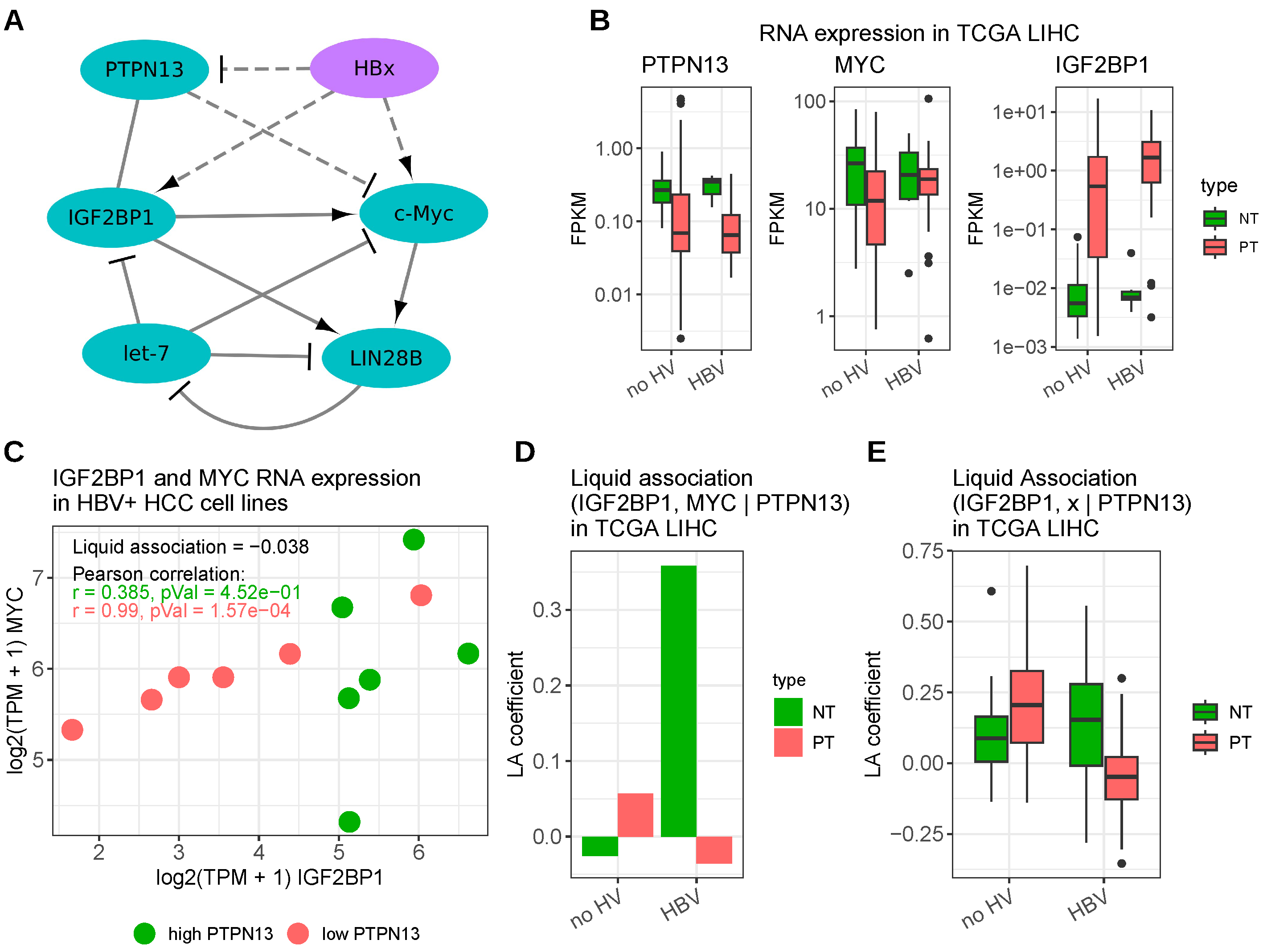
3.1. Hepatitis B Virus

Hepatitis B virus (HBV) is an enveloped DNA virus mainly infecting hepatocytes. HBV infection can result in the integration of the viral genome into the host genome and chronic infection is the main cause of liver cirrhosis and hepatocellular carcinoma (HCC) worldwide [69,70,71]. HBV’s carcinogenic potential is thought to be facilitated by different mechanisms such as mutagenesis due to the integration of viral DNA into host cancer genes, the promotion of genomic instability either via the integration of viral DNA or via the activity of certain viral proteins and due to the interference of normal cellular functions by viral proteins [72]. The regulatory HBx protein promotes HBV transcription as well as replication and contributes to the transformation of hepatocytes via multiple mechanisms. HBx directly interacts with a multitude of cellular proteins involved in proliferation, cell death, transcription and DNA repair [72]. One aspect of the HBx-mediated promotion of tumor progression is via the activation of the proto-oncogenic transcription factor c-Myc. While Li et al. described the induction of c-Myc expression via HBx by the activation of Ras/Raf/ERK1/2 cascades, Yan et al. reported that the HBx protein promotes tumor growth by facilitating c-Myc RNA stabilization via IGF2BP1 [49,73]. The latter described an interaction between HBx and the DNA methyltransferase DNMT3A leading to a hypermethylation of the promoter of the protein tyrosine phosphatase PTPN13, which, in turn, leads to a reduced PTPN13 transcription. In combination with the finding that PTPN13 directly interacts with the IGF2BP1 protein and that PTPN13 overexpression impairs the binding between IGF2BP1 and the c-Myc transcripts, Yan et al. concluded that PTPN13 protein competes with c-Myc RNA for IGF2BP1 association resulting in elevated c-Myc mRNA decay. Thus, the epigenetic silencing of PTPN13 mediated by HBV’s HBx protein enhances HCC proliferation by increasing c-MYC mRNA stability in an IGF2BP1-dependent manner [49]. In addition, Yan et al. described a dose-independent upregulation of IGF2BP1 RNA and protein by HBx [49], whereas Wang et al. observed a downregulation of let-7 miRNA family members by HBx [74]. Moreover, You et al. discovered that the LIN28B promoter was activated by HBx via c-Myc [75]. Intriguingly, IGF2BP1 has been shown to bind and thus stabilize LIN28B RNA and both, this stabilization and the activation of LIN28B via c-Myc were shown to repress let-7 miRNAs [31,76]. Altogether, this points to a regulatory network in which HBx, c-Myc, IGF2BP1 and LIN28B act synergistically to suppress let-7 action or biogenesis. Thus, the expression of all these factors is promoted by viral infection and fosters cell proliferation (Figure 1A).
The downregulation of PTPN13 in HCC upon HBV infection could be recapitulated from TCGA HCC RNA-seq data [53]. However, the difference in RNA expression levels between patients with and without hepatitis B infection was rather moderate; however, there was an obviously lower PTPN13 expression in primary tumors compared to normal tissues. C-Myc expression was higher in HBV-positive HCC samples compared to non-infected samples; however, generally elevated in normal tissue samples. IGF2BP1 expression was significantly elevated in tumor samples and in both, tumor and non-tumor tissues, IGF2BP1 abundance appeared to be increased by HBV infection (Figure 1B).
Liquid association (LA), a technique to assess the degree of interference in an expression correlation between two genes by a third gene [77], indeed revealed a negative influence of the PTPN13 expression on the correlation between IGF2BP1 and c-Myc (negative LA coefficient) in HCC-derived, HBV-positive cell lines. This was further supported by a very strong Pearson correlation between IGF2BP1 and c-Myc in the cell lines showing a low PTPN13 RNA expression compared to cell lines with higher PTPN13 expression (cell lines separated by median PTPN13 RNA expression; Figure 1C). However, although negative LA coefficients were also observed in HBV-positive HCC-patient samples, the non-tumor tissues of HBV-infected patients as well as tumor samples from non-infected patients showed a positive LA coefficient, suggesting that the interference of PTPN13 on the binding between the IGF2BP1 protein and c-Myc RNA is not only dependent on the expression levels of PTPN13, but also involves additional, difficult to identify, regulatory constraints (Figure 1D). The hypothesis that PTPN13 binding to IGF2BP1 competes with c-Myc binding leads to the tempting speculation that the binding of PTPN13 to IGF2BP1 reduces the interaction between IGF2BP1 and other target transcripts as well. To test this hypothesis, the LA coefficients of IGF2BP1 and a set of 117 published target candidates in dependence of PTPN13 were calculated. The criteria for selecting genes as target candidates comprised a consistent downregulation upon IGF2BP1 knockdown in several cancer-derived cell lines, a positive expression correlation with IGF2BP1 in tumors as well as reported IGF2BP1 binding sites [35]. Positive as well as negative LA coefficients were obtained for distinct target candidates; however, in general, LA coefficients were lowest in the HBV positive tumor samples (Figure 1E). Thus, PTPN13 might indeed interfere with the binding of IGF2BP1 and some of its targets; however, further investigations are required. Patient survival in HCC tends to be negatively associated with IGF2BP1 levels, but an additional influence of HBV on survival probabilities could not be conclusively assessed from TCGA data. The HBV-positive cohort was rather small (n = 20), the logrank test derived p-value was not significant and the Kaplan–Meier curves crossed, thus, allowing only rather unassertive conclusions (Figure S2A,C).
Figure 1. Interaction between HBV and IGF2BP1. (A) Scheme depicting the published interactions between HBV’s HBx protein and host cell components, cf. [31,49,74,75,76]. Solid line—direct interaction; dashed line—indirect interaction. (B) Distribution of RNA expression values of PTPN13, c-Myc and IGF2BP1 derived from HCC-RNA-seq samples (TCGA, cohort LIHC). (C) IGF2BP1 and c-Myc RNA expression in RNA-seq samples of HCC cell lines (data derived from CCLE data accessed via DepMap [57,58]; HBV-positivity was assessed from Expasy Cellosaurus [78]). (D) Liquid association (LA) coefficients of the correlation between IGF2BP1 and c-Myc in dependence of PTPN13 in TCGA HCC samples. (E) LA coefficient distributions of the correlation between IGF2BP1 and 117 published target candidates in dependence of PTPN13 in TCGA HCC samples. no HV—samples without detected hepatitis virus infection; HBV—samples with exclusively detected hepatitis B infection; NT—normal tissue; and PT—primary tumor tissue.
Viruses 15 01431 g001

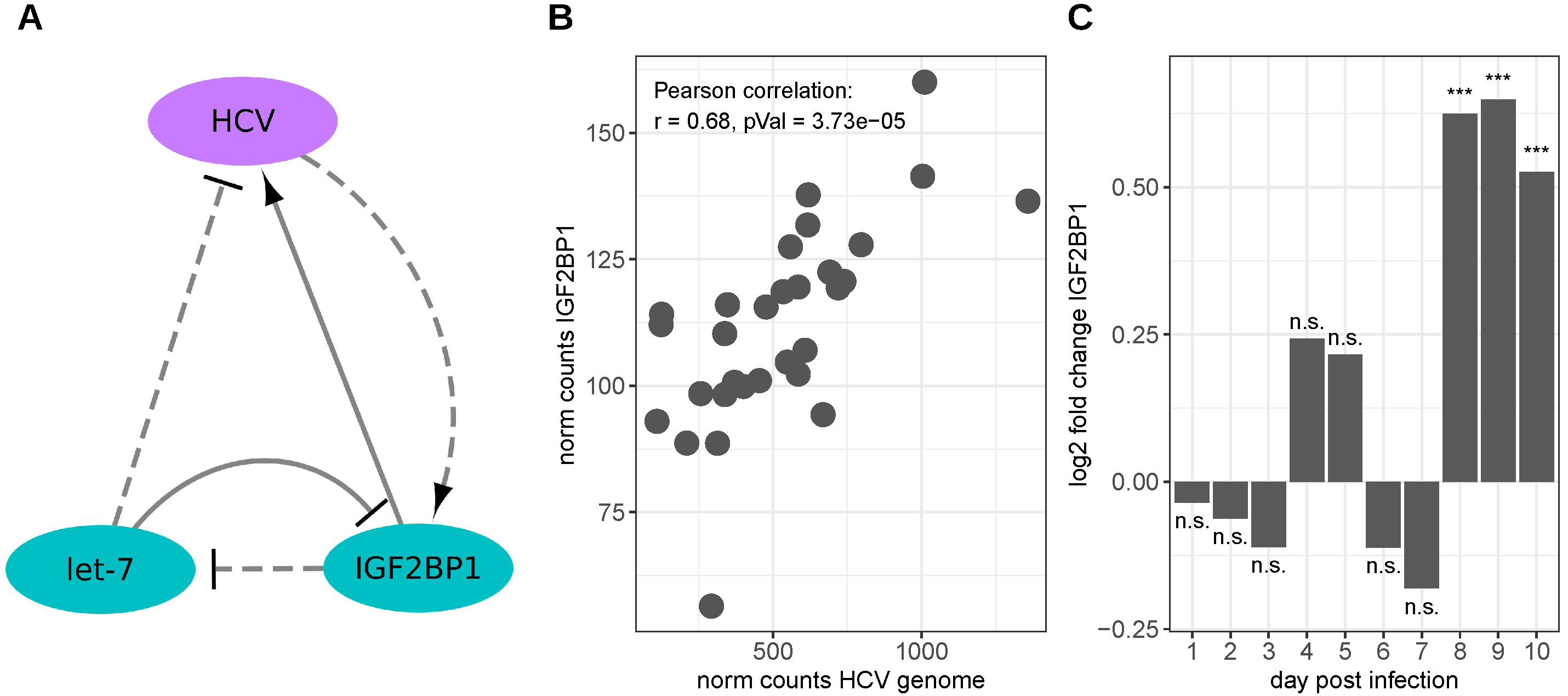
3.2. Hepatitis C Virus

The hepatitis C virus (HCV) is the main causative agent of non-A and non-B hepatitis [45] and besides HBV, constitutes another major risk factor for the development of liver cirrhosis and hepatocellular carcinoma [71,79,80]. The primary target cells of the virus are hepatocytes [81].
The positive-strand RNA genome of HVC contains an internal ribosomal entry site (IRES) in its 5’ untranslated region (5’UTR), facilitating cap-independent translation via the direct recruitment of the 40S ribosomal subunit and the eukaryotic initiation factor 3 (eIF3) [82,83].
In 2004, Lu et al. identified IGF2BP1 (CRDBP) as well as its paralogs IGF2BP2 and IGF2BP3 as HCV IRES-interacting proteins [42]. The binding of IGF2BP1 to the HCV-IRES was confirmed by Weinlich et al. in 2009. Furthermore, these authors found that IGF2BP1 can also bind to the HCV 3′UTR in vitro and that IGF2BP1 knockdown in HCC-derived Huh-7 cells leads to strongly decreased HCV IRES-mediated translation rates that were even further reduced in the absence of the HCV 3′UTR. However, the stability of the used reporter mRNAs was not reduced upon IGF2BP1 knockdown. Consistently, the addition of IGF2BP1 to translation competent extracts of primary rat hepatocytes increased the translation rates of an HCV reporter mRNA containing the HCV 5′ and 3′UTRs. In addition, a more than two-fold increase in the IGF2BP1 protein level was observed in Huh-7 cells, expressing an HCV replicon containing the nonstructural proteins NS3, NS4A, NS4B, NS5A and NS5B [45]. Moreover, Cheng et al. reported the anti-HCV activity of the let-7 miRNA family, as well as the negative regulation of IGF2BP1 expression by these miRNAs [84], providing further support for a regulatory role of an IGF2BP1-let-7 antagonism [31,85]. Figure 2A depicts the relationships between the HCV genome, IGF2BP1 and let-7 miRNAs. Another aspect of IGF2BP1 binding to the HCV RNA was reported by Li et al. They reported a competition in binding to the 5′ end of the HCV RNA between IGF2BP1 and miR-122, a liver-specific miRNA reported to promote HCV replication and translation [86,87,88,89]. After siRNA-mediated IGF2BP1-knockdown in Huh7.5 cells expressing HCV genomic RNA, significant reductions were observed in both HCV replication as well as HCC cell proliferation [86]. Bradrick et al. confirmed the binding of IGF2BP1 in the region of the miR-122 binding sites and, furthermore, found that the binding of IGF2BP1 was compromised by the deletion of the miR-122 binding sites; however, point mutations in these sites had no apparent effect on IGF2BP1 binding, suggesting the miR-122 independent binding of IGF2BP1 [89].
While there was no obvious difference in IGF2BP1 RNA expression in the TCGA HCC samples with and without detected HCV infection (Figure S3A), a positive correlation between IGF2BP1 RNA expression and the RNA levels of HCV can be seen in publicly available RNA-seq samples of HCV-infected Huh7.5.1 cells (Figure 2B, the data for which were obtained from [90]). Moreover, IGF2BP1 expression levels reached a moderate yet significant elevation after 8 days of infection in these cells (Figure 2C). Although not significant, survival analyses in the TCGA HCC cohort indicated a strong negative impact of IGF2BP1 expression on patients’ overall survival (Figure S2B).

3.3. Human Papillomavirus

Human papillomaviruses (HPVs) are a diverse set of more than 200 types of dsDNA viruses. They comprise five evolutionary groups (Alpha, Beta, Gamma, Mu, Nu) of which the Alpha group contains members causing rather harmless warts, but also the so-called high-risk (HR-) HPVs that are associated with several cancer types, including cervical cancer, several other anogenital cancers, as well as head and neck, lung and breast cancer [91,92,93,94]. Almost 100% of cervical cancers are attributable to HR-HPVs with HPV16 and 18 accounting for 71% of cases worldwide [95] and in total are estimated to cause more than 5% of all human cancers [96]. Nonetheless, cancer progression is a rather rare event following persistent HPV infection [97]. Notably, several studies have shown that HPV infection in head and neck cancers is associated with a better prognosis [98,99,100,101]. Cancer progression upon HR-HPV infection is caused by the increased expression of the viral oncoproteins E6 and E7 that activate the cell cycle, inhibit apoptosis, and allow for the accumulation of DNA damage [97]. HPV16 E7 can bind members of the well-known tumor suppressor and cell cycle regulatory retinoblastoma protein family (Rb), leading to the degradation of these proteins [38,39,40]. The binding/degradation of Rb releases and activates E2F transcription factors that drive the expression of S-phase genes [102]. Furthermore, E7 can directly bind and activate E2F1 in an Rb-independent manner [103]. HPV E6 protein complements the actions of E7 by targeting p53 for degradation and thus, preventing cell growth inhibition [102].
Similar to HPVs’ E7, IGF2BP1 supports E2F1 activity, albeit via different mechanisms. Müller et al. could show that IGF2BP1 promotes G1/S cell cycle transition by stabilizing mRNAs encoding positive regulators of this checkpoint including E2F1 and E2F-driven transcripts [37]. Wang et al. reported the m6A-dependent binding of HPV16 E7 mRNA by IGF2BP1, leading to a stabilization and, thus, increased the expression of this RNA in HPV16 positive cell lines. Moreover, this E7-IGF2BP1 complex was shown to be vulnerable to heat stress, since exposing cells to moderate heat led to a decrease in E7 as well as IGF2BP1 and to an upregulation of Rb, p53 and p21 proteins. In consequence, heat treatment was shown to reduce the proliferation and migration in HPV16-positive cells and reduced tumor growth in cell line-based xenograft mouse models [43]. Figure 3A depicts the described relations between E7 and IGF2BP1. Interestingly, besides the downregulation of p53 and Rb in HPV-positive lung cancer tissues, Hussen et al. observed a downregulation of PTPN13 and let-7 miRNAs [104]. Thus, it is tempting to speculate that regulatory circuits involving PTPN13, let-7 miRNAs, c-Myc and IGF2BP1 are also relevant in the HPV-associated malignancies (cf. Section 3.1). Regarding patient prognosis, Laban et al. observed a significant association between IGF2BP1 (IMP-1) antibody response and the shorter overall survival of patients with HPV-positive head and neck squamous cell carcinomas (HNSCC), which was not seen in HPV-negative patients [105].
A strong positive correlation between HPV E7 and IGF2BP1 RNA could be recapitulated from RNA-seq data in a panel (n = 10) of different cervical squamous carcinoma-derived cell lines expressing either HPV16 (CaSki, n = 4; SiHa, n = 3) or HPV18 (HeLa, n = 3) proteins (data derived from [106], Figure 3B). Furthermore, a slightly elevated, yet not significant, expression of IGF2BP1 RNA was observed in HPV-positive cervical cancer samples deposited by the TCGA (median FPKM = 0.08), and compared to HPV-negative samples (median FPKM = 0.03, Figure 3C). Considering the complete cervical cancer cohort, IGF2BP1 failed to yield conclusive results in terms of survival probability. However, HPV status remained unknown for many of these samples (Figure S4A). Considering only HPV-positive samples indicated a strong, yet not significant trend that IGF2BP1 is associated with reduced survival, whereas studies in the few HPV-negative samples remained non-conclusive (Figure S4B,C).

4. Human Non-Oncogenic Viruses

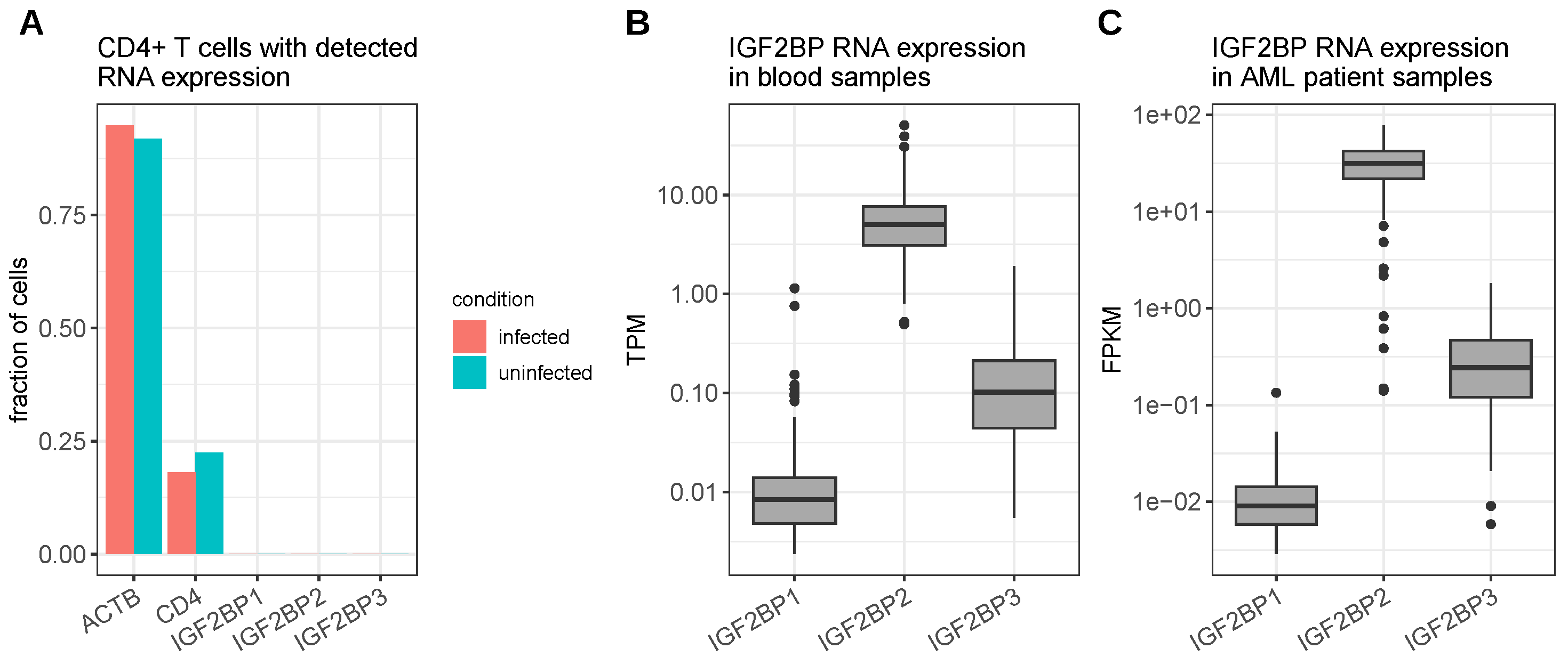
4.1. Human Immunodeficiency Virus Type 1

The human immunodeficiency virus type 1 (HIV-1) is the causative agent of the acquired immunodeficiency syndrome (AIDS). HIV-1 can infect CD4+ T cells, macrophages and dendritic cells [107]. The major structural component of HIV virions is the Gag protein. This protein is essential for virus assembly and release. It is further involved in membrane binding, the incorporation of the viral genome and Env proteins into the virions as well as in budding and release [108].
Zhou et al. discovered an association between IGF2BP1 and the HIV-1 Gag protein. The binding between the two proteins was shown to be primarily facilitated by IGF2BP1’s KH3 and KH4 domains and a zinc finger motif in the Gag nucleocapsid (NC) domain [47]. KH domain-dependent IGF2BP1-Gag binding was confirmed by Milev et al. [109]. Furthermore, IGF2BP1 expression was shown to inhibit the production of virus particles; however, effective inhibition relied on the presence of all IGF2BP1 domains. Moreover, Zhou et al. observed the incorporation of IGF2BP1 into virus particles and that the infectivity of virions produced in IGF2BP1 overexpressing cells was reduced in a primarily KH domain-dependent manner [47]. In a following work, Zhou et al. found that IGF2BP1 also interacts with HIV-1’s regulatory Rev protein [48]. The Rev protein binds and mediates the cytoplasmic transport of late-phase partially spliced viral RNAs as well as the unspliced whole RNA genome that is packaged into the budding virions [110]. The association between Rev and IGF2BP1 resulted in a re-localization of a substantial amount of Rev from nucleoli to the cytoplasm where it co-localized with IGF2BP1. This re-localization resulted in a disturbed ratio of the (multiply) spliced and unspliced viral RNAs found in the cytoplasm [48]. In addition, Zhou et al. infected a CD4+ T cell line (SupT1), ectopically expressing FLAG-IGF2BP1, with wild-type HIV-1, and observed a reduced production of the virus. Interestingly, although the authors could confirm the inhibitory role of IGF2BP1 on HIV-1 virus production, they also found that IGF2BP2 showed an even stronger effect on HIV-1 inhibition, whereas IGF2BP3 did not exert an effect on infectivity [48]. For the interaction studies, Gag and Rev were transfected into non-hematopoietic cells (HEK293T and HeLa) and the T cell line used for investigating the effect of IGF2BP1 on virus production was engineered to ectopically produce the protein, raising the question about the physiological relevance of the interactions between IGF2BP1 and HIV-1.
Single-cell RNA-seq data of primary CD4+ T cells either uninfected or infected with HIV-1 indicate barely any expression of IGF2BP1 and its paralogs (Figure 4A). Bulk RNA-seq data of blood cells, obtained from the GTEx project [60], accordingly, show a very low RNA expression of IGF2BP1. Furthermore, an upregulation of IGF2BP1 in leukemia, as frequently observed in other cancer types, cannot be seen in the TCGA acute myeloid leukemia cohort RNA-seq data. However, although the IGF2BP2 expression was also barely detected in single T cells, a moderate to high expression of IGF2BP2 mRNA was observed in the blood bulk RNA-seq and leukemia dataset, suggesting that the reported inhibitory effect of the IGF2BP proteins on HIV in T cells might be more relevant for IGF2BP2 (Figure 4B,C). According to single-cell RNA-seq data provided by the human protein atlas [111], a macrophage population found in the kidney shows moderate IGF2BP1 expression. In addition, this macrophage subpopulation was also positive for CD4, CXCR4 as well as CCR5, i.e., the receptors utilized by HIV-1 to enter the cells. However, these macrophages also express IGF2BP2 at even higher levels (Figure S5A,B). Thus, such kidney-residing macrophages might indeed be susceptible for an interplay between IGF2BP1/2 and HIV-1; however, this needs further investigation.

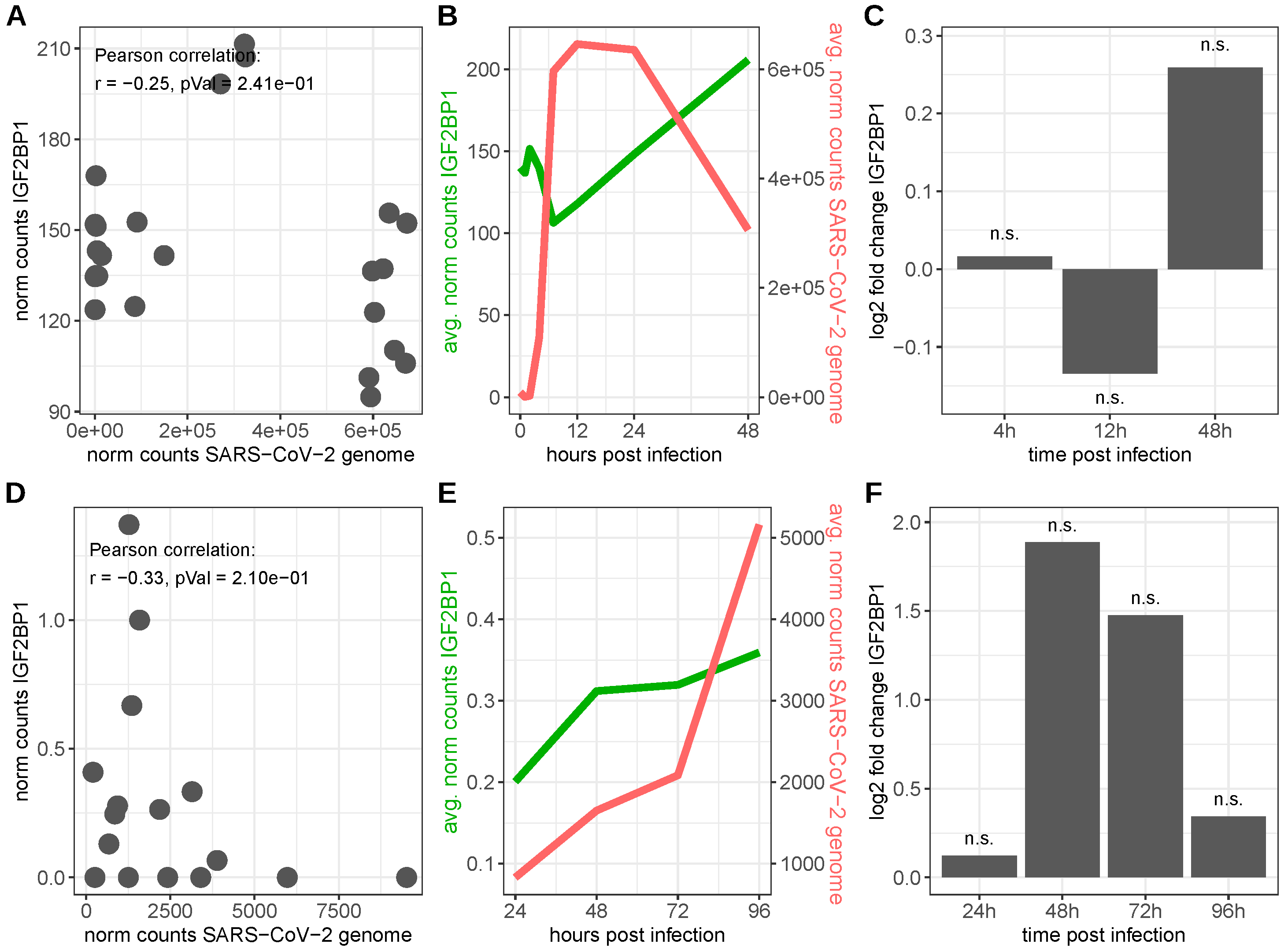
4.2. Severe Acute Respiratory Syndrome Coronavirus 2

The severe acute respiratory syndrome coronavirus 2 (SARS-CoV-2) is a single-stranded RNA virus, causing the coronavirus disease 19 (COVID-19) that became a worldwide pandemic in 2020. In most cases, SARS-CoV-2 infection causes mild cold symptoms; however, a significant proportion of patients develop severe symptoms, including acute respiratory distress (ARDS), pneumonia, renal failure, cardiac complications and even multi-organ failure. Furthermore, reports about persistent symptoms that may last for several months following an acute SARS-CoV-2 infection, designated as post-COVID syndrome, are accumulating [113,114,115]. The structural spike (S) protein of SARS-CoV-2 is equipped with a receptor-binding domain (RBD) mediating direct contact with the angiotensin-converting enzyme 2 (ACE2) receptor of susceptible host cells. However, for viral entry, the spike protein has to be primed by host cell proteases TMPRSS2 or cathepsin B/L [116]. Since SARS-CoV-2 enters the body via the respiratory tract, airway and alveolar epithelial cells as well as vascular endothelial cells and alveolar macrophages belong to the first targets of viral entry. However, cells from multiple extra-pulmonary tissues also express ACE2 and TMPRSS2 and thus represent possible target cells for SARS-CoV-2 [114,117,118].
Zhang et al. observed a stimulation of IGF2BP1 RNA expression upon SARS-CoV-2 infection in cell lines and patient lung samples. In addition, they found that the knockdown as well as knockout of IGF2BP1 in different cell lines led to reduced levels of viral RNA in these cells after infection with SARS-CoV-2. Importantly, IGF2BP1 was shown to bind and stabilize a subgenomic RNA encoding the SARS-CoV-2 S protein in vitro and thus, promoting S protein translation [44].
The inspection of two independent publicly available bulk RNA-seq datasets of SARS-CoV-2 infected colon adenocarcinoma derived Caco-2 and primary human bronchial epithelial cells (HBECs), respectively, led to inconclusive results. Considerable amounts of SARS-CoV-2 genomic RNA were detectable upon the infection in both cell lines; however, the observed amounts varied tremendously. Around 30% of all sequencing reads obtained from the Caco-2 cells were mapped to the SARS-CoV-2 genome at 7, 12 and 24 h after infection. In contrast, less than 1% of sequencing reads could be mapped to the virus genome in infected HBEC samples. IGF2BP1 RNA levels also varied by about three orders of magnitude between the two cell lines. The normalized expression values of IGF2BP1 transcripts and SARS-CoV-2 genomic RNA exhibited negative, yet not significant, Pearson correlation in infected cells from both cell lines (Figure 5A,D). Nonetheless, IGF2BP1 RNA levels tended to increase over time in infected cells, whereas the levels of SARS-CoV-2 genomic RNA started to decrease in Caco-2 cells between 12 and 24 h after infection, while they continued to increase in HBECs until the final measurements (96 h, Figure 5B,E). Compared to mock-infected (Caco-2)/uninfected (HBECs) cells, IGF2BP1 levels started to increase in both cell lines and reached almost four-fold higher levels (log2 fold change = 1.89) 48 h after infection in the HBECs. However, this increase was not significant (FDR adjusted p-value = 1), due to high variation in the expression in the biological replicates. Despite IGF2BP1 levels continuing to rise in infected cells, the rise in the expression of IGF2BP1 compared to uninfected HBEC cells was attenuated, since IGF2BP1 levels also increased in the uninfected cells (Figure 5C,F). However, IGF2BP1 RNA expression levels in the HBECs in total were rather low and, thus, detected differences might be unreliable and the consequence of data noise. Considering cell-type-specific RNA expression obtained from human protein atlas single-cell RNA-seq data, human kidney cells displayed salient co-expression patterns. Distinct sub-populations of proximal tubular cells express at least moderate levels of IGF2BP1 as well as ACE2, TMPRSS2 and cathepsin-B (CTSB) RNA (Figure S5A,C). Thus, due to a basal expression of IGF2BP1 in healthy human kidneys, they could potentially be organs vulnerable for infection by SARS-CoV-2 that might be intensified by IGF2BP1. However, further investigations are required. Moreover, it needs to be addressed if SARS-CoV-2 infections could lead to IGF2BP1 de novo synthesis, which might explain the observed elevated levels in patient lung samples.

4.3. Other Human Viruses

Besides SARS-CoV-2, Zhang et al. observed binding between IGF2BP1 and two other RNA viruses, namely Zika virus (ZIKV) and Ebola virus (EBOV) [44]. Similarly to SARS-CoV-2, the authors found a significant reduction in viral ZIKV RNA levels in infected cells upon IGF2BP1 knockdown and knockout. However, in contrast to SARS-CoV-2, they did not observe significant differences in the degradation rates of the ZIKV genomic RNA upon IGF2BP1 knockout; thus, they concluded that there was no stabilization effect, despite the fact that they observed that IGF2BP1 also promoted the translation of this virus [44]. The binding of IGF2BP1 to EBOV RNA was shown by Zhang et al. and Fang et al. However, while Zhang et al. did not observe an impact of IGF2BP1 knockdown on EBOV RNA levels, Fang et al. reported a significant reduction in infection rates after knocking down IGF2BP1 independently with two out of three used siRNAs [44,120].

5. Non-Human Viruses

Besides the above-reported human-pathogenic viruses, IGF2BP1 homologs were reported to promote the viral spread of several viruses infecting other animals.
Chen et al. reported the direct binding of IGF2BP1 to the 3′UTR of Duck hepatitis A virus type 1 (DHV-1/DHAV), a highly fatal, rapidly spreading virus infecting young ducklings [121], which led to the increased IRES-mediated translation efficiency of viral proteins in a duck embryo epithelial cell line. Furthermore, they reported that the presence of DHV-1 proteins in the cytoplasm of the duck embryo epithelial cell line led to increased IGF2BP1 protein levels [46]. The nonstructural p17 protein of the avian reovirus (ARV), another important poultry pathogen, was shown to interact with IGF2BP1 and the over-expression of IGF2BP1 in chicken cells was associated with an increase in viral replication. Furthermore, the mRNA levels of IGF2BP1 were increased upon ARV infection in chicken cells [122]. Another poultry-infecting virus with a reported connection to IGF2BP1 is the Marek’s disease virus (MDV), a highly contagious herpesvirus capable of inducing T-cell lymphomas in chicken [123]. IGF2BP1 expression was found to be increased in a chicken inbred line, highly susceptible to MDV, whereas, in a MDV-resistant cell line, IGF2BP1 RNA expression was hardly detectable. The difference in RNA expression was concordant with the increased activating histone methylation marks (H3K4me3) around the IGF2BP1 transcription start site found in the MDV susceptible chicken samples. Upon MDV infection, IGF2BP1 expression was significantly reduced in thymus cells of these animals [124]. However, it remained open, if the elevated IGF2BP1 expression was connected to higher susceptibility to the virus. Mai and Gao observed an IGF2BP1-dependent increase in the production of infectious murine leukemia virus (MLV) vectors, a retroviral vector system widely used for gene transfer. By binding and stabilizing the viral genomic RNA, IGF2BP1 increased its incorporation into virions. However, for the wild-type virus, only a modest effect was measured, since, although still increasing the incorporation of genomic RNA, IGF2BP1 showed an inhibitory effect on the production of viral proteins [125]. Similarly to HIV-1, IGF2BP1 protein was found to also be incorporated into the virion particles of this retrovirus [47,125]. Jefferson et al. identified IGF2BP1 as a binding partner of the N-terminal protease Npro of the classical swine fever virus (CSFV)/Pestivirus C by the GST pulldown assay and subsequent mass spectrometry. Since the number of detected peptides was increased in the presence of zinc, the authors concluded that the binding involved the interaction with the TRASH motif contained in the interaction domain of Npro [126].

6. Discussion

The RNA-binding protein IGF2BP1 has been reported to interact with RNA and proteins from a variety of viruses from different families, such as Hepadnaviridae (HBV), Flaviviridae (HCV, ZIKV, CSFV), Papillomaviridae (HPV), Retroviridae (HIV-1, MLV), Coronaviridae (SARS-CoV-2), Filoviridae (EBOV), Picornaviridae (DHV-1), Reoviridae (ARV) and Herpesviridae (MDV). Associations with IGF2BP1 include the binding of viral transcripts leading to their stabilization (HPV, SARS-CoV2, MLV; [43,44,125]), but also stability-independent enhanced translation (HCV, DHV-1; [45,46]). Furthermore, the binding of IGF2BP1 to viral proteins led to alterations in virus production, infectivity, or replication (HIV-1, ARV; [47,122]). Binding to IGF2BP1 usually results in proviral effects [43,44,45,46,49,86,120,122,126]. Only the binding to proteins of HIV-1 was reported to reduce virus production and infectivity [47,48]. Besides the putative influence on the natural infection process, an inhibitory effect of IGF2BP1 on HIV-1 production, facilitated by binding to Gag and Rev proteins, might also impact research and gene therapy approaches using lentiviral vectors comprising HIV-derived genes. Regarding the extent of reported virus families, the diversity of utilized molecular mechanisms and IGF2BP1’s function in promoting cell proliferation, it seems likely that even more virus species benefit from elevated IGF2BP1 expression. However, since IGF2BP1 is an oncofetal protein [19,20,21,22,23], questions about the physiological relevance of the reported interactions between IGF2BP1 and non-tumor viruses, primarily investigated in tumor-derived or embryonic cell lines, needs to be addressed by further analyses. Remarkably, however, upon infection with HBV, HCV, SARS-CoV-2, DHV-1 and ARV, increasing levels of IGF2BP1 RNA and/or protein have been observed [44,45,46,49,122]. This leads to the unsettling conclusion that infection with these, in part so far, not described as tumor-promoting viruses, might drive the expression of a protein that is known to be involved in the development and progression of multiple types of tumors [24,25,26,27,28,29]. The underlying mechanisms leading to the enhanced IGF2BP1 expression were not investigated and, thus, still need to be elaborated. On the other hand, as for cancer, the oncofetal expression pattern of IGF2BP1 could be exploited for developing novel anti-viral therapies, since the depletion/inactivation of this protein in adult tissues should have only little effects on normal cellular physiology. For example, the opposing functions of IGF2BP1 and miRNAs of the let-7 family on the spreading of viruses such as HBV, HCV and HPV might be utilized, e.g., by enhancing let-7 expression. Interferon alpha and Interleukin-28B treatment have been shown to increase cellular let-7 levels, decrease the IGF2BP1 expression and exert an anti-HCV effect [84]. Furthermore, small molecule inhibitors of IGF2BP1 function, including BTYNB or Cucurbitacin B, that have recently shown promising anti-tumor effects [37,127,128], should be tested for their impact on the production and infectivity of the virus species reported to benefit from IGF2BP1 expression.
In conclusion, IGF2BP1 can be recruited by a variety of viruses from distinct families to promote their spreading, and thus, anti-tumor strategies aiming to inhibit the expression or function of IGF2BP1 should be tested for anti-viral effects as well. Table 1 briefly summarizes the effects that IGF2BP1 exerts on the components of the described viruses as well as the impact of these viruses on IGF2BP1.

7. Future Directions

Despite only few studies dedicated to IGF2BP1–virus cross-talk, the broad range of virus families covered by these reports suggests a widespread role of IGF2BP1 in viral infection and the impact of viruses on IGF2BPs, especially in cancer. Recently developed data repositories, such as NCBI’s sequence read archive (SRA, https://www.ncbi.nlm.nih.gov/sra) or the European nucleotide archive (ENA, https://www.ebi.ac.uk/ena/), provide tremendous amounts of publicly available high-throughput sequencing data that could be a promising starting point for the discovery of new IGF2BP–virus interactions. The examination of appropriate RNA-seq datasets, provided by these repositories, could deliver new insights about the presence of viruses, e.g., in cancerous tissues but also about changes in IGF2BP expression upon virus infection. Correlations between IGF2BP1 or its paralogs and viral RNAs, derived from these studies, can be used to hypothesize about putative direct interactions and indirect cross-talk impacting the viral life cycle. Furthermore, CLIP-seq (cross-linking and immunoprecipitation) datasets could provide valuable information about viral RNAs bound to IGF2BPs. Additionally, binding motifs derived from these datasets can be used to scan viral genomes for the occurrences of putative IGF2BP binding sites. Indirect interactions such as the downregulation of PTPN13 by viral proteins leading to an IGF2BP1-dependent upregulation of c-Myc should also be further exploited. While investigated in detail for HBV [49], Hussen et al. described a reduction in PTPN13 levels upon HPV infection; however, they did not provide a mechanistic explanation [104]. Moreover, we observed a downregulation of PTPN13 as well as a negative influence on the association between IGF2BP1 and c-Myc in HCV-infected TCGA HCC samples—both effects were stronger pronounced than in HBV-positive samples (Figure S3). Thus, further investigations regarding interactions between viruses, IGF2BP1, PTPN13 and c-Myc levels are pending. Respective studies could reveal potential therapeutic options, especially in HCV/HPV-associated cancers. Recent reports provide strong evidence that RBP–RNA interactions are per se druggable and disrupting these interactions may provide benefits for viral infection-associated cancerous diseases [129].

Supplementary Materials

The following supporting information can be downloaded at: https://www.mdpi.com/article/10.3390/v15071431/s1, Figure S1: IGF2BP1 RNA expression; Figure S2: Kaplan-Meier curves describing overall survival probabilities depending on IGF2BP1 RNA expression in hepatocarcinoma derived RNA-seq data, provided by the TCGA (cohort LIHC); Figure S3: RNA expression and liquid association in in hepatocarcinoma derived RNA-seq data (HCC); Figure S4: Kaplan-Meier curves describing overall survival probabilities depending on IGF2BP1 RNA expression in cervical carcinoma derived RNA-seq data, provided by the TCGA (cohort CESC); Figure S5: Average RNA expression in distinct kidney resided cell types derived from single cell RNA-seq data provided by the Human protein atlas (www.proteinatlas.org).

Author Contributions

Conceptualization, M.G. and S.H.; formal analysis, M.G.; writing—original draft preparation, M.G.; writing—review and editing, S.H.; visualization, M.G. All authors have read and agreed to the published version of the manuscript.

Funding

This research was funded by the Deutsche Forschungsgemeinschaft (DFG) (GRK2751 to S.H and M.G.; RU5433 to S.H.).

Institutional Review Board Statement

Not applicable.

Informed Consent Statement

Not applicable.

Data Availability Statement

Bulk-RNA-seq data from individual projects were downloaded from NCBI GEO (https://www.ncbi.nlm.nih.gov/geo/). HCV: GSE126831, HPV: GSE158033 SARS-CoV-2: GSE217504 and GSE175779. Single-cell RNA-seq data of CD4+ T cells were downloaded from PanglaoDB (https://panglaodb.se/): SRS3034950, SRS3034951, SRS3034952 and SRS3034953.

Conflicts of Interest

The authors declare no conflict of interest.

References

  1. Nagy, P.D.; Pogany, J. The dependence of viral RNA replication on co-opted host factors. Nat. Rev. Microbiol. 2011, 10, 137–149. [Google Scholar] [CrossRef] [PubMed]
  2. Purdy, J.G.; Luftig, M.A. Reprogramming of cellular metabolic pathways by human oncogenic viruses. Curr. Opin. Virol. 2019, 39, 60–69. [Google Scholar] [PubMed]
  3. Saha, A.; Kaul, R.; Murakami, M.; Robertson, E.S. Tumor viruses and cancer biology: Modulating signaling pathways for therapeutic intervention. Cancer Biol. Ther. 2010, 10, 961–978. [Google Scholar] [CrossRef] [Green Version]
  4. White, M.K.; Pagano, J.S.; Khalili, K. Viruses and human cancers: A long road of discovery of molecular paradigms. Clin. Microbiol. Rev. 2014, 27, 463–481. [Google Scholar] [PubMed] [Green Version]
  5. Cook, L. Polyomaviruses. Microbiol. Spectr. 2016, 4. [Google Scholar] [CrossRef]
  6. Nevels, M.; Täuber, B.; Spruss, T.; Wolf, H.; Dobner, T. “Hit-and-run” transformation by adenovirus oncogenes. J. Virol. 2001, 75, 3089–3094. [Google Scholar]
  7. Bartel, D.P. Metazoan MicroRNAs. Cell 2018, 173, 20–51. [Google Scholar] [CrossRef] [Green Version]
  8. Dass, D.; Dhotre, K.; Chakraborty, M.; Nath, A.; Banerjee, A.; Bagchi, P.; Mukherjee, A. MiRNAs in Herpesvirus infection: Powerful regulators in small packages. Viruses 2023, 15, 429. [Google Scholar] [CrossRef]
  9. Megahed, F.; Tabll, A.; Atta, S.; Ragheb, A.; Smolic, R.; Petrovic, A.; Smolic, M. MicroRNAs: Small molecules with significant functions, particularly in the context of viral hepatitis B and C infection. Medicina 2023, 59, 173. [Google Scholar]
  10. Alshahrani, S.H.; Alameri, A.A.; Kahar, F.; Alexis Ramírez-Coronel, A.; Fadhel Obaid, R.; Alsaikhan, F.; Zabibah, R.S.; Qasim, Q.A.; Altalbawy, F.M.A.; Fakri Mustafa, Y.; et al. Overview of the role and action mechanism of microRNA-128 in viral infections. Microb. Pathog. 2023, 176, 106020. [Google Scholar] [CrossRef]
  11. Rashid, F.; Zaongo, S.D.; Song, F.; Chen, Y. The diverse roles of miRNAs in HIV pathogenesis: Current understanding and future perspectives. Front. Immunol. 2022, 13, 1091543. [Google Scholar] [CrossRef] [PubMed]
  12. He, L.; Li, H.; Wu, A.; Peng, Y.; Shu, G.; Yin, G. Functions of N6-methyladenosine and its role in cancer. Mol. Cancer 2019, 18, 176. [Google Scholar] [CrossRef] [PubMed] [Green Version]
  13. Liu, J.; Xu, Y.P.; Li, K.; Ye, Q.; Zhou, H.Y.; Sun, H.; Li, X.; Yu, L.; Deng, Y.Q.; Li, R.T.; et al. A methylome of SARS-CoV-2 in host cells. Cell Res. 2021, 31, 404–414. [Google Scholar] [CrossRef] [PubMed]
  14. Lichinchi, G.; Zhao, B.S.; Wu, Y.; Lu, Z.; Qin, Y.; He, C.; Rana, T.M. Dynamics of Human and Viral RNA Methylation during Zika Virus Infection. Cell Host Microbe 2016, 20, 666–673. [Google Scholar] [CrossRef] [Green Version]
  15. Tirumuru, N.; Zhao, B.S.; Lu, W.; Lu, Z.; He, C.; Wu, L. N(6)-methyladenosine of HIV-1 RNA regulates viral infection and HIV-1 Gag protein expression. Elife 2016, 5, e15528. [Google Scholar] [CrossRef]
  16. Lichinchi, G.; Gao, S.; Saletore, Y.; Gonzalez, G.M.; Bansal, V.; Wang, Y.; Mason, C.E.; Rana, T.M. Dynamics of the human and viral m(6)A RNA methylomes during HIV-1 infection of T cells. Nat. Microbiol. 2016, 1, 16011. [Google Scholar] [CrossRef]
  17. Gokhale, N.S.; McIntyre, A.B.R.; McFadden, M.J.; Roder, A.E.; Kennedy, E.M.; Gandara, J.A.; Hopcraft, S.E.; Quicke, K.M.; Vazquez, C.; Willer, J.; et al. N6-Methyladenosine in Flaviviridae Viral RNA Genomes Regulates Infection. Cell Host Microbe 2016, 20, 654–665. [Google Scholar] [CrossRef] [Green Version]
  18. Tan, B.; Gao, S.J. RNA epitranscriptomics: Regulation of infection of RNA and DNA viruses by N6-methyladenosine (m6A)). Rev. Med. Virol. 2018, 28, e1983. [Google Scholar] [CrossRef] [Green Version]
  19. Ioannidis, P.; Mahaira, L.; Papadopoulou, A.; Teixeira, M.R.; Heim, S.; Andersen, J.A.; Evangelou, E.; Dafni, U.; Pandis, N.; Trangas, T. CRD-BP: A c-Myc mRNA stabilizing protein with an oncofetal pattern of expression. Anticancer Res. 2003, 23, 2179–2183. [Google Scholar]
  20. Degrauwe, N.; Suvà, M.L.; Janiszewska, M.; Riggi, N.; Stamenkovic, I. IMPs: An RNA-binding protein family that provides a link between stem cell maintenance in normal development and cancer. Genes Dev. 2016, 30, 2459–2474. [Google Scholar] [CrossRef] [Green Version]
  21. Hattori, A.; Buac, K.; Ito, T. Regulation of Stem Cell Self-Renewal and Oncogenesis by RNA-Binding Proteins. Adv. Exp. Med. Biol. 2016, 907, 153–188. [Google Scholar] [PubMed]
  22. Bell, J.L.; Wächter, K.; Mühleck, B.; Pazaitis, N.; Köhn, M.; Lederer, M.; Hüttelmaier, S. Insulin-like growth factor 2 mRNA-binding proteins (IGF2BPs): Post-transcriptional drivers of cancer progression? Cell. Mol. Life Sci. 2013, 70, 2657–2675. [Google Scholar] [CrossRef] [PubMed] [Green Version]
  23. Ioannidis, P.; Kottaridi, C.; Dimitriadis, E.; Courtis, N.; Mahaira, L.; Talieri, M.; Giannopoulos, A.; Iliadis, K.; Papaioannou, D.; Nasioulas, G.; et al. Expression of the RNA-binding protein CRD-BP in brain and non-small cell lung tumors. Cancer Lett. 2004, 209, 245–250. [Google Scholar] [CrossRef] [PubMed]
  24. Köbel, M.; Weidensdorfer, D.; Reinke, C.; Lederer, M.; Schmitt, W.D.; Zeng, K.; Thomssen, C.; Hauptmann, S.; Hüttelmaier, S. Expression of the RNA-binding protein IMP1 correlates with poor prognosis in ovarian carcinoma. Oncogene 2007, 26, 7584–7589. [Google Scholar] [CrossRef] [PubMed] [Green Version]
  25. Müller, S.; Bley, N.; Glaß, M.; Busch, B.; Rousseau, V.; Misiak, D.; Fuchs, T.; Lederer, M.; Hüttelmaier, S. IGF2BP1 enhances an aggressive tumor cell phenotype by impairing miRNA-directed downregulation of oncogenic factors. Nucleic Acids Res. 2018, 46, 6285–6303. [Google Scholar] [CrossRef]
  26. Glaß, M.; Michl, P.; Hüttelmaier, S. RNA Binding Proteins as Drivers and Therapeutic Target Candidates in Pancreatic Ductal Adenocarcinoma. Int. J. Mol. Sci. 2020, 21, 4190. [Google Scholar] [CrossRef]
  27. Gutschner, T.; Hämmerle, M.; Pazaitis, N.; Bley, N.; Fiskin, E.; Uckelmann, H.; Heim, A.; Groß, M.; Hofmann, N.; Geffers, R.; et al. Insulin-like growth factor 2 mRNA-binding protein 1 (IGF2BP1) is an important protumorigenic factor in hepatocellular carcinoma. Hepatology 2014, 59, 1900–1911. [Google Scholar] [CrossRef]
  28. Bell, J.L.; Turlapati, R.; Liu, T.; Schulte, J.H.; Hüttelmaier, S. IGF2BP1 harbors prognostic significance by gene gain and diverse expression in neuroblastoma. J. Clin. Oncol. 2015, 33, 1285–1293. [Google Scholar] [CrossRef] [Green Version]
  29. Li, S.; Jiang, M. Elevated insulin-like growth factor 2 mRNA binding protein 1 levels predict a poor prognosis in patients with breast carcinoma using an integrated multi-omics data analysis. Front. Genet. 2022, 13, 994003. [Google Scholar] [CrossRef]
  30. Müller, S.; Glaß, M.; Singh, A.K.; Haase, J.; Bley, N.; Fuchs, T.; Lederer, M.; Dahl, A.; Huang, H.; Chen, J.; et al. IGF2BP1 promotes SRF-dependent transcription in cancer in a m6A- and miRNA-dependent manner. Nucleic Acids Res. 2019, 47, 375–390. [Google Scholar] [CrossRef] [Green Version]
  31. Busch, B.; Bley, N.; Müller, S.; Glaß, M.; Misiak, D.; Lederer, M.; Vetter, M.; Strauß, H.G.; Thomssen, C.; Hüttelmaier, S. The oncogenic triangle of HMGA2, LIN28B and IGF2BP1 antagonizes tumor-suppressive actions of the let-7 family. Nucleic Acids Res. 2016, 44, 3845–3864. [Google Scholar] [CrossRef] [PubMed]
  32. Leeds, P.; Kren, B.T.; Boylan, J.M.; Betz, N.A.; Steer, C.J.; Gruppuso, P.A.; Ross, J. Developmental regulation of CRD-BP, an RNA-binding protein that stabilizes c-myc mRNA in vitro. Oncogene 1997, 14, 1279–1286. [Google Scholar] [CrossRef] [Green Version]
  33. Boyerinas, B.; Park, S.M.; Murmann, A.E.; Gwin, K.; Montag, A.G.; Zillhardt, M.; Hua, Y.J.; Lengyel, E.; Peter, M.E. Let-7 modulates acquired resistance of ovarian cancer to Taxanes via IMP-1-mediated stabilization of multidrug resistance 1. Int. J. Cancer 2012, 130, 1787–1797. [Google Scholar] [CrossRef] [PubMed] [Green Version]
  34. Elcheva, I.; Goswami, S.; Noubissi, F.K.; Spiegelman, V.S. CRD-BP protects the coding region of betaTrCP1 mRNA from miR-183-mediated degradation. Mol. Cell 2009, 35, 240–246. [Google Scholar] [CrossRef] [PubMed] [Green Version]
  35. Glaß, M.; Misiak, D.; Bley, N.; Müller, S.; Hagemann, S.; Busch, B.; Rausch, A.; Hüttelmaier, S. IGF2BP1, a Conserved Regulator of RNA Turnover in Cancer. Front. Mol. Biosci. 2021, 8, 632219. [Google Scholar] [CrossRef]
  36. Huang, H.; Weng, H.; Sun, W.; Qin, X.; Shi, H.; Wu, H.; Zhao, B.S.; Mesquita, A.; Liu, C.; Yuan, C.L.; et al. Recognition of N6-methyladenosine by IGF2BP proteins enhances mRNA stability and translation. Nat. Cell Biol. 2018, 20, 285–295. [Google Scholar] [CrossRef] [PubMed]
  37. Müller, S.; Bley, N.; Busch, B.; Glaß, M.; Lederer, M.; Misiak, C.; Fuchs, T.; Wedler, A.; Haase, J.; Bertoldo, J.B.; et al. The oncofetal RNA-binding protein IGF2BP1 is a druggable, post-transcriptional super-enhancer of E2F-driven gene expression in cancer. Nucleic Acids Res. 2020, 48, 8576–8590. [Google Scholar] [CrossRef]
  38. Dyson, N.; Howley, P.M.; Münger, K.; Harlow, E. The human papilloma virus-16 E7 oncoprotein is able to bind to the retinoblastoma gene product. Science 1989, 243, 934–937. [Google Scholar] [CrossRef] [PubMed]
  39. Huh, K.; Zhou, X.; Hayakawa, H.; Cho, J.Y.; Libermann, T.A.; Jin, J.; Harper, J.W.; Munger, K. Human papillomavirus type 16 E7 oncoprotein associates with the cullin 2 ubiquitin ligase complex, which contributes to degradation of the retinoblastoma tumor suppressor. J. Virol. 2007, 81, 9737–9747. [Google Scholar] [CrossRef] [Green Version]
  40. Zhang, B.; Chen, W.; Roman, A. The E7 proteins of low- and high-risk human papillomaviruses share the ability to target the pRB family member p130 for degradation. Proc. Natl. Acad. Sci. USA 2006, 103, 437–442. [Google Scholar] [CrossRef] [PubMed] [Green Version]
  41. Berk, A.J. Recent lessons in gene expression, cell cycle control, and cell biology from adenovirus. Oncogene 2005, 24, 7673–7685. [Google Scholar] [CrossRef] [Green Version]
  42. Lu, H.; Li, W.; Noble, W.S.; Payan, D.; Anderson, D.C. Riboproteomics of the hepatitis C virus internal ribosomal entry site. J. Proteome Res. 2004, 3, 949–957. [Google Scholar] [CrossRef] [Green Version]
  43. Wang, L.; Zhan, G.; Maimaitiyiming, Y.; Su, Y.; Lin, S.; Liu, J.; Su, K.; Lin, J.; Shen, S.; He, W.; et al. m6A modification confers thermal vulnerability to HPV E7 oncotranscripts via reverse regulation of its reader protein IGF2BP1 upon heat stress. Cell Rep. 2022, 41, 111546. [Google Scholar] [CrossRef] [PubMed]
  44. Zhang, S.; Huang, W.; Ren, L.; Ju, X.; Gong, M.; Rao, J.; Sun, L.; Li, P.; Ding, Q.; Wang, J.; et al. Comparison of viral RNA-host protein interactomes across pathogenic RNA viruses informs rapid antiviral drug discovery for SARS-CoV-2. Cell Res. 2022, 32, 9–23. [Google Scholar] [CrossRef] [PubMed]
  45. Weinlich, S.; Hüttelmaier, S.; Schierhorn, A.; Behrens, S.E.; Ostareck-Lederer, A.; Ostareck, D.H. IGF2BP1 enhances HCV IRES-mediated translation initiation via the 3’UTR. RNA 2009, 15, 1528–1542. [Google Scholar] [CrossRef] [PubMed] [Green Version]
  46. Chen, J.; Zhang, R.; Lan, J.; Lin, S.; Li, P.; Gao, J.; Wang, Y.; Xie, Z.J.; Li, F.C.; Jiang, S.J. IGF2BP1 Significantly Enhances Translation Efficiency of Duck Hepatitis A Virus Type 1 without Affecting Viral Replication. Biomolecules 2019, 9, 594. [Google Scholar] [CrossRef] [PubMed] [Green Version]
  47. Zhou, Y.; Rong, L.; Lu, J.; Pan, Q.; Liang, C. Insulin-like growth factor II mRNA binding protein 1 associates with Gag protein of human immunodeficiency virus type 1, and its overexpression affects virus assembly. J. Virol. 2008, 82, 5683–5692. [Google Scholar] [CrossRef] [Green Version]
  48. Zhou, Y.; Rong, L.; Zhang, J.; Aloysius, C.; Pan, Q.; Liang, C. Insulin-like growth factor II mRNA binding protein 1 modulates Rev-dependent human immunodeficiency virus type 1 RNA expression. Virology 2009, 393, 210–220. [Google Scholar] [CrossRef] [Green Version]
  49. Yan, Y.; Huang, P.; Mao, K.; He, C.; Xu, Q.; Zhang, M.; Liu, H.; Zhou, Z.; Zhou, Q.; Zhou, Q.; et al. Anti-oncogene PTPN13 inactivation by hepatitis B virus X protein counteracts IGF2BP1 to promote hepatocellular carcinoma progression. Oncogene 2021, 40, 28–45. [Google Scholar] [CrossRef]
  50. R Core Team. R: A Language and Environment for Statistical Computing; R Foundation for Statistical Computing: Vienna, Austria, 2023. [Google Scholar]
  51. Wickham, H. ggplot2: Elegant Graphics for Data Analysis; Springer: New York, NY, USA, 2016. [Google Scholar]
  52. Shannon, P.; Markiel, A.; Ozier, O.; Baliga, N.S.; Wang, J.T.; Ramage, D.; Amin, N.; Schwikowski, B.; Ideker, T. Cytoscape: A software environment for integrated models of biomolecular interaction networks. Genome Res. 2003, 13, 2498–2504. [Google Scholar] [CrossRef]
  53. Cancer Genome Atlas Research Network. Comprehensive and integrative genomic characterization of hepatocellular carcinoma. Cell 2017, 169, 1327–1341.e23. [Google Scholar] [CrossRef] [Green Version]
  54. Cancer Genome Atlas Research Network; Albert Einstein College of Medicine; Analytical Biological Services; Barretos Cancer Hospital; Baylor College of Medicine; Beckman Research Institute of City of Hope; Buck Institute for Research on Aging; Canada’s Michael Smith Genome Sciences Centre; Harvard Medical School; Helen F. Graham Cancer Center &Research Institute at Christiana Care Health Services; et al. Integrated genomic and molecular characterization of cervical cancer. Nature 2017, 543, 378–384. [Google Scholar] [CrossRef] [PubMed] [Green Version]
  55. Ho, Y.Y. LiquidAssociation: LiquidAssociation, R Package Version 1.53.0. 2022. Available online: https://bioconductor.org/packages/release/bioc/html/LiquidAssociation.html(accessed on 21 June 2023).
  56. Therneau, T.M. A Package for Survival Analysis in R, R package version 3.5-5. 2023. Available online: https://cran.r-project.org/web/packages/survival/vignettes/survival.pdf(accessed on 21 June 2023).
  57. Barretina, J.; Caponigro, G.; Stransky, N.; Venkatesan, K.; Margolin, A.A.; Kim, S.; Wilson, C.J.; Lehár, J.; Kryukov, G.V.; Sonkin, D.; et al. The Cancer Cell Line Encyclopedia enables predictive modelling of anticancer drug sensitivity. Nature 2012, 483, 603–607. [Google Scholar] [CrossRef] [PubMed] [Green Version]
  58. Shi, B.; Ding, J.; Qi, J.; Gu, Z. Characteristics and prognostic value of potential dependency genes in clear cell renal cell carcinoma based on a large-scale CRISPR-Cas9 and RNAi screening database DepMap. Int. J. Med. Sci. 2021, 18, 2063–2075. [Google Scholar] [CrossRef]
  59. Morgan, M.; Shepherd, L. ExperimentHub: Client to Access Experimenthub Resources, R Package Version 2.7.1. 2023. Available online: https://bioconductor.org/packages/release/bioc/html/ExperimentHub.html(accessed on 21 June 2023).
  60. GTEx Consortium. The Genotype-Tissue Expression (GTEx) project. Nat. Genet. 2013, 45, 580–585. [Google Scholar] [CrossRef]
  61. Martin, M. Cutadapt removes adapter sequences from high-throughput sequencing reads. EMBnet J. 2011, 17, 10–12. [Google Scholar] [CrossRef]
  62. Kim, D.; Langmead, B.; Salzberg, S.L. HISAT: A fast spliced aligner with low memory requirements. Nat. Methods 2015, 12, 357–360. [Google Scholar] [CrossRef] [Green Version]
  63. Danecek, P.; Bonfield, J.K.; Liddle, J.; Marshall, J.; Ohan, V.; Pollard, M.O.; Whitwham, A.; Keane, T.; McCarthy, S.A.; Davies, R.M.; et al. Twelve years of SAMtools and BCFtools. Gigascience 2021, 10, giab008. [Google Scholar] [CrossRef]
  64. Liao, Y.; Smyth, G.K.; Shi, W. featureCounts: An efficient general purpose program for assigning sequence reads to genomic features. Bioinformatics 2014, 30, 923–930. [Google Scholar] [CrossRef] [Green Version]
  65. Aken, B.L.; Achuthan, P.; Akanni, W.; Amode, M.R.; Bernsdorff, F.; Bhai, J.; Billis, K.; Carvalho-Silva, D.; Cummins, C.; Clapham, P.; et al. Ensembl 2017. Nucleic Acids Res. 2017, 45, D635–D642. [Google Scholar] [CrossRef]
  66. Robinson, M.D.; McCarthy, D.J.; Smyth, G.K. edgeR: A Bioconductor package for differential expression analysis of digital gene expression data. Bioinformatics 2010, 26, 139–140. [Google Scholar] [CrossRef] [PubMed] [Green Version]
  67. Robinson, M.D.; Oshlack, A. A scaling normalization method for differential expression analysis of RNA-seq data. Genome Biol. 2010, 11, R25. [Google Scholar] [CrossRef] [PubMed] [Green Version]
  68. Hao, Y.; Hao, S.; Andersen-Nissen, E.; Mauck, W.M.; Zheng, S.; Butler, A.; Lee, M.J.; Wilk, A.J.; Darby, C.; Zager, M.; et al. Integrated analysis of multimodal single-cell data. Cell 2021, 184, 3573–3587. [Google Scholar] [CrossRef] [PubMed]
  69. Tsukuda, S.; Watashi, K. Hepatitis B virus biology and life cycle. Antivir. Res. 2020, 182, 104925. [Google Scholar] [CrossRef] [PubMed]
  70. Megahed, F.A.K.; Zhou, X.; Sun, P. The Interactions between HBV and the Innate Immunity of Hepatocytes. Viruses 2020, 12, 285. [Google Scholar] [CrossRef] [PubMed] [Green Version]
  71. Perz, J.F.; Armstrong, G.L.; Farrington, L.A.; Hutin, Y.J.; Bell, B.P. The contributions of hepatitis B virus and hepatitis C virus infections to cirrhosis and primary liver cancer worldwide. J. Hepatol. 2006, 45, 529–538. [Google Scholar] [CrossRef] [PubMed]
  72. Levrero, M.; Zucman-Rossi, J. Mechanisms of HBV-induced hepatocellular carcinoma. J. Hepatol. 2016, 64, S84–S101. [Google Scholar] [CrossRef]
  73. Li, W.; Miao, X.; Qi, Z.; Zeng, W.; Liang, J.; Liang, Z. Hepatitis B virus X protein upregulates HSP90alpha expression via activation of c-Myc in human hepatocarcinoma cell line, HepG2. Virol. J. 2010, 7, 45. [Google Scholar] [CrossRef] [Green Version]
  74. Wang, Y.; Lu, Y.; Toh, S.T.; Sung, W.K.; Tan, P.; Chow, P.; Chung, A.Y.; Jooi, L.L.; Lee, C.G. Lethal-7 is down-regulated by the hepatitis B virus x protein and targets signal transducer and activator of transcription 3. J. Hepatol. 2010, 53, 57–66. [Google Scholar] [CrossRef]
  75. You, X.; Liu, F.; Zhang, T.; Lv, N.; Liu, Q.; Shan, C.; Du, Y.; Kong, G.; Wang, T.; Ye, L.; et al. Hepatitis B virus X protein upregulates Lin28A/Lin28B through Sp-1/c-Myc to enhance the proliferation of hepatoma cells. Oncogene 2014, 33, 449–460. [Google Scholar] [CrossRef] [Green Version]
  76. Chang, T.C.; Zeitels, L.R.; Hwang, H.W.; Chivukula, R.R.; Wentzel, E.A.; Dews, M.; Jung, J.; Gao, P.; Dang, C.V.; Beer, M.A.; et al. Lin-28B transactivation is necessary for Myc-mediated let-7 repression and proliferation. Proc. Natl. Acad. Sci. USA 2009, 106, 3384–3389. [Google Scholar] [CrossRef] [PubMed] [Green Version]
  77. Li, K.C. Genome-wide coexpression dynamics: Theory and application. Proc. Natl. Acad. Sci. USA 2002, 99, 16875–16880. [Google Scholar] [CrossRef] [Green Version]
  78. Bairoch, A. The Cellosaurus, a Cell-Line Knowledge Resource. J. Biomol. Tech. 2018, 29, 25–38. [Google Scholar] [CrossRef]
  79. El-Serag, H.B. Hepatocellular carcinoma and hepatitis C in the United States. Hepatology 2002, 36, 74–83. [Google Scholar]
  80. Dubuisson, J.; Cosset, F.L. Virology and cell biology of the hepatitis C virus life cycle: An update. J. Hepatol. 2014, 61, S3–S13. [Google Scholar] [CrossRef] [PubMed] [Green Version]
  81. Zeisel, M.B.; Barth, H.; Schuster, C.; Baumert, T.F. Hepatitis C virus entry: Molecular mechanisms and targets for antiviral therapy. Front. Biosci. (Landmark Ed.) 2009, 14, 3274–3285. [Google Scholar] [CrossRef] [PubMed] [Green Version]
  82. Tsukiyama-Kohara, K.; Iizuka, N.; Kohara, M.; Nomoto, A. Internal ribosome entry site within hepatitis C virus RNA. J. Virol. 1992, 66, 1476–1483. [Google Scholar] [CrossRef] [Green Version]
  83. Kieft, J.S.; Zhou, K.; Jubin, R.; Doudna, J.A. Mechanism of ribosome recruitment by hepatitis C IRES RNA. RNA 2001, 7, 194–206. [Google Scholar] [CrossRef] [Green Version]
  84. Cheng, M.; Si, Y.; Niu, Y.; Liu, X.; Li, X.; Zhao, J.; Jin, Q.; Yang, W. High-throughput profiling of alpha interferon- and interleukin-28B-regulated microRNAs and identification of let-7s with anti-hepatitis C virus activity by targeting IGF2BP1. J. Virol. 2013, 87, 9707–9718. [Google Scholar] [CrossRef] [Green Version]
  85. Boyerinas, B.; Park, S.M.; Shomron, N.; Hedegaard, M.M.; Vinther, J.; Andersen, J.S.; Feig, C.; Xu, J.; Burge, C.B.; Peter, M.E. Identification of let-7-regulated oncofetal genes. Cancer Res. 2008, 68, 2587–2591. [Google Scholar] [CrossRef] [Green Version]
  86. Li, Y.; Masaki, T.; Shimakami, T.; Lemon, S.M. hnRNP L and NF90 interact with hepatitis C virus 5’-terminal untranslated RNA and promote efficient replication. J. Virol. 2014, 88, 7199–7209. [Google Scholar] [CrossRef] [Green Version]
  87. Jopling, C.L.; Yi, M.; Lancaster, A.M.; Lemon, S.M.; Sarnow, P. Modulation of hepatitis C virus RNA abundance by a liver-specific MicroRNA. Science 2005, 309, 1577–1581. [Google Scholar] [CrossRef] [Green Version]
  88. Jangra, R.K.; Yi, M.; Lemon, S.M. Regulation of hepatitis C virus translation and infectious virus production by the microRNA miR-122. J. Virol. 2010, 84, 6615–6625. [Google Scholar] [CrossRef] [Green Version]
  89. Bradrick, S.S.; Nagyal, S.; Novatt, H. A miRNA-responsive cell-free translation system facilitates isolation of hepatitis C virus miRNP complexes. RNA 2013, 19, 1159–1169. [Google Scholar] [CrossRef] [PubMed] [Green Version]
  90. Lupberger, J.; Croonenborghs, T.; Roca Suarez, A.A.; Van Renne, N.; Jühling, F.; Oudot, M.A.; Virzì, A.; Bandiera, S.; Jamey, C.; Meszaros, G.; et al. Combined Analysis of Metabolomes, Proteomes, and Transcriptomes of Hepatitis C Virus-Infected Cells and Liver to Identify Pathways Associated With Disease Development. Gastroenterology 2019, 157, 537–551. [Google Scholar] [CrossRef] [PubMed] [Green Version]
  91. Doorbar, J.; Quint, W.; Banks, L.; Bravo, I.G.; Stoler, M.; Broker, T.R.; Stanley, M.A. The biology and life-cycle of human papillomaviruses. Vaccine 2012, 30 (Suppl. S5), 55–70. [Google Scholar] [CrossRef] [PubMed]
  92. van Boerdonk, R.A.; Daniels, J.M.; Bloemena, E.; Krijgsman, O.; Steenbergen, R.D.; Brakenhoff, R.H.; Grünberg, K.; Ylstra, B.; Meijer, C.J.; Smit, E.F.; et al. High-risk human papillomavirus-positive lung cancer: Molecular evidence for a pattern of pulmonary metastasis. J. Thorac. Oncol. 2013, 8, 711–718. [Google Scholar] [CrossRef] [Green Version]
  93. Hasegawa, Y.; Ando, M.; Kubo, A.; Isa, S.; Yamamoto, S.; Tsujino, K.; Kurata, T.; Ou, S.H.; Takada, M.; Kawaguchi, T. Human papilloma virus in non-small cell lung cancer in never smokers: A systematic review of the literature. Lung Cancer 2014, 83, 8–13. [Google Scholar] [CrossRef]
  94. Khodabandehlou, N.; Mostafaei, S.; Etemadi, A.; Ghasemi, A.; Payandeh, M.; Hadifar, S.; Norooznezhad, A.H.; Kazemnejad, A.; Moghoofei, M. Human papilloma virus and breast cancer: The role of inflammation and viral expressed proteins. BMC Cancer 2019, 19, 61. [Google Scholar] [CrossRef] [Green Version]
  95. de Martel, C.; Plummer, M.; Vignat, J.; Franceschi, S. Worldwide burden of cancer attributable to HPV by site, country and HPV type. Int. J. Cancer 2017, 141, 664–670. [Google Scholar] [CrossRef] [Green Version]
  96. Berman, T.A.; Schiller, J.T. Human papillomavirus in cervical cancer and oropharyngeal cancer: One cause, two diseases. Cancer 2017, 123, 2219–2229. [Google Scholar] [CrossRef] [PubMed]
  97. Graham, S.V. The human papillomavirus replication cycle, and its links to cancer progression: A comprehensive review. Clin. Sci. 2017, 131, 2201–2221. [Google Scholar] [CrossRef] [PubMed] [Green Version]
  98. Ang, K.K.; Harris, J.; Wheeler, R.; Weber, R.; Rosenthal, D.I.; Nguyen-Tân, P.F.; Westra, W.H.; Chung, C.H.; Jordan, R.C.; Lu, C.; et al. Human papillomavirus and survival of patients with oropharyngeal cancer. N. Engl. J. Med. 2010, 363, 24–35. [Google Scholar] [CrossRef] [PubMed] [Green Version]
  99. Licitra, L.; Perrone, F.; Bossi, P.; Suardi, S.; Mariani, L.; Artusi, R.; Oggionni, M.; Rossini, C.; Cantù, G.; Squadrelli, M.; et al. High-risk human papillomavirus affects prognosis in patients with surgically treated oropharyngeal squamous cell carcinoma. J. Clin. Oncol. 2006, 24, 5630–5636. [Google Scholar] [CrossRef] [Green Version]
  100. Li, W.; Thompson, C.H.; O’Brien, C.J.; McNeil, E.B.; Scolyer, R.A.; Cossart, Y.E.; Veness, M.J.; Walker, D.M.; Morgan, G.J.; Rose, B.R. Human papillomavirus positivity predicts favourable outcome for squamous carcinoma of the tonsil. Int. J. Cancer 2003, 106, 553–558. [Google Scholar] [CrossRef]
  101. Mellin, H.; Dahlgren, L.; Munck-Wikland, E.; Lindholm, J.; Rabbani, H.; Kalantari, M.; Dalianis, T. Human papillomavirus type 16 is episomal and a high viral load may be correlated to better prognosis in tonsillar cancer. Int. J. Cancer 2002, 102, 152–158. [Google Scholar] [CrossRef]
  102. Moody, C.A.; Laimins, L.A. Human papillomavirus oncoproteins: Pathways to transformation. Nat. Rev. Cancer 2010, 10, 550–560. [Google Scholar] [CrossRef] [PubMed]
  103. Hwang, S.G.; Lee, D.; Kim, J.; Seo, T.; Choe, J. Human papillomavirus type 16 E7 binds to E2F1 and activates E2F1-driven transcription in a retinoblastoma protein-independent manner. J. Biol. Chem. 2002, 277, 2923–2930. [Google Scholar] [CrossRef] [Green Version]
  104. Hussen, B.M.; Ahmadi, G.; Marzban, H.; Fard Azar, M.E.; Sorayyayi, S.; Karampour, R.; Nahand, J.S.; Hidayat, H.J.; Moghoofei, M. The role of HPV gene expression and selected cellular MiRNAs in lung cancer development. Microb. Pathog. 2021, 150, 104692. [Google Scholar] [CrossRef]
  105. Laban, S.; Gangkofner, D.S.; Holzinger, D.; Schroeder, L.; Eichmüller, S.B.; Zörnig, I.; Jäger, D.; Wichmann, G.; Dietz, A.; Broglie, M.A.; et al. Antibody Responses to Cancer Antigens Identify Patients with a Poor Prognosis among HPV-Positive and HPV-Negative Head and Neck Squamous Cell Carcinoma Patients. Clin. Cancer Res. 2019, 25, 7405–7412. [Google Scholar] [CrossRef] [Green Version]
  106. Yu, L.; Lobanov, A.; Zheng, Z.M. Reply to Wang et al., Assessment of the Abundance and Potential Function of Human Papillomavirus Type 16 Circular E7 RNA. mBio 2022, 13, e0075822. [Google Scholar] [CrossRef]
  107. Murakami, T.; Ono, A. HIV-1 entry: Duels between Env and host antiviral transmembrane proteins on the surface of virus particles. Curr. Opin. Virol. 2021, 50, 59–68. [Google Scholar] [CrossRef] [PubMed]
  108. Murakami, T. Roles of the interactions between Env and Gag proteins in the HIV-1 replication cycle. Microbiol. Immunol. 2008, 52, 287–295. [Google Scholar] [CrossRef]
  109. Milev, M.P.; Brown, C.M.; Mouland, A.J. Live cell visualization of the interactions between HIV-1 Gag and the cellular RNA-binding protein Staufen1. Retrovirology 2010, 7, 41. [Google Scholar] [CrossRef] [Green Version]
  110. Rausch, J.W.; Le Grice, S.F. HIV Rev Assembly on the Rev Response Element (RRE): A Structural Perspective. Viruses 2015, 7, 3053–3075. [Google Scholar] [CrossRef] [Green Version]
  111. Uhlén, M.; Fagerberg, L.; Hallström, B.M.; Lindskog, C.; Oksvold, P.; Mardinoglu, A.; Sivertsson, Å.; Kampf, C.; Sjöstedt, E.; Asplund, A.; et al. Proteomics. Tissue-based map of the human proteome. Science 2015, 347, 1260419. [Google Scholar] [CrossRef] [PubMed]
  112. Cancer Genome Atlas Research Network; Ley, T.J.; Miller, C.; Ding, L.; Raphael, B.J.; Mungall, A.J.; Robertson, A.G.; Hoadley, K.; Triche, T.J., Jr.; Laird, P.W.; et al. Genomic and epigenomic landscapes of adult de novo acute myeloid leukemia. N. Engl. J. Med. 2013, 368, 2059–2074. [Google Scholar] [PubMed] [Green Version]
  113. Huseynov, A.; Akin, I.; Duerschmied, D.; Scharf, R.E. Cardiac Arrhythmias in Post-COVID Syndrome: Prevalence, Pathology, Diagnosis, and Treatment. Viruses 2023, 15, 389. [Google Scholar] [CrossRef]
  114. Harrison, A.G.; Lin, T.; Wang, P. Mechanisms of SARS-CoV-2 Transmission and Pathogenesis. Trends Immunol. 2020, 41, 1100–1115. [Google Scholar] [CrossRef]
  115. Azevedo, R.B.; Botelho, B.G.; Hollanda, J.V.G.; Ferreira, L.V.L.; Junqueira de Andrade, L.Z.; Oei, S.S.M.L.; Mello, T.S.; Muxfeldt, E.S. COVID-19 and the cardiovascular system: A comprehensive review. J. Hum. Hypertens. 2021, 35, 4–11. [Google Scholar] [CrossRef]
  116. Hoffmann, M.; Kleine-Weber, H.; Schroeder, S.; Krüger, N.; Herrler, T.; Erichsen, S.; Schiergens, T.S.; Herrler, G.; Wu, N.H.; Nitsche, A.; et al. SARS-CoV-2 Cell Entry Depends on ACE2 and TMPRSS2 and Is Blocked by a Clinically Proven Protease Inhibitor. Cell 2020, 181, 271–280. [Google Scholar] [CrossRef] [PubMed]
  117. Sun, K.; Gu, L.; Ma, L.; Duan, Y. Atlas of ACE2 gene expression reveals novel insights into transmission of SARS-CoV-2. Heliyon 2021, 7, e05850. [Google Scholar] [CrossRef] [PubMed]
  118. Bao, R.; Hernandez, K.; Huang, L.; Luke, J.J. ACE2 and TMPRSS2 expression by clinical, HLA, immune, and microbial correlates across 34 human cancers and matched normal tissues: Implications for SARS-CoV-2 COVID-19. J. Immunother. Cancer 2020, 8, e001020. [Google Scholar] [CrossRef] [PubMed]
  119. Subramaniyan, B.; Larabee, J.L.; Bodas, M.; Moore, A.R.; Burgett, A.W.G.; Myers, D.A.; Georgescu, C.; Wren, J.D.; Papin, J.F.; Walters, M.S. Characterization of the SARS-CoV-2 Host Response in Primary Human Airway Epithelial Cells from Aged Individuals. Viruses 2021, 13, 1603. [Google Scholar] [CrossRef] [PubMed]
  120. Fang, J.; Pietzsch, C.; Ramanathan, P.; Santos, R.I.; Ilinykh, P.A.; Garcia-Blanco, M.A.; Bukreyev, A.; Bradrick, S.S. Staufen1 Interacts with Multiple Components of the Ebola Virus Ribonucleoprotein and Enhances Viral RNA Synthesis. mBio 2018, 9, e01771-18. [Google Scholar] [CrossRef] [PubMed] [Green Version]
  121. Kim, M.C.; Kwon, Y.K.; Joh, S.J.; Kwon, J.H.; Lindberg, A.M. Differential diagnosis between type-specific duck hepatitis virus type 1 (DHV-1) and recent Korean DHV-1-like isolates using a multiplex polymerase chain reaction. Avian Pathol. 2008, 37, 171–177. [Google Scholar] [CrossRef] [Green Version]
  122. Zhang, C.; Liu, X.; Zhao, F.; Zhang, Q.; Zuo, W.; Guo, M.; Zhang, X.; Wu, Y. Identification and Functional Analyses of Host Proteins Interacting with the p17 Protein of Avian Reovirus. Viruses 2022, 14, 892. [Google Scholar] [CrossRef]
  123. Couteaudier, M.; Denesvre, C. Marek’s disease virus and skin interactions. Vet. Res. 2014, 45, 36. [Google Scholar] [CrossRef] [Green Version]
  124. Mitra, A.; Luo, J.; Zhang, H.; Cui, K.; Zhao, K.; Song, J. Marek’s disease virus infection induces widespread differential chromatin marks in inbred chicken lines. BMC Genom. 2012, 13, 557. [Google Scholar] [CrossRef] [Green Version]
  125. Mai, Y.; Gao, G. Expression of IMP1 enhances production of murine leukemia virus vector by facilitating viral genomic RNA packaging. PLoS ONE 2010, 5, e15881. [Google Scholar] [CrossRef] [Green Version]
  126. Jefferson, M.; Donaszi-Ivanov, A.; Pollen, S.; Dalmay, T.; Saalbach, G.; Powell, P.P. Host factors that interact with the pestivirus N-terminal protease, Npro, are components of the ribonucleoprotein complex. J. Virol. 2014, 88, 10340–10353. [Google Scholar] [CrossRef] [PubMed] [Green Version]
  127. Mahapatra, L.; Andruska, N.; Mao, C.; Le, J.; Shapiro, D.J. A Novel IMP1 Inhibitor, BTYNB, Targets c-Myc and Inhibits Melanoma and Ovarian Cancer Cell Proliferation. Transl. Oncol. 2017, 10, 818–827. [Google Scholar] [CrossRef] [PubMed]
  128. Liu, Y.; Guo, Q.; Yang, H.; Zhang, X.W.; Feng, N.; Wang, J.K.; Liu, T.T.; Zeng, K.W.; Tu, P.F. Allosteric Regulation of IGF2BP1 as a Novel Strategy for the Activation of Tumor Immune Microenvironment. ACS Cent. Sci. 2022, 8, 1102–1115. [Google Scholar] [CrossRef] [PubMed]
  129. Bertoldo, J.B.; Müller, S.; Hüttelmaier, S. RNA-binding proteins in cancer drug discovery. Drug Discov. Today 2023, 28, 103580. [Google Scholar] [CrossRef] [PubMed]
Figure 2. Interaction between HCV and IGF2BP1. (A) Scheme depicting the published interactions between the HCV genomic RNA and the host cell components, cf. [31,42,45,84,85]. Solid line—direct interaction, dashed line—indirect interaction. (B) Expression levels of IGF2BP1 and HCV RNA in Huh7.5.1 cells infected with HCV (data derived from [90]). (C) IGF2BP1 fold changes in HCV-infected Huh7.5.1 cells compared to uninfected cells (data derived from [90]). n.s.: FDR adjusted p-value ≥ 0.05, ***: FDR adjusted p-value < 0.001.
Viruses 15 01431 g002
Figure 3. Interaction between HPV and IGF2BP1. (A) Scheme depicting published interactions between the HCV’s E7 protein and host cell components, cf. [37,38,39,40,43,102,103]. Solid line—direct interaction. (B) RNA expression levels of IGF2BP1 and HPV E7 in HPV-positive cervical cancer cell lines (data derived from [106]). (C) IGF2BP1 expression values in cervical cancer samples (TCGA cohort CESC [54]). ns.: Wilcox-test p-value ≥ 0.05.
Viruses 15 01431 g003
Figure 4. IGF2BP RNA expression in blood cells and leukemia. (A) Fraction of CD4+ T cells uninfected or infected with HIV-1 with detectable RNA expression (normalized read counts > 0) of the indicated proteins (data derived from single-cell RNA-seq; SRA accession: SRP134859). (B) RNA expression levels of the human IGF2BP proteins in bulk RNA-seq data of blood cells (data derived from GTEx [60]). (C) RNA expression levels of the human IGF2BP proteins in the bulk RNA-seq data of peripheral blood cells from acute myeloid leukemia patients (data derived from the TCGA LAML cohort [112]).
Viruses 15 01431 g004
Figure 5. IGF2BP1 RNA expression in SARS-CoV-2-infected cells. Upper row: SARS-CoV-2-infected Caco-2 cells (data derived from GEO series GSE217504), lower row: SARS-CoV-2-infected primary human bronchial epithelial cells (HBECs, data derived from [119]). (A,D) RNA expression levels of IGF2BP1 and the SARS-CoV-2 genome in SARS-CoV-2-infected cells. (B,E) Time-point-averaged RNA expression levels of IGF2BP1 and the SARS-CoV-2 genome at the indicated time after infection. (C,F) IGF2BP1 fold changes in SARS-CoV-2 infected cells compared to mock-infected (Caco-2)/uninfected (HBECs) cells. n.s.: FDR adjusted p-value ≥ 0.05.
Viruses 15 01431 g005
Table 1. Effects of IGF2BP1–virus interactions.
VirusImpact of IGF2BP1 on VirusImpact of Virus on IGF2BP1
HBV Binding to c-Myc mRNA ↑ [49]
RNA/protein expression ↑ [49]
HCVTranslation ↑ [45]Protein expression ↑ [45]
Replication ↑ [86]
HPVStability of E7 RNA ↑ [43]Heat stability ↓ [43]
HIV-1Virus production↓ [47,48]
Infectivity ↓ [47,48]
SARS-CoV-2RNA levels ↑ [44]RNA expression ↑ [44]
Stability of S RNA ↑ [44]
ZIKVRNA levels ↑ [44]
EBOVInfectivity ↑ [120]
DHV-1Translation ↑ [46]Protein expression ↑ [46]
ARVReplication ↑ [122]RNA expression ↑ [122]
MDV RNA expression ↓ [124]
MLVstability of genomic RNA ↑ [125]
Protein levels ↓ [125]
Disclaimer/Publisher’s Note: The statements, opinions and data contained in all publications are solely those of the individual author(s) and contributor(s) and not of MDPI and/or the editor(s). MDPI and/or the editor(s) disclaim responsibility for any injury to people or property resulting from any ideas, methods, instructions or products referred to in the content.

