Article

Abundance of Poleroviruses within Tasmanian Pea Crops and Surrounding Weeds, and the Genetic Diversity of TuYV Isolates Found

1
New Town Research Laboratories, Tasmanian Institute of Agriculture, University of Tasmania, 13 St. Johns Avenue, New Town, Hobart, TAS 7008, Australia
2
Guangdong Provincial Key Laboratory of High Technology for Plant Protection, Plant Protection Research Institute, Guangdong Academy of Agricultural Sciences, Guangzhou 510640, China
3
Department of Agriculture and Fisheries (Queensland), Bundaberg Research Facility, 49 Ashfield Road, Bundaberg, QLD 4670, Australia
*
Author to whom correspondence should be addressed.
Viruses 2022, 14(8), 1690; https://doi.org/10.3390/v14081690
Submission received: 30 May 2022 / Revised: 28 July 2022 / Accepted: 28 July 2022 / Published: 30 July 2022
(This article belongs to the Special Issue State-of-the-Art Plant Virus Research in Australasia)

Abstract

:
The genus Polerovirus contains positive-sense, single-stranded RNA plant viruses that cause significant disease in many agricultural crops, including vegetable legumes. This study aimed to identify and determine the abundance of Polerovirus species present within Tasmanian pea crops and surrounding weeds that may act as virus reservoirs. We further sought to examine the genetic diversity of TuYV, the most commonly occurring polerovirus identified. Pea and weed samples were collected during 2019–2020 between October and January from thirty-four sites across three different regions (far northwest, north, and midlands) of Tasmania and tested by RT-PCR assay, with selected samples subject to next-generation sequencing. Results revealed that the presence of polerovirus infection and the prevalence of TuYV in both weeds and pea crops varied across the three Tasmanian cropping regions, with TuYV infection levels in pea crops ranging between 0 and 27.5% of tested plants. Overall, two species members from each genus, Polerovirus and Potyvirus, one member from each of Luteovirus, Potexvirus, and Carlavirus, and an unclassified virus from the family Partitiviridae were also found as a result of NGS data analysis. Analysis of gene sequences of the P0 and P3 genes of Tasmanian TuYV isolates revealed substantial genetic diversity within the collection, with a few isolates appearing more closely aligned with BrYV isolates. Questions remain around the differentiation of TuYV and BrYV species. Phylogenetic inconsistency in the P0 and P3 ORFs supports the concept that recombination may have played a role in TuYV evolution in Tasmania. Results of the evolutionary analysis showed that the selection pressure was higher in the P0 gene than in the P3 gene, and the majority of the codons for each gene are evolving under purifying selection. Future full genome-based analyses of the genetic variations will expand our understanding of the evolutionary patterns existing among TuYV populations in Tasmania.

1. Introduction

Vegetables are a major dietary source of many essential nutrients and fibre in a balanced human diet. The Australian vegetable industry is a significant part of the national economy, providing both fresh and processed vegetables and vegetable products for domestic consumption and for export. With a gross value of $4.1 billion, vegetable production was Australia’s sixth highest-value agricultural industry, with exports accounting for $385 million of the country’s agricultural export revenue in 2017–18 [1]. In Tasmania, the vegetable sector contributes substantially to the state’s economy, with a gross value of $299 million in 2019–20 and with up to $21 million of export revenue [2]. However, numerous plant pathogens, including viruses, can infect vegetables, posing a significant threat to crop growth, yield, quality, and market value.
Green peas are grown on significant areas of Tasmanian land and are known to host a variety of viruses from various families, including Luteoviridae, Solemoviridae, Potyviridae and others (Table 1) [3,4]. Chlorosis, vein clearing, mottling, dwarfing, enations, and necrosis are some of the symptoms caused by these viruses in single or mixed infections [5,6,7,8,9]. Among the viruses most commonly found infecting green pea in Tasmania are members of the Polerovirus genus, in particular Turnip yellows virus (TuYV) [3]. The Polerovirus genus (family Solemoviridae) is genetically diverse, with 32 approved or tentative species [10] that share biological characteristics such as persistent transmission by insect vectors (primarily aphids) and phloem-limitation in the plant hosts [11,12,13]. Examination of virus abundance, distribution, diversity, and alternative hosts will be critical for developing appropriate management strategies [3,14].
TuYV was first reported in the United Kingdom as a European strain of Beet western yellows virus (BWYV) based on biology and serology [15]. TuYV and BWYV were later classified as separate species by the International Committee on Taxonomy of Viruses (ICTV) based on different host ranges [16]. TuYV has a monopartite linear single-stranded RNA (ssRNA) genome that contains seven overlapping open reading frames (ORF0 to ORF5 and ORF3a). These ORFs encode proteins associated with different functions, including suppression of the host silencing response and virus accumulation (P0), virus replication (P1–P2), systemic infection and phloem-limitation (P3a and P4) and capsid proteins and vector recognition (P3 and P3–P5) [17,18,19,20,21,22,23]. P3 is the major coat protein (CP) essential for RNA stability and virion assembly. The P3–P5 fusion of CP with the P5 readthrough domain (RTD) is a minor component of the capsid. The RTD, exposed on the virion surface, is not required for infection initiation or virion assembly but is necessary for vector transmission and virus circulation in plants [23,24].
In general, the Polerovirus genus is poorly understood in terms of variation and conservation. A number of proteins within the genus have been identified as multifunctional proteins that are typically highly disordered and hypervariable [25]. It has been demonstrated that P0 and the RTD of P3–P5 appear to be hypervariable regions and are likely viral species determinants, whereas P2 and P3 are the most genetically stable cistrons in the Polerovirus genome.
The error-prone nature of RNA-dependent RNA polymerase (RdRp) creates a significant potential for genetic variation in RNA plant viruses, including TuYV [11,26]. Mutation and recombination are the two most common types of errors that are thought to cause genetic variation. Evolutionary factors such as genetic drift and selection, including selection pressures associated with virus-vector selection, host plant selection, and maintaining functional structures, could all influence genetic variation [27]. Several molecular studies have demonstrated significant levels of genetic diversity within TuYV isolates infecting different crops, including oilseed rape, pulses, beetroot etc. [3,14,28,29,30]. Furthermore, P0 and P3 play a key role in TuYV virulence [17,31]. Therefore, the mode of evolution of these cistrons can reveal information about the epidemiological dynamics of TuYV.
TuYV has a wide host range that includes numerous crops and weed species from different plant families like Brassicaceae, Fabaceae, Chenopodiaceae, and Compositae [15,32,33,34,35,36]. The wide host range increases the potential reservoirs of TuYV inoculum and acts as a “green bridge” for both virus and its aphid vectors [37]. The diverse range of hosts and vectors are the main factors aiding the worldwide distribution of TuYV. Transmission of TuYV occurs by aphids in a persistent, circulative manner, with varying degrees of efficiency [38,39]. Myzus persicae (green peach aphid) is often considered the most important vector due to its abundance, wide host range and transmission efficiency [39].
The objective of this study was to determine the prevalence, incidence, and genetic diversity of polerovirus species associated with pea crops and surrounding putative weed or pasture virus reservoir hosts in Tasmania. The outcome of this study will provide Tasmanian pea growers with a clearer understanding of what viruses may pose a threat to their crops, their putative inoculum sources and whether those potential pathogens may require management.

2. Materials and Methods

2.1. Field Survey and Sampling

Leaf samples from green pea plants and weed species found in the proximity of these pea fields were collected during the cropping period from October 2019 to January 2020 from commercial pea crops in three different regions (far northwest, northwest, and central north) of Tasmania (Figure 1A). For weed sampling, 36 sites were visited at or just prior to planting of the pea crop, and at least ten samples were collected for each species. Overall, 820 weed samples were collected with 3–5 different weed species per site. Pea leaf samples were collected at crop maturity (between the 10th and 12th week from planting) from 22 of the 36 sites, with 100 leaf samples taken at random per site. All leaf samples (weeds and pea) were grouped into lots of five prior to virus testing. Details of the types and numbers of collected weed samples are summarized (Figure 1B). The metadata were recorded, including coordinates of sites, sample, season, plant host, symptoms, and the average seasonal temperature for that region (Table S1). Samples were stored frozen at −80 °C until subsequent RT-PCR testing.

2.2. RNA Extraction and Detection of Poleroviruses through RT-PCR

Total RNA was extracted from 100 mg of each 5-leaf lot (to ensure sufficient total RNA yield for further processing, thus allowing detection of low-titre viruses) using a PowerLyzer 24 homogeniser (QIAGEN, Chadstone, VIC, Australia) and RNeasy plant mini kit (Qiagen, Hilden, Germany) following the manufacturer’s protocol. cDNA was synthesised using the iScript Reverse Transcription Supermix Kit (BioRad Bio-Rad, Hercules, CA, USA) following the manufacturer’s protocol in a 20 μL reaction volume containing 2.5 μL total RNA, 4 μL of 5× RT Supermix made up to volume with sterile distilled, and DEPC (diethyl pyrocarbonate)-treated water. The reaction was incubated at 22 °C for 5 min (priming) and 46 °C for 20 min (reverse transcription), followed by enzyme inactivation at 95 °C for 1 min.
Initially, the samples were screened by PCR using universal Luteoviridae primers (C2F1 + C2F2 and C2R1 + 2) [40], and any positive samples were selected and screened further using species-specific primer sets for TuYV [41], BWYV [3] and Phasey bean mild yellows virus (PBMYV) [42,43] (Table 2). Details, including primer sequences, amplicon size, target, and thermocycling conditions, are given in Table 2. All assays were performed in the following reaction mix: 1× HotStarTaq Master Mix (Qiagen), 10 μM of each primer pair, 1.75 μL cDNA template and made up to a total volume of 20 μL with sterile distilled water. PCR products were visualised by electrophoresis in a 1% agarose gel stained with SYBR Safe DNA Gel Stain (Invitrogen) in 1× lithium borate buffer, using a 1-kb and 100-bp molecular weight marker (Genedirex).
Amplicons of the appropriate sizes were then excised and purified using a QIAquick Gel Extraction Kit Protocol (Qiagen). Elution was done using 30 μL elution buffer (10 mM Tris HCl, pH 8.5) and sent for bidirectional sequencing by the Australian Genome Research Facility (AGRF) and Central Science Laboratories (CSL), University of Tasmania, Australia. A further 51 luteovirid-positive PCR products of unknown isolates (which showed negative results with species-specific primers) were also purified and sequenced.

2.3. Next-Generation Sequencing (NGS)

Leaf samples of different plants of the same species, representing all three Tasmanian regions, were pooled into six samples (Table S2). The total RNA of these six samples was extracted using an RNeasy plant mini kit and sent to the Department of Jobs, Precincts and Regions, AgriBio, Bundoora, Victoria for NGS. RNA-Seq libraries were prepared using a TruSeq Stranded Total RNA Sample Preparation kit with Ribozero, quantified using qubit and 2200 TapeStation system (Agilent Technologies, Santa Clara, CA, USA) and run-on Illumina NovaSeq with a paired read length of 2 × 151 bp. Trimming of adaptor and primer sequences from the reads was carried out using the BBDuK plugin package [44] in Geneious Prime 2021.2 (Biomatters Ltd., Auckland, New Zealand). Following that, de novo assembly was done using the Velvet plugin implemented in Geneious Prime with default settings. The resulting contigs were then sorted by length and blasted (BLASTn and BLASTx) against GenBank database. Simultaneously, the mapping of contigs and reads to a collection of polerovirus reference genomes (available in GenBank) was carried out, and results were also analysed in Geneious Prime. For validation, the NGS sequences were also analysed using Virfind, a virus detection pipeline [45], to screen for all possible viruses in the databases.

2.4. Data Analysis and Phylogenetic Tree Analysis

The number of single and multiple virus infections was determined and tabulated for tested samples from all locations. The virus incidence within each crop was estimated from grouped samples [46].
All the nucleotide sequences were edited and analysed with Geneious Prime and compared to the sequences already published in the NCBI GenBank database using BLASTn and BLASTx. To prepare multiple sequence alignments, complete nucleotide sequences of both P3 and P0 of all known poleroviruses isolates as well as of all available TuYV and BrYV isolates were retrieved from GenBank and aligned with the sequences obtained in this study (Table S3) using the MUSCLE option in Geneious Prime. All alignments were manually reviewed and adjusted as needed before being subjected to further analyses.
The phylogenetic trees were constructed in MEGAX using the maximum likelihood method with 1000 bootstrap values [47]. Visualisation and editing of phylogenetic trees were done using iToL [48]. Finally, sequence identity among TuYV isolates was determined using Geneious Prime.

2.5. Recombination Analysis

The sequence datasets of P0 and P3 were individually analysed to determine evidence for any recombination events. The recombination analysis was carried out using RDP [49], GENECONV [50], BOOTSCAN [51], MAXCHI [52], CHIMAERA [53], SISCAN [54] and 3SEQ [55] methods implemented in the recombination detection program v.4 [56]. Aligned sequences were subjected to the above-mentioned methods using default parameters. p-values below a Bonferroni-corrected cutoff of 0.05 were considered statistically significant. The recombination signals detected by at least three different methods were considered reliable.

2.6. Nucleotide Diversity and Haplotype Variability Indices

The average pairwise number of nucleotide differences per site (nucleotide diversity, π) was estimated for all samples of each gene (P0 and P3) using DnaSP v.5 [57]. The statistically significant differences in the mean nucleotide diversity from both datasets were determined by calculating their 95% bootstrap confidence intervals. In addition, the nucleotide diversity was determined using a 100-nucleotide sliding window with a 10 nucleotide step size. The number of segregating sites (S), the number of haplotypes (H), and haplotype diversity (Hd) were also calculated for all datasets using DnaSP v.5 [58].

2.7. Detection of Sites under Positive and Negative Selection Pressure

Using four different approaches, potential negatively and positively selected sites in the P0 and P3 coding regions were identified. These were single-likelihood ancestor counting (SLAC), fixed-effects likelihood, random-effects likelihood, and partitioning for robust inference of selection [59]. All four analyses were conducted on the Datamonkey web server (www.datamonkey.org, accessed on 10 October 2021) [60]. Genetic algorithm recombination detection (GARD) [61] was implemented to look for recombination breakpoints in datasets in order to avoid ambiguous results. The dN/dS ratios were estimated using the SLAC method based on inferred GARD-corrected phylogenetic trees to compare the selection pressures acting on the P0 and P3 genes of TuYV.

3. Results

3.1. Virus Incidence and Prevalence

Although no obvious visual symptoms associated with virus infection were noted during sample collection, PCR results showed that luteovirid infections were prevalent both in weeds and pea crops across the different locations surveyed (Table 3 and Table 4). Notably, TuYV was an abundantly detected virus amongst the tested plants. RT-PCR results indicated that out of 164 weed samples, 66 were found to be positive when tested with Luteoviridae universal primers. Out of 66 Luteoviridae positive weed samples, 29 tested positive with the TuYV species-specific primers.
Likewise, luteovirid infections were also recorded in 151 of 440 pea samples tested by PCR. Of those, 137 pea samples tested positive with the TuYV species-specific primers. Notably, none of the weed and pea samples were detected by BWYV and PBMYV-specific primers (Table 3). Additionally, 37 weeds and 14 peas luteovirid positive samples were not amplified by the species-specific PCRs used. Subsequent sequence analysis (of these luteovirid PCR products) using Blastn showed that 24 (weeds) and 5 (pea) were infected by the Soybean dwarf virus (SbDV). However, the nucleotide sequences of 13 (weeds) and 9 (pea) remain undetermined. Overall, TuYV infection was found in 18 of 22 pea crops, with incidences ranging from 2.1% (in the Fairfield district) to 27.5% (in the Palmerston region) (Table 4).

3.2. NGS of Selected Samples

The total numbers of raw reads obtained with Illumina sequencing for the six samples, U1, U2, U3, U4, U5, and U6 were 22,351,000, 16,807,356, 18,774,804, 17,655,072, 17,311,114, and 19,718,506, respectively. After quality trimming using BBDuK, these numbers were reduced to 22,228,228, 16,721,940, 18,745,222, 17,604,404, 17,257,356, and 19,933,492, respectively. These remaining reads were used to generate Polerovirus contig sequences by de novo assembly and mapping against 131 reference sequences of Polerovirus genomes. For sample U1-U6, 670, 436, 716, 796, 262 and 9349 reads produced 16, 12, 10, 16, 16, and 22 Polerovirus contigs, respectively. A BLASTn/BLASTx search of GenBank databases revealed the existence of contigs corresponding to TuYV, Brassica yellows virus (BrYV), Turnip mosaic virus (TuMV), Raphanus sativus cryptic virus (RsCV), SbDV, White clover mosaic virus (WClMV), Red clover vein mosaic virus (RCVMV), and Clover yellow vein virus (ClYVV) (Table 5). For TuYV, TuMV, RsCV, SbDV, RCVMV, and ClYVV, the obtained contigs covered the complete/near-complete genome of each virus, while for BrYV and WClMV, contigs covered 70.4 and 28.9% of their total genomes, respectively (Table 5). The sequence of the BrYV isolates (accession no: OM469309) was validated by RT-PCR, followed by Sanger sequencing of the amplicons obtained [62]. Other sequences remain tentative until further validation.

3.3. Nucleotide Sequence and Phylogenetic Analysis of Isolates Testing Positive by TuYV Species-Specieis PCR

3.3.1. P0 of Tasmanian Isolates with P0 of All Reported Poleroviruses

The P0-based phylogenetic analysis of Tasmanian isolates with globally reported Polerovirus species revealed that the overall nucleotide sequence identity ranged between 21.4 and 100%. Tasmanian TuYV isolates shared close sequence similarity with a published strain of TuYV (NC_003743; 84.3–94.0%) and BrYV (NC_016038; 82.5–96.1%) but not with any other polerovirus species tested (Table S4; Figure S1).

3.3.2. P0 of Tasmanian Isolates with P0 of Worldwide Reported TuYV and BrYV Isolates

Given the possible association of some isolates with the BrYV P0 sequence, we realigned the 75 P0 sequences of Tasmanian isolates from this study with 66 TuYV and 19 BrYV isolates retrieved from GenBank. The overall percentage of sequence identities ranged between 83.3 and 100% (Table S5).
The phylogenetic analysis based on the P0 gene of Tasmanian and worldwide reported isolates revealed the formation of 11 distinct groups based on the country of their origin (Figure 2). These groups further subdivided the TuYV and BrYV P0 populations based on the host plant they infect. For instance, a majority (74 isolates) of the Australian TuYV population (including the isolates from this study) were found to be associated with pea, followed by B. napus (16 isolates), R. raphanistrum (12 isolates), T. repens (6 isolates), Arctotheca calendula (4 isolates), Cicer arietinum (4 isolates) and Lens culinaris (2 isolates). As for other host plants such as Beta vulgaris, Diuris sp., Sinapis arvensis, Trifolium fragiferum, Trigonella foenum and Vicia sativa, each host was associated with one isolate (Figure 2). The Serbian population (six isolates) was subdivided into four groups and were reported from B. napus (two isolates), Brassica oleracea (two isolates), B. nigra (one isolate), and Sinapis alba (one isolate). Four TuYV isolates from Greece came from B. napus, while three isolates from China originated from B. pekinensis, Raphanus sativus, and N. tabacum. Two isolates from Iran were from B. napus and Sinapis arvensis, while the only isolates from Germany and Poland were from Physalis pubescens and B. napus, respectively. The only isolate from France was from Lactuca sativa. The origin of two isolates remained unknown due to a lack of host plant information. Similarly, the BrYV population from China (ten isolates) was found to be associated with B. napus (four isolates), followed by Brassica campestris (two isolates), Nicotiana tabacum (two isolates), R. raphanistrum (one isolate), and Brassica rapa (one isolate). Eight BrYV isolates from Japan were subdivided into five groups and were reported from B. napus (three isolates), B. rapa (two isolates), Sinapis alba (one isolate), B. oleracea (two isolates) and B. napus (two isolates). The only South Korean isolate was from B. rapa.
The phylogenetic analysis tree separated all the isolates into three major clades, A, B, and C (excluding the outgroup). Clade A included six already-reported TuYV isolates from different countries (four from Serbia and one each from Greece and Poland). Clade B includes all 19 BrYV (10 from China, 8 from Japan, and 1 from Korea) isolates along with 5 Tasmanian isolates from this study and 19 previously reported isolates (12 from Australia, 3 from China, and 2 from Serbia) (Figure 2). A total of 7 isolates out of these 12 Australian isolates were reported from Tasmania previously [3]. Clade B could be further subdivided into two clades (B1 and B2), with 16 of 19 BrYV worldwide isolates residing in B1 with three previously reported TuYV isolates. Five Tasmanian isolates were present in clade B2 with the three previously reported BrYV and 16 TuYV isolates. Clade C included 111 TuYV isolates in total, which could be further subdivided into seven sub-clades C1, C2, C3, C4, C5, C6 and C7. Clade C1 included five Tasmanian isolates from this study and a previously reported Australian isolate. Another five Tasmanian isolates (this study) clustered with three previously reported isolates from Australia in clade C2. Clade C3 includes 9 Tasmanian isolates, along with 12 Australian, 2 Iranian and 2 Greek isolate. A further ten Tasmanian isolates from this study clustered with another one previously reported isolate from Australia in clade C4. Clade C5 included 14 Tasmanian (this study) and 7 previously reported Australian isolates. Another nine Tasmanian isolates were residing in sub-clade C6. Finally, all remaining 17 Tasmanian isolates were grouped into sub-clade C7 along with 11 Australian and one each from Greek and French isolate (Figure 2).

3.3.3. P3 of Tasmanian Isolates with P3 of All Reported Poleroviruses

The sequence analysis of the P3 gene of Tasmanian isolates with the P3 gene of all reported Polerovirus species revealed that diversity within Tasmanian isolates ranged from 91.2 to 100%. Comparisons to the other polerovirus species showed Tasmanian TuYV isolates were closely related to TuYV (NC_003743), BrYV (NC_016038), BWYV (NC_004756), Faba bean polerovirus 1 (FBPV-1) (NC_055495), (Beet mild yellowing virus) BMYV (NC_003491), and Beet chlorosis virus (BChV) (NC_002766), sharing a percentage of sequence similarities ranging from 90.7 to 98.2% (Table S6). The phylogenetic analysis separated the Tasmanian and worldwide isolates into three major clades, A, B, and C, with all the Tasmanian isolates residing with the six closely related viruses (mentioned above) in clade C (Figure S2).

3.3.4. P3 of Tasmanian Isolates with P3 of Worldwide Reported TuYV and BrYV Isolates

The P0 analyses suggested Tasmanian isolates are unrelated to BWYV, FBPV-1, BMYV and BChV but may share some similarities to BrYV, and so P3 data were realigned and analyzed with P3 genes of 86 TuYV and 19 BrYV isolates retrieved from GenBank, revealing significant genomic diversity with an overall sequence identity ranging from 88.1–100% (Table S7).
The phylogenetic analysis based on the P3 gene of Tasmanian and worldwide reported isolates divided a total of 130 Australian isolates (including from this study) into 14 unique groups. Of these, 67 TuYV isolates originated from pea, followed by B. napus (16), B. oleracea (14), R. raphanistrum (12), T. repens (6), A. calendula (4), Cicer arietinum (4), Lens culinaris (2), Beta vulgaris (1), Diuris (1), Sinapis arvensis (1), Trifolium fragiferum (1), and Vicia sativa (1). Of the 15 German isolates, 13 were associated with pea. Physalis pubescens and Raphanus raphanistrum, were associated with one isolate each. Three TuYV isolates from Iran were from Medicago sativa, while two isolates from Colombia originated from Arachis pitoi. Two isolates from Poland were from B. napus, while two isolates from Pakistan originated from B. oleracea and Spinacia oleracea. The only isolates from Egypt and Morocco were from Vicia faba, and the only isolates from China and the United Kingdom were from Nicotiana tabacum and B. napus, respectively (Figure 3). The origin of the two isolates remained unknown due to the lack of information on the host plant.
Further analysis showed that the tree separated all the isolates into eight major clades (A to H) (excluding the outgroup). Clade A included only one Tasmanian isolate (92P), whereas seven TuYV (two each from Australia, Pakistan, and France and one from China) isolates clustered together with six BrYV (four from China and two from Japan) in clade B. Clade C contained 19 Tasmanian (this study), 8 Australian, 6 German, and 1 isolate from Poland, and 1 South Korean BrYV isolate. A total of 4 Tasmanian isolates shared clade E with 16 Australian isolates. Clade F contained 10 Tasmanian isolates, sharing this clade with 11 previously reported isolates from Australia, followed by nine from Germany, three from Iran and each isolate from Egypt, Morocco, Poland, and the UK. However, two isolates from this clade have missing information about their origin and host. Thirty Tasmanian isolates clustered with three already reported Australian TuYV isolates in clade G. Clade H included 14 Tasmanian isolates. Finally, all remaining 10 Tasmanian isolates clustered with 11 Australian and 2 Colombian isolates (Figure 3). As has been previously shown, P3 sequence analyses did not discriminate between TuYV and BrYV isolates.

3.4. Recombination Analysis

The phylogenetic analysis showed that the clustering of Tasmanian TuYV isolates varied between P0 and P3 and that both trees were not congruent. These findings point to possible recombination events in the P0 and P3 of several Tasmanian TuYV isolates.
The recombination analysis results suggested that the 36W isolate was a recombinant of haplotypes represented by the isolates 225P and 345P (p ≤ 7.386 × 10⁻³), detected within the P0 gene. One event of recombination within P3 genes was detected, indicating 88P as a (putative or moderate) recombinant of haplotype represented by the isolates 92P and 162W (p ≤ 3.451 × 10⁻⁷) (Table 6).
The analyses also revealed the recombination breakpoints i.e., putative recombination hot spots within the P0 gene (in five sequences), at positions 32–390 (without gaps) in the alignment (corresponding to nucleotides 34–409 of the P0 gene) (Figure 4A). The recombination hotspots within the P3 gene were detected (in 34 sequences) at positions 23–310 (without gaps) in the alignment (corresponding to nucleotide 24–312 within the gene) (Figure 4B).

3.5. Nucleotide Diversity and Haplotype Variability Indices

The analysis of genetic diversity within the P0 and the P3 genes confirmed that both genes were variable with a high number of mutations, a high number of polymorphic sites and very high haplotype diversity but low nucleotide diversity. However, the P0 gene had greater genetic diversity as compared to the P3 gene (Figure 5 and Table 7).

3.6. Detection of Sites under Positive and Negative Selection Pressure

In order to gain a better understanding of the potential role of selection pressure on the genomic variation observed between analysed P0 and P3 datasets, the non-synonymous to synonymous substitutions (dN/dS) for each gene were compared. The average dN/dS ratio for both genes (P0 and P3) remained low, i.e., dN/dS < 1, implying that the observed genomic variation on both P0 and P3 is being driven by negative selection (Table 8). The average dN/dS ratio was 0.6198 and 0.4245 for the P0 and P3, respectively, which showed the selection pressure was higher in the P0 gene than in the P3 gene (Figure 5A; Table 8). A total of 201/222 sites in the P0 gene sequence were found to be under negative selection pressure, with the values ranging between 0.039 and 0.982. Likewise, 145/158 negatively selected sites for the P3 gene were found, with values ranging between 0.056 and 0993. Interestingly, a few sites under positive selection pressure were also detected, though the proportion remained lower (20/222 for P0 and 12/158 for the P3 gene) than that of sites under negative selection pressure (Figure 5B; Table 8).

4. Discussion

In this study, we undertook a comprehensive survey of Tasmanian pea crops and neighbouring weed populations that may be potential virus reservoirs using RT-PCR and NGS approaches. Our findings will facilitate a broader understanding of the distribution, diversity and genomic variability of viral populations associated with pea cropping in Tasmania.
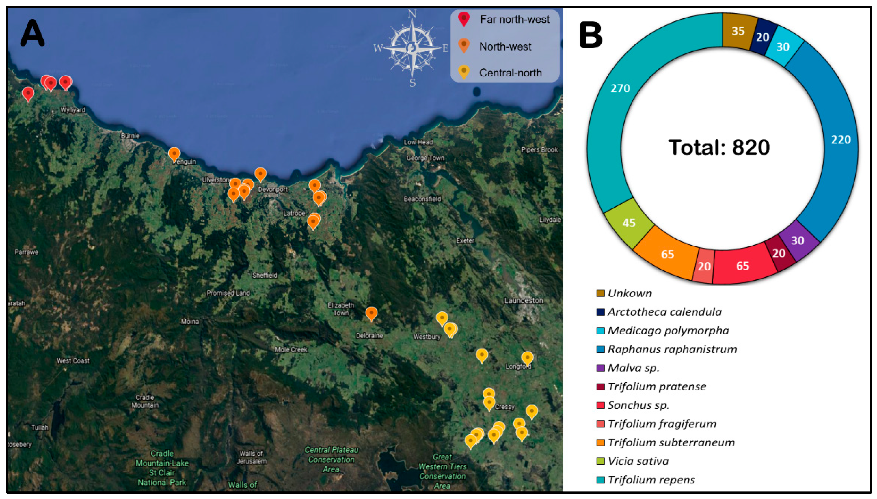
We showed that TuYV is the most prevalent and widely distributed Polerovirus in all surveyed regions, with virus infection levels in crops ranging between 0 and 27.5% of tested plants. The virus was found in 18 of the 22 pea fields located in all three cropping regions. This finding corroborates an earlier report of TuYV incidence in Tasmanian pea crops [3], where polerovirus (predominantly TuYV) infections were detected in pea crops across three seasons. However, in contrast to the data presented in this manuscript, Polerovirus prevalence did not exceed 60% of surveyed crops, and incidence did not exceed 6.7% in any field in the prior survey. Within the weed hosts neighbouring pea crops, we found that the TuYV infections were greater in R. raphanistrum and A. calendula, followed by T. repens, Vicia sativa, Trifolium fragiferum, and Sonchus sp. TuYV is known to have a wide host range that includes weeds, legume pasture species and other plants from the families Brassicaceae, Fabaceae, Amaranthaceae and Asteraceae [14,33]. It has also been reported across Australia to infect pulse crops (including peas), canola and various other weed species on a regular basis [14,36]. A further distinction from the previous Tasmanian survey was a failure to confirm the presence of the newly described PBMYV [63] in Tasmanian pea crops, which was previously found at low infection levels corresponding to 9 of 28 polerovirus infections detected [3].
The rate of TuYV infection varied according to the type of host plant and the location of the sampling sites. However, sampling bias must be considered when discussing the distribution of TuYV and the variation in its infection rate across plant species. We hypothesise that several other factors might also be involved in the variable incidence of the virus, such as stage of infection (early or late), types of aphid vectors and their population dynamics, crop management practices, climatic conditions, etc. However, additional studies are imperative to better understand the specific impact of these factors on TuYV incidence.
In addition, our NGS data confirmed several other viral reads, including an almost full genome of viruses representing five different genera and one unassigned virus. The genus Polerovirus and Potyvirus were each represented by two species members, followed by one member from each of Luteovirus, Potexvirus, and Carlavirus. The unclassified virus was from the family Partitiviridae (Table 5). However, except for BrYV, RT-PCR was not performed to further confirm the presence of the viruses detected by NGS [62]. Notably, three of these viruses (BrYV, RCVMV and RsCV) represent previously undescribed viruses in Tasmania [4].
BrYV belongs to the genus Polerovirus and is closely related to but appears to be distinctive from TuYV in terms of the P0 and P5 cistrons sequences [64,65,66]. It has been reported in Asia [64,65,66], and a recent report from Australia described the concatenated ORF-based phylogenetic analysis of collected isolates and demonstrated the paraphyletic relation between TuYV and BrYV [14]. The host range of BrYV is similar to that of TuYV [67]. However, BrYV isolates have also been subdivided into three genotypes (BrYV-A, -B, and -C) based on sequence similarity and phylogeny, with greater divergence seen in P0, P1 and P2 than in P3, P4 and P5 [65,66].
RCVMV is aphid-transmissible in a non-persistent manner; it can also be transmitted mechanically and by seed [68,69,70,71,72]. It was first reported in the United States (USA) [70] and then more widely [73,74,75,76], most recently in New Zealand [77]. Depending on the host species and virus isolate, RCVMV can cause vein mosaic, vein chlorosis, and plant stunting, as well as latent infections [73,76,78,79]. Though RCVMV has been shown to cause leaf chlorosis and yield loss in susceptible varieties of Trifolium pratense [80], assessment of the potential impact of RCVMV on clover requires further studies. That is because other viruses (e.g., AMV, Bean yellow mosaic virus (BYMV), ClYVV, or WClMV, all present in Tasmania) also naturally infect clover and are often found in mixed infections [77,81]. Although the presence of RCVMV in Tasmania has been confirmed (unpublished data), future surveys of legume and pasture crops should be conducted to determine its prevalence.
RsCV is an unclassified member of the family Partitiviridae, and members of this family are known to infect their hosts asymptomatically, including fungi, plants, some protozoa, and possibly some higher animals [82,83,84]. Members have two essential, double-stranded RNA genome segments ranging from 1.4 to 3.0 kb with RNA1 encoding RdRp, while RNA2 encodes CP [82]. Among those, plant-infecting partitiviruses are commonly referred to as cryptoviruses. Their host range includes radish, alfalfa, beet, broad bean, carrot, Brassica spp., white clover, red clover, rose, carnation, hop trefoil, Italian ryegrass, meadow fescue, spinach pear, and pine [84]. They are seed-transmitted with high efficiency, but there is no systemic infection or cell-to-cell moment due to the lack of a movement protein, so they move vertically through their host’s cell division processes instead [85].
In order to determine the genetic variability of Tasmanian isolates identified by TuYV species-specific PCR, two distinct segments in the genome were sequenced: ORF0, which codes for a protein (P0) involved in post-transcriptional gene silencing, symptom expression, and host range specificity [86,87], and ORF3, which codes for the coat protein (P3) [88,89]. Seventy-five Tasmanian isolates shared nucleotide identities ranging from 83.2–100% for P0 and from 91.2–100% for P3. Furthermore, the P0 and P3 sequences of the Tasmanian isolates shared 84.6–100% and 89.3–100% nucleotide identities with already published TuYV isolates from GenBank. These results are consistent with previous studies of genetic variation in the P0 and P3 genes from the UK [26,87]. One of the findings showed that the isolates shared 91.7–100% nucleotide identity for P0 and 94–100% for the P3 gene. The range of nucleotide identities was 86.9–98.8% and 93.5–99.8% for the P0 and P3 genes, respectively, when compared with the published isolates from GenBank [26]. In another study, the European TuYV isolates shared 81.1–100% of the P0 gene and 90.6–100% nucleotide identities [
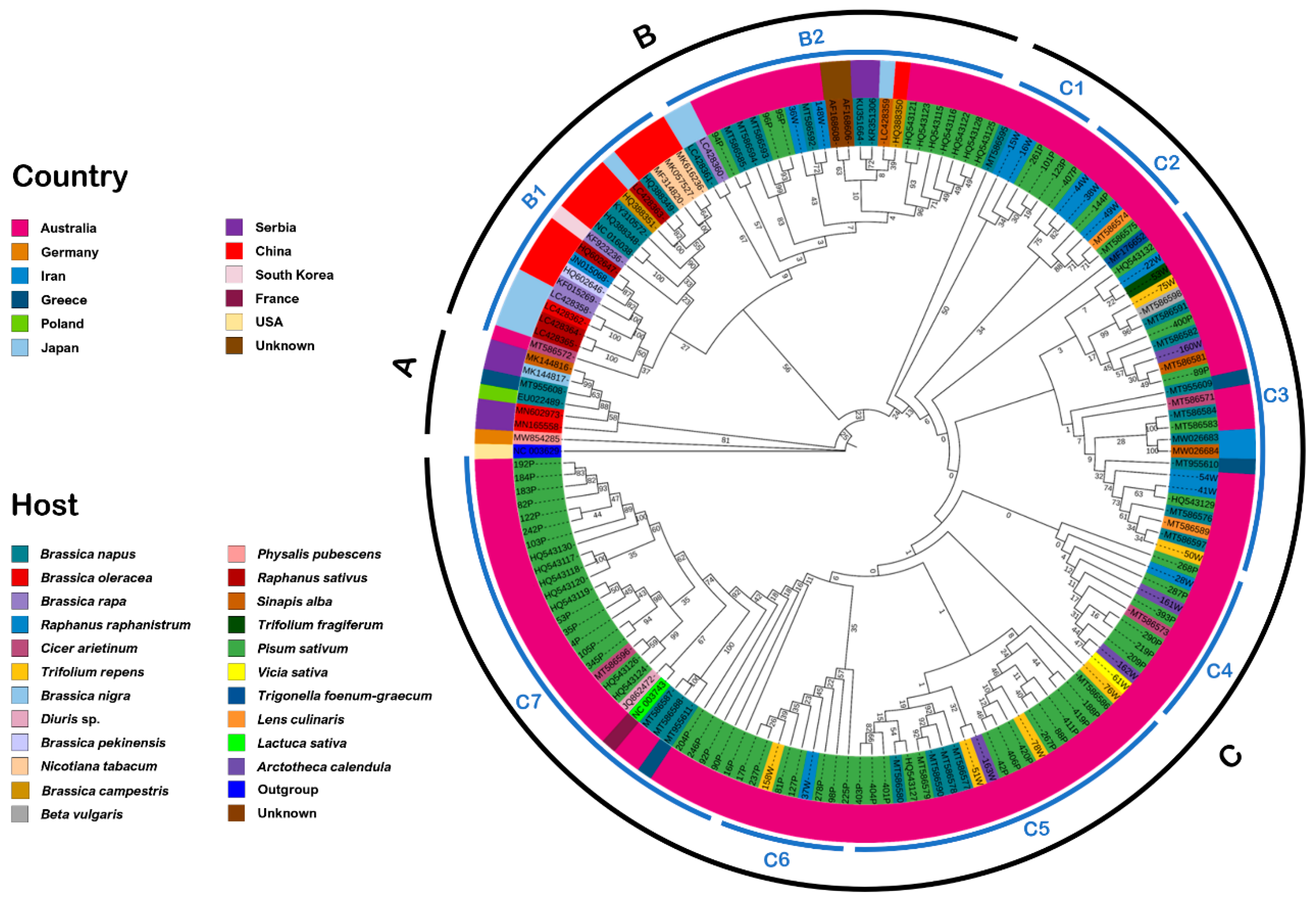
87]. The genetic variation of these ORFs indicated that the genomes of polerovirus within these regions are more diverse than previously assumed. When isolates identified as BrYV were included in the phylogenies, we found evidence for discrimination of TuYV and BrYV isolates within the P0 gene sequences, but no such clear differentiation was found with P3 sequences. Of the new Tasmanian isolates tested, five appeared to cluster with BrYV rather than TuYV in the P0 analysis and may be considered variants of this virus. Notably, subsequent testing of these variant isolates by BrYV species-specific PCR [90] did not result in positive amplification. Whilst evidence for the separation of BrYV and TuYV as distinct species exists from this and prior studies [62,64,65,66], suggestions that these viruses may represent a single highly variable species have been made [14]. Further whole genome sequencing of diverse isolates would be valuable to test these two propositions.
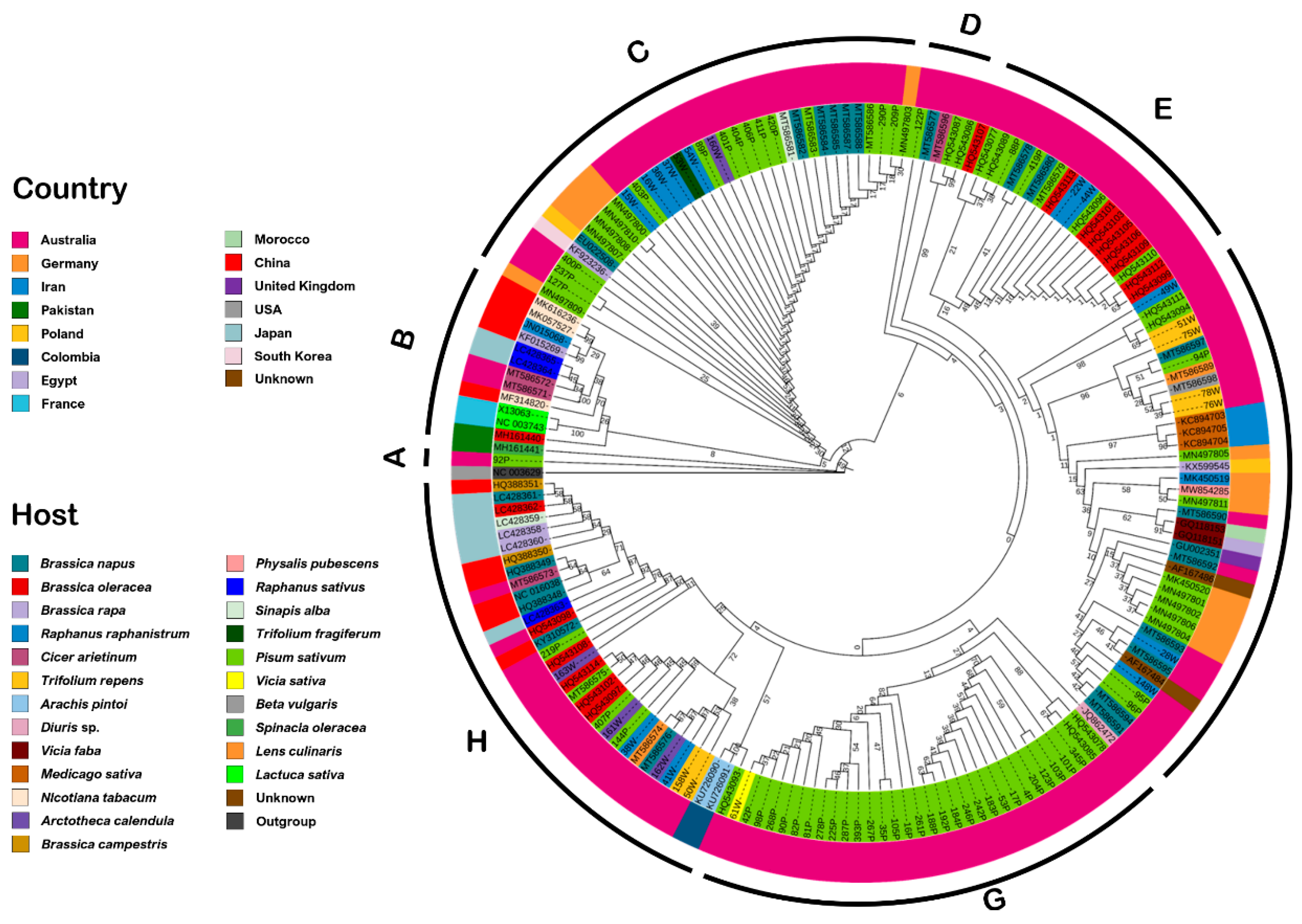
Phylogenetic analysis also highlighted similarities in sequence identity between isolates obtained from weed plants in the vicinity of pea crops and those found within the crops themselves (Figure 2 and Figure 3), pointing to the likelihood of virus exchanges between these wild and agricultural plant populations. Additional experimental analyses, including vector and virus monitoring, are required to determine the transmission rate and pattern.
Recombination is more common among RNA viruses than DNA and is a major driving force that greatly contributes to the evolution of viral populations [91]. Recombination governs genomic diversity and facilitates viral adaptation to varying environments (new host and environmental adaptation), ultimately resulting in the emergence of new/resistant-breaking/virulent variants or strains [92,93]. As previously proposed, poleroviruses tend to show higher recombination rates that further contribute to the emergence of new species and their evolution [94]. The evolution of the genus Polerovirus is marked by both intraspecific, homologous and interspecific, non-homologous recombination [14,95], and generally, in Luteoviridae, the recombination breakpoints are often at the boundaries of the gene rather than within the gene [96]. This implies that recombination events in the genome do not occur randomly; rather, these are associated with specific hotspots in the viral genome. Our findings suggested that recombination could be important and may play an important role in the evolution of these viruses. Interestingly, one isolate identified as a BrYV variant based on P0 sequence analysis was subsequently identified as a putative recombinant isolate in recombination analysis. This may suggest genetic exchange between these viruses species may be occurring. We found that recombination breakpoints were detected among 5/75 and 34/75 sequences of P0 and P3 genes, respectively (Table 6), which strongly suggests that the P0 and P3 genes have distinct evolutionary histories. Additionally, the considerable phylogenetic incongruence in P0 and P3 found in our work supports the concept that recombination might have played a role in the evolution of TuYV. Notably, recombination in genes or mutation in these proteins can affect the biological functions of the viral proteins. For instance, previous studies suggest that host range of different TuYV isolates may be influenced by genetic variation within the P0 gene [26,87]. On the other hand, P3 has a biologically active region that plays a vital role in CP subunit interactions, plant–virus interactions and aphid–virus recognition. Furthermore, the viral particle’s assembly is required for vector transmission and determines which insect vectors can transmit the virus [97,98,99]. Thus, recombination and mutation might affect critical biological functions governed by the P3 gene. The P5 readthrough component of the capsid protein has been shown to be highly variable within sequenced TuYV species and across polerovirus species [87,100]. The biological significance of variation in the P5 gene remains unclear at this stage but could be associated with specificity to different vector species. We did not include the analyses of the P5 gene in this study, which should be considered in future research to acquire a more in-depth understanding of the recombination-driven genome variation.
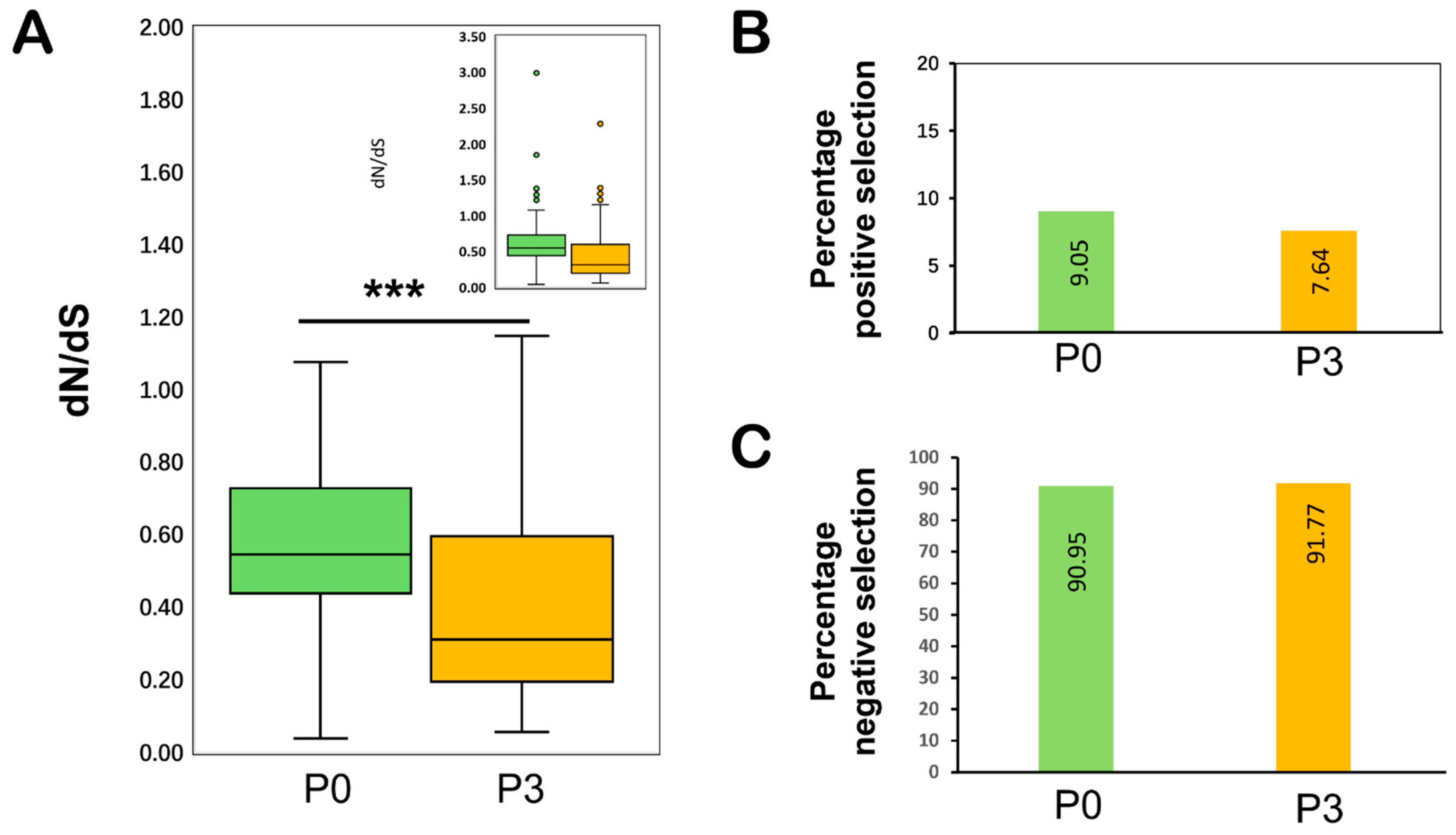
Our results showed that both P0 and P3 are primarily evolving under purifying selection pressure. Our findings revealed that with an average dN/dS ratio of <1, the majority of the codons remained under negative selection (Figure 5 and Table 8), and the overall contribution of negatively selected sites remained >90% (91% for P0 and 92.4% for P3). This is consistent with the previous research that concluded that both genes of TuYV (P0 and P3) were evolving under strong negative selection pressure [26]. Negative selection in the TuYV genome, which is required to keep the encoded protein functional (as in P3 and P0 of TuYV), may have helped to eliminate deleterious variants. TuYV’s P0 gene is involved in RNA-silencing suppression [17], and mutations in this gene are expected to pose a significant impact on virus fitness, limiting genetic diversity and thus influencing dN/dS ratio estimates [101,102].
Taken together, this study advances our current understanding of the genetically diversified and evolving populations of TuYV infecting Tasmanian pea crops. Our findings also provide a practical framework necessary for understanding the current diversity and distribution of poleroviruses in three selected regions of Tasmania. It also provides important information on epidemiological aspects and management of viral diseases of pea crops. Future studies based on the full genome-based analyses of the genetic variations will expand our understanding of the evolutionary patterns existing among TuYV populations in Tasmania.

Supplementary Materials

The following supporting information can be downloaded at: https://www.mdpi.com/article/10.3390/v14081690/s1, Figure S1: P0 of Tasmanian isolates with P0 of all reported Poleroviruses; Figure S2: CP of Tasmanian isolates with CP of all reported Poleroviruses; Table S1: The metadata recorded, during the field survey 2019–2020; Table S2: Details of six samples selected for NGS; Table S3: List of isolates included in phylogenetic analysis; Table S4: Nucleotide sequence identities of P0 of Tasmanian isolates with P0 of 36 Poleroviruses; Table S5: Nucleotide sequence identities of P0 of Tasmanian isolates with P0 of worldwide TuYV and BrYV isolates; Table S6: Nucleotide sequence identities of CP of Tasmanian isolates with CP of 36 Poleroviruses; Table S7: Nucleotide sequence identities of CP of Tasmanian isolates with CP of worldwide TuYV and BrYV isolates.

Author Contributions

Conceptualization, M.U. and C.R.W.; methodology, M.U., R.S.T., T.T. and C.R.W.; software, bioinformatics analysis, and validation, M.U. and T.F.; writing—original draft preparation, M.U.; writing—review and editing, R.S.T., T.F., T.T. and C.R.W.; supervision, R.S.T., T.T. and C.R.W.; project administration, C.R.W.; funding acquisition, C.R.W. All authors have read and agreed to the published version of the manuscript.

Funding

This research was funded by Hort Innovation, using the Vegetable Industry research and development levy and contributions from the Australian Government (project VG16086: Area-wide management of vegetable diseases: virus and bacteria). Hort Innovation is the grower-owned, not-for-profit research and development corporation for Australian horticulture.

Institutional Review Board Statement

Not applicable.

Informed Consent Statement

Not applicable.

Data Availability Statement

The data is contained within the article or Supplementary Materials.

Acknowledgments

We thank the growers and Simplot Australia Pty Ltd. for access to the commercial field sites for sample collection and Cliff Kinoti, Agriculture Victoria Research, Department of Jobs, Precints and Regions for providing the NGS services.

Conflicts of Interest

The authors declare no conflict of interest.

References

  1. ABARES. Agricultural Commodities: June Quarter 2019; Australian Bureau of Agricultural and Resource Economics and Sciences: Canberra, Australia, 2019.
  2. Tasmanian Agri-Food. Scorecard; AgriGrowth Tasmania: Hobart, Australia, 2021. [Google Scholar]
  3. Wilson, C.; Lambert, S.; Dann, A.; Cross, P.; Hay, F. Occurrence of viruses within Tasmanian vegetable crops and identification of a novel Polerovirus infecting pea. Australas. Plant Pathol. 2012, 41, 311–319. [Google Scholar] [CrossRef]
  4. Guy, P.; Cross, P.; Wilson, C. A review of the plant virus and viroid records for Tasmania. Australas. Plant Pathol. 2020, 49, 479–492. [Google Scholar] [CrossRef]
  5. Musil, M. Uber das Vorkommen des Virus des Blattrollens der Erbse in der Siowakei. (On the occurrence of a leaf roll virus in pea in Slovakia.). Biologia 1966, 21, 133–138. [Google Scholar]
  6. Bos, L.; Hampton, R.; Makkouk, K. Viruses and Virus Diseases of Pea, Lentil, Faba Bean and Chickpea. In World Crops: Cool Season Food Legumes; Springer: Berlin/Heidelberg, Germany, 1988; pp. 591–615. [Google Scholar]
  7. Kraft, J.M.; Pfleger, L.F. (Eds.) Compendium of Pea Diseases and Pests; American Phytopathological Society: Saint Paul, MN, USA, 2008. [Google Scholar]
  8. Gaafar, Y.; Grausgruber-Gröger, S.; Ziebell, H. Vicia faba, V. sativa and Lens culinaris as new hosts for Pea necrotic yellow dwarf virus in Germany and Austria. New Dis. Rep. 2016, 34, 28. [Google Scholar] [CrossRef]
  9. Gaafar, Y.Z.; Ziebell, H. Two divergent isolates of turnip yellows virus from pea and rapeseed and first report of turnip yellows virus-associated RNA in Germany. Microbiol. Resour. Announc. 2019, 8, e00214–e00219. [Google Scholar] [CrossRef]
  10. Walker, P.J.; Siddell, S.G.; Lefkowitz, E.J.; Mushegian, A.R.; Adriaenssens, E.M.; Alfenas-Zerbini, P.; Davison, A.J.; Dempsey, D.M.; Dutilh, B.E.; García, M.L. Changes to virus taxonomy and to the International Code of Virus Classification and Nomenclature ratified by the International Committee on Taxonomy of Viruses (2021). Arch. Virol. 2021, 166, 2633–2648. [Google Scholar] [CrossRef]
  11. Garcia-Ruiz, H.; Holste, N.M.; LaTourrette, K. Poleroviruses (Luteoviridae). In Encyclopedia of Virology, 4th ed.; Bamford, D.H., Zuckerman, M., Eds.; Academic Press: Oxford, UK, 2021; pp. 594–602. [Google Scholar]
  12. Dietzgen, R.G.; Mann, K.S.; Johnson, K.N. Plant virus–insect vector interactions: Current and potential future research directions. Viruses 2016, 8, 303. [Google Scholar] [CrossRef]
  13. Hipper, C.; Brault, V.; Ziegler-Graff, V.; Revers, F. Viral and cellular factors involved in phloem transport of plant viruses. Front. Plant Sci. 2013, 4, 154. [Google Scholar] [CrossRef]
  14. Filardo, F.; Nancarrow, N.; Kehoe, M.; McTaggart, A.R.; Congdon, B.; Kumari, S.; Aftab, M.; Trębicki, P.; Rodoni, B.; Thomas, J.; et al. Genetic diversity and recombination between turnip yellows virus strains in Australia. Arch. Virol. 2021, 166, 813–829. [Google Scholar] [CrossRef]
  15. Duffus, J.E.; Russell, G. Serological and host range evidence for the occurrence of beet western yellows virus in Europe. Phytopathology 1970, 60, 199–1202. [Google Scholar] [CrossRef]
  16. Mayo, M. ICTV at the Paris ICV: Results of the plenary session and the binomial ballot. Arch. Virol. 2002, 147, 2254–2260. [Google Scholar] [CrossRef]
  17. Pfeffer, S.; Dunoyer, P.; Heim, F.; Richards, K.; Jonard, G.; Ziegler-Graff, V. P0 of beet Western yellows virus is a suppressor of posttranscriptional gene silencing. J. Virol. 2002, 76, 6815–6824. [Google Scholar] [CrossRef] [PubMed]
  18. Bortolamiol, D.; Pazhouhandeh, M.; Marrocco, K.; Genschik, P.; Ziegler-Graff, V. The Polerovirus F box protein P0 targets ARGONAUTE1 to suppress RNA silencing. Curr. Biol. 2007, 17, 1615–1621. [Google Scholar] [CrossRef]
  19. Barry, J.K.; Miller, W.A. A −1 ribosomal frameshift element that requires base pairing across four kilobases suggests a mechanism of regulating ribosome and replicase traffic on a viral RNA. Proc. Natl. Acad. Sci. USA 2002, 99, 11133–11138. [Google Scholar] [CrossRef] [PubMed]
  20. Nixon, P.L.; Rangan, A.; Kim, Y.-G.; Rich, A.; Hoffman, D.W.; Hennig, M.; Giedroc, D.P. Solution structure of a luteoviral P1–P2 frameshifting mRNA pseudoknot. J. Mol. Biol. 2002, 322, 621–633. [Google Scholar] [CrossRef]
  21. Smirnova, E.; Firth, A.E.; Miller, W.A.; Scheidecker, D.; Brault, V.; Reinbold, C.; Rakotondrafara, A.M.; Chung, B.Y.-W.; Ziegler-Graff, V. Discovery of a small non-AUG-initiated ORF in poleroviruses and luteoviruses that is required for long-distance movement. PLoS Pathog. 2015, 11, e1004868. [Google Scholar] [CrossRef] [PubMed]
  22. Stevens, M.; Freeman, B.; Liu, H.Y.; Herrbach, E.; Lemaire, O. Beet poleroviruses: Close friends or distant relatives? Mol. Plant Pathol. 2005, 6, 1–9. [Google Scholar] [CrossRef]
  23. Brault, V.; Périgon, S.; Reinbold, C.; Erdinger, M.; Scheidecker, D.; Herrbach, E.; Richards, K.; Ziegler-Graff, V. The polerovirus minor capsid protein determines vector specificity and intestinal tropism in the aphid. J. Virol. 2005, 79, 9685–9693. [Google Scholar] [CrossRef]
  24. Peter, K.A.; Gildow, F.; Palukaitis, P.; Gray, S.M. The C terminus of the polerovirus p5 readthrough domain limits virus infection to the phloem. J. Virol. 2009, 83, 5419–5429. [Google Scholar] [CrossRef] [PubMed]
  25. Rodamilans, B.; Shan, H.; Pasin, F.; García, J.A. Plant viral proteases: Beyond the role of peptide cutters. Front. Plant Sci. 2018, 9, 666. [Google Scholar] [PubMed]
  26. Asare-Bediako, E. Brassicaceae: Turnip Yellows Virus Interactions. Ph.D. Thesis, University of Warwick, Coventry, UK, 2011. [Google Scholar]
  27. García-Arenal, F.; McDonald, B.A. An analysis of the durability of resistance to plant viruses. Phytopathology 2003, 93, 941–952. [Google Scholar] [CrossRef]
  28. De Miranda, J.; Stevens, M.; de Bruyne, E.; Smith, H.; Bird, C.; Hull, R. Beet luteovirus coat protein sequence variation. Ann. Appl. Biol. 1995, 127, 113–124. [Google Scholar] [CrossRef]
  29. Schubert, J.; Rabenstein, F.; Graichen, K.; Richter, K. Comparison of the 5′-end nucleotide sequences of luteoviruses from oilseed rape and sugar beet. Arch. Phytopathol. Plant Prot. 1998, 31, 519–530. [Google Scholar] [CrossRef]
  30. Hauser, S.; Stevens, M.; Mougel, C.; Smith, H.G.; Fritsch, C.; Herrbach, E.; Lemaire, O. Biological, serological, and molecular variability suggest three distinct polerovirus species infecting beet or rape. Phytopathology 2000, 90, 460–466. [Google Scholar] [CrossRef] [PubMed]
  31. Torres, M.W.; Corrêa, R.L.; Schrago, C.G. Analysis of differential selective forces acting on the coat protein (P3) of the plant virus family Luteoviridae. Genet. Mol. Res. 2005, 4, 790–802. [Google Scholar]
  32. Walkey, D.; Pink, D. Studies on resistance to beet western yellows virus in lettuce (Lactuca sativa) and the occurrence of field sources of the virus. Plant Pathol. 1990, 39, 141–155. [Google Scholar] [CrossRef]
  33. Stevens, M.; Smith, H.; Hallsworth, P. The host range of beet yellowing viruses among common arable weed species. Plant Pathol. 1994, 43, 579–588. [Google Scholar] [CrossRef]
  34. Thurston, M.; Pallett, D.; Cortina-Borja, M.; Edwards, M.L.; Raybould, A.; Cooper, J. The incidence of viruses in wild Brassica nigra in Dorset (UK). Ann. Appl. Biol. 2001, 139, 277–284. [Google Scholar] [CrossRef]
  35. Pallett, D.; Thurston, M.; Cortina-Borja, M.; Edwards, M.L.; Alexander, M.; Mitchell, E.; Raybould, A.; Cooper, J. The incidence of viruses in wild Brassica rapa ssp. sylvestris in southern England. Ann. Appl. Biol. 2002, 141, 163–170. [Google Scholar] [CrossRef]
  36. Coutts, B.; Hawkes, J.; Jones, R. Occurrence of Beet western yellows virus and its aphid vectors in over-summering broad-leafed weeds and volunteer crop plants in the grainbelt region of south-western Australia. Aust. J. Agric. Res. 2006, 57, 975–982. [Google Scholar] [CrossRef]
  37. Freeman, A.J.; Aftab, M. Effective management of viruses in pulse crops in south eastern Australia should include management of weeds. Australas. Plant Pathol. 2011, 40, 430–441. [Google Scholar] [CrossRef]
  38. Schliephake, E.; Graichen, K.; Rabenstein, F. Investigations on the vector transmission of the Beet mild yellowing virus (BMYV) and the Turnip yellows virus (TuYV)/Untersuchungen zur Vektoruebertragung des Milden Ruebenvergilbungsvirus (Beet mild yellowing virus) und des Wasserruebenvergilbungsvirus (Turnip yellows virus). Z. Pflanzenkrankh. Pflanzenschutz/J. Plant Dis. Prot. 2000, 107, 81–87. [Google Scholar]
  39. Stevens, M.; Smith, H.; Hallsworth, P. Detection of the luteoviruses, beet mild yellowing virus and beet western yellows virus, in aphids caught in sugar-beet and oilseed rape crops, 1990–1993. Ann. Appl. Biol. 1995, 127, 309–320. [Google Scholar] [CrossRef]
  40. Chomič, A.; Pearson, M.; Clover, G.; Farreyrol, K.; Saul, D.; Hampton, J.G.; Armstrong, K.F. A generic RT-PCR assay for the detection of Luteoviridae. Plant Pathol. 2010, 59, 429–442. [Google Scholar] [CrossRef]
  41. Beuve, M.; Stevens, M.; Liu, H.-Y.; Wintermantel, W.M.; Hauser, S.; Lemaire, O. Biological and molecular characterization of an American sugar beet-infecting Beet western yellows virus isolate. Plant Dis. 2008, 92, 51–60. [Google Scholar] [CrossRef]
  42. Appiah, A.S. Studies on Groundnut Rosette Disease in Ghana and Genomic Analysis of a Novel Phasey Bean Virus in Australia. Ph.D. Thesis, University of Tasmania, Hobart, Australia, 2017. [Google Scholar]
  43. Sharman, M.; Appiah, A.S.; Filardo, F.; Nancarrow, N.; Congdon, B.S.; Kehoe, M.; Aftab, M.; Tegg, R.S.; Wilson, C.R. Biology and genetic diversity of phasey bean mild yellows virus, a common virus in legumes in Australia. Arch. Virol. 2021, 166, 1575–1589. [Google Scholar] [CrossRef]
  44. Bushnell, B. BBTools: A Suite of Bioinformatic Tools Used for DNA and RNA Sequence Data Analysis. 2016. Available online: https://jgi.doe.gov/data-and-tools/bbtools/ (accessed on 1 May 2022).
  45. Ho, T.; Tzanetakis, I.E. Development of a virus detection and discovery pipeline using next generation sequencing. Virology 2014, 471, 54–60. [Google Scholar] [CrossRef]
  46. Gibbs, A.; Gower, J. The use of a multiple-transfer method in plant virus transmission studies—Some statistical points arising in the analysis of results. Ann. Appl. Biol. 1960, 48, 75–83. [Google Scholar] [CrossRef]
  47. Kumar, S.; Stecher, G.; Tamura, K. MEGA7: Molecular evolutionary genetics analysis version 7.0 for bigger datasets. Mol. Biol. Evol. 2016, 33, 1870–1874. [Google Scholar] [CrossRef]
  48. Letunic, I.; Bork, P. Interactive Tree of Life (iTOL) v5: An online tool for phylogenetic tree display and annotation. Nucleic Acids Res. 2021, 49, W293–W296. [Google Scholar] [CrossRef]
  49. Martin, D.; Rybicki, E. RDP: Detection of recombination amongst aligned sequences. Bioinformatics 2000, 16, 562–563. [Google Scholar] [CrossRef] [PubMed]
  50. Sawyer, S. GENECONV: A Computer Package for the Statistical Detection of Gene Conversion. 1999. Available online: http://www.math.wustl.edu/~sawyer (accessed on 29 April 2022).
  51. Salminen, M.O.; Carr, J.K.; Burke, D.S.; McCutchan, F.E. Identification of breakpoints in intergenotypic recombinants of HIV type 1 by bootscanning. AIDS Res. Hum. Retrovir. 1995, 11, 1423–1425. [Google Scholar] [CrossRef]
  52. Smith, J.M. Analyzing the mosaic structure of genes. J. Mol. Evol. 1992, 34, 126–129. [Google Scholar] [CrossRef] [PubMed]
  53. Posada, D.; Crandall, K.A. Evaluation of methods for detecting recombination from DNA sequences: Computer simulations. Proc. Natl. Acad. Sci. USA 2001, 98, 13757–13762. [Google Scholar] [CrossRef] [PubMed]
  54. Gibbs, M.J.; Armstrong, J.S.; Gibbs, A.J. Sister-Scanning: A Monte Carlo procedure for assessing signals in recombinant sequences. Bioinformatics 2000, 16, 573–582. [Google Scholar] [CrossRef]
  55. Boni, M.F.; Posada, D.; Feldman, M.W. An exact nonparametric method for inferring mosaic structure in sequence triplets. Genetics 2007, 176, 1035–1047. [Google Scholar] [CrossRef] [PubMed]
  56. Martin, D.P.; Murrell, B.; Golden, M.; Khoosal, A.; Muhire, B. RDP4: Detection and analysis of recombination patterns in virus genomes. Virus Evol. 2015, 1, vev003. [Google Scholar] [CrossRef]
  57. Librado, P.; Rozas, J. DnaSP v5: A software for comprehensive analysis of DNA polymorphism data. Bioinformatics 2009, 25, 1451–1452. [Google Scholar] [CrossRef] [PubMed]
  58. Rozas, J.; Sánchez-DelBarrio, J.C.; Messeguer, X.; Rozas, R. DnaSP, DNA polymorphism analyses by the coalescent and other methods. Bioinformatics 2003, 19, 2496–2497. [Google Scholar] [CrossRef] [PubMed]
  59. Scheffler, K.; Martin, D.P.; Seoighe, C. Robust inference of positive selection from recombining coding sequences. Bioinformatics 2006, 22, 2493–2499. [Google Scholar] [CrossRef]
  60. Pond, S.L.K.; Frost, S.D. Datamonkey: Rapid detection of selective pressure on individual sites of codon alignments. Bioinformatics 2005, 21, 2531–2533. [Google Scholar] [CrossRef] [PubMed]
  61. Kosakovsky Pond, S.L.; Posada, D.; Gravenor, M.B.; Woelk, C.H.; Frost, S.D. GARD: A genetic algorithm for recombination detection. Bioinformatics 2006, 22, 3096–3098. [Google Scholar] [CrossRef] [PubMed]
  62. Umar, M.; Farooq, T.; Tegg, R.S.; Thangavel, T.; Wilson, C.R. Genomic Characterisation of an Isolate of Brassica Yellows Virus Associated with Brassica Weed in Tasmania. Plants 2022, 11, 884. [Google Scholar] [CrossRef] [PubMed]
  63. Sharman, M.; Kehoe, M.; Coutts, B.; van Leur, J.; Filardo, F.; Thomas, J. Two complete genome sequences of phasey bean mild yellows virus, a novel member of the luteoviridae from Australia. Genome Announc. 2016, 4, e01569-15. [Google Scholar] [CrossRef] [PubMed]
  64. Lim, S.; Yoo, R.H.; Igori, D.; Zhao, F.; Kim, K.H.; Moon, J.S. Genome sequence of a recombinant brassica yellows virus infecting Chinese cabbage. Arch. Virol. 2015, 160, 597–600. [Google Scholar] [CrossRef]
  65. Xiang, H.-Y.; Dong, S.-W.; Shang, Q.-X.; Zhou, C.-J.; Li, D.-W.; Yu, J.-L.; Han, C.-G. Molecular characterization of two genotypes of a new polerovirus infecting brassicas in China. Arch. Virol. 2011, 156, 2251–2255. [Google Scholar] [CrossRef]
  66. Zhang, X.Y.; Xiang, H.Y.; Zhou, C.J.; Li, D.W.; Yu, J.L.; Han, C.G. Complete genome sequence analysis identifies a new genotype of brassica yellows virus that infects cabbage and radish in China. Arch. Virol. 2014, 159, 2177–2180. [Google Scholar] [CrossRef]
  67. Yoshida, N.; Tamada, T. Host range and molecular analysis of Beet leaf yellowing virus, Beet western yellows virus-JP and Brassica yellows virus in Japan. Plant Pathol. 2019, 68, 1045–1058. [Google Scholar] [CrossRef]
  68. Freeman, A. Red Clover Vein Mosaic Carlavirus. In National Diagnostic Protocol; Department of Primary Industries, Primary Industries Research Victoria: Knoxfield, VIC, Australia, 2007. [Google Scholar]
  69. Kraft, J.M.; Larsen, R.C.; Inglis, D.A. Diseases of Pea. In The Pathology of Food and Pasture Legumes; CAB International: Oxford, UK, 1998; pp. 325–370. [Google Scholar]
  70. Osborn, H. Vein-mosaic virus of red clover. Phytopathology 1937, 27, 1051. [Google Scholar]
  71. Sander, E. Biological properties of red clover vein mosaic virus. Phytopathology 1959, 49, 748–754. [Google Scholar]
  72. Weber, K.; Hampton, R. Transmission of two purified carlaviruses by the pea aphid [Acyrthosiphon pisum]. Phytopathology 1980, 70, 631–633. [Google Scholar] [CrossRef]
  73. Bos, L.; Maat, D.; Markov, M. A biologically highly deviating strain of red clover vein mosaic virus, usually latent in pea (Pisum sativum), and its differentiation from pea streak virus. Neth. J. Plant Pathol. 1972, 78, 125–152. [Google Scholar] [CrossRef]
  74. Edwardson, J.R.; Christie, R.G. CRC Handbook of Viruses Infecting Legumes; CRC Press: Boca Raton, FL, USA, 2018. [Google Scholar]
  75. Varma, A.; Gibbs, A.; Woods, R. A comparative study of red clover vein mosaic virus and some other plant viruses. J. Gen. Virol. 1970, 8, 21–32. [Google Scholar] [CrossRef] [PubMed]
  76. Wetter, C.; Quantz, L.; Brandes, J. Verwandtschaft zwischen dem Stauchevirus der Erbse und dem Rotkleeadernmosaik-Virus (red clover vein mosaic virus). J. Phytopathol. 1959, 35, 201–204. [Google Scholar] [CrossRef]
  77. Fletcher, J.; Tang, J.; Blouin, A.; Ward, L.; MacDiarmid, R.; Ziebell, H. Red clover vein mosaic virus—A novel virus to New Zealand that is widespread in legumes. Plant Dis. 2016, 100, 890–895. [Google Scholar] [CrossRef]
  78. Graves, C.; Hagedorn, D. The red clover vein-mosaic virus in Wisconsin. Phytopathology 1956, 46, 257–260. [Google Scholar]
  79. Hagedorn, D.; Bos, L.; van der Want, J. The red clover vein-mosaic virus in the Netherlands. Tijdschr. Over Plantenziekten 1959, 65, 13–23. [Google Scholar] [CrossRef]
  80. Khan, M.A.; Maxwell, D.; Smith, R. Inheritance of resistance to red clover vein mosaic virus in red clover. Phytopathology 1978, 68, 1084–1086. [Google Scholar] [CrossRef]
  81. Sherwood, R.T. Viruses of white clover in pastures of Pennsylvania, New York, and Vermont. Plant Dis. 1997, 81, 817–820. [Google Scholar] [CrossRef]
  82. King, A.M.; Adams, M.J.; Carstens, E.B.; Lefkowitz, E.J. Virus Taxonomy: Classification and Nomenclature of Viruses. In Virus Taxonomy: Classification and Nomenclature of Viruses; Elsevier: Amsterdam, The Netherlands, 2012; p. 1327. [Google Scholar]
  83. Liu, H.; Fu, Y.; Xie, J.; Cheng, J.; Ghabrial, S.A.; Li, G.; Yi, X.; Jiang, D. Discovery of novel dsRNA viral sequences by in silico cloning and implications for viral diversity, host range and evolution. PLoS ONE 2012, 7, e42147. [Google Scholar] [CrossRef]
  84. Li, L.; Liu, J.; Zhang, Q.; Fu, R.; Zhu, X.; Li, C.; Chen, J. Seed-borne viral dsRNA elements in three cultivated Raphanus and Brassica plants suggest three cryptoviruses. Can. J. Microbiol. 2016, 62, 287–295. [Google Scholar] [CrossRef] [PubMed]
  85. Ghabrial, S.; Nibert, M.; Maiss, E.; Lesker, T.; Baker, T.; Tao, Y. Family Partitiviridae. In Virus Taxonomy; Elsevier Academic Press: Amsterdam, The Netherlands, 2012; pp. 523–534. [Google Scholar]
  86. Pazhouhandeh, M.; Dieterle, M.; Marrocco, K.; Lechner, E.; Berry, B.; Brault, V.; Hemmer, O.; Kretsch, T.; Richards, K.E.; Genschik, P. F-box-like domain in the polerovirus protein P0 is required for silencing suppressor function. Proc. Natl. Acad. Sci. USA 2006, 103, 1994–1999. [Google Scholar] [CrossRef]
  87. Newbert, M.J. The Genetic Diversity of Turnip Yellows Virus in Oilseed Rape (Brassica napus) in Europe, Pathogenic Determinants, New Sources of Resistance and Host Range. Ph.D. Thesis, University of Warwick, Coventry, UK, 2016. [Google Scholar]
  88. Brault, V.; Mutterer, J.; Scheidecker, D.; Simonis, M.; Herrbach, E.; Richards, K.; Ziegler-Graff, V. Effects of point mutations in the readthrough domain of the beet western yellows virus minor capsid protein on virus accumulation in planta and on transmission by aphids. J. Virol. 2000, 74, 1140–1148. [Google Scholar] [CrossRef] [PubMed]
  89. Delfosse, V.C.; Barrios Barón, M.P.; Distéfano, A.J. What we know about poleroviruses: Advances in understanding the functions of polerovirus proteins. Plant Pathol. 2021, 70, 1047–1061. [Google Scholar] [CrossRef]
  90. Zhang, X.; Peng, Y.; Wang, Y.; Zhang, Z.; Li, D.; Yu, J.; Han, C. Simultaneous detection and differentiation of three genotypes of Brassica yellows virus by multiplex reverse transcription-polymerase chain reaction. Virol. J. 2016, 13, 189. [Google Scholar] [CrossRef] [PubMed]
  91. Sanjuán, R.; Domingo-Calap, P. Genetic diversity and evolution of viral populations. In Reference Module in Life Sciences; Elsevier: Amsterdam, The Netherlands, 2019. [Google Scholar]
  92. Nagy, P.D. Recombination in Plant RNA Viruses. In Plant Virus Evolution; Springer: Berlin/Heidelberg, Germany, 2008; pp. 133–156. [Google Scholar]
  93. Garcia-Ruiz, H.; Diaz, A.; Ahlquist, P. Intermolecular RNA recombination occurs at different frequencies in alternate forms of brome mosaic virus RNA replication compartments. Viruses 2018, 10, 131. [Google Scholar] [CrossRef]
  94. Dombrovsky, A.; Glanz, E.; Lachman, O.; Sela, N.; Doron-Faigenboim, A.; Antignus, Y. The complete genomic sequence of Pepper yellow leaf curl virus (PYLCV) and its implications for our understanding of evolution dynamics in the genus Polerovirus. PLoS ONE 2013, 8, e70722. [Google Scholar] [CrossRef]
  95. Filardo, F.F.; Sharman, M. Siratro latent polerovirus (SLPV): A new polerovirus from Australia with a non-functional open reading frame 0. Australas. Plant Pathol. 2019, 48, 491–501. [Google Scholar] [CrossRef]
  96. Pagán, I.; Holmes, E.C. Long-term evolution of the Luteoviridae: Time scale and mode of virus speciation. J. Virol. 2010, 84, 6177–6187. [Google Scholar] [CrossRef] [PubMed]
  97. Brault, V.; Bergdoll, M.; Mutterer, J.; Prasad, V.; Pfeffer, S.; Erdinger, M.; Richards, K.; Ziegler-Graff, V. Effects of point mutations in the major capsid protein of beet western yellows virus on capsid formation, virus accumulation, and aphid transmission. J. Virol. 2003, 77, 3247–3256. [Google Scholar] [CrossRef]
  98. Kaplan, I.B.; Lee, L.; Ripoll, D.R.; Palukaitis, P.; Gildow, F.; Gray, S.M. Point mutations in the potato leafroll virus major capsid protein alter virion stability and aphid transmission. J. Gen. Virol. 2007, 88, 1821–1830. [Google Scholar] [CrossRef] [PubMed]
  99. Lee, L.; Kaplan, I.B.; Ripoll, D.R.; Liang, D.; Palukaitis, P.; Gray, S.M. A surface loop of the potato leafroll virus coat protein is involved in virion assembly, systemic movement, and aphid transmission. J. Virol. 2005, 79, 1207–1214. [Google Scholar] [CrossRef]
  100. Holste, N. Polerovirus Genomic Variation and Mechanisms of Silencing Suppression by P0 Protein. Master’s Thesis, University of Nebraska-Lincoln, Lincoln, NE, USA, 2020. [Google Scholar]
  101. Holmes, E.C. Error thresholds and the constraints to RNA virus evolution. Trends Microbiol. 2003, 11, 543–546. [Google Scholar] [CrossRef] [PubMed]
  102. Simmonds, P.; Smith, D. Structural constraints on RNA virus evolution. J. Virol. 1999, 73, 5787–5794. [Google Scholar] [CrossRef]
Figure 1. (A) Map of the three different pea crop sampling regions (far northwest, northwest, and central north) in Tasmania. (B) Details of weeds and pasture species samples collected from all sites prior to pea crop sowing.
Viruses 14 01690 g001
Figure 2. Phylogenetic analysis (maximum likelihood algorithm) based on nucleotide sequences of P0 of Tasmanian isolates. The phylogenetic trees show the evolutionary relationship of P0 of Tasmanian isolates with P0 of worldwide reported TuYV and BrYV isolates. The inner ring indicates the host, and the outer ring the country of origin.
Viruses 14 01690 g002
Figure 3. Phylogenetic analysis (maximum likelihood algorithm) based on nucleotide sequences of P3 of Tasmanian isolates. The phylogenetic trees show the evolutionary relationship of P3 of Tasmanian isolates with P3 of worldwide reported TuYV and BrYV isolates. The inner ring indicates the host, and the outer ring indicates the country of origin.
Viruses 14 01690 g003
Figure 4. Results of the RDP recombination analysis for the (A) P0 gene and (B) P3 gene. Lines indicate the percentage of similarity per alignment.
Viruses 14 01690 g004
Figure 5. (A) Selection pressure was estimated by the calculation of non-synonymous to synonymous substitution ratios (dN/dS). Box plots correspond to dN/dS ratio for the TuYV-encoded P0 gene and P3 gene. The median values are represented by the horizontal lines inside the box, while inset shows the data with outliers indicated by small circles. Asterisks denote significance: *** p < 0.001. (B) Percentage of sites evolving under positive selection pressure in TuYV-encoded P0 gene and P3 gene. (C) Percentage of negatively selected sites among TuYV-encoded P0 gene and P3 gene.
Viruses 14 01690 g005
Table 1. List of viruses recorded in Tasmania infecting green peas [4].
SpeciesGenus
Alfalfa mosaic virusAlfamovirus
Cucumber mosaic virusCucumovirus
Pea enation mosaic virusEnamovirus
Phasey bean mild yellows virusPolerovirus
Soybean dwarf virusLuteovirus
Subterranean clover stunt virusNanovirus
Tomato spotted wilt virusOrthotospovirus
Turnip yellows virusPolerovirus
Table 2. Primer sequences, genomic target, primer position, expected PCR product size and cycle conditions used in this study.
Primer NameSequence 5′ to 3′TargetPositionProduct Size (bp)PCR ConditionsReference
C2F1 + C2F2TCACKTTCGGGCCGAGTLuteoviridae (partial ORF3) 1481 × 95 °C (15 min), 15 × [94 °C (30 s), 65 °C (30 s) reduce by 1 °C per cycle, 72 °C (30 s)], 25 × [94 °C (30 s), 50 °C (30 s), 72 °C (30 s)], 1 × 72 °C (7 min)[40]
TCACKTTCGGGCCGTCT
C2R1 + 2TCMAGYTCGTAAGCGATKG
TuYVCP+ATGAATACGGTCGTGGGTAGGAGTuYV ORF334835631 × 95 °C (15 min), 35 × [94 °C (30 s), 55 °C (30 s), 72 °C (1 min)], 1 × 72 °C (10 min)[41]
TuYVCP−CCAGCTATCGATGAAGAACCATTG4045
TuYVOrf0FACAAAAGAAACCAGGAGGGAATCCTTATuYV ORF01780As for TuYVCP+/TuYVCP− except 55 °C annealing[29]
TuYVOrf0RTCATACAAACATTTCGGTGTAGAC781
BWYVCPFCAGTAGCCGGTATTTACTTAGTCTACCBWYV ORF33472648As for TuYVCP+/TuYVCP− except 56 °C annealing[3]
BWYVCPRGGCACTTCATAGTGATTCTAAAAGAA4119
PhB7FGATCCTTGTGCAAGTTTGTTPartial 5′ UTR and 5′ end of ORF074601 × 94 °C (1 min), 30 × [94 °C (30 s), 58 °C (60 s), 72 °C (3 min)], 1 × 72 °C (10 min)[42,43]
PhB455RGAATGAGACCTTTGTAAGTA455
Table 3. Prevalence of virus infections (Luteoviridae universal primers and species-specific primers) in weed species neighbouring pea crops.
Weed SpeciesTotal TestedLuteoviridaeSbDVTuYVBWYVPBMYVUndeterminedNo Virus Detected
Unknown70000007
Arctotheca calendula44040000
Medicago polymorpha60000006
Rapistrum raphanistrum441801300526
Malva sp.61000015
Trifolium pratense40000004
Sonchus sp.1320100111
Trifolium fragiferum42010012
Trifolium subterraneum1310000112
Vicia sativa95310014
Trifolium repens543321900321
Total164662429001398
Table 4. Prevalence of virus infections (Luteoviridae universal primers and species-specific primers) in Tasmanian pea crops and TuYV incidence calculated using the Gibbs and Gower method (1960).
SiteU01U02U03U04U05U07U08U10U11U12U15U16U20U22U23U26U27U32U33U34U35U36Total
Luteoviridae8500983810616802401181413108151
SbDV00000100000100000120005
BWYV00000000000000000000000
PBMYV00000000000000000000000
TuYV850097381061660240117611108137
TuYV incidence (%)9.75.60.00.011.38.33.29.712.96.927.56.90.02.14.40.014.88.36.914.812.99.7-
undetermined00000000000100000062009
Total number of samples tested is 440.
Table 5. Details of viruses detected through NGS.
VirusesGenusGenome LengthMax Contigs Length
(nt)
Type of AnalysisSamples ID
1Turnip yellows virusPolerovirus5641 bp1349, 5452, 457, 5588BlastnU1, U4, U5, U6
2Brassica yellows virusPolerovirus5666 bp3994BlastnU1,
3Turnip mosaic virusPotyvirus9835 bp9829BlastnU1,
4Raphanus sativus cryptic virusunclassified Partitiviridae1866 bp1700BlastnU1,
5Soybean dwarf virusLuteovirus5853 bp5842, 2919BlastnU2, U3
6White clover mosaic virus genomePotexvirus5845 bp1695, 352, 245BlastnU2, U3, U4
7Red clover vein mosaic virusCarlavirus8604 bp8631, 463BlastnU2, U3
8Clover yellow vein virusPotyvirus9584 bp9137BlastnU2,
Table 6. Recombination within and between P0 and P3 genes.
Analysed RegionSequences Detected with Recomb. EventRecombinant 1Recombination Breakpoints “In Alignment (Without Gaps)”Parental SequencesDetection Methods 2p-Value 3
BeginEndMajorMinor
P0536W34 (32)409 (390)225P345PRMCS37.386 × 10−3
P33488P24 (23)312 (310)92P162WMS33.451 × 10−7
1 Numbering begins at the first nucleotide after the cleavage site at the replication origin and increases clockwise. 2 R, RDP; G, GeneConv; B, Bootscan; M, MaxChi; C, CHIMAERA; S, SisScan; 3, 3SEQ. 3 The described p-value corresponds to the program in bold, underlined type and is the lowest p-value calculated for the event in question.
Table 7. Molecular diversity within P0 and P3 sequences of TuYV isolates.
DatasetNumber of SequencesTotal Number of SitesSEtaHHdπθwTajima’s D
P075707182208650.9920.048290.05873−0.61202
P37548897110420.9480.040170.05046−0.69341
S, number of polymorphic (segregating) sites; Eta, total number of mutations; H, number of haplotypes; Hd, haplotype diversity; π, nucleotide diversity; θw, Watterson’s theta.
Table 8. Estimation of average dN/dS ratios, positive and negative selection pressures within P0 and P3 genes of TuYV isolates.
ORFTotal Number of CodonsAvg. dN/dS RatioPositive SelectionNegative Selection
Total SitesAvg.Min.Max.Total SitesAvg.Min.Max.
P02220.6198201.3031.0162.9882020.5520.0390.982
P31580.4245121.3041.0022.2771460.3520.0560.993
Publisher’s Note: MDPI stays neutral with regard to jurisdictional claims in published maps and institutional affiliations.

