Rotational Dynamics of The Transmembrane Domains Play an Important Role in Peptide Dynamics of Viral Fusion and Ion Channel Forming Proteins—A Molecular Dynamics Simulation Study

Abstract
1. Introduction
2. Materials and Methods
2.1. Sequences Used for MD Simulations and Preparation of Proteins
- -
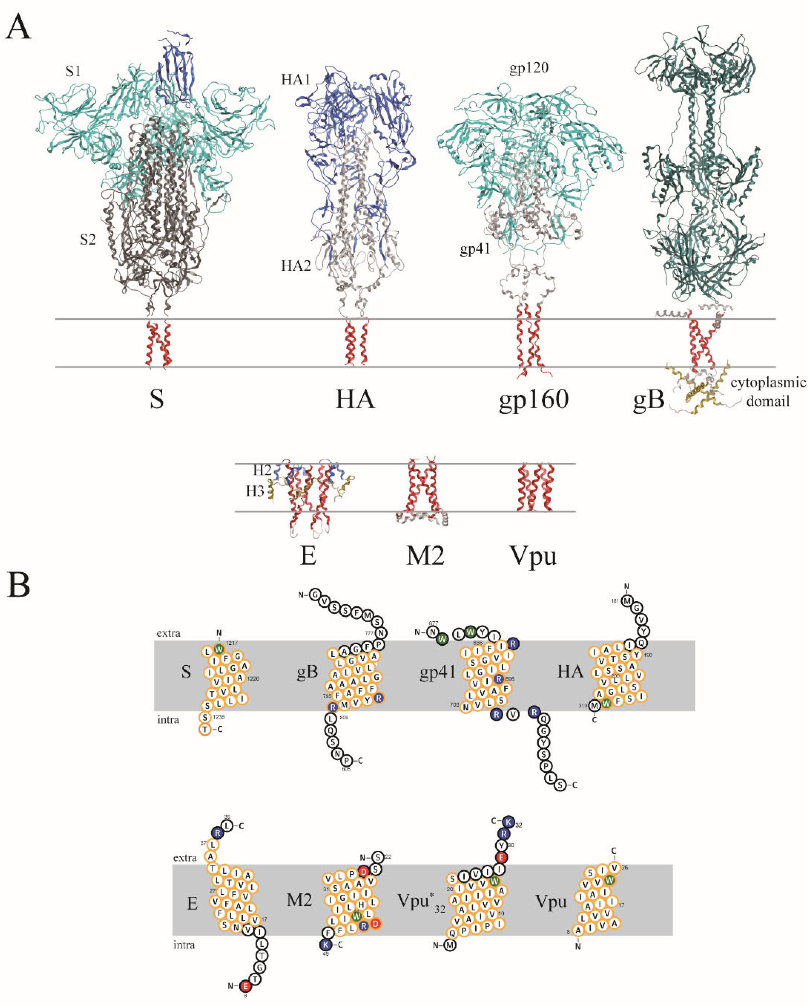
- S protein of SARS-CoV-2 (PDB ID: 7LC8 [10]), named S:
- ➢
- WLGFIAG LIAIVLVTIL LSSTTSC
- -
- haemagglutinin of influenza A virus (PDB ID: 6HJQ [12]), named HA:
- ➢
- MGVYQILAIY STVASSLVLL VSLGAISFWM without parts of the linker at the N terminal side (175–184, GVKLESMGVY),
- -
- glycoprotein 41 of HIV-1 (PDB ID: 5JYN [11]), named gp41:
- ➢
- NWLWYIRIFI IIVGSILGLR IVFAVLSLVN RVRQGYSPLS
- -
- glycoprotein B of HSV-1 (PDB ID: 5V2S [14]), named gB:
- ➢
- GVSSFMSNPF GALAVGLLVL AGLAAAFFAF RYVMRLQSNP
- -
- E protein of SARS-CoV (PDB ID: 5X29 [42]), named E:
- ➢
- ETGTLIVNSV LLFLAFVVFL LVTLAILTAL RL
and in its extended form, named E58:- ➢
- ETGTLIVNSV LLFLAFVVFL LVTLAILTAL RLAAYAANIV NVSLVKPTVY VY SRVKNL
- -
- M2 of influenza A virus (PDB ID: 2L0J [13]), named M2:
- ➢
- SSDPLVVAAS IIGILHLILW ILDRLFFK
- -
- experimental structure of Vpu of HIV-1 (PDB ID: 1PI7 [43]), Vpu, as well as two different lengths of Vpu (UniProt ID: P05919), named Vpu*32 and Vpu*53 as ideal helices, depending on the number of amino acids (32 or 53):
- Vpu
- ➢
- AIVALVVAII IAIVVWSIV
- Vpu*32
- ➢
- MQPIPIVAIV ALVVAIIIAI VVWSIVIIEY RK
- Vpu*53
- ➢
- MQPIPIVAIV ALVVAIIIAI VVWSIVIIEY RKILRQRKID RLIDRLIERA EDS
2.2. MD Simulations
2.3. Diffusion Coefficient (DC)
2.4. Generation of Artificial Oligomeric Structures
2.5. Data Analysis
2.6. Hard and Software
3. Results
3.1. Helicity
3.2. Translational and Rotational DCs
3.3. Rotational Dynamics
4. Discussion
4.1. Model Evaluation
4.2. Data Evaluation
5. Conclusions
Supplementary Materials
Author Contributions
Funding
Acknowledgments
Conflicts of Interest
References
- Rey, F.A.; Lok, S.-M. Common Features of Enveloped Viruses and Implications for Immunogen Design for Next-Generation Vaccines. Cell 2018, 172, 1319–1334. [Google Scholar] [CrossRef] [Scilit] [PubMed]
- OuYang, B.; Dong, Y.; Chou, J.J. Structural and Functional Properties of Viral Membrane Proteins. In Advances in Membrane Proteins; Cao, Y., Ed.; Springer: Singapore, 2018; pp. 147–181. [Google Scholar]
- Mehnert, T.; Routh, A.; Judge, P.J.; Lam, Y.H.; Fischer, D.; Watts, A.; Fischer, W.B. Biophysical characterisation of Vpu from HIV-1 suggests a channel-pore dualism. Proteins 2008, 70, 1488–1497. [Google Scholar] [CrossRef] [Scilit] [PubMed]
- Liao, Y.; Lescar, J.; Tam, J.P.; Liu, D.X. Expression of SARS-coronavirus envelope protein in Escherichia coli cells alters membrane permeability. Biochem. Biophys. Res. Commun. 2004, 325, 374–380. [Google Scholar] [CrossRef] [Scilit] [PubMed]
- Torres, J.; Wang, J.; Parthasarathy, K.; Liu, D.X. The transmembrane oligomers of coronavirus protein E. Biophys. J. 2005, 88, 1283–1290. [Google Scholar] [CrossRef] [Scilit]
- Chen, C.-P.; Lin, M.-H.; Chan, Y.-T.; Chen, L.-C.; Ma, C.; Fischer, W.B. Membrane protein assembly: Two cytoplasmic phosphorylated serine sites of Vpu from HIV-1 affect oligomerization. Sci. Rep. 2016, 6, 28866. [Google Scholar] [CrossRef] [Scilit]
- Wilson, L.; McKinlay, C.; Gage, P.; Ewart, G. SARS coronavirus E protein forms cation-selective ion channels. Virology 2004, 330, 322–331. [Google Scholar] [CrossRef] [Scilit]
- Chen, C.-C.; Krüger, J.; Sramala, I.; Hsu, H.-J.; Henklein, P.; Chen, Y.-M.A.; Fischer, W.B. ORF 8a of severe acute respiratory syndrome coronavirus forms an ion channel: Experiments and molecular dynamics simulations. Biochim. Biophys. Acta 2011, 1808, 572–579. [Google Scholar] [CrossRef] [Scilit]
- Pavlović, D.; Neville, D.C.A.; Argaud, O.; Blumberg, B.; Dwek, R.A.; Fischer, W.B.; Zitzmann, N. The hepatitis C virus p7 protein forms an ion channel that is inhibited by long-alkyl-chain iminosugar derivatives. Proc. Natl. Acad. Sci. USA 2003, 100, 6104–6108. [Google Scholar] [CrossRef] [Scilit]
- Fu, Q.; Chou, J.J. A Trimeric Hydrophobic Zipper Mediates the Intramembrane Assembly of SARS-CoV-2 Spike. J. Am. Chem. Soc. 2021, 143, 8543–8546. [Google Scholar] [CrossRef] [Scilit]
- Dev, J.; Park, D.; Fu, Q.; Chen, J.; Ha, H.J.; Ghantous, F.; Herrmann, T.; Chang, W.; Liu, Z.; Frey, G.; et al. Structural basis for membrane anchoring of HIV-1 envelope spike. Science 2016, 353, 172–175. [Google Scholar] [CrossRef] [Scilit]
- Benton, D.J.; Nans, A.; Calder, L.J.; Turner, J.; Neu, U.; Lin, Y.P.; Ketelaars, E.; Kallewaard, N.L.; Corti, D.; Lanzavecchia, A.; et al. Influenza hemagglutinin membrane anchor. Proc. Natl. Acad. Sci. USA 2018, 115, 10112–10117. [Google Scholar] [CrossRef] [Scilit]
- Sharma, M.; Yi, M.; Dong, H.; Qin, H.; Peterson, E.; Busath, D.D.; Zhou, H.-X.; Cross, T.A. Insight into the mechanism of the influenza A proton channel from a structure in a lipid bilayer. Science 2010, 330, 509–512. [Google Scholar] [CrossRef] [Scilit] [PubMed]
- Cooper, R.S.; Georgieva, E.R.; Borbat, P.P.; Freed, J.H.; Heldwein, E.E. Structural basis for membrane anchoring and fusion regulation of the herpes simplex virus fusogen gB. Nat. Struct. Mol. Biol. 2018, 25, 416–424. [Google Scholar] [CrossRef] [Scilit] [PubMed]
- Harrison, S.C. Viral membrane fusion. Nat. Struct. Mol. Biol. 2008, 15, 690–697. [Google Scholar] [CrossRef] [Scilit] [PubMed]
- Barrett, C.T.; Dutch, R.E. Viral Membrane Fusion and the Transmembrane Domain. Viruses 2020, 12, 693. [Google Scholar] [CrossRef] [Scilit] [PubMed]
- Miyauchi, K.; Curran, A.R.; Long, Y.; Kondo, N.; Iwamoto, A.; Engelman, D.M.; Matsuda, Z. The membrane-spanning domain of gp41 plays a critical role in intracellular trafficking of the HIV envelope protein. Retrovirology 2010, 7, 95. [Google Scholar] [CrossRef] [Scilit]
- Smith, E.C.; Smith, S.E.; Carter, J.R.; Webb, S.R.; Gibson, K.M.; Hellman, L.M.; Fried, M.G.; Dutch, R.E. Trimeric transmembrane domain interactions in paramyxovirus fusion proteins: Roles in protein folding, stability, and function. J. Biol. Chem. 2013, 288, 35726–35735. [Google Scholar] [CrossRef] [Scilit]
- Webb, S.; Nagy, T.; Moseley, H.; Fried, M.; Dutch, R. Hendra virus fusion protein transmembrane domain contributes to pre-fusion protein stability. J. Biol. Chem. 2017, 292, 5685–5694. [Google Scholar] [CrossRef] [Scilit]
- Webb, S.R.; Smith, S.E.; Fried, M.G.; Dutch, R.E. Transmembrane Domains of Highly Pathogenic Viral Fusion Proteins Exhibit Trimeric Association In Vitro. mSphere 2018, 3, e00047-18. [Google Scholar] [CrossRef] [Scilit]
- Carrasco, L. Membrane leakiness after viral infection and a new approach to the development of antiviral agents. Nature 1978, 272, 694–699. [Google Scholar] [CrossRef] [Scilit]
- Nieva, J.L.; Madan, V.; Carrasco, L. Viroporins: Structure and biological functions. Nat. Rev. Microbiol. 2012, 10, 563–574. [Google Scholar] [CrossRef] [Scilit] [PubMed]
- Lamb, R.A.; Lai, C.-J. Conservation of the influenza virus membrane proteins (M1) amino acid sequence and an open readig frame of RNA segment 7 encoding a second protein (M2) in H1N1 and H3N2 strains. Virology 1981, 112, 746–751. [Google Scholar] [CrossRef] [Scilit]
- Lamb, R.A.; Zebedee, S.L.; Richardson, C.D. Influenza virus M2 protein is an integral membrane protein expressed on the infected-cell surface. Cell 1985, 40, 627–633. [Google Scholar] [CrossRef] [Scilit]
- Strebel, K.; Klimkait, T.; Martin, M.A. Novel gene of HIV-1, vpu, and its 16-kilodalton product. Science 1988, 241, 1221–1223. [Google Scholar] [CrossRef] [Scilit] [PubMed]
- Cohen, E.A.; Terwilliger, E.F.; Sodroski, J.G.; Haseltine, W.A. Identification of a protein encoded by the vpu gene of HIV-1. Nature 1988, 334, 532–534. [Google Scholar] [CrossRef] [Scilit] [PubMed]
- Gonzales, M.E.; Carrasco, L. Viroporins. FEBS Lett. 2003, 552, 28–34. [Google Scholar] [CrossRef] [Scilit]
- Pinto, L.H.; Dieckmann, G.R.; Gandhi, C.S.; Papworth, C.G.; Braman, J.; Shaughnessy, M.A.; Lear, J.D.; Lamb, R.A.; DeGrado, W.F. A functionally defined model for the M2 proton channel of influenza A virus suggests a mechanism for its ion selectivity. Proc. Natl. Acad. Sci. USA 1997, 94, 11301–11306. [Google Scholar] [CrossRef] [Scilit]
- Pielak, R.M.; Chou, J.J. Influenza M2 proton channels. Biochim. Biophys. Acta 2011, 1808, 522–529. [Google Scholar] [CrossRef] [Scilit]
- Kalita, M.M.; Fischer, W.B. Decoupled side chain and backbone dynamics for proton translocation—M2 of influenza A. J. Mol. Model. 2017, 23, 212. [Google Scholar] [CrossRef] [Scilit]
- Ramadurai, S.; Holt, A.; Krasnikov, V.; van der Bogaart, G.; Killian, J.A.; Poolman, B. Lateral diffusion of membrane proteins. J. Am. Chem. Soc. 2009, 131, 12650–12656. [Google Scholar] [CrossRef] [Scilit]
- Alenghat, F.J.; Golan, D.E. Membrane protein dynamics and functional implications in mammalian cells. Curr. Top. Membr. 2013, 72, 89–120. [Google Scholar] [PubMed]
- Metzler, R.; Jeon, J.H.; Cherstvy, A.G. Non-Brownian diffusion in lipid membranes: Experiments and simulations. Biochim. Biophys. Acta 2016, 1858, 2451–2467. [Google Scholar] [CrossRef] [Scilit] [PubMed]
- Khoshnood, A.; Jalali, M.A. Anomalous diffusion of proteins in sheared lipid membranes. Phys. Rev. E Stat. Nonlin. Soft Matter Phys. 2013, 88, 032705. [Google Scholar] [CrossRef] [Scilit] [PubMed]
- Baker, M.K.; Abrams, C.F. Dynamics of lipids, cholesterol, and transmembrane α-helices from microsecond molecular dynamics simulations. J. Phys. Chem. B 2014, 118, 13590–13600. [Google Scholar] [CrossRef] [Scilit]
- Campagnola, G.; Nepal, K.; Schroder, B.W.; Peersen, O.B.; Krapf, D. Superdiffusive motion of membrane-targeting C2 domains. Sci. Rep. 2015, 5, 17721. [Google Scholar] [CrossRef] [Scilit]
- Goose, J.E.; Sansom, M.S.P. Reduced lateral mobility of lipids and proteins in crowded membranes. PLoS Comp. Biol. 2013, 9, e1003033. [Google Scholar] [CrossRef] [Scilit]
- Vögele, M.; Hummer, G. Divergent Diffusion Coefficients in Simulations of Fluids and Lipid Membranes. J. Phys. Chem. B 2016, 120, 8722–8732. [Google Scholar] [CrossRef] [Scilit]
- Vögele, M.; Köfinger, J.; Hummer, G. Hydrodynamics of Diffusion in Lipid Membrane Simulations. Phys. Rev. Lett. 2018, 120, 268104. [Google Scholar] [CrossRef] [Scilit]
- Linke, M.; Köfinger, J.; Hummer, G. Rotational Diffusion Depends on Box Size in Molecular Dynamics Simulations. J. Phys. Chem. Lett. 2018, 9, 2874–2878. [Google Scholar] [CrossRef] [Scilit]
- Von Bülow, S.; Siggel, M.; Linke, M.; Hummer, G. Dynamic cluster formation determines viscosity and diffusion in dense protein solutions. Proc. Natl. Acad. Sci. USA 2019, 116, 9843–9852. [Google Scholar] [CrossRef] [Scilit]
- Surya, W.; Li, Y.; Torres, J. Structural model of the SARS coronavirus E channel in LMPG micelles. Biochim. Biophys. Acta 2018, 1860, 1309–1317. [Google Scholar] [CrossRef] [Scilit] [PubMed]
- Park, S.H.; Mrse, A.A.; Nevzorov, A.A.; Mesleh, M.F.; Oblatt-Montal, M.; Montal, M.; Opella, S.J. Three-dimensional structure of the channel-forming trans-membrane domain of virus protein “u” (Vpu) from HIV-1. J. Mol. Biol. 2003, 333, 409–424. [Google Scholar] [CrossRef] [Scilit] [PubMed]
- Xiang, Z.; Soto, C.S.; Honig, B. Evaluating conformational free energies: The colony energy and its application to the problem of loop prediction. Proc. Natl. Acad. Sci. USA 2002, 99, 7432–7437. [Google Scholar] [CrossRef] [Scilit] [PubMed]
- Soto, C.S.; Fasnacht, M.; Zhu, J.; Forrest, L.; Honig, B. Loop modeling: Sampling, filtering, and scoring. Proteins 2008, 70, 834–843. [Google Scholar] [CrossRef] [Scilit] [PubMed]
- Gues, N.; Peitsch, M.C. SWISS-MODEL and the Swiss-PdbViewer: An environment for comparative protein modeling. Electrophoresis 1997, 18, 2714–2723. [Google Scholar]
- Henklein, P.; Kinder, R.; Schubert, U.; Bechinger, B. Membrane interactions and alignment of structures within the HIV-1 Vpu cytoplasmic domain: Effect of phosphorylation of serines 52 and 56. FEBS Lett. 2000, 482, 220–224. [Google Scholar] [CrossRef] [Scilit]
- Sramala, I.; Lemaitre, V.; Faraldo-Gómez, J.D.; Vincent, S.; Watts, A.; Fischer, W.B. Molecular dynamics simulations on the first two helices of Vpu from HIV-1. Biophys. J. 2003, 84, 3276–3284. [Google Scholar] [CrossRef] [Scilit]
- Yesylevskyy, S.O. ProtSqueeze: Simple and effective automated tool for setting up membrane protein simulations. J. Chem. Inf. Model. 2007, 47, 1986–1994. [Google Scholar] [CrossRef] [Scilit]
- Wolf, M.G.; Hoefling, M.; Aponte-Santamaría, C.; Grubmüller, H.; Groenhof, G. g_membed: Efficient insertion of a membrane protein into an equilibrated lipid bilayer with minimal perturbation. J. Comput. Chem. 2010, 31, 2169–2174. [Google Scholar] [CrossRef] [Scilit]
- Camley, B.A.; Lerner, M.G.; Pastor, R.W.; Brown, F.L.H. Strong influence of periodic boundary conditions on lateral diffusion in lipid bilayer membranes. J. Chem. Phys. 2015, 143, 243113. [Google Scholar] [CrossRef] [Scilit]
- Venable, R.M.; Ingólfsso, H.I.; Lerner, M.G.; Perrin, B.S., Jr.; Camley, B.A.; Marrink, S.J.; Brown, F.L.H.; Pastor, R.W. Lipid and Peptide Diffusion in Bilayers: The Saffman-Delbrück Model and Periodic Boundary Conditions. J. Phys. Chem. B 2017, 121, 3443–3457. [Google Scholar] [CrossRef] [Scilit] [PubMed]
- Vögele, M.; Köfinger, J.; Hummer, G. Finite-Size-Corrected Rotational Diffusion Coefficients of Membrane Proteins and Carbon Nanotubes from Molecular Dynamics Simulations. J. Phys. Chem. 2019, 123, 5099–5106. [Google Scholar] [CrossRef] [Scilit] [PubMed]
- Pranami, G.; Lamm, M.H. Estimating error in diffusion coefficients derived from molecular dynamics simulations. J. Chem. Theory Comput. 2015, 11, 4586–4592. [Google Scholar] [CrossRef] [Scilit] [PubMed]
- Omasits, U.; Ahrens, C.H.; Müller, S.; Wollscheid, B. Protter: Interactive protein feature visualization and integration with experimental proteomic data. Bioinformatics 2014, 30, 884–886. [Google Scholar] [CrossRef] [Scilit]
- Kalita, M.M.; Griffin, S.; Chou, J.J.; Fischer, W.B. Genotype-specific differences in structural features of hepatitis C virus (HCV) p7 membrane protein. Biochim. Biophys. Acta 2015, 1848, 1383–1392. [Google Scholar] [CrossRef] [Scilit]
- Shai, Y. Mode of action of membrane active antimicrobial peptides. Biopolymers 2002, 66, 236–248. [Google Scholar] [CrossRef] [Scilit]
- Pan, J.; Tristram-Nagle, S.; Nagle, J.F. Alamethicin aggregation in lipid membranes. J. Membr. Biol. 2009, 231, 11–27. [Google Scholar] [CrossRef] [Scilit]
- Pieta, P.; Mirza, J.; Lipkowski, J. Direct visualization of the alamethicin pore formed in a planar phospholipid matrix. Proc. Natl. Acad. Sci. USA 2012, 109, 21223–21227. [Google Scholar] [CrossRef] [Scilit]
- Van Meer, G.; Voelker, D.R.; Feigenson, G.W. Membrane lipids: Where they are and how they behave. Nat. Rev. Mol. Cell Biol. 2008, 9, 112–124. [Google Scholar] [CrossRef] [Scilit]
- Van Meer, G.; de Kroon, A.I.P.M. Lipid map of the mammalian cell. J. Cell Sci. 2011, 124 Pt 1, 5–8. [Google Scholar] [CrossRef] [Scilit]
- Mandala, V.S.; McKay, M.J.; Shcherbakov, A.A.; Dregni, A.J.; Kolcouris, A.; Hong, M. Structure and drug binding of the SARS-CoV-2 envelope protein transmembrane domain in lipid bilayers. Nat. Struct. Biol. 2020, 27, 1202–1208. [Google Scholar] [CrossRef] [Scilit] [PubMed]
- Spencer, R.H.; Rees, D.C. The alpha-helix and the organization and gating of channels. Annu. Rev. Biophys. Biomol. Struct. 2002, 31, 207–233. [Google Scholar] [CrossRef] [Scilit] [PubMed]
- Miyazawa, A.; Fujiyoshi, Y.; Unwin, N. Structure and gating mechanism of the acetylcholine receptor pore. Nature 2003, 423, 949–955. [Google Scholar] [CrossRef] [Scilit]
- Du, J.; Lü, W.; Cheng, Y.; Gouaux, E. Glycine receptor mechanism elucidated by electron cryo-microscopy. Nature 2015, 526, 224–229. [Google Scholar] [CrossRef] [Scilit] [PubMed]
- Labro, A.J.; Snyders, D.J. Being flexible: The voltage-controllable activation gate of kv channels. Front. Pharmacol. 2012, 3, 168. [Google Scholar] [CrossRef] [Scilit] [PubMed]
- Dai, J.; Zhou, H.-X. General rules for the arrangements and gating motions of pore-lining helices in homomeric ion channels. Nat. Commun. 2014, 5, 4641. [Google Scholar] [CrossRef] [Scilit] [PubMed]
- Marchesi, A.; Gao, X.; Adaixo, R.; Rheinberger, J.; Stahlberg, H.; Nimigean, C.; Simon Scheuring, S. An iris diaphragm mechanism to gate a cyclic nucleotide-gated ion channel. Nat. Commun. 2018, 9, 3978. [Google Scholar] [CrossRef] [Scilit] [PubMed]

Publisher’s Note: MDPI stays neutral with regard to jurisdictional claims in published maps and institutional affiliations. |
© 2022 by the authors. Licensee MDPI, Basel, Switzerland. This article is an open access article distributed under the terms and conditions of the Creative Commons Attribution (CC BY) license (https://creativecommons.org/licenses/by/4.0/).
Share and Cite
Wang, C.-W.; Fischer, W.B. Rotational Dynamics of The Transmembrane Domains Play an Important Role in Peptide Dynamics of Viral Fusion and Ion Channel Forming Proteins—A Molecular Dynamics Simulation Study. Viruses 2022, 14, 699. https://doi.org/10.3390/v14040699
Wang C-W, Fischer WB. Rotational Dynamics of The Transmembrane Domains Play an Important Role in Peptide Dynamics of Viral Fusion and Ion Channel Forming Proteins—A Molecular Dynamics Simulation Study. Viruses. 2022; 14(4):699. https://doi.org/10.3390/v14040699
Chicago/Turabian StyleWang, Chia-Wen, and Wolfgang B. Fischer. 2022. "Rotational Dynamics of The Transmembrane Domains Play an Important Role in Peptide Dynamics of Viral Fusion and Ion Channel Forming Proteins—A Molecular Dynamics Simulation Study" Viruses 14, no. 4: 699. https://doi.org/10.3390/v14040699
APA StyleWang, C.-W., & Fischer, W. B. (2022). Rotational Dynamics of The Transmembrane Domains Play an Important Role in Peptide Dynamics of Viral Fusion and Ion Channel Forming Proteins—A Molecular Dynamics Simulation Study. Viruses, 14(4), 699. https://doi.org/10.3390/v14040699