Brief Report

Distinct Plasma Concentrations of Acyl-CoA-Binding Protein (ACBP) in HIV Progressors and Elite Controllers

1
Infectious Disease and Immunity in Global Health Program, Research Institute of McGill University Health Centre, Montreal, QC H4A 4J1, Canada
2
Chronic Viral Illness Service, McGill University Health Centre, Montreal, QC H4A 4J1, Canada
3
CIHR Canadian HIV Trials Network, Vancouver, BC V6Z 1Y6, Canada
4
Division of Infectious Diseases, Geneva University Hospitals, 1205 Geneva, Switzerland
5
Division of Experimental Medicine, McGill University, Montreal, QC H4A 3J1, Canada
6
Instituto de Investigaciones Biomédicas en Retrovirus y SIDA (INBIRS), CONICET-Universidad de Buenos Aires, Buenos Aires C1121ABG, Argentina
7
Centre de Recherche du Centre Hospitalier de l’Université de Montréal, Montréal, QC H2X 0A9, Canada
8
Département de Microbiologie, Immunologie et Infectiologie, Université de Montréal, Montreal, QC H3T 1J4, Canada
9
Division of Clinical Immunology, McGill University Health Centre, Montreal, QC H4A 3J1, Canada
10
Centre de Recherche des Cordeliers, Equipe labellisée par la Ligue Contre le Cancer, Université de Paris, Sorbonne Université, Inserm U1138, Institut Universitaire de France, 75006 Paris, France
11
Metabolomics and Cell Biology Platforms, Institut Gustave Roussy, 94805 Villejuif, France
12
Department of Biology, Institut du Cancer Paris CARPEM, Hôpital Européen Georges Pompidou, AP-HP, 75015 Paris, France
13
Division of Hematology, McGill University Health Centre, Montreal, QC H4A 3J1, Canada
*
Authors to whom correspondence should be addressed.
Viruses 2022, 14(3), 453; https://doi.org/10.3390/v14030453
Submission received: 28 January 2022 / Revised: 18 February 2022 / Accepted: 21 February 2022 / Published: 23 February 2022

Abstract

:
HIV elite controllers (ECs) are characterized by the spontaneous control of viral replication, and by metabolic and autophagic profiles which favor anti-HIV CD4 and CD8 T-cell responses. Extracellular acyl coenzyme A binding protein (ACBP) acts as a feedback inhibitor of autophagy. Herein, we assessed the circulating ACBP levels in ECs, compared to people living with HIV (PLWH) receiving antiretroviral therapy (ART) or not. We found lower ACBP levels in ECs compared to ART-naïve or ART-treated PLWH (p < 0.01 for both comparisons), independently of age and sex. ACBP levels were similar in ECs and HIV-uninfected controls. The expression of the protective HLA alleles HLA-B*27, *57, or *58 did not influence ACBP levels in ECs. ACBP levels were not associated with CD4 or CD8 T-cell counts, CD4 loss over time, inflammatory cytokines, or anti-CMV IgG titers in ECs. In ART-treated PLWH, ACBP levels were correlated with interleukin (IL)-1β levels, but not with other inflammatory cytokines such as IL-6, IL-8, IL-32, or TNF-α. In conclusion, ECs are characterized by low ACBP plasma levels compared to ART-naïve or ART-treated PLWH. As autophagy is key to anti-HIV CD4 and CD8 T-cell responses, the ACBP pathway constitutes an interesting target in HIV cure strategies.

1. Introduction

The recent description of the Esperanza patient who achieved a possible spontaneous HIV sterilizing cure represents a beacon of hope for people living with HIV (PLWH) [1,2,3,4].
Elite controllers (ECs) can control HIV replication without antiretroviral therapy (ART) for a significant period of time. Studies of ECs led to a critical understanding of the factors associated with HIV control, offering possible clues for a HIV cure [3,5].
EC status has been shown to rely on a complex set of genetic and immunometabolic characteristics [6,7]. Protective human leukocyte antigen (HLA) alleles have been associated with robust anti-HIV CD4 and CD8 T-cell responses [8,9,10,11]. Moreover, metabolic pathways in immune cells are emerging as key factors for viral control in ECs [12,13,14]. Notably, our group has shown that the metabolic inputs of autophagy and lipophagy contribute to the HIV-specific CD4 and CD8 T-cell response in ECs [15,16]. Angin et al. first showed that HIV-specific CD8 T-cells display a restrictive glucose dependency in the majority of PLWH, which is a distinct metabolic defect not observed in ECs [17]. We confirmed that the superior CD8 T-cell responses observed in ECs are linked to their ability to use lipid and glutamine as metabolic inputs during autophagic processes [15,16]. Autophagy is involved in cellular homeostasis and cytoprotection, with an anti-aging effect. Autophagy involves master regulators of cellular metabolism, such as the mammalian target of rapamycin (mTOR) and the 5′ AMP-activated protein kinase (AMPK), combined with the selective removal of protein aggregates and organelles such as mitochondria, the endoplasmic reticulum, peroxisomes, lysosomes, and lipid droplets [18]. Autophagy and lipophagy, a form of selective autophagy, are regulated by different factors including acyl-coA-binding protein (ACBP). This protein, also known as diazepam binding inhibitor (DBI), is involved in intracellular bioenergetic reactions and serves as an extracellular feedback inhibitor of autophagy [19,20,21]. ACBP is conserved in eukaryotes and is expressed by all nucleated cells, underscoring its importance as a cellular metabolic hub. Within cells, ACBP binds to activated, acyl-coA-bound medium-chain fatty acids and shuttles them between cellular organelles for energy production. While intracellular ACBP has been shown to promote autophagy, lipophagy, and oxidative phosphorylation [22,23], secreted extracellular ACBP inhibits autophagy and stimulates appetite to increase nutrient uptake [19,20,21,24]. Hence, ACBP can be considered a metabolic and neuroendocrine factor that regulates bioenergetic and cellular functions in a context-dependent manner [21,24].
Given the influence of ACBP on cellular metabolism, and the distinctive immunometabolic features of ECs, we measured circulating ACBP in ECs, ART-naïve, and ART-treated PLWH. We found that ECs are characterized by low plasma ACBP concentrations.

2. Materials and Methods

2.1. Study Design

Blood samples were selected from PLWH who participated in the Canadian cohort of HIV-infected slow progressors (CIHR/CTN 247), and the HIV pathogenesis biobank [25]. From this cohort, we identified 37 ECs who maintained a HIV plasma viral load (VL) below 50 copies/mL in the absence of ART for at least 6 months, with CD4 T-cell counts greater than 200 cells/μL. Samples from ECs were collected between 2006 and 2020.
Samples from 27 ART-naïve participants in the chronic phase of the infection (at least 6 months after the estimated date of HIV acquisition) were obtained from the Montréal primary HIV infection cohort, the chronic viral illness service (CVIS) biobank, and the HIV pathogenesis biobank [26]. In addition, samples from 55 adult ART-treated PLWH were included from the Canadian HIV and aging cohort study, and the HIV pathogenesis biobank [27]. All participants had a VL below 50 copies/mL of plasma, and CD4 T-cell counts above 200 cells/μL. Samples from 31 adult HIV-uninfected donors from the Canadian HIV and aging cohort study, the CVIS biobank, and the HIV pathogenesis biobank were included as controls. Samples from ART-naïve, ART-treated, and control participants were collected during 2003–2012, 2011–2016, and 2002–2018 periods, respectively.

2.2. Laboratory Measurements

Blood samples were collected from participants and plasma was stored at −80°C until use, as previously described [25]. The quantification of plasma HIV VL was carried out using the RealTime HIV-1 assay (Abbott Laboratories, Chicago, IL, USA). Absolute CD4 and CD8 T-cell counts were measured by clinical labs using flow cytometry. HLA typing was performed by Sanger sequencing or next-generation sequencing using kits from GenDx (Utrecht, The Netherlands) [28].
ACBP plasma levels were measured for all samples in November 2021, using a human ACBP ELISA kit (Abnova Taiwan corporation, Taiwan) according to the supplier’s instructions. The markers of inflammation interleukin (IL)-1β, IL6, IL-8, and tumor necrosis factor alpha (TNF-α) were quantified in plasma using the Meso Scale Discovery U-Plex Pro-Inflammatory Combo 4 kit (Meso Scale Discovery, Rockville, MD, USA). CMV IgG titers and total IL-32 were measured by ELISA as previously described [25,29,30]. All measurements were performed in duplicate.

2.3. Calculation of CD4 T-Cell Count Change

For ECs enrolled in the CIHR/CTN 247 study, sequential CD4 T-cell counts were prospectively combined with historical data from medical charts for each study participant. The rate of CD4 T-cell count change over time was computed using a linear regression analysis. The results were reported as annual changes in CD4 T-cell counts. The annual slope of CD4 T-cell count change was assessed for significant difference from “0” [29].

2.4. Statistical Analyses

GraphPad Prism 9.3.0 (GraphPad, La Jolla, CA, USA) and SPSS 24.0 (IBM SPSS, Chicago, IL, USA) were used for statistical analyses. The statistical significance of differences between groups was assessed using nonparametric Kruskal–Wallis’ tests with Dunn’s post-tests. A Spearman’s rank test was used to assess correlations. An α level of 5% was considered statistically significant (p-value). Multivariable linear regression analyses were performed using SPSS 24.0.

2.5. Ethical Considerations

Ethical approval was obtained from the McGill University Health Centre Research Ethics Board (REB), as well as from all the REBs of the institutions participating and recruiting individuals included in this study. All study participants provided written informed consent.

3. Results

3.1. Study Population

A total of 150 participants were included in this study. The ECs were younger than the ART-treated PLWH (45.5 vs. 54-year-old, p = 0.004), and exhibited higher CD4 T-cell counts (640, 310, and 546, p < 0.001 and 0.007 respectively) and higher CD4/CD8 ratios (1.0, 0.39, and 0.74, p < 0.001 and 0.006 respectively) than the ART-naïve and ART-treated PLWH. A similar percentage of female and male participants was observed between the groups (23, 23, 11, and 26 percent females in the ECs, ART-treated, ART-naïve, and control participants, respectively) (Supplementary Table S1). The EC participants remained aviremic for a median of 6 years (0.7–27).
The ART-naïve PLWH had a detectable VL (median 4.7 log10 copies/mL), whereas all the ART-treated PLWH and ECs had a VL below 50 copies/mL (1.6 log10 copies/mL). The ART-treated participants received treatment for a median of 13.7 years. In the EC group, 17 out of 37 participants harbored protective HLA-B*27, *B57, or *B58 alleles (Supplementary Tables S1 and S2).

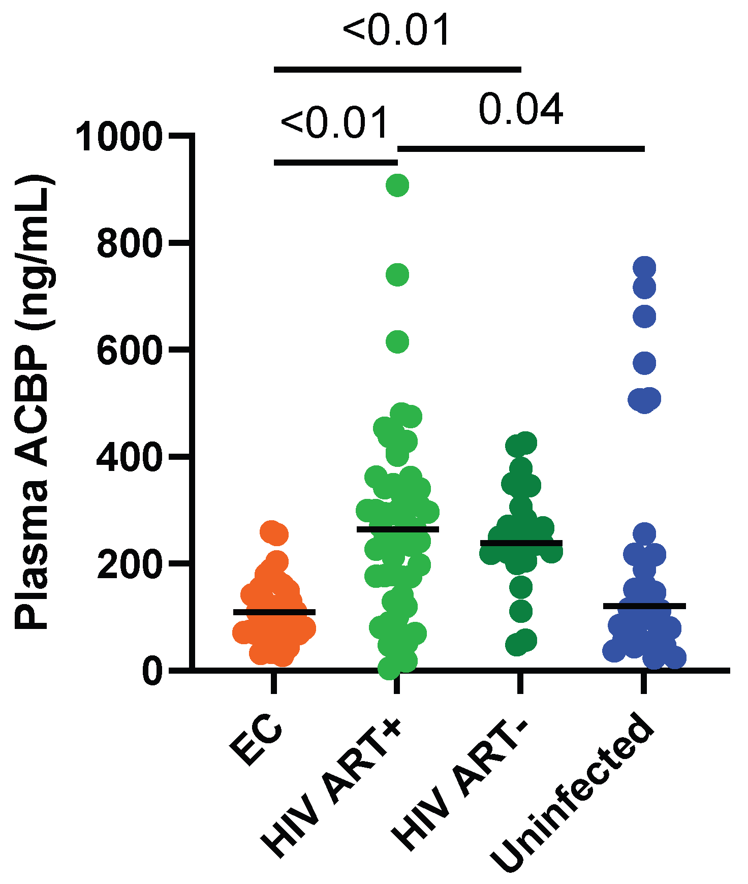
3.2. Circulating ACBP Levels Were Lower in ECs Compared to ART-Treated PLWH

Among PLWH, ACBP levels were lower in the ECs compared to the ART-naïve or ART-treated PLWH, with medians of 109.8, 238.7, and 264.6 ng/mL, respectively (p < 0.0001 for both comparisons) (Figure 1). Multivariable analyses showed that differences in age and sex could not explain these differences in ACBP levels. ACBP levels were similar in the ECs and uninfected controls (109.8 vs. 121.3 ng/mL, p = 0.33) (Figure 1). The ECs receiving statins or benzodiazepines had similar levels to those who did not take such medications (2 ECs were prescribed statins: ACBP levels 109.8 and 43.8 ng/mL; 1 EC was prescribed benzodiazepines: ACBP level 43.8 ng/mL). Three and seven participants, respectively, from the ART-treated and uninfected groups had particularly high levels of ACBP. However, their clinical characteristics such as diabetes status, weight, BMI, or age did not differ from the rest of the groups they belonged to (Supplementary Table S2). In the ECs, the duration of infection and follow-up were not associated with ACBP levels (r = −0.03, p = 0.88, Supplementary Table S2), and the ECs controlling infection for less than 5, 5 to 10, or more than 10 years had similar ACBP levels (Supplementary Figure S1)

3.3. Markers of HIV Disease Progression Were Not Associated with ACBP Levels in ECs

ACBP levels were not associated with age in the ECs, ART-naïve, or ART-treated PLWH (r = −0.13, p = 0.44; r = −0.014, p = 0.94; r = 0.12, p = 0.37, respectively), whereas they were correlated with age in the uninfected controls (r = 0.46, p < 0.01). ACBP levels were not associated with CD4 T-cell counts, CD8 T-cell counts, or CD4/CD8 ratios, which are considered markers of HIV disease progression and risk factors for non-AIDS comorbidities [31,32,33,34]. However, ACBP levels were negatively correlated with CD4 and CD8 T-cell counts in the ART-naïve PLWH (Table 1).
The ECs harboring protective HLA alleles had similar ACBP levels to those who did not (80 vs. 117 ng/mL, p = 0.22, Supplementary Table S2 and Supplementary Figure S2).
The ECs with slopes of CD4 count change significantly below “0” had similar ACBP levels to those with stable or increasing CD4 T-cell counts (109.8 vs. 110.1 ng/mL, p = 0.89, Supplementary Figure S3). Moreover, the ACBP level of the ECs was not associated with the slope of CD4 T-cell count change (correlation with annual CD4 slope r = 0.32, p = 0.07, Table 1). We previously found that the annual rate of CD4 T-cell count change was associated with CMV co-infection and anti-CMV IgG titers [29]. We did not find an association between ACBP levels and anti-CMV IgG titers in any group (Table 1). Moreover, we did not find any correlation between ACBP and IL-32 levels, a biomarker for control failure [25], in the ECs or ART-treated PLWH (r = 0.38, p = 0.13, and r = 0.03, p = 0.88, respectively).
Although ACBP levels have been linked with weight and BMI in obese people, we did not find any such association in our study groups (Table 1) [24].
In the ART-treated PLWH, we observed a significant positive correlation between ACBP and IL-1β concentrations (r = 0.29, p = 0.03, Supplementary Figure S4A), but not with IL-6, IL-8, or TNF-α levels (Supplementary Figure S4B–D). Such an association was not observed in the other groups.

4. Discussion

Our study demonstrates that ECs have significantly lower plasma ACBP levels compared to ART-naïve and ART-treated PLWH. These findings are consistent with the differences observed in T-cell metabolic pathways between ECs and other PLWH [15,16]. Lower plasma ACBP levels have been hypothesized in ECs, as extracellular ACBP inhibits autophagy and anti-HIV T-cell responses are favored by autophagy [15,16,24]. We observed a tendency of ECs with protective HLA to have lower ACBP levels, further suggesting that extracellular ACBP is linked with wearker anti-HIV responses.
ACBP levels in ECs were not influenced by participant characteristics such as age, sex, weight, or BMI. Furthermore, the HIV VL, treatment status and CD4 T-cell count of PLWH did not correlate with ACBP levels. ART-naive and ART-treated PLWH had similar levels of ACBP, suggesting that ART has no impact on ACBP levels [35]. However, both ART-naïve and ART-treated PLWH had higher ACBP plasma levels than ECs, suggesting that low ACBP is a patient-intrinsic, treatment-independent characteristic of ECs.
ECs have been shown to present with lower levels of inflammation compared to ART-naive and ART-treated PLWH [12,36,37]. However, other groups found elevated levels of the inflammatory markers IP-10 and TNF-α in ECs compared to controls, and those levels were not different from those of ART-treated PLWH [38]. In the group of ART-treated PWLH, we found an association between the inflammatory cytokine IL-1β and ACBP. This suggests that specific pro-inflammatory pathways could be responsible for ACBP induction in PLWH. Indeed, circulating ACBP levels were elevated in patients undergoing gut surgery, correlated with clinical signs of inflammation and high TNF-α levels [39]. Moreover, ACBP levels are elevated in the cerebrospinal fluid and the plasma of patients with Alzheimer’s disease and other neuroinflammatory conditions [40,41].
ACBP has been shown to promote food intake in animal models, and ACBP levels were higher in obese people [22,23,24]. Although weight gain is an issue for ART-treated PLWH, we did not observe any significant correlation between ACBP and either weight or BMI in our study, which may be underpowered for this kind of analysis.
Due to the small EC sample size, we were not able to optimally match participants and controls for age and sex. We founds that the usage of lipid-lowering agents or benzodiazepines did not influence ACBP levels in ECs. For the other groups, we did not have information on their treatment outside of antiretroviral drugs. Moreover, ACBP levels were only analyzed in plasma samples. The tissue expression of ACBP, notably in the liver and adipose tissue, should be compared in ECs vs. ART-naïve and ART-treated PLWH [42]. The comparatively low ACBP plasma levels of ECs could reflect a reduced secretion of ACBP, as well as a lower expression of intracellular ACBP. The cellular source and the mechanisms underlying the production of ACBP in ART-naïve and ART-treated PLWH, compared to ECs and uninfected controls, should be explored in future studies.

5. Conclusions

In summary, we detected lower ACBP plasma concentrations in ECs compared to ART-naïve or ART-treated PLWH. Low plasma ACBP levels could be used as a marker of ECs, reflecting this population’s particularly efficient immunometabolism. It would be interesting to stimulate autophagy by neutralizing the extracellular ACBP in ART-treated PLWH [24], and to investigate whether such a manipulation could facilitate the control of viral infections in suitable preclinical models. Exploring the ACBP pathway could also unravel new therapeutic targets to promote anti-HIV CD4 and CD8 T-cell responses in PLWH.

Supplementary Materials

The following supporting information can be downloaded at: https://www.mdpi.com/article/10.3390/v14030453/s1. Supplementary Table S1: Characteristics of study participants; Supplementary Table S2: HLA types and plasma ACBP levels of HIV elite controller participants; Supplementary Figure S1: Plasma ACBP levels in ECs controlling for less than 5, 5 to 10, or more than 10 years; Supplementary Figure S2: Plasma ACBP levels in ECs harboring or not protective (prot) HLA; Supplementary Figure S3: Plasma ACBP levels in ECs with CD4 decay (declining CD4 count) or not (stable/increasing CD4 counts); Supplementary Figure S4: Correlations between plasma levels of ACBP and IL-1β, IL-6, IL-8, and TNF-α in ART-treated PLWH.

Author Contributions

S.I. performed the experiments, analyzed the data, wrote the first draft, and revised the final draft of the manuscript. L.R., J.L., B.F., S.B., S.K., T.M., C.B., M.D., M.E.-F., C.L.T. and N.F.B. contributed to the experiments, data analysis, and critical review of the first and final drafts of the manuscript. M.D., C.L.T. and J.-P.R. recruited participants. J.-P.R. designed the study, contributed to data analysis, and critically reviewed the first and final drafts of the manuscript. G.K. discussed the results and edited the manuscript. All authors have read and agreed to the published version of the manuscript.

Funding

Our work was funded by the Fonds de la Recherche Québec-Santé (FRQ-S): Réseau SIDA/Maladies Infectieuses and Thérapie Cellulaire; the Canadian Institutes of Health Research (CIHR; Grants HOP 103230, PTJ 166049, and DC0190GP); the Vaccines and Immunotherapies Core of the CIHR Canadian HIV Trials Network (CTN; Grant CTN 247); the Canadian Foundation for AIDS Research (CANFAR; Grant 02-512); the CIHR-funded Canadian HIV Cure Enterprise (CanCURE) Team Grant HB2-164064. L.R. is a post-doctoral fellow supported by the “Fonds de perfectionnement” of the Geneva University Hospitals, Switzerland, and the Swiss National Science Foundation. S.I. is a post-doctoral fellow supported by the FRQ-S and CIHR/CTN. The Canadian HIV and Aging Cohort Study is supported by operating grants TCO-125276 and HAL-157985 from the Canadian Institutes of Health Research, and receives support from the CIHR HIV Clinical Trial Network (project CTN 272.) MD is supported by a clinician-researcher salary award from the Fonds de recherche du Québec- Santé CT is the Pfizer/Université de Montral Chair on HIV translational research. G.K. is supported by the Ligue Contre le Cancer (équipe labellisée); the Agence National de la Recherche (ANR); the Institut National du Cancer (INCa); the Institut Universitaire de France; LabEx Immuno-Oncology (ANR-18-IDEX-0001); and the Seerave foundation. This study contributes to the IdEx Université de Paris ANR-18-IDEX-0001. J.-P.R is the holder of the Louis Lowenstein Chair in Hematology and Oncology, McGill University.

Institutional Review Board Statement

The study was conducted in accordance with the Declaration of Helsinki, and approved by the Institutional Review Board of the McGill University Health Centre (studies 2019-5170, 2013-2134) and the Centre de recherche du Centre Hospitalier de l’Université de Montréal (CRCHUM) (studies 2013-2134, MP-02-2012-3068 and CE 11.063-BSP).

Informed Consent Statement

Informed consent was obtained from all subjects involved in the study.

Data Availability Statement

Data are available upon fair requisition to Jean-Pierre Routy at [email protected].

Acknowledgments

The authors are highly grateful to the study participants for their contributions, as well as Angie Massicotte, Josée Girouard, Stéphanie Matte, Louise Gilbert, Mario Legault, Cezar Iovi, and Olfa Debbeche for study coordination and assistance.

Conflicts of Interest

Drs. Tremblay, Kroemer, and Routy declared grants and honoraria obtained outside of the submitted work. Dr. Kroemer is the inventor of a patent covering therapeutic targeting of ACBP/DBI for the treatment of aging, age-related diseases, and metabolic disorders. None of the other authors have conflicts of interest to disclose.

References

  1. Blankson, J.N. The Esperanza Patient: More Hope for a Sterilizing HIV-1 Cure. Ann. Intern. Med. 2021. [Google Scholar] [CrossRef] [PubMed]
  2. Turk, G.; Seiger, K.; Lian, X.; Sun, W.; Parsons, E.M.; Gao, C.; Rassadkina, Y.; Polo, M.L.; Czernikier, A.; Ghiglione, Y.; et al. A Possible Sterilizing Cure of HIV-1 Infection Without Stem Cell Transplantation. Ann. Intern. Med. 2021. [Google Scholar] [CrossRef] [PubMed]
  3. May, M.E.; Kwaa, A.K.; Blankson, J.N. HIV-1 reservoirs in elite controllers: Clues for developing a functional cure? Future Microbiol. 2017, 12, 1019–1022. [Google Scholar] [CrossRef] [PubMed] [Green Version]
  4. Kanazawa, J.; Gianella, S.; Concha-Garcia, S.; Taylor, J.; Kaytes, A.; Christensen, C.; Patel, H.; Ndukwe, S.; Rawlings, S.A.; Hendrickx, S.; et al. Ethical and practical considerations for HIV cure-related research at the end-of-life: A qualitative interview and focus group study in the United States. BMC Med. Ethics 2022, 23, 2. [Google Scholar] [CrossRef]
  5. Deeks, S.G.; Archin, N.; Cannon, P.; Collins, S.; Jones, R.B.; de Jong, M.; Lambotte, O.; Lamplough, R.; Ndung’u, T.; Sugarman, J.; et al. Research priorities for an HIV cure: International AIDS Society Global Scientific Strategy 2021. Nat. Med. 2021, 27, 2085–2098. [Google Scholar] [CrossRef]
  6. Saag, M.; Deeks, S.G. How do HIV elite controllers do what they do? Clin. Infect. Dis. 2010, 51, 239–241. [Google Scholar] [CrossRef] [Green Version]
  7. Collins, D.R.; Gaiha, G.D.; Walker, B.D. CD8(+) T cells in HIV control, cure and prevention. Nat. Rev. Immunol. 2020, 20, 471–482. [Google Scholar] [CrossRef]
  8. Fellay, J.; Shianna, K.V.; Ge, D.; Colombo, S.; Ledergerber, B.; Weale, M.; Zhang, K.; Gumbs, C.; Castagna, A.; Cossarizza, A.; et al. A whole-genome association study of major determinants for host control of HIV-1. Science 2007, 317, 944–947. [Google Scholar] [CrossRef] [Green Version]
  9. Miura, T.; Brockman, M.A.; Schneidewind, A.; Lobritz, M.; Pereyra, F.; Rathod, A.; Block, B.L.; Brumme, Z.L.; Brumme, C.J.; Baker, B.; et al. HLA-B57/B*5801 human immunodeficiency virus type 1 elite controllers select for rare gag variants associated with reduced viral replication capacity and strong cytotoxic T-lymphocyte [corrected] recognition. J. Virol. 2009, 83, 2743–2755. [Google Scholar] [CrossRef] [Green Version]
  10. Migueles, S.A.; Sabbaghian, M.S.; Shupert, W.L.; Bettinotti, M.P.; Marincola, F.M.; Martino, L.; Hallahan, C.W.; Selig, S.M.; Schwartz, D.; Sullivan, J.; et al. HLA B*5701 is highly associated with restriction of virus replication in a subgroup of HIV-infected long term nonprogressors. Proc. Natl. Acad. Sci. USA 2000, 97, 2709–2714. [Google Scholar] [CrossRef] [Green Version]
  11. Okulicz, J.F.; Lambotte, O. Epidemiology and clinical characteristics of elite controllers. Curr. Opin. HIV AIDS 2011, 6, 163–168. [Google Scholar] [CrossRef]
  12. Chowdhury, F.Z.; Ouyang, Z.; Buzon, M.; Walker, B.D.; Lichterfeld, M.; Yu, X.G. Metabolic pathway activation distinguishes transcriptional signatures of CD8+ T cells from HIV-1 elite controllers. AIDS 2018, 32, 2669–2677. [Google Scholar] [CrossRef]
  13. Tarancon-Diez, L.; Rodriguez-Gallego, E.; Rull, A.; Peraire, J.; Vilades, C.; Portilla, I.; Jimenez-Leon, M.R.; Alba, V.; Herrero, P.; Leal, M.; et al. Immunometabolism is a key factor for the persistent spontaneous elite control of HIV-1 infection. EBioMedicine 2019, 42, 86–96. [Google Scholar] [CrossRef] [Green Version]
  14. Saez-Cirion, A.; Sereti, I. Immunometabolism and HIV-1 pathogenesis: Food for thought. Nat. Rev. Immunol. 2021, 21, 5–19. [Google Scholar] [CrossRef]
  15. Loucif, H.; Dagenais-Lussier, X.; Avizonis, D.; Choiniere, L.; Beji, C.; Cassin, L.; Routy, J.P.; Fritz, J.H.; Olagnier, D.; van Grevenynghe, J. Autophagy-dependent glutaminolysis drives superior IL21 production in HIV-1-specific CD4 T cells. Autophagy 2021, 1–18. [Google Scholar] [CrossRef]
  16. Loucif, H.; Dagenais-Lussier, X.; Beji, C.; Cassin, L.; Jrade, H.; Tellitchenko, R.; Routy, J.P.; Olagnier, D.; van Grevenynghe, J. Lipophagy confers a key metabolic advantage that ensures protective CD8A T-cell responses against HIV-1. Autophagy 2021, 17, 3408–3423. [Google Scholar] [CrossRef]
  17. Angin, M.; Volant, S.; Passaes, C.; Lecuroux, C.; Monceaux, V.; Dillies, M.A.; Valle-Casuso, J.C.; Pancino, G.; Vaslin, B.; Le Grand, R.; et al. Metabolic plasticity of HIV-specific CD8(+) T cells is associated with enhanced antiviral potential and natural control of HIV-1 infection. Nat. Metab. 2019, 1, 704–716. [Google Scholar] [CrossRef]
  18. Deretic, V.; Kroemer, G. Autophagy in metabolism and quality control: Opposing, complementary or interlinked functions? Autophagy 2021, 1–10. [Google Scholar] [CrossRef]
  19. Alquier, T.; Christian-Hinman, C.A.; Alfonso, J.; Faergeman, N.J. From benzodiazepines to fatty acids and beyond: Revisiting the role of ACBP/DBI. Trends Endocrinol. Metab. 2021, 32, 890–903. [Google Scholar] [CrossRef]
  20. Arya, R.; Sundd, M.; Kundu, S. Structural And Functional Aspects Of Acyl-Coenzyme A Binding Proteins (ACBPs): A Comprehensive Review. J. Proteins Proteom. 2012, 3, 61–71. [Google Scholar]
  21. Lopez-Otin, C.; Kroemer, G. Hallmarks of Health. Cell 2021, 184, 33–63. [Google Scholar] [CrossRef] [PubMed]
  22. Charmpilas, N.; Ruckenstuhl, C.; Sica, V.; Buttner, S.; Habernig, L.; Dichtinger, S.; Madeo, F.; Tavernarakis, N.; Bravo-San Pedro, J.M.; Kroemer, G. Acyl-CoA-binding protein (ACBP): A phylogenetically conserved appetite stimulator. Cell Death Dis. 2020, 11, 7. [Google Scholar] [CrossRef] [PubMed]
  23. Pedro, J.M.B.; Sica, V.; Madeo, F.; Kroemer, G. Acyl-CoA-binding protein (ACBP): The elusive ‘hunger factor’ linking autophagy to food intake. Cell Stress 2019, 3, 312–318. [Google Scholar] [CrossRef]
  24. Bravo-San Pedro, J.M.; Sica, V.; Martins, I.; Pol, J.; Loos, F.; Maiuri, M.C.; Durand, S.; Bossut, N.; Aprahamian, F.; Anagnostopoulos, G.; et al. Acyl-CoA-Binding Protein Is a Lipogenic Factor that Triggers Food Intake and Obesity. Cell Metab. 2019, 30, 754–767.e759. [Google Scholar] [CrossRef] [PubMed] [Green Version]
  25. El-Far, M.; Kouassi, P.; Sylla, M.; Zhang, Y.; Fouda, A.; Fabre, T.; Goulet, J.P.; van Grevenynghe, J.; Lee, T.; Singer, J.; et al. Proinflammatory isoforms of IL-32 as novel and robust biomarkers for control failure in HIV-infected slow progressors. Sci. Rep. 2016, 6, 22902. [Google Scholar] [CrossRef] [PubMed] [Green Version]
  26. Mehraj, V.; Cox, J.; Lebouche, B.; Costiniuk, C.; Cao, W.; Li, T.; Ponte, R.; Thomas, R.; Szabo, J.; Baril, J.G.; et al. Socio-economic status and time trends associated with early ART initiation following primary HIV infection in Montreal, Canada: 1996 to 2015. J. Int. AIDS Soc. 2018, 21, e25034. [Google Scholar] [CrossRef] [PubMed] [Green Version]
  27. Durand, M.; Chartrand-Lefebvre, C.; Baril, J.G.; Trottier, S.; Trottier, B.; Harris, M.; Walmsley, S.; Conway, B.; Wong, A.; Routy, J.P.; et al. The Canadian HIV and aging cohort study—Determinants of increased risk of cardio-vascular diseases in HIV-infected individuals: Rationale and study protocol. BMC Infect. Dis. 2017, 17, 611. [Google Scholar] [CrossRef] [PubMed] [Green Version]
  28. Bernard, N.F. Natural killer (NK) cell receptor-HLA ligand genotype combinations associated with protection from HIV infection: Investigation of how protective genotypes influence anti HIV NK cell functions. AIDS Res. Ther. 2017, 14, 38. [Google Scholar] [CrossRef] [Green Version]
  29. Isnard, S.; Ramendra, R.; Lin, J.; Kant, S.; Fombuena, B.; Ouyang, J.; Peng, X.; El Far, M.; Tremblay, C.; Bernard, N.F.; et al. Anti-CMV IgG is linked to CD4 T-cell count decay in HIV elite controllers. Clin. Infect. Dis. 2020, 1, 144–147. [Google Scholar] [CrossRef]
  30. Ramendra, R.; Isnard, S.; Lin, J.; Fombuena, B.; Ouyang, J.; Mehraj, V.; Zhang, Y.; Finkelman, M.; Costiniuk, C.; Lebouche, B.; et al. Cytomegalovirus Seropositivity Is Associated With Increased Microbial Translocation in People Living With Human Immunodeficiency Virus and Uninfected Controls. Clin. Infect. Dis. 2020, 71, 1438–1446. [Google Scholar] [CrossRef] [Green Version]
  31. Caby, F.; Guihot, A.; Lambert-Niclot, S.; Guiguet, M.; Boutolleau, D.; Agher, R.; Valantin, M.A.; Tubiana, R.; Calvez, V.; Marcelin, A.G.; et al. Determinants of a Low CD4/CD8 Ratio in HIV-1-Infected Individuals Despite Long-term Viral Suppression. Clin. Infect. Dis. 2016, 62, 1297–1303. [Google Scholar] [CrossRef] [Green Version]
  32. Han, W.M.; Apornpong, T.; Kerr, S.J.; Hiransuthikul, A.; Gatechompol, S.; Do, T.; Ruxrungtham, K.; Avihingsanon, A. CD4/CD8 ratio normalization rates and low ratio as prognostic marker for non-AIDS defining events among long-term virologically suppressed people living with HIV. AIDS Res. Ther. 2018, 15, 13. [Google Scholar] [CrossRef]
  33. Serrano-Villar, S.; Sainz, T.; Lee, S.A.; Hunt, P.W.; Sinclair, E.; Shacklett, B.L.; Ferre, A.L.; Hayes, T.L.; Somsouk, M.; Hsue, P.Y.; et al. HIV-infected individuals with low CD4/CD8 ratio despite effective antiretroviral therapy exhibit altered T cell subsets, heightened CD8+ T cell activation, and increased risk of non-AIDS morbidity and mortality. PLoS Pathog. 2014, 10, e1004078. [Google Scholar] [CrossRef] [Green Version]
  34. Lu, W.; Mehraj, V.; Vyboh, K.; Cao, W.; Li, T.; Routy, J.P. CD4:CD8 ratio as a frontier marker for clinical outcome, immune dysfunction and viral reservoir size in virologically suppressed HIV-positive patients. J. Int. AIDS Soc. 2015, 18, 20052. [Google Scholar] [CrossRef]
  35. Capeau, J. Weight and antiretrovirals: A new episode in a long series. Lancet HIV 2021, 8, e663–e664. [Google Scholar] [CrossRef]
  36. Gonzalo-Gil, E.; Ikediobi, U.; Sutton, R.E. Mechanisms of Virologic Control and Clinical Characteristics of HIV+ Elite/Viremic Controllers. Yale J. Biol. Med. 2017, 90, 245–259. [Google Scholar]
  37. Kamya, P.; Tsoukas, C.M.; Boulet, S.; Routy, J.-P.; Thomas, R.; Côté, P.; Boulassel, M.R.; Lessard, B.; Kaul, R.; Ostrowski, M.; et al. T cell Activation does not drive CD4 decline in longitudinally followed HIV-infected Elite Controllers. AIDS Res. Ther. 2011, 8, 20. [Google Scholar] [CrossRef] [Green Version]
  38. Noel, N.; Boufassa, F.; Lecuroux, C.; Saez-Cirion, A.; Bourgeois, C.; Dunyach-Remy, C.; Goujard, C.; Rouzioux, C.; Meyer, L.; Pancino, G.; et al. Elevated IP10 levels are associated with immune activation and low CD4(+) T-cell counts in HIV controller patients. AIDS 2014, 28, 467–476. [Google Scholar] [CrossRef]
  39. Clavier, T.; Tonon, M.C.; Foutel, A.; Besnier, E.; Lefevre-Scelles, A.; Morin, F.; Gandolfo, P.; Tuech, J.J.; Quillard, M.; Veber, B.; et al. Increased plasma levels of endozepines, endogenous ligands of benzodiazepine receptors, during systemic inflammation: A prospective observational study. Crit. Care 2014, 18, 633. [Google Scholar] [CrossRef] [Green Version]
  40. Ferrarese, C.; Appollonio, I.; Frigo, M.; Meregalli, S.; Piolti, R.; Tamma, F.; Frattola, L. Cerebrospinal fluid levels of diazepam-binding inhibitor in neurodegenerative disorders with dementia. Neurology 1990, 40, 632–635. [Google Scholar] [CrossRef]
  41. Conti, E.; Andreoni, S.; Tomaselli, D.; Storti, B.; Brovelli, F.; Acampora, R.; Da Re, F.; Appollonio, I.; Ferrarese, C.; Tremolizzo, L. Serum DBI and biomarkers of neuroinflammation in Alzheimer’s disease and delirium. Neurol. Sci. 2021, 42, 1003–1007. [Google Scholar] [CrossRef] [PubMed]
  42. Bourgeois, C.; Gorwood, J.; Olivo, A.; Le Pelletier, L.; Capeau, J.; Lambotte, O.; Bereziat, V.; Lagathu, C. Contribution of Adipose Tissue to the Chronic Immune Activation and Inflammation Associated With HIV Infection and Its Treatment. Front. Immunol. 2021, 12, 670566. [Google Scholar] [CrossRef] [PubMed]
Figure 1. Plasma ACBP levels are lower in ECs compared to ART-naïve and ART-treated PLWH. Statistical significance measured via Kruskal–Wallis’s test. Black bars represent medians.
Viruses 14 00453 g001
Table 1. Correlations between ACBP levels and HIV-disease progression markers.
ACBP vs.ECHIV ART+HIV ART-Uninfected Controls
rprprprp
CD4 count−0.080.62−0.060.64−0.480.010.330.22
CD8 count−0.140.41−0.060.68−0.380.048−0.530.04
CD4/CD8 ratio0.210.210.030.80−0.120.560.68<0.01
CD4 decay0.320.07NDNDND
Weight−0.250.24−0.0020.990.140.69ND
BMI−0.030.900.110.440.230.50ND
CMV IgG titers−0.190.310.050.79−0.060.80.120.66
Presence of protective HLA0.2 *NDNDND
* Mann–Whitney’s test between participants with vs. without protective HLA-B27, -B57, or -B58. ND: not done. Significant correlations are indicated in bold. ACBP: acyl-coA binding protein; BMI: body mass index; CMV: cytomegalovirus; Ig: immunoglobulin; HLA: human leukocyte antigen. Correlations performed with Spearman’s test.
Publisher’s Note: MDPI stays neutral with regard to jurisdictional claims in published maps and institutional affiliations.

