Article

Adeno-Associated Virus (AAV-DJ)—Cryo-EM Structure at 1.56 Å Resolution

1
Department of Biochemistry & Molecular Biology, Oregon Health & Science University, Portland, OR 97239, USA
2
Department of Biomedical Engineering, Oregon Health & Science University, Portland, OR 97239, USA
3
Department of Biochemistry, University of Missouri, Columbia, MO 65211, USA
*
Author to whom correspondence should be addressed.
These authors contributed equally to this work.
Viruses 2020, 12(10), 1194; https://doi.org/10.3390/v12101194
Submission received: 18 September 2020 / Revised: 14 October 2020 / Accepted: 15 October 2020 / Published: 20 October 2020
(This article belongs to the Special Issue In Memory of Michael Rossmann)

Abstract

:
Adeno-associated virus is the leading viral vector for gene therapy. AAV-DJ is a recombinant variant developed for tropism to the liver. The AAV-DJ structure has been determined to 1.56 Å resolution through cryo-electron microscopy (cryo-EM). Only apoferritin is reported in preprints at 1.6 Å or higher resolution, and AAV-DJ nearly matches the highest resolutions ever attained through X-ray diffraction of virus crystals. However, cryo-EM has the advantage that most of the hydrogens are clear, improving the accuracy of atomic refinement, and removing ambiguity in hydrogen bond identification. Outside of secondary structures where hydrogen bonding was predictable a priori, the networks of hydrogen bonds coming from direct observation of hydrogens and acceptor atoms are quite different from those inferred even at 2.8 Å resolution. The implications for understanding viral assembly mean that cryo-EM will likely become the favored approach for high resolution structural virology.

1. Introduction

Interest in the structures of viruses goes back to the dawn of molecular biology and the hope for insights into the nature of living things from their crystallization [1]. By the early 1960s, the first atomic structures of proteins were revealed [2], and thoughts were turning to viruses. Symmetry became clear as nature’s key to the assembly of protective protein coats that could encapsidate virus-encoding genomes [3]. A young Michael Rossmann was harnessing symmetry as a means toward protein structure determination [4] that would, many years later, lead to the largest crystal structures, those of viruses. Faster progress towards understanding viral assembly, albeit at lower resolution, came through the development of electron microscopy (EM) approaches [5]. It would be another decade before technical developments by Rossmann and others led to the first virus high resolution crystal structures [6,7]. Both technologies have continued to develop and have been the foundations on which structural virology is built. Broadly, crystallography provides atomic detail where possible, and EM provides access to interesting structures that are too large or heterogenous. Their complementarity was illustrated in a joint study locating proteins within the adenoviral capsid [8], and recently, the numbers of viruses subject to both approaches has risen dramatically.
There are now approaching 1000 structures of viral assemblies (mostly icosahedral) in the Protein Data Bank [9], nearly 400 of which are X-ray crystallographic, and about 550 are by cryo-EM. At high resolution, there are three X-ray crystallographic structures beyond 1.9 Å: Coxsackievirus A24 at 1.4 Å [10], Satellite Tobacco Mosaic Virus [11] and Satellite Tobacco Necrosis Virus [12], both at 1.45 Å resolution. The EM structures are often at lower resolution, reflecting both past history of the technology, and its application to larger assemblies, those less symmetrical, and in complex with host molecules, all of which increase the challenges of crystallization and diffraction methods. The 2018 publication of a sub-2Å AAV2 structure indicated that EM technology was fast catching up [13]. The capabilities of the new cryo-EM technology [14] has been widely embraced, sometimes championed, by crystallographers, including those vested in crystallographic development, like Michael Rossmann [15]. Our own experimental analysis of the gene therapy vector, AAV-DJ got somewhat ahead of capabilities for the refinement of atomic structures against the highest resolution cryo-EM data. Here we report an analysis, now complete at 1.56 Å resolution. We describe the refinement process and what can be seen from cryo-EM at this near-atomic resolution.
AAV is a small single-stranded DNA parvovirus that attracted attention, because it infected humans without causing recognizable disease [16]. Three decades of development have culminated in the use of recombinant forms as vectors for the first FDA-approved in vivo gene therapies of genetic diseases, most notably in 2019 for a treatment of spinal muscular atrophy (SMA) [17,18]. AAV-DJ is a recombinant chimeric mix of natural serotypes 2, 8 and 9, selected from a randomized library for liver tropism and immune evasion [19]. Like the natural type-species, AAV-2, it attaches to extracellular heparanoids and became a model system in our laboratory for studying the glycan interactions [20,21,22]. Before the cryo-EM “resolution revolution” [23], studies of viral interactions with di- to penta-saccharides pushed the then-limits, and it was helpful to have the increased EM contrast and superior alignment of recombinantly expressed virus like particles (VLPs) over otherwise identical DNA-containing wild-type viruses.
Given its relevance to gene therapy, it is not surprising that the initial 3.2 Å crystal structure of AAV-2 [24] has been followed by 11 crystal structures of 6 natural serotype variants, some at resolutions of 2.6 Å [25,26]. There are 40 structures of AAV variants or complexes in the EM data bank, ranging in resolution mostly from 1.86 Å [13], then from 2.4 to 20 Å [27], three-quarters of which supported deposition of atomic coordinates. AAV is proving to be amenable to high resolution cryo-EM, even though heterogeneity would have suggested that it not be the best candidate. Alternate splicing and start codons result in three viral proteins in ~1:1:10 ratio of VP1:VP2:VP3, where VP1 and VP2 are N-terminally extended, the larger VP1 including a phospholipase A2 domain implicated on entry in endosomal escape [28]. While a fraction of VP2 N-termini could occupy pores on the 5-fold axes, the VP1-unique regions are thought to be usually sequestered inside the capsid until needed. Possible locations for VP1 and VP2-unique N-terminal regions have been proposed from diffuse structures in low resolution cryo-EM [29], but neither crystallography nor cryo-EM has shown any part of the VP1/2-unique regions definitively at high resolution. It is not known whether the minor capsid proteins occupy special locations in the otherwise icosahedrally-symmetric shell, and, by convention, residues for all subunits are numbered from the start of VP1, the first 200+ invisible.
The final step in structure determination, by crystallography or cryo-EM, is atomic refinement, in which the model is computationally adjusted for better fit to the experimental data while retaining excellent stereochemical geometry. In recent years, an adaptation of the crystallographic refinement package, Phenix, has become very popular in cryo-EM, and is noted for both excellent stereochemical restraints and speed [30]. The latter is achieved with target function that drives atoms along the gradient in the Coulombic potential map that is very fast, because it involves evaluations only at atom centers. For robust refinement of model disorder parameters and experimental resolution, the RSRef refinement method was more appropriate, with a real-space target function that compares potential calculated from the atomic model and electron scattering factors at all surrounding grid points in the map [31]. High resolution refinement with RSRef is currently implemented as an extension of the CNS suite [32]. Software updates were needed for then unchartered EM resolutions, for the inclusion of hydrogens, alternative rotamers and other features of high resolution structure.

2. Materials and Methods

2.1. Sample Preparation

Empty capsids of AAV-DJ were expressed in Sf9 insect cells, a baculovirus expression system, and purified using three rounds of CsCl density gradient ultracentrifugation, followed by heparin affinity chromatography, as previously described [27,33]. Capsid VLPs were dialyzed into 50 mM HEPES, 25 mM MgCl2, 25 mM NaCl, pH = 7.4. EM copper grids were R2/2 200 mesh from Quantifoil (Jena, Germany). AAV-DJ (3 μL at 0.6 mg/mL) was applied to copper grids that had been glow discharged in 75/25 percent Ar/O. Two aliquots were applied to the grid (to increase particle density) with manual blotting between. Grids were then vitrified in liquid nitrogen-cooled ethane using an FEI Vitrobot (FEI, Hillsboro, OR, USA) with a single 3 s blot of force 1 at 100% humidity.

2.2. Image Acquisition

Data were collected using a pixel size of 0.514 Å on a FEI Titan Krios (Thermo Fisher, Inc., Hillsboro, OR, USA) at 300 kV, using a Falcon 3 camera (Thermo Fisher, Inc.) with a total dose of ~30 e2 fractionated across 200 frames. The camera dose rate was ~0.5 e/pixel/s. Defocus was randomly in the nominal range of 0.8 to 2.6 μm. Images were acquired in EPU (Thermo Fisher, Inc.) without the use of image shift. Coma-free alignment and objective astigmatism where corrected using Sherpa (Thermo Fisher, Inc.).

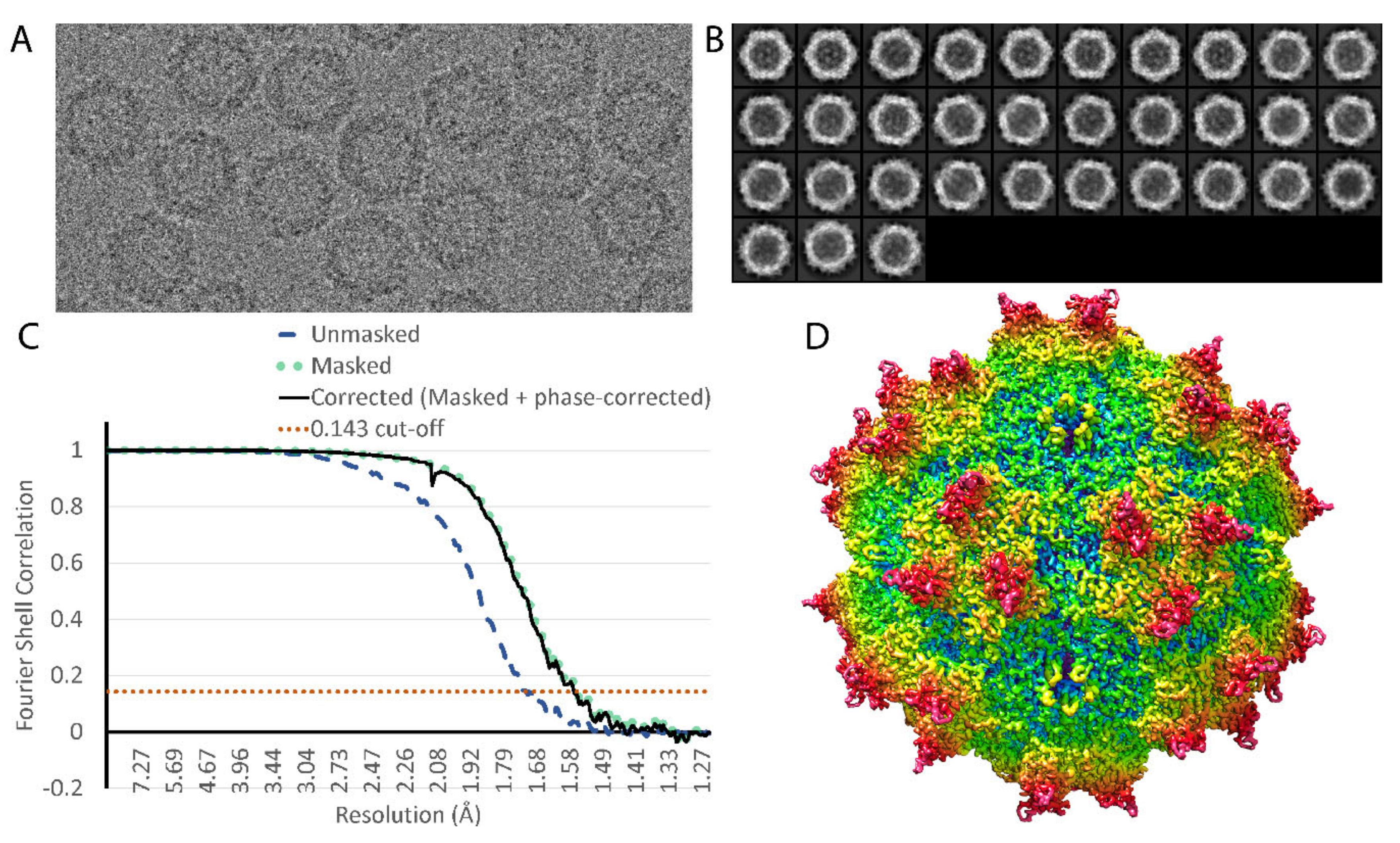
2.3. Image Processing

The AAV-DJ native dataset yielded 2241 movies. This initial dataset was refined to a resolution of ~2.2 Å before anisotropic magnification was observed to be preventing further progress. Distortion was estimated as ~0.9% and correction applied to the raw movies using magdistort binaries [34]. Processing was done within Relion 3.0 [35], with motion correction performed using Motioncor2 [36] and contrast transfer function (CTF) estimation done using Gctf [37]. DoGPicker [38] was used to pick ~70,000 potential particles and 4 rounds of 2D classification generated templates that were then used to re-pick 75,316 potential particles in Relion (Figure 1, Table 1). From here, multiple rounds of 2D classification and 3D classification with C1 symmetry were used to remove outliers, resulting in 48,209 particles after deduplication. These particles refined to ~2.2 Å with I1 symmetry, and subsequent refinements of beam tilt and per-particle CTF brought the resolution to 1.8 Å, further particle-polishing and subsequent re-refinement of CTF brought the resolution to 1.70 Å and a final reconstruction using Ewald’s sphere correction [39] ended at 1.56 Å. The map used for modeling was sharpened using the volume whitening routine in cisTEM [40].

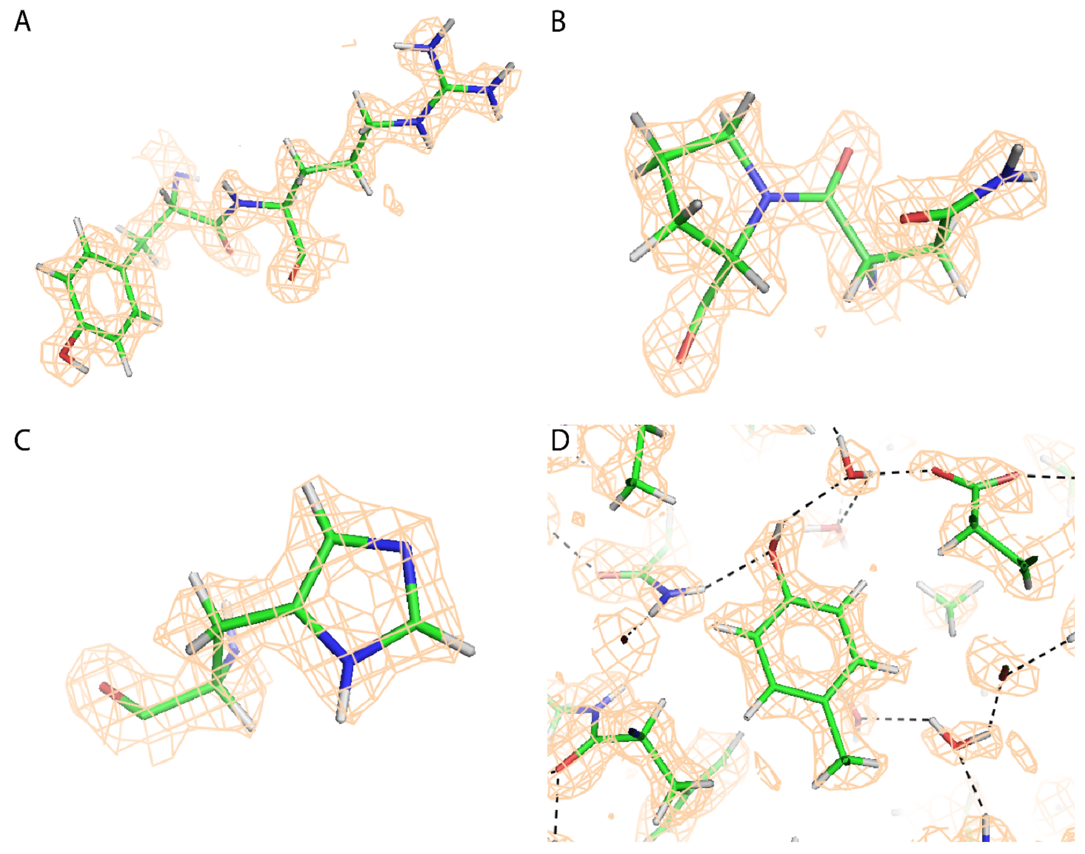
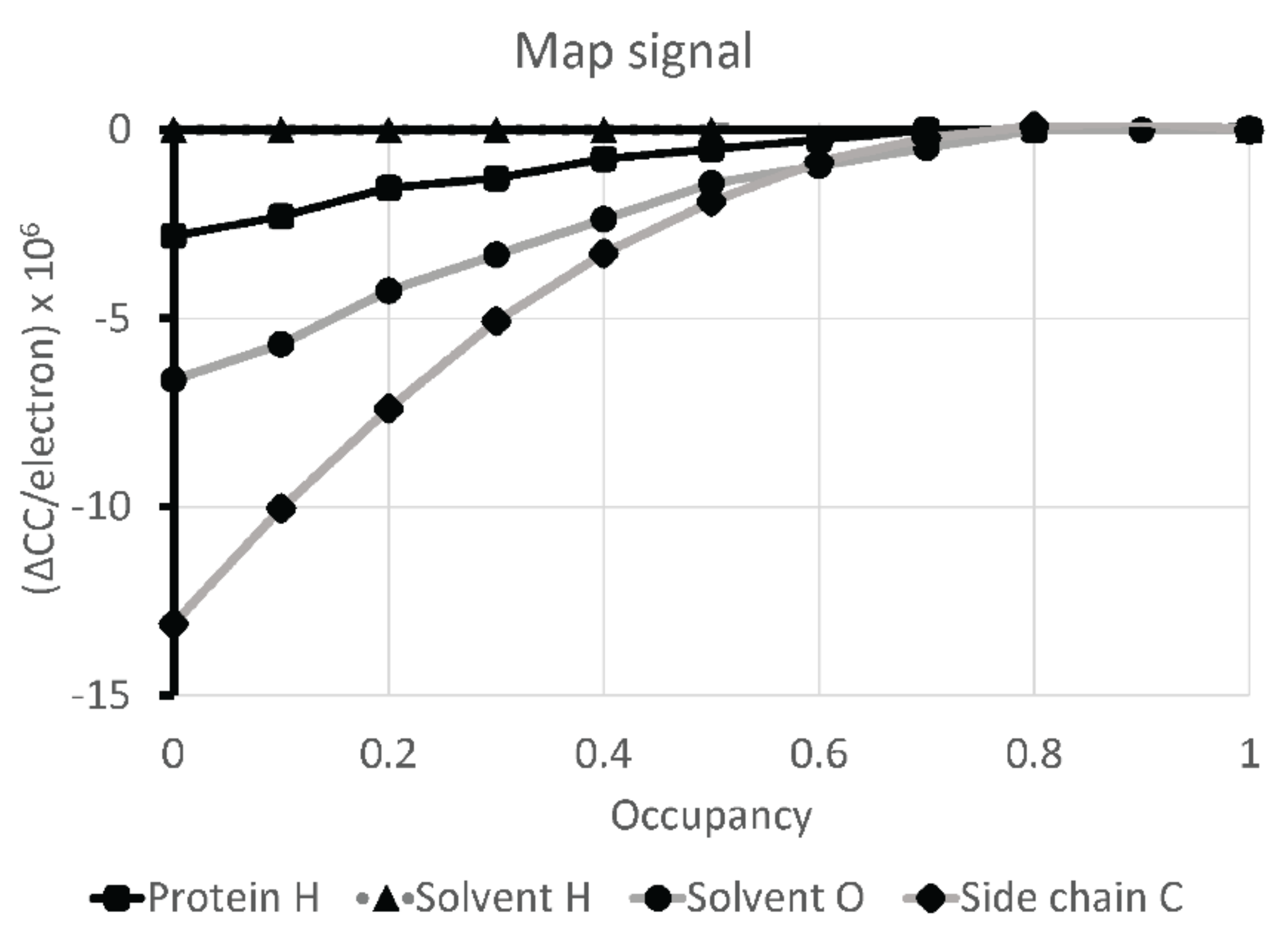
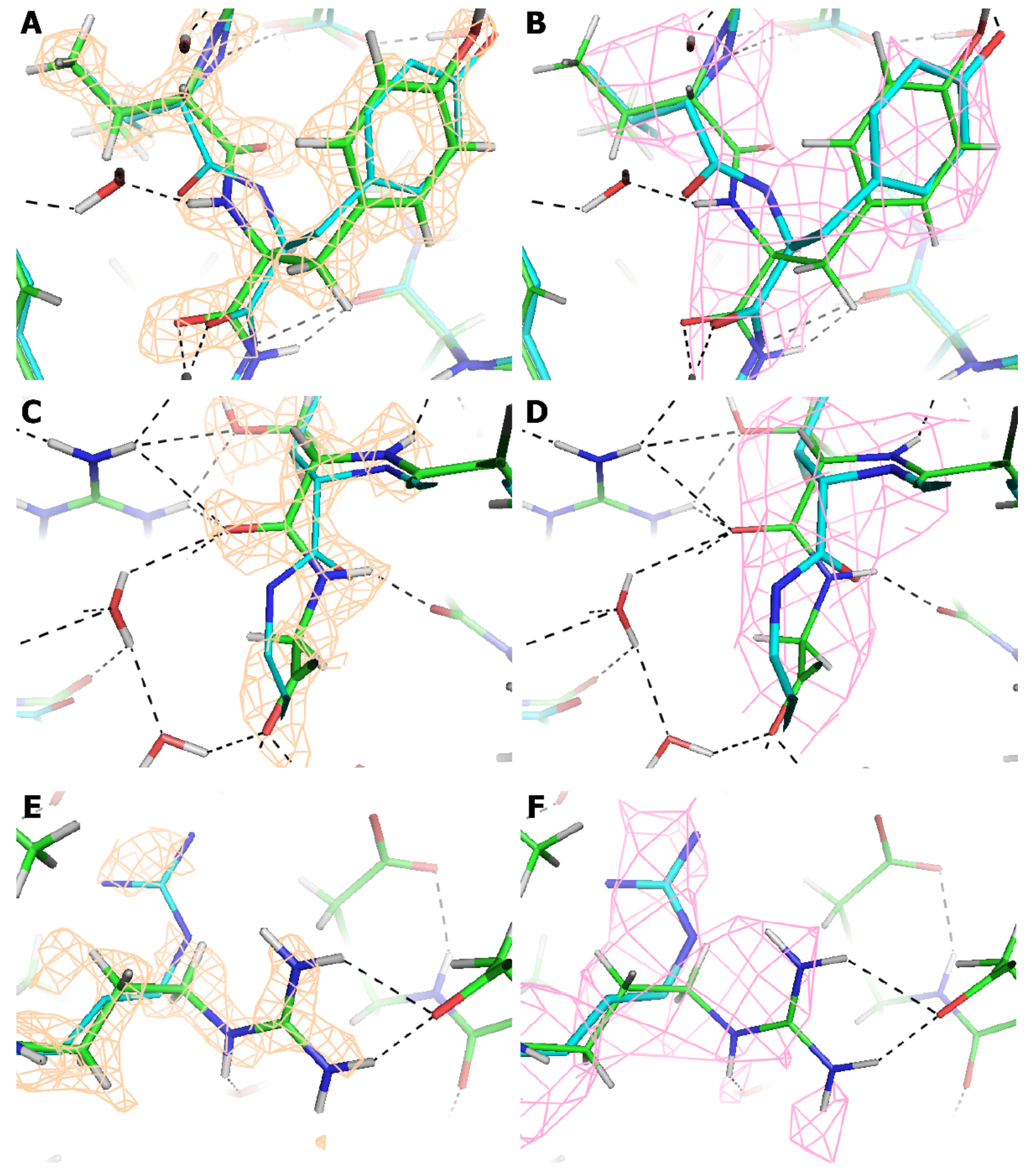
2.4. Atomic Modeling and Refinement
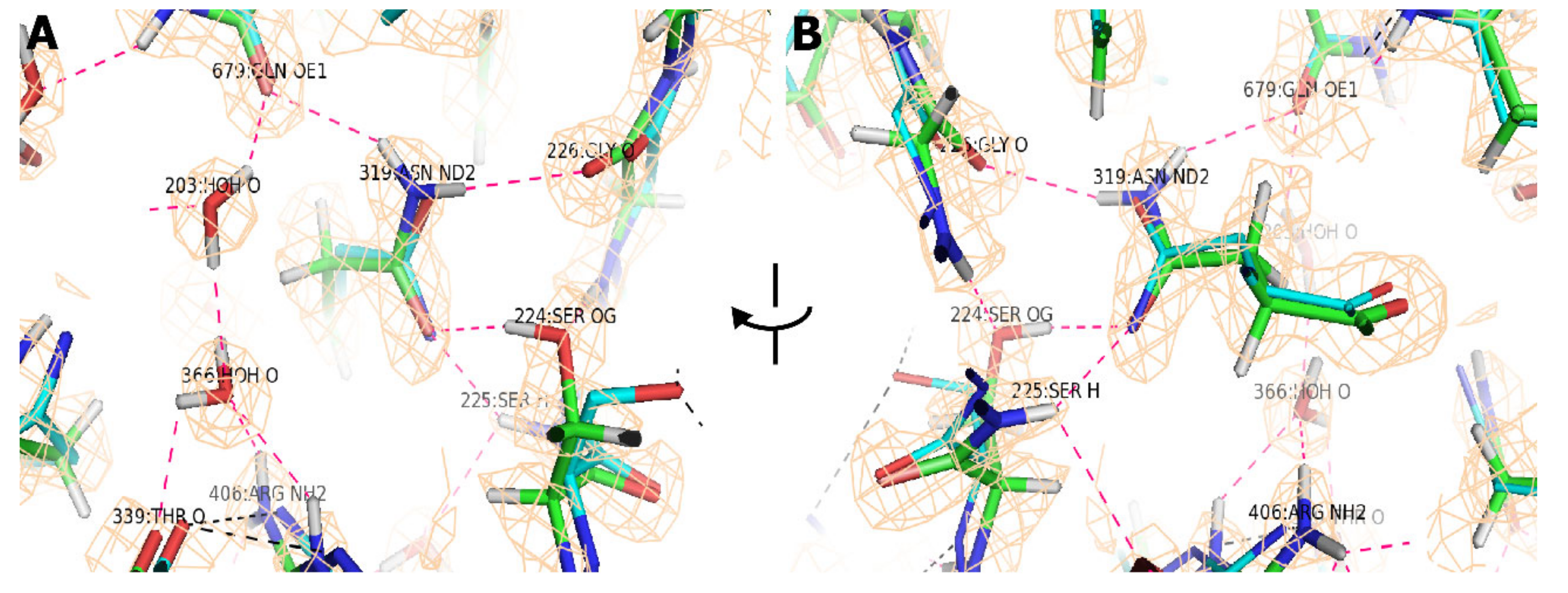
Envelope corrections and resolution, together with the effective EM magnification (at the start), were least-squares refined periodically against the emerging atomic model, using RSRef [31]. Calibration of the magnification was against the prior 2.8 Å cryo-EM structure of a glycan complex [22], and ultimately back to an external crystallographic standard, the AAV2 crystal structure [24]. Resolution is refined by optimizing the parameters of a Butterworth low-pass filter applied to the atomic model, until its density optimally agrees with the experimental reconstruction.
Starting from the prior 2.8 Å structure of a glycan complex, the atomic model was refined by optimization of the agreement between model-calculated and the sharpened experimental Coulombic potential map, using the real-space RSRef method described in the Introduction [31]. The first batch used simulated-annealing torsion angle dynamics [32] with a slow cooling protocol starting at a temperature of 5000 K (assessed as better than 10,000 K). Coot [41] “manual” rebuilding was alternated with RSRef refinement, using the unsharpened map where the sharpened map was unclear. The second and subsequent batches of refinement used gradient descent optimization instead of simulated annealing. Prior to the third batch, solvent waters were modeled into unoccupied peaks >1.8 σ where there was the potential for hydrogen bonding. Bound ions and riding hydrogens [42] were also added. The latter necessitated the addition of hydrogen electron scattering factors to RSRef [43], and use of the “allhdg5-4” topology and parameter files for CNS force field stereochemical restraints [44]. Before the next iteration of RSRef, hydrogen positions were optimized using an Amber force field as implemented in Phenix geometry minimization [45], harmonically restraining all non-hydrogen atoms. The energy minimized histidine protonation states were assessed: five were confirmed in the map, four were ambiguous, and one needed reconfiguring, along with surrounding waters. These protonation states were fixed by declarations as “HISD” or “HISE” to CNS. All atoms were refined on subsequent RSRef optimization, but water molecules, whose hydrogens were ill-defined in the map, remained close to their Amber-optimized orientations.
Segments of five loops were disordered in the sharpened reconstruction, backbone falling below 1 σ: residues 217–222, 264–268, 327–333, 453–458, and 705–708, totaling 24 of 532 (4%) amino acids. Residues 217–220 had not been observed in prior AAV-DJ structures. At least backbone density was clear in the unsharpened reconstruction, so the final batch of refinement was divided into two, first refining these segments into the unsharpened reconstruction while all other atom positions were harmonically restrained strongly. Then the selections were inverted for refinement of all but the less-ordered segments to the sharpened reconstruction. Alternate side chain rotamers were rechecked, and nine were retained with user-estimated relative occupancies.
The above represents a conservative model parameterization, because, with cryo-EM, cross-validation is not available as a check on over-fitting. Individual atomic B-factors were approached similarly. In the absence of cross-validation, the restraint weight on the similarity of neighboring B-factors (0.018) was adjusted to yield a root mean square deviation (RMSD) less than the crystal structures of AAV-2 and AAV-3B (1.6 Å2) [24,26].
RSRef supports the refinement not only of individual atomic B-factors, but an imaging envelope correction (overall B-factor), and resolution, implemented as a 5th order Butterworth low-pass filter [31,46]. These are covariant and cannot all be refined together. For a model-based estimate of resolution, it was refined against the 249 atoms of the β-barrel at the core of the VP subunit, subject to a constraint that <B> ≥ 0.0. This embodies the premise that the best estimate of experimental resolution would come from the parts of structure least likely to be disordered. Indeed, the mean B-factor for these atoms is 3.3 Å2, close enough to 0.0 to indicate that map-sharpening had been near optimal and that a model-based resolution estimate would be a good approximation.
The observability of solvent molecules, hydrogens, et cetera, in the reconstruction was quantified in the following way. Model-map correlation coefficients were calculated as the occupancies of classes of atoms were varied: protein hydrogens, solvent hydrogens, solvent oxygens and (for comparison) protein backbone and protein side chain carbons. Correlation coefficients were estimated from the calculated Coulombic potential at all grid points in the experimental reconstruction within 2 Å of any atom, such that the same map volume was used for all calculations. Occupancies were varied between 1.0 and 0.0, normalizing the change in correlation coefficient by the total number of electrons in the atom set.
Accession numbers: An atomic model will be available from the protein data bank (http://www.rcsb.org) PDBid 7KFR. EM reconstructions will be available from the EMDataBank (http://www.ebi.ac.uk/pdbe/emdb/) with accession number EMD-22854.

3. Results

By FSC0.143 gold standard [47], the resolution is estimated to be 1.56 Å (Figure 1, Table 1). It is only recently with pre-print reports of apo-ferritin assemblies that this resolution has been exceeded for cryo-TEM of biomolecular assemblies [48,49]. There have been many measures of resolution in cryo-EM [50], and with the technological advances in recent years, there is growing appreciation of molecular features resolvable at the more common circa 3 Å resolution, we are in unchartered territory near 1.5 Å. The community has been shown how the Coulombic potential (EM maps) can differ from electron density (crystallography) [51]. Furthermore, EM resolution is conventionally measured in terms of signal-noise, in reciprocal space, and so it is an “average” between regions that may have substantial variation in local effective resolution [52]. A refined atomic structure allows a real-space evaluation with refinement of the parameters of a low pass filter applied to calculation of Coulombic potential from the atomic model for best agreement with the experimental reconstruction. An FSC0.143 calculated between two half data sets corresponds to the resolution where Cref, the cross-correlation between a full set and a noise-free reference, falls to 0.5 [53]. Although there is not strict equivalence, one might therefore expect correspondence between FSC0.143 and d0.5, the point at which a fitted 5th order Butterworth low pass filter half attenuates Fourier contributions to calculation of the Coulombic potential from the atomic coordinates [31]. When optimized using the full atomic model, d0.5 refines to 1.44 Å.
Local resolution is also a combination of multiple effects including (a) molecular disorder, (b) sample preparation, (c) microscope transfer/point spread functions as well as (d) alignment and other errors in computational reconstruction [54,55]. They can be difficult to tease apart, because of covariance of atomic displacement parameters (B-factors), envelope, or sharpening corrections and other resolution attenuation. It was postulated that these could be factored into molecular (a) and experimental (b–d) components if a significant part of the structure was highly ordered, as evidenced by mean refined B-factor approaching zero (without an envelope correction). The 249 atoms within the nine strands, βA–βG, of the core subunit jellyroll approximate this, with mean B, <B> = 3.3 Å2. The refined d0.5 is only marginally different at 1.43 Å, suggesting that on completion of refinement, the resolution estimate reflects the best experimental resolution, and that the molecular disorder is parameterized through variation in the atomic B-factors. Note that the model-referenced estimate (1.44 Å) differs somewhat from the FSC0.143, reflecting both differences in their means of calculation, and that the most ordered parts of the structure are seen with greater clarity than average.
A priority was to establish what could be interpreted with confidence in EM reconstructions at this resolution. This started during the atomic refinement, because we needed to know how to appropriately parameterize the model. Inspection of the map clearly showed the presence of a first hydration shell of solvent water molecules bound to many of the polar protein groups (Figure 2D). In the absence of cross-validation methods, modeling was conservative, adding solvent molecules only where peaks in the map exceeded 1.8 σ, totaling 265 per protein subunit or an average of one water per two amino acids. Presumptive divalent ions could also be seen, coordinated by histidine ring nitrogens. Unexpected at this resolution was the prevalence of features corresponding to hydrogen atoms. In X-ray crystallography, 1.2 Å resolution is generally considered the starting point for modeling hydrogens, with complete structures expected only beyond 1 Å resolution. Indications of hydrogens had been reported in an EM structure at 1.86 Å resolution [13], but now, as we approach 1.5 Å resolution, hydrogen locations through most of the protein are clear to model (Figure 2D).
As resolution improves, one expects a gradual strengthening of features that would be impossible to see at lower resolution. The strengths of features per atom type are calibrated in Figure 3 to those of side-chain carbons, and we see that protein hydrogens are at about a quarter of full signal, solvent oxygens are at half, and that, on average, there is no signal for the more disordered solvent hydrogens. Their placement depends completely upon the Amber molecular mechanics calculation.
At the end of atomic refinement (Table 2), deviations from ideal geometry are very modest, both for attributes that are stereochemically restrained (bond lengths, angles, contacts) and those that are unrestrained and used as a read-out on model quality (Ramachandran φ, ψ and side chain rotamer outliers). For a reconstruction that is optimally sharpened so that it reveals the detailed features, high resolution noise is amplified, so cross-correlation coefficients (CC) are lowered. For the unsharpened map, the overall CC is 0.90 when d0.5 refined to 2.7 Å using map grid points within 1.8 Å of any atom. Against the sharpened map, the overall CC is 0.80 for grid points within 2.0 Å of atoms. However, the quality varies from excellent in the β-barrel (CC = 0.88 for grid points within 2 Å of the 249 atoms) to marginal, the marginal regions includingthe 24 disordered loop residues with breaks in backbone continuity that are only breached in the unsharpened map.
While the locations of hydrogens are predominantly clear, they are not as well enclosed within a map isocontour as other atoms. There are multiple causes. Hydrogen atoms have only a single electron/proton, so their Coulombic potential is weaker than non-hydrogen protein atoms, even if the experimentally observed signal were not one quarter of that to be expected (above). Furthermore, it has been noted by others that the orbitals of a σ-bonded hydrogen are not centered on the nucleus, but skewed towards the covalent bond [56,57]. This leads to an appearance in X-ray structures of hydrogens being 10% closer to their covalently bonded partner, and EM structures may see some of a similar effect. Some choose to display and restrain towards a shortened bond, but Figure 2 shows nucleus-defined atom centers.
Now with a very high resolution structure as a yardstick, it is enlightening to analyze the accuracy of prior AAV-DJ structures at 2.8 Å and 4.5 Å, [20,22], the former representing a resolution that is often a goal in the emerging post “resolution revolution” era of cryo-EM, while the latter represents the 5 Å average of structures deposited to EMDB in 2019–2020. It is sobering that the root mean square deviation (RMSD) between the 2.8 Å and 1.56 Å structures is as high as 0.9 Å, given estimates that stereochemical restraints in protein crystallography support accuracy 5–8-fold beyond the nominal experimental resolution [31,58,59,60]. Upon analysis, the AAV-DJ comparison is driven by 10% of the atoms that are outliers with an RMSD of 2.3 Å, while 90% have an RMSD of 0.35 Å (Table 3). If it is assumed that the deviations can be attributed to resolution-proportional and independent errors in the two structures, the estimated coordinate errors for the 90% are 0.17 Å and 0.31 Å, 9-fold better than the respective nominal 1.56 Å and 2.8 Å resolutions. Outliers in comparing crystal structures have been reported, but the fraction outside the expected distribution [58] is greater for our cryo-EM structures.
The largest outliers (5–10 Å) are in side-chains, usually where there is little indication in any of the maps as to which rotamer should be chosen. Figure 4E,F show a 5 Å change in an arginine side chain that is likely somewhat disordered, but where the higher resolution map gave clear indication of the need for a change. Evidence is not only in the map for the guanidinium, but also from the tetrahedral hydrogens that confirm the positions of Cβ through Cδ, consistent only with the 1.56 Å resolution structure. As for backbone outliers, about 30% are in less ordered parts of AAV-DJ, in the 5% of the structure that was refined against the unsharpened reconstruction, because it was clearer than the sharpened map. Other backbone outliers come from the many regions in the 2.8 Å map where the map has a tubular backbone without definition of carbonyl oxygens. In retrospect, we see that the peptide bonds were flipped in the 2.8 Å atomic model. Figure 4 illustrates two regions. Figure 4A clearly shows the correct peptide orientation. At 2.8 Å (Figure 4B) it would be difficult to distinguish correct from incorrect. Panels C and D show an example of where the 2.8 Å structure followed that of homologous AAVs, but it was not clear that a correction was needed until the 1.56 Å resolution reconstruction. There are many other peptide bonds where the 2.8 Å map gives no indication of carbonyl orientation, but where the change in the 1.56 Å resolution structure is more subtle than a peptide flip. Even through not optimally configured at 2.8 Å, it is only a few percent of NH-OC backbone-backbone hydrogen bonds that are not correctly identified at 2.8 Å, providing that generous angular cut-offs are used [61]. So, secondary structure is predominantly identified correctly at 2.8 Å. This feeds back to improve model quality, because, once recognized, even absent formal restraints, our a priori understanding of idealized secondary structure informs improved atom placement, mitigating ambiguity in the map.
The improved resolution has greater impact upon the understanding of atomic interactions, where they are not clear in the lower resolution map, and there is not a priori information as to their nature. Figure 5 explains a typical situation where the hydrogen bonding network is completely different when seen at high resolution. At 2.8 Å the water molecules could not be modeled, and the rotamer of Asn319 had side chain dihedral angle, χ2 flipped, i.e., rotated by 180°. The correct asparagine rotamer is inferred from the interactions with Ser224-Gly226. This was not possible at 2.8 Å, because of backbone deviations of 0.52 Å (not particularly atypical with an RMSD of 0.42 Å). The advantage at 1.56 Å is clear definition of both the carbonyl oxygen of Gly226 and the amino hydrogen of Ser225 which precisely define the backbone location and settle the ambiguity at 2.8 Å resolution in rotamer choice and hydrogen bond interactions.

4. Discussion

Although the AAV-DJ structure represents a milestone, there is reason for optimism that it might prove, in due course, to lack exceptionality. While this is a robust viral capsid with the icosahedral symmetry that is highly advantageous in cryo-EM reconstruction, it is a relatively heterogenous virus with variant capsid proteins in unpredictable locations (see Introduction). Furthermore, the results were achieved with a microscope configuration that has become standard over the last couple of years. Approximately the same number of particles (47,000; 2.6 × 10⁶ asymmetric units) were used as in an earlier (2017) 2.8 Å structure determination [22], in other words, this is a standard data collection regime without extraordinary efforts to maximize the resolution. The results reported here have not depended on the very latest microscope developments that have supported apoferritin structures beyond 1.5 Å [48,49].
The structure contains many parts that reveal detailed features, such as hydrogen locations, with clarity that we would not have expected from X-ray structures at corresponding resolutions. However, comparisons of the different AAV-DJ structures show larger discrepancies than we might expect at 1.6–2.8 Å resolution. With AAV-DJ, about 10% of the reconstruction is disordered enough to affect atomic modeling. This appears to be a higher proportion than in crystal structures, and only about half become clear with high resolution EM data. It is not clear that biologist “consumers” of atomic structures will evaluate the experimental map directly, so it becomes a priority to develop annotations of the local confidence in EM-derived atomic models, so that detailed interpretations are not extended from robust regions into parts where conclusions might be fickle.
What biochemical insights come from observation of hydrogens? Recognizable tetrahedral shape removes ambiguity in modeling aliphatic groups (Figure 2A), and can provide unequivocal evidence for infrequent conformations such as cis-peptides (Figure 2B). In these cases, the positions of hydrogens are implicitly defined (completely) by the locations of neighboring heavy atoms. However, it is not just “riding” hydrogens that are observable. For much of the structure, those whose positions depend upon rotation about a next-nearest bond are also indicated. This can be significant for hydroxyl groups, where rotation about the hydroxyl bond determines which neighboring atoms could be hydrogen bond acceptors (or donors). Examples of tyrosine hydroxyl groups are shown in Figure 2A,D where the map indicates the direction of the hydrogen.
At this resolution of ~1.5 Å, we are also getting some indication of the presence, or not, of titratable protons. For seven of thirteen histidines in an AAV-DJ VP3, the map indicates which nitrogen(s) of a histidine side chain are protonated (Figure 2C); two others show the entire imidazole ring, but without distinctive hydrogens at Nε or Nδ; while four exhibit disorder that precludes imidazole proton assignment with confidence. None of the seven are seen in the fully protonated HisH+ form which is expected to be a minor fraction at the buffer pH 7.4, unless perturbed by the local environment. Two of the seven appear predominantly in the N3 tautomer (hydrogen at Nε) which is favored 4:1, absent perturbation, due to a slightly more basic pKa of the N3 tautomer [62], while five are the N1 tautomeric form (hydrogen at Nδ), a ratio indicative of some perturbation. For six of the seven distinctive histidines, post facto, we see strong chemical rationale for the tautomer due to one or more of the following: (1) a glutamate or aspartate carboxylate oxygen within 3Å hydrogen-bonding distance of Nε or Nδ and raising its pKa; (2) metal ion coordination by one nitrogen, lowering its pKa and raising the other [63,64]; (3) hydrogen bonding with a partner that is unambiguously a donor (amide nitrogen) or acceptor (carbonyl oxygen), implying, respectively, the absence or presence of a hydrogen at that histidine nitrogen; and (4) steric clash if protonated. One histidine is well solvated at both nitrogens, with no obvious perturbation, and it is in the favored N3 tautomeric form. There are no cases where map features are inconsistent with analysis of pK-perturbing interactions. Thus, while hydrogens are not clear for all histidines, all indications are that features seen are real and not false positives. Thus, we are observing, at 1.56 Å resolution, the effects of local environmental pKa perturbations of the nitrogen atoms of half of the histidines. This has exciting implications when high resolution studies of enzymes and channels become more routine, and one can imagine pH-dependent studies to analyze titratable protonation states.
The X-ray crystal structures of five different viruses/VLPs and their variants have been reported at better than 2 Å with three at slightly beyond 1.5 Å—Coxsackievirus A24v, Satellite Tobacco Mosaic Virus (STMV) and Satellite Tobacco Necrosis Virus (STNV) [10,11,12]. For none of these highest resolution virus structures has it been possible to visualize hydrogens or refine their atomic positions. A first indication of hydrogen atoms came with a 1.86 Å cryo-EM structure of an AAV variant [13]. Here, at 1.56 Å, the AAV-DJ structure is refined with a full complement of hydrogens, most of which are clearly defined in the map. This heralds a new era in virus structure with direct experimental observation leading to significantly improved characterization of the hydrogen bonding networks that are key to tertiary structure and assembly. This is coming from cryo-EM (rather than X-ray crystallography), because of the greater contribution of nuclear charge to the scattering of electrons compared to X-ray scattering which depends upon a single electron for a hydrogen atom [49,65,66]. At the forefront of EM resolution are structures of high symmetry, the 24-mer apoferritin and the 60-mer AAV, with distinct advantages in terms of protomer orientation distribution, alignment determination and potential for signal averaging. Their example is demonstrating that for other samples, instrumentation need not be limiting. Indeed the structure of a pentameric membrane protein has recently been reported at 1.7 Å resolution [48].

5. Conclusions

Even for those of us who came of age after development of protein crystallography, the progress in cryo-EM has been astonishing. Michael Rossmann and others of his generation took on the then seemingly impossible challenge of extending X-ray crystallography to mega-Dalton viral assemblies. It was their vision and success that became the foundation for structural virology. Notwithstanding his investment in crystallographic methodology, he was among the first to embrace complementary application of both approaches. As cryo-EM now becomes the choice even for high resolution virus structure, one suspects that he might have envisioned this before the rest of us, and that he would now be embracing the progress.

Author Contributions

Conceptualization, M.S.C. and C.K.Y.; methodology, C.K.Y.; software, M.S.C.; validation, C.K.Y. and M.S.C.; investigation, Q.X. and C.K.Y.; data curation, C.K.Y., Q.X. and M.S.C.; writing—original draft preparation, M.S.C.; writing—review and editing, C.K.Y., Q.X. and M.S.C.; visualization, M.S.C.; supervision, M.S.C.; funding acquisition, M.S.C. All authors have read and agreed to the published version of the manuscript.

Funding

This work was funded by NIH R35 GM122564 (M.S.C.) and U24 GM129547 (J.E. Gouaux, J. Evans, S. Reichow and M.S.C.).

Acknowledgments

We thank reviewer 1 for suggesting that we point out the implications of our results for future pH-dependent analyses of titratable protonation states.

Conflicts of Interest

The authors declare no conflict of interest.

References

  1. Stanley, W.M. Isolation of a crystalline protein possessing the properties of tobacco-mosaic virus. Science 1935, 81, 644–645. [Google Scholar] [CrossRef] [PubMed] [Green Version]
  2. Kendrew, J.C.; Dickerson, R.E.; Strandberg, B.E.; Hart, R.G.; Davies, D.R.; Phillips, D.C.; Shore, V.C. Structure of Myoglobin: A Three-Dimensional Fourier Synthesis at 2 Å. Resolution. Nature 1960, 185, 422–427. [Google Scholar] [CrossRef] [PubMed]
  3. Caspar, D.L.D.; Klug, A. Physical principles in the construction of regular viruses. In Cold Spring Harbor Symposium in Quantitative Biology; Cold Spring Harbor Laboratory Press, Cold Spring Harbor: New York, NY, USA, 1962; Volume 27, pp. 1–24. [Google Scholar]
  4. Rossmann, M.G.; Blow, D.M. Determination of Phases by the Conditions of Non-Crystallgraphic Symmetry. Acta Cryst. 1963, 16, 39–45. [Google Scholar] [CrossRef]
  5. De Rosier, D.J.; Klug, A. Reconstruction of Three Dimensional Structures from Electron Micrographs. Nature 1968, 217, 130–134. [Google Scholar] [CrossRef]
  6. Harrison, S.C.; Olson, A.; Schutt, C.E.; Winkler, F.K.; Bricogne, G. Tomato bushy stunt virus at 2.9 Å resolution. Nature 1978, 276, 368–373. [Google Scholar] [CrossRef]
  7. Abad-Zapatero, C.; Abdel-Meguid, S.S.; Johnson, J.E.; Leslie, A.G.W.; Rayment, I.; Rossmann, M.G. Structure of southern bean mosaic virus at 2.8 A resolution. Nature 1980, 286, 33–39. [Google Scholar] [CrossRef]
  8. Stewart, P.L.; Fuller, S.D.; Burnett, R.M. Difference imaging of adenovirus: Bridging the resolution gap between X-ray crystallography and electron microscopy. EMBO J. 1993, 12, 2589–2599. [Google Scholar] [CrossRef]
  9. Berman, H.M.; Westbrook, J.; Feng, Z.; Gilliland, G.; Bhat, T.N.; Weissig, H.; Shindyalov, I.N.; Bourne, P.E. The Protein Data Bank. Nucleic Acids Res. 2000, 28, 235–242. [Google Scholar] [CrossRef] [Green Version]
  10. Zocher, G.; Mistry, N.; Frank, M.; Hahnlein-Schick, I.; Ekstrom, J.O.; Arnberg, N.; Stehle, T. A sialic acid binding site in a human picornavirus. PLoS Pathog 2014, 10, e1004401. [Google Scholar] [CrossRef] [Green Version]
  11. Larson, S.B.; Day, J.S.; McPherson, A. Satellite tobacco mosaic virus refined to 1.4 Å resolution. Acta Crystallogr. D Biol. Crystallogr. 2014, 70, 2316–2330. [Google Scholar] [CrossRef] [Green Version]
  12. Lane, S.W.; Dennis, C.A.; Lane, C.L.; Trinh, C.H.; Rizkallah, P.J.; Stockley, P.G.; Phillips, S.E. Construction and crystal structure of recombinant STNV capsids. J. Mol. Biol. 2011, 413, 41–50. [Google Scholar] [CrossRef] [PubMed]
  13. Tan, Y.Z.; Aiyer, S.; Mietzsch, M.; Hull, J.A.; McKenna, R.; Grieger, J.; Samulski, R.J.; Baker, T.S.; Agbandje-McKenna, M.; Lyumkis, D. Sub-2 Å Ewald curvature corrected structure of an AAV2 capsid variant. Nat. Commun. 2018, 9, 3628. [Google Scholar] [CrossRef] [PubMed]
  14. Cheng, Y. Single-Particle Cryo-EM at Crystallographic Resolution. Cell 2015, 161, 450–457. [Google Scholar] [CrossRef] [PubMed] [Green Version]
  15. Sirohi, D.; Chen, Z.; Sun, L.; Klose, T.; Pierson, T.C.; Rossmann, M.G.; Kuhn, R.J. The 3.8 Å resolution cryo-EM structure of Zika virus. Science 2016, 352, 467–470. [Google Scholar] [CrossRef] [Green Version]
  16. Kerr, J.R.; Cotmore, S.F.; Bloom, M.E.; Linden, R.M.; Parrish, C.R. (Eds.) Parvoviruses; Hodder Arnold, Ltd.: London, UK, 2006. [Google Scholar]
  17. Al-Zaidy, S.A.; Kolb, S.J.; Lowes, L.; Alfano, L.N.; Shell, R.; Church, K.R.; Nagendran, S.; Sproule, D.M.; Feltner, D.E.; Wells, C.; et al. AVXS-101 (Onasemnogene Abeparvovec) for SMA1: Comparative Study with a Prospective Natural History Cohort. J. Neuromuscul. Dis. 2019, 6, 307–317. [Google Scholar] [CrossRef] [Green Version]
  18. Hoy, S.M. Onasemnogene Abeparvovec: First Global Approval. Drugs 2019, 79, 1255–1262. [Google Scholar] [CrossRef]
  19. Grimm, D.; Lee, J.S.; Wang, L.; Desai, T.; Akache, B.; Storm, T.A.; Kay, M.A. In vitro and in vivo gene therapy vector evolution via multispecies interbreeding and retargeting of adeno-associated viruses. J. Virol. 2008, 82, 5887–5911. [Google Scholar] [CrossRef] [Green Version]
  20. Lerch, T.F.; O’Donnell, J.K.; Meyer, N.L.; Xie, Q.; Taylor, K.A.; Stagg, S.M.; Chapman, M.S. Structure of AAV-DJ, a retargeted gene therapy vector: Cryo-electron microscopy at 4.5 Å resolution. Structure 2012, 20, 1310–1320. [Google Scholar] [CrossRef] [Green Version]
  21. Xie, Q.; Spilman, M.; Meyer, N.L.; Lerch, T.F.; Stagg, S.M.; Chapman, M.S. Electron microscopy analysis of a disaccharide analog complex reveals receptor interactions of adeno-associated virus. J. Struct. Biol. 2013, 184, 129–135. [Google Scholar] [CrossRef] [Green Version]
  22. Xie, Q.; Spear, J.M.; Noble, A.J.; Sousa, D.R.; Meyer, N.L.; Davulcu, O.; Zhang, F.; Linhardt, R.J.; Stagg, S.M.; Chapman, M.S. The 2.8 Å Electron Microscopy Structure of Adeno-Associated Virus-DJ Bound by a Heparinoid Pentasaccharide. Mol. Ther. Methods Clin. Dev. 2017, 5, 1–12. [Google Scholar] [CrossRef] [Green Version]
  23. Thompson, R.F.; Walker, M.; Siebert, C.A.; Muench, S.P.; Ranson, N.A. An introduction to sample preparation and imaging by cryo-electron microscopy for structural biology. Methods 2016, 100, 3–15. [Google Scholar] [CrossRef] [PubMed]
  24. Xie, Q.; Bu, W.; Bhatia, S.; Hare, J.; Somasundaram, T.; Azzi, A.; Chapman, M.S. The atomic structure of adeno-associated virus (AAV-2), a vector for human gene therapy. Proc. Natl. Acad. Sci. USA 2002, 99, 10405–10410. [Google Scholar] [CrossRef] [PubMed] [Green Version]
  25. Nam, H.J.; Lane, M.D.; Padron, E.; Gurda, B.; McKenna, R.; Kohlbrenner, E.; Aslanidi, G.; Byrne, B.; Muzyczka, N.; Zolotukhin, S.; et al. Structure of adeno-associated virus serotype 8, a gene therapy vector. J. Virol. 2007, 81, 12260–12271. [Google Scholar] [CrossRef] [PubMed] [Green Version]
  26. Lerch, T.F.; Xie, Q.; Chapman, M.S. The structure of adeno-associated virus serotype 3B (AAV-3B): Insights into receptor binding and immune evasion. Virology 2010, 403, 26–36. [Google Scholar] [CrossRef] [PubMed] [Green Version]
  27. Meyer, N.L.; Hu, G.; Davulcu, O.; Xie, Q.; Noble, A.J.; Yoshioka, C.; Gingerich, D.S.; Trzynka, A.; David, L.; Stagg, S.M.; et al. Structure of the gene therapy vector, adeno-associated virus with its cell receptor, AAVR. Elife 2019, 8, e44707. [Google Scholar] [CrossRef] [PubMed]
  28. Girod, A.; Wobus, C.E.; Zadori, Z.; Ried, M.; Leike, K.; Tijssen, P.; Kleinschmidt, J.A.; Hallek, M. The VP1 capsid protein of adeno-associated virus type 2 is carrying a phospholipase A2 domain required for virus infectivity. J. Gen. Virol. 2002, 83, 973–978. [Google Scholar] [CrossRef] [Green Version]
  29. Kronenberg, S.; Kleinschmidt, J.A.; Bottcher, B. Electron cryo-microscopy and image reconstruction of adeno-associated virus type 2 empty capsids. EMBO Rep. 2001, 2, 997–1002. [Google Scholar] [CrossRef] [Green Version]
  30. Afonine, P.V.; Poon, B.K.; Read, R.J.; Sobolev, O.V.; Terwilliger, T.C.; Urzhumtsev, A.; Adams, P.D. Real-space refinement in PHENIX for cryo-EM and crystallography. Acta Crystallogr. D Struct. Biol. 2018, 74, 531–544. [Google Scholar] [CrossRef] [Green Version]
  31. Chapman, M.S.; Trzynka, A.; Chapman, B.K. Atomic modeling of cryo-electron microscopy reconstructions--joint refinement of model and imaging parameters. J. Struct. Biol. 2013, 182, 10–21. [Google Scholar] [CrossRef] [Green Version]
  32. Brünger, A.T.; Adams, P.D.; Clore, G.M.; DeLano, W.L.; Gros, P.; Grosse-Kunstleve, R.W.; Jiang, J.-S.; Kuszewski, J.; Nilges, M.; Pannu, N.S.; et al. Crystallography and NMR system: A new software system for macromolecular structure determination. Acta Crystallogr. 1998, 54, 905–921. [Google Scholar] [CrossRef]
  33. Meyer, N.; Davulcu, O.; Xie, Q.; Silveria, M.; Zane, G.; Large, E.; Chapman, M. Expression and Purification of Adeno-associated Virus Virus-like Particles in a Baculovirus System and AAVR Ectodomain Constructs in E. coli. Bio-Protocol 2020, 10, e3513. [Google Scholar] [CrossRef]
  34. Grant, T.; Grigorieff, N. Automatic estimation and correction of anisotropic magnification distortion in electron microscopes. J. Struct. Biol. 2015, 192, 204–208. [Google Scholar] [CrossRef] [PubMed]
  35. Zivanov, J.; Nakane, T.; Forsberg, B.O.; Kimanius, D.; Hagen, W.J.; Lindahl, E.; Scheres, S.H. New tools for automated high-resolution cryo-EM structure determination in RELION-3. Elife 2018, 7, e42166. [Google Scholar] [CrossRef] [PubMed]
  36. Zheng, S.Q.; Palovcak, E.; Armache, J.P.; Verba, K.A.; Cheng, Y.; Agard, D.A. MotionCor2: Anisotropic correction of beam-induced motion for improved cryo-electron microscopy. Nat. Methods 2017, 14, 331–332. [Google Scholar] [CrossRef] [PubMed] [Green Version]
  37. Zhang, K. Gctf: Real-time CTF determination and correction. J. Struct. Biol. 2016, 193, 1–12. [Google Scholar] [CrossRef] [Green Version]
  38. Voss, N.R.; Yoshioka, C.K.; Radermacher, M.; Potter, C.S.; Carragher, B. DoG Picker and TiltPicker: Software tools to facilitate particle selection in single particle electron microscopy. J. Struct. Biol. 2009, 166, 205–213. [Google Scholar] [CrossRef] [Green Version]
  39. Wolf, M.; DeRosier, D.J.; Grigorieff, N. Ewald sphere correction for single-particle electron microscopy. Ultramicroscopy 2006, 106, 376–382. [Google Scholar] [CrossRef]
  40. Grant, T.; Rohou, A.; Grigorieff, N. cisTEM, user-friendly software for single-particle image processing. Elife 2018, 7, e35383. [Google Scholar] [CrossRef]
  41. Emsley, P.; Lohkamp, B.; Scott, W.G.; Cowtan, K. Features and development of Coot. Acta Crystallogr. D Biol. Crystallogr. 2010, 66, 486–501. [Google Scholar] [CrossRef] [Green Version]
  42. Word, J.M.; Lovell, S.C.; Richardson, J.S.; Richardson, D.C. Asparagine and glutamine: Using hydrogen atom contacts in the choice of side-chain amide orientation. J. Mol. Biol. 1999, 285, 1735–1747. [Google Scholar] [CrossRef] [Green Version]
  43. Peng, L.-M.; Ren, G.; Dudarev, S.L.; Whelan, M.J. Robust Parameterization of Elastic and Absorptive Electron Atomic Scattering Factors. Acta Crystallogr. Sect. A 1996, 52, 257–276. [Google Scholar] [CrossRef] [Green Version]
  44. Linge, J.; Nilges, M. Influence of non-bonded parameters on the quality of NMR structures: A new force field for NMR structure calculation. J. Biomol. NMR 1999, 13, 51–59. [Google Scholar] [CrossRef] [PubMed]
  45. Moriarty, N.W.; Janowski, P.A.; Swails, J.M.; Nguyen, H.; Richardson, J.S.; Case, D.A.; Adams, P.D. Improved chemistry restraints for crystallographic refinement by integrating the Amber force field into Phenix. Acta Crystallogr. Sect. D 2020, 76, 51–62. [Google Scholar] [CrossRef] [PubMed] [Green Version]
  46. Butterworth, S. On the Theory of Filter Amplifiers. Wirel. Eng. 1930, 7, 536–541. [Google Scholar]
  47. Scheres, S.H. Relion: Implementation of a Bayesian approach to cryo-EM structure determination. J. Struct. Biol. 2012, 180, 519–530. [Google Scholar] [CrossRef] [PubMed] [Green Version]
  48. Nakane, T.; Kotecha, A.; Sente, A.; McMullan, G.; Masiulis, S.; Brown, P.M.G.E.; Grigoras, I.T.; Malinauskaite, L.; Malinauskas, T.; Miehling, J.; et al. Single-particle cryo-EM at atomic resolution. bioRxiv 2020, 110189. [Google Scholar] [CrossRef]
  49. Yip, K.M.; Fischer, N.; Paknia, E.; Chari, A.; Stark, H. Breaking the next Cryo-EM resolution barrier—Atomic resolution determination of proteins! bioRxiv 2020, 106740. [Google Scholar] [CrossRef]
  50. Penczek, P.A. Fundamentals of three-dimensional reconstruction from projections. Methods Enzymol. 2010, 482, 1–33. [Google Scholar] [CrossRef] [Green Version]
  51. Bartesaghi, A.; Merk, A.; Banerjee, S.; Matthies, D.; Wu, X.; Milne, J.L.; Subramaniam, S. Electron microscopy. 2.2 A resolution cryo-EM structure of beta-galactosidase in complex with a cell-permeant inhibitor. Science 2015, 348, 1147–1151. [Google Scholar] [CrossRef] [Green Version]
  52. Kucukelbir, A.; Sigworth, F.J.; Tagare, H.D. Quantifying the local resolution of cryo-EM density maps. Nat. Methods 2014, 11, 63–65. [Google Scholar] [CrossRef] [Green Version]
  53. Rosenthal, P.B.; Henderson, R. Optimal determination of particle orientation, absolute hand, and contrast loss in single-particle electron cryomicroscopy. J. Mol. Biol. 2003, 333, 721–745. [Google Scholar] [CrossRef] [PubMed]
  54. Stagg, S.M.; Noble, A.J.; Spilman, M.; Chapman, M.S. ResLog plots as an empirical metric of the quality of cryo-EM reconstructions. J. Struct. Biol. 2014, 185, 418–426. [Google Scholar] [CrossRef] [PubMed] [Green Version]
  55. Spear, J.M.; Noble, A.J.; Xie, Q.; Sousa, D.R.; Chapman, M.S.; Stagg, S.M. The influence of frame alignment with dose compensation on the quality of single particle reconstructions. J. Struct. Biol. 2015, 192, 196–203. [Google Scholar] [CrossRef] [PubMed] [Green Version]
  56. Stewart, R.F.; Davidson, E.R.; Simpson, W.T. Coherent X-Ray Scattering for the Hydrogen Atom in the Hydrogen Molecule. J. Chem. Phys. 1965, 42, 3175–3187. [Google Scholar] [CrossRef]
  57. Cooper, R.I.; Thompson, A.L.; Watkin, D.J. Crystals enhancements: Dealing with hydrogen atoms in refinement. J. Appl. Crystallogr. 2010, 43, 1100–1107. [Google Scholar] [CrossRef]
  58. Stroud, R.M.; Fauman, E.B. Significance of structural changes in proteins: Expected errors in refined protein structures. Protein Sci. 1995, 4, 2392–2404. [Google Scholar] [CrossRef] [Green Version]
  59. Schröder, G.F.; Levitt, M.; Brunger, A.T. Super-resolution biomolecular crystallography with low-resolution data. Nature 2010, 464, 1218–1222. [Google Scholar] [CrossRef]
  60. Afonine, P.V.; Grosse-Kunstleve, R.W.; Echols, N.; Headd, J.J.; Moriarty, N.W.; Mustyakimov, M.; Terwilliger, T.C.; Urzhumtsev, A.; Zwart, P.H.; Adams, P.D. Towards automated crystallographic structure refinement with phenix.refine. Acta Crystallogr. D Biol. Crystallogr. 2012, 68, 352–367. [Google Scholar] [CrossRef] [Green Version]
  61. Kabsch, W.; Sander, C. Dictionary of Protein Structure: Pattern Recognition of Hydrogen-Bonded and Geometrical Features. Biopolymers 1983, 22, 2577–2637. [Google Scholar] [CrossRef]
  62. Tanokura, M. 1H-NMR study on the tautomerism of the imidazole ring of histidine residues. I. Microscopic pK values and molar ratios of tautomers in histidine-containing peptides. Biochim. Biophys. Acta 1983, 742, 576–585. [Google Scholar] [CrossRef]
  63. Sundberg, R.J.; Martin, R.B. Interactions of Histidine and Other Imidazole Derivates with Transition Metal Ions in Chemical and Biological Systems. Chem. Rev. 1973, 74, 471–517. [Google Scholar] [CrossRef]
  64. Wolff, N.; Deniau, C.; Létoffé, S.; Simenel, C.; Kumar, V.; Stojiljkovic, I.; Wandersman, C.; Delepierre, M.; Lecroisey, A. Histidine pK(a) shifts and changes of tautomeric states induced by the binding of gallium-protoporphyrin IX in the hemophore HasA(SM). Protein Sci. 2002, 11, 757–765. [Google Scholar] [CrossRef] [Green Version]
  65. Williams, J. Electron scattering from atomic hydrogen. III. Absolute differential cross sections for elastic scattering of electrons of energies from 20 to 680 eV. J. Phys. B At. Mol. Phys. 2001, 8, 2191. [Google Scholar] [CrossRef]
  66. Carter, C.; March, N.H.; Vincent, D. X-ray and Electron Scattering by Molecular Hydrogen. Proc. Phys. Soc. 1958, 71, 2–16. [Google Scholar] [CrossRef]
Figure 1. Cryo-EM reconstruction. (A) Motion-corrected image, integrated from frames; (B) 2D class averages calculated from 75,316 particles; (C) the gold standard FSC [47] gives an estimate of resolution at 1.56 Å; and (D) the reconstruction, rainbow colored according to distance from the center of the particle. The view is along a 2-fold symmetry axis. Spikes surrounding each 3-fold axis can be seen left and right of center, and 5-fold pores can be seen above and below center.
Viruses 12 01194 g001
Figure 2. Fit of the atomic model to the map, contoured at 1.5 σ with atoms color-coded: carbon, green; nitrogen, blue; oxygen, red; and hydrogen; grey. (A) Tyr485-Arg486: bulges are apparent for all aliphatic and guanidinyl hydrogens and for the tyrosine hydroxyl; (B) Asn520-Pro521: the tetrahedral shape of the sp3 carbons define the pucker of this proline which is clearly in cis configuration; (C) His360: the map is suggestive that there is a hydrogen at Nδ, and that the Nε is predominantly deprotonated; (D) Tyr283: the aromatic hydrogens are clearly visible, as is the hydroxyl, oriented for hydrogen bond donation to a solvent water that bridges to the carboxylate of Asp285.
Viruses 12 01194 g002
Figure 3. Signal for hydrogen atoms. As the group occupancy is changed from 1 (right) to 0 (left), the change in map-model cross-correlation coefficient (ΔCC) is plotted, normalized per electron in the atom group. For protein hydrogens, the map has signal ¼ of that expected relative to side chain carbons, while the oxygens of solvent molecules are at ½ the expected signal.
Viruses 12 01194 g003
Figure 4. Improvements in the structure between 2.8 Å and 1.56 Å resolution. Paired images show the 1.56 Å (brown, left) and 2.8 Å (pink, right) reconstructions with the refined atomic models with green Cα (1.56 Å) or cyan (2.8 Å). (A,B) Peptide bond Val613-Tyr614, both maps contoured at 2.0 σ. (C,D) Peptide at the tight turn Thr407-Gly408, both maps contoured at 1.5 σ. (E,F) Side chain of Arg391, both maps contoured at 1.1 σ.
Viruses 12 01194 g004
Figure 5. The hydrogen bond network near Asn319 defined at high resolution. Panels (A) and (B) are forward and reverse views of the same region, rotated about the vertical axis shown. The atomic models at 2.8 Å and 1.56 Å are shown with cyan and green carbons, respectively, overlaid on the 1.56 Å resolution map, contoured at 1.5 σ. The network of 12 hydrogen bonds implicated at 1.56 Å (pink dashes) is completely different from the 3 suggested at 2.8 Å (black dashes). There are three principle causes: (1) the water molecules bridging between Gln679 and Thr339/Arg406 of a neighboring subunit are apparent only at the higher resolution. Solvent hydrogen locations are not apparent in the map and were oriented by energy minimization. (2) At 2.8 Å, an incorrect rotamer was chosen for Asn319, with χ2 flipped by 180°. Even at 1.56 Å, the map gives little indication, but the changed rotamer has superior interactions with Gln679 and Ser224-Gly226. (3) There were two reasons that the latter were not recognized: (a) the Oγ of Ser225 was misplaced by 2 Å, with the correct rotamer only apparent at 1.56 Å; (b) The backbone for Ser224-Gly226 differs by 0.52 Å. In the 1.56 Å resolution map, both the carbonyl oxygen of Gly226 and the amino hydrogen of Ser225 are clear (see reverse view, panel (B)) in the map contoured at 1.5 σ, confirming the interpretation.
Viruses 12 01194 g005
Table 1. Cryo-EM data collection and processing.
Data Collection
Magnification155,000×
Voltage300 kV
Electron exposure30 e2
Defocus range−0.8 to −2.6 μm
Pixel size0.514 Å
(on refinement vs. atomic model:)0.5105 Å
Data Processing
Motion correctionRelion 3.0
Anisotropic magnification correction
Distortion angle5.3°
Percent distortion0.91%
CTF estimation
Resolution range30 to 1.56 Å
Symmetry imposedI1
Initial particle images85,341
Final particle images48,209
Map resolution1.56 Å
FSC threshold0.143
Table 2. Refinement of the atomic model.
Protein atoms/asymmetric unit (mean B-factor):8144 (5.6 Å2)
Non hydrogen4176 (5.3 Å2)
Core β-barrel249 (3.3 Å2)
Water molecules265 (11.9 Å2)
RMS bond length deviation from ideal0.019 Å
RMS bond angle deviation from ideal1.4°
Ramachandran outliers1 (0.2%)
Side chains: multiple conformer/outliers8/6 (1%)
Cross-correlation (model-map)0.80
Resolution from model-map refinement (d0.5)1.44 Å (core β-barrel)
Table 3. Comparison with lower resolution cryo-EM structures (RMS differences). For the 2.8 Å structure, both glycan-bound and unbound conformers were refined against a symmetrized partially bound reconstruction [22]. Compared here is the unbound conformer. Outliers are defined in the conventional statistical sense with differences >3rd quartile + 1.5 × (interquartile range).
AtomsCf. AAV-DJ at 4.5 Å (pdbID: 3J1Q) [20]Cf. AAV-DJ at 2.8 Å resolution (pdbID: 5UF6) [22]
All1.39 Å0.87 Å
All, after rotamer inversion of (pseudo-) symmetrical side chains [58]1.36 Å0.80 Å
After outliers removed (% atoms)1.06 (7%)0.35 Å (10%)
ackbone0.96 Å0.42 Å
After outliers removed (% atoms)0.87 (3%)0.29 Å (6%)
Cα0.95 Å0.35 Å
After outliers removed (% atoms)0.86 (3%)0.28 Å (5%)
Publisher’s Note: MDPI stays neutral with regard to jurisdictional claims in published maps and institutional affiliations.

