Review

Hepatitis C Virus as a Unique Human Model Disease to Define Differences in the Transcriptional Landscape of T Cells in Acute versus Chronic Infection
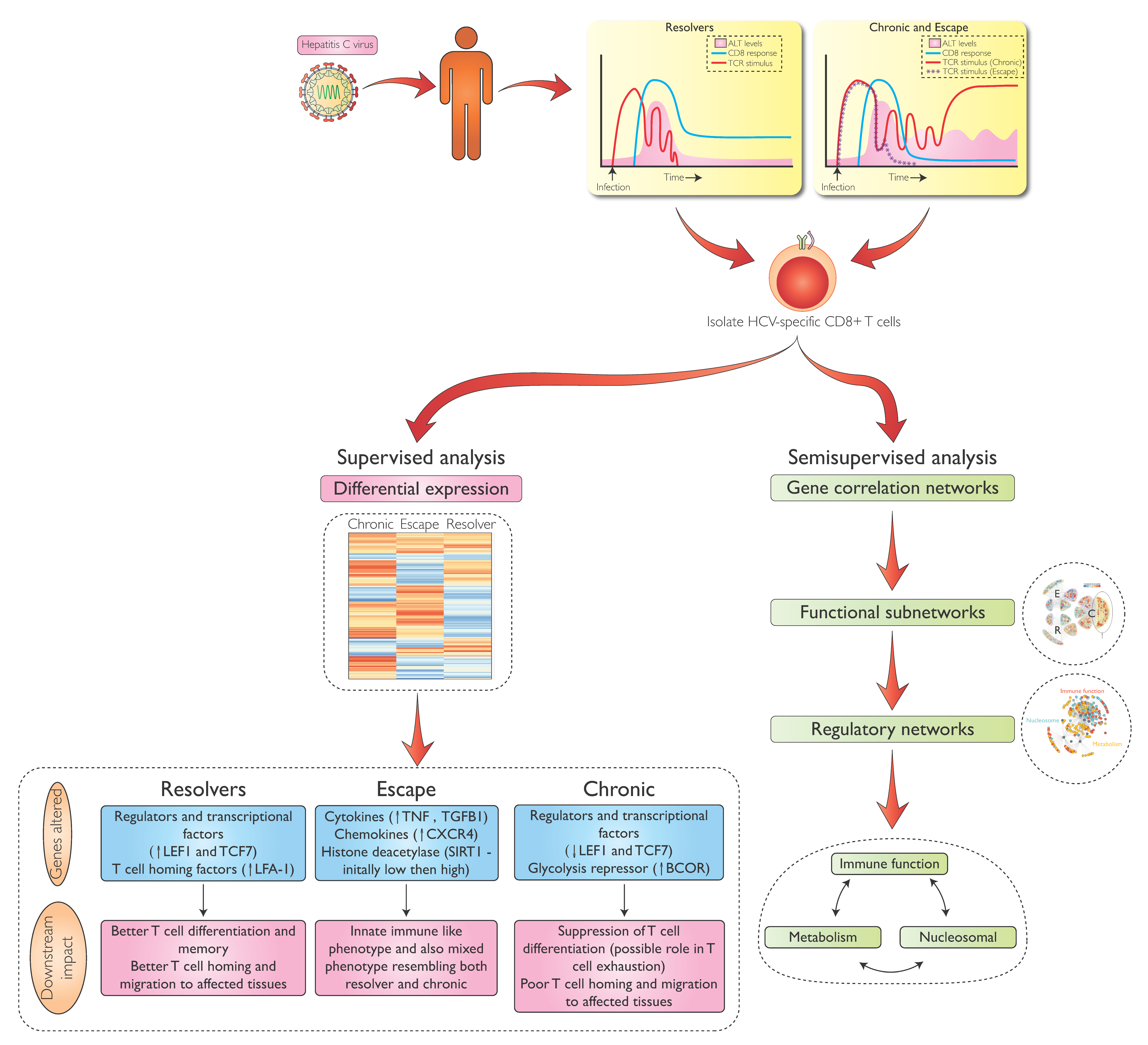
Liver Center at the Gastrointestinal Unit, Department of Medicine, Massachusetts General Hospital and Harvard Medical School, Boston, MA 02114, USA
*
Author to whom correspondence should be addressed.
Viruses 2019, 11(8), 683; https://doi.org/10.3390/v11080683
Submission received: 27 June 2019 / Revised: 21 July 2019 / Accepted: 23 July 2019 / Published: 26 July 2019

Abstract

:
The hepatitis C virus is unique among chronic viral infections in that an acute outcome with complete viral elimination is observed in a minority of infected patients. This unique feature allows direct comparison of successful immune responses with those that fail in the setting of the same human infection. Here we review how this scenario can be used to achieve better understanding of transcriptional regulation of T-cell differentiation. Specifically, we discuss results from a study comparing transcriptional profiles of hepatitis C virus (HCV)-specific CD8 T-cells during early HCV infection between patients that do and do not control and eliminate HCV. Identification of early gene expression differences in key T-cell differentiation molecules as well as clearly distinct transcriptional networks related to cell metabolism and nucleosomal regulation reveal novel insights into the development of exhausted and memory T-cells. With additional transcriptional studies of HCV-specific CD4 and CD8 T-cells in different stages of infection currently underway, we expect HCV infection to become a valuable model disease to study human immunity to viruses.

1. Introduction

Infection with hepatitis C virus (HCV) is unique among chronic viral infections in humans because a significant proportion of newly infected persons (~20%–30%) can completely eliminate the virus, typically within six months of exposure [1]. This observation supports the prospect of an HCV vaccine that can at least prevent chronicity and its harmful sequelae, such as end-stage liver disease and cancer, if sterilizing immunity is not feasible. It also makes the first six months of HCV infection a powerful and one-of-a-kind human model system to define and directly compare immune responses that do and do not control infection within the same basic parameters.
In addition to the dichotomous outcome of HCV infection, recent introduction of direct-acting antiviral drugs (DAA) has created another unique feature to utilize HCV infection as a human paradigm [2]. As the only chronic viral infection in both humans and animals that can be cured through a highly specific targeted small molecule intervention, we can now study the effects of removing the root cause of immune exhaustion (i.e., chronic antigenic stimulation) from the host-pathogen interaction.
Despite these unique opportunities for basic investigations into human immunology, our knowledge base regarding molecular regulation of successful and failing immunity against HCV remains limited. This is largely due to technical challenges, such as extremely small populations of HCV-specific T-cells in the blood [3] due to compartmentalization of activated immune cells to the site of infection, the liver [4]. However, several recent technological breakthroughs now enable analysis of HCV-specific immunity with unprecedented depth and comprehensiveness. Therefore, HCV infection could be a model human disease to understand key differences between protective and failing immunity.

2. T Cells and the Outcome of HCV Infection

Both HCV-specific CD4 and CD8 T-cells are indispensable to control HCV infection. In chimpanzees, depletion of CD4 T-cells before re-infection after previous spontaneous clearance of HCV results in low-level persistence of viremia and eventual selection of viral mutations, leading to escape from the CD8 response [5]. When CD8 T-cells are deleted, HCV replication is not even partially controlled until the depletion wanes off and HCV-specific CD8 T-cells re-emerge [6]. This strong experimental evidence from an animal model of HCV infection, which has since been abandoned for ethical reasons [7], is further corroborated by studies in humans early after HCV exposure [8,9].
Such studies widely agree that a chronic outcome of infection is directly linked to an early defect in the HCV-specific CD4 response, as proliferative responses to HCV proteins are almost universally absent even during the earliest phase of the CD4 response in patients with persisting viremia [9,10]. However, this is not based on a lack of HCV-specific CD4 T-cells, which can typically be detected at least temporarily in all subjects using function-independent assays, such as HLA class II tetramers [10]. Once chronic viremia is established, HCV-specific CD4-T cells are often completely absent or reduced to extremely small populations that are barely detectable even with the most sensitive approaches [11].
In the CD8 response, HCV-specific CD8 T-cells are also detectable in most subjects early on [12], typically at higher frequency than their CD4 counterparts [13], and their appearance in early infection often coincides with some level of control of HCV viremia [8]. After a phase that can last several months—in which virus and CD8 T-cells seem to be engaged in a continuous tug of war—there is either full and sustained control of viremia or persistent viremia with high levels of viral replication. In the latter scenario, there are signs of progressive T-cell exhaustion on HCV-specific T-cells [14] as well as the appearance of viral escape mutations in CD8 epitope regions that lead to diminished recognition of the new circulating quasispecies [15,16]. Which of these two viral escape mechanisms initiates earlier or is more important for viral persistence and whether they are a cause or consequence of viral persistence are currently unknown.

3. Hurdles to Deeper Analysis of HCV-Specific T-Cells

While HCV infection presents unique opportunities to study T cell responses in different disease outcomes in humans, there are also significant hurdles to analyze HCV-specific CD4 and CD8 T-cells—the most important being low frequency of these populations in the blood. Especially in chronic infection, very few specificities can usually be detected directly ex vivo, and even the most dominant HCV-specific CD8 response rarely exceeds 0.1% of peripheral CD8 T-cell populations, with most populations much lower in frequency [12,17]. This is in stark contrast to other chronic viral infections, such as human immunodeficiency virus (HIV), Epstein-Barr virus (EBV), and cytomegalo virus (CMV), in which typically many robust responses can be identified that together represent >10% of the total CD8 T-cell pool [18].
Given that until recently even the most advanced technologies to study cellular regulation on the molecular level (e.g., microarrays, assays for transposase-accessible chromatin using sequencing ATAC-seq) required thousands or more cells to deliver robust high-quality results, there was limited opportunity to take advantage of the explanatory potential of the HCV infection model. However, more powerful next-generation sequencing technologies now allow studies on the single cell level, and HCV investigators have learned to use magnetic bead enrichment of tetramer-binding HCV-specific T-cells for isolation of even the smallest T-cell populations [19]. This now allows analysis of HCV-specific CD8 and, in a subgroup of patients, CD4 T-cells during most stages of infection, including chronicity.

4. Transcriptional Analysis of HCV-Specific CD8 T-Cells During Early HCV Infection

Based on the interest in determining correlates of both protective and failing immunity and the observation that HCV-specific CD8 T-cells are detectable in most patients during the initial 6 months of infection at relatively high frequencies, we focused our first direct ex-vivo transcriptional analysis on CD8 T-cells during early HCV infection. In a recently published study [20], we analyzed HCV-specific CD8 T-cells for differential expression between groups of patients with resolving infection (Resolver) or chronic infection (see Figure 1). In chronic infection, we further distinguished between T cell populations that target the currently circulating HCV strain (Chronic) versus those in which circulating virus is dominated by viral variants not recognized by isolated HCV-specific CD8 T-cell populations (Escape). This allowed us to analyze HCV-specific CD8 T-cells from the same host that do or do not receive T cell receptor (TCR) signal and thus potentially discriminate between the impact of very specific and prolonged antigenic stimulation and non-specific consequences of a general chronic inflammatory environment in chronic infection.

5. Differential Gene Expression in HCV-Specific CD8 T-cells During Acute and Chronic Infection

During the initial 36 weeks of HCV infection, we identified sets of genes with both similar and differential gene expression between different outcome groups (Resolver, Chronic, Escape), using supervised differential gene expression analysis (Figure 1). Composition of these gene sets changed from the early phase (0–18 weeks) to the subsequent phase (19–36 weeks) of acute infection. For example, cells from all three groups expressed classic markers of T cell activation, such as CD74 and HLA-DRB1, at high levels during the initial phase of infection, followed by later downregulation. This reduced activation of the T cell response, irrespective of outcome, is consistent with the clinical course of persistent HCV infection—inflammation is much less pronounced in the later phase of infection compared to the early acute phase, even in chronic outcomes with continued high viral replication.
In contrast to consistent transcriptional patterns across different outcomes, we identified sets of genes that were predominantly up- or downregulated in one group compared to the others. For example, Escape samples had much higher expression of the cytokines tumor necrosis factor α (TNF) and transforming growth factor β1 (TGFB1), their respective downstream signaling components nuclear factor NF-kappa-B p105 subunit (NFKB1), jun-B (JUNB), krueppel-like factor 10 (KLF10), and mothers against decapentaplegic homolog 7 (SMAD7), as well as the chemokine receptors C-X-C chemokine receptor type 4 (CXCR4) and C-C chemokine receptor type 7 (CCR7), compared to lower expression levels in both Chronic and Resolver groups. All of these functionally related genes continued to be expressed in Escape samples at later stages of infection. Because TNF and TGFB1 signaling pathways are not normally associated with classical CD8 T-cells, our interpretation of this data is that T cells exposed to a chronic inflammatory environment without concomitant TCR stimulation adopt a more innate-like immune phenotype, a phenomenon previously described in the context of tumor immunology and hepatitis A [21,22,23]. Inclusion of Escape samples therefore adds an additional dimension to the complexity of T cell differentiation, especially given the observation of similar phenotypes in cancer, another disease in which T-cell exhaustion and dysregulation play a major role in disease pathogenesis. Differences in directly antigen-mediated and antigen-independent effects on CD8 T-cell regulation and differentiation warrant more detailed investigation.
The most important differences between outcome groups were for key genes linked to cellular differentiation programs related to T cell memory. In patients with acute resolving HCV infection, CD8 T-cells had early high expression of regulators of T cell differentiation and memory, TCF7 and its transcriptional target LEF1. Both TCF7 and LEF1 also possess intrinsic histone deacetylase (HDAC) activity, which is thought to be functionally crucial to their control of T cell lineage commitment [24]. Both TCF7 and LEF1 are expressed at low levels in Chronic samples throughout early and late acute phases of infection, suggesting that T cell differentiation might be negatively impacted very early during chronic infection. Interestingly, Escape samples exhibit a mixed expression profile for these genes, sharing higher levels of TCF7 expression similar to Resolver samples and lower levels of LEF1 expression similar to Chronic samples, suggesting that they develop a semi-differentiated phenotype that could feature entirely distinct histone acetylation patterns from both effector and memory T-cells.
For other genes, Escape samples initially shared features with one of the other groups during early infection, but later switched to a phenotype more similar to the other group. One example is the histone deacetylase SIRT1, for which Escape samples shared early low expression with Resolver samples and later expressed higher levels as in Chronic samples. This further substantiates the hypothesis that histone acetylation patterns in Escape T cells differ substantially from their antigen-cognizant counterparts in chronic infection. The deacetylase SIRT1 is of special interest, as it is an important epigenetic modifier that acts as a repressor for the TBX21 (T-bet) locus, a key transcription factor in T cell differentiation—making it an attractive target for immunotherapeutic intervention [25]. There are already small molecules under investigation that can reduce expression of another important T-cell transcription factor, BATF, while at the same time increasing SIRT1-mediated repression of TBX21 [26], resulting in increased Tscm and Tcm populations [26]. In addition to an anti-inflammatory role through regulation of TBX21, SIRT1 also plays an important role in regulation of mammalian metabolism, where it promotes fatty acid oxidation and gluconeogenesis [27].
Like SIRT1, a variety of other important metabolic regulators were expressed at consistently higher levels in Chronic samples throughout early and late acute infection, including TBK1 and BCOR. TBK1 facilitates induction of glycolytic enzymes as part of HIF1A’s positive regulation of glycolysis, which is particularly important in hypoxic conditions such as those encountered in the liver microenvironment. HIF1A itself is induced by TCR and IL-2 signaling and works to sustain Myc-dependent metabolic reprogramming [28,29] in response to TCR stimulation [28], which, in turn, is crucial to establish robust proliferation [30].
Interestingly, we also observed high expression levels of BCOR in Chronic and Escape samples. Unlike TBK1, the Bcl-6 co-repressor BCOR represses transporters and enzymes in glycolysis, dampening the metabolic program associated with effector differentiation [31,32,33,34]. This Bcl-6-dependent dampening is antagonized by T-bet, awarding this important T-cell transcription factor—which is required for specialization of Th1 cells by activating IFNG and CXCR3 expression in a DNA-binding-dependent manner [35,36,37,38]—an important role in promoting glycolysis [32,33]. However, neither Bcl-6 nor T-bet were differentially expressed in our data set, suggesting that this specific pathway of regulating T-cell glycolysis might not be a key differentiating factor in HCV infection. However, we cannot rule out the possibility that another binding partner for BCOR regulates HCV-specific T-cells, as several other such binding partners have been described in the literature recently [39,40].

6. Metabolic Regulation in HCV-Specific CD8 T-Cells

While the standard differential gene expression analysis we employed in our recently published study identified several key differences between Acute and Chronic (and Escape) CD8 T-cell populations in genes linked to established processes in T-cell differentiation [20], supervised analytic approaches have clear limitations. This is especially true for complex cross-sectional human cohort studies that compare more than two groups across multiple conditions/timeframes, given heterogeneity in both genetic backgrounds and disease and sample variability. Such studies typically benefit from more complex semi-/unsupervised analytic approaches capable of identifying data patterns that cannot be known beforehand or that are masked by the chosen comparison groups.
Therefore, we further investigated CD8 transcriptional profiles using a semi-supervised weighted gene correlation network analysis approach (WGCNA) (see Figure 1). We combined early and late samples while preserving the overall sample group structure (Chronic, Escape, Resolver) to identify clusters (also called modules) of genes with highly correlated expression patterns across samples within each group. WGCNA is based on the assumption that genes with highly correlated expression patterns across samples that share a particular condition (e.g., Chronic HCV) represent genes with a common regulator (i.e., are co-regulated) in that condition. Identifying all such clusters of co-regulated genes for a particular condition forms a condition-specific module network, which can then be checked for module preservation across conditions and correlations of module-specific patterns with other sample traits (e.g., clinical parameters) [41].
By analyzing all three sample groups individually, we identified one specific set of genes related to metabolic pathways that were exclusively co-regulated in Chronic patients and associated mostly with two highly interconnected gene modules. Expression of genes within these modules was also strongly (negatively) correlated with time from infection, meaning that gene expression decreased with duration of viral persistence. Additional significant correlations were observed with patient age and sex, as well as a detectable presence of HCV-specific CD4 T-cells, with higher expression of genes in the metabolic signature observed in younger age, females, and presence of strong HCV-specific CD4 T-cell responses. As these patient traits are strongly associated with a higher likelihood of HCV resolution, this gene signature has potential to identify mechanistic links between sex, age, and T help and HCV control.
This Chronic-exclusive metabolic gene signature, which was marked by genes with high initial expression followed by rapid downregulation, was significantly enriched for genes related to oxidative phosphorylation. Downregulation of genes related to oxidative phosphorylation during early HCV infection seems paradoxical when considering that the electron transport chain presents a highly efficient means of energy production for a cell. However, activated and effector T-cells in general highly favor glycolysis as a means of energy production, while memory T-cells are more dependent on oxidative phosphorylation (OXPHOS) to meet their energetic demands [42,43]. Therefore, the observed downregulation in OXPHOS genes along with higher expression of glycolysis-promoting genes, such as TBK1, in the Chronic group suggests that perpetual activation might prevent these cells from activating a metabolic program that favors differentiation into memory cells.
Similar patterns of metabolic dysregulation in virus-specific CD8 T-cells also have been described in other human infections, such as HIV and, more recently, HBV as well as for lymphocytic choriomeningitis virus (LCMV) infection in mice [44,45,46]. While dysregulation signatures in both LCMV and HBV center around OXPHOS, the HBV signature is even closer to that in HCV, as the mouse signature also contained a strong signal of perturbed glycolysis not present in HBV or HCV. These results suggest that central features of CD8 T-cell metabolic dysregulation are shared across different models of T-cell exhaustion, with some variation in patterns and specific pathways of dysregulation that are most likely dependent on factors such as host species, infectious agent, and viral tropism.

7. Transcriptional Regulation of HCV-Specific CD8 T-Cells Beyond Metabolism

After identifying a distinct Chronic-exclusive signature of metabolic dysregulation, we examined underlying transcriptional regulators that might drive this change in metabolic programming. We hypothesized that transcriptional regulators would likely be annotated in the same modules as their targets if expression of the former linearly affected expression of the latter. Thus, we assembled information on known transcription factor-target interactions from multiple databases and intersected them with our weighted gene correlation network. In this way, we could estimate transcription factor-target coverage for each module (i.e., the number of interactions any given transcription factor had with genes annotated in the same module). Based on this information, we used the most highly connected transcription factors for each module of interest as a seed for a regulatory network, allowing connections between transcriptional regulators and targets not just within but also across closely connected modules to more realistically represent a functional regulatory network.
This approach resulted in a concise network of regulatory interactions that was largely Chronic-exclusive and provided links between transcriptional regulators and most genes in our metabolic dysregulation signature. However, the approach also extended interactions to genes more directly related to immune function and genes crucial to nucleosomal regulation of transcription that shared similar expression kinetics with drastically reduced expression as chronic infection progressed. The regulatory genes we identified to be the most likely drivers of these early transcriptional changes in chronic samples, which we used to seed the regulatory network, included various transcription factors, co-factors, and chromatin remodeling genes, including CBX3, CREB1, FLI1, KDM5B, NELFE, RFX5, STAT1, STAT5A, and TP53.
The seed for the regulatory network also included IRF3 and FLI1, which were not annotated in the chronic gene modules of interest but were co-regulated with CREB1, STAT1, and TP53 in a closely related Resolver module. These were introduced to allow the possibility that absence of certain regulators might be partly responsible for observed dysregulation in Chronic samples. While some of these genes are known transcriptional regulators of immune function, including STAT1 and STAT5A, which are associated with effective IFN responses and T-cell memory differentiation [47], involvement in metabolic regulation is a less expected observation. For other genes, such as TP53, the opposite is true. TP53 has a well-documented role in regulation of metabolism [48], but its immune-regulatory role has only recently been appreciated [49].
When examining the inferred transcriptional targets for these transcription factors in chronic HCV, we encountered not only most metabolic genes in the identified metabolic dysregulation signature, but also a wide range of genes related to nucleosomal regulation of transcription and chromatin modification. This included 21 genes coding for various histone subunits as well as many highly immune-relevant genes, such as AKT1, CCL4, CCL5, CCR5, CD14, CD27, CD38, CD244, CX3CR1, ICAM1, ITGAL, ITGB2, ITGB7, and MYD88. This further underlines the intersection between metabolic and nucleosomal regulation that was already suggested by some findings in the supervised gene expression analysis.
The reconstructed network also contained genes crucial to T-cell homing, migration, and adhesion. Among them was ICAM1, an important facilitator of intercellular adhesion, as well as its binding partner duo ITGAL and ITGB2, which together make up LFA-1. This is interesting because LFA-1 is not only a crucial regulator of T cells leaving the bloodstream and entering infected tissues but also has recently been reported to be an important factor in fine-tuning T cell effector function [50]. Another interesting finding in this context is that the Chronic network also featured ITGB7, but not its counterpart ITGA4. Both genes, which together form another important T-cell homing complex, were present in the Resolver network, where ITGB2 was missing. Together this suggests that T cells in Chronic and Resolver samples might be subject to vastly different cell migration cues that affect their ability to successfully fight infection. Finally, the Chronic network also contained CX3CR1, another important lymphocyte homing factor whose presence and absence are indicative of effector and central memory T-cells, respectively [51]—again suggesting that rapid downregulation of CX3CR1 might indicate these cells receive improper homing signals and fail to differentiate into a more long-lived anti-inflammatory phenotype.
Together, these data suggest that CD8 T-cells in Chronic HCV undergo rapid dysregulation of genes that are crucial for orchestration and direction of adaptive T-cell responses by impacting T-cell function, differentiation, and migration. Further, this dysregulation can be linked to known clinical predictors of HCV disease outcome and seems to be rooted in either simultaneous or rapidly sequential disruption of three integrally linked functional domains: immune function, metabolism, and nucleosomal regulation of transcriptional processes. While the exact order of regulatory events in early chronic HCV remains to be determined, it is likely to hold valuable information on the causal basis for T-cell exhaustion in HCV infection and in general.
Our study not only identifies pathways that are likely implicated in failure of the CD8 T-cell response to HCV infection but also highlights their high degree of interconnectedness—underlining the importance of tackling complex immunological problems with more holistic and integrative experimental and computational approaches. For example, large parts of cellular identity are encoded in the epigenetic landscape that produces a unique pattern of chromatin accessibility and modification, allowing establishment of different cell types from a shared genetic code. Epigenetic marks are important for both T cell reprogramming in response to antigenic stimulus [52,53,54] as well as their maintenance after stimulus withdrawal [55]. More recent work has further highlighted the existence of transcriptional-epigenetic profiles in the context of T-cell exhaustion in cancer and chronic viral infections as well as their role in establishment and reversibility of the exhausted phenotype [56,57,58,59,60]. In this context, it is important to note that T-cell exhaustion is a process that can be driven both by terminal differentiation of cells as well as by strong and prolonged antigen stimulation [61,62,63]. Further, exhausted cells exhibit profound heterogeneity with regard to their proliferative capacity [64,65,66,67,68,69], which might influence their capacity for exhaustion reversal.
In addition, it has become evident that metabolites have the capacity to regulate both epigenetic and transcriptional landscapes [70,71,72]. In many cases, metabolites are the substrates used to generate epigenetic modifications. Addition and removal of these modifications are, for the most part, catalyzed by enzymes whose activities are meditated by availability of substrates, cofactors, and allosteric regulators derived from metabolic pathways [73,74,75,76,77,78,79,80,81,82,83,84,85]. The fact that the epigenome is crucial to establish distinct cellular states [86,87,88] and critically involved in immune cell fate [89] makes the intersection and interaction of metabolism and epigenetics an exciting emerging field of study, particularly in the context of immunity.

8. Conclusions

We reviewed the first study utilizing HCV infection as a unique model disease for in-depth analysis of transcriptional regulation in human virus-specific T cells associated with distinct infection outcomes. We are convinced that this study will be followed by many others that will both deepen the findings from CD8 T-cells in early HCV infection as well as extend them to different infection scenarios that can be uniquely studied in HCV infection.
We are currently aware of multiple studies by different investigators using new generation technologies to more mechanistically examine metabolic dysregulation of HCV-specific CD8 T-cells and also analyze T cells before and after therapy. Additional studies will also better characterize HCV-specific CD4 T-cells and help understand different transcriptional states of HCV-specific CD8 T-cells at the site of infection, the liver. We are looking forward to publication of these results, with the expectation that the data will further establish the unique potential of studying human immunology in HCV infection.
Ongoing and future studies will also greatly benefit from constant expansion and refinement of the analytical tools used to understand such complex human data sets, including horizontal integration of distinct but interrelated processes such as metabolism and immune effector function. We will also learn about vertical regulation of T cell processes through epigenetic, transcriptional, translational, and post-translational processes. These insights will help understand cross-talk between T cells and different immune cell types or other components of the host immune response by integrating data from many different emerging studies [90] as well as using unbiased parallel analysis of different immune cells with technologies such as 10x or Seq-Well. There has never been a more exciting time to study HCV immunology.

Funding

This work was supported by NIH U19 AI082630 and U01 AI131314 (both to G.M.L.).

Acknowledgments

We are thankful to Marcos Damasio, Hannah Drescher and Sonu Sobudhi for careful reading of the manuscript.

Conflicts of Interest

The authors declare no conflict of interest.

References

  1. Lauer, G.M.; Walker, B.D. Hepatitis C Virus Infection. N. Engl. J. Med. 2001, 345, 41–52. [Google Scholar] [CrossRef] [PubMed]
  2. Luxenburger, H.; Neumann-Haefelin, C.; Thimme, R. HCV-Specific T Cell Responses During and After Chronic HCV Infection. Viruses 2018, 10, 645. [Google Scholar] [CrossRef] [PubMed]
  3. Lauer, G.M.; Barnes, E.; Lucas, M.; Timm, J.; Ouchi, K.; Kim, A.Y.; Day, C.L.; Robbins, G.K.; Casson, D.R.; Reiser, M.; et al. High resolution analysis of cellular immune responses in resolved and persistent hepatitis C virus infection. Gastroenterology 2004, 127, 924–936. [Google Scholar] [CrossRef] [PubMed]
  4. Wong, D.K.H.; Dudley, D.D.; Afdhal, N.H.; Dienstag, J.; Rice, C.M.; Wang, L.; Houghton, M.; Walker, B.D.; Koziel, M.J. Liver-Derived CTL in Hepatitis C Virus Infection: Breadth and Specificity of Responses in a Cohort of Persons with Chronic Infection. J. Immunol. 1998, 160, 1479–1488. [Google Scholar] [PubMed]
  5. Grakoui, A.; Shoukry, N.H.; Woollard, D.J.; Han, J.-H.; Hanson, H.L.; Ghrayeb, J.; Murthy, K.K.; Rice, C.M.; Walker, C.M. HCV persistence and immune evasion in the absence of memory T cell help. Science 2003, 302, 659–662. [Google Scholar] [CrossRef] [PubMed]
  6. Shoukry, N.H.; Grakoui, A.; Houghton, M.; Chien, D.Y.; Ghrayeb, J.; Reimann, K.A.; Walker, C.M. Memory CD8+ T Cells Are Required for Protection from Persistent Hepatitis C Virus Infection. J. Exp. Med. 2003, 197, 1645–1655. [Google Scholar] [CrossRef] [PubMed]
  7. Lanford, R.E.; Walker, C.M.; Lemon, S.M. The Chimpanzee Model of Viral Hepatitis: Advances in Understanding the Immune Response and Treatment of Viral Hepatitis. ILAR J. 2017, 58, 172–189. [Google Scholar] [CrossRef] [PubMed]
  8. Thimme, R.; Oldach, D.; Chang, K.-M.; Steiger, C.; Ray, S.C.; Chisari, F.V. Determinants of Viral Clearance and Persistence during Acute Hepatitis C Virus Infection. J. Exp. Med. 2001, 194, 1395–1406. [Google Scholar] [CrossRef]
  9. Gerlach, J.T.; Diepolder, H.M.; Jung, M.C.; Gruener, N.H.; Schraut, W.W.; Zachoval, R.; Hoffmann, R.; Schirren, C.A.; Santantonio, T.; Pape, G.R. Recurrence of hepatitis C virus after loss of virus-specific CD4(+) T-cell response in acute hepatitis C. Gastroenterology 1999, 117, 933–941. [Google Scholar] [CrossRef]
  10. Schulze zur Wiesch, J.; Ciuffreda, D.; Lewis-Ximenez, L.; Kasprowicz, V.; Nolan, B.E.; Streeck, H.; Aneja, J.; Reyor, L.L.; Allen, T.M.; Lohse, A.W.; et al. Broadly directed virus-specific CD4+ T cell responses are primed during acute hepatitis C infection, but rapidly disappear from human blood with viral persistence. J. Exp. Med. 2012, 209, 61–75. [Google Scholar] [CrossRef]
  11. Schulze zur Wiesch, J.; Lauer, G.M.; Day, C.L.; Kim, A.Y.; Ouchi, K.; Duncan, J.E.; Wurcel, A.G.; Timm, J.; Jones, A.M.; Mothe, B.; et al. Broad Repertoire of the CD4+ Th Cell Response in Spontaneously Controlled Hepatitis C Virus Infection Includes Dominant and Highly Promiscuous Epitopes. J. Immunol. 2005, 175, 3603–3613. [Google Scholar] [CrossRef] [PubMed]
  12. Cox, A.L.; Mosbruger, T.; Lauer, G.M.; Pardoll, D.; Thomas, D.L.; Ray, S.C. Comprehensive analyses of CD8+ T cell responses during longitudinal study of acute human hepatitis C. Hepatology 2005, 42, 104–112. [Google Scholar] [CrossRef] [PubMed]
  13. Kasprowicz, V.; Schulze zur Wiesch, J.; Kuntzen, T.; Nolan, B.E.; Longworth, S.; Berical, A.; Blum, J.; McMahon, C.; Reyor, L.L.; Elias, N.; et al. High Level of PD-1 Expression on Hepatitis C Virus (HCV)-Specific CD8+ and CD4+ T Cells during Acute HCV Infection, Irrespective of Clinical Outcome. J. Virol. 2008, 82, 3154–3160. [Google Scholar] [CrossRef] [PubMed]
  14. Urbani, S.; Amadei, B.; Tola, D.; Massari, M.; Schivazappa, S.; Missale, G.; Ferrari, C. PD-1 Expression in Acute Hepatitis C Virus (HCV) Infection Is Associated with HCV-Specific CD8 Exhaustion. J. Virol. 2006, 80, 11398–11403. [Google Scholar] [CrossRef] [PubMed] [Green Version]
  15. Timm, J.; Lauer, G.M.; Kavanagh, D.G.; Sheridan, I.; Kim, A.Y.; Lucas, M.; Pillay, T.; Ouchi, K.; Reyor, L.L.; Zur Wiesch, J.S.; et al. CD8 Epitope Escape and Reversion in Acute HCV Infection. J. Exp. Med. 2004, 200, 1593–1604. [Google Scholar] [CrossRef] [PubMed]
  16. Cox, A.L.; Mosbruger, T.; Mao, Q.; Liu, Z.; Wang, X.-H.; Yang, H.-C.; Sidney, J.; Sette, A.; Pardoll, D.; Thomas, D.L.; et al. Cellular immune selection with hepatitis C virus persistence in humans. J. Exp. Med. 2005, 201, 1741–1752. [Google Scholar] [CrossRef]
  17. Lauer, G.M.; Lucas, M.; Timm, J.; Ouchi, K.; Kim, A.Y.; Day, C.L.; zur Wiesch, J.S.; Paranhos-Baccala, G.; Sheridan, I.; Casson, D.R.; et al. Full-Breadth Analysis of CD8+ T-Cell Responses in Acute Hepatitis C Virus Infection and Early Therapy. J. Virol. 2005, 79, 12979–12988. [Google Scholar] [CrossRef]
  18. Van Leeuwen, E.M.M.; Koning, J.J.; Remmerswaal, E.B.M.; van Baarle, D.; van Lier, R.A.W.; ten Berge, I.J.M. Differential Usage of Cellular Niches by Cytomegalovirus versus EBV- and Influenza Virus-Specific CD8+ T Cells. J. Immunol. 2006, 177, 4998–5005. [Google Scholar] [CrossRef]
  19. Nitschke, K.; Flecken, T.; Schmidt, J.; Gostick, E.; Marget, M.; Neumann-Haefelin, C.; Blum, H.E.; Price, D.A.; Thimme, R.; Diamond, M.S. Tetramer Enrichment Reveals the Presence of Phenotypically Diverse Hepatitis C Virus-Specific CD8+ T Cells in Chronic Infection. J. Virol. 2015, 89, 25–34. [Google Scholar] [CrossRef]
  20. Wolski, D.; Foote, P.K.; Chen, D.Y.; Lewis-Ximenez, L.L.; Fauvelle, C.; Aneja, J.; Walker, A.; Tonnerre, P.; Torres-Cornejo, A.; Kvistad, D.; et al. Early Transcriptional Divergence Marks Virus-Specific Primary Human CD8+ T Cells in Chronic versus Acute Infection. Immunity 2017, 47, 648–663. [Google Scholar] [CrossRef]
  21. Kim, J.; Chang, D.-Y.; Lee, H.W.; Lee, H.; Kim, J.H.; Sung, P.S.; Kim, K.H.; Hong, S.-H.; Kang, W.; Lee, J.; et al. Innate-like Cytotoxic Function of Bystander-Activated CD8+ T Cells Is Associated with Liver Injury in Acute Hepatitis A. Immunity 2018, 48, 161–173. [Google Scholar] [CrossRef] [PubMed]
  22. Barbarin, A.; Cayssials, E.; Jacomet, F.; Nunez, N.G.; Basbous, S.; Lefèvre, L.; Abdallah, M.; Piccirilli, N.; Morin, B.; Lavoue, V.; et al. Phenotype of NK-Like CD8(+) T Cells with Innate Features in Humans and Their Relevance in Cancer Diseases. Front. Immunol. 2017, 8, 1114. [Google Scholar] [CrossRef] [PubMed]
  23. Seyda, M.; Elkhal, A.; Quante, M.; Falk, C.S.; Tullius, S.G. T Cells Going Innate. Trends Immunol. 2016, 37, 546–556. [Google Scholar] [CrossRef] [PubMed] [Green Version]
  24. Xing, S.; Li, F.; Zeng, Z.; Zhao, Y.; Yu, S.; Shan, Q.; Li, Y.; Phillips, F.C.; Maina, P.K.; Qi, H.H.; et al. Tcf1 and Lef1 transcription factors establish CD8+ T cell identity through intrinsic HDAC activity. Nat. Immunol. 2016, 17, 695–703. [Google Scholar] [CrossRef] [PubMed]
  25. Kuroda, S.; Yamazaki, M.; Abe, M.; Sakimura, K.; Iwai, Y. Basic leucine zipper transcription factor, ATF-like (BATF) regulates epigenetically and energetically effector CD8 T-cell differentiation via Sirt1 expression. Proc. Natl. Acad. Sci. USA 2011, 108, 14885–14889. [Google Scholar] [CrossRef] [PubMed] [Green Version]
  26. Kagoya, Y.; Nakatsugawa, M.; Yamashita, Y.; Ochi, T.; Guo, T.; Anczurowski, M.; Saso, K.; Butler, M.O.; Arrowsmith, C.H. BET bromodomain inhibition enhances T cell persistence and function in adoptive immunotherapy models. J. Clin. Investig. 2016, 126, 3479–3494. [Google Scholar] [CrossRef] [Green Version]
  27. Gillum, M.P.; Erion, D.M.; Shulman, G.I. Sirtuin-1 regulation of mammalian metabolism. Trends Mol. Med. 2011, 17, 8–13. [Google Scholar] [CrossRef] [PubMed] [Green Version]
  28. Wang, R.; Dillon, C.P.; Shi, L.Z.; Milasta, S.; Finkelstein, D.; McCormick, L.L.; Chi, H.; Munger, J.; Green, D.R. The Transcription Factor Myc Controls Metabolic Reprogramming upon T Lymphocyte Activation. Immunity 2011, 35, 871–882. [Google Scholar] [CrossRef] [Green Version]
  29. Finlay, D.K.; Rosenzweig, E.; Sinclair, L.V.; Feijoo-Carnero, C.; Hukelmann, J.L.; Rolf, J.; Panteleyev, A.A.; Okkenhaug, K.; Cantrell, D.A. PDK1 regulation of mTOR and hypoxia-inducible factor 1 integrate metabolism and migration of CD8+ T cells. J. Exp. Med. 2012, 209, 2441–2453. [Google Scholar] [CrossRef] [Green Version]
  30. Boroughs, L.K. Metabolic pathways promoting cancer cell survival and growth. Nat. Cell Biol. 2015, 17, 351–359. [Google Scholar] [CrossRef] [Green Version]
  31. Ballesteros-Tato, A.; León, B.; Graf, B.A.; Moquin, A.; Adams, P.S.; Lund, F.E.; Randall, T.D. Interleukin-2 Inhibits Germinal Center Formation by Limiting T Follicular Helper Cell Differentiation. Immunity 2012, 36, 847–856. [Google Scholar] [CrossRef] [PubMed] [Green Version]
  32. Oestreich, K.J.; Mohn, S.E.; Weinmann, A.S. Molecular mechanisms that control the expression and activity of Bcl-6 in TH1 cells to regulate flexibility with a TFH like gene profile. Nat. Immunol. 2012, 13, 405–411. [Google Scholar] [CrossRef] [PubMed]
  33. Oestreich, K.J.; Read, K.A.; Gilbertson, S.E.; Hough, K.P.; McDonald, P.W.; Krishnamoorthy, V.; Weinmann, A.S. Bcl-6 directly represses the gene program of the glycolysis pathway. Nat. Immunol. 2014, 15, 957–964. [Google Scholar] [CrossRef] [PubMed]
  34. Pipkin, M.E.; Sacks, J.A.; Cruz-Guilloty, F.; Lichtenheld, M.G.; Bevan, M.J.; Rao, A. Interleukin-2 and Inflammation Induce Distinct Transcriptional Programs that Promote the Differentiation of Effector Cytolytic T Cells. Immunity 2010, 32, 79–90. [Google Scholar] [CrossRef] [PubMed] [Green Version]
  35. Beima, K.M.; Miazgowicz, M.M.; Lewis, M.D.; Yan, P.S.; Huang, T.H.-M.; Weinmann, A.S. T-bet binding to newly identified target gene promoters is cell type-independent but results in variable context-dependent functional effects. J. Biol. Chem. 2006, 281, 11992–12000. [Google Scholar] [CrossRef] [PubMed]
  36. Miller, S.A.; Huang, A.C.; Miazgowicz, M.M.; Brassil, M.M.; Weinmann, A.S. Coordinated but physically separable interaction with H3K27-demethylase and H3K4-methyltransferase activities are required for T-box protein-mediated activation of developmental gene expression. Genes Dev. 2008, 22, 2980–2993. [Google Scholar] [CrossRef] [PubMed] [Green Version]
  37. Kim, S.T.; Costa, G.L.; Zhang, X.; Fathman, C.G.; Glimcher, L.H. A Novel Transcription Factor, T-bet, Directs Th1 Lineage Commitment. Cell 2000, 100, 655–669. [Google Scholar] [Green Version]
  38. Sullivan, B.M.; Stemmann, C.; Satoskar, A.R.; Sleckman, B.P.; Glimcher, L.H. Distinct Effects of T-bet in TH1 Lineage Commitment and IFN-γ Production in CD4 and CD8 T Cells. Science 2002, 295, 338–342. [Google Scholar]
  39. Yoon, J.; Feng, X.; Kim, Y.-S.; Shin, D.-M.; Hatzi, K.; Wang, H.; Morse, H.C. Interferon regulatory factor 8 (IRF8) interacts with the B cell lymphoma 6 (BCL6) corepressor BCOR. J. Biol. Chem. 2014, 289, 34250–34257. [Google Scholar] [CrossRef]
  40. Yamamoto, Y.; Tsuzuki, S.; Tsuzuki, M.; Handa, K.; Inaguma, Y.; Emi, N. BCOR as a novel fusion partner of retinoic acid receptor alpha in a t(X;17)(p11;q12) variant of acute promyelocytic leukemia. Blood 2010, 116, 4274–4283. [Google Scholar] [CrossRef] [Green Version]
  41. Langfelder, P.; Horvath, S. WGCNA: An R package for weighted correlation network analysis. BMC Bioinform. 2008, 9, 559. [Google Scholar] [CrossRef]
  42. Van der Windt, G.J.W.; Everts, B.; Chang, C.-H.; Freitas, T.C.; Pearce, E.J.; Pearce, E.L. Mitochondrial respiratory capacity is a critical regulator of CD8+ T cell memory development. Immunity 2012, 36, 68–78. [Google Scholar] [CrossRef]
  43. Van der Windt, G.J.W.; O’Sullivan, D.; Everts, B.; Huang, S.C.-C.; Buck, M.D.; Chang, C.-H.; Smith, A.M.; Ai, T.; Faubert, B.; Jones, R.G.; et al. CD8 memory T cells have a bioenergetic advantage that underlies their rapid recall ability. Proc. Natl. Acad. Sci. USA 2013, 110, 14336–14341. [Google Scholar] [CrossRef] [Green Version]
  44. Trautmann, L.; Mbitikon-Kobo, F.-M.; Goulet, J.-P.; Peretz, Y.; Shi, Y.; Van Grevenynghe, J.; Procopio, F.A.; Boulassel, M.R.; Routy, J.-P.; Chomont, N.; et al. Profound metabolic, functional, and cytolytic differences characterize HIV-specific CD8 T cells in primary and chronic HIV infection. Blood 2012, 120, 3466–3477. [Google Scholar] [CrossRef]
  45. Bengsch, B.; Johnson, A.L.; Kurachi, M.; Odorizzi, P.M.; Pauken, K.E.; Attanasio, J.; Stelekati, E.; McLane, L.M.; Paley, M.A.; Delgoffe, G.M.; et al. Bioenergetic Insufficiencies Due to Metabolic Alterations Regulated by the Inhibitory Receptor PD-1 Are an Early Driver of CD8+ T Cell Exhaustion. Immunity 2016, 45, 358–373. [Google Scholar] [CrossRef]
  46. Fisicaro, P.; Barili, V.; Montanini, B.; Acerbi, G.; Ferracin, M.; Guerrieri, F.; Salerno, D.; Boni, C.; Massari, M.; Cavallo, M.C.; et al. Targeting mitochondrial dysfunction can restore antiviral activity of exhausted HBV-specific CD8 T cells in chronic hepatitis B. Nat. Med. 2017, 57, 167–336. [Google Scholar] [CrossRef]
  47. O’Shea, J.J.; Plenge, R. JAK and STAT Signaling Molecules in Immunoregulation and Immune-Mediated Disease. Immunity 2012, 36, 542–550. [Google Scholar] [CrossRef] [Green Version]
  48. Berkers, C.R.; Maddocks, O.D.K.; Cheung, E.C.; Mor, I.; Vousden, K.H. Metabolic regulation by p53 family members. Cell Metab. 2013, 18, 617–633. [Google Scholar] [CrossRef]
  49. Muñoz-Fontela, C.; Mandinova, A.; Aaronson, S.A.; Lee, S.W. Emerging roles of p53 and other tumour-suppressor genes in immune regulation. Nat. Rev. Immunol. 2016, 16, 741–750. [Google Scholar] [CrossRef] [Green Version]
  50. Verma, N.K.; Kelleher, D. Not Just an Adhesion Molecule: LFA-1 Contact Tunes the T Lymphocyte Program. J. Immunol. 2017, 199, 1213–1221. [Google Scholar] [CrossRef] [Green Version]
  51. Gerlach, C.; Moseman, E.A.; Loughhead, S.M.; Alvarez, D.; Zwijnenburg, A.J.; Waanders, L.; Garg, R.; de la Torre, J.C.; von Andrian, U.H. The Chemokine Receptor CX3CR1 Defines Three Antigen-Experienced CD8 T Cell Subsets with Distinct Roles in Immune Surveillance and Homeostasis. Immunity 2016, 45, 1270–1284. [Google Scholar] [CrossRef] [PubMed] [Green Version]
  52. Crompton, J.G.; Clever, D.; Vizcardo, R.; Rao, M.; Restifo, N.P. Reprogramming antitumor immunity. Trends Immunol. 2014, 35, 178–185. [Google Scholar] [CrossRef] [PubMed] [Green Version]
  53. Takahashi, K.; Yamanaka, S. Induction of pluripotent stem cells from mouse embryonic and adult fibroblast cultures by defined factors. Cell 2006, 126, 663–676. [Google Scholar] [CrossRef] [PubMed]
  54. Themeli, M.; Rivière, I.; Sadelain, M. New cell sources for T cell engineering and adoptive immunotherapy. Cell Stem Cell 2015, 16, 357–366. [Google Scholar] [CrossRef] [PubMed]
  55. Abdelsamed, H.A.; Moustaki, A.; Fan, Y.; Dogra, P.; Ghoneim, H.E.; Zebley, C.C.; Triplett, B.M.; Sekaly, R.-P.; Youngblood, B. Human memory CD8 T cell effector potential is epigenetically preserved during in vivo homeostasis. J. Exp. Med. 2017, 214, 1593–1606. [Google Scholar] [CrossRef] [PubMed]
  56. Doering, T.A.; Crawford, A.; Angelosanto, J.M.; Paley, M.A.; Ziegler, C.G.; Wherry, E.J. Network Analysis Reveals Centrally Connected Genes and Pathways Involved in CD8+ T Cell Exhaustion versus Memory. Immunity 2012, 37, 1130–1144. [Google Scholar] [CrossRef] [PubMed] [Green Version]
  57. Pauken, K.E.; Sammons, M.A.; Odorizzi, P.M.; Manne, S.; Godec, J.; Khan, O.; Drake, A.M.; Chen, Z.; Kurachi, M.; Barnitz, R.A.; et al. Epigenetic stability of exhausted T cells limits durability of reinvigoration by PD-1 blockade. Science 2016, 354, 1160–1165. [Google Scholar] [CrossRef] [PubMed] [Green Version]
  58. Sen, D.R.; Kaminski, J.; Barnitz, R.A.; Kurachi, M.; Gerdemann, U.; Yates, K.B.; Tsao, H.W.; Godec, J.; LaFleur, M.W.; Brown, F.D.; et al. The epigenetic landscape of T cell exhaustion. Science 2016, 354, 1165–1169. [Google Scholar] [CrossRef] [PubMed] [Green Version]
  59. Scott-Browne, J.P.; López-Moyado, I.F.; Trifari, S.; Wong, V.; Chavez, L.; Rao, A.; Pereira, R.M. Dynamic Changes in Chromatin Accessibility Occur in CD8+ T Cells Responding to Viral Infection. Immunity 2016, 45, 1327–1340. [Google Scholar] [CrossRef]
  60. Shin, H.; Wherry, E.J. CD8 T cell dysfunction during chronic viral infection. Curr. Opin. Immunol. 2007, 19, 408–415. [Google Scholar] [CrossRef]
  61. Gallimore, A.; Gallimore, A.; Glithero, A.; Glithero, A.; Godkin, A.; Godkin, A.; Tissot, A.C.; Tissot, A.C.; Plückthun, A.; Plückthun, A.; et al. Induction and Exhaustion of Lymphocytic Choriomeningitis Virus–specific Cytotoxic T Lymphocytes Visualized Using Soluble Tetrameric Major Histocompatibility Complex Class I–Peptide Complexes. J. Exp. Med. 1998, 187, 1383–1393. [Google Scholar] [CrossRef]
  62. Zajac, A.J.; Blattman, J.N.; Blattman, J.N.; Murali-Krishna, K.; Murali-Krishna, K.; Sourdive, D.J.; Sourdive, D.J.D.; Suresh, M.; Altman, J.D.; et al. Viral Immune Evasion Due to Persistence of Activated T Cells Without Effector Function. J. Exp. Med. 1998, 188, 2205–2213. [Google Scholar] [CrossRef]
  63. Schietinger, A.; Greenberg, P.D. Tolerance and exhaustion: Defining mechanisms of T cell dysfunction. Trends Immunol. 2014, 35, 51–60. [Google Scholar] [CrossRef]
  64. Blackburn, S.D.; Shin, H.; Freeman, G.J.; Wherry, E.J. Selective expansion of a subset of exhausted CD8 T cells by alphaPD-L1 blockade. Proc. Natl. Acad. Sci. USA 2008, 105, 15016–15021. [Google Scholar] [CrossRef]
  65. He, R.; Hou, S.; Liu, C.; Zhang, A.; Bai, Q.; Han, M.; Yang, Y.; Wei, G.; Shen, T.; Yang, X.; et al. Follicular CXCR5- expressing CD8(+) T cells curtail chronic viral infection. Nature 2016, 537, 412–428. [Google Scholar] [CrossRef]
  66. Im, S.J.; Hashimoto, M.; Gerner, M.Y.; Kissick, H.T.; Burger, M.C.; Lee, J.; Nasti, T.H.; Sharpe, A.H.; Freeman, G.J.; Germain, R.N.; et al. Defining CD8+ T cells that provide the proliferative burst after PD-1 therapy. Nature 2016, 537, 417–421. [Google Scholar] [CrossRef]
  67. Paley, M.A.; Kroy, D.C.; Odorizzi, P.M.; Johnnidis, J.B.; Dolfi, D.V.; Barnett, B.E.; Bikoff, E.K.; Robertson, E.J.; Lauer, G.M.; Reiner, S.L.; et al. Progenitor and Terminal Subsets of CD8+ T Cells Cooperate to Contain Chronic Viral Infection. Science 2012, 338, 1220–1225. [Google Scholar] [CrossRef]
  68. Philip, M.; Fairchild, L.; Sun, L.; Horste, E.L.; Camara, S.; Shakiba, M.; Scott, A.C.; Viale, A.; Lauer, P.; Merghoub, T.; et al. Chromatin states define tumour-specific T cell dysfunction and reprogramming. Nature 2017, 545, 452–456. [Google Scholar] [CrossRef]
  69. Wu, T.; Ji, Y.; Moseman, E.A.; Xu, H.C.; Manglani, M.; Kirby, M.; Anderson, S.M.; Handon, R.; Kenyon, E.; Elkahloun, A.; et al. The TCF1-Bcl6 axis counteracts type I interferon to repress exhaustion and maintain T cell stemness. Sci. Immunol. 2016, 1, eaai8593. [Google Scholar] [CrossRef]
  70. Carey, B.W.; Finley, L.W.S. Intracellular α-ketoglutarate maintains the pluripotency of embryonic stem cells. Nature 2015, 518, 413–416. [Google Scholar] [CrossRef]
  71. TeSlaa, T.; Chaikovsky, A.C.; Lipchina, I.; Escobar, S.L.; Hochedlinger, K.; Huang, J.; Graeber, T.G.; Braas, D.; Teitell, M.A. α-Ketoglutarate Accelerates the Initial Differentiation of Primed Human Pluripotent Stem Cells. Cell Metab. 2016, 24, 485–493. [Google Scholar] [CrossRef] [PubMed]
  72. Macias, D.; Lee, K.L.; Veliça, P.; You, J.; Chia, G.S.; Sim, J.; Doedens, A.; Abelanet, A.; Evans, C.E.; Griffiths, J.R.; et al. S-2-hydroxyglutarate regulates CD8+ T-lymphocyte fate. Nature 2016, 540, 236–241. [Google Scholar]
  73. Jasper, H.; Ho, T.T. Metabolic regulation of stem cell function in tissue homeostasis and organismal ageing. Nat. Cell Biol. 2016, 18, 823–832. [Google Scholar]
  74. Kaelin, W.G.; McKnight, S.L. Influence of metabolism on epigenetics and disease. Cell 2013, 153, 56–69. [Google Scholar] [CrossRef] [PubMed]
  75. Wagner, G.R. Nonenzymatic protein acylation as a carbon stress regulated by sirtuin deacylases. Mol. Cell 2014, 54, 5–16. [Google Scholar] [CrossRef] [PubMed]
  76. Locasale, J.W.; Cantley, L.C. Metabolic flux and the regulation of mammalian cell growth. Cell Metab. 2011, 14, 443–451. [Google Scholar] [CrossRef] [PubMed]
  77. Liberti, M.V. The Warburg Effect: How Does it Benefit Cancer Cells? Trends Biochem. Sci. 2016, 41, 211–218. [Google Scholar] [CrossRef] [Green Version]
  78. Mentch, S.J. One-carbon metabolism and epigenetics: Understanding the specificity. Ann. N. Y. Acad. Sci. 2016, 1363, 91–98. [Google Scholar] [CrossRef]
  79. Huang, Z.; Cai, L. Dietary control of chromatin. Curr. Opin. Cell Biol. 2015, 34, 69–74. [Google Scholar] [CrossRef] [Green Version]
  80. Wellen, K.E. A two-way street: Reciprocal regulation of metabolism and signalling. Nat. Rev. Mol. Cell. Biol. 2012, 13, 270–276. [Google Scholar] [CrossRef]
  81. Ryall, J.G.; Cliff, T.; Dalton, S.; Sartorelli, V. Metabolic Reprogramming of Stem Cell Epigenetics. Cell Stem Cell 2015, 17, 651–662. [Google Scholar] [CrossRef] [Green Version]
  82. Kinnaird, A.; Zhao, S.; Wellen, K.E.; Michelakis, E.D. Metabolic control of epigenetics in cancer. Nat. Rev. Cancer 2016, 16, 694–707. [Google Scholar] [CrossRef]
  83. Locasale, J.W. Serine, glycine and one-carbon units: Cancer metabolism in full circle. Nat. Rev. Cancer 2013, 13, 572–583. [Google Scholar] [CrossRef]
  84. Pavlova, N.N. The Emerging Hallmarks of Cancer Metabolism. Cell Metab. 2016, 23, 27–47. [Google Scholar] [CrossRef] [Green Version]
  85. Losman, J.-A.; Kaelin, W.G. What a difference a hydroxyl makes: Mutant IDH, (R)-2-hydroxyglutarate, and cancer. Genes Dev. 2013, 27, 836–852. [Google Scholar] [CrossRef]
  86. Hörmanseder, E.; Simeone, A.; Allen, G.E.; Bradshaw, C.R.; Figlmüller, M.; Gurdon, J.; Jullien, J. H3K4 Methylation-Dependent Memory of Somatic Cell Identity Inhibits Reprogramming and Development of Nuclear Transfer Embryos. Cell Stem Cell 2017, 21, 135–143. [Google Scholar] [CrossRef]
  87. Matoba, S.; Liu, Y.; Lu, F.; Iwabuchi, K.A.; Inoue, A.; Zhang, Y. Embryonic development following somatic cell nuclear transfer impeded by persisting histone methylation. Cell 2014, 159, 884–895. [Google Scholar] [CrossRef]
  88. Roadmap Epigenomics Consortium; Meuleman, W.; Ernst, J.; Bilenky, M.; Yen, A.; Heravi-Moussavi, A.; Kheradpour, P.; Zhang, Z.; Wang, J.; Ziller, M.J.; et al. Integrative analysis of 111 reference human epigenomes. Nature 2015, 518, 317–330. [Google Scholar] [Green Version]
  89. Phan, A.T.; Goldrath, A.W.; Glass, C.K. Metabolic and Epigenetic Coordination of T Cell and Macrophage Immunity. Immunity 2017, 46, 714–729. [Google Scholar] [CrossRef] [Green Version]
  90. Rosenberg, B.R.; Depla, M.; Freije, C.A.; Gaucher, D.; Mazouz, S.; Boisvert, M.; Bédard, N.; Bruneau, J.; Rice, C.M.; Shoukry, N.H. Longitudinal transcriptomic characterization of the immune response to acute hepatitis C virus infection in patients with spontaneous viral clearance. PLoS Pathog. 2018, 14, e1007290. [Google Scholar] [CrossRef]
Figure 1. Flow diagram for the generation of transcriptional data from HCV-specific CD8 T cells in acute HCV infection followed by the different approaches to analysis of the data (as in Wolski et al., Immunity 2017).
Viruses 11 00683 g001

