The NS Segment of H1N1pdm09 Enhances H5N1 Pathogenicity in a Mouse Model of Influenza Virus Infections
Abstract
1. Introduction
2. Materials and Methods
2.1. Ethics Statement
2.2. Laboratory Facilities, Biosafety, and Biosecurity
2.3. Cells and Viruses
2.4. In Vitro Co-Infections
2.5. Replication Kinetics
2.6. Animal Experiments
2.7. Statistical Analyses
3. Results
3.1. Genetic Characterization of Reassortant Viruses
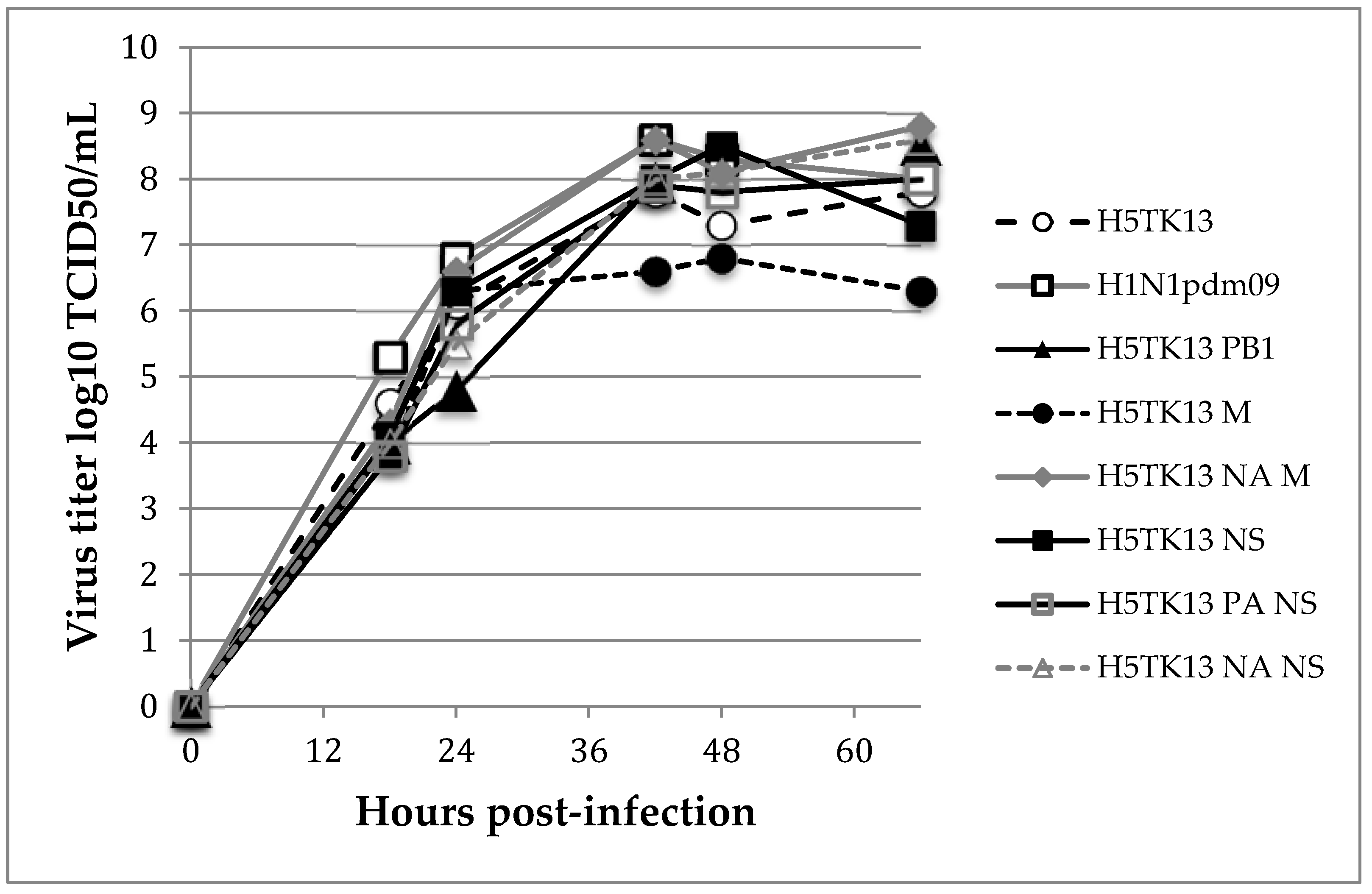
3.2. In Vitro Characterization of Reassortant Viruses
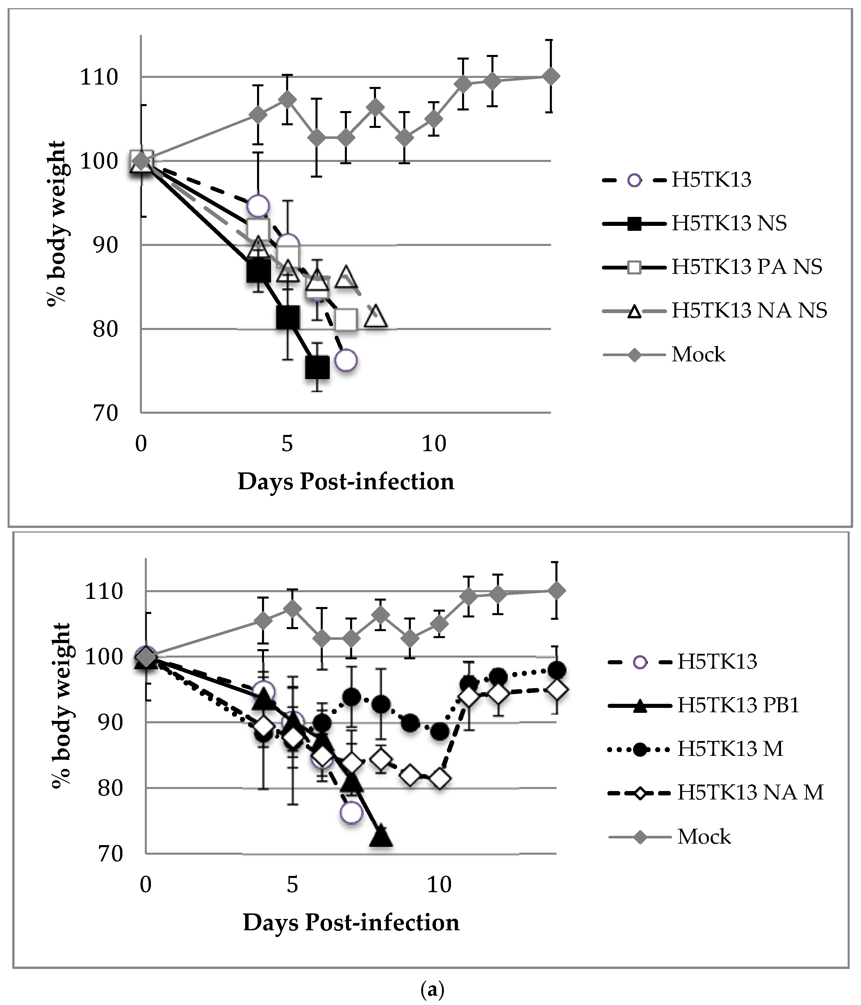
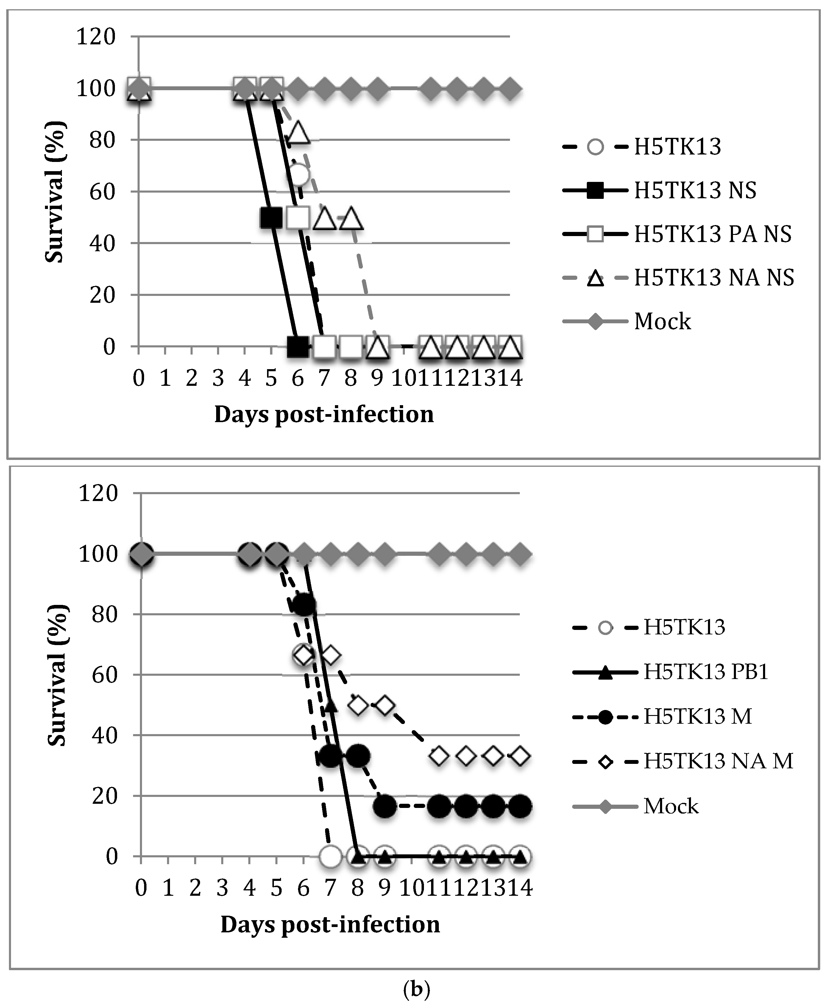
3.3. Pathogenicity of Reassortant Viruses in Mice
4. Discussion
5. Conclusions
Author Contributions
Funding
Acknowledgments
Conflicts of Interest
References
- WHO. Cumulative Number of Confirmed Human Cases of Avian Influenza A(H5N1) Reported to WHO. 2018. Available online: http://www.who.int/influenza/human_animal_interface/2018_03_02_tableH5N1.pdf?ua=1 (accessed on 8 March 2018).
- Herfst, S.; Schrauwen, E.J.A.; Linster, M.; Chutinimitkul, S.; de Wit, E.; Munster, V.J.; Sorrell, E.M.; Bestebroer, T.M.; Burke, D.F.; Smith, D.J.; et al. Airborne transmission of influenza A/H5N1 virus between ferrets. Science 2012, 336, 1534–1541. [Google Scholar] [CrossRef] [PubMed]
- Imai, M.; Watanabe, T.; Hatta, M.; Das, S.C.; Ozawa, M.; Shinya, K.; Zhong, G.; Hanson, A.; Katsura, H.; Watanabe, S.; et al. Experimental adaptation of an influenza H5 HA confers respiratory droplet transmission to a reassortant H5 HA/H1N1 virus in ferrets. Nature 2012, 486, 420–428. [Google Scholar] [CrossRef] [PubMed]
- Guan, Y.; Vijaykrishna, D.; Bahl, J.; Zhu, H.; Wang, J.; Smith, G.J.D. The emergence of pandemic influenza viruses. Protein Cell 2010, 1, 9–13. [Google Scholar] [CrossRef] [PubMed]
- Trifonov, V.; Khiabanian, H.; Greenbaum, B.; Rabadan, R. The origin of the recent swine influenza A(H1N1) virus infecting humans. Eurosurveill. 2009, 14, 6. [Google Scholar]
- Chutinimitkul, S.; Herfst, S.; Steel, J.; Lowen, A.C.; Ye, J.; van Riel, D.; Schrauwen, E.J.A.; Bestebroer, T.M.; Koel, B.; Burke, D.F.; et al. Virulence-associated substitution D222G in the hemagglutinin of 2009 pandemic influenza A(H1N1) virus affects receptor binding. J. Virol. 2010, 84, 11802–11813. [Google Scholar] [CrossRef] [PubMed]
- WHO. Influenza Update. 2017. Available online: http://www.who.int/influenza/surveillance_monitoring/updates/latest_update_GIP_surveillance/en/ (accessed on 22 December 2017).
- WHO. Updated Unified Nomenclature System for the Highly Pathogenic H5N1 Avian Influenza Viruses. 2011. Available online: http://www.who.int/influenza/gisrs_laboratory/h5n1_nomenclature/en/ (accessed on 22 December 2017).
- Ottmann, M.; Duchamp, M.B.; Casalegno, J.-S.; Frobert, E.; Moulès, V.; Ferraris, O.; Valette, M.; Escuret, V.; Lina, B. Novel influenza A(H1N1) 2009 in vitro reassortant viruses with oseltamivir resistance. Antivir. Ther. 2010, 15, 721–726. [Google Scholar] [CrossRef] [PubMed]
- Casalegno, J.-S.; Ferraris, O.; Escuret, V.; Bouscambert, M.; Bergeron, C.; Linès, L.; Excoffier, T.; Valette, M.; Frobert, E.; Pillet, S.; et al. Functional balance between the hemagglutinin and neuraminidase of influenza A(H1N1)pdm09 HA D222 variants. PLoS ONE 2014, 9, e104009. [Google Scholar] [CrossRef] [PubMed]
- Salzberg, S.L.; Kingsford, C.; Cattoli, G.; Spiro, D.J.; Janies, D.A.; Aly, M.M.; Brown, I.H.; Couacy-Hymann, E.; De Mia, G.M.; Dung, D.H.; et al. Genome analysis linking recent European and African influenza (H5N1) viruses. Emerg. Infect. Dis. 2007, 13, 713–718. [Google Scholar] [CrossRef] [PubMed]
- Duchamp, M.B.; Casalegno, J.S.; Gillet, Y.; Frobert, E.; Bernard, E.; Escuret, V.; Billaud, G.; Valette, M.; Javouhey, E.; Lina, B.; et al. Pandemic A(H1N1) 2009 influenza virus detection by real time RT-PCR: Is viral quantification useful? Clin. Microbiol. Infect. 2010, 16, 317–321. [Google Scholar] [CrossRef] [PubMed]
- Basler, C.F.; Aguilar, P.V. Progress in identifying virulence determinants of the 1918 H1N1 and the Southeast Asian H5N1 influenza A viruses. Antivir. Res. 2008, 79, 166–178. [Google Scholar] [CrossRef] [PubMed]
- Jackson, S.; Van Hoeven, N.; Chen, L.-M.; Maines, T.R.; Cox, N.J.; Katz, J.M.; Donis, R.O. Reassortment between avian H5N1 and human H3N2 influenza viruses in ferrets: A public health risk assessment. J. Virol. 2009, 83, 8131–8140. [Google Scholar] [CrossRef] [PubMed]
- Chen, L.-M.; Davis, C.T.; Zhou, H.; Cox, N.J.; Donis, R.O. Genetic compatibility and virulence of reassortants derived from contemporary avian H5N1 and human H3N2 influenza A viruses. PLoS Pathog. 2008, 4, e1000072. [Google Scholar] [CrossRef] [PubMed]
- Li, C.; Hatta, M.; Nidom, C.A.; Muramoto, Y.; Watanabe, S.; Gabriele, N.; Kawaoka, Y. Reassortment between avian H5N1 and human H3N2 influenza viruses creates hybrid viruses with substantial virulence. Proc. Natl. Acad. Sci. USA 2010, 107, 4687–4692. [Google Scholar] [CrossRef] [PubMed]
- Cline, T.D.; Karlsson, E.A.; Freiden, P.; Seufzer, B.J.; Rehg, J.E.; Webby, R.J.; Schultz-Cherry, S. Increased pathogenicity of a reassortant 2009 pandemic H1N1 influenza virus containing an H5N1 hemagglutinin. J. Virol. 2011, 85, 12262–12270. [Google Scholar] [CrossRef] [PubMed]
- Garten, R.J.; Davis, C.T.; Russell, C.A.; Shu, B.; Lindstrom, S.; Balish, A.; Sessions, W.M.; Xu, X.; Skepner, E.; Deyde, V.; et al. Antigenic and genetic characteristics of swine-origin 2009 A(H1N1) influenza viruses circulating in humans. Science 2009, 325, 197–201. [Google Scholar] [CrossRef] [PubMed]
- Karasin, A.I.; Schutten, M.M.; Cooper, L.A.; Smith, C.B.; Subbarao, K.; Anderson, G.A.; Carman, S.; Olsen, C.W. Genetic characterization of H3N2 influenza viruses isolated from pigs in North America, 1977–1999: Evidence for wholly human and reassortant virus genotypes. Virus Res. 2000, 68, 71–85. [Google Scholar] [CrossRef]
- Rameix-Welti, M.-A.; Tomoiu, A.; Dos Santos Afonso, E.; van der Werf, S.; Naffakh, N. Avian Influenza A virus polymerase association with nucleoprotein, but not polymerase assembly, is impaired in human cells during the course of infection. J. Virol. 2009, 83, 1320–1331. [Google Scholar] [CrossRef] [PubMed]
- Zhang, Y.; Zhang, Q.; Kong, H.; Jiang, Y.; Gao, Y.; Deng, G.; Shi, J.; Tian, G.; Liu, L.; Liu, J.; et al. H5N1 hybrid viruses bearing 2009/H1N1 virus genes transmit in guinea pigs by respiratory droplet. Science 2013, 340, 1459–1463. [Google Scholar] [CrossRef] [PubMed]
- Barman, S.; Ali, A.; Hui, E.K.; Adhikary, L.; Nayak, D.P. Transport of viral proteins to the apical membranes and interaction of matrix protein with glycoproteins in the assembly of influenza viruses. Virus Res. 2001, 77, 61–69. [Google Scholar] [CrossRef]
- Leser, G.P.; Lamb, R.A. Lateral Organization of Influenza Virus Proteins in the Budozone Region of the Plasma Membrane. J. Virol. 2017, 91. [Google Scholar] [CrossRef] [PubMed]
- Chou, Y.; Albrecht, R.A.; Pica, N.; Lowen, A.C.; Richt, J.A.; García-Sastre, A.; Palese, P.; Hai, R. The M segment of the 2009 new pandemic H1N1 influenza virus is critical for its high transmission efficiency in the guinea pig model. J. Virol. 2011, 85, 11235–11241. [Google Scholar] [CrossRef] [PubMed]
- Hale, B.G.; Randall, R.E.; Ortín, J.; Jackson, D. The multifunctional NS1 protein of influenza A viruses. J. Gen. Virol. 2008, 89, 2359–2376. [Google Scholar] [CrossRef] [PubMed]
- Hale, B.G.; Steel, J.; Medina, R.A.; Manicassamy, B.; Ye, J.; Hickman, D.; Hai, R.; Schmolke, M.; Lowen, A.C.; Perez, D.R.; et al. Inefficient control of host gene expression by the 2009 pandemic H1N1 influenza A virus NS1 protein. J. Virol. 2010, 84, 6909–6922. [Google Scholar] [CrossRef] [PubMed]
- Ozawa, M.; Basnet, S.; Burley, L.M.; Neumann, G.; Hatta, M.; Kawaoka, Y. Impact of amino acid mutations in PB2, PB1-F2, and NS1 on the replication and pathogenicity of pandemic (H1N1) 2009 influenza viruses. J. Virol. 2011, 85, 4596–4601. [Google Scholar] [CrossRef] [PubMed]
- Octaviani, C.P.; Ozawa, M.; Yamada, S.; Goto, H.; Kawaoka, Y. High level of genetic compatibility between swine-origin H1N1 and highly pathogenic avian H5N1 influenza viruses. J. Virol. 2010, 84, 10918–10922. [Google Scholar] [CrossRef] [PubMed]


| 1 | 2 | 3 | 4 | 5 | 6 | 7 | 8 | |
|---|---|---|---|---|---|---|---|---|
| Reassortant Virus | PB2 | PB1 | PA | HA | NP | NA | M | NS |
| H5TK13 | H5 | H5 | H5 | H5 | H5 | H5 | H5 | H5 |
| H5TK13 PB1 | H5 | H1 | H5 | H5 | H5 | H5 | H5 | H5 |
| H5TK13 M | H5 | H5 | H5 | H5 | H5 | H5 | H1 | H5 |
| H5TK13 M NA | H5 | H5 | H5 | H5 | H5 | H1 | H1 | H5 |
| H5TK13 NS | H5 | H5 | H5 | H5 | H5 | H5 | H5 | H1 |
| H5TK13 PA NS | H5 | H5 | H1 | H5 | H5 | H5 | H5 | H1 |
| H5TK13 NA NS | H5 | H5 | H5 | H5 | H5 | H1 | H5 | H1 |
| Virus and Reassortants | log10 MLD50 † | Mean Survival Time (Days) ‡ | Virus Titer § (log10 TCID50/gram ± SD) (No. Positive Mice/Total No. of Mice) | Copies × 105/mL ± SD (No. of Positive Mice/Total No. of Mice) | ||
|---|---|---|---|---|---|---|
| Lung (6/6) | Spleen # | Brain # | Serum No Significant Difference p < 0.05 | |||
| H5TK13 WT | 1.0 | 6.5 | 6.1 ± 0.3 | 2.5 (1/6) | 4.9 ± 0.6 (5/6) | 7.0 ± 1.3 (6/6) |
| H5TK13 PB1 | 2.3 | 7.5 | 5.6 ± 0.3 | <(0/6) | <(0/6) | 7.8 ± 0.5 (4/6) |
| H5TK13 M | 4.4 | 7.7 | 6.4 ± 0.4 | <(0/6) | <(0/6) | 7.2 ± 1.1 (6/6) |
| H5TK13 NA M | 4.6 | 8.8 | 6.4 ± 0.1 | <(0/6) | <(0/6) | 6.4 ± 0,8 (6/6) |
| H5TK13 NS | 0.3 | 5.5 | 6.6 ± 0.3 | 3.2 ± 0.3 (3/6) | 3.0 ± 0.6 (3/6) | 5.9 ± 0.2 (4/6) |
| H5TK13 PA NS | 1.8 | 6.5 | 6.5 ± 0.1 | <(0/6) | <(0/6) | 8.7 ± 0.9 (6/6) |
| H5TK13 NA NS | 3.7 | 8.3 | 6.8 ± 0.6 | <(0/6) | <(0/6) | 6.8 ± 0.7 (6/6) |
© 2018 by the authors. Licensee MDPI, Basel, Switzerland. This article is an open access article distributed under the terms and conditions of the Creative Commons Attribution (CC BY) license (http://creativecommons.org/licenses/by/4.0/).
Share and Cite
Ferraris, O.; Casalegno, J.-S.; Frobert, E.; Bouscambert Duchamp, M.; Valette, M.; Jacquot, F.; Raoul, H.; Lina, B.; Ottmann, M. The NS Segment of H1N1pdm09 Enhances H5N1 Pathogenicity in a Mouse Model of Influenza Virus Infections. Viruses 2018, 10, 504. https://doi.org/10.3390/v10090504
Ferraris O, Casalegno J-S, Frobert E, Bouscambert Duchamp M, Valette M, Jacquot F, Raoul H, Lina B, Ottmann M. The NS Segment of H1N1pdm09 Enhances H5N1 Pathogenicity in a Mouse Model of Influenza Virus Infections. Viruses. 2018; 10(9):504. https://doi.org/10.3390/v10090504
Chicago/Turabian StyleFerraris, Olivier, Jean-Sébastien Casalegno, Emilie Frobert, Maude Bouscambert Duchamp, Martine Valette, Frédéric Jacquot, Hervé Raoul, Bruno Lina, and Michèle Ottmann. 2018. "The NS Segment of H1N1pdm09 Enhances H5N1 Pathogenicity in a Mouse Model of Influenza Virus Infections" Viruses 10, no. 9: 504. https://doi.org/10.3390/v10090504
APA StyleFerraris, O., Casalegno, J.-S., Frobert, E., Bouscambert Duchamp, M., Valette, M., Jacquot, F., Raoul, H., Lina, B., & Ottmann, M. (2018). The NS Segment of H1N1pdm09 Enhances H5N1 Pathogenicity in a Mouse Model of Influenza Virus Infections. Viruses, 10(9), 504. https://doi.org/10.3390/v10090504


