Article

Rapid Identification of Intact Staphylococcal Bacteriophages Using Matrix-Assisted Laser Desorption Ionization-Time-of-Flight Mass Spectrometry

1
Department of Experimental Biology, Faculty of Science, Masaryk University, Kotlářská 2, 61137 Brno, Czech Republic
2
Central European Institute of Technology, Masaryk University, Kamenice 5, 62500 Brno, Czech Republic
3
National Centre for Biomolecular Research, Faculty of Science, Masaryk University, Kamenice 5, 62500 Brno, Czech Republic
*
Author to whom correspondence should be addressed.
Viruses 2018, 10(4), 176; https://doi.org/10.3390/v10040176
Submission received: 13 March 2018 / Revised: 1 April 2018 / Accepted: 2 April 2018 / Published: 4 April 2018
(This article belongs to the Special Issue Hurdles for Phage Therapy (PT) to Become a Reality)

Abstract

:
Staphylococcus aureus is a major causative agent of infections associated with hospital environments, where antibiotic-resistant strains have emerged as a significant threat. Phage therapy could offer a safe and effective alternative to antibiotics. Phage preparations should comply with quality and safety requirements; therefore, it is important to develop efficient production control technologies. This study was conducted to develop and evaluate a rapid and reliable method for identifying staphylococcal bacteriophages, based on detecting their specific proteins using matrix-assisted laser desorption ionization time-of-flight mass spectrometry (MALDI-TOF MS) profiling that is among the suggested methods for meeting the regulations of pharmaceutical authorities. Five different phage purification techniques were tested in combination with two MALDI-TOF MS matrices. Phages, either purified by CsCl density gradient centrifugation or as resuspended phage pellets, yielded mass spectra with the highest information value if ferulic acid was used as the MALDI matrix. Phage tail and capsid proteins yielded the strongest signals whereas the culture conditions had no effect on mass spectral quality. Thirty-seven phages from Myoviridae, Siphoviridae or Podoviridae families were analysed, including 23 siphophages belonging to the International Typing Set for human strains of S. aureus, as well as phages in preparations produced by Microgen, Bohemia Pharmaceuticals and MB Pharma. The data obtained demonstrate that MALDI-TOF MS can be used to effectively distinguish between Staphylococcus-specific bacteriophages.

Graphical Abstract

1. Introduction

Due to increasing antibiotic resistance, bacterial infections have become a serious problem in hospital and community environments. A possible approach to combat such infections is phage therapy, either as an alternative to antibiotics or utilizing phage-antibiotic synergy. Experimental phage therapy proved to be successful for the treatment of bacterial infections in animal models and human patients [1]. Interest in phage therapy has therefore increased [2] and applications of phages are currently being investigated extensively [3]. For the rational use of phages, it is necessary to have rapid methods of identification, particularly for new isolates as well as previously characterized phages after passaging for quality control of bacteriophage-based products.
Identification of bacteriophages is usually based on morphological characterization using electron microscopy and by genome sequencing [4]. Multiplex PCR can be used to identify well-characterized phages with defined conserved genes typical for particular groups, for example, phages of dairy bacteria [5] or staphylococcal phages [6]. However, high mosaicism and modular genomic structure of tailed phages complicates their detection [7].
This work focuses on the identification of staphylococcal phages that play an important role in biology, evolution and pathogenicity of staphylococci [8,9,10,11,12]. While lytic phages shape bacterial population dynamics, temperate phages are a driving force in bacterial evolution and can benefit the host bacteria by introducing novel traits such as virulence factors as a consequence of lysogenic conversion [13]. Bacteriophages also facilitate the horizontal transfer of bacterial DNA, including resistance genes, through transduction [14,15,16].
The taxonomy of staphylococcal phages has recently been updated by the International Committee on Taxonomy of Viruses (ICTV) [17]. All staphylococcal phages belong to the order Caudovirales, including Myoviridae, Siphoviridae and Podoviridae families. Staphylococcal phages from the family Siphoviridae are temperate and comprise 3 genera that correlate with previously described serological groups [18]. Candidates for combating staphylococcal infections are phages belonging to Myoviridae and Podoviridae families [8] as they seem unable to lysogenise host cells [11] and recent findings suggest that lytic and polyvalent staphylococcal phages belonging to the Myoviridae family are safe for therapeutic applications [19].
Fingerprinting by means of Matrix-Assisted Laser Desorption/Ionization—Time-of-Flight Mass Spectrometry (MALDI-TOF MS) is based on rapid detection of compounds ionized directly or after minimal sample treatment. The greatest success of this method has been achieved in the field of bacterial identification [20]. Protein profiles obtained by MALDI-TOF MS are used nowadays in thousands of clinical laboratories worldwide. Apart from bacteria, several other types of samples, including viruses, can be subjected to MALDI-TOF MS profiling analysis [21]. However, MALDI-TOF MS profiling is not currently used for routine identification of tailed phages.
The first successful attempt at bacteriophage MALDI-TOF MS profiling was documented by Thomas et al. [22]. This study focused on the ssRNA phage MS2 that belongs to the Leviviridae family and infects Escherichia coli. Treatment of MS2 phage particles, isolated by centrifugation and ultrafiltration, with 50% acetic acid was found to be necessary to obtain protein signals, as an acidic environment causes disassembly of phage protein structures. Acid treatment directly in the MALDI matrix solution was also found to be beneficial, as demonstrated by the successful generation of protein signals after preparing the phage sample in a MALDI matrix comprising 17% formic acid [23,24]. To improve reproducibility and sensitivity, McAlpin et al. [25] suggested a 10 min treatment of Yersinia pestis podovirus φA1122 with β-mercaptoethanol to reduce inter-molecular disulphide bonds. This procedure was found to be more efficient than acid treatment.
Despite relatively long-standing knowledge that MALDI-TOF MS fingerprinting of bacteriophages was possible, most studies in the field were aimed at method optimization and involved only a limited set of strains. The largest collection of 12 strains was assessed by Bourdin et al. [26] who used MALDI-TOF MS profiling for phage purification control after a multi-step isolation procedure. These works have demonstrated the usefulness of phage protein profiling for particular analytical purposes, however, comprehensive evaluation of sample preparation methods, MALDI-TOF mass spectral quality, reproducibility and discriminatory power have not yet been assessed critically. For this reason, we conducted a study on a relatively large set involving closely related staphylococcal phage strains, using various combinations of sample preparation techniques to establish the optimum setup and assess its applicability in identifying bacteriophages.

2. Materials and Methods

2.1. Bacterial Strains and Phages

Phages and bacterial strains used in this work are described in Table 1. The bacteriophages from the International Typing Set for human S. aureus and their propagation strains (PS) were obtained from Dr. P. Petráš (National Reference Laboratory for Staphylococci, National Institute of Public Health, Prague, Czech Republic). Laboratory lysogenised strains PS 47[53+], PS 47[77+] were prepared previously [27]. Phages 11, 80α and K and S. aureus RN4220 were kindly provided by Prof. C. Wolz (Interfaculty Inst. of Microbiology and Infection Medicine Tübingen, University of Tübingen, Tübingen, Germany). Propagation strain S. aureus RN4220 ΔtarM for podoviruses [28] was provided by Prof. A. Peschel (Department of Infection Biology, University of Tübingen, Tübingen, Germany). Phages Twort, 44AHJD and P68 were purchased from Félix d’Hérelle Reference Center for Bacterial Viruses (Université Laval, Québec, QC, Canada) and phage X2 from the National Collection of Type Cultures (Public Health England, Salisbury, UK). Phages B166 and B236 [29], 131 and 812 [30] and phage K1/420 [31] were described previously. Phage PYO was isolated from a phage cocktail containing Staphylococcus, Streptococcus, Proteus, E. coli and Pseudomonas aeruginosa phages (PYO Bacteriophagum combinierae liquidum, lot no. 000860, BioPharm, Tbilisi, Republic of Georgia). A commercial Staphylococcus bacteriophage preparation produced by Microgen (lot no. H141, 0812, 08.14, Moscow, Russia) was purchased in a pharmacy in Samara, Russia. Bacteriophage preparation Stafal® lot no. 140805201 was kindly provided by Bohemia Pharmaceuticals (Prague, Czech Republic). Phage preparation Duofag, staphylococcal phages SAU1 and SAU2 and P. aeruginosa phage PAE1 were kindly provided by MB Pharma (Prague, Czech Republic). S. aureus strains CCM 4028, CCM 4890 and CCM 8428 were obtained from the Czech Collection of Microorganisms (Masaryk University, Brno, Czech Republic).

2.2. Bacteriophage Propagation and Titration

Phages were propagated on their propagation strains (Table 1) in a liquid medium meat-peptone broth (MPB) prepared from 13 g of nutrient broth (Oxoid, CM0001), 3 g of yeast extract (Oxoid, LP0021) and 5 g of peptone (Oxoid, LP0037) dissolved in distilled water to 1000 mL (pH 7.4). Prophage-less S. aureus strains CCM 4890 [32], RN4220 [33] and CCM 8428 [34] were used for propagation of phages with known genomic sequences to eliminate the possibility of false positive signals in MALDI-TOF MS spectra caused by induced prophages. Phages 29, 42E and 79 were propagated in a soft agar layer with 0.7% (w/v) of bacteriological agar (Oxoid, LP0011) on top of solid meat-peptone agar with 1.5% (w/v) technical agar (Oxoid, LP0012). Two other types of growth media were used to test the influence of medium on mass spectral quality: brain heart infusion (BHI) broth (Oxoid, CM1135; pH 7.4) and 2× yeast-tryptone broth (2YT) consisting of 16 g of tryptone (Oxoid, LP0042), 10 g of yeast extract (Oxoid, LP0021) and 5 g of NaCl dissolved in distilled water to 1000 mL (pH 7.4).
For phage titration, ten-fold serial dilutions of phage in MPB were prepared. An overnight culture of propagation strain in MPB was mixed with 1/10 volume of 0.02 M CaCl2. Bacterial suspension (0.1 mL) was added to 3 mL of 0.7% meat-peptone soft agar cooled to 45 °C and overlaid on 1.5% meat peptone agar plates. The plates were left to dry for 10 min. Phage dilutions were dropped onto the top agar and left to dry. Plates were incubated overnight at 37 °C.

2.3. Concentrating Bacteriophage to Pellets

Phage lysate, obtained after complete lysis of the bacteria, was centrifuged at 4500× g for 30 min at 4 °C and filtered through 0.45 μm pore-sized polyethersulfone syringe filters (Techno Plastic Products, Trasadingen, Switzerland) to remove bacterial debris. Phages were pelleted by centrifugation at 54,000× g for 2.5 h at 4 °C in a JA-30.50 Ti rotor (Beckman, Brea, CA, USA). For preparation of pellets from 3 mL of phage lysates, conical tubes (part no. 358119, Beckman) and adapters (part no. 358153, Beckman) with an SW 55 Ti rotor (Beckman) were used. The resulting pellet was resuspended in 350 μL of phage buffer (5 × 10⁻² mol/L Tris pH 8.0, 10−2 mol/L CaCl2, 10−2 mol/L NaCl) overnight at 4 °C. For resuspending the pellets from 3 mL samples, 30 μL of phage buffer was used.

2.4. Purification of Phage Particles by CsCl Density Gradient Centrifugation

This procedure was carried out according to the description by Nováček et al. [31]. Phage pellets resuspended in phage buffer were used as an input material for CsCl density gradient centrifugation. Soluble proteins from resuspended pellets were removed by extraction with an equal volume of chloroform. The resulting aqueous fraction (approximately 1.2 mL) was overlaid onto a preformed CsCl (Sigma-Aldrich, St. Louis, MO, USA) density gradient (1 mL of each 1.45 g/mL 1.50 g/mL, 1.70 g/mL of CsCl in phage buffer) and centrifuged at 194,000× g for 4 h at 12 °C using a SW 55 Ti rotor (Beckman). Phage particles forming a visible zone were collected by puncturing the tube with an 0.8 mm gauge needle and syringe. Caesium chloride was removed from the phage-containing fraction by dialysis against an excess of phage buffer at 4 °C overnight using Visking dialysis tubing type 8/32”, 0.05 mm thick (part no. 1780.1, Carl Roth, Karlsruhe, Germany).

2.5. Fast Protein Liquid Chromatography (FPLC)

Bacteriophage lysates were purified using a monolithic column, CIMmultusTM QA 1 mL (Bia separations, Ajdovščina, Slovenia) and FPLC NGC (Bio-Rad, Hercules, CA, USA). Phages were purified according to column manufacturer’s recommendation with minor modifications [35]. After removing bacterial debris by centrifugation and filtration, 25 mL of phage lysate (108–9 PFU/mL) were mixed with 100 mM sodium phosphate buffer pH 7 at a ratio of 1:1. The column was equilibrated with 100 mM sodium phosphate buffer pH 7. The phage suspension was loaded onto the column and a linear gradient of 100 mM sodium phosphate, 2 M NaCl, pH 7 buffer was applied for elution of phages. NaCl was removed from the phage-containing fraction by dialysis as described following CsCl density gradient centrifugation.

2.6. Phage Purification by Ultrafiltration

Filtered phage lysate was used for tangential flow filtration using Pellicon XL 50 Ultrafiltration Cassettes (Millipore, Burlington, MA, USA) Biomax 300 (for Podoviridae and Myoviridae phages) and Biomax 500 (for Siphoviridae phages) according to the manufacturer’s instructions. All fluids were loaded onto the cassette with a peristaltic pump set at 40–50 mL per minute. The cassette was rinsed with 300 mL of sterile distilled water and then 300 mL of phage buffer. 350 mL of phage lysate was loaded onto the cassette and after concentrating/thickening of phages to 10–50 mL, phages were mixed with 1000 mL of phage buffer and thickened again to 10–50 mL.

2.7. Phage Precipitation by Polyethylene Glycol (PEG)

Phages were purified with PEG 8000 (Sigma-Aldrich) according to Sambrook et al. [36] with minor modifications. NaCl and PEG 8000 were dissolved in 30 mL of filtered phage lysate to final concentrations of 0.5 M and 10 % (w/v), respectively, by brief stirring. Phages were precipitated overnight at 4 °C, pelleted by centrifugation at 10,000× g for 15 min at 4 °C and the supernatant was discarded. Pellets were dissolved in 0.5 mL of phage buffer overnight at 4 °C. Phage particles were then separated from co-precipitated bacterial debris by centrifugation at 5000× g for 10 min at 4 °C. The residual PEG was removed by gentle extraction for 1 min with an equal volume of chloroform. The phage-containing aqueous phase was separated by centrifugation at 5000× g for 15 min, collected and filtered through 0.22 μm pore-sized polyethersulfone syringe filters (Techno Plastic Products, Trasadingen, Switzerland).

2.8. Sample Preparation for MALDI-MS

The phage samples were mixed with a MALDI matrix solution in a 1:4 v/v ratio, the resulting mixtures were applied to three positions on the stainless steel MALDI sample plate in a volume of 0.6 μL and were allowed to dry at room temperature. Ferulic acid (FerA, 12.5 mg/mL in a water:acetonitrile:formic acid, 50:33:17, v/v mixture), or, alternatively, alpha-cyano-4-hydroxycinnamic acid (HCCA, saturated solution in water:acetonitrile:trifluoroacetic acid, 47.5:50:2.5, v/v mixture) were used as the MALDI matrix solutions.
For peptide mass fingerprinting and MS/MS analyses, the phage preparations in a volume of 5 μL were incubated with 50 ng of trypsin in 25 mM ammonium bicarbonate for two hours at 40 °C. The proteolytic mixture in a volume of 1 μL was applied to an AnchorChip MALDI sample plate, mixed with 0.6 μL of the MALDI matrix (alpha-cyano-4-hydroxycinnamic acid, 2 mg/mL in a water:acetonitrile:trifluoroacetic acid, 32:66:2, v/v mixture) and allowed to dry at room temperature.

2.9. MALDI-MS Profiling Analysis

MALDI-TOF mass spectral fingerprints were obtained using an Ultraflextreme instrument (Bruker Daltonik, Bremen, Germany) operated in the linear positive mode under FlexControl 3.4 software. External calibration of the mass spectra in the linear positive mode was performed using lysozyme (its monomer, dimer and multiply protonated ions). Laser power was set to 120% of the threshold laser power for a particular type of sample. Three independent spectra comprising 1000 laser shots each were acquired from each of the wells. Within an individual well, a minimum of 200 and a maximum of 400 shots were obtained from one position. The mass spectra were recorded within the m/z range of 2–100 kDa. Mass spectra were processed using Flex Analysis (version 3.4; Bruker Daltonik). The MALDI-TOF mass spectra-based dendrogram was constructed using the Pearson’s product moment coefficient as a measure of similarity and the unweighted pair group average linked method (UPGMA) as a grouping method using the Biotyper 3.1 software (Bruker Daltonik). The protein fingerprints of phages with known genomic sequences were closely examined by comparing the m/z values with the predicted molecular weight (Mw) of phage structural proteins from the NCBI Protein database or from custom RAST annotations [37]. For matching the m/z values to Mw of phage structural proteins, a 500 ppm tolerance was taken into account. The first amino acid of a protein was identified using TermiNator [38,39] and the Mw was calculated using ExPASy ProtParam [40].

2.10. MALDI-MS/MS Analysis

Analyses of digested samples were carried out using the same instrument operated in the reflectron positive mode. Seven peptide standards (Bruker Daltonik) covering the mass range of 700–3100 Da were used for external mass calibration. Peptide maps were acquired with 800 laser shots. Peaks with minimum S/N = 10 were picked out for MS/MS analysis employing the LIFT arrangement with 600 laser shots for each peptide. The MASCOT 2.2 (MatrixScience, London, UK) search engine was used for processing the MS and MS/MS data. Database searches were carried out on the NCBIprot database (non-redundant, taxonomy All Entries; downloaded from ftp://ftp.ncbi.nih.gov/blast/db/FASTA/). For MALDI-MS data, a mass tolerance of 30 ppm was allowed for peptide mapping and 0.5 Da for MS/MS ion searches. Oxidation of methionine as an optional modification and two enzyme miscleavages were set for all searches. Peptides with statistically significant peptide scores (p < 0.05) were considered. Manual MS/MS spectral assignment validation was carried out.

3. Results

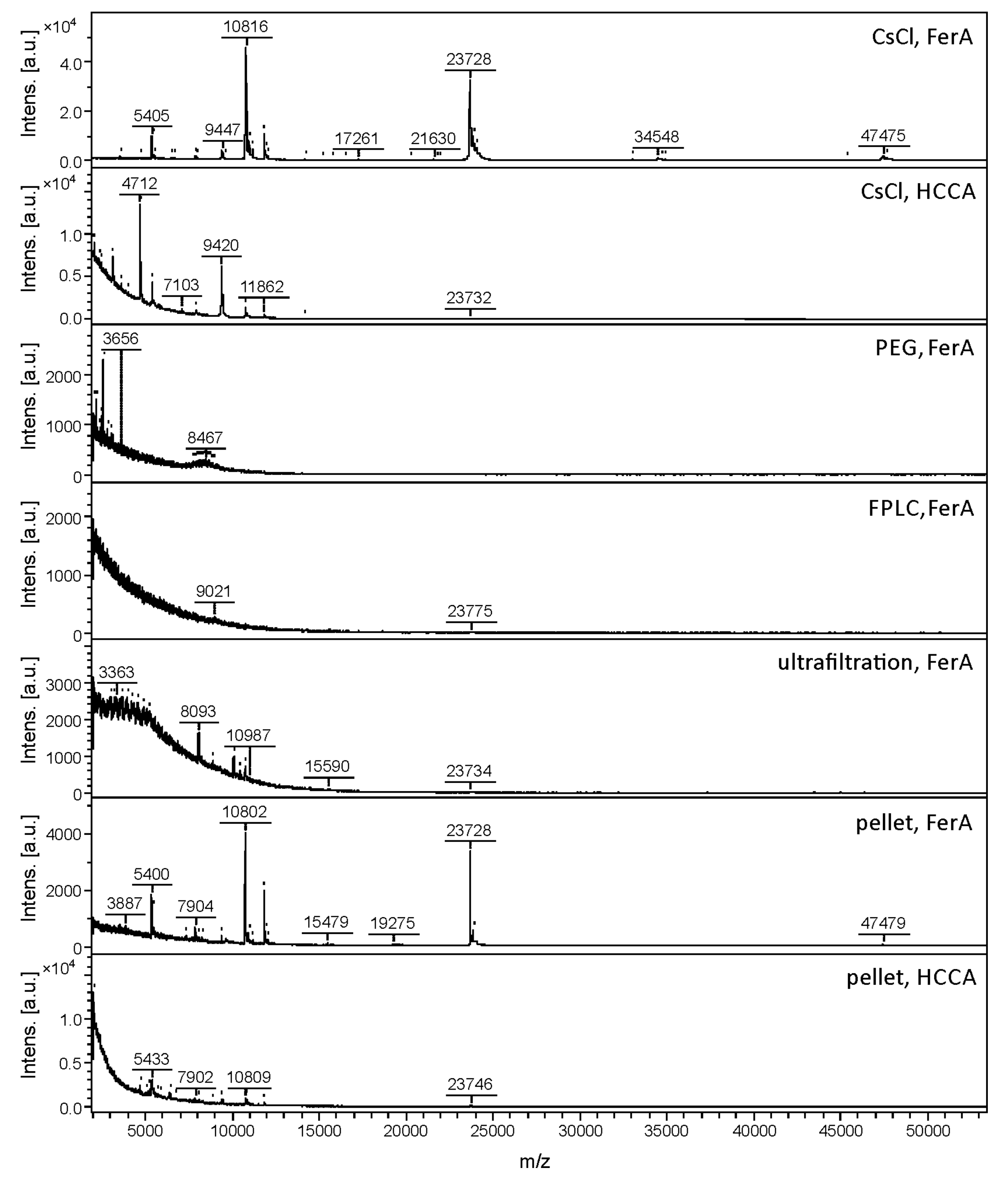
3.1. Comparison of Phage Purification Techniques for Sample Preparation and MALDI-TOF MS Method Development

Five different techniques that are commonly used for phage purification and concentration were tested together with two MALDI matrices for developing a rapid and reliable MALDI-TOF MS-based method for identification of S. aureus bacteriophages. Traditional techniques used for the concentration and purification of phages involve centrifugation, filtration, precipitation and zonal ultracentrifugation [41]. In addition to these traditional techniques, FPLC has been recently proved to be efficient for phage purification [42]. The following types of phage samples were analysed by MALDI-TOF MS: (i) phage pellets dissolved in phage buffer; (ii) phages purified by CsCl density gradient centrifugation; (iii) PEG precipitated phages; (iv) phages purified using FPLC and a poly(glycidyl methacrylate-co-ethylene dimethacrylate) monolith column modified with quaternary amine groups; and (v) phages concentrated and purified with Pellicon XL 50 ultrafiltration cassettes. As a measure of the feasibility of the sample preparation method, the mass spectral quality, in terms of the number of peaks detected, the signal-to-noise ratio and reproducibility based on three independent experiments separated by a minimum of weekly intervals, were assessed on five phage specimens belonging to five different genera from three families: phage K1/420 belonging to the Myoviridae family and Kayvirus genus; phage P68 belonging to the Podoviridae family and the P68virus genus; phages 3A, 71 and 77 belonging to the Siphoviridae family and Triavirus, Phietavirus and Biseptimavirus genera, respectively. The results are summarized in Table 2 and depicted in Figure 1.
Caesium chloride density gradient centrifugation provided reproducible MALDI-TOF mass spectra with the highest number of peaks and signal-to-noise ratios for most of the five samples assessed. This method is the most precise for phage purification and is routinely used in structural studies of phages. On the other hand, CsCl density gradient centrifugation is an expensive and time-consuming method that is inaccessible to many users and inapplicable for large scale production of phages. Therefore, MALDI-TOF MS phage identification using other affordable phage purification methods was investigated. PEG precipitation is an easy method for small-scale phage purification but the residues of PEG 8000 in samples interfered with the ionization of proteins, so this method was determined to be unsuitable for subsequent MALDI-TOF MS profiling analysis (Figure 1). When FPLC was used for phage purification, the mass spectra showed approximately 50% of signals compared to spectra of the CsCl purified phages. Similarly, ultrafiltration was not determined to be a suitable method, yielding a considerably lower number of signals (Table 2). To improve the mass spectral quality, treatment of the phages with β-mercaptoethanol for disassembling the phage particles was assessed, as described by McAlpin et al. [25]. Apart from the recommended protocol (10 min treatment at a 1:10 v/v ratio), a treatment of phage 3A purified by 500 kDa Pellicon XL 50 ultrafiltration cassette was modified to 30 or 60 min at v/v ratio of 1:5 or 1:30. However, no improvement in mass spectral quality was achieved by any of the β-mercaptoethanol treatment conditions tested (Figure S1).
The simplest setup consisting of phage pellet dissolution in phage buffer yielded surprisingly high-quality spectra comparable to those acquired from phages purified by CsCl density gradient centrifugation. Although in this case, the phage was not completely separated from bacterial cell debris, the quantity of phage particles was sufficient to provide mass spectra containing the majority of peaks (80–90%) corresponding to those detected after CsCl purification (Figure 1). Other components of the pellet, such as remaining bacterial cell debris evidently do not cause interference by suppressing ionization of the bacteriophage proteins nor by the appearance of superfluous signals unrelated to bacteriophages.
The influence of sample preparation technique on mass spectral quality was assessed in more depth by cluster analysis involving mass spectra obtained by three repeated analyses, using both CsCl density gradient centrifugation and pellet dissolution (Figure 2). The distances corresponding to variability among three repeated experiments were greater than those arising from different sample preparation methods. The influence of sample preparation method on analytical output was found to be insignificant. The simpler sample preparation procedure thus yielded satisfactory mass spectral outputs; however, to accurately determine the relationship between mass spectra and phage protein components, CsCl density gradient centrifugation was the most appropriate.
A disadvantage of the MALDI-MS profiling method using ferulic acid as the MALDI matrix was the inability to achieve automatic mass spectral acquisition due to non-homogeneous crystallization on the MALDI target. For that reason, another matrix solution (HCCA) was tested, selected on the basis of results of Bourdin et al. [26]. This matrix solution is also used for routine bacterial identification; its use in phage profiling would thus be more compatible with a microbiological laboratory workflow. The mass spectra obtained using HCCA as a matrix revealed signals in a narrower mass range (Figure 1). In addition, in some cases, almost all peaks detected corresponded to different ionization forms of the same protein caused by multiple protonation or gas-phase oligomer formation. The information content of the mass spectra was therefore limited, negatively influencing the discriminatory power of the method. In addition, the use of HCCA as a matrix was not advisable in combination with samples prepared by pellet dissolution, as all mass spectral quality parameters were significantly deteriorated in that case (see Figure 1 and Table 2). For that reason, ferulic acid as a MALDI matrix, in combination with phage pellet dissolution, was used in the remaining profiling experiments.

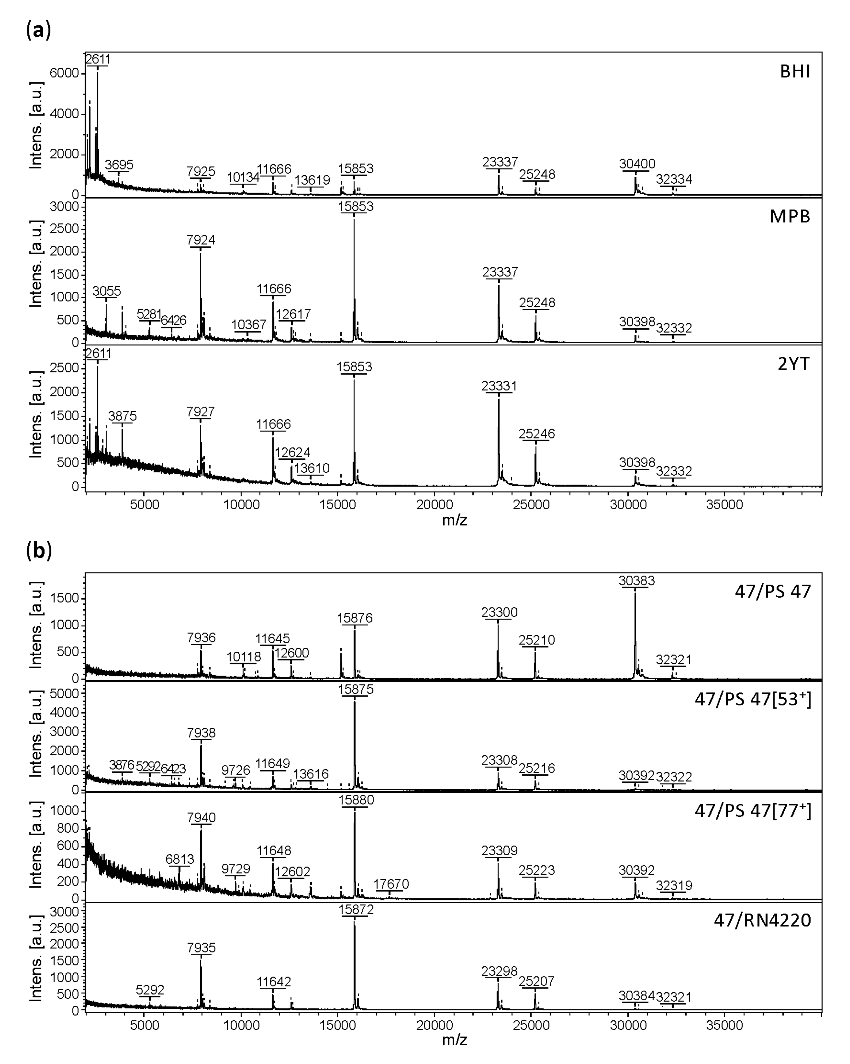
3.2. Effect of Culture Conditions on the Quality of Phage MALDI-TOF Mass Spectra

Culture conditions can affect bacterial MALDI-MS protein fingerprints. As published previously [43], there were small differences in the MALDI-TOF mass spectra of bacteria cultured under different conditions. Nevertheless, despite those differences, whole bacterial cells were identifiable by MALDI-TOF MS regardless of the medium used. However, it is not known whether phage proteomes are influenced by different phage propagation conditions. As a parameter of sample preparation, the influence of culture medium on MALDI-TOF mass spectral quality was tested. For that purpose, the five phage strains 3A, 71, 77, K1/420 and P68 used for method development were grown on prophage-less S. aureus strains in three different growth media (MPB, BHI and 2YT). As an example, the mass spectra of phage 3A are compared in Figure 3a. The main differences between the mass spectra were the relative intensities of signals in the high-mass range. Differences were also visible in the low-mass range (especially at m/z lower than 3.5 kDa). These probably resulted in more distant positions of the analyses in the dendrogram (Figure 4), equivalent to mass spectral differences due to the accuracy of the method itself (distance level equal to 1000). Similar effect of the culture conditions on the mass spectral quality was determined also for the remaining four strains 71, 77, K1/420 and P68 showing only minor qualitative differences in their mass spectra. The proportion of peaks conserved regardless of the cultivation conditions was greater than 80% in all cases including nine replicate analyses per sample.
The influence of different S. aureus strains used for phage propagation was also tested, especially for the possibility of induced prophages that could affect the protein fingerprint obtained by MALDI-TOF MS. Bacteriophage 47 was grown on prophage-less strain RN4220, on strain PS 47 (=RN1) known to carry three prophages 11, 12 and 13 and on two quadruple lysogenic strains derived from PS 47 harbouring additional prophages 53 or 77. The MALDI-TOF mass spectra shown in Figure 3b reveal that the vast majority of peaks were conserved, regardless of the propagation strain. Qualitative mass spectral differences were associated with peaks of low intensity in the low-mass range (lower than 7 kDa), representing less than 15% of all peaks observed in the individual mass spectra. The most significant differences were of a quantitative nature, where the relative intensities of peaks at m/z greater than 20 kDa and their signal-to-noise ratios, were influenced by the propagation strain.
The impact of propagation strain on the discriminatory power of the method was tested by means of cluster analysis based on MALDI-TOF mass spectra of six Triavirus strains of the Siphoviridae family that have very similar MALDI-TOF mass spectra (Figure 4). While the accuracy of the method, as reflected by the distance between three independent analyses of phage strain 3A, reached the maximum dendrogram distance (1000), mass spectral differences of phage 47 arising from different propagation strains resulted in a distance of 750. Note that due to the clustering method used (see Section 2.9), these threshold values are dependent of the collection of mass spectra used for the generation of individual dendrograms and the distance level representing the minimal similarity is always considered to be the highest distance (1000). The influence of propagation strain was thus lower than the overall accuracy of the method and its impact on the discriminatory power of the method was practically insignificant.

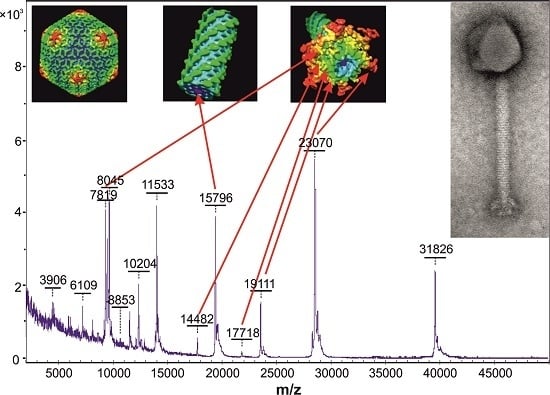
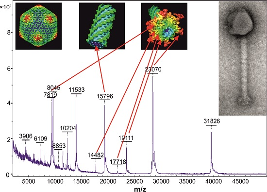
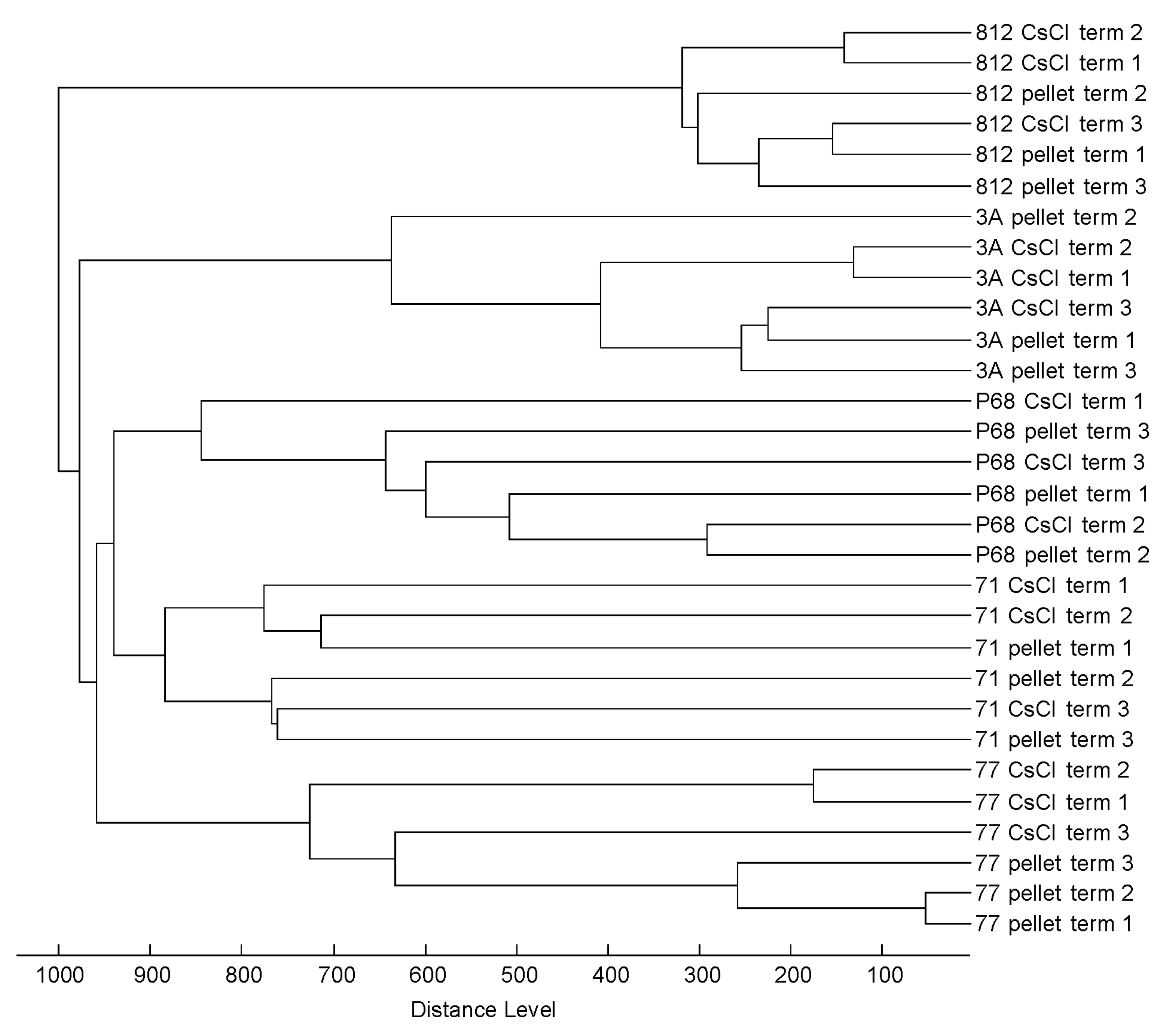
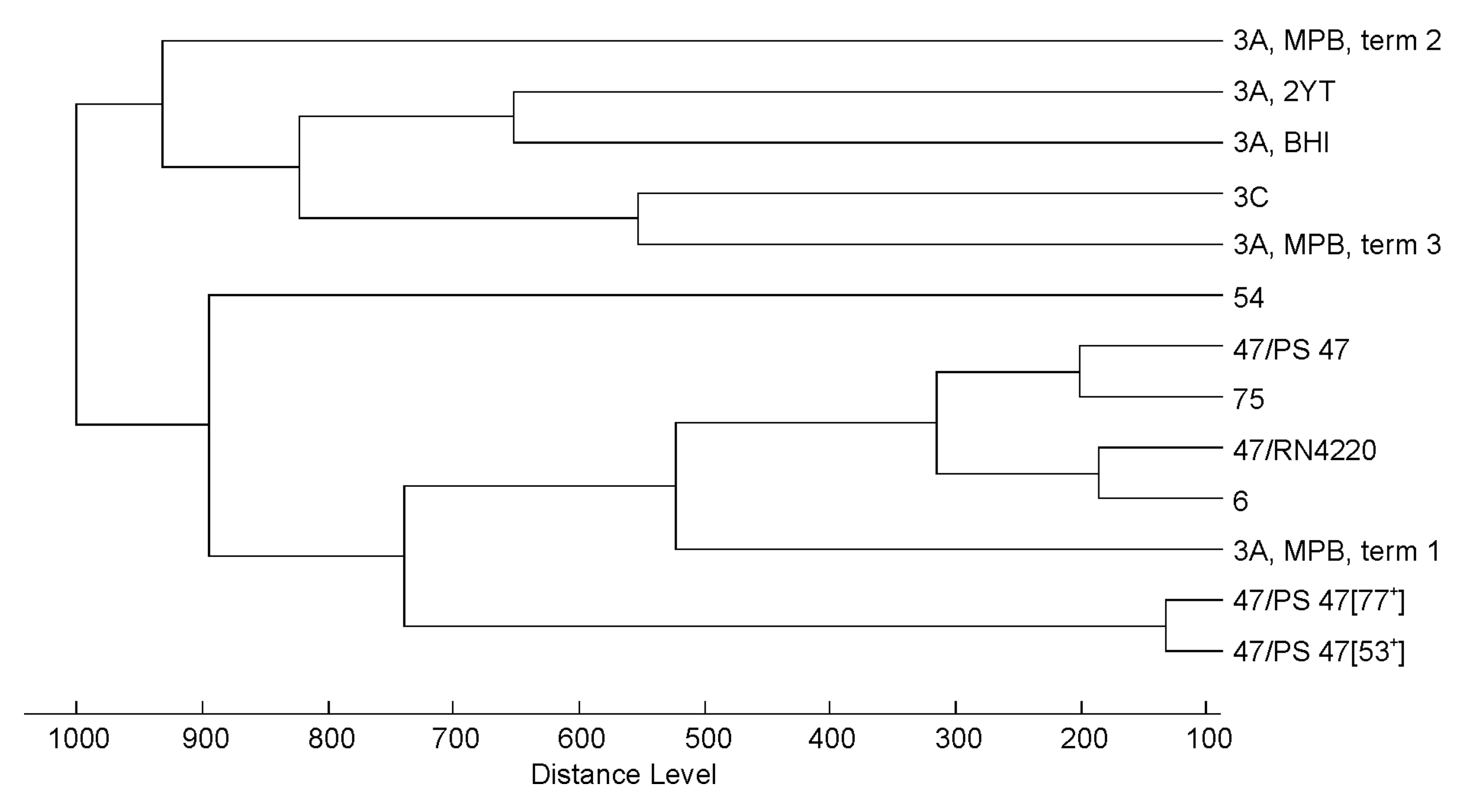
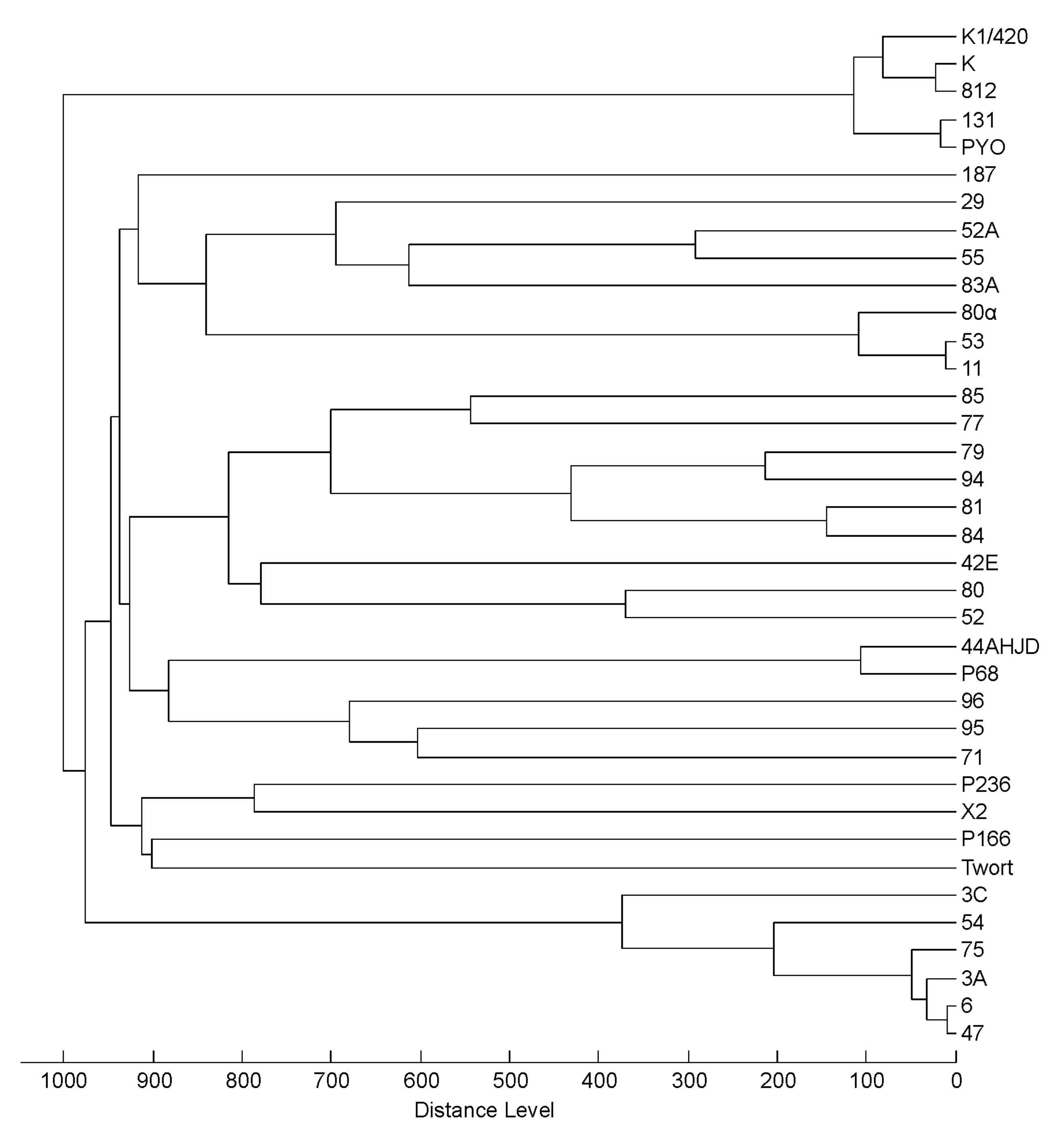
3.3. Profiling of 37 Phages by MALDI-TOF MS

Under the sample preparation arrangement proposed, 37 phages were purified and analysed. Mass spectra of Myoviridae and Siphoviridae were found to be richer than the mass spectra of Podoviridae. This could be explained by the fact that Myoviridae and Siphoviridae include more complex phages with a higher number of structural proteins of molecular weights detectable by MALDI-TOF MS. The dendrogram based on cluster analysis of the mass spectral signals (Figure 5) revealed four significant groups. The first one corresponded to phages of the Kayvirus genus (K, K1/420, 812, 131 and PYO). This grouping was supported by visual inspection of the mass spectra where all phage strains shared distinctive peaks at m/z = 10,204, 11,533, 15,796, 17,745, 19,112 and 23,067. This mass spectral profile allows unambiguous identification of kayviruses that are frequently used in phage therapy.
Six phages of the second well separated cluster (3A, 3C, 6, 47, 54 and 75) belonged to the Triavirus genus and shared several common signals in their MALDI-MS profiles (m/z = 7936, 11,645, 12,598, 15,871, 23,295, 25,208 and 30,373). The third well-defined group consisted of transducing phages from the genus Phietavirus where phages 11, 53 and 80α are closely related at the genomic level. Mass spectra of other phages from this branch did not show major similarities except phages 29 and 55, that shared the peak at m/z = 20,786, corresponding to the major tail protein. The last group consisted of the only two podoviruses P68 and 44AHJD present in this analysis. They shared peaks at m/z= 6916, 15,112 and 46,769. The profiles of other phages were not clustered into groups corresponding to their genomic relatedness. MALDI-TOF mass spectra of Myoviridae phage Twort (Twortvirus genus) and Siphoviridae phages 187 and B166 (supposed phietaviruses) showed no similarities with MALDI-TOF mass spectra of any other phages included in the clustered analysis and their genomes and proteomes also differed [29,44].
On the basis of m/z values, peaks from mass spectra were assigned to corresponding phage virion proteins annotated in databases (Table 3). The most frequently detected proteins were the major tail protein, Ig-like domain and major capsid protein. Head and tail connecting protein, baseplate protein and various hypothetical proteins were also assigned. In phage K1/420, representing Kayvirus, the identity of proteins was confirmed by MALDI-MS/MS analysis of peptides obtained by tryptic digestion of phage K1/420. The masses of proteins listed in Table 4 correspond to those observed in the MALDI-MS profiles.

3.4. Practicability of the Method

MALDI-TOF MS is known for its high sensitivity; the detection limit for peptides is reaching, in some cases, attomolar levels. To estimate the detection limit for phages, a series of 10-fold dilutions of CsCl purified phage K1/420 with an initial titre 1 × 10⁹ PFU/mL were prepared. Signals enabling phage identification were detected only in the 10-fold diluted sample. The 100-fold diluted sample yielded only one signal, corresponding to one of the dominant signals of the undiluted phage sample. Further dilution resulted in the absence of any signals (Figure S2). The detection limit of the method is therefore 1 × 10⁷ PFU/mL but practically, 1 × 10⁸ PFU/mL are needed for reliable identification.
To examine the applicability of the method, mass spectra were obtained from pellets formed from 3 mL volumes of lysates of five phages from the initial method evaluation set. Most significantly, phage titre had an impact on mass spectral quality; while no significant decrease in the number of signals compared to the standard procedure was observed for phage strains K1/420 and 3A (initial titre was 7 × 10⁹ and 1 × 10¹⁰ PFU/mL), the number of signals decreased to approximately 50% for phages 71 and 77 (initial titre was 1 × 10⁹ PFU/mL for both phages) and to less than 30% in the case of phage P68 with an initial titre of 1 × 10⁸ PFU/mL.
To examine the possibility of identifying phages in commercial phage preparations, Staphylococcus bacteriophage produced by Microgen, Stafal®, Duofag and three individual phages designated SAU1, SAU2 and PAE1 that are components of Duofag, were pelleted from a 20 mL volume and analysed by MALDI-TOF MS. The peak lists obtained were compared to a custom database containing 37 bacteriophage profiles (generated in Section 3.3) using a data treatment approach common to routine bacterial identification using Biotyper software. Similarities in the experimental mass spectra to the individual database entries were expressed by log(scores) that indicate the confidence of identification. For bacterial identification, the log(score) thresholds of 1.700 and 2.000 indicate species identification at lower and higher confidence levels, respectively. From the six phage preparations tested in nine replicates, four samples were assigned to database entries of strains of the Kayvirus genus with high confidence (the log(scores) from nine analyses were the following: SAU1: 2.06 ± 0.06, SAU2 2.02 ± 0.05, Stafal®: 2.22 ± 0.03 and Duofag: 2.01 ± 0.07) and the remaining sample from the Microgen preparation to the same database entries at a lower confidence level (log(score) = 1.87 ± 0.08). Mass spectra of phage PAE1 did not match any of the database entries (log(score) = 1.07 ± 0.11), which is a true negative result as this phage infects P. aeruginosa and differs from staphylococcal phages in our database. It should also be noted that due to the increased number of peaks in the low-mass range, most probably related to a higher complexity of the sample matrix, the identification was based only on signals at m/z > 3.5 kDa. Importantly, the score difference between the identification hits of kayviruses and the remaining hits for all samples was always greater than 0.3, which assured no ambiguity in the identification outputs.

4. Discussion

Although phage research started over 100 years ago and their therapeutic applications have been studied from the beginning [45], necessary safety requirements for phage preparations are still being discussed [2,46]. Different phage preparations are currently available on the market or under specific experimental treatment programs in Georgia, Poland, Russia, Slovakia and several clinical trials related to phage therapy have been started in the Western World. The production of bacteriophage preparations should comply with recently established requirements including phage identification as a component of quality control [2]. MALDI-TOF MS is among the recommended methods for phage identification in master and working seed lots [46].
Bacteriophage profiling by MALDI-TOF MS described in the literature has mainly been employed in bacterial identification based on phage amplification and detection of specific protein signals. Bacteriophage amplification was also found to be feasible for the identification of components in a bacterial mixture, which is difficult in direct bacterial profiling by MALDI-TOF MS. This was demonstrated by Rees and Voorhees [24] who were able to determine both components of an E. coliSalmonella ssp. mixture by detection of proteins from phages MS2 and MPSS-1 inoculated with the bacteria. Cox et al. [47] employed modelling of Y. pestis phage φA1122 and E. coli MS2 phage amplification to predict optimum growth conditions and thus to simplify the analysis workflow, which normally includes monitoring phage growth by repeated analyses at certain times. To avoid false positive identifications, undetectably low phage titres must be introduced. Therefore, Pierce et al. [48] proposed the use of isotopically labelled 15N Siphoviridae bacteriophage 53, whereas the presence of S. aureus was based on detection of an unlabelled form of phage 53 capsid protein.
Over the past decade, several variants of the method based on proteomics approaches have been described: MALDI-TOF MS analysis after microwave-assisted acid digestion of MS2 phages isolated by centrifugation and ultrafiltration represents a simple and rapid method [49]. In combination with subsequent digestion by trypsin, MALDI-TOF MS was used for detection of the amplified staphylococcal phage K, in the presence of an antibiotic, where the phage protein tryptic peptides were detected only for antibiotic resistant bacterial strains [50]. Instead of MALDI, electrospray (ESI) has also been employed. After centrifugation, ultrafiltration and acidification, the diluted solution of MS2 protein extract yielded coat protein signals after direct injection to ESI-MS. The identity of the proteins was verified by ESI-MS/MS (Top-down) by Cargile et al. [51] and Wick et al. [52]. An even more sophisticated approach is represented by LC-ESI-MS/MS of phage proteins digested by trypsin [53].
We have shown that pelleting of phages is time- and cost-effective and this technique can be used for sample preparation for MALDI-TOF MS even from a volume of 3 mL of phage lysate containing a sufficiently high titre (approximately 109 PFU/mL). The risk of obtaining limited analytical output when dealing with small sample volumes at lower titres should always be considered, especially when CsCl purification is not involved. The mass spectral quality should be visually monitored to avoid possible ambiguities arising from limited information gained from lower numbers of protein components detected. Similar detection limits were shown by Rees and Barr [50]. Although pelleted phages lack the purity of phages isolated by CsCl density gradient centrifugation, the quality of the MALDI-TOF mass spectra acquired from pellets is comparable to those acquired from CsCl purified phages. At the same time, almost no differences between phages propagated in different types of media or on different propagation strains were found even when prophages were present in the genome of a propagation strain. Prophages are induced spontaneously under various stress conditions [54,55,56]. In our previous work [57] we showed that it was possible to detect spontaneously induced phages by PCR as they can contaminate the lysates of phages propagated on lysogenic strains. The frequency of spontaneous phage induction from lysogenic bacterial strains, as demonstrated by the appearance of infecting phage particles, has been shown to be low—around 10−8 to 10−5 PFU per bacterial cell [58]. Under experimental conditions, the cell count in cultivation reaches a maximum of 108 cells per mL, therefore 103 induced phages per mL may be present. According to the experimentally set phage detection limit of MALDI-TOF MS (107 PFU/mL), induced phages cannot be detected, particularly in the presence of an excess of propagated phages. Due to ionization suppression effects, MALDI-TOF MS profiling analysis principally determinates only the majority component of the sample. Without employing other separation steps, the method cannot be used for detection of minority components, such as traces of unwanted contaminants of commercial preparations.
The results of cluster analysis of MALDI-TOF mass spectra obtained from 37 phages including 29 Siphoviridae, 6 Myoviridae and 2 Podoviridae did not always correlate with their genome-based taxonomic status. Therefore, the method is not a suitable tool for classification of unknown phage strains to established phage genera. However, due to the satisfactory degree of repeatability of the mass spectra acquired from phages propagated under different conditions, the method can be used for direct phage strain identification or classification to a group of closely related ones. The possibility of reliable phage strain identification will always rely on the uniqueness of signals observed. Strain typing would be practically possible only after comprehensive examination of a wide of range of strains to affirm the specificity of the signals. It is evident from cluster analysis involving three independent cultivations of selected phages (Figure 4), that some strains would not be distinguishable, as was demonstrated on 3A-related phages from Triavirus genus. This is due to the fact that the variability of their mass spectra induced by the analytical method itself was greater than differences between mass spectra among different phages within a particular cluster.
Cluster analysis based on MALDI-TOF mass spectra showed that phages from the Kayvirus genus were clustered separately from other phages. Distinguishing kayviruses from other phages is important as these phages have been successfully used in the treatment of staphylococcal infections in humans and animals [19,30,59,60,61,62,63]. For most of the tested phages, the MALDI-TOF MS output was strain specific but kayviruses share 99% amino acid identity in most structural virion proteins, therefore they can be considered as variants of one phage strain, where individual mutant variants differ in genes encoding non-structural proteins. It is thus logical that on the basis of their MALDI-TOF MS protein profiles, these strains remain indistinguishable. MALDI-TOF mass spectra with peaks at m/z values typical for Kayvirus genus were obtained also from pelleted phage preparations. These signals then permitted the identification of strains using a workflow that is familiar to users of MALDI-MS systems in microbial diagnostics. Interpretation of the scoring outputs that was adopted from a routine setup used in bacterial identification represents a field for specific evaluation and probably, different score thresholds should be applied for phage analysis. Kayvirus characteristic peaks were found even in the Duofag phage cocktail that consists of two staphylococcal phages and one P. aeruginosa phage.
Our findings suggest that MALDI-TOF MS could be used not only for identification of laboratory cultured phages but also in the verification of phages in ready-to-use preparations. The identification success rate is dependent mostly on phage titres, while the cultivation conditions and sample purity do not play a key role. The reliability of identification of individual strains should be evaluated in comparison with other closely related strains.

Supplementary Materials

The following are available online at https://www.mdpi.com/1999-4915/10/4/176/s1, Figure S1: Comparison of MALDI-TOF mass spectra obtained from phage 3A purified by 500 kDa Pellicon XL 50 ultrafiltration cassette before and after 10 min treatment with β-mercaptoethanol in 1:10 v/v ratio, Figure S2: MALDI-TOF mass spectra obtained from series of 10-fold dilutions of CsCl purified Kayvirus K1/420 with an initial titre 1 × 10⁹ PFU/mL.

Acknowledgments

This work was supported by the Ministry of Health of the Czech Republic (grant number NT16-29916A). The work was also supported from European Regional Development Fund-Project “CIISB4HEALTH” (No. CZ.02.1.01/0.0/0.0/16_013/0001776). Ondrej Šedo and Zbyněk Zdráhal are grateful for support from the Czech Science Foundation (project no. P206/12/G151). CIISB research infrastructure project LM2015043 funded by MEYS CR is gratefully acknowledged for the financial support of the MALDI-TOF MS measurements at the Proteomics Core Facility.

Author Contributions

Roman Pantůček and Dana Štveráková designed the study. Roman Pantůček, Ondrej Šedo, Dana Štveráková and Martin Benešík conceived and planned the experiments; Dana Štveráková, Ondrej Šedo and Martin Benešík performed the experiments; Ondrej Šedo and Dana Štveráková analysed the data; Zbyněk Zdráhal and Jiří Doškař consulted the theory and experimental design; Dana Štveráková, Ondrej Šedo and Roman Pantůček wrote the paper.

Conflicts of Interest

The authors declare no conflict of interest.

References

  1. Melo, L.D.R.; Oliveira, H.; Santos, S.B.; Sillankorva, S.; Azeredo, J. Phages against infectious diseases. In Bioprospecting; Topics in Biodiversity and Conservation; Springer: Cham, Switzerland, 2017; Volume 16, pp. 269–294. ISBN 978-3-319-47933-0. [Google Scholar]
  2. Sybesma, P.; Pirnay, J.-P. Expert round table on acceptance and re-implementation of bacteriophage therapy Silk route to the acceptance and re-implementation of bacteriophage therapy. Biotechnol. J. 2016, 11, 595–600. [Google Scholar] [CrossRef] [Green Version]
  3. Bárdy, P.; Pantůček, R.; Benešík, M.; Doškař, J. Genetically modified bacteriophages in applied microbiology. J. Appl. Microbiol. 2016, 121, 618–633. [Google Scholar] [CrossRef] [PubMed]
  4. Ackermann, H.-W. Phage classification and characterization. Methods Mol. Biol. 2009, 501, 127–140. [Google Scholar] [CrossRef] [PubMed]
  5. Del Rio, B.; Binetti, A.G.; Martín, M.C.; Fernández, M.; Magadán, A.H.; Alvarez, M.A. Multiplex PCR for the detection and identification of dairy bacteriophages in milk. Food Microbiol. 2007, 24, 75–81. [Google Scholar] [CrossRef] [PubMed]
  6. Kahánková, J.; Pantůček, R.; Goerke, C.; Růžičková, V.; Holochová, P.; Doškař, J. Multilocus PCR typing strategy for differentiation of Staphylococcus aureus siphoviruses reflecting their modular genome structure. Environ. Microbiol. 2010, 12, 2527–2538. [Google Scholar] [CrossRef] [PubMed]
  7. Brüssow, H.; Hendrix, R.W. Phage genomics: Small is beautiful. Cell 2002, 108, 13–16. [Google Scholar] [CrossRef]
  8. Deghorain, M.; Van Melderen, L. The staphylococci phages family: An overview. Viruses 2012, 4, 3316–3335. [Google Scholar] [CrossRef] [PubMed]
  9. Howard-Varona, C.; Hargreaves, K.R.; Abedon, S.T.; Sullivan, M.B. Lysogeny in nature: Mechanisms, impact and ecology of temperate phages. ISME J. 2017, 11, 1511–1520. [Google Scholar] [CrossRef] [PubMed]
  10. Zeman, M.; Mašlaňová, I.; Indráková, A.; Šiborová, M.; Mikulášek, K.; Bendíčková, K.; Plevka, P.; Vrbovská, V.; Zdráhal, Z.; Doškař, J.; et al. Staphylococcus sciuri bacteriophages double-convert for staphylokinase and phospholipase, mediate interspecies plasmid transduction and package mecA gene. Sci. Rep. 2017, 7, 46319. [Google Scholar] [CrossRef] [PubMed]
  11. Xia, G.; Wolz, C. Phages of Staphylococcus aureus and their impact on host evolution. Infect. Genet. Evol. 2014, 21, 593–601. [Google Scholar] [CrossRef] [PubMed]
  12. Goerke, C.; Pantůček, R.; Holtfreter, S.; Schulte, B.; Zink, M.; Grumann, D.; Bröker, B.M.; Doškař, J.; Wolz, C. Diversity of prophages in dominant Staphylococcus aureus clonal lineages. J. Bacteriol. 2009, 191, 3462–3468. [Google Scholar] [CrossRef] [PubMed]
  13. Brüssow, H.; Canchaya, C.; Hardt, W.-D. Phages and the evolution of bacterial pathogens: From genomic rearrangements to lysogenic conversion. Microbiol. Mol. Biol. Rev. 2004, 68, 560–602. [Google Scholar] [CrossRef] [PubMed]
  14. Haaber, J.; Leisner, J.J.; Cohn, M.T.; Catalan-Moreno, A.; Nielsen, J.B.; Westh, H.; Penadés, J.R.; Ingmer, H. Bacterial viruses enable their host to acquire antibiotic resistance genes from neighbouring cells. Nat. Commun. 2016, 7, 13333. [Google Scholar] [CrossRef] [PubMed]
  15. Mašlaňová, I.; Doškař, J.; Varga, M.; Kuntová, L.; Mužík, J.; Malúšková, D.; Růžičková, V.; Pantůček, R. Bacteriophages of Staphylococcus aureus efficiently package various bacterial genes and mobile genetic elements including SCCmec with different frequencies. Environ. Microbiol. Rep. 2013, 5, 66–73. [Google Scholar] [CrossRef] [PubMed]
  16. Mašlaňová, I.; Stříbná, S.; Doškař, J.; Pantůček, R. Efficient plasmid transduction to Staphylococcus aureus strains insensitive to the lytic action of transducing phage. FEMS Microbiol. Lett. 2016, 363, fnw211. [Google Scholar] [CrossRef] [PubMed]
  17. Adams, M.J.; Lefkowitz, E.J.; King, A.M.Q.; Harrach, B.; Harrison, R.L.; Knowles, N.J.; Kropinski, A.M.; Krupovic, M.; Kuhn, J.H.; Mushegian, A.R.; et al. Ratification vote on taxonomic proposals to the International Committee on Taxonomy of Viruses (2016). Arch. Virol. 2016, 161, 2921–2949. [Google Scholar] [CrossRef] [PubMed]
  18. Doškař, J.; Pallová, P.; Pantůček, R.; Rosypal, S.; Růžičková, V.; Pantůčková, P.; Kailerová, J.; Klepárník, K.; Malá, Z.; Boček, P. Genomic relatedness of phages of the International Typing Set and detection of serogroup A, B and F prophages in lysogenic strains. Can. J. Microbiol. 2000, 46, 1066–1076. [Google Scholar] [CrossRef] [PubMed]
  19. Cui, Z.; Guo, X.; Dong, K.; Zhang, Y.; Li, Q.; Zhu, Y.; Zeng, L.; Tang, R.; Li, L. Safety assessment of Staphylococcus phages of the family Myoviridae based on complete genome sequences. Sci. Rep. 2017, 7, 41259. [Google Scholar] [CrossRef] [PubMed]
  20. Bizzini, A.; Greub, G. Matrix-assisted laser desorption ionization time-of-flight mass spectrometry, a revolution in clinical microbial identification. Clin. Microbiol. Infect. 2010, 16, 1614–1619. [Google Scholar] [CrossRef] [PubMed]
  21. Fenselau, C.; Demirev, P.A. Characterization of intact microorganisms by MALDI mass spectrometry. Mass Spectrom. Rev. 2001, 20, 157–171. [Google Scholar] [CrossRef] [PubMed]
  22. Thomas, J.J.; Falk, B.; Fenselau, C.; Jackman, J.; Ezzell, J. Viral characterization by direct analysis of capsid proteins. Anal. Chem. 1998, 70, 3863–3867. [Google Scholar] [CrossRef] [PubMed]
  23. Madonna, A.J.; Van Cuyk, S.; Voorhees, K.J. Detection of Escherichia coli using immunomagnetic separation and bacteriophage amplification coupled with matrix-assisted laser desorption/ionization time-of-flight mass spectrometry. Rapid Commun. Mass Spectrom. 2003, 17, 257–263. [Google Scholar] [CrossRef] [PubMed]
  24. Rees, J.C.; Voorhees, K.J. Simultaneous detection of two bacterial pathogens using bacteriophage amplification coupled with matrix-assisted laser desorption/ionization time-of-flight mass spectrometry. Rapid Commun. Mass Spectrom. 2005, 19, 2757–2761. [Google Scholar] [CrossRef] [PubMed]
  25. McAlpin, C.R.; Cox, C.R.; Matyi, S.A.; Voorhees, K.J. Enhanced matrix-assisted laser desorption/ionization time-of-flight mass spectrometric analysis of bacteriophage major capsid proteins with β-mercaptoethanol pretreatment. Rapid Commun. Mass Spectrom. 2010, 24, 11–14. [Google Scholar] [CrossRef] [PubMed]
  26. Bourdin, G.; Schmitt, B.; Guy, L.M.; Germond, J.-E.; Zuber, S.; Michot, L.; Reuteler, G.; Brüssow, H. Amplification and purification of T4-like Escherichia coli phages for phage therapy: From laboratory to pilot scale. Appl. Environ. Microbiol. 2014, 80, 1469–1476. [Google Scholar] [CrossRef] [PubMed]
  27. Borecká, P.; Rosypal, S.; Pantůček, R.; Doškař, J. Localization of prophages of serological group B and F on restriction fragments defined in the restriction map of Staphylococcus aureus NCTC 8325. FEMS Microbiol. Lett. 1996, 143, 203–210. [Google Scholar] [CrossRef] [PubMed]
  28. Li, X.; Gerlach, D.; Du, X.; Larsen, J.; Stegger, M.; Kühner, P.; Peschel, A.; Xia, G.; Winstel, V. An accessory wall teichoic acid glycosyltransferase protects Staphylococcus aureus from the lytic activity of Podoviridae. Sci. Rep. 2015, 5, 17219. [Google Scholar] [CrossRef] [PubMed]
  29. Botka, T.; Růžičková, V.; Konečná, H.; Pantůček, R.; Rychlík, I.; Zdráhal, Z.; Petráš, P.; Doškař, J. Complete genome analysis of two new bacteriophages isolated from impetigo strains of Staphylococcus aureus. Virus Genes 2015, 51, 122–131. [Google Scholar] [CrossRef] [PubMed]
  30. Pantůček, R.; Rosypalová, A.; Doškař, J.; Kailerová, J.; Růžičková, V.; Borecká, P.; Snopková, Š.; Horváth, R.; Götz, F.; Rosypal, S. The polyvalent staphylococcal phage φ812: Its host-range mutants and related phages. Virology 1998, 246, 241–252. [Google Scholar] [CrossRef] [PubMed]
  31. Nováček, J.; Šiborová, M.; Benešík, M.; Pantůček, R.; Doškař, J.; Plevka, P. Structure and genome release of Twort-like Myoviridae phage with a double-layered baseplate. Proc. Natl. Acad. Sci. USA 2016, 113, 9351–9356. [Google Scholar] [CrossRef] [PubMed]
  32. Yoshizawa, Y. Isolation and characterization of restriction negative mutants of Staphylococcus aureus. Jikeikai Med. J. 1985, 32, 415–421. [Google Scholar]
  33. Novick, R. Properties of a cryptic high-frequency transducing phage in Staphylococcus aureus. Virology 1967, 33, 155–166. [Google Scholar] [CrossRef]
  34. Moša, M.; Boštík, J.; Pantůček, R.; Doškař, J. Medicament in the Form of Anti-Staphylococcus Phage Lysate, Process of Its Preparation and Use. Patent Application CZ201200668-A3, 27 September 2012. [Google Scholar]
  35. Kramberger, P.; Honour, R.C.; Herman, R.E.; Smrekar, F.; Peterka, M. Purification of the Staphylococcus aureus bacteriophages VDX-10 on methacrylate monoliths. J. Virol. Methods 2010, 166, 60–64. [Google Scholar] [CrossRef] [PubMed]
  36. Sambrook, J.; Maniatis, T.; Fritsch, E.F. Molecular Cloning: A Laboratory Manual, 2nd ed.; Cold Spring Harbor Laboratory Press: Cold Spring Harbor, NY, USA, 1987; ISBN 978-0-87969-309-1. [Google Scholar]
  37. Aziz, R.K.; Bartels, D.; Best, A.A.; DeJongh, M.; Disz, T.; Edwards, R.A.; Formsma, K.; Gerdes, S.; Glass, E.M.; Kubal, M.; et al. The RAST server: Rapid annotations using subsystems technology. BMC Genom. 2008, 9, 75. [Google Scholar] [CrossRef] [PubMed]
  38. Frottin, F.; Martinez, A.; Peynot, P.; Mitra, S.; Holz, R.C.; Giglione, C.; Meinnel, T. The proteomics of N-terminal methionine cleavage. Mol. Cell. Proteom. 2006, 5, 2336–2349. [Google Scholar] [CrossRef] [PubMed]
  39. Martinez, A.; Traverso, J.A.; Valot, B.; Ferro, M.; Espagne, C.; Ephritikhine, G.; Zivy, M.; Giglione, C.; Meinnel, T. Extent of N-terminal modifications in cytosolic proteins from eukaryotes. Proteomics 2008, 8, 2809–2831. [Google Scholar] [CrossRef] [PubMed]
  40. Gasteiger, E.; Hoogland, C.; Gattiker, A.; Duvaud, S.; Wilkins, M.R.; Appel, R.D.; Bairoch, A. Protein identification and analysis tools on the ExPASy server. In The Proteomics Protocols Handbook; Humana Press: New York, NY, USA, 2005; pp. 571–607. ISBN 978-1-59259-890-8. [Google Scholar]
  41. Bonilla, N.; Rojas, M.I.; Netto Flores Cruz, G.; Hung, S.-H.; Rohwer, F.; Barr, J.J. Phage on tap—A quick and efficient protocol for the preparation of bacteriophage laboratory stocks. PeerJ 2016, 4, e2261. [Google Scholar] [CrossRef] [PubMed]
  42. Adriaenssens, E.M.; Lehman, S.M.; Vandersteegen, K.; Vandenheuvel, D.; Philippe, D.L.; Cornelissen, A.; Clokie, M.R.J.; Garría, A.J.; De Proft, M.; Maes, M.; et al. CIM® monolithic anion-exchange chromatography as a useful alternative to CsCl gradient purification of bacteriophage particles. Virology 2012, 434, 265–270. [Google Scholar] [CrossRef] [PubMed]
  43. Valentine, N.; Wunschel, S.; Wunschel, D.; Petersen, C.; Wahl, K. Effect of culture conditions on microorganism identification by matrix-assisted laser desorption ionization mass spectrometry. Appl. Environ. Microbiol. 2005, 71, 58–64. [Google Scholar] [CrossRef] [PubMed]
  44. Kwan, T.; Liu, J.; DuBow, M.; Gros, P.; Pelletier, J. The complete genomes and proteomes of 27 Staphylococcus aureus bacteriophages. Proc. Natl. Acad. Sci. USA 2005, 102, 5174–5179. [Google Scholar] [CrossRef] [PubMed]
  45. Salmond, G.P.C.; Fineran, P.C. A century of the phage: Past, present and future. Nat. Rev. Microbiol. 2015, 13, 777–786. [Google Scholar] [CrossRef] [PubMed]
  46. Pirnay, J.-P.; Merabishvili, M.; Raemdonck, H.V.; Vos, D.D.; Verbeken, G. Bacteriophage production in compliance with regulatory requirements. Methods Mol. Biol. 2018, 1693, 233–252. [Google Scholar] [CrossRef] [PubMed]
  47. Cox, C.R.; Rees, J.C.; Voorhees, K.J. Modeling bacteriophage amplification as a predictive tool for optimized MALDI-TOF MS-based bacterial detection. J. Mass Spectrom. 2012, 47, 1435–1441. [Google Scholar] [CrossRef] [PubMed]
  48. Pierce, C.L.; Rees, J.C.; Fernández, F.M.; Barr, J.R. Viable Staphylococcus aureus quantitation using 15N metabolically labeled bacteriophage amplification coupled with a multiple reaction monitoring proteomic workflow. Mol. Cell. Proteom. 2012, 11, M111.012849. [Google Scholar] [CrossRef] [PubMed]
  49. Swatkoski, S.; Russell, S.; Edwards, N.; Fenselau, C. Analysis of a model virus using residue-specific chemical cleavage and MALDI-TOF mass spectrometry. Anal. Chem. 2007, 79, 654–658. [Google Scholar] [CrossRef] [PubMed]
  50. Rees, J.C.; Barr, J.R. Detection of methicillin-resistant Staphylococcus aureus using phage amplification combined with matrix-assisted laser desorption/ionization mass spectrometry. Anal. Bioanal. Chem. 2017, 409, 1379–1386. [Google Scholar] [CrossRef] [PubMed]
  51. Cargile, B.J.; McLuckey, S.A.; Stephenson, J.L. Identification of bacteriophage MS2 coat protein from E. coli lysates via ion trap collisional activation of intact protein ions. Anal. Chem. 2001, 73, 1277–1285. [Google Scholar] [CrossRef] [PubMed]
  52. Wick, C.H.; Elashvili, I.; Stanford, M.F.; McCubbin, P.E.; Deshpande, S.V.; Kuzmanovic, D.; Jabbour, R.E. Mass spectrometry and integrated virus detection system characterization of MS2 bacteriophage. Toxicol. Mech. Methods 2007, 17, 241–254. [Google Scholar] [CrossRef] [PubMed]
  53. Serafim, V.; Pantoja, L.; Ring, C.J.; Shah, H.; Shah, A.J. Rapid identification of E. coli bacteriophages using mass spectrometry. J. Proteom. Enzymol. 2017, 6, 1000130. [Google Scholar] [CrossRef]
  54. Goerke, C.; Köller, J.; Wolz, C. Ciprofloxacin and trimethoprim cause phage induction and virulence modulation in Staphylococcus aureus. Antimicrob. Agents Chemother. 2006, 50, 171–177. [Google Scholar] [CrossRef] [PubMed]
  55. Selva, L.; Viana, D.; Regev-Yochay, G.; Trzcinski, K.; Corpa, J.M.; Lasa, Í.; Novick, R.P.; Penadés, J.R. Killing niche competitors by remote-control bacteriophage induction. Proc. Natl. Acad. Sci. USA 2009, 106, 1234–1238. [Google Scholar] [CrossRef] [PubMed]
  56. Tang, Y.; Nielsen, L.N.; Hvitved, A.; Haaber, J.K.; Wirtz, C.; Andersen, P.S.; Larsen, J.; Wolz, C.; Ingmer, H. Commercial biocides induce transfer of prophage Φ13 from human strains of Staphylococcus aureus to livestock CC398. Front. Microbiol. 2017, 8, 2418. [Google Scholar] [CrossRef] [PubMed]
  57. Kahánková, J.; Španová, A.; Pantůček, R.; Horák, D.; Doškař, J.; Rittich, B. Extraction of PCR-ready DNA from Staphylococcus aureus bacteriophages using carboxyl functionalized magnetic nonporous microspheres. J. Chromatogr. B 2009, 877, 599–602. [Google Scholar] [CrossRef] [PubMed]
  58. Czyz, A.; Los, M.; Wrobel, B.; Wegrzyn, G. Inhibition of spontaneous induction of lambdoid prophages in Escherichia coli cultures: Simple procedures with possible biotechnological applications. BMC Biotechnol. 2001, 1, 1. [Google Scholar] [CrossRef]
  59. O’Flaherty, S.; Ross, R.P.; Meaney, W.; Fitzgerald, G.F.; Elbreki, M.F.; Coffey, A. Potential of the polyvalent anti-Staphylococcus bacteriophage K for control of antibiotic-resistant staphylococci from hospitals. Appl. Environ. Microbiol. 2005, 71, 1836–1842. [Google Scholar] [CrossRef] [PubMed]
  60. Kvachadze, L.; Balarjishvili, N.; Meskhi, T.; Tevdoradze, E.; Skhirtladze, N.; Pataridze, T.; Adamia, R.; Topuria, T.; Kutter, E.; Rohde, C.; et al. Evaluation of lytic activity of staphylococcal bacteriophage Sb-1 against freshly isolated clinical pathogens. Microb. Biotechnol. 2011, 4, 643–650. [Google Scholar] [CrossRef] [PubMed]
  61. Vandersteegen, K.; Mattheus, W.; Ceyssens, P.-J.; Bilocq, F.; De Vos, D.; Pirnay, J.-P.; Noben, J.-P.; Merabishvili, M.; Lipinska, U.; Hermans, K.; et al. Microbiological and molecular assessment of bacteriophage ISP for the control of Staphylococcus aureus. PLoS ONE 2011, 6, e24418. [Google Scholar] [CrossRef] [PubMed] [Green Version]
  62. Łobocka, M.; Hejnowicz, M.S.; Dąbrowski, K.; Gozdek, A.; Kosakowski, J.; Witkowska, M.; Ulatowska, M.I.; Weber-Dąbrowska, B.; Kwiatek, M.; Parasion, S.; et al. Genomics of staphylococcal Twort-like phages—Potential therapeutics of the post-antibiotic era. Adv. Virus Res. 2012, 83, 143–216. [Google Scholar] [CrossRef] [PubMed]
  63. Międzybrodzki, R.; Kłak, M.; Jończyk-Matysiak, E.; Bubak, B.; Wójcik, A.; Kaszowska, M.; Weber-Dąbrowska, B.; Łobocka, M.; Górski, A. Means to facilitate the overcoming of gastric juice barrier by a therapeutic staphylococcal bacteriophage A5/80. Front. Microbiol. 2017, 8. [Google Scholar] [CrossRef] [PubMed]
Figure 1. Comparison of the mass spectra of phage 77 prepared by five different sample preparation methods and by using two different matrix-assisted laser desorption ionization matrices, ferulic acid (FerA) and alpha-cyano-4-hydroxycinnamic acid (HCCA) shown for samples prepared by CsCl density gradient centrifugation and pellet dissolution.
Viruses 10 00176 g001
Figure 2. Cluster analysis based on matrix-assisted laser desorption ionization-time-of-flight (MALDI-TOF) mass spectra of three independent analyses of five phage strains prepared by CsCl density gradient centrifugation and pellet dissolution.
Viruses 10 00176 g002
Figure 3. Comparison of the mass spectra of phage 3A using three different cultivation media (a) and phage 47 grown on four different propagation strains (b).
Viruses 10 00176 g003
Figure 4. Cluster analysis of Triavirus mass spectra demonstrating the influence of the mass spectral variability of the method itself (phage 3A, term 1, term 2, term 3), variability resulting from different propagation strains (phage 47 propagated on S. aureus RN4220, PS 47, PS 47 [53+] and PS 47 [77+]) and different cultivation media (phage 3A propagated in meat-peptone broth (MPB), brain heart infusion (BHI) and 2× yeast-tryptone broth (2YT).
Viruses 10 00176 g004
Figure 5. Cluster analysis of mass spectra of all 37 phage strains involved in the study.
Viruses 10 00176 g005
Table 1. List of bacteriophages and staphylococcal strains used in this work.
FamilySerogroup/GenusPhage 1Propagation Strains (S. aureus)
SiphoviridaeA/Triavirus3A *PS 3A, CCM 4890
3C *PS 3C
6 *PS 6
42E *PS 42E
47 *RN4220, PS 47, PS 47 [53+], PS 47 [77+]
54 *PS 54
75 *PS 75
79 *PS 52A
81 * PS 81
94 *PS 94
B/Phietavirus11CCM 4890
29 *RN4220
52 *PS 52
52A *PS 52A, RN4220
53 *CCM 4890
55 *RN4220
71 *PS 71, CCM 4890
80 *PS 80
80αRN4220
83A *PS 83A
85 *PS 85, RN4220
95 *PS 95
96 *PS 96, RN4220
B166CCM 4890
B236CCM 4890
X2CCM 4890
L/Phietavirus187PS 187
F/Biseptimavirus77 *PS 77, CCM 4890
84 *PS 84
MyoviridaeD/Kayvirus131CCM 8428
812CCM 4028
KRN4220
K1/420CCM 8428
PYOCCM 8428
D/TwortvirusTwortHER 1048 2
PodoviridaeG/P68virus44AHJDRN4220 ΔtarM
P68RN4220 ΔtarM
1 Phages belonging to the International Typing Set for human Staphylococcus aureus are marked with asterisk (*). 2 Staphylococcus hyicus.
Table 2. Evaluation of the mass spectral quality in terms of signal reproducibility (peaks detected in 100% or 70–99% of mass spectra acquired for each sample) and signal-to-noise ratio of the most intense peak when using five different isolation methods and two matrix-assisted laser desorption ionization (MALDI) matrices.
PhageIsolation MethodMALDI MatrixOne TermThree Terms
100% Peaks70–99% PeaksMaximal Signal-to-Noise100% Peaks70–99% Peaks
3ACsCl gradientFerA2022504 ± 1411218
CsCl gradientHCCA32107 ± 41--
FPLCFerA4534 ± 11--
UltrafiltrationFerA1816 ± 4--
PelletFerA239517 ± 21988
PelletHCCA15444 ± 4-
71CsCl gradientFerA813397 ± 307315
CsCl gradientHCCA225 ± 1--
FPLCFerA53157 ± 72--
UltrafiltrationFerA3617 ± 1--
PelletFerA1111557 ± 10894
PelletHCCA14910 ± 1--
77CsCl gradientFerA1021591 ± 96620
CsCl gradientHCCA11364 ± 11--
FPLCFerA00N/A--
UltrafiltrationFerA6319 ± 7--
PelletFerA129247 ± 36811
PelletHCCA9615 ± 7--
K1/420CsCl gradientFerA4113398 ± 1222112
CsCl gradientHCCA173429 ± 30--
FPLCFerA00N/A--
UltrafiltrationFerA00N/A--
PelletFerA177125 ± 2387
PelletHCCA5529 ± 3--
P68CsCl gradientFerA1314452 ± 12279
CsCl gradientHCCA45281 ± 18--
FPLCFerA1911227 ± 23--
UltrafiltrationFerA5141 ± 15--
PelletFerA99248 ± 1954
PelletHCCA44399 ± 166--
Legend: N/A, not applicable; -, not analysed.
Table 3. List of proteins identified in MALDI-TOF MS spectra.
PhageNCBI Genome Accession No.Protein FunctionNCBI Protein Accession no.Mw TheoreticalMw Experimental
3ANC_007053Major tail proteinYP_23994423,33523,336
Ig-like domainYP_23994515,85515,857
UnknownYP_23995210,37210,373
42ENC_007052Major tail proteinYP_23986623,29523,290
Ig-like domainYP_23986815,87115,873
47NC_007054Major tail proteinYP_24001223,29523,298
Ig-like domainYP_24001315,87115,875
11NC_004615Major tail proteinNP_80329221,38221,376
Head-tail connector proteinNP_80328912,66012,658
80αNC_009526Major tail proteinYP_00128536721,39521,406
UnknownYP_00128536210,79010,797
53NC_007049Major tail proteinYP_23965321,39521,405
Head-tail connector proteinYP_23965012,66012,667
55NC_007060Major capsid proteinYP_24045929,48729,497
71NC_007059Head-tail connector proteinYP_24038711,75911,758
UnknownYP_24038812,85712,857
B166NC_028859Major tail proteinAKC0465920,40620,413
B236NC_028915Major capsid proteinYP_00920916829,48929,493
187NC_007047Major capsid proteinYP_23949332,99732,987
77NC_005356Major tail proteinNP_95861223,73023,731
UnknownNP_95861910,80510,801
K1/420KJ206563Tail tube proteinAHY2650215,79415,796
Baseplate proteinAHY2651819,10919,111
Putative baseplate componentAHY2652314,48014,482
Ig-like proteinAHY2655223,06923,070
Tail morphogenetic proteinAHY2655317,71817,718
Major tail proteinAHY2655478187819
812KJ206559Tail tube protein*15,79415,800
Baseplate proteinAHY2564919,10919,114
Putative baseplate componentAHY2565414,48014,479
Ig-like proteinAHY2568323,06923,073
Tail morphogenetic protein*17,71817,719
Major tail proteinAHY2568578187817
131RAST annotations were usedTail tube protein*15,79415,792
Baseplate protein*19,10919,109
Putative baseplate component*14,48014,484
Ig-like protein*23,06923,069
Tail morphogenetic protein*17,71817,719
KNC_005880.2Tail tube proteinYP_00904132315,79415,802
Baseplate proteinYP_00904133919,10919,104
Putative baseplate componentYP_00904134314,48014,488
Ig-like proteinYP_00904137223,06923,071
Tail morphogenetic proteinYP_00904137317,71817,711
TwortNC_007021Structural proteinYP_23857718,75918,763
UnknownYP_2386873923.53920
UnknownYP_23861876387639
P68NC_004679Major capsid proteinNP_81733646,76946,764
UnknownNP_81733815,11215,104
UnknownNP_81733769166915
44AHJDNC_004678Major capsid proteinNP_81731446,76946,770
UnknownNP_81731615,11215,110
UnknownNP_81731569166917
* Newly sequenced phage genomes annotated using RAST.
Table 4. List of proteins identified by MALDI-MS/MS after tryptic digestion of phage K1/420.
NCBI Accession No.Protein NamePredicted Protein FunctionMwNo. of Matched PeptidesMascot ScoreSequence Coverage
YP_240967ORF189Major tail protein78188127696%
YP_240933ORF117Unknown14,480529151%
YP_007112937F867_gp192Ig-like protein23,069318826%

