Article

Identification and Molecular Characterization of a Novel Partitivirus from Trichoderma atroviride NFCF394

1
Institute for Molecular Biology and Genetics, Chonbuk National University, 567 Baekje-daero, Jeonju, Chonbuk 54896, Korea
2
Department of Bioactive Material Sciences, Chonbuk National University, 567 Baekje-daero, Jeonju, Chonbuk 54896, Korea
*
Author to whom correspondence should be addressed.
Viruses 2018, 10(11), 578; https://doi.org/10.3390/v10110578
Submission received: 19 September 2018 / Revised: 18 October 2018 / Accepted: 21 October 2018 / Published: 23 October 2018
(This article belongs to the Special Issue Mycoviruses)

Abstract

:
An increasing number of novel mycoviruses have been described in fungi. Here, we report the molecular characteristics of a novel bisegmented double-stranded RNA (dsRNA) virus from the fungus Trichoderma atroviride NFCF394. We designated this mycovirus as Trichoderma atroviride partitivirus 1 (TaPV1). Electron micrographs of negatively stained, purified viral particles showed an isometric structure approximately of 30 nm in diameter. The larger segment (dsRNA1) of the TaPV1 genome comprised 2023 bp and contained a single open reading frame (ORF) encoding 614 amino acid (AA) residues of RNA-dependent RNA polymerase (RdRp). The smaller segment (dsRNA2) consisted of 2012 bp with a single ORF encoding 577 AA residues of capsid protein (CP). The phylogenetic analysis, based on deduced amino acid sequences of RdRp and CP, indicated that TaPV1 is a new member of the genus Alphapartitivirus in the family Partitiviridae. Virus-cured isogenic strains did not show significant changes in colony morphology. In addition, no changes in the enzymatic activities of β-1,3-glucanase and chitinase were observed in virus-cured strains. To the best of our knowledge, this is the first report of an Alphapartitivirus in T. atroviride.

1. Introduction

The genus Trichoderma is one of the core fungal genera, and ubiquitous strains are typically found in soil and root environments. Trichoderma spp. are the principal decomposers of the ecosystem, and perform specialized soil mineralizing functions and nutrient cycling functions by decomposing organic matter in various ecological niches. Trichoderma and Aspergillus spp. are major producers of a number of industrial and pharmaceutical enzymes, such as cellulase and endo-β-1,3-glucanase from T. reesei, T. harzianum, or T. longibrachiatum [1,2,3,4]. In addition, various secondary metabolites of Trichoderma spp. are applied as food and animal feed additives [5]. Moreover, Trichoderma spp., including T. harzianum and T. atroviride, are known for their potential value as a biocontrol agent through both classical mycoparaticism and other augmentative biocontrol effects [6,7]. However, these fungi are also known as the cause of green mold disease, which results in substantial losses in the yield of cultivated mushrooms.
Mycoviruses, which are fungal viruses, have been detected in all major taxa of filamentous fungi, mushrooms, and yeasts [8,9]. Fungal viruses have various genome types: (1) double-stranded RNA (dsRNA) genomes, which are taxonomically classified into seven families: Chrysoviridae, Endornaviridae, Megabirnaviridae, Quadriviridae, Partitiviridae, Reoviridae, and Totiviridae; (2) single-stranded RNA (ssRNA) genomes, which are classified into six families: Alphaflexiviridae, Barnaviridae, Gammaflexiviridae, Hypoviridae, Narnaviridae, and Mymonaviridae; and (3) circular single-stranded DNA (ssDNA) genomes, which are assigned into a newly proposed family: Gemoniviridae, and as yet unclassified genomes [10]. Although various fungal viruses have recently been described, the number of mycoviruses for which the genome has been characterized is small compared to plant and animal viruses [11]. Despite the extensive research that led to the discovery of diverse fungal viruses, more information is needed to determine the full role of mycoviruses in relation to their hosts.
Recently, the presence of various mycoviruses in Trichoderma spp. has been suggested in studies reporting variable dsRNA mycovirus incidence [12]. In addition, studies of the biological function of mycoviruses have been conducted [13,14]. Moreover, only a few mycoviruses identified in this fungus have been characterized at the molecular level. In this study, we report the dsRNA of a novel mycovirus from Trichoderma atroviride.

2. Materials and Methods

2.1. Fungal Strains and Culture Conditions

The T. atroviride strain NFCF394 infected with mycovirus was isolated from substrates showing green mold symptoms collected from Korean shiitake farms [12]. Fungal isolates were maintained at 25 ˚C in the dark on potato dextrose agar (PDA). Viral dsRNA was removed through single-sporing followed by the hyphal-tipping technique [15].

2.2. Isolation and Purification of Virus Particles and Transmission Electron Microscopy

To obtain viral particles, 30 g of mycelia was ground and mixed with 100 mM phosphate buffer (pH 7.4). After the removal of the cellular debris, the lysate was subjected to ultracentrifugation at 4 °C for 2 h to obtain the sediment, which was suspended in 100 mM phosphate buffer. The extract was further subjected to ultracentrifugation in sucrose density gradients (100 to 500 mg/mL with intervals of 100 mg/mL) [16]. The fraction containing the virus particles was carefully collected and dialyzed overnight. The dialyzed fractions were collected through ultracentrifugation for 2 h and suspended in 50 μL of 0.05 M phosphate buffer for further analysis. The structure of the virus-like particles was visualized using a transmission electron microscope (TEM) on an H-7650 instrument installed at the Center for University-Wide Research Facilities at Chonbuk National University (Hitachi, Tokyo, Japan) after negative staining with 2% uranyl acetate. The viral dsRNA elements from the crude extract were extracted with phenol, chloroform, and isoamyl alcohol, precipitated with ethanol, and visualized through agarose gel electrophoresis. Viral proteins were detected through 10 % SDS-PAGE analysis.

2.3. Nucleic Acid Extraction and Viral Genome Sequencing

dsRNA extraction and Northern hybridization analysis were performed as previously reported by Park et al. [17]. Purified dsRNA was subjected to cDNA library construction and genome sequencing using the Illumina HiSeq 2000 platform (Macrogen Inc., Seoul, Korea). The Illumina adapter sequence reads were quality checked using FastQC and trimmed using Trimmomatic (ver. 0.32). Qualified reads were assembled to generate contigs with Trinity, and abundance was estimated using RSEM software (v1.2.15) [18], to calculate the fragments per kilobase of transcript per million mapped reads (FPKM)-values. Northern blot analyses using probes corresponding to the sequences of each contig were conducted.

2.4. Rapid Amplification of cDNA Ends (RACE) Analysis

RNA ligase-mediated rapid amplification of cDNA ends (RLM-RACE) was performed to determine the 5’- and 3’-terminal sequences of dsRNA using an RLM-RACE kit (Ambion, Austin, TX, USA). Purified dsRNA was denatured in dimethyl sulfoxide and treated with calf intestine alkaline phosphatase and tobacco acid pyrophosphatase to remove free 5’ phosphates and cap structures. The 5’ RNA adapter oligonucleotide (5’-GCUGAUGGCGAUGAAUGAACACUGCGUUUGCUGGCUUUGAUGAAA-3’) was ligated to the decapped RNA using T4 RNA ligase. The ligates were subjected to random-primed reverse transcription and the 5’ end of a specific sequence was amplified. The 3’ terminus of dsRNA was ligated to a 3’ RACE adapter oligonucleotide (5’-GCGAGCACAGAATTAATACGACTCACTATAGGT12VN-3’) and subjected to RT-PCR. The resulting cDNA was amplified by PCR to determine the 3’ end sequence.

2.5. Sequence Analysis

Phylogenetic trees were constructed using the maximum-likelihood methods [19] with the software package MEGA7 [20] after performing multiple sequence alignments using CLUSTAL X (ver. 2.1) [21].

2.6. Assays of Chitinase and β-1,3-Glucanase Activity

Culture supernatants were harvested and subjected to chitinase assays according to the manufacturer’s instructions (Sigma-Aldrich, St. Louis, MO, USA). β-1,3-glucanase assays were performed in 0.05 M sodium citrate buffer (pH 4.5) with β-1,3-glucan for 2 h. The reaction was stopped by heating at 100 °C for 5 min, and the amount of reducing sugar liberated was measured using neocuproine.

3. Results and Discussion

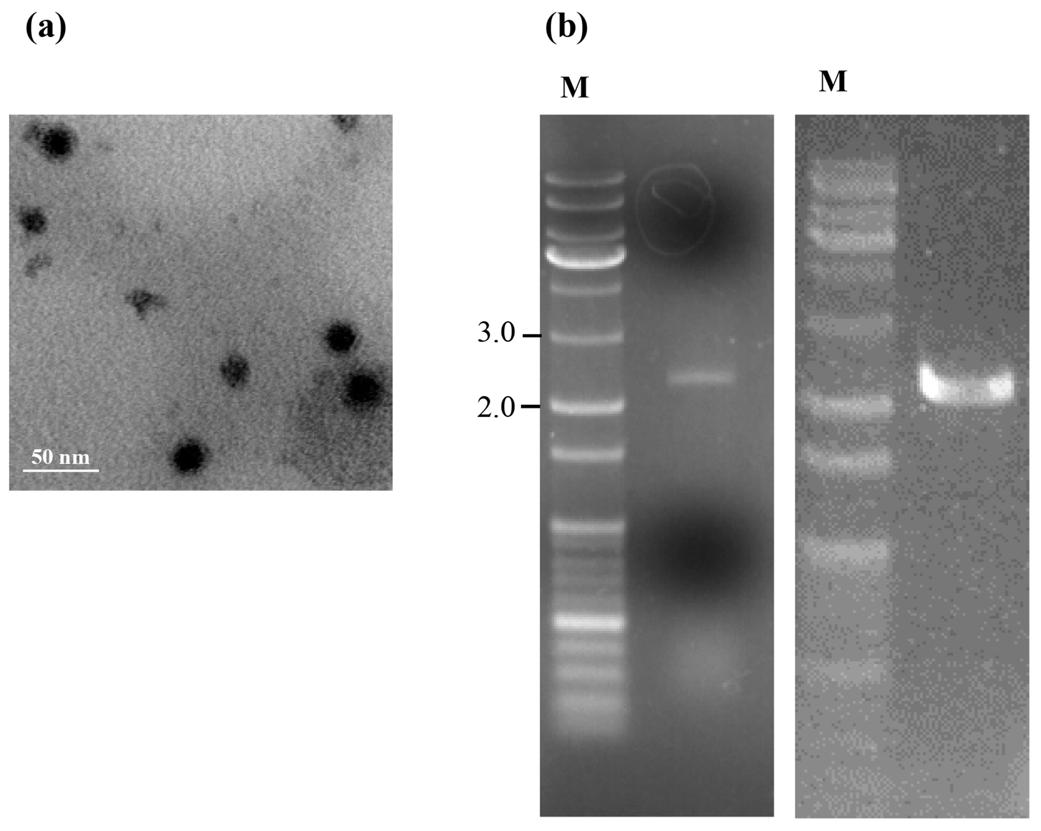
3.1. Profile of Virus Particles from T. atroviride NFCF394

Electron microscopy revealed that the isometric virus particles isolated from T. atroviride NFCF394 were isometric with a diameter of approximately 30 nm (Figure 1a), which is similar to the 25 to 40–50 nm diameter reported for members of the family Partitiviridae. Subsequently, nucleic acids extracted from the viral particles showed a broad ethidium bromide stained band at 2.0 kbp, which contained two dsRNA segments with a similar size of approximately 2.0 kbp. This agarose gel band pattern was identical to that of the dsRNA preparation from whole RNAs of infected fungal cells (Figure 1b). These data indicated that the dsRNAs identified from mycelia were indeed from the virus particles.

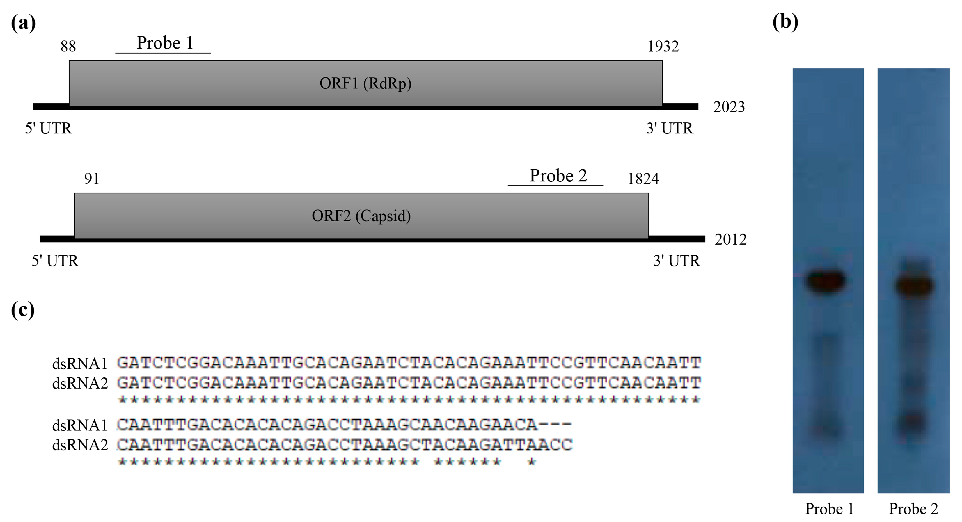
3.2. Molecular Characterization of Novel Partitivirus from T. atroviride NFCF394

The dsRNA extracted from the mycelia of T. atroviride NFCF394 was treated with DNase I and S1 nuclease, and the 2.0 kbp band was resolved with 1% agarose gel electrophoresis (Figure 1b right). The constructed cDNA library from the purified dsRNAs was subjected to next-generation sequencing (NGS) using Illumina HiSeq 2000. A total of 10,494 contigs with an average length of 379 nucleotides was produced. After quality trimming and assembly with Trinity, a total of 21 assembled contigs with significant FPKM values were obtained and further manually assembled. The assembled reads were subjected to a BLASTX search to identify the viral sequence in the NCBI protein database with an E-value cutoff of 0. The results showed two dsRNA segments identified as dsRNA1 and dsRNA2 containing a single open reading frame (ORF) for each segment (Figure 2a). To verify the contig sequences, RT-PCR analyses using corresponding primer pairs based on the two representative contigs (1948 and 1816 bp) were conducted. The resulting specific amplicons were cloned, at least three clones for each amplicon were sequenced, and the near full-length sequences obtained from NGS were verified. Northern hybridization demonstrated that both segments existed in the dsRNA band of the mycoviral genome (Figure 2b). These results confirmed that the dsRNA bands were double bands with similar sizes.
The sequence analysis of dsRNA1 revealed that it encoded an ORF (ORF1) consisting of 614 amino acids with a predicted molecular mass of 72 kDa and an isoelectric point (pI) of 8.2. Homology searches of the deduced amino acid sequence showed a high similarity to the known sequences of RdRp of Rosellinia necatrix partitivirus 7 (RnPV7), Rhizoctonia solani partitivirus 1 (RsPV1), Rosellinia necatrix partitivirus 5 (RnPV5), and Sclerotinia sclerotiorum partitivirus S (SsPV-S) (Table 1). In addition, ORF2 consisted of 577 amino acids with a predicted molecular mass of 65 kDa and pI of 6.7. ORF2 showed similarity to the known sequences of CP of Rosellinia necatrix partitivirus 7 (RnPV7) and Sclerotinia sclerotiorum partitivirus S (SsPV-S). Compared to the RdRp amino acid sequences, the CP amino acid sequence showed a lower level of similarity.
The RACE protocol was applied to determine the 5’ and 3’ terminal sequences. The full lengths of the dsRNA sequences for dsRNA1 and dsRNA2 were determined to be 2023 and 2012 bp, respectively. These sequences were deposited in GenBank (accession number MH921573 and MH921574, respectively). The 5’ untranslated region (UTR) of the coding strand of dsRNA1 was 87 nt, and the corresponding 5’ UTR of dsRNA2 was 90 nt. The nucleotide sequences at the 5’-termini of dsRNA1 and dsRNA2 shared a sequence identity of 99% (Figure 2c). A conserved sequence (GAWNW: N, any nt; W, A, or U) at the 5’-terminus of fungal Alphapartitivirus was observed at the 5’-termini of dsRNA1 and dsRNA2 (GACAAAUU). These sequences shared a characteristic feature in that the G near the 5’-termini was followed by an A, U, or C but not a G for the next five or six nucleotide positions [22]. In addition, the coding strands of dsRNA1 and dsRNA2 contained 3’ UTRs comprising 91 and 188 nt, respectively. A-rich regions interrupted by non-A residues in the 3’-termini of dsRNA1 (a sequence of 39 A residues in the 3’-terminal 50-nt) and dsRNA2 (a sequence of 26 A residues in the 3’-terminal 50-nt) were found, which is a common characteristic of Alphapartitivirus [8].

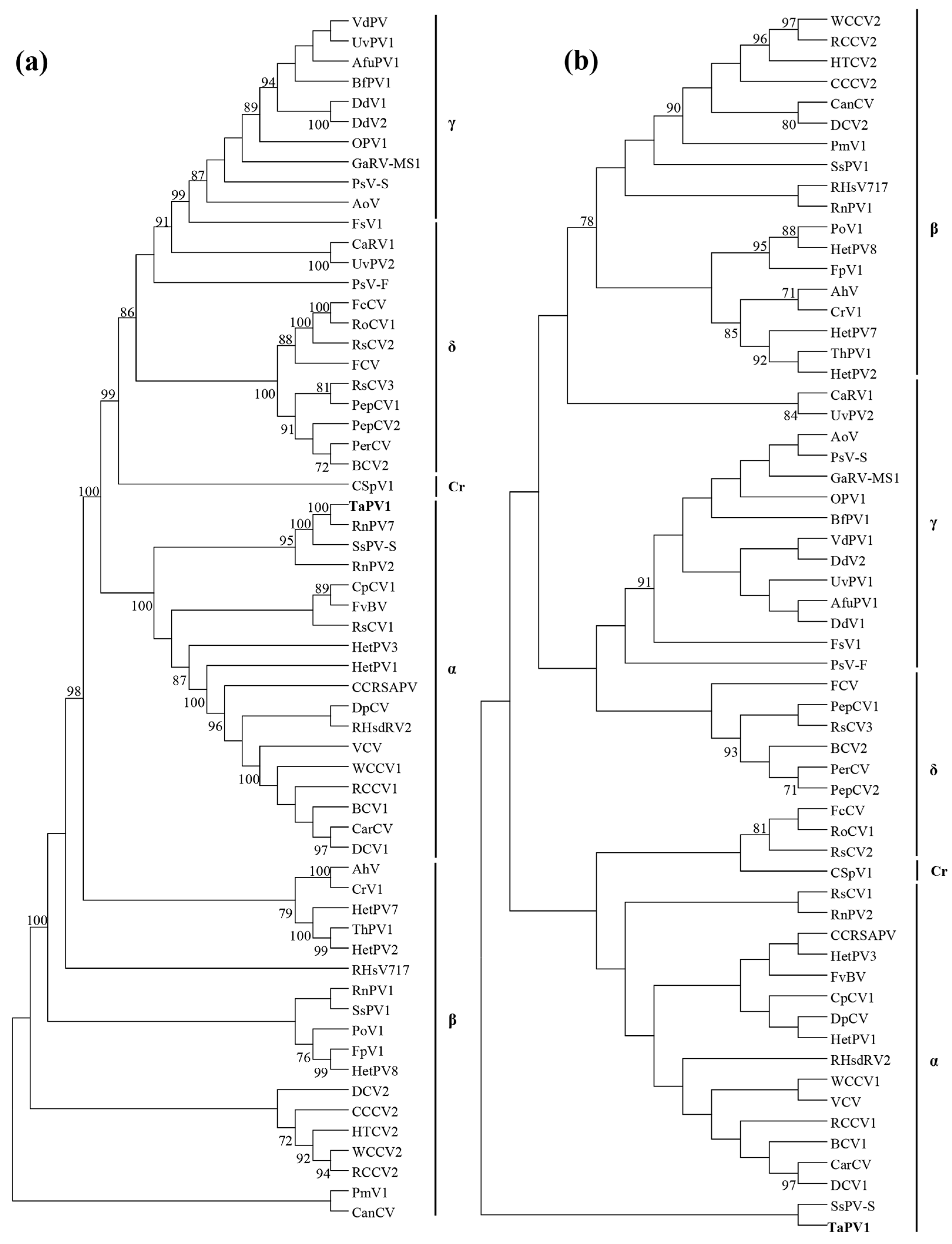
3.3. Phylogenic Analysis of Amino Acid Sequences Encoded by the ORFs

The phylogenetic analysis of RdRp with top-ranked similar sequences and selected members of Partitiviridae was performed using the maximum-likelihood method (Figure 3a). This revealed that RdRp encoded by ORF1 was affiliated with the clade encompassing the genus Alphapartitivirus with strong bootstrap support. Rosellinia necatrix partitivirus 7 and Sclerotinia sclerotiorum partitivirus S were the closest phylogenetic neighbors. In addition, the phylogenetic analysis of CP of selected members (Figure 3b) also indicated that this segment belongs to the genus Alphapartitivirus.
The genome organization of dsRNAs with bipartite segments, isometric viral particle structure, and virus size indicate that the dsRNAs belong to the family Paritiviridae. Together, the genome organization, sequence similarity of RdRp and CP, and the phylogenetic analysis suggest that the dsRNAs are genomic components of the genus Alphapartitivirus in the family Partitiviridae. Based on the cutoff values of the ICTV criterion for the genus Alphapartitivirus in the Partitiviridae demarcation (≤ 90% and ≤ 80% amino acid identities in RdRp and CP, respectively), we concluded that the dsRNA in the current study represents a novel species, of which dsRNA1 and dsRNA2 were proposed as genome segments of RdRp and CP in the family Alphapartitivirus. Thus, we named our dsRNA Trichoderma atroviride alphapartitivirus 1 (TaPV1). The genome size of TaPV1 is also interesting. The genome segment size of RdRp in TaPV1, which gave a genome length of 2023 bp, fell within the range of genome sizes (1866–2027 bp) of Alphapartitivirus members. In addition, the genome size of CP in TaPV1 (2012 bp) exhibited a substantially larger size than previous reports suggested (1708–1866 bp). Thus, the genome size of TaPV1 appears to be the largest of the fungal Alphapartitivirus organisms.

3.4. Phenotypic Characteristics of Mycovirus-Cured and -Containing Strains

T. atroviride NFCF394 was observed as white mycelium formed on the whole plate of PDA after incubation for 3 days. To determine whether the mycovirus affects the fungal host phenotype, virus-cured isogenic strains were obtained using single-spore isolation followed by hyphal tipping. Spores were harvested from 7-day-old culture plates containing actively growing T. atroviride NFCF394, and 50 expected colony-forming units were spread on a fresh PDA plate. Of the 20 single-spored progenies, TaPV1 dsRNA bands were eliminated in three single-spored progenies, suggesting the vertical transmission of TaPV1 at a rate consistent with our previous data [13]. However, no apparent difference in colony morphology was observed between TaPV1-containing and the three virus-cured strains. The activities of two representative antifungal enzymes, β-1,3-glucanase and chitinase, were also analyzed. No obvious alterations of enzymatic activities were observed between the infected and virus-cured isogenic strains (Figure S1). Comparisons of means were tested using the Student’s t-test.
Although there are many cases of mycoviruses that induce the viral-specific symptoms in the host, including reduced fungal virulence (hypovirulence) in phytopathogenic fungi [23], infection of mycoviruses is generally thought to be asymptomatic or cryptic in nature [24]. While further studies are required, no changes in fungal growth, colony morphology, or antifungal enzyme activity were observed in this study. However, to clarify the TaPV1-host interaction, studies that include viral transfection with purified viral particles will be necessary.

Supplementary Materials

Supplementary materials can be found at https://www.mdpi.com/1999-4915/10/11/578/s1.

Author Contributions

Supervision; D.-H.K.; Methodology, J.C. and H-E.Y.; Writing and Original Draft Preparation, J.C.; Writing, Review and Editing, D.-H.K.

Funding

This work was supported by the NRF grants from MSIP (2018R1A2A1A05078682).

Acknowledgments

We wish to thank the Institute of Molecular Biology and Genetics at Chonbuk National University for kindly providing the facilities for this research.

Conflicts of Interest

The authors declare that they have no conflict of interest.

References

  1. Sivasithamparam, K.; Ghisalberti, E.L. Trichoderma and Gliocladium: Basic Biology, Taxonomy and Genetics, 1st ed.; Taylor and Francis: London, UK, 1998; pp. 139–191. [Google Scholar]
  2. Kubicek, C.P.; Penttila, M.E. Trichoderma and Gliocladium: Enzymes, Biological Control and Commercial Applications, 1st ed.; Taylor and Francis: London, UK, 1998; pp. 49–71. [Google Scholar]
  3. Gupta, V.K.; Schmoll, M.; Herrera-Estrella, A.; Upadhyay, R.S.; Druzhinina, I.; Tuohy, M.G. Biotechnology and Biology of Trichoderma; Elsevier: Oxford, UK, 2014. [Google Scholar]
  4. Schuster, A.; Schmoll, M. Biology and biotechnology of Trichoderma. Appl. Microbiol. Biotechnol. 2010, 87, 787–799. [Google Scholar] [CrossRef] [PubMed]
  5. Contreras-Cornejo, H.A.; Macías-Rodríguez, L.; del-Val, E.; Larsen, J. Ecological functions of Trichoderma spp. and their secondary metabolites in the rhizosphere: Interactions with plants. FEMS Microbiol. Ecol. 2016, 92, fiw036. [Google Scholar] [CrossRef] [PubMed]
  6. Papavizas, G.C. Trichoderma and Gliocladium: Biology, ecology, and potential for biocontrol. Annu. Rev. Phytopathol. 1985, 23, 23–54. [Google Scholar] [CrossRef]
  7. Howell, C.R. Mechanisms employed by Trichoderma species in the biological control of plant diseases: The history and evolution of current concepts. Plant Dis. 2003, 87, 4–10. [Google Scholar] [CrossRef]
  8. Ghabrial, S.A.; Castón, J.R.; Jiang, D.; Nibert, M.L.; Suzuki, N. 50-plus years of fungal viruses. Virology 2015, 479–480, 356–368. [Google Scholar] [CrossRef] [PubMed]
  9. Wickner, R.B. Double-stranded and single-stranded RNA viruses of Saccharomyces cerevisiae. Annu. Rev. Microbiol. 1992, 46, 347–375. [Google Scholar] [CrossRef] [PubMed]
  10. The Online (10th) Report of the International Committee on Taxonomy of Viruses. Available online: https://talk.ictvonline.org/ictv-reports/ictv_online_report (accessed on 12 December 2016).
  11. Roossinck, M.J. Metagenomics of plant and fungal viruses reveals an abundance of persistent lifestyles. Front. Microbiol. 2015, 5, 1–3. [Google Scholar] [CrossRef] [PubMed]
  12. Yun, S.H.; Lee, S.H.; So, K.K.; Kim, J.M.; Kim, D.H. Incidence of diverse dsRNA mycoviruses in Trichoderma spp. causing green mold disease of shiitake Lentinula edodes. FEMS Microbiol. Lett. 2016. [Google Scholar] [CrossRef] [PubMed]
  13. Lee, S.H.; Yun, S.H.; Chun, J.; Kim, D.H. Characterization of a novel dsRNA mycovirus of Trichoderma atroviride NFCF028. Arch. Virol. 2017, 162, 1073–1077. [Google Scholar] [CrossRef] [PubMed]
  14. Chun, J.; Yang, H.E.; Kim, D.H. Identification of a novel partitivirus of Trichoderma harzianum NFCF319 and evidence for the related antifungal activity. Front. Plant Sci. under review.
  15. Kim, J.M.; Jung, J.E.; Park, J.A.; Park, S.M.; Cha, B.J.; Kim, D.H. Biological function of a novel chrysovirus, CnV1-Bs122, in the Korean Cryphonectria nitschkei BS122 strain. J. Biosci. Bioeng. 2013, 115, 1–3. [Google Scholar] [CrossRef] [PubMed]
  16. Wang, L.; Jiang, J.; Wang, Y.; Hong, N.; Zhang, F.; Xu, W.; Wang, G. Hypovirulence of the phytopathogenic fungus Botryosphaeria dothidea: Association with a coinfecting chrysovirus and a partitivirus. J. Virol. 2014, 88, 7517–7527. [Google Scholar] [CrossRef] [PubMed]
  17. Park, S.M.; Kim, J.M.; Chung, H.J.; Lim, J.Y.; Kwon, B.R.; Lim, J.G.; Kim, J.A.; Kim, M.J.; Cha, B.J.; Lee, S.H.; et al. Occurrence of diverse dsRNA in a Korean population of the chestnut blight fungus, Cryphonectria parasitica. Mycol. Res. 2008, 112, 1220–1226. [Google Scholar] [CrossRef] [PubMed]
  18. Li, B.; Dewey, C.N. RSEM: Accurate transcript quantification from RNA-Seq data with or without a reference genome. BMC Bioinform. 2011, 12, 323. [Google Scholar] [CrossRef] [PubMed]
  19. Felsenstein, J. Confidence limits on phylogenies: An approach using the bootstrap. Evolution 1985, 39, 783–791. [Google Scholar] [CrossRef] [PubMed]
  20. Kumar, S.; Stecher, G.; Tamura, K. MEGA7: Molecular Evolutionary Genetics Analysis version 7.0 for bigger datasets. Mol. Biol. Evol. 2016, 33, 1870–1874. [Google Scholar] [CrossRef] [PubMed] [Green Version]
  21. Thompson, J.D.; Gibson, T.J.; Plewniak, F.; Jeanmougin, F.; Higgins, D.G. The CLUSTAL_X windows interface: Flexible strategies for multiple sequence alignment aided by quality analysis tools. Nucleic Acids Res. 1987, 25, 4876–4882. [Google Scholar] [CrossRef]
  22. Nibert, M.L.; Ghabrial, S.A.; Maiss, E.; Lesker, T.; Vainio, E.J.; Jiang, D.; Suzuki, N. Taxonomic reorganization of family Partitiviridae and other recent progress in partitivirus research. Virus Res. 2014, 88, 128–141. [Google Scholar] [CrossRef] [PubMed]
  23. Van Alfen, N.K. Hypovirulence of Endothia (Cryphonectria) parasitica and Rhizoctonia solani. In Fungal Virology, 1st ed.; Buck, K.W., Ed.; CRC Press: Boca Raton, FL, USA, 1986; pp. 143–162. [Google Scholar]
  24. Pearson, M.N.; Beever, R.E.; Boine, B.; Arthur, K. Mycoviruses of filamentous fungi and their relevance to plant pathology. Mol. Plant. Pathol. 2009, 10, 115–128. [Google Scholar] [CrossRef] [PubMed]
Figure 1. Profiles of isometric viral particles isolated from T. atroviride NFCF394. (a) Purified virus particles were negative-stained with 2% uranyl acetate and examined using transmission electron microscopy. Scale bar, 50 nm. (b) Agarose gel electrophoresis of the dsRNAs extracted from viral particles (left) and mycelia (right) of virus-infected T. atroviride NFCF394. Lane M contains the DNA size standard and the numbers indicate the size in kbp.
Viruses 10 00578 g001
Figure 2. Molecular characteristics of the T. atroviride partitivirus 1 (TaPV1) genomic double-stranded RNAs (dsRNAs). (a) Schematic diagrams of the genomic organization of TaPV1 dsRNA segments. Shaded boxes are open reading frames (ORFs) encoding RNA-dependent RNA polymerase (RdRp) and coat protein (CP). Numbers indicate the total lengths of the TaPV1 genome segments and the positions of the start and stop codons. (b) Northern blot analysis of TaPV1 dsRNA1 and dsRNA2. RNAs were hybridized with probes for dsRNA1 and dsRNA2, and these probes are indicated in panel (b). (c) Alignment of the 5’-terminal untranslated regions of dsRNA1 and dsRNA2 of TaPV1. The conserved sequences are indicated with asterisks.
Viruses 10 00578 g002
Figure 3. Phylogenetic analysis of TaPV1 and selected dsRNA viruses. (a) Maximum-likelihood (ML) tree using LG + F substitution model based on the RdRp amino acid sequences of Partitiviridae. α, Alphapartitivirus; β, Betapartitivirus; γ, Gammapartitivirus; δ, Deltapartitivirus; Cr, Cryspovirus. (b) ML tree for CP amino acid sequences of Partitiviridae. α, Alphapartitivirus; β, Betapartitivirus; γ, Gammapartitivirus; δ, Deltapartitivirus; Cr, Cryspovirus. The numbers at the nodes represent bootstrap values out of 1000 replicates; values are shown only if greater than 70%. See Table S1 for a detailed listing of viruses.
Viruses 10 00578 g003
Table 1. Amino acid sequence identity (%) between T. atroviride alphapartitivirus 1 (TaPV1) ORF1, ORF2 and other viruses from the genus Alphapartitivirus.
VirusIdentity (%)Overlap
ORF1 search
RnPV7 (LC076694)60.5376/622
RsPV1 (AND83003)52.6328/623
RnPV5 (BAM36403)52.8344/652
SsPV-S (GQ280377)42.1263/625
ORF2 search
RnPV7 (BAT32943)26.7178/666
SsPV-S (GQ280378)21.9138/630

