Article

Potential of RNAi Targeting Juvenile Hormone Acid Methyltransferase (JHAMT) for Controlling Dendroctonus valens LeConte (Coleoptera: Scolytidae)


1
School of Life Sciences, Hebei University, Baoding 071002, China
2
Engineering Research Center of Ecological Safety and Conservation in Beijing-Tianjin-Hebei (Xiong’an New Area) of MOE, Hebei University, Baoding 071002, China
3
Pangquangou National Nature Reserve Administration, Lüliang 030510, China
*
Authors to whom correspondence should be addressed.
Forests 2026, 17(5), 628; https://doi.org/10.3390/f17050628
Submission received: 16 April 2026 / Revised: 15 May 2026 / Accepted: 20 May 2026 / Published: 21 May 2026
(This article belongs to the Special Issue Advances in Wood Borer Control and Management)

Abstract

Dendroctonus valens LeConte represents a major invasive pest species in China. Both larvae and adults primarily feed on the phloem of the tree trunk base and roots, disrupting nutrient transport and leading to host tree mortality, which poses a severe threat to forest ecosystems and the forestry economy. Juvenile hormone acid methyltransferase (JHAMT) is a key enzyme in insect juvenile hormone (JH) biosynthesis. In this study, we identified a JHAMT-encoding gene, DvJHAMT, in D. valens via bioinformatic analysis. RT-qPCR analysis revealed that DvJHAMT is predominantly expressed during the egg and larval stages. In the fourth-instar larvae, the highest expression levels were observed in the head and epidermis, suggesting a central regulatory role during this critical developmental period. To investigate its function via RNA interference (RNAi), a nanomaterial, star polycation (SPc), was employed for the transdermal delivery of dsRNA into the fourth-instar larvae. The results demonstrated that DvJHAMT knockdown significantly downregulated mRNA levels, resulting in marked decreases in larval survival, pupation, and eclosion rates. Notably, treatment with 0.7 µg dsDvJHAMT-SPc resulted in a 96.67% mortality rate and a reduced pupation rate of 41.67% at 34 days post-treatment. Furthermore, RNAi led to developmental deformities and significant weight loss in larvae. ELISA assays confirmed that DvJHAMT silencing led to reduced JHAMT enzyme activity and JH III titers in a dose-dependent manner. In conclusion, our findings demonstrate that DvJHAMT plays a vital role in JH biosynthesis and that its suppression exhibits potent lethal effects, suggesting that DvJHAMT is a promising candidate for RNAi-based management of D. valens.

1. Introduction

The red turpentine beetle (Dendroctonus valens LeConte), one of the main invasive pests in China, is native to North America and was introduced to China in the early 1980s. It has since caused extensive damage to pine forests [1]. It is estimated that by 2010, this invasive pest had infested over 500,000 hectares of pine forests, resulting in the mortality of tens of millions of pine trees. Such widespread destruction poses a severe threat to both forest ecological security and the forestry economy in China [2,3].
In China, the successful establishment and devastating outbreaks of D. valens are at tributed to several factors, including the prevalence of susceptible hosts like Pinus tabuliformis, favorable climatic conditions, and lack of co-evolved natural enemies. Additionally, its high reproductive and dispersal capacities, combined with its role as a vector for pathogenic fungi, further exacerbate its impact on forest health [4,5,6,7]. The beetle feeds primarily on the basal trunk cambium, disrupting the conductive tissues of trees and leading to tree mortality accelerated by pathogenic fungi [8,9]. Its potential range is likely to continue expanding.
Juvenile hormone (JH) serves as a central regulator of numerous physiological processes, including growth, development, molting, metamorphosis, and reproduction [10,11,12,13,14]. JH is an acyclic sesquiterpenoid synthesized in the insect corpora allata (CA) via a multi-step enzymatic pathway [15]. Its biosynthesis is a two-stage enzymatic process. First, acetyl-CoA is converted to farnesyl pyrophosphate (FPP) through a series of reactions in the mevalonate (MVA) pathway [16,17]. During the second stage, FPP serves as the precursor for JH biosynthesis. In this stage, Juvenile hormone acid methyltransferase (JHAMT) catalyzes the methylation of juvenile hormone acid (JHA) to produce biologically active JH and is a rate-limiting enzyme in JH biosynthesis [16,18].
JHAMT belongs to the S-adenosylmethionine (SAM)-dependent methyltransferase superfamily and catalyzes the transfer of a methyl group from SAM to the carboxyl group of JH acid [19]. To date, JHAMT has been successfully cloned and functionally characterized in a variety of insect species, including Drosophila melanogaster, Tribolium castaneum, Leptinotarsa decemlineata, and Monolepta hieroglyphica [20,21,22,23].
In recent years, RNA interference (RNAi) technology has been demonstrated to be a useful tool for study of insect gene function and development of novel pest control strategy [24]. Encapsulation of dsRNA with nanomaterials significantly enhances its stability in both the environment and within the insect body, thereby offering a key strategy for improving the efficacy of RNAi-based gene function study or pest control technologies [25]. JHAMT is conserved among insects and is absent in vertebrate genomes, therefore it is regarded as an ideal target for developing eco-friendly pest management approaches [24,25]. In recent years, RNAi technology has been demonstrated to be a useful tool for the study of insect gene function and development of novel pest control strategies [24]. Functional studies showed that knockdown or knockout of JHAMT significantly disrupts insect growth, development, reproduction, and even diapause, further underscoring its pivotal role in physiological regulation [26,27,28,29]. Nanomaterial-aided RNA interference of insect JHAMT is a promising prospect in insect pest control. For instance, foliar spraying of chitosan nanoparticles-dsRNA complexes targeting JHAMT achieved efficient gene silencing and 100% larval mortality in the cotton bollworm (Helicoverpa armigera) [30]. The nanomaterial star polycation (SPc), characterized by high efficiency, low cost, and low cytotoxicity, has shown great potential as a dsRNA carrier in pest control applications [31,32]. Encapsulation of dsRNA with nanomaterials significantly enhances its stability in both the environment and within the insect body, thereby offering a key strategy for improving the efficacy of RNAi-based gene function study or pest control technologies [33]. This provides a viable technical basis for developing RNAi-based control strategies targeting DvJHAMT.
To obtain a better understanding of the role of DvJHAMT in the growth and development of D. valens, we systematically investigated the function of JHAMT in this study. Molecular cloning, phylogeny, and expression profiles of DvJHAMT were performed in D. valens. Subsequently, RNAi-mediated knockdown of DvJHAMT was performed, and the changes in both JHAMT enzyme activity and juvenile hormone III (JH III) titer were quantified using enzyme-linked immunoassays (ELISA). Alongside observations of changes in the phenotype of the insect, we are capable of acquiring more knowledge on the role of DvJHAMT in JH biosynthesis and its impact on insect development. These findings would contribute to the development of novel, environment-friendly strategies to control D. valens.

2. Materials and Methods

2.1. Insect Collection and Rearing

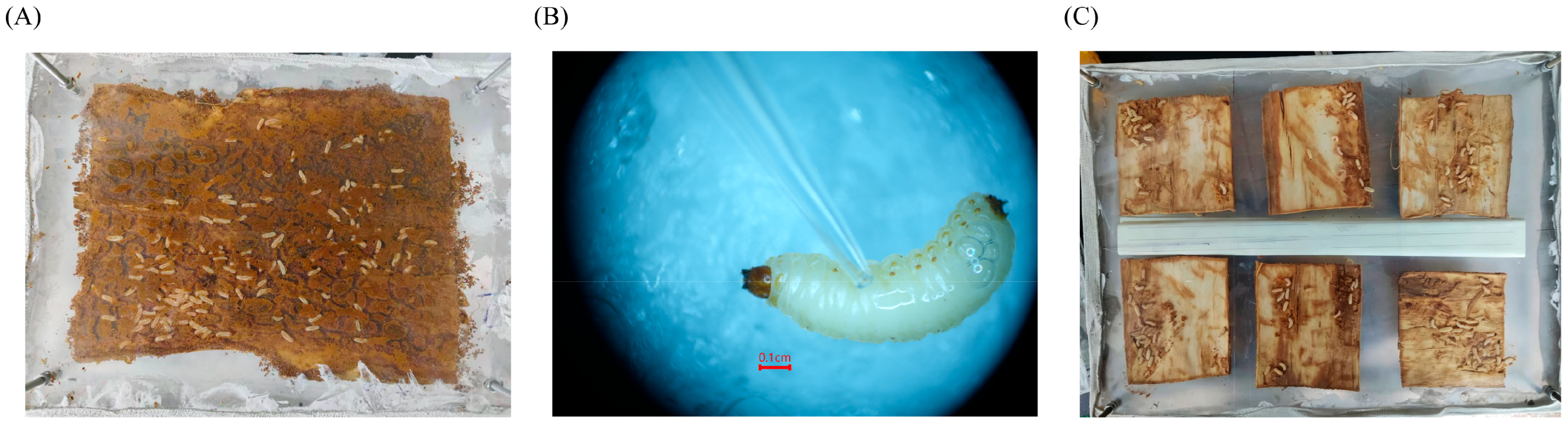
Fourth-instar larvae of D. valens were collected in May and September 2025 from Longxing Forest Farm (122°20′ E, 47°23′ N) in Lüliang, Shanxi Province, China. To facilitate indoor rearing, a sandwich bark assembly was constructed. Fresh pine bark (Pinus tabuliformis) with intact phloem was stripped and sectioned into square piece (approx. 20 × 10 cm). These bark sections were sandwiched between two clean, transparent acrylic plates and secured at the plant corners with screws to simulate the compressive pressure found within the host tree. To prevent escape, the edges were sealed with a combination of gauze and plaster, with small ventilation holes drilled into the plaster (Figure 1A). The rearing units were maintained in constant darkness at 25 ± 3°C and 50% relative humidity, mimicking the sub-cortical microenvironment of pine trees to support normal larval development.

2.2. Molecular Cloning of DvJHAMT

The cDNA sequence of DvJHAMT was identified through BLAST alignment against the D. valens genome [34] and transcriptome datasets [35]. Gene-specific primers were designed using Primer Premier 6.0 (Premier Biosoft International, Palo Alto, CA, USA) to amplify the complete open reading frame (ORF) of DvJHAMT. The primer sequences, along with their respective melting temperatures ( T m ) and GC content, are detailed in Table S1.
Total RNA was extracted using the TransZol Up Plus RNA Kit (TransGen Biotech, Beijing, China) following the manufacturer’s protocol. RNA concentration and integrity were assessed using a Micro Drop SE nucleic acid protein analyzer (Shanghai BIO-DL Science Instrument Co., Ltd., Shanghai, China) and agarose gel electrophoresis. Subsequently, RT-PCR was performed to amplify the DvJHAMT gene. The resulting PCR products were electrophoresed on a 1% (w/v) agarose gel, and target fragments of the expected size were recovered and purified using the Gel DNA Extraction Mini Kit (Vazyme Biotech, Nanjing, China). The purified DNA was ligated into a pTOPO-TA (Aidlab Biotechnologies Co., Ltd., Beijing, China) vector and transformed into Escherichia coli DH5α competent cells. Positive clones were selected, and recombinant plasmids were isolated using the Plasmid Miniprep Kit (Beiwo Medical, Hangzhou, China). Finally, the inserts were sequenced and verified by Sangon Biotech (Shanghai, China).

2.3. Bioinformatic Analysis

The ORF of the DvJHAMT gene was determined by analyzing the cDNA sequence with ORF Finder (https://www.ncbi.nlm.nih.gov/orffinder/, accessed on 10 January 2025). The molecular weight (MW, kDa) and isoelectric point (pI) of the protein were predicted using the ExPASy Proteomics Server (http://expasy.org/, accessed on 20 July 2025). Subcellular localization of the protein was predicted via the Cell-PLoc (http://www.csbio.sjtu.edu.cn/bioinf/Cell-PLoc/, accessed on 10 July 2024). Hydrophobicity/hydrophilicity analysis of the protein was performed using the Protscale online tool (https://web.expasy.org/protscale/, accessed on 20 July 2025). Homologous JHAMT sequences were retrieved from the NCBI GenBank. Multiple sequence alignment was performed using DNAMAN Version 9 (Lynnon Biosoft, San Ramon, CA, USA). A phylogenetic tree was subsequently constructed using the neighbor-joining (NJ) method in MEGA11 (Molecular Evolutionary Genetics Analysis, Tempe, AZ, USA) with 1000 bootstrap replicates. To visualize and annotate the resulting tree, iTOL (https://itol.embl.de/, accessed on 15 November 2025) was used. Table S1 provides the GenBank accession numbers for all sequences used in this study.

2.4. DvJHAMT Expression Profiling

Total RNA was extracted from D. valens individuals of different developmental stages, including eggs, larvae, pupae, and adults, all of which were collected directly from infested pine trees in the field (Taiyue Mountain, Shanxi Province, China). The extraction was performed using the TransZol Up Plus RNA Kit according to the manufacturer’s instructions. Briefly, due to the substantial variation in body size and tissue mass among different developmental stages, the number of individuals per RNA extraction sample was adjusted to ensure sufficient RNA yield: ten eggs, five first-instar larvae, one individual of 2nd–4th-instar larvae, one pupa, or one adult. Additionally, tissue samples including head, gut, Malpighian tubules, fat body, and epidermis, were collected from 20 fourth-instar larvae under a stereomicroscope for subsequent total RNA extraction and cDNA synthesis. All samples were analyzed with three biological replicates. RNA concentration and integrity were assessed using a Micro Drop SE nucleic acid-protein analyzer (Shanghai BIO-DL Science Instrument Co., Ltd., Shanghai, China) and agarose gel electrophoresis. Subsequently, 1 μg of total RNA from each sample was used as a template for cDNA synthesis, which was performed following the instructions of the HiFiScript gDNA Removal RT MasterMix (CWBIO, Taizhou, China).
Total RNA was extracted from D. valens at various developmental stages using the TransZol Up Plus RNA Kit (TransGen Biotech, Beijing, China) according to the manufacturer’s protocol. Sampling was standardized as follows: 10 eggs, 5 first-instar larvae, and 1 individual each for the second to fourth larval instars, pupae, and adults. For tissue-specific expression analysis, the head, gut, Malpighian tubules, fat body, and epidermis were dissected from 20 individuals of the fourth-instar larvae under a stereomicroscope. All samples were prepared in three biological replicates. A Micro Drop SE nucleic acid-protein analyzer was used to assess RNA concentration and purity, while integrity was verified via 1% agarose gel electrophoresis. Subsequently, first-strand cDNA was synthesized from the total RNA using the HiFiScript gDNA Removal RT MasterMix (CWBIO, Taizhou, China), which includes a genomic DNA removal step to ensure template purity.
The spatio-temporal expression profile of DvJHAMT was analyzed via A SYBR Green I-based RT-qPCR method. The tubulin (TUB) [35] gene was utilized as an internal reference for normalization. All RT-qPCR primers are listed in Table S1. Relative expression levels were calculated using the 2−ΔΔCT method [36,37]. Each experimental condition was represented by three biological replicates, with three technical replicates performed for each reaction to ensure analytical reproducibility.

2.5. Synthesis of Double-Stranded RNA

To generate double-stranded RNA (dsRNA), primers targeting a specific region of the DvJHAMT coding sequence (CDS) were designed using Primer Premier 6.0. To minimize potential off-target effects, a 304 bp fragment (nucleotides 91–394 of the CDS) was selected. This sequence showed no significant homology to other genomic regions of D. valens (E-value > 1 × 10⁻⁵; no continuous identical sequences 20 nt) based on NCBI BLASTn analysis against the D. valens genome. The T7 promoter sequence (5′-TAATACGACTCACTATAGGG-3′) was appended to the 5′ ends of both the forward and reverse primers. A fragment of the Enhanced Green Fluorescent Protein (EGFP) gene was amplified from the pEGFP-N2 plasmid (NCBI Accession No. NG_017013.2) and used as a non-endogenous control. Synthesis of dsRNA was performed using the T7 RNAi Transcription Kit (Vazyme Biotech, Nanjing, China) according to the manufacturer’s instructions. The resulting dsRNA products were purified by ethanol precipitation, resuspended in nuclease-free water, and quantified using a spectrophotometer. The integrity of the dsRNA was verified by 1% agarose gel electrophoresis.

2.6. RNA Interference

The Star Polycation (SPc) nanocarrier used in this study was obtained from Prof. Jie Shen (China Agricultural University). The preparation and characterization of SPc followed the previously described protocols [38]. To prepare the SPc/dsRNA complex, purified dsRNA was mixed with SPc at a mass ratio of 1:1.5 and incubated at room temperature for 15 min. The successful encapsulation of dsRNA within the SPc nanocarrier was confirmed via agarose gel electrophoresis, where the immobilization of dsRNA in the loading well indicated complete complex formation. The final concentrations of the dsDvJHAMT-SPc complexes were adjusted to 0.3, 0.5, and 0.7 µg/µL.
Healthy fourth-instar larvae of uniform body weight were selected for the RNAi experiments. Larvae were briefly anesthetized at low temperature (4 °C) before treatment. A 1 µL droplet of the dsDvJHAMT-SPc formulation was applied to the dorsal abdominal plate of each larva using a micro-pipette (Eppendorf, Hamburg, Germany) (Figure 1B). Control larvae were treated with an equivalent dose of a dsEGFP-SPc complex. Each treatment group consisted of three biological replicates, with 23 larvae per replicate. Following a 30 min absorption period to ensure complete transdermal delivery, the larvae were returned to collective rearing under standard conditions (Figure 1C). It should be noted that larvae were maintained in groups, and developmental parameters such as body weight, deformity rate, and mortality were assessed at the group level rather than by tracking individual larvae over time. Therefore, the data represent population-level trends across independent biological replicates.
To evaluate the biological impact of DvJHAMT silencing, developmental indicators including mortality, deformity rate, body weight, pupation rate, and emergence rate were recorded daily. Morphological changes were documented using an SZMN stereomicroscope (SOPTOP, Ningbo, China).
To assess the duration and magnitude of the RNAi effect, total RNA was extracted from one randomly selected larva per biological replicate at 24, 48, and 72 h post-treatment. Following cDNA synthesis, RT-qPCR was performed to determine the transcript levels of DvJHAMT. Relative expression levels were calculated using the 2−ΔΔCt method [36,37], with three biological and three technical replicates for each time point.

2.7. Quantification of JH Titers and JHAMT Protein Levels

To analyze the effects of topical dsDvJHAMT-SPc application on JH biosynthesis and JHAMT enzyme activity in D. valens larvae, JH titers and JHAMT enzyme activity were quantified using enzyme-linked immunosorbent assays (ELISA). Samples were collected from larvae in both the dsDvJHAMT-SPc and dsEGFP-SPc groups on days 1, 2, 3, 5, 10, and 15 post-RNAi treatment. The assays were performed using an Insect Juvenile Hormone (JH) ELISA Kit and an Insect Juvenile Hormone Acid Methyltransferase (JHAMT) ELISA Kit (EnzymeLink Biotechnology Co., Ltd., Shanghai, China) according to the manufacturer’s instructions. For each time point and treatment, five biological replicates were analyzed. Optical absorbance at 450 nm was measured using a Sunrise F50 microplate reader (Tecan Trading AG, Zürich, Switzerland), and concentrations were calculated based on a standard curve generated with the provided standards.

2.8. Statistical Analysis

Data processing and visualization were performed using GraphPad Prism 6.0 (GraphPad Software Inc., San Diego, CA, USA). All data are presented as the mean ± standard error of the mean (SEM). Differences between two experimental groups were analyzed using Student’s t-test. For comparisons among multiple groups, a one-way analysis of variance (ANOVA) was performed, followed by Tukey’s post hoc test for multiple comparisons. Survival data were analyzed using the Kaplan–Meier method, and comparisons be-tween groups were performed using log-rank tests. Statistical significance was set at p ≤ 0.05. Statistical significance was set at p ≤ 0.05. In the figures, significant differences are indicated by different lowercase letters above the bars.

3. Results

3.1. Bioinformatics Analysis of DvJHAMT

Based on the D. valens genome and transcriptome data combined with our sequencing analysis, the cDNA fragment of the DvJHAMT (GenBank: PX861299) contains an ORF of 846 bp encoding a protein of 281 amino acids. The predicted molecular weight is 32.48 kDa, with a theoretical isoelectric point (pI) of 8.84. Amino acid composition analysis showed that leucine (10.3%) and serine (7.5%) are the most abundant residues. No signal peptide cleavage site was identified, and DvJHAMT is primarily distributed in the cytoplasm.
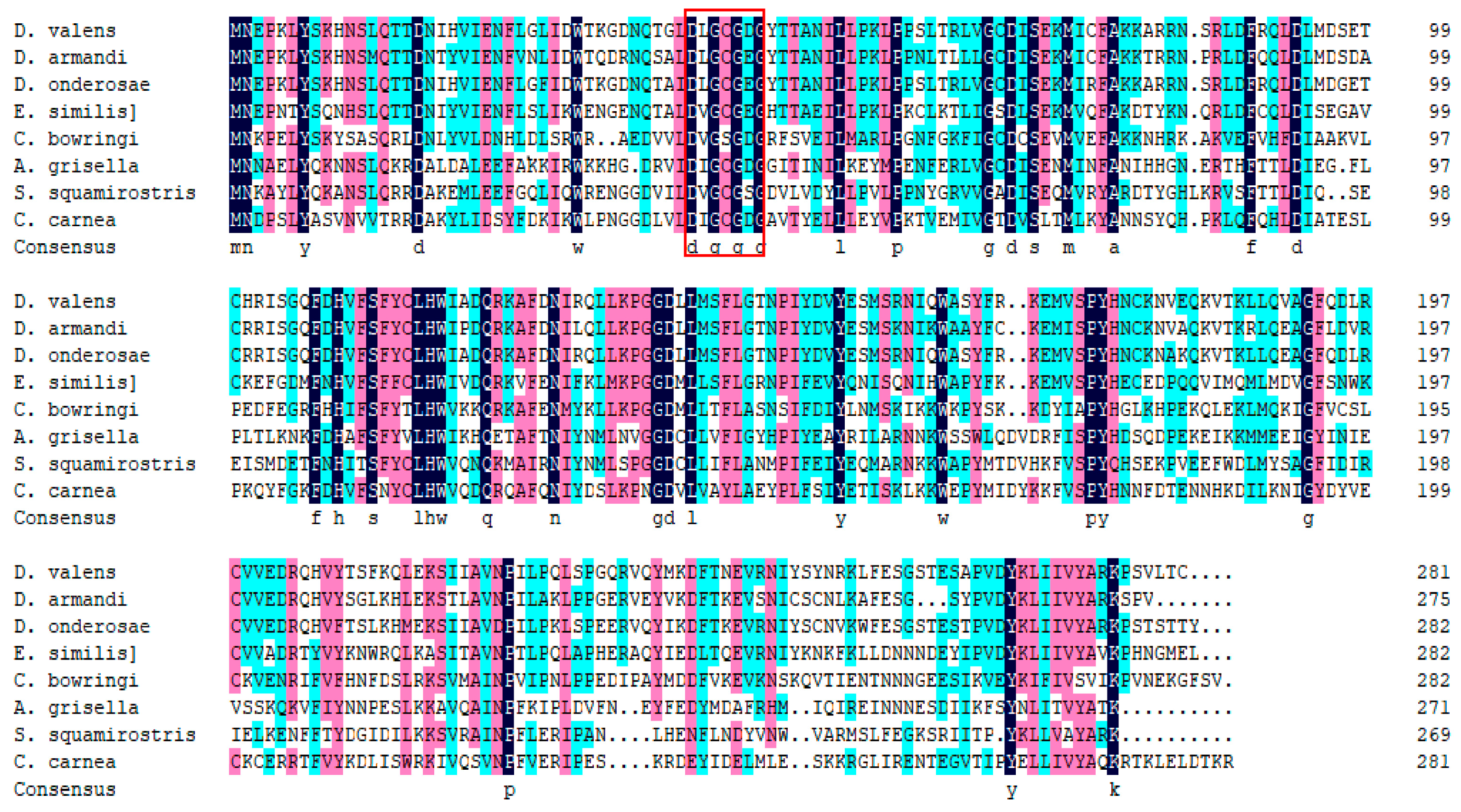
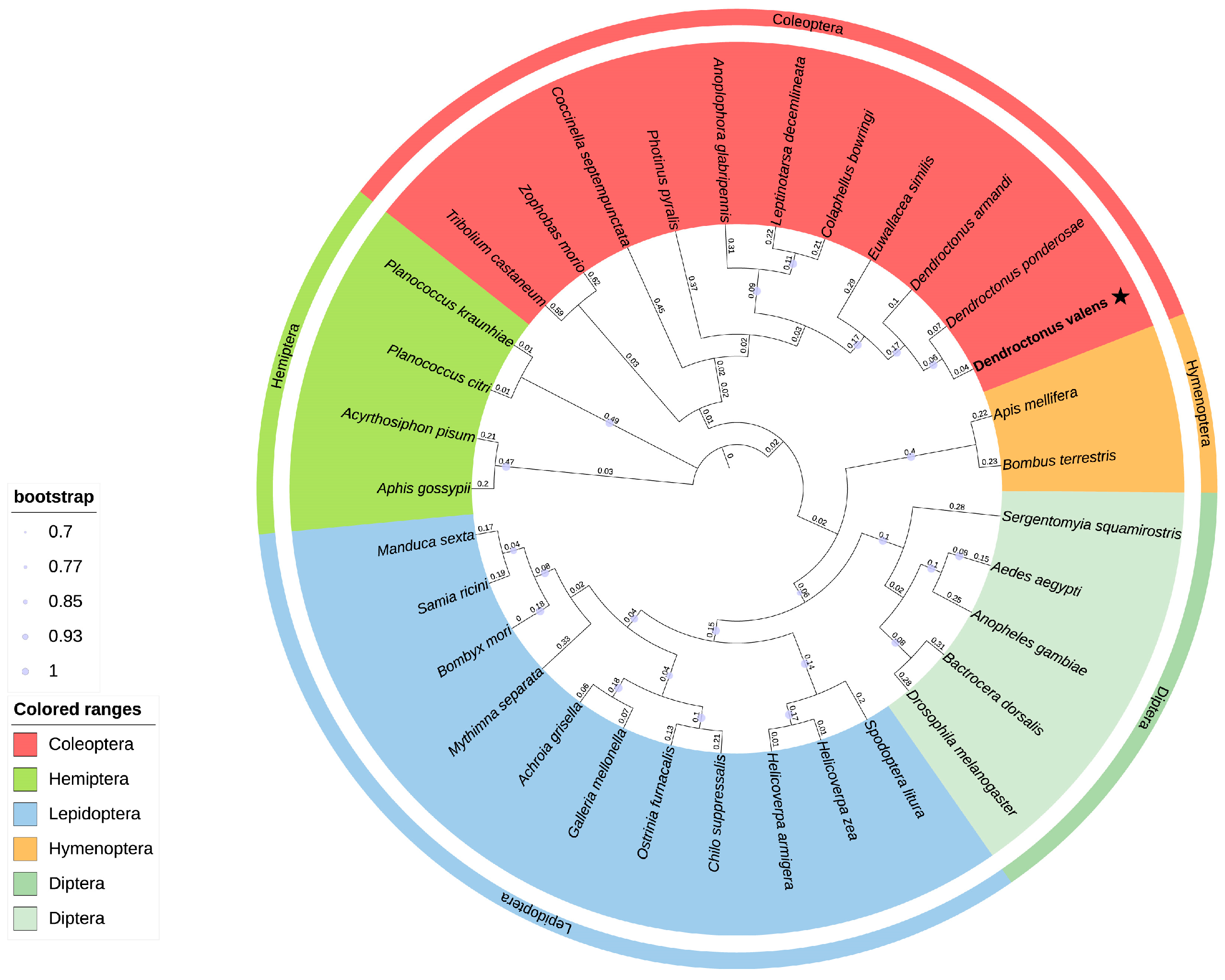
Sequence alignment of DvJHAMT with seven JHAMTs from seven other insect species was performed using DNAMAN9 (Figure 2). The SAM-binding motif, hh(D/E)hGXGXG (where h represents a hydrophobic residue, X represents any amino acid) [39], was found within the first motif (amino acid residues 41–47) of DvJHAMT. Phylogenetic analysis further showed that the DvJHAMT protein shares the highest similarity with JHAMTs from D. armandi and D. ponderosae, clustering within the Coleoptera clade (Figure 3). These results indicate that DvJHAMT is closely related to JHAMT proteins from other coleopteran insects.

3.2. Spatiotemporal Expression Pattern of DvJHAMT

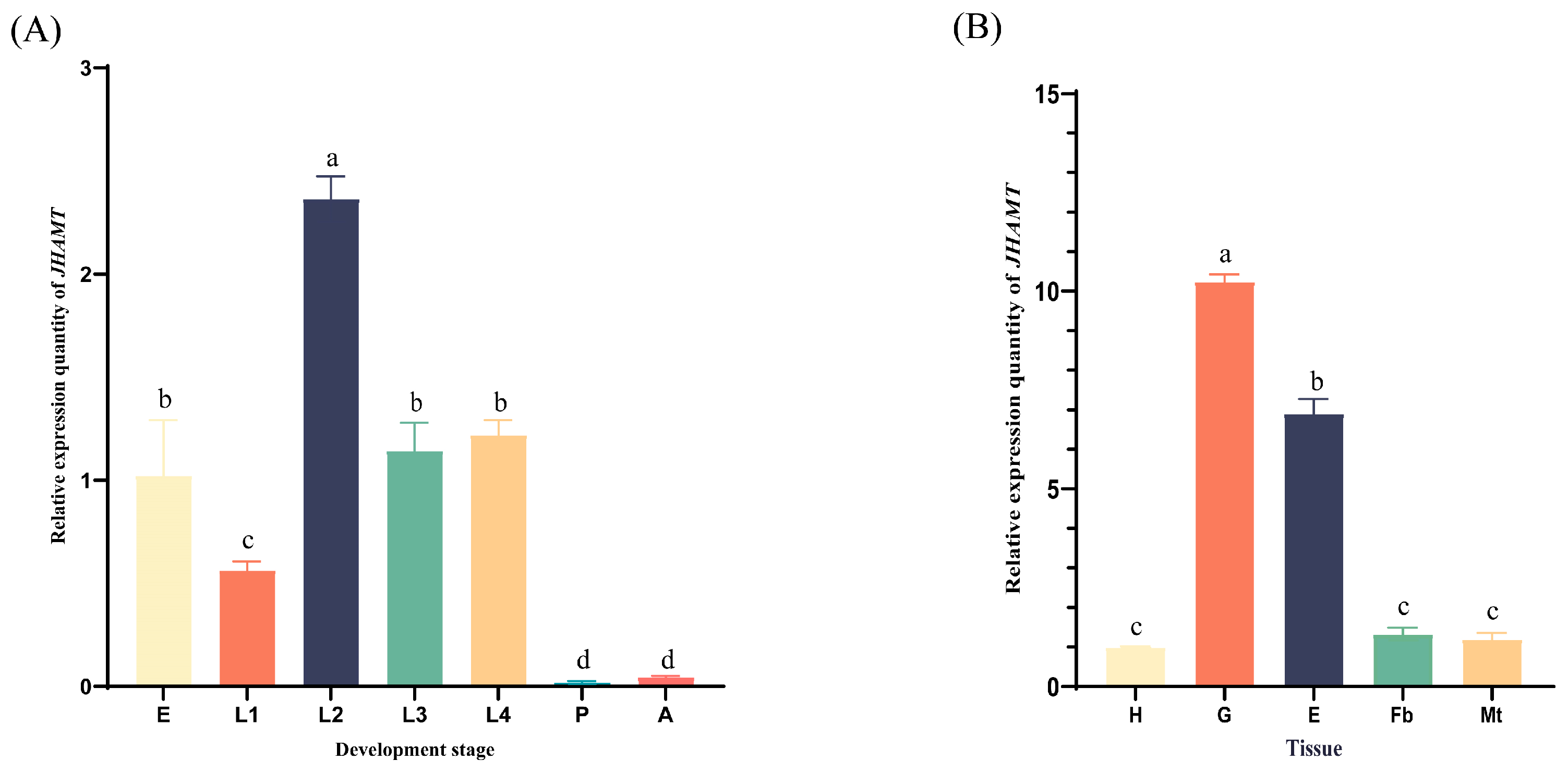
The relative expression levels of the DvJHAMT gene across different developmental stages of D. valens were analyzed using RT-qPCR. The results indicated that transcription levels varied significantly among stages. Significant expression was detected in eggs and all larval instars, whereas expression in the pupal and adult stages was minimal (Figure 4A). The highest relative expression level of DvJHAMT was found in the second instar larval stage, followed by the third and the fourth larval stages.
Analysis of different larval tissues showed that DvJHAMT was expressed in all examined tissues, but with significant variation in expression levels. The highest relative expression was observed in the gut, where it was approximately 1.5-fold higher than in the epidermis, 10.2-fold higher than in the head, 7.7-fold higher than in the Malpighian tubules, and 8.5-fold higher than in the fat body (Figure 4B).

3.3. Silencing Efficiency of DvJHAMT

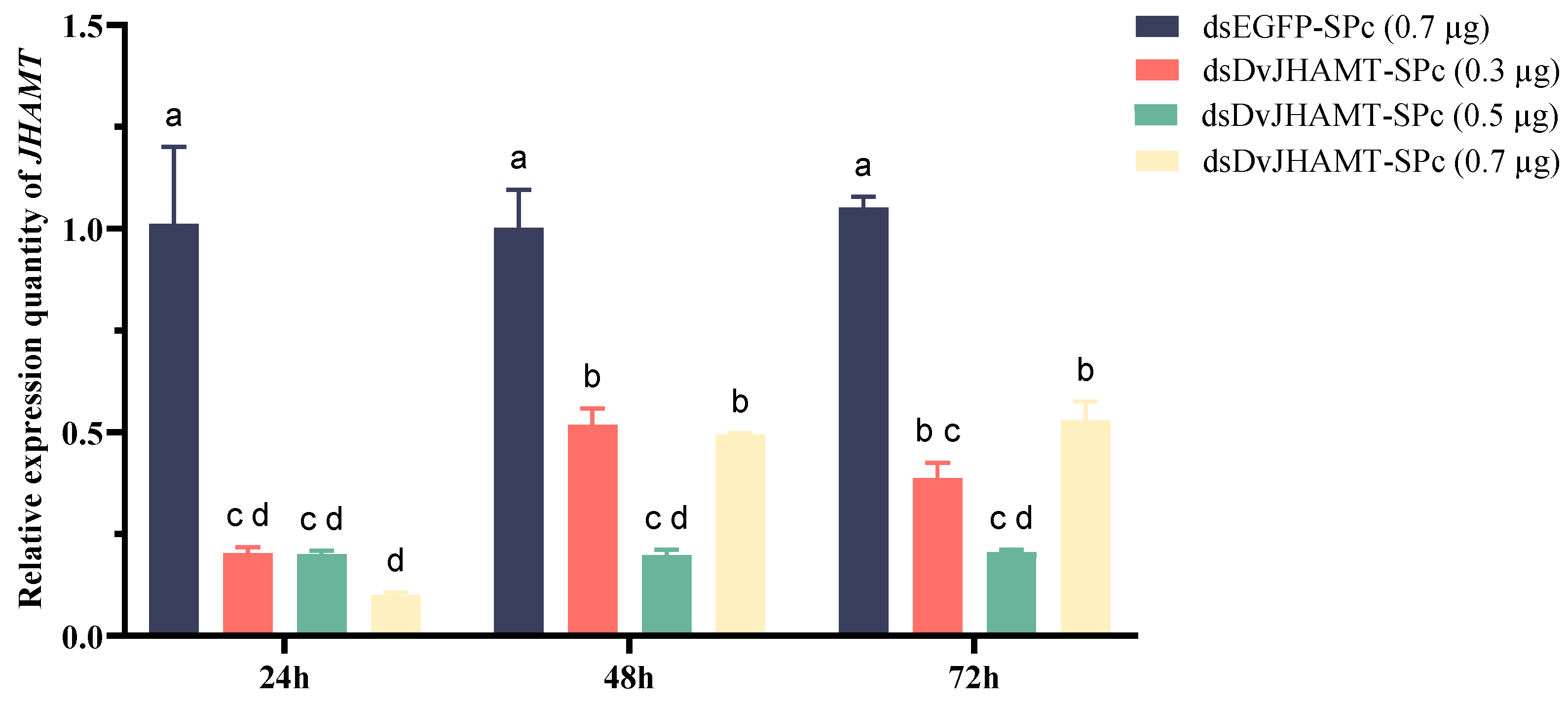
To evaluate RNAi silencing efficiency of the target gene, transcript levels of DvJHAMT were measured by RT-qPCR at 24, 48, and 72 h after topical application of different concentrations of dsDvJHAMT-SPc (Figure 5). The results demonstrated that all dsDvJHAMT-SPc treatments significantly reduced the relative expression of DvJHAMT compared to the dsEGFP-SPc control group (p < 0.001). At 24 h post-application, treatments with 0.3 µg, 0.5 µg, and 0.7 µg of dsDvJHAMT-SPc resulted in downregulations of 59.6%, 78.3%, and 90.1%, respectively, exhibiting a clear dose-dependent effect. This inhibitory effect gradually diminished over time. By 72 h, the downregulation levels were 42.1%, 58.6%, and 47.1%, with no significant differences observed among dsDvJHAMT-SPc groups. Two-way ANOVA confirmed that the dsDvJHAMT treatment significantly suppressed gene expression in both a dose- and time-dependent manner.

3.4. Effects of DvJHAMT Silencing on Pupation and Emergence of D. valens

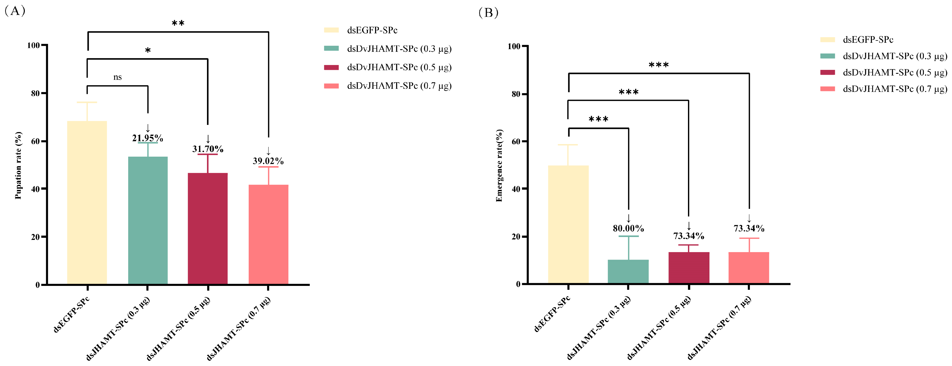
In the dsEGFP-SPc control groups (0.3 µg, 0.5 µg, and 0.7 µg), the first pupae appeared on days 17, 15, and 17, while the first adults emerged on days 29, 31, and 28. By contrast, the dsDvJHAMT-SPc groups entered the pupal stage much earlier, on days 13, 8, and 7. The timing of adult emergence in these groups was similar to the controls, occurring on days 29, 30, and 28. Statistical analysis showed that the onset of pupation in the dsDvJHAMT-SPc groups (7–13 days) was significantly accelerated compared to the controls (15–17 days) (p < 0.05), with the degree of acceleration positively correlated with the dose. However, there was no significant difference in the timing of adult emergence across all groups (ranging from 28 to 31 days). These results indicate that silencing the JHAMT gene primarily shortens the larval developmental duration and significantly extends the pupal stage.
In the control groups, the majority of larvae successfully pupated (68.33%) and emerged as adults (50.00%). Conversely, the pupation rates in all dsDvJHAMT-SPc treatment groups were significantly reduced (Figure 6A). The pupation rates for the 0.3 µg and 0.5 µg dsDvJHAMT-SPc groups dropped to approximately 53.33% and 46.67%, respectively. The most pronounced inhibitory effect was observed at the 0.7 µg dose, where the pupation rate fell to 41.67% (a 39.02% reduction; p < 0.05). Furthermore, the adult emergence rates decreased to 10.00%, 13.33%, and 13.33% (Figure 6B).

3.5. Effect of DvJHAMT Silencing on the Body Weight of D. valens

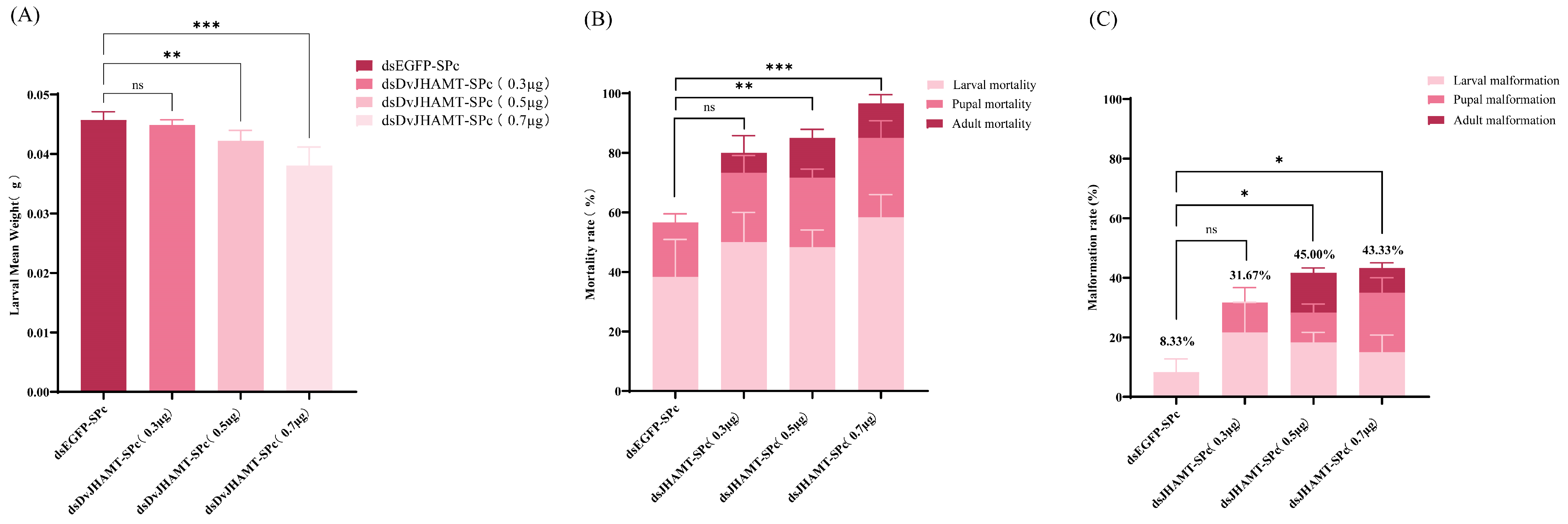
The effect of dsDvJHAMT on larval growth was investigated by applying different concentrations of dsDvJHAMT-SPc topically. At a single time point (day 12) post-treatment, larval body weight was assessed from group-reared larvae. Compared to the dsEGFP-SPc control, the 0.5 µg and 0.7 µg treatments exhibited slight but statistically significant reductions in larval body weight, with corresponding decreases of 7.71% and 16.57% (p < 0.01). No obvious difference was observed in the 0.3 µg treatment group (Figure 7A). These results demonstrate that dsDvJHAMT-SPc inhibits larval weight gain in a dose-dependent manner.

3.6. Effect of DvJHAMT Silencing on Development and Survival of D. valens

The mortality of D. valens was monitored for 35 days following RNAi treatment (Figure 6). Survival distributions were compared using the Kaplan–Meier method with log-rank tests. Survival rates in all three treatment groups declined significantly compared to the control (log-rank test, p < 0.01 for all treatment groups vs. control). Specifically, significant differences in mortality emerged starting from day 18 for the 0.3 µg and 0.5 µg groups (p < 0.01), and as early as day 12 for the 0.7 µg group (p < 0.05). The cumulative mortality rates for the 0.3, 0.5, and 0.7 µg dosages reached 80.00%, 85.00%, and 96.67%, respectively (Figure 7B), with the corresponding corrected mortality rates being 53.85%, 65.38%, and 92.31%.
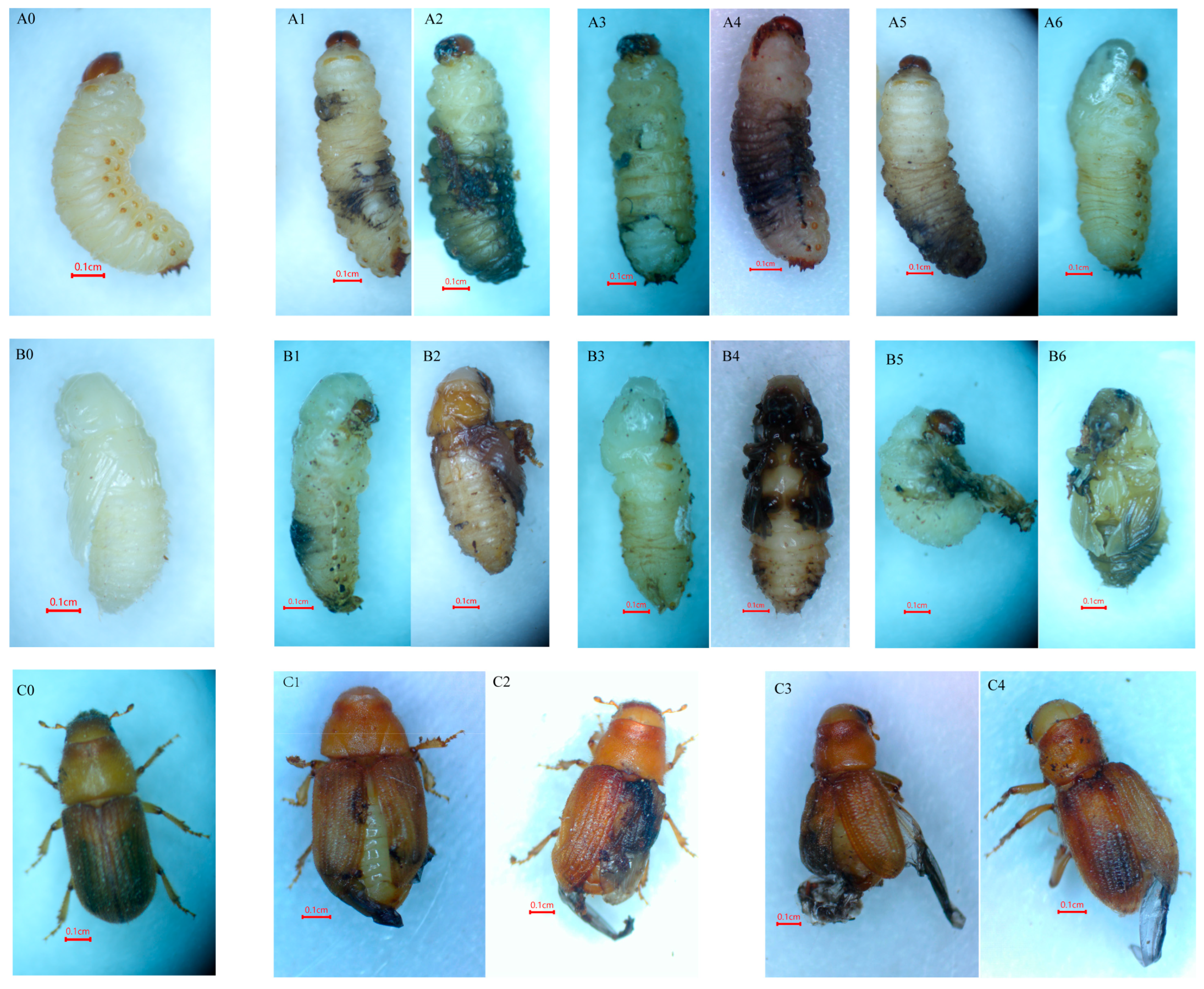
Furthermore, the treatment induced deformity rates of 31.67%, 41.67%, and 43.33% (Figure 7C), reflecting a potent lethal effect (p < 0.01). In summary, six types of lethal phenotypes were observed in dsDvJHAMT-SPc treatment groups (Figure 8). Larvae exhibited failed molting (Figure 8: A1, A3, A6), as well as darkened cuticles, tissue softening, and shrinkage (Figure 8: A2, A4, A5). Deformity was also observed during the pupal stage, characterized by typical lethal phenotypes such as incomplete transition from larva to pupa, resulting in “pupae” retaining larval head structures (Figure 8: B1, B3, B5), and darkened pupae (Figure 8: B2, B4, B6). Adults emerging after treatment with 0.5 µg and 0.7 µg doses exhibited deformity including failure of hindwing retraction and abnormal forewing closure (Figure 8: C1C4). In contrast, larvae in the dsEGFP-SPc control group displayed a normal white cuticle and developed normally into adults (Figure 8: A0, B0, C0).

3.7. Changes in JHAMT Enzyme Activity and JH III Titer

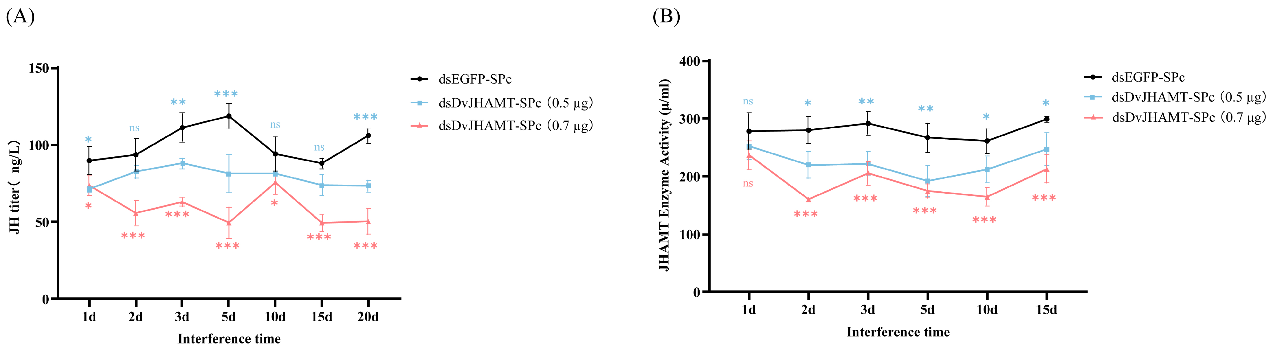
To further investigate the physiological function of DvJHAMT in D. valens larvae, the JHAMT enzyme activity and JH III titer were measured in fourth-instar larvae following RNAi (Figure 8). The results indicated that dsDvJHAMT-SPc treatment dose-dependently reduced both JHAMT enzyme activity and JH III titers compared to the dsEGFP-SPc groups. Specifically, the maximum suppression of JH III titer occurred on day 5 (p < 0.0001). The 0.5 µg and 0.7 µg dosage groups showed reductions of 31.6% and 58.3% (Figure 9A). JHAMT protein levels were also markedly downregulated, with the strongest inhibition detected on day 2 (p < 0.05). Corresponding decreases reached 21.6% and 42.8% in the two dosage groups (Figure 9B).

4. Discussion

JH plays an irreplaceable and central role in the regulation of insect growth, development, and reproduction [40,41]. As a sesquiterpenoid hormone unique to insects, JH works together with 20-hydroxyecdysone (20E) to control embryonic development, larval growth, and metamorphosis. While 20E primarily triggers periodic molting, JH finely regulates developmental timing and reproductive maturation by maintaining the larval state and preventing premature metamorphosis [42]. The physiological effects of JH are closely tied to its titer in vivo, which is precisely regulated through biosynthesis and degradation pathways [43]. Particularly during the critical transition from larva to pupa, JH titer decreases significantly while 20E levels rise; this shift in hormonal balance serves as a key signal for initiating metamorphic development. The biosynthesis of JH depends on the mevalonate pathway [44,45]. Within this pathway, JHAMT catalyzes the methylation of JH acid or its inactive precursors to form biologically active JH, making JHAMT the final rate-limiting enzyme in JH synthesis [20,45]. Consequently, JHAMT is not only a key enzyme determining JH biosynthetic flux but also an important regulatory node linking developmental signals with hormonal output.
Cloned and identified in various insect species, the JHAMT gene represents a key enzyme in JH biosynthesis [19,20,21,23,28,30,39]. With in-depth research on its expression regulation mechanisms, enzymatic properties, and functional diversity across different insect species, JHAMT has been recognized as a key target for understanding the physiological functions of the JH pathway [46]. Sequence analysis revealed that DvJHAMT, along with all other coleopteran JHAMTs, contains a conserved SAM-binding domain, suggesting its methyltransferase activity [45,47]. Previous studies have confirmed that this SAM-binding site is essential for the methylation of JH acid into active JH by JHAMT [48]. Phylogenetic analysis based on JHAMT sequences from representative species of Hemiptera, Lepidoptera, Coleoptera, Hymenoptera, and Diptera demonstrated that DvJHAMT shares the closest genetic relationship with JHAMT homologs from other coleopteran insects, forming a distinct monophyletic clade.
The efficacy of RNAi of the JHAMT gene critically depends on the efficiency of dsRNA delivery. Conventional methods, such as microinjection and feeding, exhibit notable limitations. In this study, we used a combined approach integrating transdermal delivery with SPc nanomaterial-based delivery. This strategy is operationally simple and non-invasive. Moreover, the SPc nanocarrier protects dsRNA and enhances its delivery efficiency, thereby overcoming the constraints of traditional methods, enabling stable and highly efficient gene silencing, and facilitating the elucidation of JHAMT’s biological functions [33,49].
Current research indicates that Coleoptera generally exhibit higher sensitiveness to dsRNA than Lepidoptera [24]. For example, T. castaneum exhibits lethal phenotypes even at minimal doses (0.1–0.5 µg) of dsRNA through both injection and feeding routes [50], whereas lepidopteran species such as Lymantria dispar demand substantially higher concentrations (1–3 µg) via injection to elicit robust gene silencing [51]. Treatment of the coleopteran D. valens larvae with different doses of dsRNA demonstrated that a dose as low as 0.3 µg dsDvJHAMT-SPc already caused significant downregulation of JHAMT expression (59.6%) and accelerated development. In the treatment groups, elevating the dose to 0.7 µg triggered an extremely robust gene silencing effect (with a 90.1% reduction in gene expression) and severe developmental defects (e.g., a sharp decline in pupation and eclosion rates), which exhibited a clear dose-dependent pattern. These findings further confirm that D. valens shares the high RNAi sensitivity typical of many coleopteran insects. Moreover, the results indicate that dsDvJHAMT-SPc treatment effectively disrupted normal larval development, a phenomenon also observed in other insect species [52,53].
Studies have demonstrated that RNA-mediated silencing of the BgJHAMT gene leads to a sharp decline in JH synthesis [54]. RNAi-induced knockdown of the AiJHAMT gene in A. ipsilon reduces JH titer in larvae, impairs larval growth and survival, and disrupts pupation [30]. Similarly, silencing of the AtJHAMT gene severely hinders ovarian development and markedly lowers JH titer in A. tumida [28]. In L. decemlineata, silencing of the LdJHAMT1 and LdJHAMT2 genes reduces hemolymph JH titer, shortens larval development duration, reduces pupation and adult emergence rates, and impairs reproduction [55]. In this study, following treatment with dsDvJHAMT-SPc, both the JH III titer and JHAMT enzymatic activity in D. valens larvae were significantly reduced, resulting in increased mortality and malformation rates, and reduced body weight. Concurrently, the duration of larval development was shortened by 4–10 days, with pupation occurring earlier than in the control group. However, the pupal stage was correspondingly prolonged, resulting in the overall total developmental cycle (from 4th instar larva to adult) remaining unchanged. In addition, the treated larvae displayed a range of developmental abnormalities, including cuticular melanization, tissue shrinkage, and incomplete larval-pupal metamorphosis in some individuals, as well as severe wing malformation and eclosion failure in adults. These phenotypes may be associated with reduced juvenile hormone levels modulating the expression of cuticle synthesis-related genes [56,57]. We could conduct transcriptomic and metabolomic analyses of RNAi-treated larvae and further investigate the effects on reproduction in D. valens.
This study systematically elucidates the function of JHAMT and its application potential in pest control. These findings not only contribute to a deeper understanding of the evolutionary and physiological mechanisms underlying insect endocrine regulation but also provide a new molecular foundation and rationale for developing environmentally friendly green management technologies through specific interference with pest development or reproduction. We successfully achieved efficient gene silencing under laboratory conditions using the transdermal delivery method. However, this method faces several challenges in complex field environments. As a bark-boring pest, D. valens larvae are concealed beneath the bark, making it difficult to deliver dsRNA directly to the target insects. Moreover, naked dsRNA can be degraded by environmental RNases, and the manual, insect-by-insect treatment cannot be performed on a large scale. Therefore, achieving targeted delivery and reducing the cost of large-scale application are critical issues that urgently need to be addressed. In the future, exploring the integration of RNAi technology with strategies such as soil-drench applications, biocontrol agent-mediated delivery (entomopathogenic fungi like Bacillus thuringiensis), and plant-mediated host-induced gene silencing (HIGS) to develop efficient and stable delivery systems applicable to field environments represents a key next step toward the practical application of this technology.

5. Conclusions

Targeting the forest quarantine pest D. valens, this study employed transdermal delivery to deliver dsDvJHAMT using SPc nanomaterial, effectively silencing the key juvenile hormone synthesis gene DvJHAMT. Larvae treated with RNAi exhibited growth retardation, developmental deformity, and increased mortality, along with significantly reduced pupation and eclosion rates. Furthermore, both JHAMT enzyme activity and JH III titer were markedly decreased in the treated larvae, further confirming the critical role of JH in regulating insect growth and development.
In conclusion, this study utilized the SPc nanomaterial-based transdermal delivery system to silence the key juvenile hormone (JH) biosynthesis gene, DvJHAMT, in the forest quarantine pest D. valens. RNAi-mediated knockdown of DvJHAMT resulted in significant growth retardation, developmental deformities, and increased mortality, alongside markedly reduced pupation and emergence rates. These phenotypic changes were consistent with the significant reduction in both JHAMT enzyme activity and JH III titers, further confirming the essential role of JH in regulating insect development. Our findings offer a solid theoretical basis for developing novel, eco-friendly pest management strategies targeting the JH biosynthetic pathway in D. valens.

Supplementary Materials

The following supporting information can be downloaded at: https://www.mdpi.com/article/10.3390/f17050628/s1. Table S1: Information on insect JHAMT proteins.

Author Contributions

Conceptualization, J.W. and J.L.; methodology, J.L., J.W., Q.C., Y.S. and D.K.; formal analysis, Q.C., J.L. and Y.S.; investigation, Q.C., Y.S., J.W. and J.L.; resources, J.L., J.W. and J.H.; data curation, J.L. and Q.C.; writing—original draft preparation, Q.C. and J.L.; writing—review and editing, J.L. and J.W.; visualization, Q.C., J.L., J.H. and J.W.; supervision, J.L. and J.W.; project administration, J.W.; funding acquisition, J.W. All authors have read and agreed to the published version of the manuscript.

Funding

This research was supported by the National Key Research and Development Program of China, funded by the Ministry of Science and Technology of the People’s Republic of China (Grant No. 2023YFC2604800).

Data Availability Statement

All data in this study, such as the gene entry numbers, are available on the NCBI (National Center for Biotechnology Information) website.

Acknowledgments

The SPc nanoparticles utilized in this paper were generously provided by Jie Shen from China Agricultural University.

Conflicts of Interest

The authors declare no conflicts of interest.

References

  1. Bi, P.; Yu, L.; Zhou, Q.; Kuang, J.; Tang, R.; Ren, L.; Luo, Y. Early detection of Dendroctonus valens infestation with UAV-based thermal and hyperspectral images. Remote Sens. 2024, 16, 3840. [Google Scholar] [CrossRef]
  2. Yan, Z.L.; Sun, J.H.; Don, O.; Zhang, Z.N. The red turpentine beetle, Dendroctonus valens LeConte (Scolytidae): An exotic invasive pest of pine in China. Biodivers. Conserv. 2005, 14, 1735–1760. [Google Scholar] [CrossRef]
  3. Sun, J.; Lu, M.; Gillette, N.E.; Wingfield, M.J. Red turpentine beetle: Innocuous native becomes invasive tree killer in China. Annu. Rev. Entomol. 2013, 58, 293–311. [Google Scholar] [CrossRef]
  4. Liu, F.H.; Ye, F.Y.; Cheng, C.H.; Kang, Z.W.; Kou, H.R.; Sun, J.H. Symbiotic microbes aid host adaptation by metabolizing a deterrent host pine carbohydrate d-pinitol in a beetle-fungus invasive complex. Sci. Adv. 2022, 8, eadd5051. [Google Scholar] [CrossRef]
  5. Lu, M.; Sun, J.H. Red turpentine beetle Dendroctonus valens LeConte. In Biological Invasions and Its Management in China; Springer: Berlin/Heidelberg, Germany, 2017; pp. 219–228. [Google Scholar]
  6. Cheng, C.; Xu, L.; Xu, D.; Lou, Q.; Lu, M.; Sun, J. Does cryptic microbiota mitigate pine resistance to an invasive beetle-fungus complex? Implications for invasion potential. Sci. Rep. 2016, 6, 33979. [Google Scholar] [CrossRef]
  7. Mann, A.J.; Barnum, R.M.; Held, B.W.; Bushley, K.E.; Aukema, B.H.; Blanchette, R.A. Fungal and bacterial communities of the red turpentine beetle (Dendroctonus valens LeConte) in the Great Lakes Region, USA. Forests 2025, 16, 1604. [Google Scholar] [CrossRef]
  8. Owen, D.R.; Wood, D.L.; Parmeter, J.R. Association between Dendroctonus valens and black stain root disease on ponderosa pine in the Sierra Nevada of California. Can. Entomol. 2005, 137, 595–600. [Google Scholar] [CrossRef]
  9. Mason, C.J.; Hanshew, A.S.; Raffa, K.F. Contributions by host trees and insect activity to bacterial communities in Dendroctonus valens (Coleoptera: Curculionidae) galleries, and their high overlap with other microbial assemblages of bark beetles. Environ. Entomol. 2016, 45, 348–356. [Google Scholar] [CrossRef] [PubMed]
  10. Liu, S.; Li, K.; Gao, Y.; Liu, X.; Chen, W.; Ge, W.; Feng, Q.; Palli, S.R.; Li, S. Antagonistic actions of juvenile hormone and 20-hydroxyecdysone within the ring gland determine developmental transitions in Drosophila. Proc. Natl. Acad. Sci. USA 2018, 115, 139–144. [Google Scholar] [CrossRef]
  11. Zhang, X.; Li, S.; Liu, S. Juvenile hormone studies in Drosophila melanogaster. Front. Physiol. 2022, 12, 785320. [Google Scholar] [CrossRef]
  12. Inui, T.; Sezutsu, H.; Daimon, T. MicroRNA let-7 is required for hormonal regulation of metamorphosis in the silkworm, Bombyx mori. Insect Biochem. Mol. Biol. 2022, 145, 103784. [Google Scholar] [CrossRef]
  13. Tsang, S.S.K.; Law, S.T.S.; Li, C.; Qu, Z.; Bendena, W.G.; Tobe, S.S.; Hui, J.H.L. Diversity of insect sesquiterpenoid regulation. Front. Genet. 2020, 11, 1027. [Google Scholar] [CrossRef]
  14. Oi, C.A.; Ferreira, H.M.; da Silva, R.C.; Bienstman, A.; Nascimento, F.S.D.; Wenseleers, T. Effects of juvenile hormone in fertility and fertility-signaling in workers of the common wasp Vespula vulgaris. PLoS ONE 2021, 16, e0250720. [Google Scholar] [CrossRef]
  15. Jindra, M.; Belles, X.; Shinoda, T. Molecular basis of juvenile hormone signaling. Curr. Opin. Insect Sci. 2015, 11, 39–46. [Google Scholar] [CrossRef]
  16. Smykal, V.; Dolezel, D. Evolution of proteins involved in the final steps of juvenile hormone synthesis. J. Insect Physiol. 2023, 145, 104487. [Google Scholar] [CrossRef]
  17. Kenny, N.J.; Quah, S.; Holland, P.W.H.; Tobe, S.S.; Hui, J.H.L. How are comparative genomics and the study of microRNAs changing our views on arthropod endocrinology and adaptations to the environment? Gen. Comp. Endocrinol. 2013, 188, 16–22. [Google Scholar] [CrossRef]
  18. Maligeppagol, M.; Navale, P.M.; Asokan, R.; Krishna, V.; Sharath Chandra, G.; Prasad Babu, K.; Latha, J.; Krishna Kumar, N.K.; Ellango, R. Transgenic tomato expressing dsRNA of juvenile hormone acid O-methyl transferase gene of Helicoverpa armigera (Lepidoptera: Noctuidae) affects larval growth and its development. J. Asia-Pac. Entomol. 2017, 20, 559–567. [Google Scholar]
  19. Shinoda, T.; Itoyama, K. Juvenile hormone acid methyltransferase: A key regulatory enzyme for insect metamorphosis. Proc. Natl. Acad. Sci. USA 2003, 100, 11986–11991. [Google Scholar] [CrossRef] [PubMed]
  20. Niwa, R.; Niimi, T.; Honda, N.; Yoshiyama, M.; Itoyama, K.; Kataoka, H.; Shinoda, T. Juvenile hormone acid O-methyltransferase in Drosophila melanogaster. Insect Biochem. Mol. Biol. 2008, 38, 714–720. [Google Scholar] [CrossRef]
  21. Minakuchi, C.; Namiki, T.; Yoshiyama, M.; Shinoda, T. RNAi-mediated knockdown of juvenile hormone acid O-methyltransferase gene causes precocious metamorphosis in the red flour beetle Tribolium castaneum. FEBS J. 2008, 275, 2919–2931. [Google Scholar] [CrossRef] [PubMed]
  22. Guo, W.; Bai, C.; Wang, Z.; Wang, P.; Fan, Q.; Mi, X.; Wang, L.; He, J.; Pang, J.; Luo, X.; et al. Double-stranded RNAs high-efficiently protect transgenic potato from Leptinotarsa decemlineata by disrupting juvenile hormone biosynthesis. J. Agric. Food Chem. 2018, 66, 11990–11999. [Google Scholar] [CrossRef] [PubMed]
  23. Song, X.; Zhang, D.W.; Yang, Y.; Kou, J.F.; Liu, C.; Yi, C.Q.; Tang, Z.Y.; Liu, W.T.; Chen, R.; Guo, W.H.; et al. Structure-function analysis of MhieJHAMT: A key enzyme in Monolepta hieroglyphica juvenile hormone biosynthesis. Int. J. Biol. Macromol. 2025, 318, 145020. [Google Scholar] [CrossRef]
  24. Lu, J.; Shen, J. Target genes for RNAi in pest control: A comprehensive overview. Entomol. Gen. 2024, 44, 95–114. [Google Scholar] [CrossRef]
  25. Kola, V.S.; Renuka, P.; Madhav, M.S.; Mangrauthia, S.K. Key enzymes and proteins of crop insects as candidate for RNAi based gene silencing. Front. Physiol. 2015, 6, 119. [Google Scholar] [CrossRef]
  26. Tian, Z.; Guo, S.; Li, J.X.; Zhu, F.; Liu, W.; Wang, X.P. Juvenile hormone biosynthetic genes are critical for regulating reproductive diapause in the cabbage beetle. Insect Biochem. Mol. Biol. 2021, 139, 103654. [Google Scholar] [CrossRef]
  27. Gu, Y.; Yang, X.; Liu, S.; Chen, X.; Liu, R.; Gao, J.; Zhong, Y.; Li, X.; Han, W. RNAi-mediated knockdown of juvenile hormone acid methyltransferase depresses reproductive performance in female Aethina tumida. Pestic. Biochem. Physiol. 2025, 211, 106420. [Google Scholar] [CrossRef]
  28. Daimon, T.; Uchibori, M.; Nakao, H.; Sezutsu, H.; Shinoda, T. Knockout silkworms reveal a dispensable role for juvenile hormones in holometabolous life cycle. Proc. Natl. Acad. Sci. USA 2015, 112, E4226–E4235. [Google Scholar] [CrossRef]
  29. Zhang, J.; Yu, J.M.; Shi, X.X.; Liu, D.Y.; Deng, Q.; Peng, J.N.; Li, M.Y.; Liu, S. Knockdown of the juvenile hormone acid O-methyltransferase gene impairs development of Agrotis ipsilon (Lepidoptera: Noctuidae). J. Asia-Pac. Entomol. 2025, 28, 102410. [Google Scholar] [CrossRef]
  30. Kolge, H.; Kadam, K.; Galande, S.; Lanjekar, V.; Ghormade, V. New frontiers in pest control: Chitosan nanoparticles-shielded dsRNA as an effective topical RNAi spray for gram podborer biocontrol. ACS Appl. Bio Mater. 2021, 4, 5145–5157. [Google Scholar] [CrossRef] [PubMed]
  31. Wang, Z.; Li, M.; Kong, Z.; Wang, E.; Zhang, B.; Lv, J.; Xu, X. Star polycation mediated dsRNA improves the efficiency of RNA interference in Phytoseiulus persimilis. Nanomaterials 2022, 12, 1770. [Google Scholar] [CrossRef] [PubMed]
  32. Long, G.J.; Liu, X.Z.; Guo, H.; Zhang, M.Q.; Gong, L.L.; Ma, Y.F.; Dewer, Y.; Mo, W.J.; Ding, L.W.; Wang, Q.; et al. Oral-based nanoparticle-wrapped dsRNA delivery system: A promising approach for controlling an urban pest, Blattella germanica. J. Pest. Sci. 2024, 97, 739–755. [Google Scholar] [CrossRef]
  33. Li, M.; Sun, X.; Yin, M.; Shen, J.; Yan, S. Recent advances in nanoparticle-mediated co-delivery system: A promising strategy in medical and agricultural field. Int. J. Mol. Sci. 2023, 24, 5121. [Google Scholar] [CrossRef]
  34. Liu, Z.; Xing, L.; Huang, W.; Liu, B.; Wan, F.; Raffa, K.F.; Hofstetter, R.W.; Qian, W.; Sun, J. Chromosome-level genome assembly and population genomic analyses provide insights into adaptive evolution of the red turpentine beetle, Dendroctonus valens. BMC Biol. 2022, 20, 190. [Google Scholar] [CrossRef]
  35. Zhao, D.; Zheng, C.; Shi, F.; Xu, Y.; Zong, S.; Tao, J. Expression analysis of genes related to cold tolerance in Dendroctonus valens. PeerJ 2021, 9, e10864. [Google Scholar] [CrossRef] [PubMed]
  36. Schmittgen, T.D.; Livak, K.J. Analyzing real-time PCR data by the comparative CT method. Nat. Protoc. 2008, 3, 1101–1108. [Google Scholar] [CrossRef] [PubMed]
  37. Livak, K.J.; Schmittgen, T.D. Analysis of relative gene expression data using real-time quantitative PCR and the 2−ΔΔCT method. Methods 2002, 25, 402–408. [Google Scholar] [CrossRef]
  38. Yang, Y.X.; Jiang, Q.H.; Peng, M.; Zhou, Z.Y.; Du, X.G.; Yin, M.Z.; Shen, J.; Yan, S. A star polyamine-based nanocarrier delivery system for enhanced avermectin contact and stomach toxicity against green peach aphids. Nanomaterials 2022, 12, 1445. [Google Scholar] [CrossRef] [PubMed]
  39. Zhou, Q.H.; Zhang, Q.; Yang, R.L.; Yuan, G.R.; Wang, J.J.; Dou, W. RNAi-mediated knockdown of juvenile hormone acid O-methyltransferase disrupts larval development in the oriental fruit fly, Bactrocera dorsalis (Hendel). Pestic. Biochem. Physiol. 2022, 188, 105285. [Google Scholar] [CrossRef]
  40. Cusson, M.; Sen, S.E.; Shinoda, T. Juvenile hormone biosynthetic enzymes as targets for insecticide discovery. In Advanced Technologies for Managing Insect Pests; Springer: Berlin/Heidelberg, Germany, 2013; pp. 31–55. [Google Scholar]
  41. Yin, Y.; Qiu, Y.W.; Huang, J.; Tobe, S.S.; Chen, S.S.; Kai, Z.P. Enzymes in the juvenile hormone biosynthetic pathway can be potential targets for pest control. Pest. Manag. Sci. 2020, 76, 1071–1077. [Google Scholar] [CrossRef]
  42. Jindra, M.; Palli, S.R.; Riddiford, L.M. The juvenile hormone signaling pathway in insect development. Annu. Rev. Entomol. 2013, 58, 181–204. [Google Scholar] [CrossRef]
  43. Xu, H.; Zhang, Y.; Zhang, L.; Wang, Z.; Guo, P.; Zhao, P. Structural characterization and functional analysis of juvenile hormone diol kinase from the silkworm, Bombyx mori. Int. J. Biol. Macromol. 2021, 167, 570–577. [Google Scholar] [CrossRef] [PubMed]
  44. Belles, X.; Martin, D.; Piulachs, M.D. The mevalonate pathway and the synthesis of juvenile hormone in insects. Ann. Rev. Entomol. 2005, 50, 181–199. [Google Scholar] [CrossRef] [PubMed]
  45. Guo, P.; Zhang, Y.; Zhang, L.; Xu, H.; Zhang, H.; Wang, Z.; Jiang, Y.; Molloy, D.; Zhao, P.; Xia, Q. Structural basis for juvenile hormone biosynthesis by the juvenile hormone acid methyltransferase. J. Biol. Chem. 2021, 297, 101234. [Google Scholar] [CrossRef]
  46. Zhang, R.; Zhong, Z. Juvenile hormone acid O-methyltransferase (JHAMT): A key gene in juvenile hormone signaling pathway in insects. Arch. Insect Biochem. Physiol. 2026, 121, e22026. [Google Scholar]
  47. Defelipe, L.A.; Dolghih, E.; Roitberg, A.E.; Nouzova, M.; Mayoral, J.G.; Noriega, F.G.; Turjanski, A.G. Juvenile hormone synthesis: “esterify then epoxidize” or “epoxidize then esterify”? Insights from the structural characterization of juvenile hormone acid methyltransferase. Insect Biochem. Mol. Biol. 2011, 41, 228–235. [Google Scholar] [CrossRef]
  48. Xie, X.; Tao, T.; Liu, M.; Zhou, Y.; Liu, Z.; Zhu, D. The potential role of juvenile hormone acid methyltransferase in methyl farnesoate (MF) biosynthesis in the swimming crab, Portunus trituberculatus. Anim. Reprod. Sci. 2016, 168, 40–49. [Google Scholar] [CrossRef] [PubMed]
  49. Yu, N.; Christiaens, O.; Liu, J.; Niu, J.; Cappelle, K.; Caccia, S.; Huvenne, H.; Smagghe, G. Delivery of dsRNA for RNAi in insects: An overview and future directions. Insect Sci. 2013, 20, 4–14. [Google Scholar] [CrossRef]
  50. Cao, M.; Gatehouse, J.A.; Fitches, E.C. A systematic study of RNAi effects and dsRNA stability in Tribolium castaneum and Acyrthosiphon pisum, following injection and ingestion of analogous dsRNAs. Int. J. Mol. Sci. 2018, 19, 1079. [Google Scholar] [CrossRef]
  51. Ji, W.; Xie, Q.; Chang, X.; Shi, J. Silencing of juvenile hormone-related genes through RNA interference leads to molt failure and high mortality in the spongy moth. Insect Sci. 2025, 32, 717–729. [Google Scholar] [CrossRef]
  52. Christiaens, O.; Prentice, K.; Pertry, I.; Ghislain, M.; Bailey, A.; Niblett, C.; Gheysen, G.; Smagghe, G. RNA interference: A promising biopesticide strategy against the African Sweetpotato Weevil Cylas brunneus. Sci. Rep. 2016, 6, 38836. [Google Scholar] [CrossRef]
  53. Nouzova, M.; Edwards, M.J.; Michalkova, V.; Ramirez, C.E.; Ruiz, M.; Areiza, M.; DeGennaro, M.; Fernandez-Lima, F.; Feyereisen, R.; Jindra, M.; et al. Epoxidation of juvenile hormone was a key innovation improving insect reproductive fitness. Proc. Natl. Acad. Sci. USA 2021, 118, e2109381118. [Google Scholar] [CrossRef] [PubMed]
  54. Dominguez, C.V.; Maestro, J.L. Expression of juvenile hormone acid O-methyltransferase and juvenile hormone synthesis in Blattella germanica. Insect Sci. 2018, 25, 787–796. [Google Scholar] [CrossRef] [PubMed]
  55. Fu, K.Y.; Li, Q.; Zhou, L.T.; Meng, Q.W.; Lu, F.G.; Guo, W.C.; Li, G.Q. Knockdown of juvenile hormone acid methyl transferase severely affects the performance of Leptinotarsa decemlineata (Say) larvae and adults. Pest. Manag. Sci. 2016, 72, 1231–1241. [Google Scholar] [CrossRef]
  56. Shi, J.F.; Mu, L.L.; Guo, W.C.; Li, G.Q. Identification and hormone induction of putative chitin synthase genes and splice variants in Leptinotarsa decemlineata (SAY). Arch. Insect Biochem. Physiol. 2016, 92, 242–258. [Google Scholar] [CrossRef]
  57. Charles, J.P. The regulation of expression of insect cuticle protein genes. Insect Biochem. Mol. Biol. 2010, 40, 205–213. [Google Scholar] [CrossRef] [PubMed]
Figure 1. Rearing and dsRNA-SPc treatment of D. valens. (A) Larvae reared under standard conditions; (B) Topical application of dsRNA-SPc to a fourth-instar larva (scale bar = 0.1 cm); (C) Larval morphology at one day post-treatment.
Forests 17 00628 g001
Figure 2. Multiple sequence alignment of JHAMT homologs. Sequences are from D. valens, D. armandi, D. ponderosae, Euwallacea similis (E. similis), Colaphellus bowringi (C. bowringi), Achroia grisella (A. grisella), Sergentomyia squamirostris (S. squamirostris), and Chrysoperla carnea (C. carnea). Residues are shaded according to their conservation level: black indicates 100% identity across all sequences, pink represents identity ≥ 75%, and blue represents identity ≥ 50%. The red box highlights the conserved SAM-binding motif. Table S1 lists the GenBank accession numbers.
Forests 17 00628 g002
Figure 3. Phylogenetic analysis of JHAMT in different insect species. Using the neighbor-joining approach, a tree was constructed; The numbers represent the tree confidence calculated by bootstrap analysis with 1000 replicates. The DvJHAMT is indicated by black solid star. Table S1 lists the GenBank accession numbers.
Forests 17 00628 g003
Figure 4. Developmental stage and tissue specificity of DvJHAMT expression in D. valens. (A) Expression across life stages: E (egg), L1–L4 (1–4 instars larvae), P (pupa), A (adult); (B) Expression in fifth-instar larval tissues: H (head), G (gut), E (epidermis), Mt (Malpighian tubules), and Fb (fat body). RT-qPCR data were normalized to TUB. Values: mean ± SEM, n = 3. Different lowercase letters indicate statistically significant differences (p < 0.05, one-way ANOVA with Tukey’s HSD test).
Forests 17 00628 g004
Figure 5. The expression level of DvJHAMT after different doses of dsDvJHAMT. Relative expression levels were measured at 24, 48, and 72 h post-application of dsDvJHAMT-SPc complexes at 0.3, 0.5, and 0.7 µg/µL. The dsEGFP-SPc complex served as the control at each time point. RT-qPCR data normalized to TUB. Data are presented as the mean ± SEM (n = 3 independent replicates). Different lowercase letters indicate statistically significant differences (p < 0.05, two-way ANOVA with Tukey’s HSD test).
Forests 17 00628 g005
Figure 6. Impact of dsDvJHAMT-SPc RNAi on pupation and emergence rates of D. valens. (A) Pupation rate; (B) Emergence rate. The control group received dsEGFP-SPc. Values are the mean ± SEM from three independent biological replicates. Significant differences from control were determined by one-way ANOVA with Tukey’s multiple comparisons test (ns, not significant; * p < 0.05, ** p < 0.01, *** p < 0.001).
Forests 17 00628 g006
Figure 7. Effects of dsDvJHAMT-SPc on larval development parameters. (A) Larval body weight at 12 days post-treatment; (B) Mortality rate; (C) Deformity rate. The control group received dsEGFP-SPc. Significant differences from control were determined by one-way ANOVA with Tukey’s multiple comparisons test (ns, not significant; * p < 0.05, ** p < 0.01, and *** p < 0.001).
Forests 17 00628 g007
Figure 8. Representative phenotypes of pupation and eclosion in D. valens larvae following topical application of dsEGFP-SPc or dsDvJHAMT-SPc at different concentrations. Larvae treated with dsEGFP-SPc developed normally (A0, B0, C0), whereas larvae treated with dsDvJHAMT-SPc at dosage level of 0.3 µg (A1, A2, B1, and B2), 0.5 µg (A3, A4, B3, B4, C1, and C2), or 0.7 µg (A5, A6, B5, B6, C3, and C4) exhibited lethal deformity (scale bar = 0.1 cm).
Forests 17 00628 g008
Figure 9. Effects of dsDvJHAMT-SPc treatments on JH III titers and JHAMT enzyme activity in fourth-instar larvae of D. valens. (A) JH III titers; (B) JHAMT enzyme activity. dsEGFP-SPc treatment groups were used as the control. Data are presented as mean ± SEM from 6 biological replicates per time point. Statistical significance was determined by two-way ANOVA with Tukey’s multiple comparisons test (ns, not significant; * p < 0.05, ** p < 0.01, and *** p < 0.001).
Forests 17 00628 g009
Disclaimer/Publisher’s Note: The statements, opinions and data contained in all publications are solely those of the individual author(s) and contributor(s) and not of MDPI and/or the editor(s). MDPI and/or the editor(s) disclaim responsibility for any injury to people or property resulting from any ideas, methods, instructions or products referred to in the content.