
Communication

First Report of Fruit Rot of Snake Fruit (Salacca zalacca) Caused by Thielaviopsis euricoi Based on Morphological and Phylogenetic Analyses

1
State Key Laboratory of Green Pesticide, Integrative Microbiology Research Centre, Guangdong Provincial Key Laboratory of Microbial Signals and Disease Control, College of Plant Protection, South China Agricultural University, Guangzhou 510642, China
2
Guangzhou Customs Technology Center, Guangzhou 510623, China
3
Key Laboratory of Biodiversity Conservation of National Forestry and Grassland Administration, Ecology and Nature Conservation Institute, Chinese Academy of Forestry, Beijing 100091, China
*
Author to whom correspondence should be addressed.
Forests 2026, 17(2), 230; https://doi.org/10.3390/f17020230
Submission received: 9 January 2026 / Revised: 5 February 2026 / Accepted: 7 February 2026 / Published: 8 February 2026
(This article belongs to the Special Issue Identification and Molecular Phylogeny of Forest Microorganisms)

Abstract

Snake fruit (Salacca zalacca (Gaertner) Voss) is renowned for its taste and nutritional value; however, the snake fruit rot diseases caused by fungal pathogens can lead to significant economic losses. In this study, a fungal isolate was obtained from the rotten snake fruit with brown rot symptoms at the fruit apex upon removal of the peel. The causal agent was identified as Thielaviopsis species based on cultural and morphometric characteristics. Multi-locus phylogenetic analyses of the internal transcribed spacer (ITS) region, translation elongation factor 1-alpha (tef1-α) and beta-tubulin (tub2) genes revealed that the isolates Te 5742 clustered within Thielaviopsis euricoi Bat. & A. F. Vital. Furthermore, pathogenicity tests fulfilled Koch’s postulates, confirming T. euricoi as the pathogen responsible for the observed rot. This is the first report of T. euricoi causing fruit rot on snake fruit worldwide and will provide information on monitoring and management of snake fruit diseases in the garden and market.

1. Introduction

Snake fruit (Salacca zalacca (Gaertner) Voss) is a prominent tropical fruit known for its distinctive sour and sweet flavor profile and high antioxidant activity, which exceeds that of other common tropical fruits such as mangosteen (Garcinia mangostana L.) and avocado (Persea americana Mill.) [1,2,3]. With the increasing global demand for fruit consumption, it holds significant potential for the international market [2,4]. However, the rising incidence of diseases, particularly fruit rots caused by pathogens such as Peniophora salaccae N. Suwannar., S. Boukaew, & J. Kumla, Thielaviopsis paradoxa (De Seynes) Höhn., and T. ethacetica Went, poses a serious threat to the sustainable development of the snake fruit industry. As reported, T. paradoxa was found as the predominant pathogen causing snake fruit rot in Indonesian traditional markets, and P. salaccae was identified as a novel causal agent of this disease [5,6]. The rotten symptoms are often observed at the fruit apex, while it remains unclear how the rot diseases initially happened.
The genus Thielaviopsis Went was established in 1893 based on T. ethacetica and accommodates the anamorphs of described Ceratocystis Ellis & Halst. species [7,8]. However, the sexual states of several species have not been observed, including T. euricoi (Bat. & A.F. Vital) A.E. Paulin, T.C. Harr. & McNew, T. musarum (R.S. Mitchell) Riedl, T. australis (J. Walker & Kile) Z.W. De Beer, T.A. Duong & M.J. Wingf., and T. neocaledoniae (Kiffer & Delon) Z.W. De Beer, T.A. Duong & M.J. Wingf. [7,9]. Discriminating between Thielaviopsis species based solely on morphology is challenging due to the absence of sexual structures in some species and the overlapping morphological characteristics among different species [9]. Therefore, multigene phylogenetic analyses based on ITS, tub2, and tef1-α gene sequences are indispensable for accurate species identification [8,9].
Thielaviopsis species are well-known soilborne pathogens affecting monocotyledonous plants. Symptoms caused by Thielaviopsis species are typically shown by necrosis tissue, the charcoal-like black appearance, and the bending of the terminal bud [10,11,12]. For instance, T. paradoxa and T. punctulata have been reported as the causal agents of black scorch in palm species (Arecaceae) [10,11]. T. paradoxa and T. ethacetica have been confirmed as pathogens causing fruit rot in snake fruit, calling people’s attention to the control of Thielaviopsis species in the snake fruit garden [5]. Notably, T. euricoi (syn. Hughesiella euricoi) was first described by Batista & A.F. Vital in 1956 from air samples [13]; to date, it has not been reported as a plant pathogen.
In the present study, we observed fruit rot on snake fruit caused by an isolate morphologically resembling T. euricoi. Given the limited information regarding the spectrum of pathogens affecting snake fruit, the aims of this study were to identify the causal agent using morphological and phylogenetic analyses methods and to demonstrate its pathogenicity on snake fruit. This study lays the groundwork for future disease management and informs the quarantine strategies of various countries aimed at preventing the introduction of this pathogen via snake fruit.

2. Materials and Methods

2.1. Sample Collection and Isolation

In April 2025, snake fruit (Salacca zalacca) carried by travelers entering via the port of Hong Kong, China, was intercepted by Guangzhou Customs in accordance with regulations prohibiting the import of plants by passengers. The specific origin of the fruit was undetermined. The samples were maintained at 4 °C and transported to the laboratory of the Guangzhou Customs Technology Center for further analysis. Once we received the snake fruit samples, we conducted the symptom examination and made records. The isolations were collected from symptomatic tissue and purified using the tissue isolation method [11]. The resulting pure cultures were deposited in Guangzhou Customs Technology Center (Guangzhou, China).

2.2. Morphological Identification and Characterization

Colony morphology and pigmentation were assessed on potato dextrose agar (PDA), malt extract agar (MEA), and oatmeal agar (OA) plates incubated at 25 ± 2 °C (12 h light/12 h dark cycle) for 7 days [14]. Morphological characteristics were examined and captured using a Leica M205 C stereomicroscope and a Leica K7 digital camera equipped with Leica Application Suite X (LAS X) software v.3.7.6 (Leica, Wetzlar, Germany). The dimensions of 50 conidia were measured for each isolate (n = 50). Additionally, synnemata observed on the surface of rotting snake fruit were examined and photographed.

2.3. Sequence Data

Genomic DNA of 7-day-old colonies was extracted using the Plant Genomic DNA Kit (Tiangen Biotech, Beijing, China) following the manufacturer’s instructions. Polymerase chain reactions (PCR) were performed to amplify the internal transcribed spacer (ITS) region, the translation elongation factor 1-alpha (tef1-α), and the partial beta-tubulin (tub2) genes using primer pairs ITS5/ITS4, EF1F/EF2R, and Bt1a/Bt1b, respectively [15,16,17]. The PCR products were sequenced by Tianyi Huiyuan Biological Technology Co., Ltd. (Guangzhou, China). All sequences obtained in the study were deposited in GenBank (Table 1).

2.4. Phylogenetic Analyses

The sequences obtained for ITS, tub2, and tef1-α were aligned with reference sequences retrieved from GenBank (Table 1) using MEGA software v.7.0 with default settings. Maximum likelihood (ML) phylogenetic analysis was performed using MEGA v.6.0 (or 7.0/11.0) based on the GTR substitution model. Branch support was evaluated using 1000 nonparametric bootstrap replicates [11].

2.5. Pathogenicity Trials

Pathogenicity tests were conducted on healthy, surface-sterilized snake fruit incubated at 25 ± 2 °C with 90% relative humidity in the dark. Both conidial suspension and PDA plug inoculation methods were employed [11,12,18]. For inoculum preparation, isolate Te 5742 was cultured on PDA for 7 days. A conidial suspension was filtered by using sterile water through sterile gauze and adjusted to the concentration of 1 × 10⁵ conidia/mL [11].
For the suspension assay, 10 µL of the conidial suspension was dropped onto the surface of intact (unpeeled) fruit, with sterile water serving as the control [19]. Mycelial plugs (5 mm diameter) excised from the margin of 7-day-old colonies on PDA were placed onto the surface of both intact (unpeeled) and peeled snake fruit. Sterile PDA plugs were used as controls. Each treatment included three replicates consisting of 10 fruits each. Disease incidence (number of rotted fruits) and symptoms were recorded at 24, 48, and 72 h post-inoculation (hpi). To fulfill Koch’s postulates, the pathogen was re-isolated from symptomatic snake fruit tissues [11] and confirmed by morphological characteristics and tef1-α sequence analysis.

3. Results

3.1. Symptom Description and Morphological Characteristics

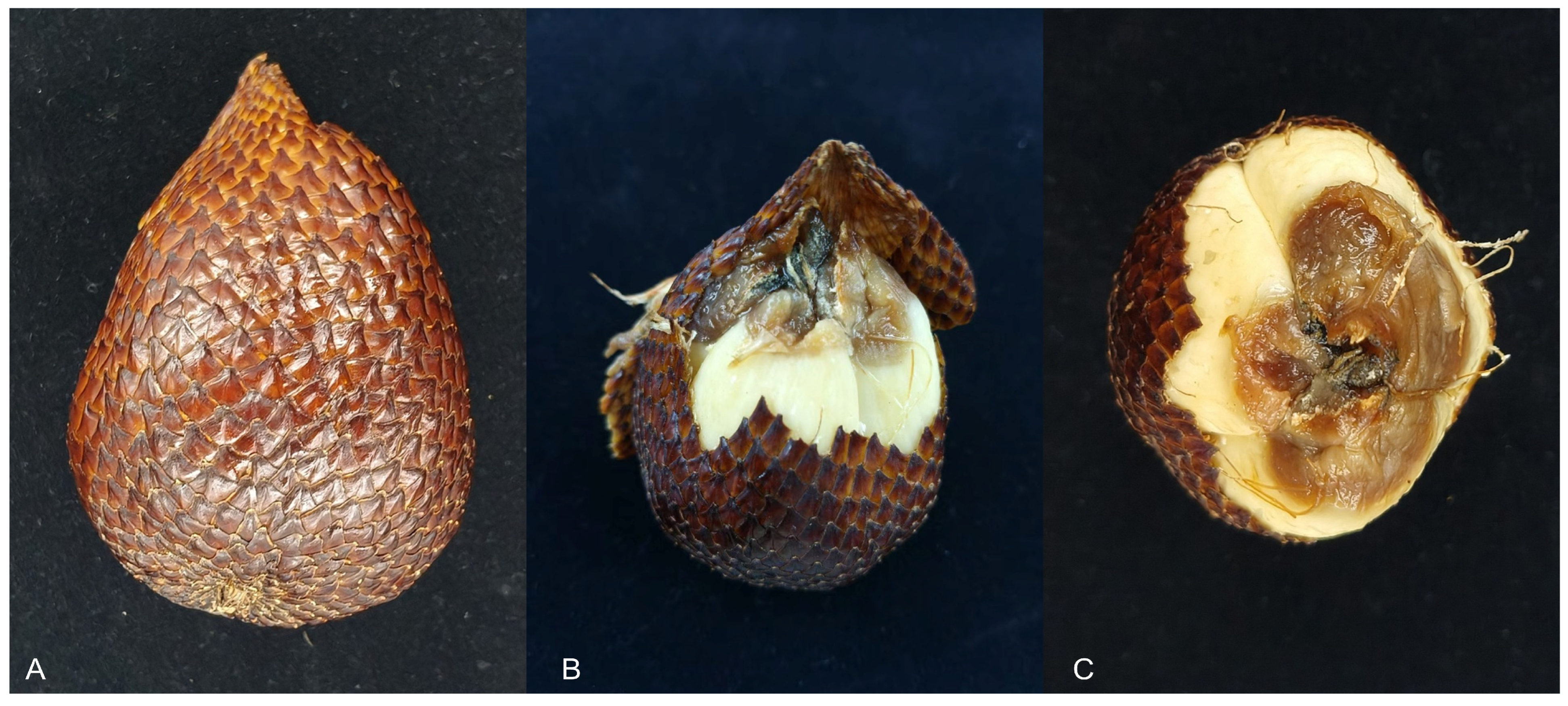
Intact snake fruit appeared asymptomatic (Figure 1A); however, brown rot was observed at the fruit apex upon removal of the peel. Fungal structures were visible near the residual stigma at the top of the fruit (Figure 1B,C). A total of four fungal isolates were obtained for their morphological similarity and purified from the rotten snake fruit tissue. Among these, isolate Te 5742 was selected as the representative strain for further characterization.
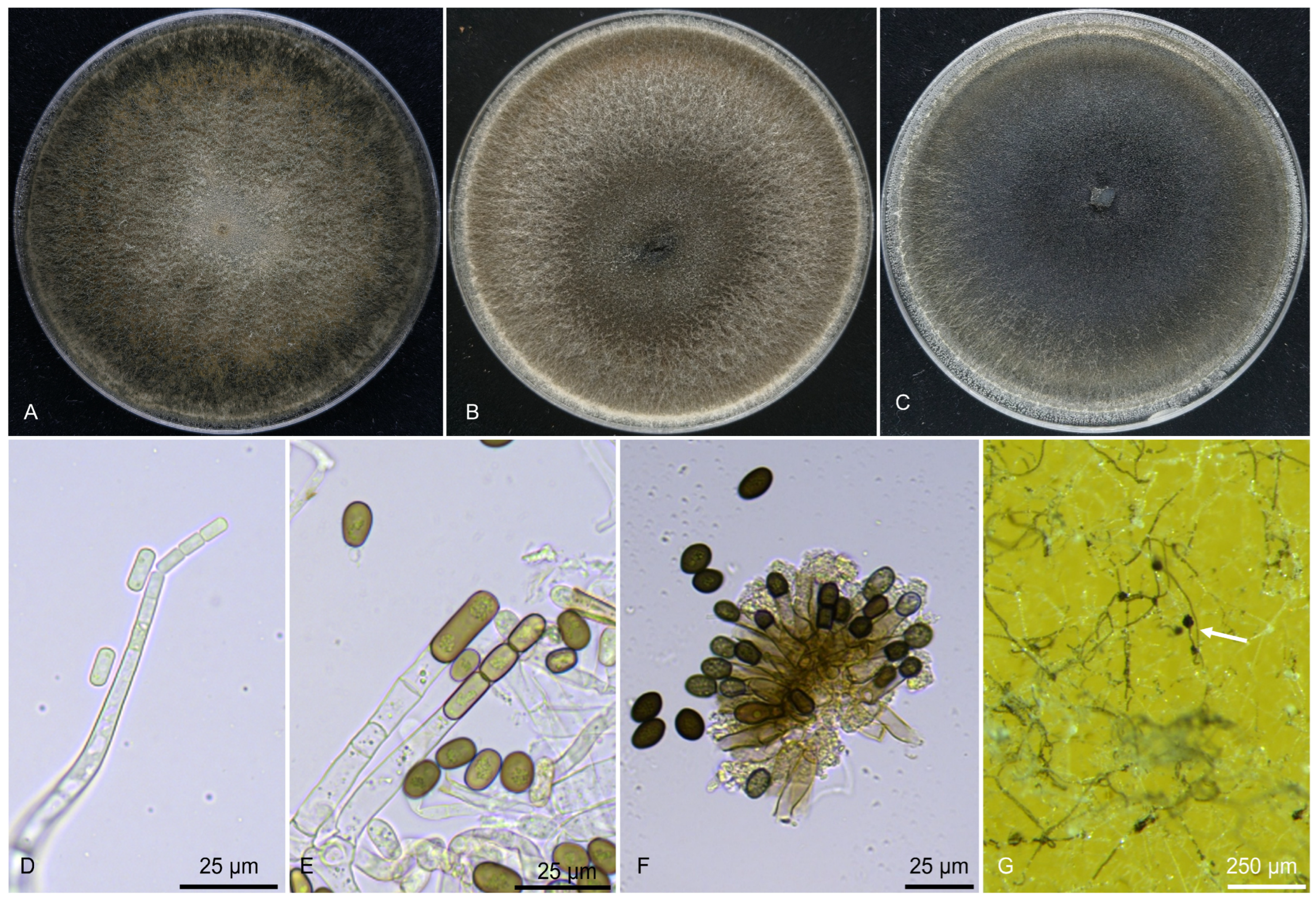
Isolate Te 5742 grew rapidly, covering the 90 mm petri dish within 48 h on PDA in 25 ± 2 °C. Colonies were initially hyaline to white, transitioning to fuscous black or greyish sepia with age, and sporulated abundantly on PDA, MEA, and OA (Figure 2A–C). A strong fruity aroma was noted on PDA cultures. Conidiophores were hyaline, flask, phialidic, and mononematous, exhibiting enteroblastic conidium ontogeny (Figure 2D). Primary conidia were hyaline, aseptate, and cylindrical (Figure 2D), measuring 8.59–11.80 × 4.04–5.49 μm (n = 50). Secondary conidia were produced holoblastically, aseptate, initially hyaline but becoming greyish sepia, and typically oblong with thick walls at maturity. Two distinct size ranges were observed for secondary conidia (Figure 2E): 9.79–12.67 × 5.69–8.81 μm and 15.29–23.28 × 6.57–12.05 μm (n = 50). Aleurioconidia (chlamydospores) were produced holoblastically, singly, or in short chains; these were dark mouse-gray, thick-walled, and subglobose to globose (Figure 2F), measuring 10.22–15.58 × 7.13–9.97 μm (n = 50). Synnemata observed on the host surface possessed a globose apex (Figure 2G). Ascomata were not observed.

3.2. Molecular Characterization and Phylogenetic Analyses

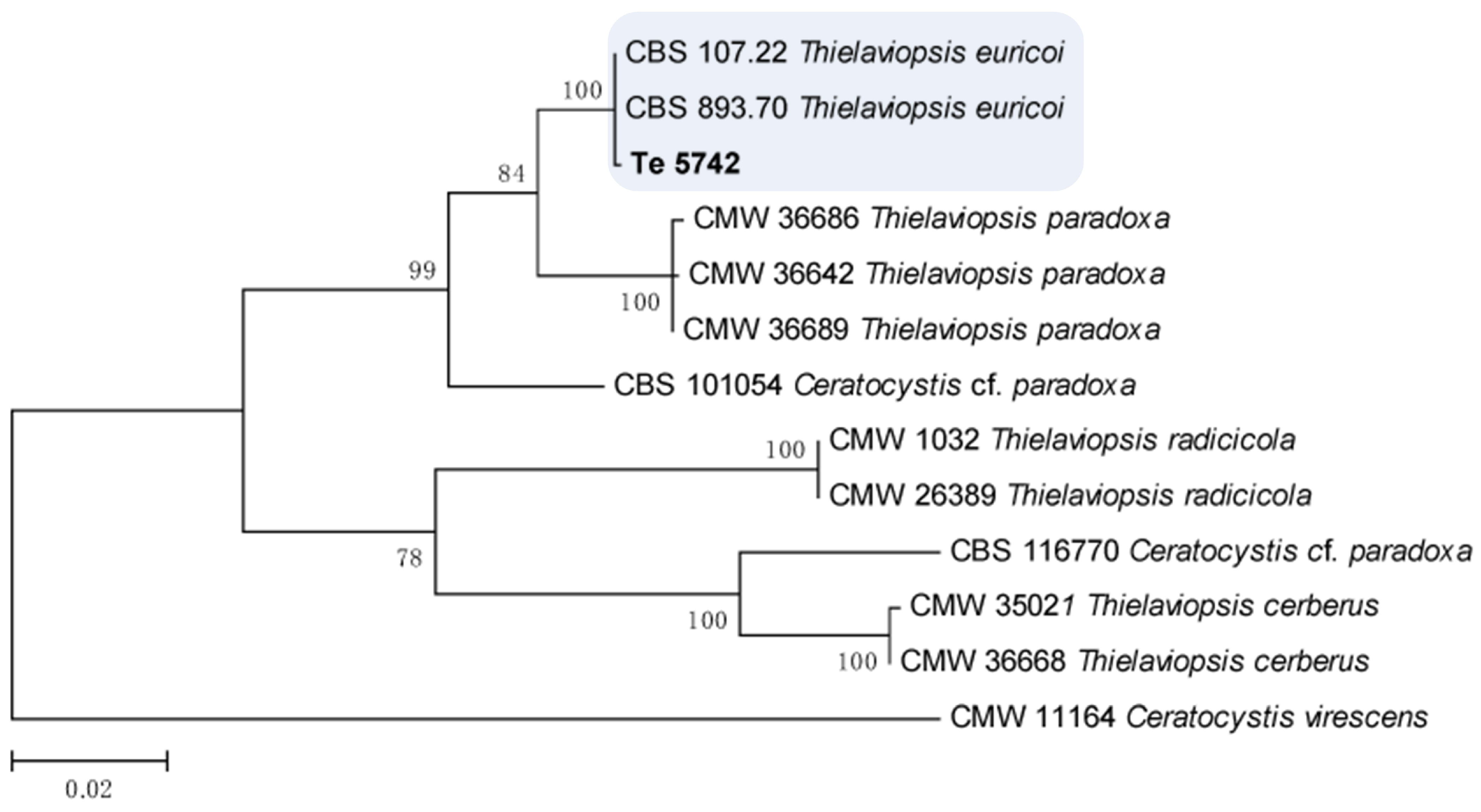
The ITS, tub2, and tef1-α sequences of isolate Te 5742 were deposited in GenBank (Table 1). The concatenated dataset consisted of 13 strains, with Ceratocystis virescens (CMW 11164) serving as an outgroup. Phylogenetic analysis revealed that isolate Te 5742 formed a well-supported clade with Thielaviopsis euricoi-type strains (CBS 893.70 and CBS 107.22) with 100% bootstrap support (Figure 3).

3.3. Pathogenicity Tests

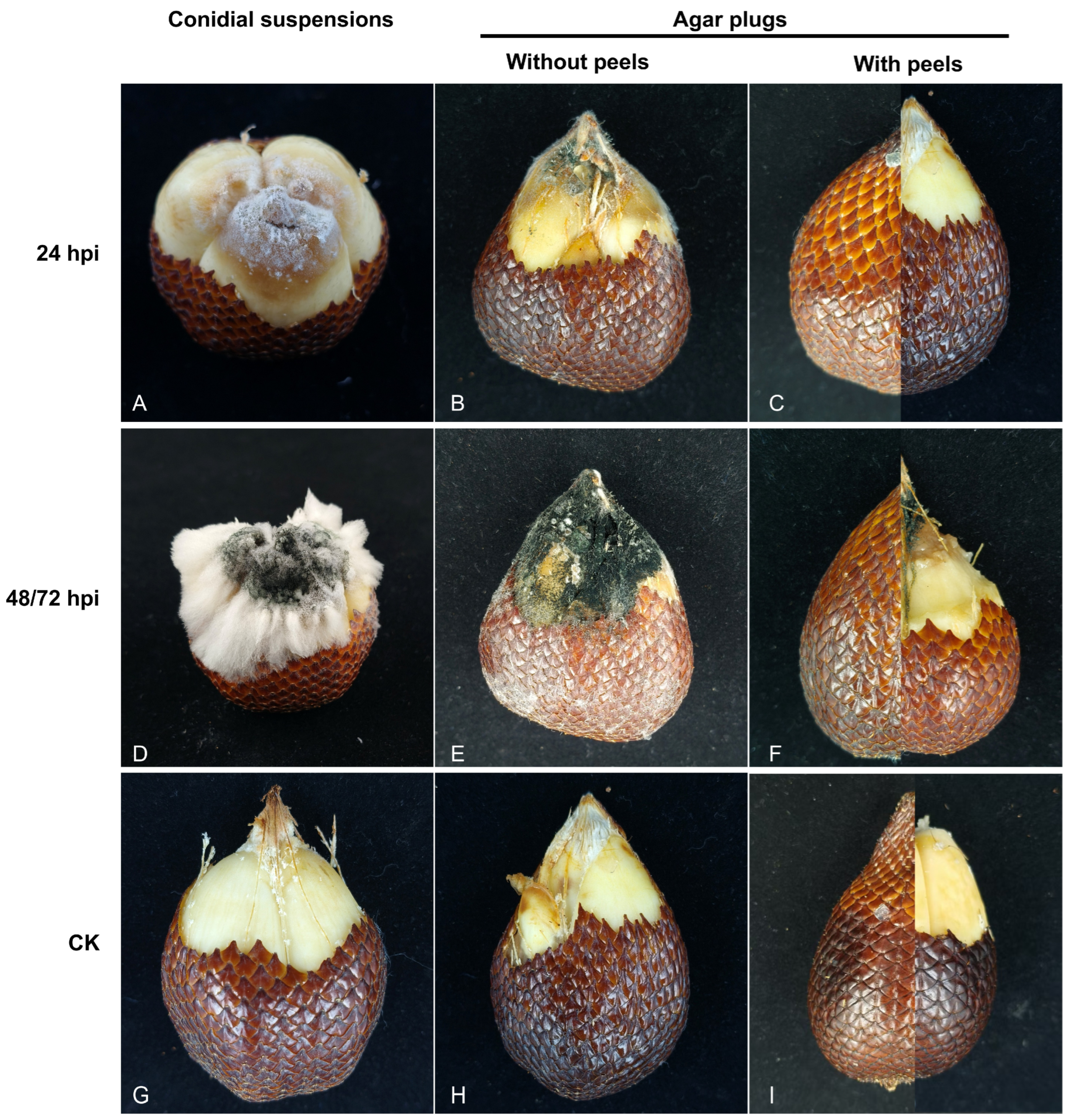
Pathogenicity assays demonstrated that rot symptoms initiated on peeled snake fruit at 24 h post-inoculation (hpi) in both conidial suspension and PDA plug treatments. In contrast, intact (unpeeled) fruit remained asymptomatic at this stage (Figure 4A–C). By 48 hpi, peeled fruit treated with the conidial suspension was covered with profuse white mycelium, while that treated with PDA plugs exhibited dark conidial production (Figure 4D,E). Interestingly, symptoms on intact (unpeeled) fruit were delayed; mycelial growth was not observed until 72 hpi in the PDA plug treatment (Figure 4F). The control fruits remained symptomless (Figure 4G–I). The isolate Te 5742 caused rot symptoms in all inoculated snake fruits, and the disease incidences recorded at 24, 48, and 72 hpi were all 100%. The pathogen was re-isolated from the inoculated snake fruit and identified as T. euricoi based on consistent morphological characteristics and identical tef1-α sequences, thus fulfilling Koch’s postulates.

4. Discussion

Morphological identification within the genus Thielaviopsis is challenging due to overlapping sporulation characteristics. For instance, T. ovoidea (Nag Raj & W.B. Kendr.) A.E. Paulin, T.C. Harr. & McNew, T. populi (Veldeman ex Kiffer & Delon) A.E. Paulin, T.C. Harr. & McNew, and T. thielavioides are morphologically similar, distinguished only by minor differences in aleurioconidia [7]. Specifically, species within the Ceratocystis paradoxa complex (C. musarum, C. paradoxa, C. radicicola, and T. euricoi) exhibit limited diagnostic morphological differences. In the present study, a key diagnostic feature of isolate Te 5742 was the production of aleurioconidia either singly or in short chains, differentiating it from C. radicicola, where they are borne exclusively singly [9]. Consistent with previous descriptions [7,9], ascomata were not observed in our isolate. Notably, we observed secondary conidia in two distinct size ranges, a feature that has not been previously described for T. euricoi [7,9], suggesting potential intraspecific variation. Additionally, while many Ceratocystis species are known to emit intense fruity scents to attract insect vectors [20,21], this characteristic was not mentioned in the original description of T. euricoi by Mbenoun et al. [9].
Given these morphological ambiguities, multigene phylogenies are critical for the accurate identification of Thielaviopsis species. Previous studies have demonstrated that ITS, β-tubulin, and tef1-α gene sequences are effective in delimiting species complexes across the genus [9,11,12]. Our phylogenetic analyses placed isolate Te 5742 within the T. euricoi clade with high support, corroborating our morphological identification. These results further confirm the utility of combined DNA sequence data in revising phylogenetic relationships and defining species boundaries within Ceratocystis sensu lato [12].
The genus Thielaviopsis includes numerous economically important pathogens of monocotyledonous plants, such as pineapple (Ananas comosus), banana (Musa acuminata), and palms (Arecaceae) [8]. Historically, T. euricoi strains have been collected primarily from Arecaceae hosts, such as coconut palm (Cocos nucifera); however, its pathogenicity had not been previously confirmed. In this study, we isolated T. euricoi (strain Te 5742) as the new pathogen on rotting snake fruit. This finding aligns with the growing recognition of Thielaviopsis species as emerging threats to snake fruit production [5]. The initial symptom showed that the brown rot on the flesh was similar to previously reported snake fruit rot diseases caused by T. paradoxa and P. salaccae [5,6]. In the present study, the pathogenicity test results showed that peeled fruit was more easily infected by the pathogen. It can be explained by the protective role of the fruit peel, which acts as a physical barrier to impede fungal pathogens, and is also consistent with the previous research results [5]. Although not consistently observed, profuse white mycelium emerged on some snake fruit inoculated with PDA plugs after extended incubation (>72 hpi). We speculate that the abundant production of white hyphae is associated with spores actively utilizing host-derived nutrients for development. Until now, there is still limited information about how the Thielaviopsis species disperses and carries out infection on snake fruit. Targeted monitoring for the T. euricoi-caused rotten disease should be established in snake fruit orchards.
In conclusion, this is the first report worldwide of T. euricoi causing fruit rot on snake fruit. Although the precise geographical origin of the intercepted fruit could not be determined, the severe symptoms observed in our pathogenicity tests highlight a significant risk. The finding is a valuable addition to the existing knowledge about the fungal pathogens causing snake fruit rot, and the government should pay attention to the quarantine of imported snake fruit. The development of T. euricoi rapid detection technology in the future is necessary.

5. Conclusions

In this study, we identified Thielaviopsis euricoi as a new pathogen of a previously undescribed fruit rot on snake fruit. This conclusion is supported by morphological characterization, multi-locus phylogenetic analysis, and pathogenicity assays fulfilling Koch’s postulates. Our work constitutes the first global record of this fungal species as a pathogen of snake fruit. These findings provide a scientific foundation for future taxonomic studies, the development of disease management strategies for Thielaviopsis species, and the quarantine priorities in snake fruit trade.

Author Contributions

Conceptualization, C.S.; methodology, M.W. and L.F.; software, C.S. and N.J.; validation, Z.C. and H.L.; formal analysis, M.W., X.Y. and L.F.; writing—original draft preparation, C.S., M.W. and L.F.; writing—review and editing, C.S., Z.C. and H.L.; visualization, C.S., X.Y. and N.J.; supervision, H.L.; project administration, H.L.; funding acquisition, C.S. All authors have read and agreed to the published version of the manuscript.

Funding

This research was funded by RESEARCH PROJECT OF GENERAL ADMINISTRATION OF CUSTOMS, grant number 2025HK041.

Data Availability Statement

Accession numbers for the DNA sequence data are available in NCBI GenBank and are listed in Table 1.

Conflicts of Interest

The authors declare no conflicts of interest.

References

  1. Aralas, S.; Mohamed, M.; Bakar, M.F.A. Antioxidant properties of selected salak (Salacca zalacca) varieties in Sabah, Malaysia. Nutr. Food Sci. 2009, 39, 243–250. [Google Scholar] [CrossRef][Green Version]
  2. Supapvanich, S.; Megia, R.; Ding, P. Salak (Salacca zalacca (Gaertner) Voss); Woodhead Publishing: Cambridge, UK, 2011; pp. 334–352. [Google Scholar]
  3. Haruenkit, R.; Poovarodom, S.; Leontowicz, H.; Leontowicz, M.; Sajewicz, M.; Kowalska, T.; Delgado-Licon, E.; Rocha-Guzman, N.E.; Gallegos-Infante, J.A.; Trakhtenberg, S.; et al. Comparative study of health properties and nutritional value of durian, mangosteen, and snake fruit: Experiments In Vitro and In Vivo. J. Agric. Food Chem. 2007, 55, 5842–5849. [Google Scholar] [CrossRef] [PubMed]
  4. Sukewijaya, I.M.; Rai, I.N.; Mahendra, M.S. Development of salak Bali as an organic fruit. Asian J. Food Agro-Ind. 2009, 2, S37–S43. [Google Scholar]
  5. Ahmad, R.Z.; Setyabudi, D.A.; Wulandari, N.F. The mold causing agent of rotten snake fruit (Salacca zalacca (gaertn.) from traditional fruit markets. IOP Conf. Ser. Earth Environ. Sci. 2018, 197, 012031. [Google Scholar] [CrossRef]
  6. Boukaew, S.; Kumla, J.; Suwannarach, N. Peniophora salaccae sp. nov. (Russulales, Basidiomycota), a snake fruit (Salacca zalacca) pathogen from southern Thailand. Phytotaxa 2024, 662, 137–149. [Google Scholar] [CrossRef]
  7. Paulin-Mahady, A.E.; Harrington, T.C.; Mcnew, D. Phylogenetic and taxonomic evaluation of Chalara, Chalaropsis, and Thielaviopsis anamorphs associated with Ceratocystis. Mycologia 2002, 94, 62–72. [Google Scholar] [CrossRef] [PubMed]
  8. De Beer, Z.W.; Duong, T.A.; Barnes, I.; Wingfield, B.D.; Wingfield, M.J. Redefining Ceratocystis and allied genera. Stud. Mycol. 2014, 79, 187–219. [Google Scholar] [CrossRef] [PubMed]
  9. Mbenoun, M.; De Beer, Z.W.; Wingfield, M.J.; Wingfield, B.D.; Roux, J. Reconsidering species boundaries in the Ceratocystis paradoxa complex including a new species from oil palm and cacao in Cameroon. Mycologia 2014, 106, 757–784. [Google Scholar] [CrossRef] [PubMed]
  10. Álvarez, E.; Llano, G.A.; Loke, J.B.; Chacon, M.I. Characterization of Thielaviopsis paradoxa isolates from oil palms in Colombia, Ecuador and Brazil. J. Phytopathol. 2012, 160, 690–700. [Google Scholar] [CrossRef]
  11. Alhudaib, K.A.; El-Ganainy, S.M.; Almaghasla, M.I.; Sattar, M.N. Characterization and control of Thielaviopsis punctulata on date palm in Saudi Arabia. Plants 2022, 11, 250. [Google Scholar] [CrossRef] [PubMed]
  12. Ayika, M.G.; Rosano, A.; Valiente, J.; Chakrabarti, S.; Rollins, J.A.; Dhillon, B. Characterizing the palm pathogenic Thielaviopsis species from Florida. J. Fungi 2024, 10, 247. [Google Scholar] [CrossRef] [PubMed]
  13. Batista, A.C.; Vital, A.F. Hughesiella—Novo e curiosos genero de fungos Dematiaceae. An. Soc. Biol. Pernamb. 1956, 14, 141–144. [Google Scholar]
  14. Rayner, R.W. A Mycological Colour Chart; Commonwealth Mycological Institute: Surrey, UK, 1970. [Google Scholar]
  15. White, T.J.; Bruns, T.; Lee, S.; Taylor, J. Amplification and direct sequencing of fungal ribosomal RNA genes for phylogenetics. PCR Protoc. Guide Methods Appl. 1990, 18, 315–322. [Google Scholar]
  16. Lousie, G.N.; Donaldson, G.C. Development of primer sets designed for use with the PCR to amplify conserved genes from filamentous ascomycetes. Appl. Environ. Microb. 1995, 61, 1323–1330. [Google Scholar]
  17. Jacobs, K.; Bergdahl, D.R.; Wingfield, M.J.; Halik, S.; Seifert, K.A.; Bright, D.E.; Wingfield, B.D. Leptographium wingfieldii introduced into North America and found associated with exotic Tomicus piniperda and native bark beetles. Mycol. Res. 2004, 108, 411–418. [Google Scholar] [CrossRef] [PubMed]
  18. Santos, P.H.D.; Mussi-Dias, V.; Freire, M.G.M.; Carvalho, B.M.; Silveira, S.F. Diagrammatic scale of severity for postharvest black rot (Ceratocystis paradoxa) in coconut palm fruits. Summa Phytopathol. 2017, 43, 269–275. [Google Scholar] [CrossRef][Green Version]
  19. Li, Y.; Li, A.; Jiang, N. Identification of the nut rot pathogen affecting Castanopsis carlesii based on morphological and phylogenetic analyses. Forests 2025, 16, 627. [Google Scholar] [CrossRef]
  20. Mailula, D.M.; Wingfield, B.D.; Nest, M.A.V.D.; Hammerbacher, A. Production of fusel alcohols and fusel acetates by pathogenic fungi in the Ceratocystidaceae. Fungal Ecol. 2025, 76, 101427. [Google Scholar] [CrossRef]
  21. Suwandi, S.; Irsan, C.; Hamidson, H.; Umayah, A.; Asriyani, K.D. Identification and characterization of Ceratocystis fimbriata causing lethal wilt on the Lansium tree in Indonesia. Plant Pathol. J. 2021, 37, 124–136. [Google Scholar] [CrossRef]
Figure 1. Symptoms of snake fruit rot disease of Thielaviopsis euricoi. (A) Appearance of the intercepted snake fruit. (B,C) Front and top views of rotten snake fruit after removing the peels, respectively.
Forests 17 00230 g001
Figure 2. Morphology of T. euricoi. (AC) Colony on PDA, MEA, and OA after 7 d. (D) Primary conidia oozing out of flask-shaped phialidic conidiophores. (E) Secondary conidia. (F) Aleurioconidia. (G) Synnemata (indicated by white arrow).
Forests 17 00230 g002
Figure 3. Phylogenetic maximum likelihood tree resulting from the combined dataset of ITS, β-tubulin, and tef1-α gene sequences. Numbers above the branches indicate ML bootstraps (≥50%). Isolate obtained from this study is marked in bold.
Forests 17 00230 g003
Figure 4. The symptoms of Thielaviopsis euricoi isolate Te 5742 inoculated on snake fruit. (AC) Symptoms of rotten snake fruit inoculated by the conidial suspension and PDA plugs at 24 hpi (hours post-inoculation). (D,E) Symptoms of rotten snake fruit inoculated by the conidial suspension and PDA plugs at 48 hpi, respectively. (F) Symptoms of rotten snake fruit inoculated by PDA plugs at 72 hpi. (GI) CK, Symptoms of snake fruit inoculated with sterilized distilled water or pure PDA plugs as negative controls.
Forests 17 00230 g004
Table 1. Details of fungal isolates used for phylogenetic analysis in this study.
Species NameCulture AccessionHostCountryGenBank Accession Numbers
ITStef1tub2
Ceratocystis cf. paradoxaCBS 116770Palm speciesEcuadorJX518334JX518302JX518366
CBS 101054Rosa sp.NetherlandsJX518333JX518301JX518365
C. virescensCMW 11164Fagus americanumUSADQ520639EF070413EF070441
Thielaviopsis cerberusCMW 35021Theobroma cacaoCameroonJX518355JX518323JX518387
CMW 36668Theobroma cacaoCameroonJX518348JX518380JX518316
T. euricoiCBS 893.70NABrazilJX518335JX518303JX518367
CBS 107.22Cocos nuciferaNAJX518367JX518304JX518368
Te 5742Salacca zalaccaNAPX736385PX776694PX776695
T. paradoxaCMW 36642Elaeis guineensisCameroonJX518346JX518314JX518378
CMW36686Elaeis guineensisCameroonJX518352JX518320JX518384
CMW 36689Elaeis guineensisCameroonJX518342JX518310JX518374
T. radicicolaCMW 1032Phoenix dactyliferaUSAKF612023KF612024KF612025
CMW 26389Lawsonia inermisMauritaniaKF953932KF953931KF917202
Note: Newly generated sequences are shown in bold. NA, not applicable.
Disclaimer/Publisher’s Note: The statements, opinions and data contained in all publications are solely those of the individual author(s) and contributor(s) and not of MDPI and/or the editor(s). MDPI and/or the editor(s) disclaim responsibility for any injury to people or property resulting from any ideas, methods, instructions or products referred to in the content.