Article

Production and Storage of Male-Sterile Somatic Embryos of Sugi (Japanese Cedar, Cryptomeria japonica) at Temperatures Above Freezing

1
Department of Forest Molecular Genetics and Biotechnology, Forestry and Forest Products Research Institute, Matsunosato 1, Tsukuba 305-8687, Japan
2
Department of Agriculture, Faculty of Agriculture, Niigata University, Ikarashi 8050, Niigata 950-2181, Japan
*
Author to whom correspondence should be addressed.
Forests 2025, 16(9), 1431; https://doi.org/10.3390/f16091431
Submission received: 15 July 2025 / Revised: 31 August 2025 / Accepted: 5 September 2025 / Published: 7 September 2025
(This article belongs to the Section Genetics and Molecular Biology)

Abstract

Sugi-pollinosis poses a significant socioeconomic and public health concern in Japanese society. Consequently, the use of male-sterile plants (pollen-free plants or PFPs) is anticipated in reforestation efforts. In this context, we developed an improved, simplified method for efficiently propagating sugi PFPs. In the present study, we compared the efficiency of different embryogenic cell lines (ECLs) in producing somatic embryos and examined how effectively these embryos germinate and convert into plantlets. We also evaluated the germination potential of somatic embryos stored for various durations at temperatures above freezing and room temperature. The production efficiency of somatic embryos ranged from 129.6 to 504.1 per plate, with an average of 349.8 across the ECLs tested. The overall average germination and conversion rates of somatic embryos were found to be 93.9% and 92.4%, respectively. Furthermore, although differences were observed among the evaluated genotypes, our five-year study demonstrated that sugi somatic embryos could be stored at 25 °C, 15 °C, or 5 °C for 6, 12, or 24 months, respectively, without a notable decline in germination capacity. The developed method enhances flexibility in plant production scheduling and facilitates the optimal timing for transferring somatic seedlings to the field.

1. Introduction

Japan ranks among the top three countries in the Northern Hemisphere with the highest forest coverage, at 68.4% [1]. The sugi (Japanese cedar, Cryptomeria japonica D. Don), an endemic conifer in the Cupressaceae family, is the most important forest tree species in Japan. It occupies approximately 4.5 million hectares, accounting for 44% of the nation’s total forest area [2]. Despite its commercial value, sugi forests release substantial amounts of pollen each spring, triggering allergic reactions in approximately 40% of the population [3]. This represents a significant socioeconomic and public health issue in Japanese society. Sugi pollen allergy is regarded as a national affliction, with estimated daily economic losses from absenteeism exceeding 230 billion Japanese yen [4]. As such, the use of male-sterile plants (pollen-free plants or PFPs) in reforestation is expected to help mitigate sugi-pollinosis.
In this context, several studies have reported the production of PFPs through a combination of marker-assisted selection and micropropagation techniques via somatic embryogenesis (SE) for the clonal mass propagation of sugi somatic seedlings [5,6,7,8,9,10]. A simplified and improved propagation system for sugi PFPs via SE was later published, detailing all stages of the somatic seedling production process [11,12]. However, major obstacles to practical application remain, including various bottlenecks related to genotype dependence in SE initiation, somatic embryo production, germination frequency, and plant conversion. The ultimate goal of a successful production process is the large-scale production of high-quality somatic embryos, resulting in high germination rates and the subsequent production of vigorous planting material [13]. Therefore, the key criteria for applying SE protocols are the efficient production of somatic embryos across multiple genotypes and the generation of somatic plants with strong field performance. Our experimental results confirm that the genetic origin of embryogenic cell lines (ECLs) significantly influences somatic embryo production in sugi. They also highlight the importance of selecting highly responsive “Yokozuna” cell genotypes (“champion” cell genotypes) to optimize the efficiency of SE protocols [11].
This study aimed to improve existing protocols by gathering additional data on somatic embryo production efficiency, germination rates, somatic seedling conversion rates, and the germination performance of somatic embryos derived from various ECLs following storage at above-freezing temperatures for up to five years. Although cryopreservation is the ideal method for preserving genetic material, it involves costly equipment and specialized procedures. Moreover, short- to medium-term storage of somatic embryos allows flexibility in scheduling somatic seedling production, facilitating the timely transfer of plants to the field. Thus, maintaining the germination capacity of somatic embryos for 6 to 24 months under simple storage conditions would be a practical enhancement to our methodology for producing sugi PFPs.

2. Materials and Methods

2.1. SE Initiation, Maintenance, Proliferation, and Selection of Male-Sterile ECLs

Seven male-sterile embryogenic cells (ECs) were induced from entire megagametophytes of immature seeds collected in July 2016 from the full-sib seed family ‘Shindai 3’ × ‘Suzu 2’, which carries the male sterility allele ms1, as described by Maruyama et al. [6]. Embryogenic masses (EMs) were maintained and proliferated through regular subculturing at 2-week intervals on a maintenance/proliferation medium. This medium contained half the standard concentration of basal salts [14] and was supplemented with 3 μM 2,4-dichlorophenoxyacetic acid (2,4-D), 1 μM 6-benzylaminopurine (BAP), 30 g L−1 sucrose, 1.5 g L−1 glutamine, and 3 g L−1 gellan gum (Wako Pure Chemical, Osaka, Japan). Twelve EM clumps per plate were cultured in the dark at 25 °C. Male-sterile ECLs were selected according to the methodology described by Ueno et al. [15], and the genotypes and male sterility of these lines were confirmed using the protocol outlined by Tsuruta et al. [8].

2.2. Production of Somatic Embryos

The methodology described by Maruyama et al. [7] was used for somatic embryo production. Two-week-old proliferated EMs from male-sterile ECLs were cultured in clumps (five masses per 90 × 20 mm plate, each weighing 100 mg; total 500 mg per plate) on a maturation medium containing the basal salt concentration of the standard EM medium [14]. The medium was supplemented with 30 g L−1 maltose, 2 g L−1 activated charcoal (AC), 100 µM abscisic acid, and a mixture of amino acids (in g L−1: glutamine 2, asparagine 1, arginine 0.5, citrulline 0.079, ornithine 0.076, lysine 0.055, alanine 0.04, and proline 0.035). It also contained 175 g L−1 polyethylene glycol (average molecular weight: 7300–9300; Wako Pure Chemical, Osaka, Japan) and 3.3 g L−1 gellan gum for solidification. Plates were sealed with Parafilm® and incubated in darkness at 25 °C for eight weeks. The number of cotyledonary embryos was counted per plate, with seven to ten replicates for each line.

2.3. Somatic Embryo Germination and Conversion

Cotyledonary embryos derived from male-sterile ECLs were individually picked from the maturation medium using tweezers and placed horizontally on a germination medium. This medium was based on the maintenance/proliferation medium, supplemented with 20 g L−1 sucrose, 2 g L−1 AC, and 10 g L−1 agar, but without any plant growth regulators. The somatic embryos were cultured at 25 °C under a photon flux density of 45–65 µmol m−2 s−1, provided by white fluorescent lamps (100 V, 40 W), for 16 h per day. Somatic embryo germination (i.e., root emergence) and plantlet conversion (i.e., root and epicotyl emergence) were assessed after four and eight weeks of culture, respectively. Germination and conversion rates were calculated per plate as the number of germinants and converted plantlets, respectively, divided by the number of cotyledonary embryos tested. The average of all plates was used to determine the final germination and conversion rates, with ten replicates per line.

2.4. Storage of Somatic Embryos and Post-Storage Germination Test

The ability of somatic embryos to germinate following storage at room temperature and temperatures above the freezing point for various durations was investigated using embryos derived from line SSD-18. Plates containing cotyledonary embryos on maturation medium were sealed with three layers of Parafilm®, placed in a Ziploc® freezer bag (Asahi Kasei Co., Ltd., Tokyo, Japan), and stored in the dark for different time periods (0, 6, 12, 18, 24, 30, 36, 48, and 60 months) at 25 °C (room temperature), 15 °C and 5 °C. In addition, the germination response of somatic embryos from seven male-sterile ECLs was evaluated after a 24-month storage period at 5 °C. Germination tests were conducted on germination medium under the culture conditions described in Section 2.3 following storage. The germination rate was calculated using the average value from five to ten plates per storage condition.

2.5. Statistical Analysis

Differences in the number of mature cotyledonary embryos, as well as germination and conversion frequencies among ECLs, were assessed using generalized linear models and generalized linear mixed models (GLMMs). Poisson and binomial distributions were applied for the error distribution relating to the number of mature embryos per plate and for germination and conversion (1, 0), respectively. In the GLMMs, the plate was treated as a random effect. Pairwise differences were identified using the Tukey–HSD test. All statistical analyses were performed using R software [16], with the packages “lme4” [17] and “multcomp” [18].

3. Results and Discussion

3.1. Maintenance and Proliferation of Male-Sterile ECLs

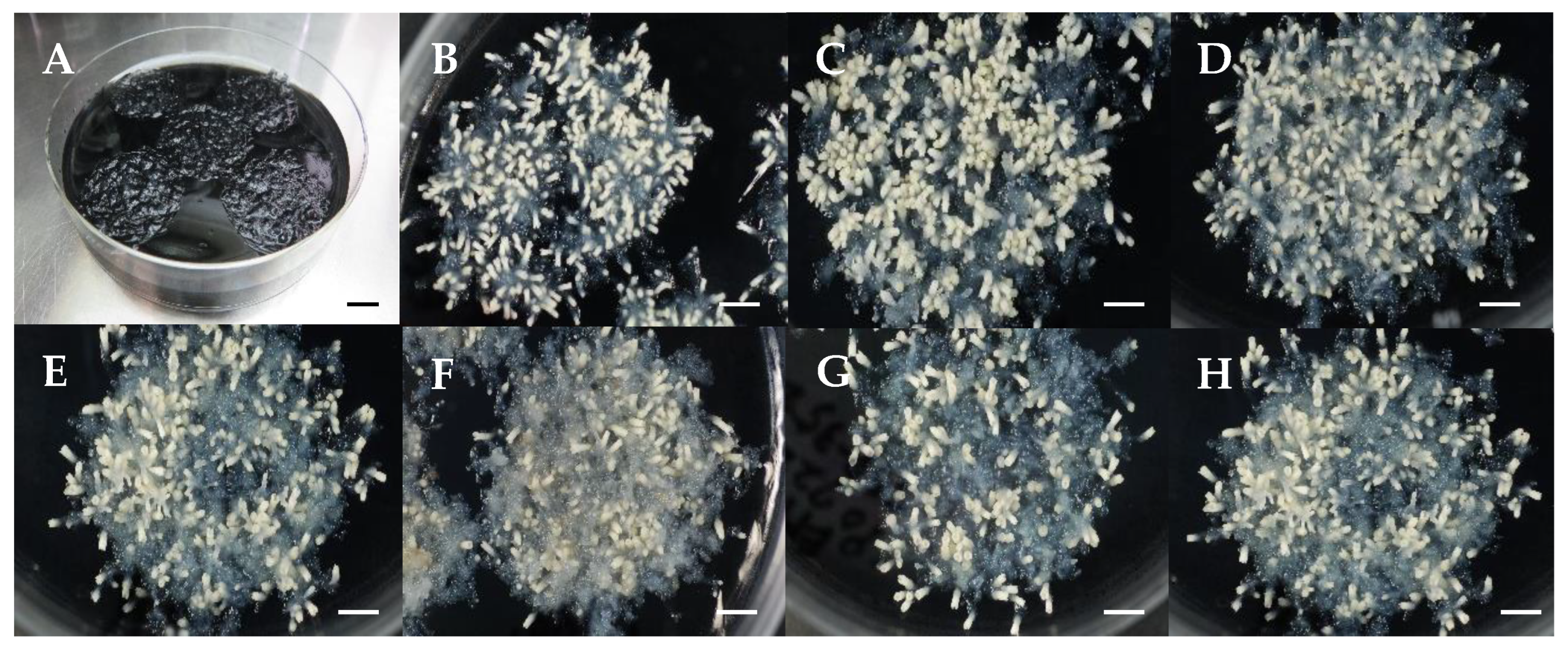
Maintaining and proliferating EMs through biweekly subculturing enabled continuous growth across the seven ECLs tested. After two weeks of culture, no remarkable differences in mass proliferation were observed with the naked eye. However, morphological variations were noted among the ECLs. For example, the EMs of the SSD-018 (Figure 1B), SSD-112 (Figure 1E), and SSD-352 (Figure 1H) lines were friable and white, whereas those of the SSD-073 (Figure 1C), SSD-113 (Figure 1F), and SSD-182 (Figure 1G) lines were also friable but exhibited a yellowish-white coloration. In contrast, EMs from the SSD-100 line (Figure 1D) were yellowish-white with a mucilaginous texture. Morphological differences are commonly reported among ECs derived from different lines and even within the same genotype [19,20,21,22,23,24]. Moreover, some studies suggest that these morphological traits may be associated with proliferation capacity and the potential for somatic embryo formation [25,26,27,28]. Nevertheless, despite the observed morphological differences among certain ECLs, our study found no noticeable variation in their proliferation capacity (Figure 1B–H), and all lines subsequently produced cotyledonary embryos (Figure 2). Therefore, while morphological characteristics may occasionally indicate highly responsive ECLs, this does not appear to apply uniformly across all genotypes.

3.2. Production of Somatic Embryos from Different Male-Sterile ECLs
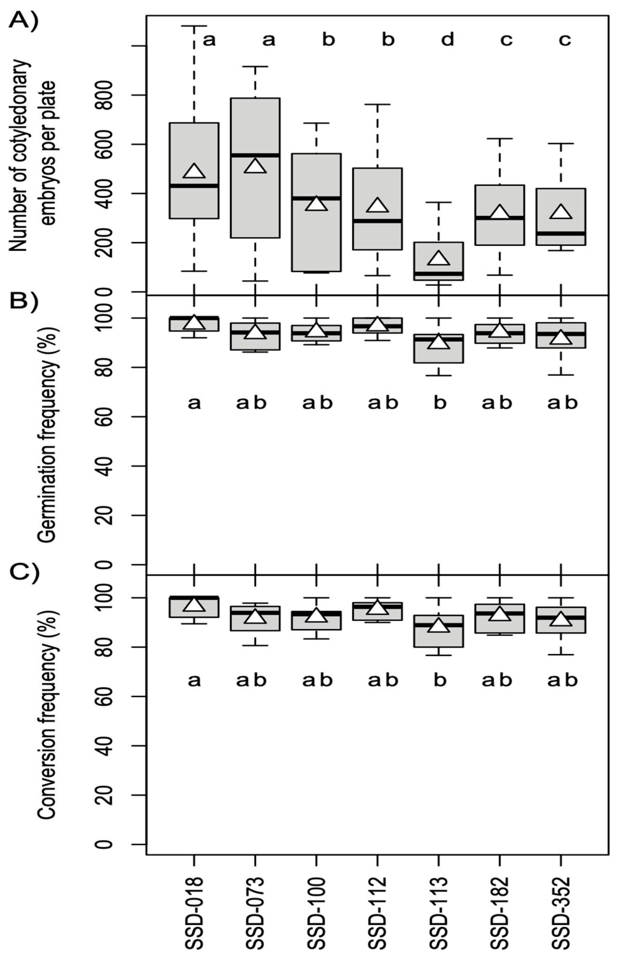
Cotyledonary embryos developed from the EMs after 6–8 weeks of culture on the maturation medium (Figure 3). As shown in Figure 2A, somatic embryo production was confirmed in all ECLs tested, with an overall average of 349.8 embryos produced per plate (equivalent to approximately 700 embryos per 1 g). No significant morphological differences related to genotype were observed in the size or shape of the somatic embryos (Figure 3B–H). However, analysis of the results revealed differences in production efficiency among genotypes (p < 0.05, GLMM, Figure 2A). The SSD-073 line yielded the highest average number of somatic embryos per plate (504.1 ± 351.2), while the SSD-113 line produced the fewest (129.6 ± 125.9). These values correspond to approximately 1000 and 260 cotyledonary embryos per gram of fresh EMs, respectively (calculation based on the number of embryos obtained per 500 mg per plate).
Figure 2. Box plot showing the efficiency of cotyledonary embryo production (A), germination (B), and conversion (C) in different sugi (Cryptomeria japonica) lines. The bold line in each box indicates the median; the triangle represents the mean. Different letters indicate statistically significant differences at the 5% level.
Forests 16 01431 g002
Figure 3. Somatic embryo production in sugi (Japanese cedar, Cryptomeria japonica). (A) Embryogenic masses transferred to maturation medium. (BH) Somatic embryo production after eight weeks from lines SSD-018 (B), SSD-073 (C), SSD-100 (D), SSD-112 (E), SSD-113 (F), SSD-182 (G), and SSD-352 (H). Bars: 10 mm.
Forests 16 01431 g003
It is widely reported that cotyledonary embryo production varies significantly among ECLs [25,29,30,31]. In conifers, somatic embryo production is influenced by several key factors, including medium composition, culture conditions, and genotype origin [32,33,34,35,36,37]. Studies on SE in sugi suggest that the genetic origin of ECs is the most critical factor influencing somatic embryo production efficiency [7,11]. Therefore, the development of markers to identify “Yokozuna” (“champion”) cells, those with the highest potential for producing high-quality embryos, is essential for the practical application of SE protocols.

3.3. Germination and Conversion of Somatic Embryos

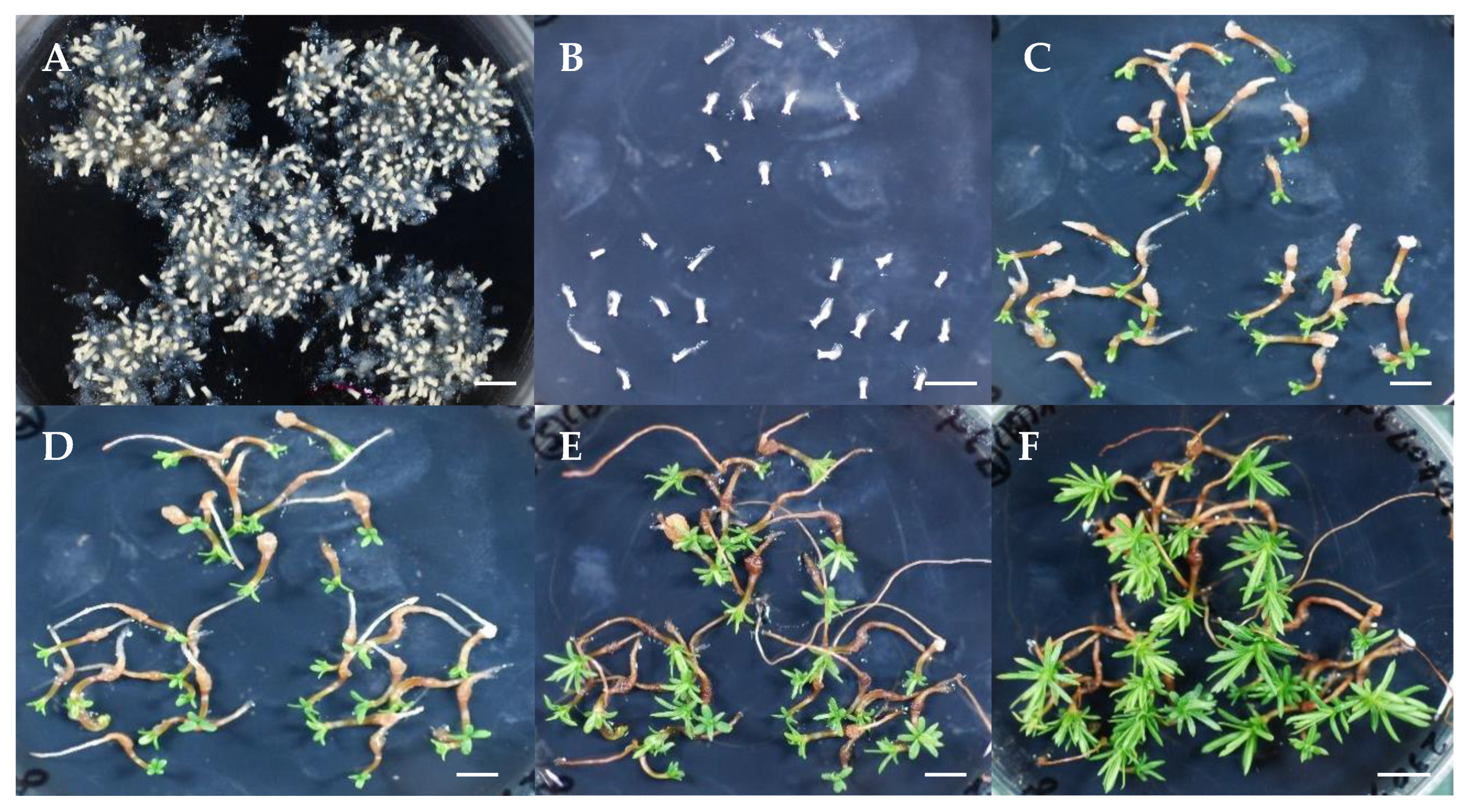
Cotyledonary embryos germinated readily upon transfer to germination medium, regardless of genotype. Root and epicotyl emergence were observed after 1–2 weeks (Figure 4C,D) and 3–4 weeks (Figure 4E) of culture, respectively. High germination frequencies were recorded across all ECLs, ranging from 89.6% ± 7.7% (SSD-113) to 97.5% ± 3.4% (SSD-018), with an overall average of 93.9% after four weeks of culture (Figure 2B). Significant difference was only observed between SSD-018 and SSD-113 (p < 0.05, GLMM, Figure 2). Nearly all germinated embryos subsequently converted into plantlets (Figure 4F). After 8 weeks of culture, the average conversion rate was 92.5%, ranging from 88.1% ± 8.5% (SSD-113) to 96.6% ± 4.6% (SSD-018) (Figure 2C). No genotype-related morphological differences were observed during the germination and conversion processes of somatic embryos.
Sugi somatic embryos derived from male-sterile ECLs germinated readily without the need for post-maturation treatment, which is reportedly essential for Japanese pines [38,39,40,41] and other conifer species [25,42,43,44,45,46]. In the present study, nearly all germinated embryos developed into plants, confirming the high quality of the somatic embryos produced. The efficient production and subsequent conversion of these embryos into vigorous planting stock is fundamental to the practical application of SE technology in mass propagation, breeding programs, and deployment strategies in multivarietal forestry [25,47]. Additionally, an efficient plant regeneration system via SE serves as a powerful platform for studying molecular, genetic, and physiological processes, as well as for facilitating genetic engineering [11].

3.4. Germination of Stored Somatic Embryos

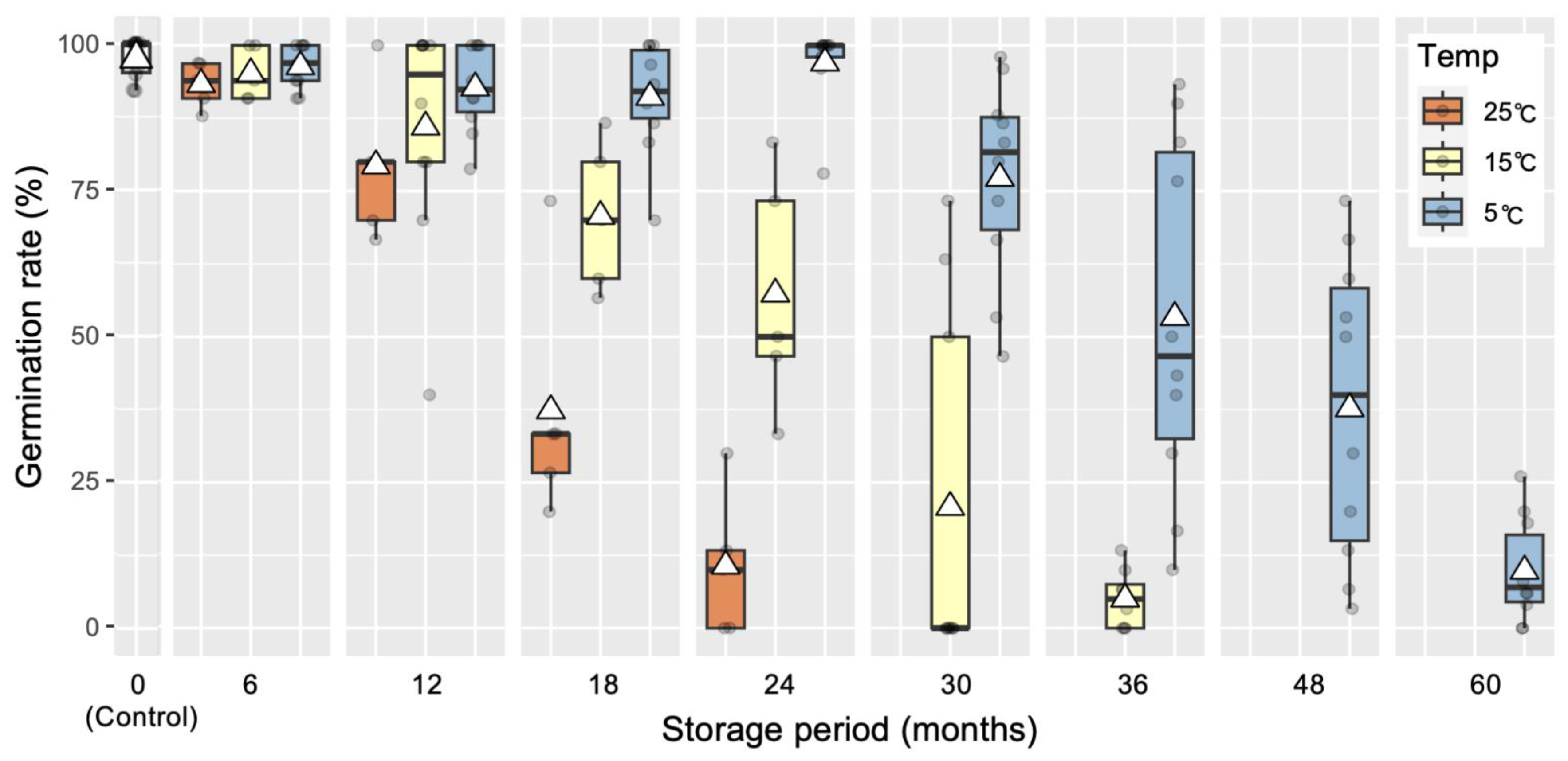
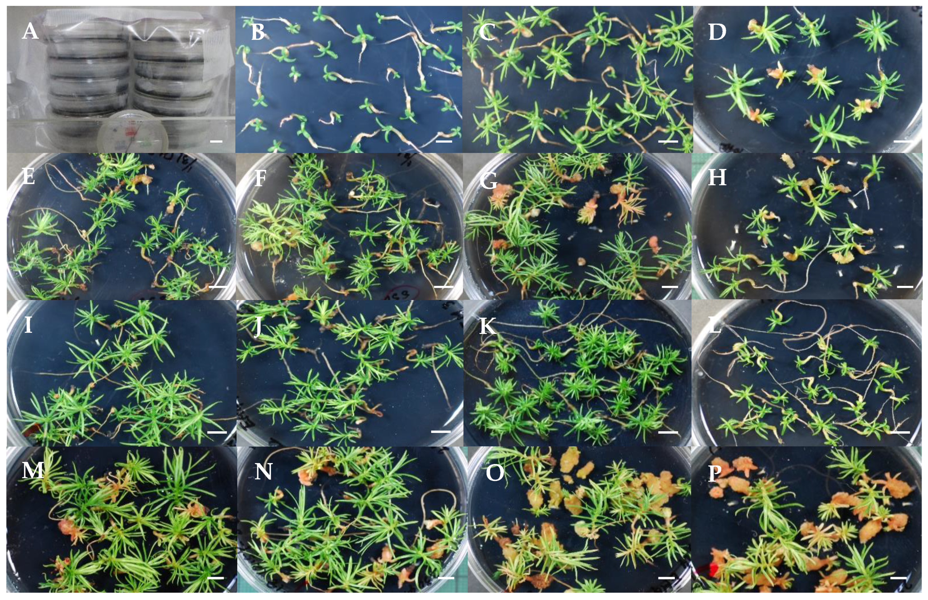
As shown in Figure 5, Figure 6 and Figure 7, differences in somatic embryo germination frequency were observed depending on storage temperature and duration. The most favorable results for maintaining germination capacity over time were achieved when embryos were stored at 5 °C. After 6 (Figure 7I), 12 (Figure 7J), 18 (Figure 7K), and 24 (Figure 7L) months, most stored embryos exhibited normal germination, with rates ranging from 91.1% to 97.0%, comparable to the germination response of non-stored control embryos (Figure 7B). However, after 30 (Figure 7M), 36 (Figure 7N), 48 (Figure 7O), and 60 (Figure 7P) months, germination gradually declined to 77.2%, 53.3%, 37.7%, and 9.8%, respectively. Notably, following 36 months of storage, sporadic callus formation was observed in some embryos or germinants (Figure 7N). This phenomenon became more pronounced after 48 months (Figure 7O) and further increased after 60 months of storage (Figure 7P). Although this study did not specifically investigate the cause, it is assumed that prolonged storage may lead to physiological changes possibly involving hormone levels, lipid composition, protein degradation, or other metabolic alterations that promote callus formation in somatic embryos.
In contrast, the lowest viability over time was observed in embryos stored at 25 °C. Although average germination rates remained relatively high at 93.3% and 79.3% after 6 (Figure 7C) and 12 (Figure 7D) months, respectively, these rates declined sharply to 37.3% after 18 months and further dropped to 10.7% after 24 months. In comparison, embryos stored at 15 °C exhibited higher germination frequencies over the same period. The average germination rates were 95.2%, 86.0%, and 70.7% after 6 (Figure 7E), 12 (Figure 7F), and 18 (Figure 7G) months, respectively. However, these rates decreased to 57.3% after 24 months (Figure 7H) and further declined to 20.7% after 30 months. By 36 months, germination capacity had nearly vanished, with a rate of only 5%.
In summary, considering that, for practical applications, the average germination capacity of somatic embryos after storage should not fall below 85%, we conclude that sugi somatic embryos can be stored at 25 °C, 15 °C, or 5 °C for up to 6, 12, or 24 months, respectively. The mean germination capacities following storage at 25 °C for 6 months, 15 °C for 12 months, and 5 °C for 24 months were 93.3% ± 3.9%, 86.0% ± 19.6%, and 97.0% ± 22.3%, respectively.
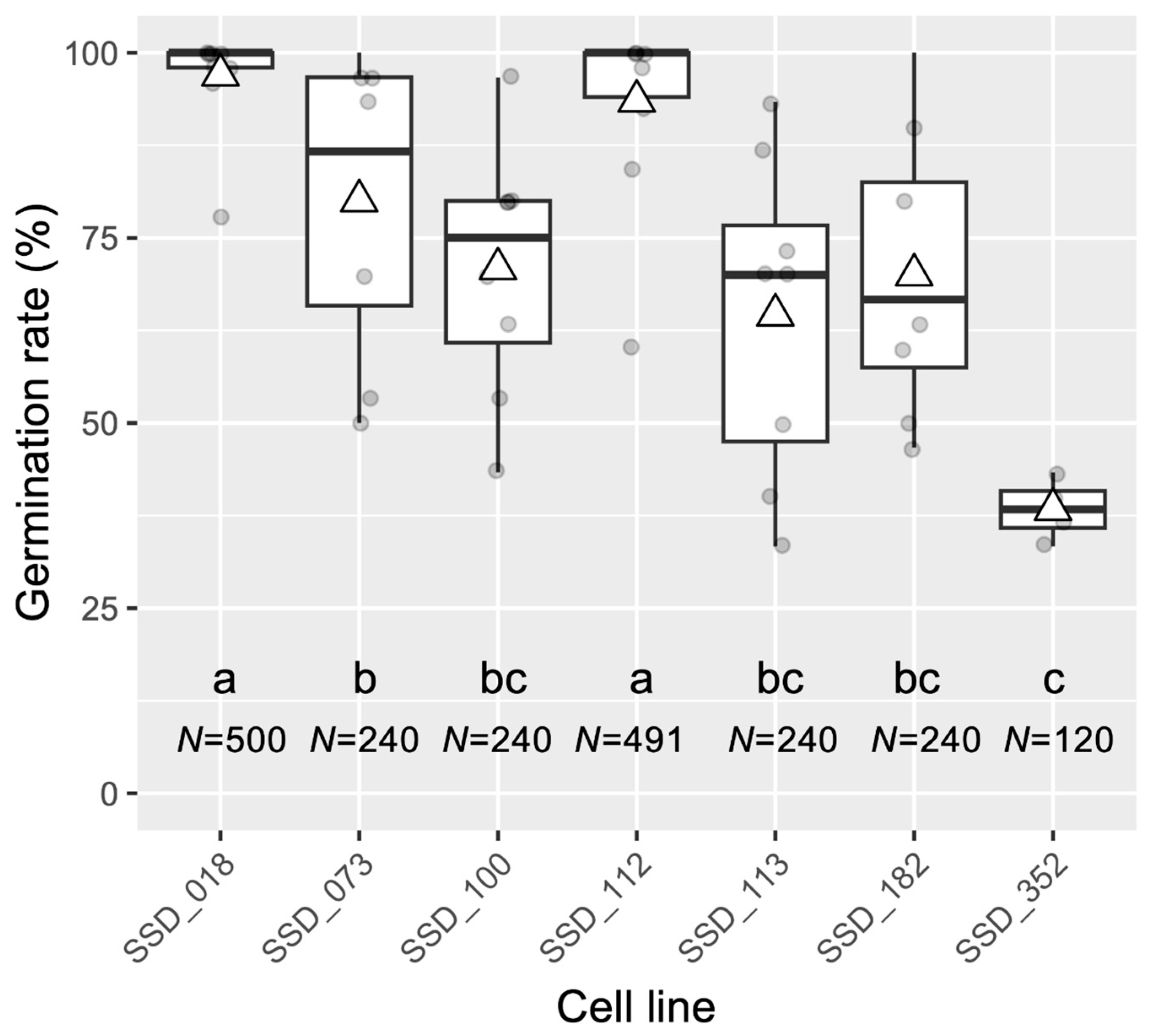
However, as shown in Figure 6, variation in germination response was observed among ECLs following storage at 5 °C for 24 months. The highest germination rates were obtained with embryos derived from lines SSD-018 and SSD-112, which maintained viability at 97.0% and 93.5%, respectively. In contrast, the lowest rate (38.3%) was recorded for embryos from the SSD-352 genotype. The remaining ECLs exhibited intermediate germination frequencies ranging from 64.6% to 80.0%. Overall, Figure 5, Figure 6 and Figure 7 demonstrate that sugi somatic embryos can be stored at 5 °C for at least two years without a significant loss of germination capacity. However, due to observed genotype-dependent variation, the optimal storage period at 5 °C likely ranges from 12 to 24 months, depending on the specific ECL.
Cryopreservation is considered the most effective method for the long-term storage of genetic material. Accordingly, many studies on woody species, including Japanese cedar [14,48,49], have focused on this approach, particularly using ECs at early developmental stages. This preference is due to the higher tolerance of early-stage embryogenic cells to liquid nitrogen storage compared to more differentiated stages, such as cotyledonary somatic embryos [50]. However, cryopreservation requires costly equipment and specialized procedures. Since our objective is to introduce flexibility into the seedling production schedule, our methodology can accommodate this by maintaining somatic embryo germination capacities of approximately 90% for 12 to 24 months when stored at 5 °C. This storage strategy allows for easy regulation of the optimal field transfer period and facilitates year-round workload distribution. Consistent with our findings, Torres-Viñals et al. [51] reported a germination rate exceeding 90% for grapevine somatic embryos after 30 days of storage at 4 °C. Similarly, Jayasankar et al. [52] observed a 90% conversion rate in the same species using desiccated somatic embryos stored for 42 months at 4 °C. El-Dawayati et al. [53] successfully stored clusters of Phoenix dactylifera somatic embryos at 15 °C for up to eight months. Shigeta et al. [54] reported a germination frequency above 95% for encapsulated carrot somatic embryos stored for three months at 4 °C. Likewise, celery somatic embryos stored at 4 °C for 6 months retained viability and produced morphologically normal plants, comparable to those regenerated from non-stored embryos [55]. However, Bornman et al. [56] reported that cold storage negatively affected the germination of naked and encapsulated Picea abies somatic embryos when stored for one month or longer. Similarly, Reeves et al. [57] found a 38% survival rate after acclimatization for Pinus radiata emblings derived from somatic embryos stored at 5 °C for 5 to 6 months. Välimäki et al. [58] also stored Picea abies cotyledonary embryos for up to six months at 2 °C and concluded that storage should not exceed eight weeks for optimal results.
The somatic embryo storage method described in our study is simple, inexpensive, and highly practical, requiring only a conventional refrigerator for storage at 5 °C. However, variability among genotypes must be considered an inherent factor in the production of somatic embryos. This is an important consideration when planning the production of somatic plants for practical use.
Finally, the summarized conditions and results of our methodology are shown in Table 1. Although more research is needed to refine and consolidate our methodology, we believe the results of this study can serve as a reference for applying it to the production of somatic plants in other conifers.

4. Conclusions

This study presents an efficient and stable propagation system for PFPs of Japanese cedar through SE, following early selection of male-sterile ECLs. High somatic embryo production efficiency, germination, and plantlet conversion were achieved across multiple lines. Furthermore, although variability among genotypes was observed, our study demonstrated that somatic embryos can be stored for six to 24 months without a significant reduction in their germination capacity. This approach offers greater flexibility in somatic seedling production schedules and allows for even distribution of the workload throughout the year. Despite the need for a future economic study to support this perspective, we believe that the methodology described here provides practical insights that will accelerate the widespread production of sugi PFPs in Japan.

Author Contributions

Conceptualization and methodology, T.E.M., M.T., S.U. and Y.M.; funding acquisition and project administration, T.E.M.; plant material preparation, T.E.M., Y.M. and S.U.; data curation, T.E.M. and M.T.; experiments and data analysis, T.E.M. and M.T.; writing—original draft, T.E.M.; writing—review and editing, T.E.M., M.T., S.U. and Y.M. All authors have read and agreed to the published version of the manuscript.

Funding

This research was supported in part by grants from the Bio-oriented Technology Research Advancement Institution (BRAIN) (The Science and Technology Research Promotion Program for Agriculture, Forestry and Fisheries and Food Industry (No. 28013BC)) and JSPS KAKENHI (No. 21K19154 and No. 23K21218).

Data Availability Statement

The original contributions presented in the study are included in the article, further inquiries can be directed to the corresponding author.

Acknowledgments

We thank the Niigata Prefectural Forestry Research Institute for their support in preparing the seed material.

Conflicts of Interest

The authors declare no conflicts of interest.

Abbreviations

The following abbreviations are used in this manuscript:
ECsEmbryogenic cells
ECLsEmbryogenic cell lines
EMsEmbryogenic masses
GLMMGeneralized linear mixed models
SESomatic embryogenesis

References

  1. FAO. Global Forest Resources Assessment 2020 Main Report; Food and Agriculture Organization of the United Nations: Rome, Italy, 2020; 186p. [Google Scholar]
  2. Forestry Agency. Statistical Handbook of Forest and Forestry; Forestry Agency, Ministry of Agriculture, Forestry and Fisheries: Tokyo, Japan, 2020; pp. 8–9. (In Japanese)
  3. Matsubara, A.; Sakashita, M.; Gotoh, M.; Kawashima, K.; Matsuoka, T.; Kondo, S.; Yamada, T.; Takeno, S.; Takeuchi, K.; Urashima, M.; et al. Epidemiological Survey of Allergic Rhinitis in Japan 2019. Nippon. Jibiinkoka Gakkai Kaiho 2020, 123, 485–490, (In Japanese with English abstract). [Google Scholar] [CrossRef]
  4. Panasonic Corporation Communication Design Center. PR Times Press Release. Available online: https://prtimes.jp/main/html/rd/p/000001014.000024101.html (accessed on 2 February 2025).
  5. Maruyama, E.T.; Ueno, S.; Hirayama, S.; Kaneeda, T.; Moriguchi, Y. Somatic embryogenesis and plant regeneration from sugi (Japanese cedar, Cryptomeria japonica D. Don, Cupressaceae) seed families by marker assisted selection for the male sterility allele ms1. Plants 2020, 9, 1029. [Google Scholar] [CrossRef] [PubMed]
  6. Maruyama, E.T.; Ueno, S.; Hosoi, Y.; Miyazawa, S.; Mori, H.; Kaneeda, T.; Bamba, Y.; Itoh, Y.; Hirayama, S.; Kawakami, K.; et al. Somatic embryogenesis initiation in sugi (Japanese cedar, Cryptomeria japonica D. Don): Responses from male-fertile, male-sterile, and polycross-pollinated-derived seed explants. Plants 2021, 10, 398. [Google Scholar] [CrossRef] [PubMed]
  7. Maruyama, E.T.; Ueno, S.; Mori, H.; Kaneeda, T.; Moriguchi, Y. Factors influencing somatic embryo maturation in sugi (Japanese cedar, Cryptomeria japonica (Thunb. ex L.f.) D. Don). Plants 2021, 10, 874. [Google Scholar] [CrossRef] [PubMed]
  8. Tsuruta, M.; Maruyama, T.E.; Ueno, S.; Hasegawa, Y.; Moriguchi, Y. Marker-assisted selection for pollen-free somatic plants of sugi (Japanese cedar, Cryptomeria japonica): A simple and effective methodology for selecting male-sterile mutants with ms1-1 and ms1-2. Front. Plant Sci. 2021, 12, 748110. [Google Scholar] [CrossRef]
  9. Tsuruta, M.; Maruyama, T.E.; Ueno, S.; Kaneeda, T.; Moriguchi, Y. Plant regeneration and in vitro growth performance of male-sterile somatic plantlets of sugi (Japanese cedar, Cryptomeria japonica) derived from different embryogenic cell lines. Forests 2021, 12, 1592. [Google Scholar] [CrossRef]
  10. Tsuruta, M.; Ueno, S.; Maruyama, T.E.; Hakamata, T.; Moriguchi, Y. Application of STH-PAS, a novel chromatographic visualization system of PCR products for rapid screening of male-sterile lines in Cryptomeria japonica. Bull. For. For. Prod. Res. Inst. 2022, 463, 223–227. [Google Scholar]
  11. Maruyama, T.E.; Tsuruta, M.; Ueno, S.; Kawakami, K.; Bamba, Y.; Moriguchi, Y. An Improved and Simplified Propagation System for Pollen-Free Sugi (Cryptomeria japonica) via Somatic Embryogenesis. Front. Plant Sci. 2022, 13, 825340. [Google Scholar] [CrossRef]
  12. FFPRI (Forestry and Forest Products Research Institute). Manual for the Propagation of Pollen-Free Sugi (Cryptomeria japonica) via Somatic Embryogenesis; Department of Forest Molecular Genetics and Biotechnology, Ed.; Asahi Publishers: Ibaraki, Japan, 2024; Version 1.2; pp. 11–15. (In Japanese)
  13. Klimaszewska, K.; Cyr, D.R. Conifer somatic embryogenesis: I. Development. Dendrobiology 2002, 48, 31–39. [Google Scholar]
  14. Maruyama, E.; Tanaka, T.; Hosoi, Y.; Ishii, K.; Morohoshi, N. Embryogenic cell culture, protoplast regeneration, cryopreservation, biolistic gene transfer and plant regeneration in Japanese cedar (Cryptomeria japonica D. Don). Plant Biotechnol. 2000, 17, 281–296. [Google Scholar] [CrossRef]
  15. Ueno, S.; Uchiyama, K.; Moriguchi, Y.; Ujino-Ihara, T.; Matsumoto, A.; Wei, F.J.; Saito, M.; Higuchi, Y.; Futamura, N.; Kanamori, H.; et al. Scanning RNA-Seq and RAD-Seq approach to develop SNP markers closely linked to MALE STERILITY 1 (MS1) in Cryptomeria japonica D. Don. Breed. Sci. 2019, 69, 19–29. [Google Scholar] [CrossRef]
  16. R Core Team. R: A Language and Environment for Statistical Computing; R Foundation for Statistical Computing: Vienna, Austria, 2024; Available online: https://www.R-project.org/ (accessed on 29 November 2024).
  17. Bates, D.; Maechler, M.; Bolker, B.; Walker, S. Fitting linear mixed-effects models using lme4. J. Stat. Softw. 2015, 67, 1–48. [Google Scholar] [CrossRef]
  18. Hothorn, T.; Bretz, F.; Westfall, P. Simultaneous inference in general parametric models. Biom. J. 2008, 50, 346–363. [Google Scholar] [CrossRef]
  19. Egertsdotter, U.; von Arnold, S. Classification of embryogenic cell lines of Picea abies as regards protoplast isolation and culture. J. Plant Physiol. 1993, 141, 222–229. [Google Scholar] [CrossRef]
  20. Bercetche, J.; Pâques, M. Somatic embryogenesis in maritime pine (Pinus pinaster). In Somatic Embryogenesis in Woody Plants, Vol. 3 Gymnosperms; Jain, S., Gupta, P., Newton, R., Eds.; Kluwer Academic Publishers: Dordrecht, The Netherlands, 1995; pp. 222–242. [Google Scholar]
  21. Filonova, L.H.; Bozhkov, P.V.; von Arnold, S. Developmental pathway of somatic embryogenesis in Picea abies as revealed by time-lapse tracking. J. Exp. Bot. 2000, 343, 249–264. [Google Scholar] [CrossRef] [PubMed]
  22. Salajová, T.; Salaj, J. Somatic embryogenesis in Pinus nigra: Embryogenic tissue initiation, maturation and regeneration ability of established cell lines. Biol. Plant. 2005, 49, 333–339. [Google Scholar] [CrossRef]
  23. Reeves, C.; Hargreaves, C.; Trontin, J.-F.; Lelu-Walter, M.-A. Simple and efficient protocols for the initiation and proliferation of embryogenic tissue of Douglas-fir. Trees 2018, 32, 175–190. [Google Scholar] [CrossRef]
  24. Klimaszewska, K. Somatic embryogenesis in Picea mariana (Mill.). In Somatic Embryogenesis in Woody Plants, Vol. 3 Gymnosperms; Jain, S., Gupta, P., Newton, R., Eds.; Kluwer Academic Publishers: Dordrecht, The Netherlands, 1995; pp. 67–79. [Google Scholar]
  25. Klimaszewska, K.; Trontin, J.F.; Becwar, M.R.; Devillard, C.; Park, Y.S.; Lelu-Walter, M.A. Recent progress in somatic embryogenesis of four Pinus spp. Tree For. Sci. Biotechol. 2007, 1, 11–25. [Google Scholar]
  26. Guo, H.-H.; Wu, J.-F.; Chen, C.-X.; Wang, H.-M.; Zhao, Y.-L.; Zhan, C.-J.; Jia, Y.-H.; Liu, F.; Ning, T.-Y.; Chu, Z.-H.; et al. Identificacion and characterization of cell cultures with various embryogenic/regenerative potential in cotton based on morphological, cytochemical, and cytogenetical assessment. J. Integrat. Agric. 2019, 18, 1–8. [Google Scholar] [CrossRef]
  27. Peng, C.; Gao, F.; Wang, H.; Tretyakova, I.N.; Nosov, A.M.; Shen, S.; Yang, L. Morphological and physiological indicators for screening cell lines with high potential for somatic embryo maturation at an early stage of somatic embryogenesis in Pinus koraiensis. Plants 2022, 11, 1867. [Google Scholar] [CrossRef]
  28. Lian, Y.; Li, J.; Xu, X.; Yang, Q.; Gao, M.; Chen, Y.; Guan, C. Genotype-dependent induction of embryogenic callus and programmed cell death in Korean pine. BMC Plant Biol. 2025, 25, 741. [Google Scholar] [PubMed]
  29. Ramarosandratana, A.; Harvengt, L.; Bouvet, A.; Calvayrac, R.; Pâques, M. Effects of carbohydrate source, polyethylene glycol and gellan gum concentration on embryonal-suspensor mass (ESM) proliferation and maturation of maritime pine somatic embryos. Vitr. Cell. Develop. Biol. Plant 2001, 37, 29–34. [Google Scholar] [CrossRef]
  30. Miguel, C.; Gonçalves, S.; Tereso, S.; Marum, L.; Maroco, J.; Oliveira, M. Somatic embryogenesis from 20 open-pollinated families of Portuguese plus trees of maritime pine. Plant Cell Tissue Organ Cult. 2004, 76, 121–130. [Google Scholar] [CrossRef]
  31. Lelu-Walter, M.A.; Bernier-Cardou, M.; Klimaszewska, K. Simplified and improved somatic embryogenesis for clonal propagation of Pinus pinaster. Plant Cell Rep. 2006, 25, 767–776. [Google Scholar] [CrossRef] [PubMed]
  32. Tautorus, T.E.; Fowke, L.C.; Dunstan, D.I. Somatic embryogenesis in conifers. Can. J. Bot. 1991, 69, 1873–1899. [Google Scholar] [CrossRef]
  33. Stasolla, C.; Kong, L.; Yeung, E.C.; Thorpe, T.E. Maturation of somatic embryos in conifers: Morphogenesis, physiology, biochemistry, and molecular biology. Vitr. Cell Dev. Biol.-Plant 2002, 38, 93–105. [Google Scholar] [CrossRef]
  34. Stasolla, C.; Yeung, E.C. Recent advances in conifer somatic embryogenesis: Improving somatic embryo quality. Plant Cell Tissue Organ Cult. 2003, 74, 15–35. [Google Scholar] [CrossRef]
  35. Elmer, K.H.S. Factors regulating somatic embryogenesis in plants. In Somatic Embryogenesis and Gene Expression; Aslam, J., Srivastava, P.S., Sharma, M.P., Eds.; Narosa Publishing House: New Delhi, India, 2013; pp. 56–81. [Google Scholar]
  36. Egertsdotter, U. Plant physiological and genetical aspects of the somatic embryogenesis process in conifers. Scand. J. For. Res. 2019, 34, 360–369. [Google Scholar] [CrossRef]
  37. Izuno, A.; Maruyama, T.E.; Ueno, S.; Ujino-Ihara, T.; Moriguchi, Y. Genotype and transcriptome effects on somatic embryogenesis in Cryptomeria japonica. PLoS ONE 2020, 15, e0244634. [Google Scholar] [CrossRef]
  38. Maruyama, E.; Hosoi, Y.; Ishii, K. Somatic embryo production and plant regeneration of Japanese black pine (Pinus thunbergii). J. For. Res. 2005, 10, 403–407. [Google Scholar] [CrossRef]
  39. Maruyama, E.; Hosoi, Y.; Ishii, K. Propagation of Japanese red pine (Pinus densiflora Zieb. et Zucc.) via somatic embryogenesis. Prop. Ornam. Plants 2005, 5, 199–204. [Google Scholar]
  40. Hosoi, Y.; Maruyama, T.E. Plant regeneration from embryogenic tissue of Pinus luchuensis Mayr, and endemic species in Ryukyu Island, Japan. Plant Biotechnol. 2012, 29, 401–406. [Google Scholar] [CrossRef]
  41. Maruyama, E.T.; Hosoi, Y. Progress in somatic embryogenesis of Japanese pines. Front. Plant Sci. 2019, 10, 31. [Google Scholar] [CrossRef] [PubMed]
  42. Roberts, D.R.; Sutton, B.C.S.; Flinn, B.S. Synchronous and high frequency germination of interior spruce somatic embryos following partial drying at high relative humidity. Can. J. Bot. 1990, 68, 1086–1090. [Google Scholar] [CrossRef]
  43. Lelu, M.-A.; Klimaszewska, K.; Pflaum, G.; Bastien, C. Effect of maturation duration on desiccation tolerance in hybrid larch (Larix × leptoeuropaea Dengler) somatic embryos. Vitr. Cell Dev. Biol.-Plant 1995, 31, 15–20. [Google Scholar] [CrossRef]
  44. Lara-Chavez, A.; Flinn, B.S.; Egertsdotter, U. Initiation of somatic embryogenesis from immature zygotic embryos of Oocarpa pine (Pinus oocarpa Schiede ex Schlectendal). Tree Physiol. 2011, 31, 539–554. [Google Scholar] [CrossRef] [PubMed]
  45. Liao, Y.K.; Juan, I.-P. Improving the germination of somatic embryos of Picea morrisonicola Hayata: Effects of cold storage and partial drying. J. For. Res. 2015, 20, 114–124. [Google Scholar] [CrossRef]
  46. Pullman, G.S.; Olson, K.; Fisher, T.; Egertsdotter, U.; Frampton, J.; Bucalo, K. Fraser fir somatic embryogenesis: High frequency initiation, maintenance, embryo development, germination and cryopreservation. New For. 2016, 47, 453–480. [Google Scholar] [CrossRef]
  47. Park, Y.S. Implementation of conifer somatic embryogenesis in clonal forestry: Technical requirements and deployment considerations. Ann. For. Sci. 2002, 59, 651–656. [Google Scholar] [CrossRef]
  48. Maruyama, E.T.; Ishii, K.; Hosoi, Y. Cryopreservation of sugi embryogenic cells by simple freezing and vitrification methods. In Cryopreservation of Plant Cell and Organs; Takao, N., Hirai, D., Matsumoto, T., Tanaka, D., Eds.; National Institute of Agrobiological Sciences: Ibaraki, Japan, 2006; pp. 145–146. (In Japanese) [Google Scholar]
  49. Taniguchi, T.; Konagaya, K.; Nanasato, Y. Somatic embryogenesis in artificially pollinated seed families of 2nd generation plus trees and cryopreservation of embryogenic tissue in Cryptomeria japonica D. Don (sugi). Plant Biotechnol. 2020, 37, 239–245. [Google Scholar] [CrossRef]
  50. Ballesteros, D.; Martínez, M.T.; Sánchez-Romero, C.; Montalbán, I.; Sales, E.; Moncaleán, P.; Arrillaga, I.; Corredoira, E. Current status of the cryopreservation of embryogenic material of woody species. Front. Plant Sci. 2024, 14, 1337152. [Google Scholar] [CrossRef]
  51. Torres-Viñals, M.; Sabaté-Casaseca, S.; Aktouche, N.; Grenan, S.; Lopez, G.; Porta-Falguera, M.; Torregrosa, L. Large-scale production of somatic embryos as a source of hypocotyl explants for Vitis vinifera micrografting. Vitis 2004, 43, 163–168. [Google Scholar]
  52. Jayasankar, S.; Van Aman, M.; Cordts, J.; Dhekney, S.; Li, Z.T.; Gray, D.J. Low temperature storage of suspension culture-derived grapevine somatic embryos and regeneration of plants. Vitr. Cell. Dev. Biol. Plant 2005, 41, 752. [Google Scholar] [CrossRef]
  53. El-Dawayati, M.M.; Abdel baki, M.A.; Abdelgalil, L.M. Effect of Different conservation periods with different sucrose concentrations on conserving somatic embryos clusters of Date Palm (Phoenix dactylifera L.) under minimal growth conditions. Food Nutr. J. 2017, 2, 133. [Google Scholar]
  54. Shigeta, J.; Mori, T.; Sato, K. Storage of encapsulated somatic embryos of carrot. Biotech. Techn. 1993, 7, 165–168. [Google Scholar] [CrossRef]
  55. Nadel, B.L.; Altman, A.; Ziv, M. Cold storage and efficient conversion of somatic celery embryos into transplantable plants. Sci. Hort. 1990, 44, 9–16. [Google Scholar] [CrossRef]
  56. Bornman, C.H.; Dickens, O.S.P.; van der Merwe, C.F.; Coetzee, J.; Botha, A.-M. Somatic embryos of Picea abies behave like isolated zygotic embryos in vitro with greatly reduced physiological vigour. S. Afr. J. Bot. 2003, 69, 176–185. [Google Scholar] [CrossRef]
  57. Reeves, C.; Tikkinen, M.; Aronen, T.; Krajnakova, J. Application of cold storage and short in vitro germination for somatic embryos of Pinus radiata and P. sylvestris. Plants 2023, 12, 2095. [Google Scholar] [CrossRef] [PubMed]
  58. Välimäki, S.; Teyssier, C.; Tikkinen, M.; Delile, A.; Boizot, N.; Varis, S.; Lelu-Walter, M.-A.; Aronen, T. Norway spruce somatic embryogenesis benefits from proliferation of embryogenic tissues on filter discs and cold storage of cotyledonary embryos. Front. Plant Sci. 2022, 13, 1031686. [Google Scholar] [CrossRef] [PubMed]
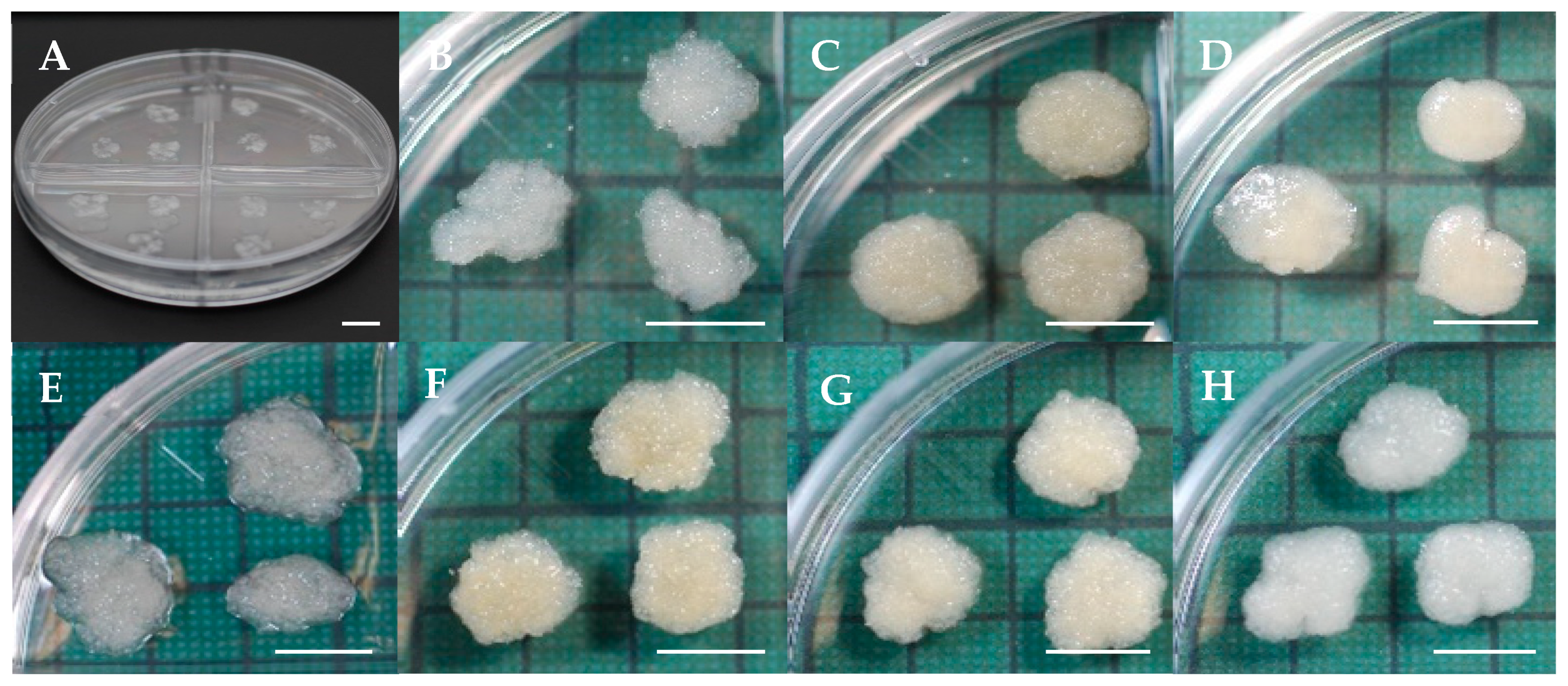
Figure 1. Proliferation of embryogenic masses in sugi (Japanese cedar, Cryptomeria japonica). (A) Embryogenic masses after transfer to proliferation medium. (BH) Proliferation after 2 weeks from lines SSD-018 (B), SSD-073 (C), SSD-100 (D), SSD-112 (E), SSD-113 (F), SSD-182 (G), and SSD-352 (H). Bars: 10 mm.
Forests 16 01431 g001
Figure 4. Germination and plantlet conversion of somatic embryos in sugi (Japanese cedar, Cryptomeria japonica). (A) Mature somatic embryos; (B) Embryos transferred to germination medium; (C) Response after 1 week; (D) Two weeks; (E) Four weeks; (F) Eight weeks. Bars: 10 mm.
Forests 16 01431 g004
Figure 5. Germination response of sugi (Japanese cedar, Cryptomeria japonica) somatic embryos after different storage periods at 25 °C, 15 °C, and 5 °C. The bold line in each box indicates the median, the triangle represents the mean, and the vertical line indicates the standard deviation.
Forests 16 01431 g005
Figure 6. Germination response of sugi (Japanese cedar, Cryptomeria japonica) somatic embryos after 24 months of storage at 5 °C, shown for different lines. The bold line in each box indicates the median, the triangle represents the mean, and the vertical line indicates the standard deviation. Different letters indicate statistically significant differences at the 5% level. N represents the number of somatic embryos tested.
Forests 16 01431 g006
Figure 7. Germination response of sugi (Japanese cedar, Cryptomeria japonica) somatic embryos after storage at 25 °C, 15 °C, and 5 °C for various durations. (A) Stored somatic embryos; (B) Germination of non-stored embryos (control) after 2 weeks on germination medium; (C,D) Germination after 6 and 12 months of storage at 25 °C, respectively; (EH) Germination after 6, 12, 18, and 24 months at 15 °C, respectively; (IP) Germination after 6, 12, 18, 24, 30, 36, 48, and 60 months at 5 °C, respectively. Bars: 10 mm.
Forests 16 01431 g007
Table 1. Summarized conditions and results in the production and storage of male-sterile somatic embryos of sugi (Japanese cedar, C. japonica) at temperatures of 25 °C, 15 °C, and 5 °C.
StagesSummarized ConditionsSummarized ResultsReferences
Embryogenic culture initiationCulture of megagametophyte explants (isolated from seeds collected in mid-July 2016 from the full-sib seed family ‘Shindai 3’ × ‘Suzu 2’) in the dark at 25 °C.Initiation rate: 14.1%Maruyama et al. 2021a [6]
Maintenance and proliferation of EMsSubculture EMs on maintenance/proliferation medium in the dark at 25 °C every 2 weeks.Friable and white EMs: SSD-018, SSD-112, SSD-352; Friable and yellowish-white EMs: SSD-073, SSD-113; Mucilaginous and yellowish-white EMs: SSD-100.This report (Figure 1)
Discrimination of male-sterile ECLsDNA extraction from embryogenic cells by InstaGene and DNA marker diagnosis of MS1 genotype.Discrimination probability of male-sterile ECLs: 100%.Ueno et al. 2019 [5]; Tsuruta et al. 2021a [8]
Production of somatic embryosCulture of EMs on maturation medium (5 masses per plate, 100 mg each) in the dark at 25 °C.Average number of mature embryos per plate: 349.8; Range: 129.6–504.1.This report
(Figure 2A)
Somatic embryo germination and conversionCulture somatic embryos on germination medium at 25 °C under a photon flux density of 45–65 µmol m−2 s−1 for 16 h per day.Average germination rate: 93.9%; Range: 89.6%–97.5%. Average conversion rate: 92.5%; Range: 88.1%–96.6%.This report
(Figure 2B,C)
Storage of somatic embryos and post-storage germination test of embryos derived from line SSD-018Plates containing cotyledonary embryos on maturation medium were sealed with three layers of Parafilm®, placed in a Ziploc® freezer bag, and stored in the dark at 25 °C, 15 °C, and 5 °C for 0–60 months (M).
Germination test was conducted on germination medium at 25 °C under a photon flux density of 45–65 µmol m−2 s−1 for 16 h per day.
Germination rate after storage at 25 °C for: 6 M (93.3%), 12 M (79.3%), 18 M (37.3%), and 24 M (10.7%). At 15 °C for: 6 M (95.2%), 12 M (86.0%), 18 M (70.7%), 24 M (55.3%), 30 M (20.7%), and 36 M. At 5 °C for: 6 M (96.4%), 12 M (92.7%), 18 M (91.1%), 24 M (97.0%), 30 M (77.2%), 36 M (53.3%), 48 M (37.7%), and 60 M (9.8%).This report
(Figure 5)
Storage of somatic embryos and post-storage germination test of embryos derived from different ECLsThe same conditions described above apply to the storage of somatic embryos at 5 °C for 24 months (M).Germination rate of different ECLs after storage at 5 °C for 24 M: SSD-018 (97,0%), SSD-073 (80%), SSD-100 (70.8%), SSD-112 (93.5%), SSD-113 (64.6%), SSD-182 (70%), and SSD-352 (38.3%).This report
(Figure 6)
Disclaimer/Publisher’s Note: The statements, opinions and data contained in all publications are solely those of the individual author(s) and contributor(s) and not of MDPI and/or the editor(s). MDPI and/or the editor(s) disclaim responsibility for any injury to people or property resulting from any ideas, methods, instructions or products referred to in the content.