Article

Enhanced Detection of Phytophthora Species at P. pluvialis Outbreak Sites in Commercial Forests Across Britain

1
Forest Research, Northern Research Station, Roslin EH25 9SY, Scotland, UK
2
The James Hutton Institute, Invergowrie, Dundee DD2 5DA, Scotland, UK
3
Forest Research, Alice Holt Lodge, Farnham GU10 4LH, England, UK
*
Author to whom correspondence should be addressed.
Forests 2025, 16(9), 1419; https://doi.org/10.3390/f16091419
Submission received: 4 June 2025 / Revised: 25 August 2025 / Accepted: 27 August 2025 / Published: 4 September 2025
(This article belongs to the Special Issue Pathogenic Fungi in Forest)

Abstract

Invasive Phytophthora species are increasingly impacting UK landscapes. Most recently, cryptic outbreaks of P. pluvialis Reeser, Sutton & E. Hansen have occurred on western hemlock and Douglas fir at several forest sites across Britain. To better understand the ubiquity and life cycle of this pathogen in British forests and the assemblages of co-inhabiting Phytophthora species, metabarcoding and baiting methodologies were applied to soil, stream water, and rainwater samples collected over a full calendar year from seventeen sites across Britain. Thirty-five Phytophthora species were detected across all sites, substrates, and detection methods, with most detections occurring in stream water by metabarcoding. The three most frequently detected species were (1) P. pluvialis, (2) P. gonapodyides H.E. Petersen & Buisman and (3) P. ramorum Werres, De Cock & Man in ‘t’ veld. Other species detected included the regulated pathogens P. austrocedri Greslen & Hansen, P. kernoviae Brasier, Beales & S.A. Kirk and P. lateralis Tucker & Milbraith, as well as P. ornamentata Scanu, Linald & T. Jung, a new species record for the UK. Phytophthora pluvialis was most frequently detected in March, with rainfall trap metabarcoding data suggesting that aerial dissemination occurs predominantly in late winter/early spring. Consistent detections of P. pluvialis in soil by metabarcoding indicate the potential for soilborne transfer of this pathogen by animal or human vectors, including equipment or machinery in forest operations. The study’s findings are discussed in relation to understanding how P. pluvialis spreads and the approaches needed to address key knowledge gaps in relation to inoculum sources. The results provide a baseline for Phytophthora diversity in British commercial forests, facilitating a greater understanding of typical and unusual trends in species assemblages. This study also consolidates the value of metabarcoding as an effective surveillance tool for Phytophthora in commercial forests.

1. Introduction

Phytophthora is an oomycete genus comprising around 240 described species [1,2]. Phytophthoras are fungus-like organisms taxonomically positioned within the Kingdom Stramenopila and include both root- and foliar-infecting plant pathogens. Phytophthora produces water-disseminated motile zoospores, formed in spore sacs known as sporangia, as well as resting spores, which can persist in plant debris and soil for extended periods, and so much of their life cycle is conducted in water and soil [3]. Phytophthora pathogens are increasingly having an impact on UK landscapes, largely due to the introduction and spread of new invasive species through imported plant material and the circulation of this material in the plant trade [4]. Examples include P. ramorum, the causal agent of the sudden death disease of Japanese larch (Larix kaempferi (Lambert) Carrière) in commercial forestry plantations across the UK and the Republic of Ireland [5], and P. austrocedri which is killing native juniper (Juniperus communis) in natural woodlands in Britain [6].
UK production forestry has found itself in a particularly vulnerable position following the epidemic of P. ramorum on larches, leaving Sitka spruce (Picea sitchensis (Bongard) Carrière) as one of the few remaining species choices for acid upland sites. Particularly concerning, therefore, was the finding in September 2021 of a new invasive Phytophthora pathogen, P. pluvialis, infecting western hemlock (Tsuga heterophylla (Rafinesque) Sargent) [7] and Douglas fir (Pseudotsuga menziesii (Mirbel) Franco) in southwest England. Both these hosts are being considered for their increased potential as future production forestry species in the UK. Phytophthora pluvialis was first described during environmental sampling of streams, soil, and rainfall traps in forests of native mixed tanoak (Notholithocarpus densiflorus (Hooker & Arnott) Manos, Cannon & S.H. Oh) and Douglas fir in Oregon, USA [8]. Following its original description, P. pluvialis was subsequently shown to cause needle cast and twig cankers on seedling trees of Douglas fir under experimental conditions in Oregon, involving artificial inoculation or exposure beneath the canopy of Douglas fir plantations [8]. The pathogen also causes a serious disease known as ‘red needle cast’ on pine (Pinus radiata D. Don) in New Zealand, where it is assumed to have been introduced [9].
Since the first findings in southwest England, P. pluvialis has been subsequently detected at more forest sites in northern England, Wales, and western Scotland, mainly on infected western hemlock but also on Douglas fir and, at a single location, on Japanese larch. The symptoms can be subtle and include single, partially girdled branches on western hemlock and defoliation of Douglas fir, with the latter symptom masked by co-occurrence of other needle pathogens, including Nothophaeocryptopus gaeumannii (T. Rohde) Videira, C. Nakashima, U. Braun & Crous (Swiss needle cast) and Rhizoctonia butinii Oberwinkler, R. Bauer, Garnica & R. Kirschner. The presence of older cankers on many infected western hemlock trees indicates that P. pluvialis might have been present, undetected, for several years. However, its distribution in the wider environment and means of introduction and spread from site to site, and within sites, is cryptic. Methods to enhance surveillance for P. pluvialis and other Phytophthora species in UK forestry are needed as an early warning of their presence, to allow greater understanding of their environmental distribution, and to enable rapid deployment of measures to mitigate their impacts and prevent further spread.
A sampling and metabarcoding methodology for the detection of all known Phytophthora species in environmental DNA samples has been developed and successfully applied for the detection of Phytophthora in soil [10,11], rivers and rainwater traps [12], and in roots and water samples in plant nurseries [4,13,14]. This method also has the potential to identify novel (as yet undescribed) Phytophthora species and can be combined with more traditional baiting approaches, which aim to obtain live cultures of the pathogens and therefore provide absolute proof of presence and their viability [15].
The overall aim of this study was to achieve a better understanding of the ubiquity and life cycle of P. pluvialis in British forests and the assemblages of co-inhabiting Phytophthora species to better understand the risks posed by these pathogens to commercial forestry. An established metabarcoding and baiting methodology was applied to soil, stream water, and rainwater samples collected from seventeen sites distributed across England, Wales, and Scotland, most of which were confirmed P. pluvialis outbreak sites. Sampling was undertaken over a full calendar year with the following specific objectives: (i) to investigate where and how P. pluvialis resides and spreads in the local environment by testing soil, stream water, and rainwater; (ii) to explore whether P. pluvialis detections coincide with seasonal patterns; and (iii) to identify assemblages of co-existing Phytophthora species in soil, stream water, and rainwater to better understand species’ ubiquity in these substrates across the three home countries and identify any new species records for the UK.

2. Materials and Methods

2.1. Site Selection and Sampling Strategy

Seventeen sites located across England, Wales, and Scotland were selected for sampling, with sites within the same forest distinguished by ‘a’ or ‘b’ after the forest site code (Table 1 and Figure 1). All sites contained forest roads (e.g., Figure 2a) and/or public footpaths, as well as watercourses. Fourteen sites were selected based on being P. pluvialis outbreak sites containing infected western hemlock and/or Douglas fir; thirteen of these sites were commercial forestry plantations, and one was a public garden (Table 1). Two additional commercial forestry sites were included where P. pluvialis was not known to be present at the start of the study: WHa in Wales, located in open terrain at the upper edge of an adjacent forest area, and SR in Scotland (Table 1). A non-commercial forestry negative control site located in southeast England (EAH), with no known P. pluvialis infections, was also included in the study (Table 1 and Figure 1). At this site, stream water only was sampled for metabarcoding analysis, whereas at all other sites, stream water, rainwater, and soil were sampled for metabarcoding analyses, with stream water additionally being sampled for baiting. At all sites, sampling was conducted from September/October 2022 until September/October 2023. Within each site, rainwater and soil sampling points were chosen based on being within a 10 m radius of each other and adjacent to the canopy of a focal western hemlock or Douglas fir tree, where present, whereas stream water sampling points varied according to each site. Details of sampling point locations at each site are presented in Table 1. Clean, disposable gloves and cleaned, surface-sterilised tools were used to collect each different sample.

2.2. Stream Water Sampling by Baiting

Stream water was sampled by baiting monthly for the first ten months of the project. Bait raft bags were constructed from nylon mesh secured with staples and contained two polystyrene blocks, which served as floats (Figure 2b). Bait leaves were rhododendron ‘Cunningham White’ variety from stock plants purchased from commercial plant nurseries and maintained by the sampling teams in each region. Western hemlock shoots collected from local, visibly healthy trees were added as a second type of bait leaf from spring 2023 onwards. Five visibly healthy, non-wounded rhododendron leaves (and later, western hemlock shoots with needles) were placed into each bait raft, and the open end was sealed with staples (Figure 2b). At the same time, two leaves of each bait type were bagged separately and placed in a fridge at 5 °C as non-exposed control leaves. Bait rafts were placed into a stream and anchored to a fixed point with a string (Figure 2c) for two weeks, after which time the bait leaves were collected and sent immediately to the laboratory for processing, along with the non-exposed control leaves.
Tissue sections (~5–7 mm2) were cut from the margins of any visible developing grey to brown lesions (e.g., Figure 2d) on baits or from the clean control leaves. Excised tissues were rinsed in sterile distilled water (SDW), surface sterilised in 70% ethanol, rinsed again in SDW, and placed on Phytophthora-specific Synthetic Mucor Agar (SMA) [16] with the addition of antibiotics [17]. Plates were monitored daily, and developing hyaline, smooth, swollen, nodose to tuberculate hyphae without cross-walls were subcultured as putative Phytophthora onto V8 juice agar or carrot agar and incubated at 15 °C until visual identification of single colonies was possible. DNA was extracted using the Nucleospin Plant II mini kit (740770, Machery Nagel, Düren, Germany) protocol for plants, with the following modification: mycelium (scraped from the surface of a colony growing on V8 agar) was ground in an Eppendorf using a mini pestle and sterile glass beads. In step 2a, buffer PL1 was added, and the samples were incubated at 65 °C for 1 h. Steps 3–7 were unchanged.
Isolates obtained by baiting were identified by Sanger sequencing of the Internal Transcribed Spacer (ITS) region of the nuclear ribosomal DNA (rDNA) using the forward primer ITS6 (5′ GAAGGTGAAGTCGTAACAAGG 3′) and the reverse primer ITS4 (5′ TCCTCCGCTTATTGATATGC 3′) [18,19] with Bioron Hotstart taq (Bioron, Römerberg, Germany). PCR thermal cycling parameters were as follows: 1 cycle of 95 °C for 2 min; 30 cycles of 95 °C for 20 s, 55 °C for 25 s, 72 °C for 50 s, and a final cycle of 72 °C for 10 min. The ~900 bp product was verified by running 4 µL (3 µL sample/1 µL purple gel loading dye) on a 1% agarose gel, followed by clean-up by ExoSAP-IT™ (ThermoFisher Scientific, Inchinnan, Renfrew, UK) before sequencing. Raw sequences were aligned and edited using Sequencher version 5.4.6 for Windows and searched against published ITS sequences in the GenBank NCBI nucleotide database using BLASTN+ 2.16.0 [20]. Species identity was based on a 100% or 99% match across the entire sequence length to verifiable sequences derived from voucher specimens or published taxonomic papers.

2.3. Stream Water and Rainwater Sampling for Metabarcoding

Stream water was sampled for metabarcoding every two months at the same point on each stream used for baiting. At each sampling, three 5 L replicate sub-samples of stream water were pre-filtered through muslin cloth and pumped through a Merck Millipore Ltd. (Darmstadt, Germany), 47 mm diameter mixed cellulose ester filter (Sigma-Aldrich, Burlington, MA, USA) of 1.2 µm pore size held in a 47 mm polycarbonate in-line filter holder (Pall Corporation, New York, NY, USA) using a pressure sprayer (Hozelock, Birmingham, UK) (Figure 2e). Up to three filters per 5 l subsample were collected (depending on the rate at which filters blocked) (Figure 2f) and placed in a 15 mL buffer tube containing 8 mL of Longmire lysis buffer (100 mM Tris, 100 mM EDTA, 10 mM NaCl, 0.5% SDS [21]). Buffer tubes containing filters were stored at 5–10 °C (in a cool box in the field) before being sent to the laboratory, where they were stored at room temperature for up to a week before DNA extraction. DNA extracts were stored at −20 °C.
Rainwater was sampled on a fortnightly basis using one rainfall trap per site positioned approximately 1 m from the canopy edge of a western hemlock or Douglas fir tree, where present (Table 1 and Figure 2a,g). Rainfall traps consisted of a single 2-litre plastic bottle with a plastic funnel secured into the neck of the bottle with strong tape. The bottle was placed in a metal mesh basket attached to a post at approximately 1.5 m height (Figure 2a,g). At each rainwater collection time point, the entire sample was filtered as described above for the stream water. Bottles were discarded after each sampling period and replaced with fresh bottles.
Sample collection equipment (rainwater bottles, funnels, pressure sprayers, hose ends with filter nozzles, buckets, and bait rafts) was cleaned before sampling by soaking for at least 30 min in a chlorine solution containing one chlorine tablet (Palintest™ Instachlor tablets PR-150, Palintest Ltd., Gateshead, UK) in 5 L of water, followed by thorough rinsing in tap water. For pressure sprayers, the chlorinated water was pumped through the pressure sprayer, and the filter holder was unscrewed before rinsing. Before each sampling visit, 5 L of mains tap water was pumped through the clean pressure sprayer as a blank control to provide a baseline to assess cross-contamination between samples. This single ‘blank’ filter was placed in a separate buffer tube and processed as for environmental water samples.

2.4. Soil Sampling

Soil was sampled from each site every two months at sampling points that depended on site factors, e.g., under the drip zone of infected trees (if present), around rain traps, or from adjacent footpaths, forest operation trails, or the banks of watercourses (Table 1). For each sample, using a disinfected trowel to first scrape away the litter layer, approximately 100 g of soil was collected at 10–30 cm depth at four points and pooled into a sealable plastic bag to a total weight of ~400 g. If sampling was from around an infected tree, then 100 g soil samples were collected at four points around the tree, at approximately 0.3–1 m distance from the main stem, and then pooled. Sampling from paths involved scraping soil/debris from the path surface. All stones were removed if present, and the soil was stored at 5 °C for no more than a few days before being sent to the laboratory, where they were stored at −20 °C before DNA extraction. Soil sampling tools were cleaned with Propellar™ arboricultural disinfectant (Evans Chemicals, Redruth, UK) between samples.

2.5. Processing of Samples for Metabarcoding

All replicate sub-samples were processed separately for metabarcoding. For all water samples, tubes of Longmire’s buffer with filters were shaken at 300 rpm for 10 min (Eppendorf ThermomixerTM C, Eppendorf, Hamburg, Germany) prior to DNA extraction from 1.5 mL aliquots using the DNeasy Blood & Tissue Kit (Qiagen Sciences, Germantown, MD, USA) and starting at step 1c (cultured cells) with Longmire lysis buffer replacing PBS and eluting DNA in the final step with 50 µL buffer (DNeasy Blood and Tissue Handbook, Qiagen, June 2023 [22]). Soil samples were oven-dried in aluminium trays at 32 °C for approximately a week, depending on water content. Dried soil was mixed thoroughly and ground using 1.5 cm ball bearings in 50 mL canisters with a Retsch MM400 mill (Retsch, Han, NRW, Germany). DNA was extracted from three 250 mg subsamples using the PowerSoil Pro™ DNA isolation kit (Qiagen, Manchester, UK) according to the manufacturer’s instructions. Post-DNA extraction clean-up was carried out using the Zymo DNA Clean & Concentrator™ (Zymo Research, Irvine, CA, USA) according to the manufacturer’s instructions.
The ~260 bp ITS1 region of rDNA was amplified using nested PCR with the forward primer 18Ph2 (5′ GGATAGACTGTTGCAATTTTCAGT 3′) and the reverse primer 5.8S-1 (5′ GCARRGACTTTCGTCCCYRC 3′) in the 1st round and the forward primer ITS6 (5′ GAAGGTGAAGTCGTAACAAGG 3′) and the reverse primer 5.8S-1 in the 2nd round, as described in the protocol of Scibetta et al. [23] to prioritise Phytophthora over Pythium and as validated previously for detection of forest Phytophthoras [4,10,13]. Amendments to the protocol include the use of KAPA HiFi HotStart ReadyMix PCR kit (Roche Diagnostics, Burgess Hill, UK) and a reaction volume of 12.5 µL, with each reaction containing 4.5 µL PCR-grade water, 6.25 µL Kapa HiFi Hotstart ReadyMix (source as above), 0.375 µL (10 µM) of each forward and reverse primer, and 1 µL DNA or 1 µL round 1 reaction product. Amplification conditions were also altered from the Scibetta protocol [23] with initial denaturation at 95 °C for 3 min (1st and 2nd round), followed by 30 cycles of 98 °C for 20 s, 61 °C for 25 s, and 72 °C for 40 s with a final cycle of 72 °C for 1 min (1st round) and 25 cycles of 98 °C for 20 s, 61 °C for 25 s, and 72 °C for 25 s with a final cycle of 72 °C for 1 min (2nd round). When the initial PCR amplification was successful, the round 1 product was re-amplified in a 29 µL reaction, together with a set of synthetic sequence controls, which act as a check for cross-contamination during Illumina plate preparation and sequencing [24], using the 2nd round primers MIS_ITS6_F (TCGTCGGCAGCGTCAGATGTGTATAAGAGACAGGAAGGTGAAGTCGTAACAAGG) and MIS_5.8S-1R (GTCTCGTGGGCTCGGAGATGTGTATAAGAGACAGGCARRGACTTTCGTCCCYRC) with MiSeq overhang adapters that allow index attachment.
Libraries with universal adapters, allowing versatility of index attachment, were prepared following the Illumina 16S metagenomic sequencing library preparation protocol (Illumina Technologies, San Diego, CA, USA [25]). Product clean-up prior to index attachment was with AMPure XP magnetic beads (Beckman-Coulter UK Ltd., Little Chalfont, UK) (20 µL beads to 25 µL PCR product), followed by two washes with 200 µL 80% ethanol. After removal of the supernatant, the beads were air-dried for up to 10 min, and 52 µL of 10 mM tris HCl, pH 8.5, was added per sample. Index PCR was performed on 5 µL of cleaned DNA using unique combinations of Illumina Nextera XT Index 1 and Index 2 primers (Illumina XT DNA library preparation kits A-D, Illumina, San Diego, CA, USA) at 5 µL of each primer per sample, 10 µL of PCR-grade water, and 25 µL of KAPA HiFi HotStart ReadyMix PCR kit (Roche Diagnostics, Burgess Hill, UK). PCR conditions were 95 °C for 3 min, 8 cycles of 95 °C for 30 s, 55 °C for 30 s, and 72 °C for 30 s, and a final extension of 72 °C for 5 min. The final library clean-up was with 56 µL of AMPure XP beads, followed by two washes with 200 µL 80% ethanol. After the final wash, the beads were allowed to dry (as above) before adding 27 µL of 10 mM tris HCl, pH 8.5, per sample, from which 20 µL was transferred into individual 0.5 mL low-bind Eppendorfs to represent one library.
Libraries were quality checked for fragment size using a TapeStation 4200 (Agilent Technologies, Santa Clara, CA, USA and quantified with Qubit (Qubit 2 fluorometer, Invitrogen, London, UK), normalised (diluted) to 8 nM, and pooled for 250 bp paired-end sequencing on a single flow cell of an Illumina MiSeq using the MiSeq v. 2 500 bp kit or the Nextseq 2000 kit (Illumina Technologies, San Diego, California, USA) as recommended. Each pooled library was loaded at 3 pM, with a 40% PhiX control library included. Following quality control and de-multiplexing, unique sequences per sample were assigned to species using the Phytophthora classifier (THAPBI-PICT version 1.0.20; [24]). Default settings for Illumina MiSeq were a 1 bp difference for a species match and 3 bp for genus (setting -m 1s3g), and for the Illumina NextSeq 2000 plates, mild UNOISE2 denoising [26] was applied to a harsher minimum abundance threshold of 1000 reads per unique read per sample (settings --denoise unoise-l --unoise_alpha 6 -a 1000). Replicates were pooled for reporting of results so that each individual detection is defined as the presence of that species in at least one of the three replicates within a sample taken at a single time period.

3. Results

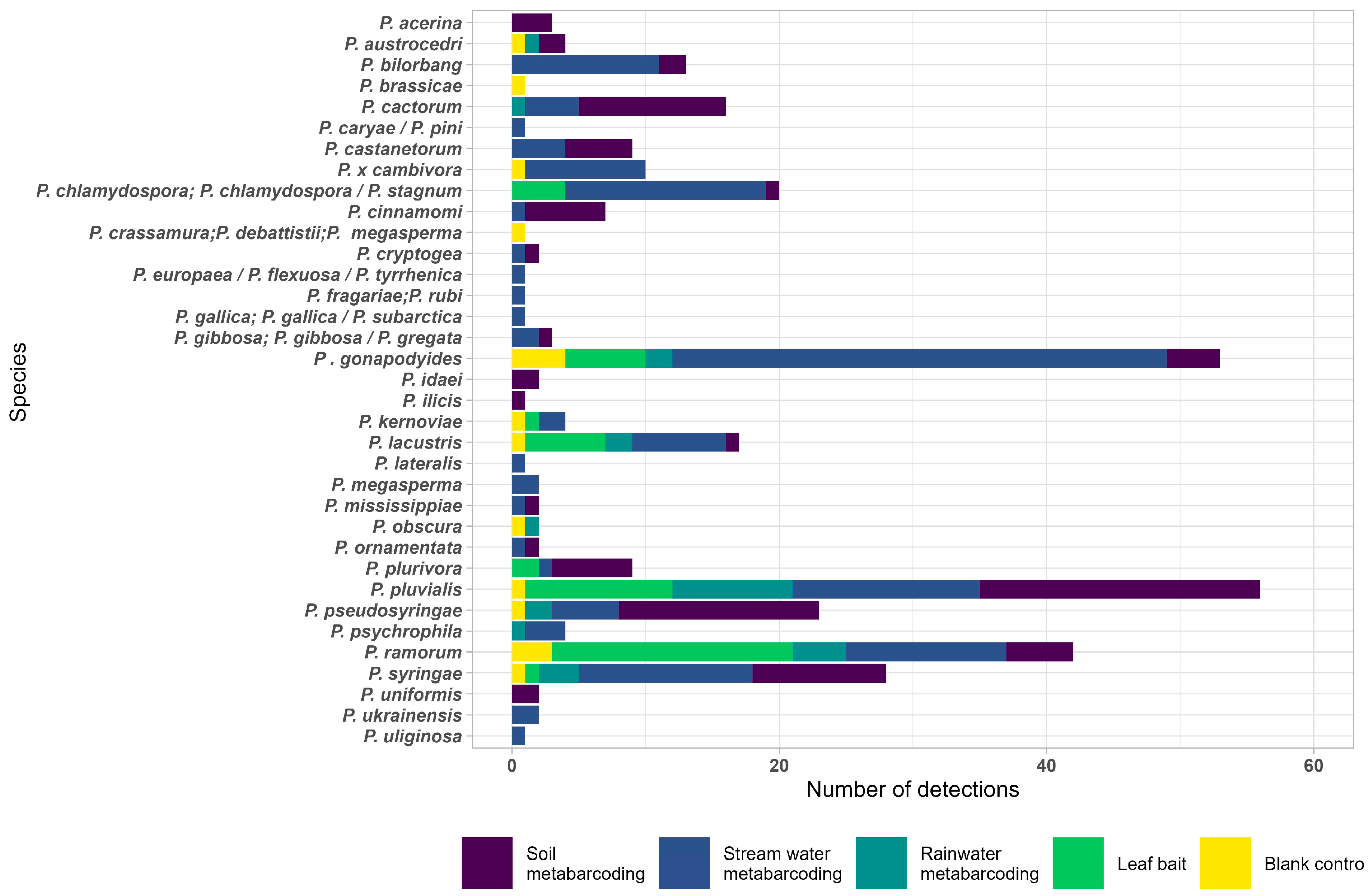
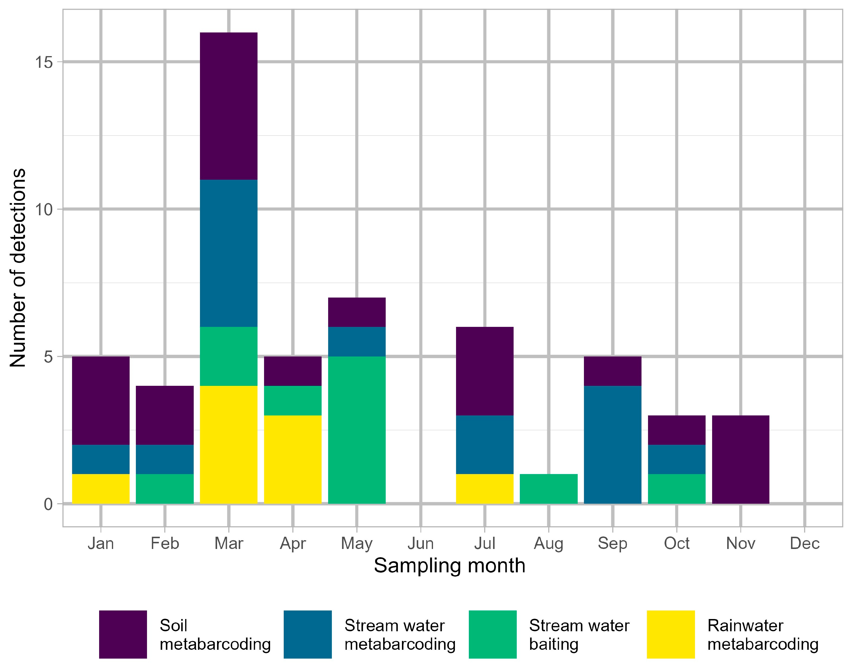
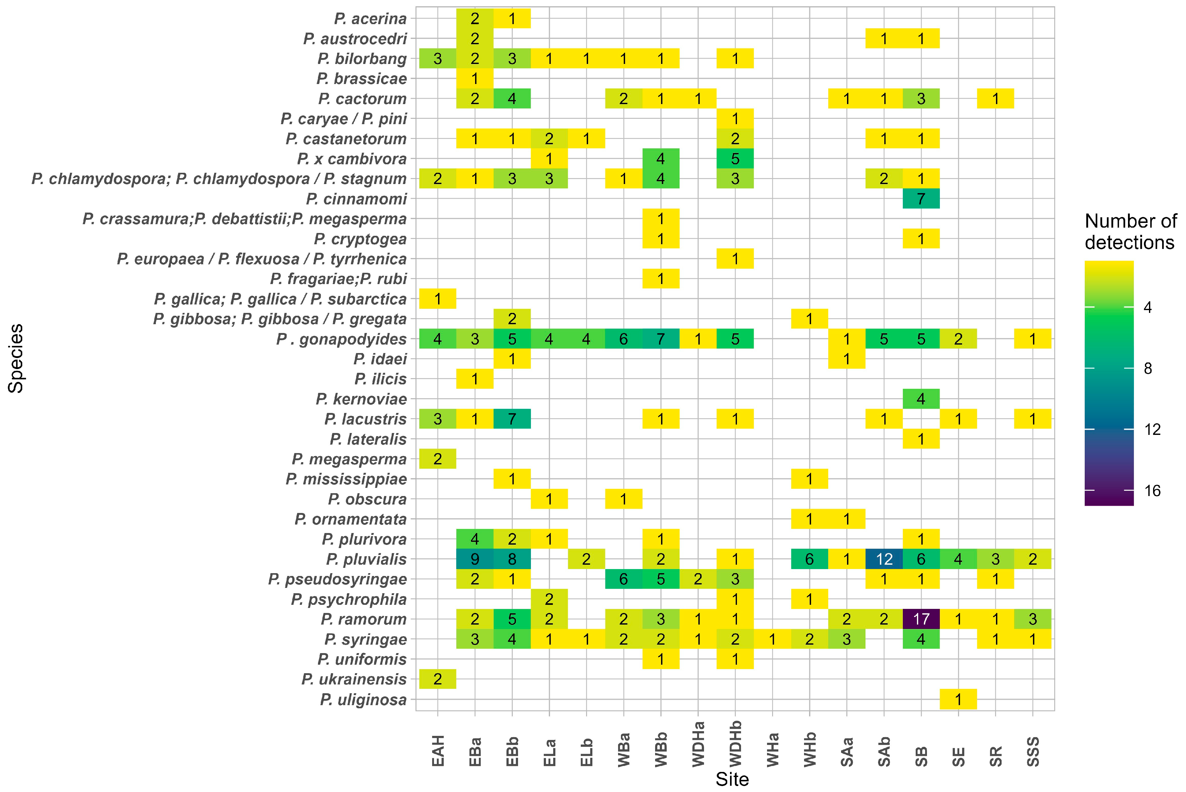
Thirty-five Phytophthora species were detected across all sites, substrates, and detection methods, with most detections occurring in stream water by metabarcoding (Figure 3). The three most frequently detected species were P. pluvialis (56 detections), P. gonapodyides (51 detections), and P. ramorum (42 detections) (Figure 3). For P. pluvialis, there were 21 detections in soil, 14 detections in stream water by metabarcoding, 11 detections in stream water by baiting, and 9 detections in rainwater (Figure 3). Most detections of P. pluvialis occurred in March, with detections in all substrates during this month (Figure 4). Notably, P. pluvialis was detected in soil in nine months of the year and was most frequently detected by leaf baiting in May (Figure 4). There were 28 detections of P. pluvialis across all the Scotland sites, 19 detections of the pathogen at the England sites (except the negative control site), and 9 detections at the Wales sites. The Phytophthora species most frequently detected in the same samples as P. pluvialis were (in order of frequency of co-detections) P. gonapodyides, P. syringae (Berk.) Kleb, P. cinnamomi Rands, P. cactorum (Lebert & Cohn) J. Schr., P. plurivora Jung & Burgess, P. ramorum, and P. pseudosyringae T. Jung & Delatour. The Phytophthora species detected at each site are illustrated in Figure 5, and results are presented for all Phytophthora detections by method and country (see below). Supplementary Table S1 provides the number of Illumina sequence reads for each species detected by metabarcoding in each sample replicate, along with site, date, and substrate. Note that the downy mildew species detected in each sample are also presented in Supplementary Table S1, although downy mildews are not discussed in this paper. A number of blank control samples were positive in the nested PCR, with the majority containing downy mildew or unknown species (Supplementary Table S1). Thirteen blank control samples also contained Phytophthora taxa, with a total of twelve Phytophthora species detected in these blank controls (Supplementary Table S1 and Figure 3).

3.1. Phytophthora Detection in Stream Water by Baiting

Overall, a total of 862 rhododendron leaf samples and 65 western hemlock samples were processed for the baiting analyses, with only rhododendron leaves yielding isolates. Across all sites, there were 49 isolates from leaf baits representing eight different Phytophthora species. This represented an isolation rate per leaf bait of 5%. Phytophthora pluvialis was isolated from five of the seventeen sites: EBa and EBb in England and SAb, SE, and SR in Scotland (Table 2), the latter site being one at which the pathogen had not been previously reported. Except for one detection in August (SE) and one in October (SR), all baiting detections of P. pluvialis occurred between February and May (Table 2). The regulated pathogen P. ramorum was detected by baiting at eight sites across England, Wales, and Scotland, with detections occurring in all months of the year except for July and August (Table 2). Another regulated pathogen, P. kernoviae, was also detected by baiting at SB in Scotland (Table 2). Other non-regulated Phytophthora pathogens of woody hosts detected by baiting were P. plurivora at ELa in England and WBb in Wales and P. syringae at ELb in England (Table 2). Additionally, ‘riverine’ Phytophthora species baited from stream water at several sites included P. chlamydospora Brasier & Hansen, P. gonapodyides, and P. lacustris Brasier, Cacciola, Nechwatal, Jung & Bakonyi (Table 2). ‘Riverine’ Phytophthoras are typically Clade 6 species known for their water-inhabiting lifestyles and are not generally regarded as serious plant pathogens.

3.2. Phytophthora Detection in Stream Water by Metabarcoding

Of the 474 replicate stream water samples collected during the study, 241 (51%) were positive for Phytophthora and closely related oomycetes in the nested PCR assay and were processed for metabarcoding. Across all sites, there were 152 Phytophthora detections in stream water by metabarcoding, representing 27 different species.

3.2.1. England—Stream Water

Phytophthora pluvialis was detected in stream water by metabarcoding at both EBa (January and July 2023) and EBb (September 2022 and February 2023). Phytophthora ramorum was detected at ELb in January 2023. Other Phytophthora species known to be pathogenic on woody hosts included P. castanetorum T. Jung, M. Horta Jung, Bakonyi & Scanu, detected at both EBa and ELa, P. pseudosyringae at EBa, as well as P. × cambivora (Petri) Buisman, P. psychrophila T. Jung & E.M. Hansen, and P. syringae at ELa; notably, the latter three species were all detected in a single stream water sample along with P. castanetorum. Riverine species detected at the EB and EL sites included P. bilorbang Aghighi & Burgess, P. chlamydospora × stagnum Yang, Richardson & Hong, P. gonapodyides, P. lacustris Brasier, Cacciola, Nechw., T. Jung & Bakonyi, and P. mississippiae Yang, Copes & Hong. At the negative control EAH site, the following eight riverine species of Phytophthora were detected: P. bilorbang, P. chlamydospora × stagnum X. Yang & C. X. Hong, P. gallica Jung & Nechwatal, P. gonapodyides, P. lacustris, P. megasperma Drechsler, P. subarctica T. Jung, T. Corcobado, J. Oliva & I. Milenković, and P. ukrainensis Milenković, T. Jung, T. Corcobado & I. Matsiakh.

3.2.2. Wales—Stream Water

Phytophthora pluvialis was detected in stream water by metabarcoding at WDHb in May 2023 and at WBb in October 2022. Phytophthora ramorum was detected at WBb in January 2023. Other pathogens of woody hosts detected in stream water at the Wales sites included P. cactorum, P. × cambivora, P. pseudosyringae, P. psychrophila, P. syringae, and P. uniformis (Brasier & S.A. Kirk) Ex Abad et al.. One stream water sample from WDHb contained DNA matching nine Phytophthora species, including two not recorded elsewhere in this study: P. flexuosa/P. tyrrhenica/europaea and P. caryae/P. pini (these two species complexes cannot be differentiated from species based on the ITS1 barcode used here). This same stream water sample also contained DNA from several unknown Phytophthora species. Stream water from WHa and WHb had low Phytophthora diversity, including only riverine species Phytophthora gibbosa/gregata and P. mississippiae and unidentified Phytophthora species.

3.2.3. Scotland—Stream Water

Phytophthora pluvialis was detected in stream water by metabarcoding at SAb (September 2022, March and July 2023), SB (March and September 2023), SR (September 2022 and March 2023), and SSS (March 2023). Phytophthora ramorum was detected at SAa (September 2022), SAb (March 2023), SB (September 2022 and February, March, and September 2023), SR (March 2023), and SSS (February and March 2023). Notably, at SB, a single stream water sample collected in September 2023 contained DNA of ten Phytophthora species, including, in addition to P. pluvialis and P. ramorum, the regulated species P. lateralis and P. kernoviae as well as known pathogens of woody hosts, P. cinnamomi, P. cryptogea Pethybridge & Lafferty, P. plurivora, and P. syringae, plus two riverine species. Also of note is the detection of P. ornamentata, a species not previously recorded in the UK, at SAa, in the same sample as P. ramorum and P. syringae. The oak pathogen P. uliginosa T. Jung & E.M. Hansen was detected at SE. Other species detected at the Scottish sites included riverine Phytophthoras.

3.3. Phytophthora Detection in Rainwater by Metabarcoding

Of the 748 replicate rainwater samples collected during the study, only 144 (19%) were positive for Phytophthora and closely related oomycetes in the nested PCR assay and processed for metabarcoding. Across the project, there were 28 Phytophthora detections in rainwater, representing eleven different species. Phytophthora pluvialis was not detected in rainwater at the England sites but was detected at WHb in Wales in January, March (together with P. psychrophila), April, and July 2023. In Scotland, P. pluvialis was detected in rainwater at SAb, SE, and SSS, all in the same exposure period, which was the fortnight leading up to 2nd March 2023. The pathogen was additionally detected at SAb in April 2023. All these detections of P. pluvialis occurred at very high read counts (Supplementary Table S1). Phytophthora ramorum was detected in rainwater at EBa (May 2023) and SB (February and September 2023), and P. austrocedri was detected at EBa and SAb, the latter finding in a rainwater blank sample at high read counts (Supplementary Table S1). Other known pathogens detected in rainwater across all sites included P. brassicae de Cock & Man in ‘t Veld, P. obscura Grünwald & Werres, P. pseudosyringae, and P. syringae. Phytophthora pseudosyringae was also detected in a rainwater blank collected at the same time period as a rainwater sample containing the pathogen. Many rainwater samples yielded only downy mildews or unknown species.

3.4. Phytophthora Detection in Soil by Metabarcoding

Of the 318 replicate soil samples processed in this study, 142 (45%) were found to be positive for Phytophthora or closely related oomycetes in the nested PCR. Across all sites, there were 100 Phytophthora detections in soil, representing 21 different species.

3.4.1. England—Soil

Phytophthora pluvialis was detected in soil at EBa (November 2022 and January, March, May, and September 2023), EBb (October and November 2022, January and March 2023), and ELb (January and March 2023). Some of these detections occurred at very high read counts (Supplementary Table S1). Other Phytophthora pathogens detected in soil samples across the England sites included P. acerina B. Ginetti and the regulated pathogen P. austrocedri, as well as P. cactorum, P. castanetorum, P. ideai Kennedy and Duncan, P. ilicis Buddenhagen & Young, P. plurivora, and P. syringae. The riverine species P. bilorbang, P. gibbosa T. Jung, M.J.C. Stukely & T.I. Burgess, P. gonapodyides, and P. lacustris were also detected.

3.4.2. Wales—Soil

Phytophthora pluvialis was detected in a single soil sample from WBb in April 2023. Phytophthora ramorum was detected at WDHa in January 2023. In many soil samples from the Wales sites, the dominant species detected was P. pseudosyringae. Other species detected included P. castanetorum, P. cryptogea, P. mississippiae, P. ornamentata, and P. syringae.

3.4.3. Scotland—Soil

Phytophthora pluvialis was detected in soil at SAa (September 2023), SAb (March and July 2024), SB (November 2022, February, March, and July 2023), and SE (March 2023). Phytophthora ramorum was detected in soil at SB (November 2022), SE (June 2023), and SSS (October 2022), and P. austrocedri was also detected at SB. Six SB soil samples contained very high reads of P. cinnamomi (Supplementary Table S1). Other species detected in soil across the Scotland sites included P. cactorum, P. castanetorum, P. idaei, P. pseudosyringae, and P. syringae.

3.5. Detections of Unknown Phytophthora spp. and Nothophytophthora spp.

DNA matching the unknown species Phytophthora taxon 92a15fa, P. taxon d6452d7, P. taxon catala2015sp2/sp5/sp9, and P. taxon catala2017sp4 was detected in streams in all three countries during this study. Phytophthora taxon Catala2015sp5 was also detected in a rainwater sample at WDHa in Wales, and P. taxon catala2018sp9 was detected in soil from EBb in England. Another closely related oomycete genus, Nothophytophthora, was also detected in stream water by baiting and metabarcoding at sites across all three countries. These detections included N. lirii O’Hanlon, I. Milenković & T. Jung, N. intricata T. Jung, Scanu, Bakonyi & M. Horta Jung, and N. irlandica O’Hanlon, I. Milenković & T. Jung, as well as unidentified Nothophytophthora spp. Nothophytophthora was not detected in soil or rainwater. Elongisporangium undulatum (Petersen) Uzuhasi, Tojo & Kakish, a fast-growing soil-inhabiting oomycete species, was detected by baiting of stream water at SAb in Scotland.

4. Discussion

This study is the first to investigate the prevalence of Phytophthora in commercial forestry environments across Britain and was initiated following the recent spate of outbreaks of P. pluvialis. It has successfully generated information on the abundance, life cycle, and likely means of spread of P. pluvialis and co-existing Phytophthora species in different environmental substrates that can inform future risk mitigation and surveillance strategies. The results can also be viewed as an approximate ‘baseline’ for Phytophthora diversity in commercial forests so that ‘typical’ and ‘abnormal’ trends in species assemblages can be identified in the future.

4.1. Detection of Phytophthora Pluvialis and Implications for Spread and Surveillance

Phytophthora pluvialis was detected in soil and/or water at all the known outbreak sites except ELa in England, where the infected trees had been felled before the project started. At the paired site, ELb, which had also been subject to sanitary felling, P. pluvialis was detected in soil in March 2023 but not earlier in the study, indicating the continued presence of an active inoculum source in the area. Phytophthora pluvialis was detected by metabarcoding and baiting of stream water at SR in Scotland, a site with no confirmed infected trees at the start of the study, but where infected trees were located subsequently. This may indicate long-distance dispersal from a source, although infection may have been present earlier, but trees were asymptomatic, as was the case in Belgium [27]. This detection demonstrates the successful utility of the methods applied here for surveillance of P. pluvialis.
Phytophthora pluvialis has a growth optimum at 15–20 °C, with slow growth at 5 °C and no growth above 25 °C, and it is partially caducous [8], enabling sporangia to be transmitted by wind and rain. In this study, the detection of P. pluvialis at very high read counts in rainwater collected from four of the sites in March and April and during the same late-February, two-week exposure period at three Scotland sites indicates that inoculum release and aerial dissemination occur predominantly in late winter/early spring with rain. This aligns with a New Zealand study of inoculum production by P. pluvialis from needles of infected P. radiata, on which it sporulates abundantly [9]. In the P. radiata stands of New Zealand, P. pluvialis inoculum production peaked during periods of wet weather at low temperatures [28]. Given the requirement for needle wetness for infection and sporulation of P. pluvialis on P. radiata needles, there is reduced detection of the pathogen during warm and dry conditions in the summer, with summer droughts shown to result in decreased expression of disease on P. radiata the following year [29]. This seasonal pattern of disease has allowed the development of a climate model to predict outbreaks of P. pluvialis on P. radiata in New Zealand [29]. Similar research could be carried out in the UK to better understand and predict P. pluvialis outbreaks in relation to temperature and rainfall patterns.
In this study, all rainwater traps yielding P. pluvialis were located within 5 m of an infected western hemlock tree, which is presumed to have been the inoculum source. Phytophthora pluvialis was found to sporulate on needles of all known hosts in Britain, which include western hemlock, Douglas fir, and larch, with Douglas fir supporting the greatest amount of sporulation [30]. However, the study involved artificial inoculation of excised material, and a high initial inoculation load (>104 zoospores/mL) was required to cause infection. The number of sporangia subsequently produced by P. pluvialis was much lower compared with the sporulation potential of P. ramorum on larch, leading to uncertainty about the main host driving P. pluvialis epidemics in Britain [30]. However, information is still needed on the capacity of P. pluvialis to sporulate on infected hosts under field conditions and the critical environmental conditions required to induce sporulation, as well as the distance of spread from an infected host. This will help answer the outstanding question as to whether longer-distance aerial dispersal facilitates introduction to new sites. Rainwater, collected over two weeks, was not the most efficient sampling medium for metabarcoding due to the proliferation of downy mildews, which are amplified by the PCR primers. However, rain trapping could be applied over a shorter period in late winter and early spring at specific distances from an infected host, with concurrent monitoring of environmental conditions, to better understand dispersal distances and weather factors conducive to inoculum production.
Soil was an effective substrate for surveillance of P. pluvialis, with soil detections occurring at eight of the sites and across nine months of the year. The pathogen is self-fertile (homothallic) and produces abundant oospores in culture [8]. These thick-walled resting spores facilitate longer-term survival in soil and plant debris, although they have yet to be confirmed in nature in Britain. Stream water was also an effective substrate for detecting P. pluvialis, with detections by metabarcoding occurring at nine of the sites and over seven months of the year. Baiting of stream water was a very useful complement to metabarcoding with lesions on rhododendron, but not western hemlock, baits in this project, demonstrating the presence of the living pathogen. But baiting was only effective at five of the infected sites, with May being a particularly successful month for baiting P. pluvialis from streams.
The original inoculum sources for the P. pluvialis outbreaks in Britain remain unknown. The pathogen was not detected in an extensive survey of British plant nurseries, including forest nurseries, conducted from 2016 to 2022 [4], nor in soils at Scottish public gardens and woodlands surveyed by metabarcoding from 2014 to 2016 [10,11]. One of the sites included in the study by Riddell et al. [10] was the public garden site surveyed here, suggesting that P. pluvialis was introduced to the site after 2016 or was introduced earlier but remained highly localised. The results of this study suggest that P. pluvialis may arrive at a new site via aerial dissemination of inoculum or vector-mediated transfer in infested soil or plant material. Continued regular surveillance is advised to enable a rapid response to disease outbreaks, with the use of good biosecurity practices to ensure equipment, tools, and boots are clean when moving between sites. There is much uncertainty about the main susceptible host driving infections in British forests, especially since hosts may be infected but asymptomatic for disease, as found during surveys around Douglas fir in Belgium [27]. In this study, the pattern of foliar infections on western hemlock being predominantly on the lower branches of understorey trees alongside paths/forest roads and watercourses suggests that once P. pluvialis has been introduced to a site, local spread of the pathogen may occur, with lower branch infections probably arising from short-distance dispersal of P. pluvialis (e.g., rain splash) from inoculum reservoirs in soil and water.

4.2. Other Phytophthora Species Detected and Implications for Forest Health

Phytophthora ramorum, which was detected at sampling sites in England, Wales, and Scotland, is a regulated pathogen causing significant mortality of larch across the UK [31]. This pathogen now appears to be ubiquitous at many forest sites in the western part of Britain. It is noteworthy that the most effective detection method for P. ramorum included in situ baiting in streams using rhododendron leaves, as well as filtering stream water for DNA analysis. The detection of P. ramorum during all months except August also suggests that P. ramorum is biologically active across much of the year in Britain, in contrast to the situation in the western US, where P. ramorum is active mainly in late winter/early spring [32]. In Britain, where rainfall can occur across the entire year, rainfall episodes have been closely correlated with sporulation and infection [31].
Several other known Phytophthora pathogens of woody hosts were detected in this study, including P. austrocedri, which is responsible for the decline of native juniper populations across northern Britain [6]. This pathogen was one of the most abundant Phytophthora species detected in soils at public gardens and other highly disturbed sites in Scotland [10]. Two other regulated pathogens, P. kernoviae and P. lateralis, were detected at the public garden site in this study in addition to P. austrocedri, P. ramorum, and P. pluvialis. The abundance of P. cinnamomi at the public garden site and its lack of detection at the other sites is also notable, as it has likely been introduced to the site through the planting of exotics over many years. This highly destructive warm-temperature pathogen is impacting forest ecosystems on several continents, and its distribution in the wider UK environment is expected to expand with climate change [33,34]. This study demonstrates how public gardens, with their diverse tree collections, frequent planting and renovation schemes, and high visitor footfall, are reservoirs of Phytophthora diversity and can act as ‘sentinel’ sites for monitoring invasive tree pathogens.
Phytophthora ornamentata was detected at two sites in this study, one in Wales and one in Scotland, and appears to be a new UK record. It is a clade 6 species first described in Sardinia and found to be pathogenic to seedlings of Sardinian native species, leading to the conclusion that it may be associated with the locally observed declining Mediterranean maquis vegetation [35]. This species was not detected in the survey of British nurseries [4], and further investigation at the two sites at which it was detected in this study found no evidence of disease on vegetation. It is possible that P. ornamentata is more widely distributed in British forests, and further metabarcoding surveillance may help to shed light on its niche. Having been observed in the Mediterranean region on host species not present in the UK, it is not currently regarded as a high-risk species in Britain’s current climate.
Other Phytophthora species detected include ubiquitous pathogens of woody hosts in the UK, such as P. cactorum, P. plurivora, P. syringae, and P. × cambivora [34]. Phytophthora plurivora is an example of a pathogen that has undergone recent global expansion due to its international spread in traded diseased plant material, enabling the pathogen to colonise new environments and hosts [36]. This species was formerly considered part of the P. citricola species complex and is thought to be involved in widespread declines of beech (Fagus sylvatica) and oak species (Quercus spp.) in continental Europe [37,38]. Phytophthora uniformis, one of the parent species of the alder-killing hybrid P. × alni, was detected in stream water at two of the Wales sites. It is also pathogenic to Alnus spp. across Europe but is less aggressive than the hybrid P. × alni [39]. Phytophthora castanetorum, detected at sites in all three countries, was recently described from the rhizosphere of sweet chestnut (Castanea sativa) forests in Portugal and Italy and is regarded as a weak pathogen [40]. It is also commonly detected in UK nurseries, where it was found most often in association with Chamaecyparis and Quercus [4].
Phytophthora pseudosyringae was particularly abundant in soils at two of the Wales sites. This fits with the frequent detection of this pathogen on larch in Wales during P. ramorum surveys (Anna Harris, personal communication). Phytophthora pseudosyringae is an aerially disseminated species first reported in Britain in 2009, causing disease on Nothofagus spp. [41] and was assumed at the time to be invasive. Since then, however, the pathogen has been detected on an increasing number of hosts, including woody hosts such as Fagus and Larix, on which it causes cankers, and in different environments and substrates, including soils and stream water [10,12]. It is also highly abundant in British plant nurseries, particularly in water samples [4]. Thus, due to its ubiquitous nature, it is now speculated that P. pseudosyringae may have been present in this country for longer than previously thought.
Stream water was also an effective substrate for detecting Phytophthora, and in some cases yielded large numbers of species in a single sample, which supports the findings of previous projects [4,10,15]. Other prevalent Phytophthora species detected from stream water by baiting or in soil by metabarcoding include the riverine clade 6 species P. chlamydospora, P. gonapodyides, and P. lacustris, which flourish in aquatic habitats and may be regarded as weak pathogens or adapted to plant litter breakdown [42,43]. It should be noted here, however, that P. gonapodyides has been isolated from lesions on diseased trees of various species in Britain, although its role as a pathogen in these cases is unclear [10]. Phytophthora bilorbang is also regarded as a saprotroph of leaf debris and occasionally as an opportunistic pathogen [35]. The clade 10 species P. gallica/P. subarctica and P. ukrainensis were detected by metabarcoding in stream water at the control site, negative for P. pluvialis disease outbreaks. These species have also been detected in water samples collected in a survey of British plant nurseries conducted from 2016 to 2022 [4]. Phytophthora subarctica and P. ukrainensis have only been described very recently from surveys of watercourses in northern Sweden and Ukraine, respectively [44]. They likely have water-associated lifestyles as saprotrophs and opportunistic pathogens of riparian plants, typical of species in this clade [45].
Some of the paired sites in this study showed contrasting diversities of Phytophthora, most notably the Wales sites’ WHa (1 taxon) and WHb (6 taxa), and WDHa (5 taxa) and WDHb (14 taxa). WHa, where only a single detection of P. syringae was recorded, is close to the source of a river with no trees located upstream, only peat and upland ground vegetation. The nearby forested WHb site contains multiple forest roads and footpaths, and it yielded eleven detections of six Phytophthora species, including P. ramorum and P. pluvialis. WDHa is located at a small stream feeding into the larger catchment upstream of WDHb. The area around site b contains a wider mix of conifer and broadleaved species compared with site a, and the site b catchment includes a nearby farm and holiday accommodation.
There were thirteen detections of Phytophthora species by metabarcoding in the water blank control samples, representing twelve taxa. It is likely that these detections represent DNA carry-over from previous samples rather than cross-contamination during Illumina plate preparation, as the synthetic sequence controls acted as a check for this. The fact that the great majority of blank control samples did not contain Phytophthora indicates that the methods used to clean equipment generally worked well. These findings also illustrate the importance of having blank control samples to assess with accuracy between-sample contamination and review results accordingly.

4.3. Unidentified Phytophthora Species and Other Oomycete Genera

Many unknown Phytophthoras were detected in streams in all three countries during this study. These as yet undescribed ‘species’ were initially identified from stream water eDNA in northern Spain [14]. The same unknown species were also detected in water sampled from British plant nurseries [4]. Until these species are isolated into culture, their host associations are unknown. However, data collected so far suggest they may have a water-associated rather than a pathogenic lifestyle, similar to many clade 6 Phytophthoras [43].
Nothophytophthora sp. irlandica and N. sp. lirii were found in streams in all three countries in this project. These species were first described from forest streams in Ireland and Northern Ireland [46], with the authors speculating about their possible introduction through the nursery trade from Chile. Nothophytophthora intricata was also isolated in this study alongside the known tree pathogens, Phytophthora × cambivora and P. plurivora. N. intricata was originally described by Jung et al. in 2017 [47] from declining horse chestnut trees in a flood plain in Germany, although its role, if any, in the decline was not determined. Jung [47] also described several other Nothophytophthora species, noting the wide distribution of this genus around the world and attributing the lack of database records to the difficulty of their isolation in the presence of faster-growing Phytophthora and Pythium. Landa et al. [11] also detected Nothophytophthora spp. in soils at five woodland sites in Britain using metabarcoding and recommended further studies into the possible pathogenicity of these species.

5. Conclusions

This study has further consolidated the effectiveness of metabarcoding, combined with baiting, as a surveillance tool for Phytophthora in commercial forests. Taken individually, metabarcoding and baiting detection methodologies each have their benefits. For example, metabarcoding, based on DNA detection, yields a larger diversity of species, not all of which may be viable, whereas baiting indicates the presence of live species but may underestimate diversity due to fast-growing species being favoured. However, important knowledge gaps remain, such as understanding where the P. pluvialis outbreaks originated from, recognising asymptomatic trees, identifying the main sporulating hosts contributing to epidemics, and how this pathogen arrives at new sites. Determining this missing information may aid in preventing future outbreaks of invasive Phytophthora species in British forests. Future studies should be carried out to detect and quantify natural sporulation of P. pluvialis on known hosts under field conditions, including estimates of the distance of spread from an infected host. Most effective would be the siting of rainfall traps at specific distances from a focal infection source, as well as asymptomatic trees, and collecting rainwater for metabarcoding analysis during March on a weekly basis to avoid accumulation of downy mildews, which may occur over a two-week trapping period. The potential for P. pluvialis to survive and be disseminated in soil by animal or human vectors, including equipment and machinery used in forest operations, should also be investigated. Metabarcoding of soil samples should continue to be deployed across key forest and ‘sentinel’ sites (such as botanic gardens) as an effective, broad-spectrum method for the routine surveillance and early detection of Phytophthora pathogens.

Supplementary Materials

The following supporting information can be downloaded at https://www.mdpi.com/article/10.3390/f16091419/s1, Supplementary Table S1: Phytophthora and other oomycete species detected by metabarcoding in soil, rainwater, and stream water at each of seventeen sites located in England, Wales, and Scotland. The table shows site code, year, month, and day of sampling, substrate sampled, and the number of sequence reads per species detected per sample replicate in red-highlighted cells. Note that there were some unknown water samples for which the date of sampling and sample type were not recorded.

Author Contributions

Conceptualization, S.G. and A.P.-S.; methodology, S.G.; software, P.J.A.C.; validation, P.J.A.C., S.G., and D.F.-M.; formal analysis, P.J.A.C., A.M., S.G. and D.F.-M.; investigation, A.M., D.C., H.D. and D.F.-M.; resources, A.M., D.F.-M. and A.P.-S.; data curation, P.J.A.C., S.G. and A.M.; writing—original draft preparation, S.G.; writing—review and editing, all authors; visualisation, S.G.; supervision, S.G., A.M. and D.F.-M.; project administration, S.G. and A.M.; funding acquisition, S.G. All authors have read and agreed to the published version of the manuscript.

Funding

The project was supported by a grant funded jointly by the Department for Environment, Food and Rural Affairs, Natural Resources Wales, and Scottish Forestry (Project number TH12223FR12). The authors acknowledge Research Computing at the James Hutton Institute for providing computational resources and technical support for the “UK’s Crop Diversity Bioinformatics HPC” (BBSRC grants BB/S019669/1 and BB/X019683/1), use of which has contributed to the results reported within this paper.

Data Availability Statement

The raw Illumina MiSeq and NextSeq FASTQ reads of data that support the findings of this study are available from Zenodo [https://doi.org/10.5281/zenodo.15578673, accessed on 26 August 2025]. Some of the FASTQ files also appear on the ENA/SRA under PRJEB76241.

Acknowledgments

The authors wish to thank Ben Jones, Barnaby Wylder, Joe McMinn, Sean McDill, Lucy Turner, Matthew Wilkinson, Alistair McLeod, Mick Biddle, and all the sampling teams in each country who assisted with site selection, established each sampling location, and carried out sampling for the duration of the project. The authors also thank Finnley Tierney-Kitchener for assistance with sample processing, Pete Hedley and Jenny Morris of the Core Technologies group at JHI for Illumina sequencing, Joe Beesley for producing Figure 1, and Barnaby Wylder for permitting use of photos in Figure 2.

Conflicts of Interest

The authors declare no conflicts of interest.

References

  1. Abad, Z.G.; Burgess, T.I.; Bourret, T.; Bensch, K.; Cacciola, S.O.; Scanu, B.; Mathew, R.; Kasiborski, B.; Srivastava, S.; Kageyama, K.; et al. Phytophthora: Taxonomic and Phylogenetic Revision of the Genus. Stud. Mycol. 2023, 106, 259–348. [Google Scholar] [CrossRef]
  2. Jung, T.; Milenković, I.; Balci, Y.; Janoušek, J.; Kudláček, T.; Nagy, Z.; Baharuddin, B.; Bakonyi, J.; Broders, K.D.; Cacciola, S.O.; et al. Worldwide Forest Surveys Reveal Forty-Three New Species in Phytophthora Major Clade 2 with Fundamental Implications for the Evolution and Biogeography of the Genus and Global Plant Biosecurity. Stud. Mycol. 2024, 107, 251–388. [Google Scholar] [CrossRef] [PubMed]
  3. Erwin, D.C.; Ribeiro, O.K. Phytophthora Diseases Worldwide; APS Press: St. Paul, MN, USA, 1996. [Google Scholar]
  4. Green, S.; Cooke, D.E.L.; Barwell, L.; Purse, B.V.; Cock, P.; Frederickson-Matika, D.; Randall, E.; Keillor, B.; Pritchard, L.; Thorpe, P.; et al. The Prevalence of Phytophthora in British Plant Nurseries; High-Risk Hosts and Substrates and Opportunities to Implement Best Practice. Plant Pathol. 2025, 74, 696–717. [Google Scholar] [CrossRef]
  5. Webber, J.F.; Mullett, M.; Brasier, C.M. Dieback and Mortality of Plantation Japanese Larch (Larix kaempferi) Associated with Infection by Phytophthora Ramorum. New Dis. Rep. 2010, 22, 19. [Google Scholar] [CrossRef]
  6. Green, S.; Elliot, M.; Armstrong, A.; Hendry, S.J. Phytophthora Austrocedrae Emerges as a Serious Threat to Juniper (Juniperus communis) in Britain. Plant Pathol. 2015, 64, 456–466. [Google Scholar] [CrossRef]
  7. Pérez-Sierra, A.; Chitty, R.; Eacock, A.; Jones, B.; Biddle, M.; Crampton, M.; Lewis, A.; Olivieri, L.; Webber, J.F. First Report of Phytophthora pluvialis in Europe Causing Resinous Cankers on Western Hemlock. New Dis. Rep. 2022, 45, e12064. [Google Scholar] [CrossRef]
  8. Reeser, P.; Sutton, W.; Hansen, E.; Reeser, P.; Sutton, W.; Hansen, E. Phytophthora pluvialis, a New Species from Mixed Tanoak-Douglas-Fir Forests of Western Oregon, U.S.A. N Am. Fungi 2013, 8, 1–8. [Google Scholar] [CrossRef]
  9. Dick, M.A.; Williams, N.M.; Karl-Friedrich, M.B.; Gardner, J.F.; Bulman, L.S. Pathogenicity of Phytophthora pluvialis to Pinus radiata and Its Relation with Red Needle Cast Disease in New Zealand. N. Z. J. For. Sci. 2014, 44, 6. [Google Scholar] [CrossRef]
  10. Riddell, C.E.; Frederickson-Matika, D.; Armstrong, A.C.; Elliot, M.; Forster, J.; Hedley, P.E.; Morris, J.; Thorpe, P.; Cooke, D.E.L.; Pritchard, L.; et al. Metabarcoding Reveals a High Diversity of Woody Host-Associated Phytophthora spp. In Soils at Public Gardens and Amenity Woodlands in Britain. PeerJ 2019, 7, e6931. [Google Scholar] [CrossRef]
  11. Landa, B.B.; Arias-Giraldo, L.F.; Henricot, B.; Montes-Borrego, M.; Shuttleworth, L.A.; Pérez-Sierra, A. Diversity of Phytophthora Species Detected in Disturbed and Undisturbed British Soils Using High-Throughput Sequencing Targeting ITS RRNA and COI MtDNA Regions. Forests 2021, 12, 229. [Google Scholar] [CrossRef]
  12. Riddell, C.E.; Dun, H.F.; Elliot, M.; Armstrong, A.C.; Clark, M.; Forster, J.; Hedley, P.E.; Green, S. Detection and Spread of Phytophthora austrocedri within Infected Juniperus communis Woodland and Diversity of Co-Associated Phytophthoras as Revealed by Metabarcoding. For. Pathol. 2020, 50, e12602. [Google Scholar] [CrossRef]
  13. Green, S.; Cooke, D.E.L.; Dunn, M.; Barwell, L.; Purse, B.; Chapman, D.S.; Valatin, G.; Schlenzig, A.; Barbrook, J.; Pettitt, T.; et al. PHYTO-THREATS: Addressing Threats to UK Forests and Woodlands from Phytophthora; Identifying Risks of Spread in Trade and Methods for Mitigation. Forests 2021, 12, 1617. [Google Scholar] [CrossRef]
  14. Català, S.; Pérez-Sierra, A.; Abad-Campos, P. The Use of Genus-Specific Amplicon Pyrosequencing to Assess Phytophthora Species Diversity Using EDNA from Soil and Water in Northern Spain. PLoS ONE 2015, 10, e0119311. [Google Scholar] [CrossRef]
  15. Schiffer-Forsyth, K.; Frederickson Matika, D.; Hedley, P.E.; Cock, P.J.A.; Green, S. Phytophthora in Horticultural Nursery Green Waste—A Risk to Plant Health. Horticulturae 2023, 9, 616. [Google Scholar] [CrossRef]
  16. Elliott, C.G.; Hendrie, M.R.; Knights, B.A. The Sterol Requirement of Phytophthora cactorum. J. Gen. Microbiol. 1966, 42, 425–435. [Google Scholar] [CrossRef]
  17. Brasier, C.M.; Beales, P.A.; Kirk, S.A.; Denman, S.; Rose, J. Phytophthora kernoviae sp. Nov., an Invasive Pathogen Causing Bleeding Stem Lesions on Forest Trees and Foliar Necrosis of Ornamentals in the UK. Mycol. Res. 2005, 109, 853–859. [Google Scholar] [CrossRef]
  18. White, T.J.; Bruns, T.; Lee, S.J.W.T.; Taylor, J. Amplification and Direct Sequencing of Fungal Ribosomal RNA Genes for Phylogenetics. In PCR Protocols: A Guide to Methods and Applications; Academic Press: London, UK, 1990; Volume 18, pp. 315–322. [Google Scholar]
  19. Cooke, D.E.L.; Drenth, A.; Duncan, J.M.; Wagels, G.; Brasier, C.M. A Molecular Phylogeny of Phytophthora and Related Oomycetes. Fungal Genet. Biol. 2000, 30, 17–32. [Google Scholar] [CrossRef]
  20. Altschul, S.F.; Gish, W.; Miller, W.; Myers, E.W.; Lipman, D.J. Basic Local Alignment Search Tool. J. Mol. Biol. 1990, 215, 403–410. [Google Scholar] [CrossRef]
  21. Longmire, J.L.; Maltbie, M.; Baker, R.J. Use of Lysis Buffer in DNA Isolation and Its Implication for Museum Collections; Museum of Texas Tech University: Lubbock, TX, USA, 1997. [Google Scholar]
  22. QIAGEN N.V. DNeasy® Blood and Tissue Handbook; 06/2023 HB-2061-003; QIAGEN N.V.: Hilden, Germany, 2023. [Google Scholar]
  23. Scibetta, S.; Schena, L.; Chimento, A.; Cacciola, S.O.; Cooke, D.E.L. A Molecular Method to Assess Phytophthora Diversity in Environmental Samples. J. Microbiol. Methods 2012, 88, 356–368. [Google Scholar] [CrossRef]
  24. Cock, P.J.A.; Cooke, D.E.L.; Thorpe, P.; Pritchard, L. THAPBI PICT—A Fast, Cautious, and Accurate Metabarcoding Analysis Pipeline. PeerJ 2023, 11, e15648. [Google Scholar] [CrossRef] [PubMed]
  25. Illumina Support Centre. Preparing 16S Ribosomal RNA Gene Amplicons for the Illumina MiSeq System; Illumina Support Centre: San Diego, CA, USA, 2013; pp. 1–28. [Google Scholar]
  26. Edgar, R.C. UNOISE2: Improved Error-Correction for Illumina 16S and ITS Amplicon Sequencing. bioRxiv 2016, 081257. [Google Scholar] [CrossRef]
  27. Pirronitto, S.; Paquet, F.; Gaucet, V.; Chandelier, A. First Report of Phytophthora pluvialis in Douglas Fir Plantations in Belgium. New Dis. Rep. 2024, 49, e12244. [Google Scholar] [CrossRef]
  28. Fraser, S.; Gomez-Gallego, M.; Gardner, J.; Bulman, L.S.; Denman, S.; Williams, N.M. Impact of Weather Variables and Season on Sporulation of Phytophthora pluvialis and Phytophthora kernoviae. For. Pathol. 2020, 50, e12588. [Google Scholar] [CrossRef]
  29. Watt, M.S.; Holdaway, A.; Watt, P.; Pearse, G.D.; Palmer, M.E.; Steer, B.S.C.; Camarretta, N.; McLay, E.; Fraser, S. Early Prediction of Regional Red Needle Cast Outbreaks Using Climatic Data Trends and Satellite-Derived Observations. Remote Sens. 2024, 16, 1401. [Google Scholar] [CrossRef]
  30. Harris, A. Sporulation Potential of Phytophthora pluvialis on Epidemiology Significant Conifer Species Grown in the UK. In End of Year Report to DEFRA Future Proofing Plant Health; Department for Environment, Food and Rural Affairs (DEFRA): London, UK, 2024. [Google Scholar]
  31. Dun, H.F.; MacKay, J.J.; Green, S. Expansion of Natural Infection of Japanese Larch by Phytophthora ramorum Shows Trends Associated with Seasonality and Climate. Plant Pathol. 2024, 73, 419–430. [Google Scholar] [CrossRef]
  32. Fichtner, E.J.; Lynch, S.C.; Rizzo, D.M. Survival, Dispersal, and Potential Soil-Mediated Suppression of Phytophthora ramorum in a California Redwood-Tanoak Forest. Phytopathology 2009, 99, 608–619. [Google Scholar] [CrossRef]
  33. Burgess, T.I.; Scott, J.K.; Mcdougall, K.L.; Stukely, M.J.C.; Crane, C.; Dunstan, W.A.; Brigg, F.; Andjic, V.; White, D.; Rudman, T.; et al. Current and Projected Global Distribution of Phytophthora cinnamomi, One of the World’s Worst Plant Pathogens. Glob. Change Biol. 2017, 23, 1661–1674. [Google Scholar] [CrossRef] [PubMed]
  34. Cooke, D.E.L. Threats Posed by Phytophthora to Scottish Plant Health; a Review of Previous Findings, Pathways of Entry and Further Spread and the Status of Diagnostic Techniques; Report to Scottish Forestry; Forest Research: Edinburgh, UK, 2015. [Google Scholar]
  35. Scanu, B.; Linaldeddu, B.T.; Deidda, A.; Jung, T. Diversity of Phytophthora Species from Declining Mediterranean Maquis Vegetation, Including Two New Species, Phytophthora crassamura and P. ornamentata sp. Nov. PLoS ONE 2015, 10, e0143234. [Google Scholar] [CrossRef]
  36. Schoebel, C.N.; Stewart, J.; Gruenwald, N.J.; Rigling, D.; Prospero, S. Population History and Pathways of Spread of the Plant Pathogen Phytophthora plurivora. PLoS ONE 2014, 9, e85368. [Google Scholar] [CrossRef]
  37. Jung, T. Beech Decline in Central Europe Driven by the Interaction between Phytophthora Infections and Climatic Extremes. For. Pathol. 2009, 39, 73–94. [Google Scholar] [CrossRef]
  38. Jung, T.; Blaschke, H.; Obwald, W. Involvement of Soilborne Phytophthora Species in Central European Oak Decline and the Effect of Site Factors on the Disease. Plant Pathol. 2000, 49, 706–718. [Google Scholar] [CrossRef]
  39. Redondo, M.A.; Stenlid, J.; Oliva, J. Genetic Variation Explains Changes in Susceptibility in a Naïve Host against an Invasive Forest Pathogen: The Case of Alder and the Phytophthora alni Complex. Phytopathology 2020, 110, 517–525. [Google Scholar] [CrossRef] [PubMed]
  40. Jung, T.; Jung, M.H.; Cacciola, S.O.; Cech, T.; Bakonyi, J.; Seress, D.; Mosca, S.; Schena, L.; Seddaiu, S.; Pane, A.; et al. Multiple New Cryptic Pathogenic Phytophthora Species from Fagaceae Forests in Austria, Italy and Portugal. IMA Fungus 2017, 8, 219–244. [Google Scholar] [CrossRef]
  41. Scanu, B.; Jones, B.; Webber, J.F. A New Disease of Nothofagus in Britain Caused by Phytophthora pseudosyringae. New Dis. Rep. 2012, 25, 27. [Google Scholar] [CrossRef]
  42. Brasier, C.M.; Cooke, D.E.L.; Duncan, J.M.; Hansen, E.M. Multiple New Phenotypic Taxa from Trees and Riparian Ecosystems in Phytophthora gonapodyides-P. Megasperma ITS Clade 6, Which Tend to Be High-Temperature Tolerant and Either Inbreeding or Sterile. Mycol. Res. 2003, 107, 277–290. [Google Scholar] [CrossRef]
  43. Jung, T.; Stukely, M.J.C.; Hardy, G.E.S.J.; White, D.; Paap, T.; Dunstan, W.A.; Burgess, T.I. Multiple New Phytophthora Species from ITS Clade 6 Associated with Natural Ecosystems in Australia: Evolutionary and Ecological Implications. Persoonia Mol. Phylogeny Evol. Fungi 2011, 26, 13–39. [Google Scholar] [CrossRef]
  44. Jung, T.; Milenković, I.; Corcobado, T.; Májek, T.; Janoušek, J.; Kudláček, T.; Tomšovský, M.; Nagy, Z.; Durán, A.; Tarigan, M.; et al. Extensive Morphological and Behavioural Diversity among Fourteen New and Seven Described Species in Phytophthora Clade 10 and Its Evolutionary Implications. Persoonia Mol. Phylogeny Evol. Fungi 2022, 49, 1–57. [Google Scholar] [CrossRef]
  45. Corcobado, T.; Cech, T.L.; Daxer, A.; Ďatková, H.; Janoušek, J.; Patra, S.; Jahn, D.; Hüttler, C.; Milenković, I.; Tomšovský, M.; et al. Phytophthora, Nothophytophthora and Halophytophthora Diversity in Rivers, Streams and Riparian Alder Ecosystems of Central Europe. Mycol. Prog. 2023, 22, 50. [Google Scholar] [CrossRef] [PubMed]
  46. O’Hanlon, R.; Destefanis, M.; Milenković, I.; Tomšovsky, M.; Janoušek, J.; Bellgard, S.E.; Weir, B.S.; Ček, T.K.; Jung, M.H.; Jung, T. Two New Nothophytophthora Species from Streams in Ireland and Northern Ireland: Nothophytophthora irlandica and N. Lirii sp. Nov. PLoS ONE 2021, 16, e0250527. [Google Scholar] [CrossRef] [PubMed]
  47. Jung, T.; Scanu, B.; Bakonyi, J.; Seress, D.; Kovács, G.M.; Durán, A.; Von Stowasser, E.S.; Schena, L.; Mosca, S.; Thu, P.Q.; et al. Nothophytophthora Gen. Nov., a New Sister Genus of Phytophthora from Natural and Semi-Natural Ecosystems. Persoonia Mol. Phylogeny Evol. Fungi 2017, 39, 143–174. [Google Scholar] [CrossRef]
Figure 1. Locations of the seventeen sites surveyed in this study. Site code: the first letter indicates country (E = England, W = Wales, S = Scotland), followed by the site identifier code, as described in Table 1.
Forests 16 01419 g001
Figure 2. Illustration of site and sampling methodology. (a) Western hemlock infected with P. pluvialis with adjacent forest track and rainwater trap, (b) bait raft containing rhododendron leaves, (c) two bait rafts floating in a stream, (d) rhododendron leaves with necrotic lesions after two-week exposure period, (e) pressure sprayer with filter holder used for filtering water samples, (f) discoloured cellulose filter post-filtration, (g) rainfall trap close to canopy of infected western hemlock.
Forests 16 01419 g002
Figure 3. Phytophthora species detected across all seventeen sites, and the number of detections of each species in soil by metabarcoding, stream water by metabarcoding, rainwater by metabarcoding, stream water by baiting, or in the blank controls.
Forests 16 01419 g003
Figure 4. Number of detections of P. pluvialis in soil by metabarcoding, stream water by metabarcoding, stream water by baiting, and rainwater by metabarcoding in all months of the sampling year.
Forests 16 01419 g004
Figure 5. Heat-map with the number of combined detections by metabarcoding and baiting superimposed for each Phytophthora species at each site. Site code—the first letter indicates country (E = England, W = Wales, S = Scotland), the following capitals being a site identifier with lowercase a/b identifying two sampling locations within the site (note: not all sites had two sampling locations).
Forests 16 01419 g005
Table 1. Country location of sites sampled in this study and details for focal tree species, rainwater, soil, and stream sampling points. Abbreviations: site code-the first letter indicates country (E = England, W = Wales, S = Scotland), the following capitals being a site identifier with lowercase a/b identifying two sampling locations within the site (note: not all sites had two sampling locations). * WH = western hemlock, DF = Douglas fir, JL = Japanese larch.
SiteCountry of LocationFocal Tree Species * and Whether Infected with P. pluvialis (y/n)Rainwater Trap (RWT) Distance to Canopy of Nearest Focal TreeSoil Sampling Points in Relation to RWTStream Sampling Distance to Focal Tree
EAH England, HampshireWH (n), DF (n)Not sampledNot sampled1.4 km from WH and 2.1 km from DF
EBa England, CumbriaWH (y), DF (y),
JL (y)
0.5 m0.5 m from RWT; 5 m from stream sampling point; 2 m from footpath5 m
EBb England, CumbriaWH (y), DF (y)0.5 m2 m from RWT and footpath; 15 m from stream sampling point15 m
ELa England, CornwallWH (y), DF (y)1 m1 m from RWT and under canopy of symptomatic tree20 m
ELb England, CornwallWH (y), DF (y)1 m1 m from RWT and under canopy of symptomatic tree5 m
WBa Wales,
Carmarthenshire
WH (y),1 m1 m adjacent to RWT and under canopy of symptomatic tree101 m downhill from sample
WBb Wales,
Carmarthenshire
WH (y),1 m1 m adjacent to RWT and under canopy of symptomatic tree530 m upstream from (a) sample
WDHa Wales, Gwynedd/PowysWH (y), DF (y)1 m1 m adjacent to RWT and under canopy of symptomatic tree15 m
WDHb Wales, Gwynedd/PowysWH (y)1 m1 m adjacent to RWT and under canopy of symptomatic tree35 m
WHa Wales, Powys>4 km upstream from infected WH at Hafren B. No trees in sampling area or upstream.No focal treeWithin 5 m of RWTNo focal tree
WHb Wales, PowysWH (y)1 m1 m adjacent to RWT and under canopy of symptomatic tree35 m
SAa Scotland, ArgyllWH (y), DF (y)0.5 m4 m15 m
SAb Scotland, ArgyllWH (y), DF (y)0.5 m0.5 m–1 m at cardinal points from infected tree20 m
SB Scotland, Argyll (public garden)WH (y)1 m1 m from base of infected tree and 2–4 m from RWT15 m
SE Scotland, ArgyllWH (y)5 m0.5 m–1 m at cardinal points from infected tree40 m
SR Scotland, InvernessshireWH (n), DF (n)4 m1 m at cardinal points from base of nearest infected tree20 m
SSS Scotland, Ross and CromartyWH (y), DF (y)3 m1 m at cardinal points from base of infected tree20 m
Table 2. Species detected by baiting at each site listed in chronological order of first detection, number of fortnightly sampling periods in which the species was detected, and month and year of detection. Abbreviations: site code—the first letter indicates country (E = England, W = Wales, S = Scotland), the following capitals being a site identifier with lowercase a/b identifying two sampling locations within the site (note: not all sites had two sampling locations).
Site Species Detected by Baiting Number of DetectionsMonth and Year of Sampling Period
England
EAH Not tested-Not tested
EBa Phytophthora pluvialis1May 2023
Nothophytophthora sp. 1May 2023
Phytophthora chlamydospora1August 2023
EBb Phytophthora lacustris5September and October 2022, June and September (×2) 2023
Phytophthora chlamydospora2September 2022, November 2022
Phytophthora ramorum4October and November 2022, May (×2) 2023
Phytophthora pluvialis2May (×2) 2023
Nothophytophthora sp.1September 2023
ELa Phytophthora ramorum1September 2022
Phytophthora chlamydospora1October 2022
Phytophthora plurivora1September 2023
ELb Phytophthora gonapodyides2October 2022, September 2023
Phytophthora syringae1October 2022
Wales
WBa Phytophthora ramorum2November 2022, April 2023
WBb Phytophthora ramorum2February and May 2023
Phytophthora plurivora1September 2023
WDHa No species detected
WDHb Phytophthora ramorum1October 2022
WHa No species detected
WHb No species detected
Scotland
SAa Phytophthora ramorum1January 2023
Nothophytophthora sp1October 2023
SAb Phytophthora lacustris1November 2023
Phytophthora gonapodyides2October 2022, January 2023
Phytophthora pluvialis5February, March, April, May (×2) 2023
Phytophthora ramorum1April 2023
Elongisporangium undulatum1September 2023
Nothophytophthora sp.1October 2023
SB Phytophthora ramorum6November and December 2022, February, March, May and June 2023
Phytophthora kernoviae1September 2022
SE Phytophthora pluvialis2March and August 2023
Phytophthora gonapodyides1June 2023
SR Nothophytophthora sp.1October 2023
Phytophthora pluvialis1October 2023
SSS No species detected
Disclaimer/Publisher’s Note: The statements, opinions and data contained in all publications are solely those of the individual author(s) and contributor(s) and not of MDPI and/or the editor(s). MDPI and/or the editor(s) disclaim responsibility for any injury to people or property resulting from any ideas, methods, instructions or products referred to in the content.