Article

Genome-Wide Identification of Rubber Tree LRR-RLK Genes and Functional Characterization of HbPSKR2 (HbLRR-RLK174)

1
National Key Laboratory for Tropical Crop Breeding, Ministry of Agriculture and Rural Affairs Key Laboratory of Biology and Genetic Resources of Rubber Tree, State Key Laboratory Breeding Base of Cultivation and Physiology for Tropical Crops, Rubber Research Institute, Chinese Academy of Tropical Agricultural Sciences, Haikou 571101, China
2
College of Advance Agricultural Sciences, Zhejiang A&F University, Hangzhou 311300, China
3
Wuhan BioRun Biosciences Co., Ltd., Wuhan 430205, China
4
Sanya Research Institute, Chinese Academy of Tropical Agricultural Sciences, Sanya 572024, China
*
Author to whom correspondence should be addressed.
Forests 2025, 16(3), 552; https://doi.org/10.3390/f16030552
Submission received: 12 February 2025 / Revised: 16 March 2025 / Accepted: 18 March 2025 / Published: 20 March 2025
(This article belongs to the Section Genetics and Molecular Biology)

Abstract

:
As one of the largest gene families in plants, the Leucine-Rich Repeat Receptor-Like Kinase (LRR-RLK) genes are involved in important biological processes, such as plant growth and development and response to bio-/abiotic stresses. The rubber tree (Hevea brasiliensis Müll. Arg.) is the primary commercial source of natural rubber globally. In this study, 274 LRR-RLK genes were comprehensively identified and classified into 21 subclades of the rubber tree genome. Members belonging to the same subclade exhibited comparable gene structures and possessed conserved protein motifs. Gene duplication analysis detected 35 tandem duplication genes and 81 segmental duplication genes. Cis-element analysis of HbLRR-RLK promoters identified light, hormone, stress, and development-related cis-elements. Tissue-specific expression profiling revealed that 73% (200/274) of HbLRR-RLKs were expressed in at least one of seven analyzed tissues. Protein–protein interaction (PPI) network identified 584 potential interactions among the HbLRR-RLKs. Additionally, subcellular localization analysis suggested that HbPSKR2 (HbLRR-RLK174) is a plasma membrane-localized receptor, and the gene could restore the short-root phenotype of the atpskr mutant in Arabidopsis. These results provide a comprehensive structure to facilitate analysis of the evolution and functional diversification of LRR-RLKs in the rubber tree.

1. Introduction

Transmembrane signal transduction is of great significance in biological processes in organisms. Receptor-like kinases (RLKs) located on the cell membrane can sense extracellular molecules, including proteins, peptides, RNA, and plant hormones [1], which have different properties. RLKs then transduce these extracellular signals into the cytoplasm, triggering the complex molecular regulation network [2]. The first RLK was identified in maize in 1990 [3]. Since then, thousands of RLKs have been reported [4]. Structural features of RLKs include an extracellular receptor domain (ECD) that senses signals, a transmembrane domain (TM) that anchors the protein within the membrane, and a cytoplasmic serine/threonine kinase domain (KD) that transduces signals to the cytoplasm via autophosphorylation [5,6]. The ECD regions are important for the recognition of various extracellular signals and can be used to classify RLKs into seven families: LRR-RLKs, G-lectin-RLKs, Wall-associated kinase-RLKs, Domain of Unknown Function 26-RLKs, L-lectin-RLKs, Lysin motif-RLKs, and Malectin-containing RLKs [7].
LRR-RLK is one of the largest families of RLKs in plants. The distinctive feature of its ECD is the tandemly arranged LRR motifs. Each LRR motif contains 22–24 amino acid residues with seven conserved amino acid residues XXLXLX [8]. The structure of the LRR unit includes an α-helix and a β-sheet. The conserved residues of the β-sheet serve as a surface for protein–protein interaction [9]. LRR-RLK can be further divided into 21 subclades based on the amino acid in the KD. These are subclades I, II, III, IV, V, VI, VII-1, VII-2, VII-3, VIII-1, VIII-2, IX, X-1, X-2, X-3, XI, XII, XIII-1, XIII-2, XIV, and XV [10]. Previous studies have shown that the subclades members are involved in important biological processes, such as plant development and response to bio-/abiotic stresses. For instance, CLV, which belongs to subclade XI is a key regulator of the CLAVATA signaling pathway, regulating meristem cell division and development [11]; RUL belongs to clade III, is an activator of cambium, and positively regulates the production of secondary vascular tissues in Arabidopsis [12]; FLS belongs to subclade XII, is involved in specific perception of bacterial flagellin flg22 peptide, and triggers the defense response to pathogens [13].
The phytosulfokine receptor (PSKR), which belongs to subclade X, is the unique receptor for phytosulfokine (PSK) [14]. The extracellular domain of PSKR comprises 21 LRRs, of which LRR 17 and 18 are the recognition sites for PSK. The binding of PSK is crucial for the regulation of PSKR activity. Wang et al. reported that the two sulfated groups of PSK bind directly to the recognition sites of LRRs, forming an anti-β-sheet structure in space, which further promotes the binding ability of the intracellular kinase domain with other proteins [15]. As a member of LRR-RLKs, the role of PSKR mainly involves regulation of plant development and stress response. In Arabidopsis, overexpression of PSKR significantly increases the root length, while pskr mutants display a short-root phenotype [16]. The activation of SlPSKR1 in Solanum lycopersicum L. increases intracellular Ca2+ concentration, promoting the formation of the SlPSKR1-CaM-YUC module to activate the resistance of tomato to Phytophthora infestans (Mont.) de Bary [17].
Natural rubber (NR) is a type of macromolecule polyisoprene chain [18], widely used in industrial fields due to its superior resilience, efficient heat dispersion, and excellent resistance [19]. Though more than 2500 plants produce NR, the rubber tree (Hevea brasiliensis Müll. Arg.) is the sole source of NR worldwide [20]. Laticifer is a specialized tissue in the bark of the rubber tree that serves as the site for NR biosynthesis and storage [21]. The milky latex is the cytoplasm of the laticifer cell, containing 30%–50% NR [22]. During production, the latex is collected from the severed laticifer by tapping and used for NR extraction. Laticifer is differentiated from the cambium and activated by JA [23]. We recently demonstrated that PSK is involved in laticifer differentiation through the JA signaling pathway and cloned the HbPSKR2 gene [24]. In the present research, we conducted a genome-wide analysis of the LRR-RLK family in the rubber tree genome and functionally identified HbPSKR2 (HbLRR-RLK174) via heterologous expression in Arabidopsis.

2. Materials and Methods

2.1. Genome-Wide Identification of LRR-RLKs in Rubber Tree (Hevea brasiliensis)

To identify the members of the LRR-RLK family in the rubber tree CATAS8-79 genome (https://doi.org/10.5281/zenodo.7123623, accessed on 15 March 2025), hidden Markov model (HMM) analysis was first used to identify putative proteins. The LRR domain was searched using LRR_1 (PF00560), and the kinase domain was searched using PKinase (PF00069). The intersection of the two was used to obtain the candidate target protein set. The HMM was obtained from the PFAM database (version 37.2, http://pfam.xfam.org/, accessed on 15 March 2025). Subsequently, each HMM model was employed as a probe to conduct a BLASTP search against the rubber tree protein database using HMMER (version 3.0, http://www.hmmer.org/, accessed on 15 March 2025), and the threshold expectation value was set at 1 × 10⁻⁵. Protein sequences of Arabidopsis LRR-RLK members were obtained from the TAIR database (https://www.arabidopsis.org/, accessed on 15 March 2025) for query. Local BLASTP analysis was performed on the rubber tree protein data and the results with an E value of less than 1 × 10⁻¹⁰ were retained for further analysis. All hits obtained using HMM and BLASTP were merged, and the presence of LRR and RLK domains in each candidate sequence was verified using SMART (version 9, http://smart.embl-heidelberg.de/, accessed on 15 March 2025), PFAM, and CDD search (version 3.2.1, https://www.ncbi.nlm.nih.gov/Structure/cdd/wrpsb.cgi, accessed on 15 March 2025). The presence of transmembrane domains in these sequences was verified using TMHMM (version 2.0, https://services.healthtech.dtu.dk/services/TMHMM-2.0/, accessed on 15 March 2025). Finally, the online tool Phobius (version 1.01, http://phobius.sbc.su.se/, accessed on 15 March 2025) was used to predict the signal peptides for all members, and the online tool ExPASy ProtParam (https://web.expasy.org/protparam/, accessed on 15 March 2025) was used to predict the biophysical properties of potential HbLRR-RLKs, including molecular weight, isoelectric point, and amino acid number.

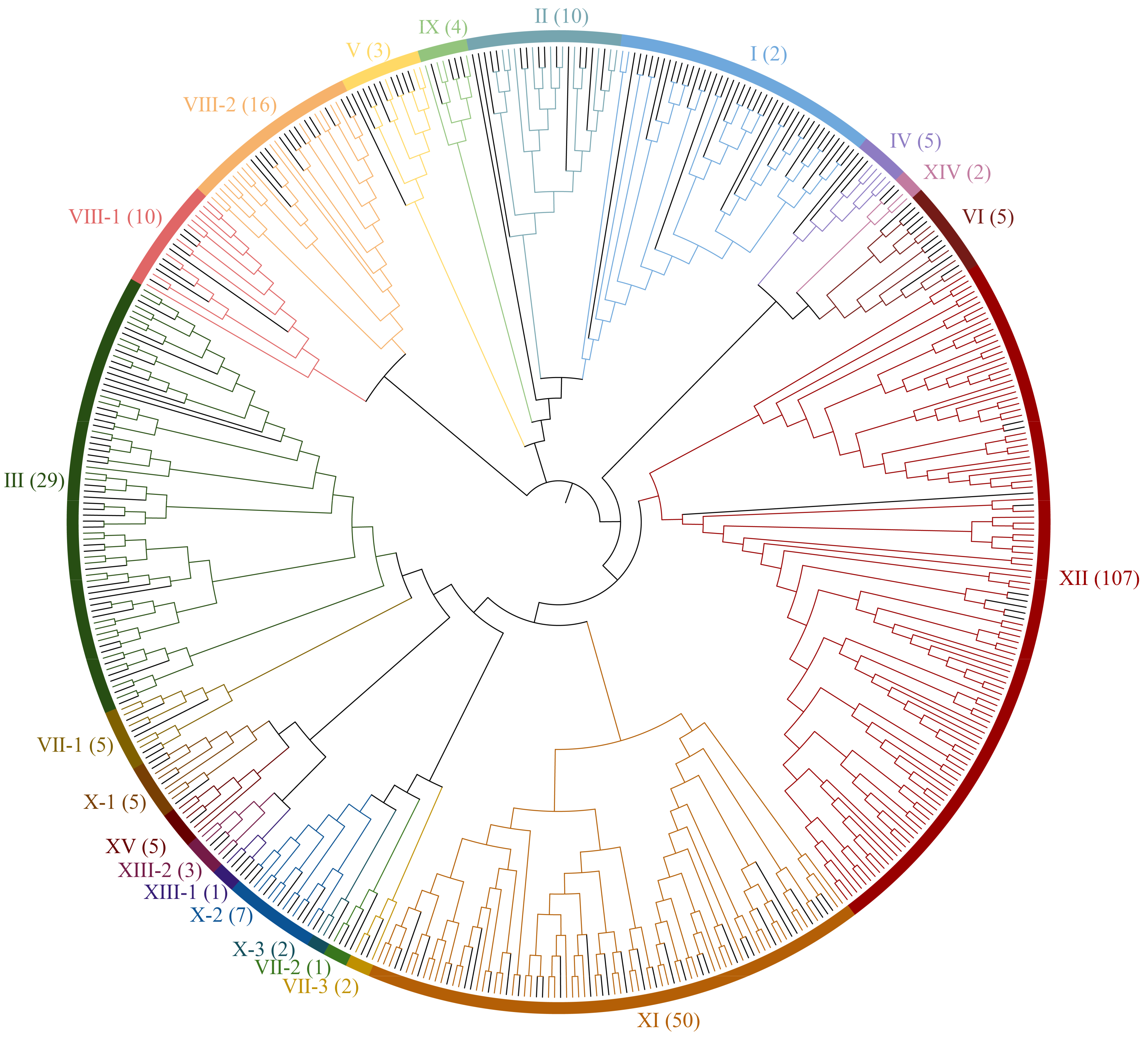
2.2. Phylogenetic Analysis

Multiple sequence alignment of 225 AtLRR-RLKs and 274 HbLRR-RLKs was performed using ClustalX (version 2.1, http://www.clustal.org/clustal2/, accessed on 15 March 2025). Using the neighbor-joining (NJ) method, the unrooted phylogenetic tree of AtLRR-RLKs and HbLRR-RLKs was constructed in the Mega 11 software, with 1000 bootstrap replicates. The tree was displayed using the ITOL online tool (version 6, http://itol.embl.de/help.cgi, accessed on 15 March 2025).

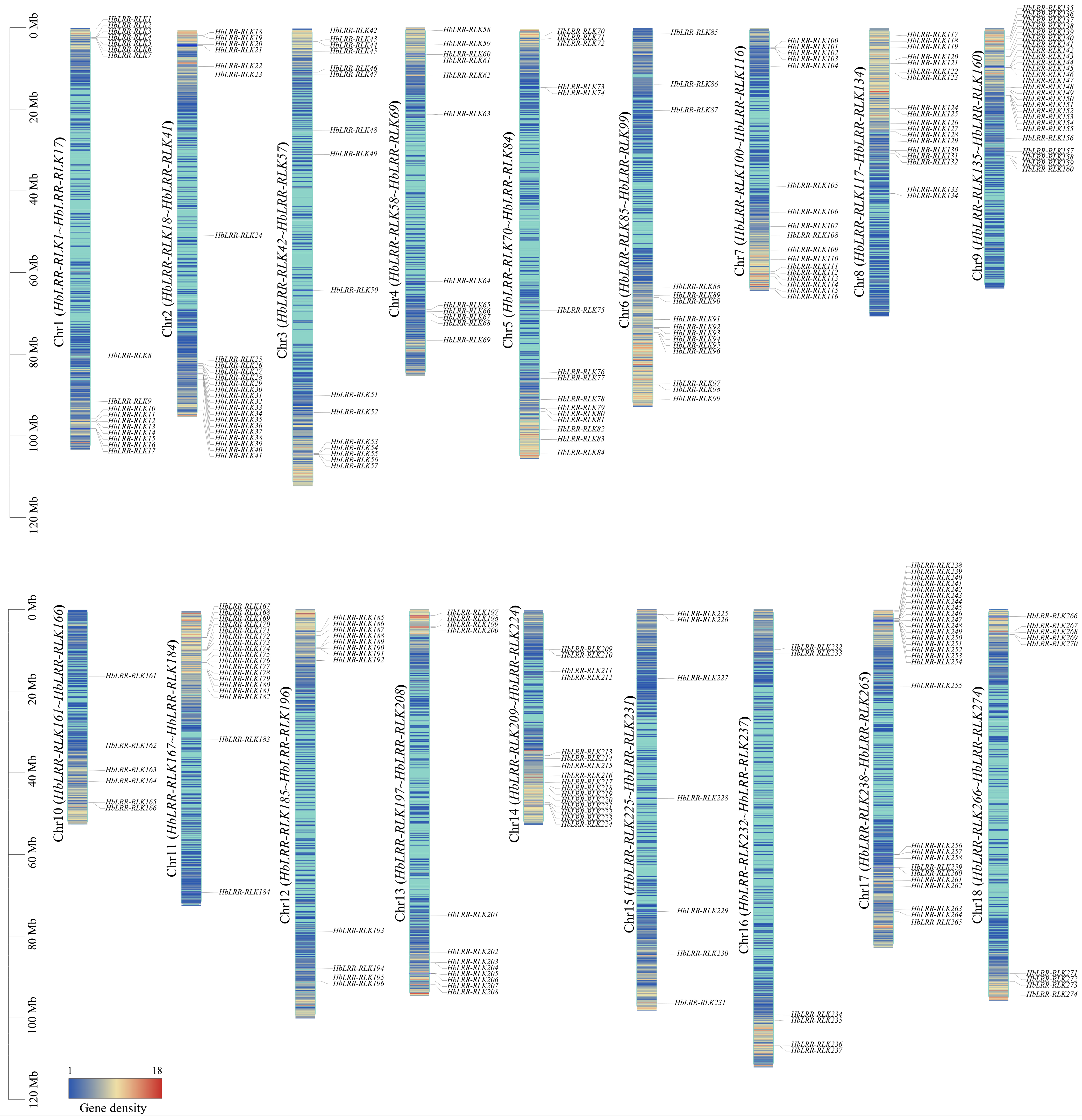
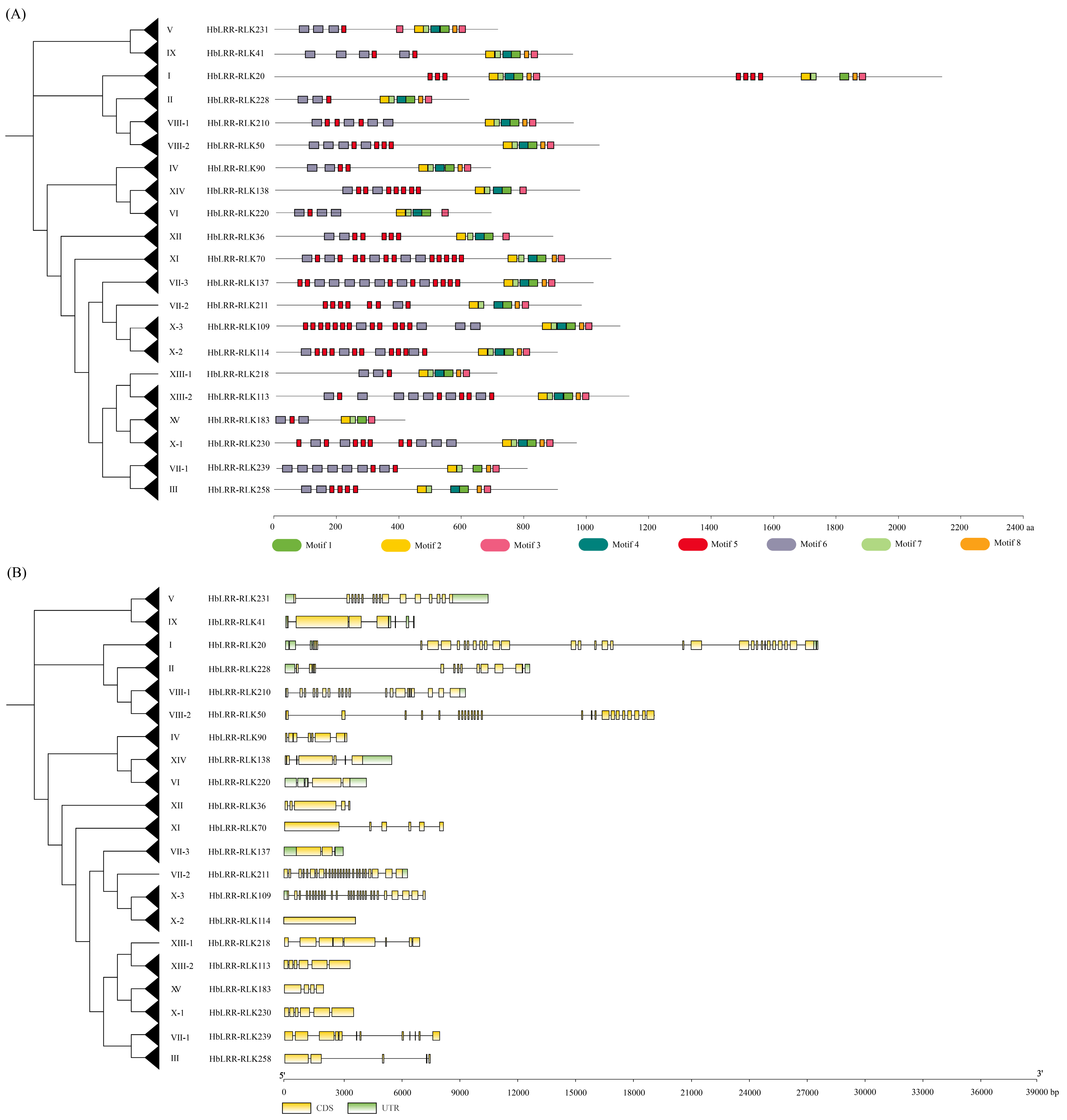
2.3. Chromosomal Localization, Conserved Motif, and Gene Structure Analysis

To build a distribution map for LRR-RLK genes in rubber tree, HbLRR-RLKs’ genomic information was retrieved from the rubber tree genome database. The chromosomal locations of HbLRR-RLKs were displayed using TBtools tool (version 2.056) [25]. The online MEME software (version 5.5.7, https://meme-suite.org/meme/, accessed on 15 March 2025) was used to predict the conserved motifs of HbLRR-RLKs based on the following parameters: site distribution was set to Zero or One Occurrence Per Sequence (zoops), and the number of motifs returned was set to 8. Other optional parameters were set to the default values. To further study the evolution of introns, TBtools was used to draw the HbLRR-RLK gene structures based on the annotation information.

2.4. HbLRR-RLK Duplication and Synteny Analysis

To identify gene duplication, the protein sequences of 274 HbLRR-RLKs were aligned using BLASTP (E value < 1 × 10⁻⁵). The MCScanX toolkit integrated with TBtools (version 2.056) was used to detect the duplication pattern of HbLRR-RLK. Two or more genes with a distance of less than 200 kb and more than 70% similarity were defined as tandem duplications. A synteny plot was drawn using the circos program to illustrate duplicated rubber tree gene pairs and the synteny blocks of orthologous LRR-RLK genes between Hevea brasiliensis and Arabidopsis thaliana (L.) Heynh (https://www.ncbi.nlm.nih.gov/datasets/genome/GCF_000001735.4/, accessed on 15 March 2025), Populus tomentosa Carr. (https://www.ncbi.nlm.nih.gov/datasets/genome/GCA_018804465.1/, accessed on 15 March 2025), and Manihot esculenta Crantz (https://www.ncbi.nlm.nih.gov/datasets/genome/GCF_001659605.2/, accessed on 15 March 2025). To evaluate gene divergence following duplication, we calculated the Ks (synonymous) and Ka (non-synonymous) substitution rates for each duplicated gene from the nucleotide sequence using the KaKs_Calculator (version 2.0) software.

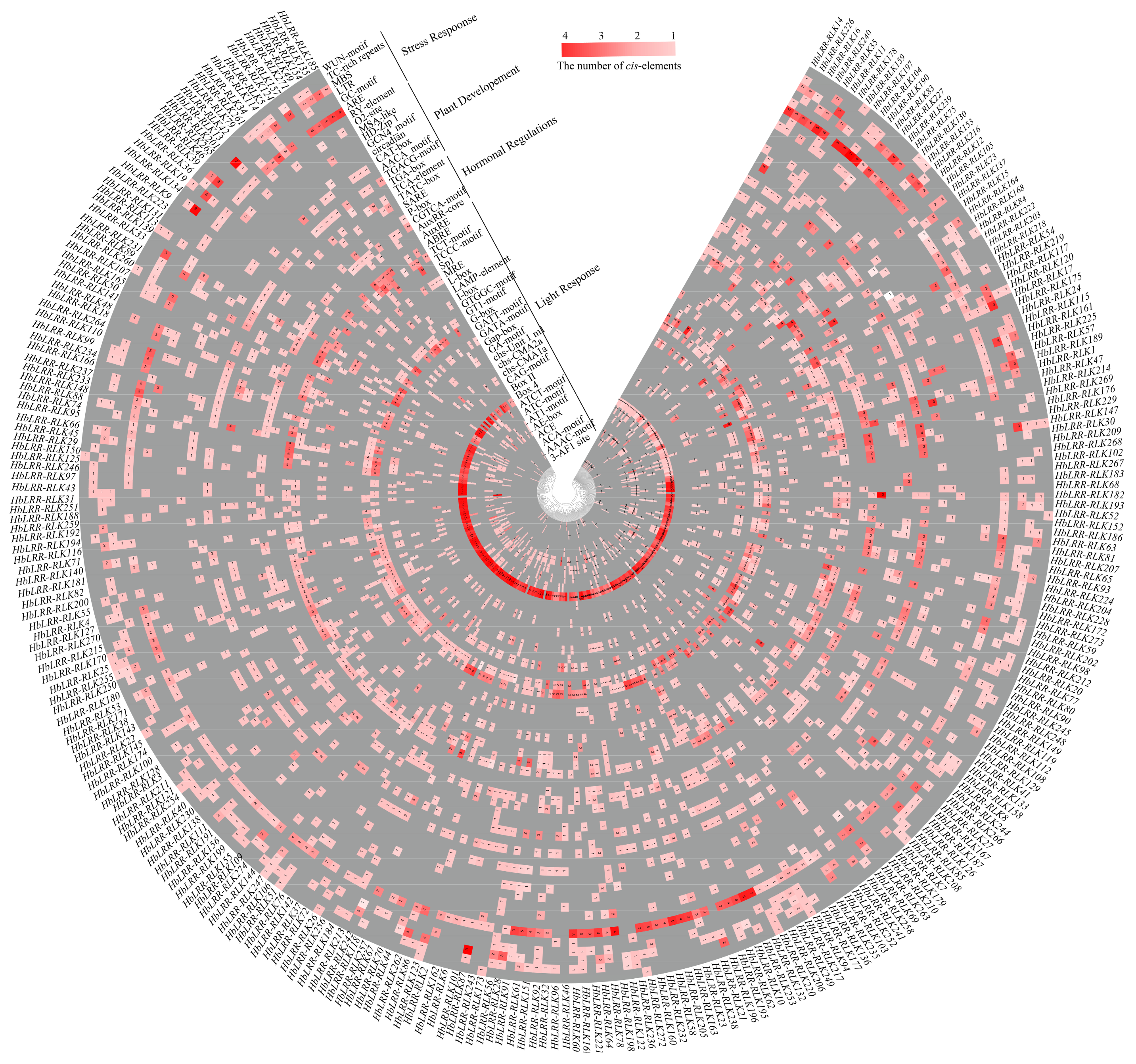
2.5. Analysis of Cis-Regulatory Elements HbLRR-RLKs

To investigate the cis-acting elements, the 2 kb promoter sequences of the HbLRR-RLK genes were extracted from the rubber tree genome and uploaded to the online database PlantCARE (https://bioinformatics.psb.ugent.be/webtools/plantcare/html/, accessed on 15 March 2025) with default parameters. Consensus sequences (such as TATA-box, CAAT-box, etc.) were filtered out, and functional elements were retained for further analysis.

2.6. Tissue-Specific HbLRR-RLKs Expression Patterns

Published transcriptome data for seven tissues (cambium, inner bark, primary latex, secondary latex, female flower, male flower, and leaf) were downloaded from the National Genomics Data Center (NGDC, https://ngdc.cncb.ac.cn/, accessed on 15 March 2025) under project number PRJCA004986. After removing adapters, the clean reads were mapped to the CATAS8-79 genome using HISAT2 software (version 2.2.1) [26], and the FPKM (fragments per kilobase of exon per million mapped fragments) value of each gene was analyzed using StringTie software (version 2.2.3) [27]. The expression levels of HbLRR-RLKs were obtained and visualized using TBtool (version 2.056) [25].

2.7. HbLRR-RLK Protein–Protein Interactions

The protein–protein interaction network of HbLRR-RLKs was generated using STRING (version 12.0, https://string-db.org, accessed on 15 March 2025) with the default parameters (required score: 0.4; FDR stringency: 0.05). Arabidopsis thaliana was used as the reference organism. The network was further visualized and analyzed using a grid layout model in Cytoscape software (version 3.9.1).

2.8. Subcellular Localization of HbPSKR2

The HbPSKR2 fragments were amplified, digested with KpnI and BamHI, and ligated into the pBinGFP4 vector digested with the same restriction endonuclease, generating the pBin-Pro35S::HbPSKR2:eGFP recombinant vector. NAA60 is a target protein located on the plasma membrane [28]. The pBin-Pro35S::NAA60:eRFP recombinant vector was maintained in our library. Both the pBin-Pro35S::HbPSKR2:eGFP and the pBin-Pro35S::NAA60:eRFP recombinant vectors were transiently introduced into Nicotiana benthamiana Domin using Agrobacterium tumefaciens (GV3101)-mediated transformation [29]. The subcellular localization of the proteins was observed under a Zeiss LSM 900 confocal microscope (Zeiss, Jena, Germany) at a magnification of 200×. The primers used for subcellular localization were listed in Supplementary Table S6.

2.9. Overexpression of HbPSKR2 in Arabidopsis

Arabidopsis thaliana (Columbia ecotype) was used for transformation. The Arabidopsis atpskr2 mutant (SALK No.: SALK_024464C) was obtained from Arashare (http://www.arashare.cn, accessed on 15 March 2025), a non-profit Arabidopsis mutant resource sharing center. The seedlings were planted in a growth chamber with a 16 h light/8 h dark cycle at 21 °C. The CDS sequence of HbPSKR2 was subcloned into the entry vector pDONR207 using Gateway™ BP Clonase™ II enzyme (Invitrogen, Waltham, MA, USA), then recombined into the destination vector pB7WG2D using Gateway™ LR Clonase™ II (Invitrogen, Waltham, MA, USA). The recombinant vector PB7WG2D-HbPSKR2 was transformed into Agrobacterium tumefaciens (GV3101) for plant transformation. The genetic transformation of Arabidopsis thaliana was performed using the floral dip method [30]. Transgenic seeds (T1) were selected on 1/2 Murashige and Skoog (MS) solid media, containing 0.4% gelzan (Sigma-Aldrich, Saint Louis, MO, USA), 2% sucrose, and 30 mg/L of hygromycin. Positive T1 plants were detected via direct polymerase chain reaction (PCR) amplification using the Plant Tissue PCR Kit (Yeasen, Shanghai, China) following the manufacturer’s protocol. The primers used for transgenic determination were listed in Supplementary Table S6.

2.10. Quantitative Real-Time PCR (qRT-PCR) Assay of the HbPSKR2 Gene

Total RNA was extracted using a Polysaccharide Polyphenol Plant Total RNA Extraction Kit (Tiangen Biotech Co., Beijing, China) according to the manufacturer’s instructions. The OD260 and OD280 values of total RNA were measured using the NanoDrop2000 ultramicroscopic spectrophotometer (Thermo Fisher, Waltham, MA, USA). The first strand of the cDNA was synthesized based on the RevertAid First Strand cDNA Synthesis Kit (Thermo Fisher, Waltham, MA, USA). Quantitative real-time (qRT)-PCR amplifications were performed using a Bio-Rad CFX384 PCR system and TB Green Premix Ex Taq (Takara, Dalian, China), fully following to the minimum information for publication of quantitative real-time PCR experiments guidelines [31]. The reaction procedure was as follows: 95 °C for 5 min, followed by 45 cycles of 95 °C for 20 s, 60 °C for 60 s, and 72 °C for 60 s. The melt curve was collected from 55 to 95 °C, increasing by 0.5 °C every 30 s. Relative expression levels were calculated using the 2−ΔΔCt method [32], with ACTIN3 as the internal reference gene [33]. The primers used for transgenic determination are listed in Supplementary Table S6.

2.11. Phenotypic Analysis of Transgenic Arabidopsis

The T3 homozygous generation of the highest transgenic expression line was used for root phenotypic analysis [34]. The transgenic seeds were surface sterilized for 2 min with 75% ethanol and rinsed 5 times with double-distilled water. Then, the seeds were placed on Petri plates containing 1/2 MS solid medium with 0.4% gelzan and 2% sucrose. The Petri plates were kept in a growth chamber with a 16 h light/8 h dark cycle at 21 °C for 7 days. The root length was measured from the hypocotyl to the root tip.

2.12. Statistical Analysis

Statistical analysis results are presented as mean ± standard deviation of three independent biological replicates. Student’s t-test was used to determine significant differences between groups.

3. Results

3.1. Characterization of LRR-RLKs in Rubber Tree

To identify the LRR-RLK family in the rubber tree, a BLASTP search was conducted using LRR-RLK protein sequences from Arabidopsis thaliana as queries. Following the confirmation of both LRR and RLK domains through Pfam analysis, a total of 274 LRR-RLK members (designated HbLRR-RLK1 to HbLRR-RLK274) were identified and systematically named based on their chromosomal positions (Figure 1, Table S1). These genes were distributed across 18 chromosomes, with chromosome 9 harboring the most members (26) and chromosomes 10 and 16 containing the least (six each). The molecular weights of the HbLRR-RLKs ranged from 45.52 kDa (HbLRR-RLK183) to 254.95 kDa (HbLRR-RLK11), and their isoelectric points (pI) varied from 5.10 (HbLRR-RLK115) to 9.36 (HbLRR-RLK22). Signal peptide (SP) analysis revealed that 48.91% (143/274) of the HbLRR-RLKs contained SPs, suggesting their potential involvement in extracellular signal transduction pathways.

3.2. Phylogenetic Analysis of Rubber Tree HbLRR-RLKs

To explore the evolutionary relationships of the LRR–RLK family members, we constructed an unrooted neighbor–joining (NJ) phylogenetic tree using 274 HbLRR-RLKs and 225 AtLRR-RLKs from Arabidopsis. The phylogenetic analysis classified the HbLRR-RLKs into 21 subclades based on the nomenclature of their Arabidopsis homologs (Figure 2, Table S2). The subclades exhibited significant variations in size, with subclade XII being the largest, comprising 107 HbLRR-RLKs and nine AtLRR-RLKs, while subclade X-3 was the smallest, containing only two HbLRR-RLKs and one AtLRR-RLK. Conserved domain analysis identified eight motifs in HbLRR-RLKs (Figure S1), with motifs 5 and 6 annotated as LRR domains and motifs 1, 2, 3, 4, 7, and 8 annotated as serine/threonine kinase catalytic domains, underscoring their functional roles in signal transduction (Figure 3A and Figure S2A). The exon-intron structure analysis revealed substantial variations among subclades but high conservation within subclades. For instance, subclades VII-3 and X-3 contained fewer than three exons, whereas subclades I, II, V, VIII-1, VIII-2, XIII-1, and XIII-2 contained more than ten exons (Figure 3B and Figure S2B).

3.3. HbLRR-RLK Gene Duplication and Synteny Analysis

Gene duplication events were analyzed using TBtools software (version 2.056) to explore the evolutionary mechanisms driving the expansion of the HbLRR-RLK family. We detected seven tandem duplication events on chromosomes 1, 9, 11, and 17 (Table S3). Intraspecific collinearity analysis revealed that 29.56% (81/274) of the HbLRR-RLKs were involved in 48 segmental duplication events (Figure 4A, Table S3). The Ka/Ks ratios of homologous gene pairs ranged from 0.34 to 0.71, suggesting that they have undergone purifying selection, maintaining their functional integrity while allowing for subtle diversification.
To further explore the expansion mechanisms, a comparative synteny analysis was performed between the rubber tree and three other species (Arabidopsis thaliana, Populus tomentosa, and Manihot esculenta). We identified 186 collinear LRR-RLK gene pairs between the rubber tree and cassava, followed by 137 pairs with poplar and 103 pairs with Arabidopsis, reflecting the closer genetic relationship between the rubber tree and cassava (Figure 4B). Notably, the HbLRR-RLKs on chromosomes 2 and 9 exhibited no synteny with the other three species, suggesting unique evolutionary trajectories for these genes in the rubber tree.

3.4. HbLRR-RLKs Cis-Regulatory Element Analysis

To explore the regulation of HbLRR-RLK expression, the 2 kb region upstream of the translation start sites was analyzed using PlantCARE. A total of 23 functional cis-elements were identified and categorized into four groups: light response, hormone response, developmental regulation, and stress response (Figure 5, Table S4). Box 4, CGTCA-motif, TGACG-motif, and ARE were the most abundant elements in their respective categories (Figure S3). Notably, the number of cis-elements varied significantly among HbLRR-RLK promoters, with the HbLRR-RLK82 promoter containing the most (51) and the HbLRR-RLK134 promoter containing the least (seven). This variability suggests diverse regulatory mechanisms governing the expression of individual HbLRR-RLKs.

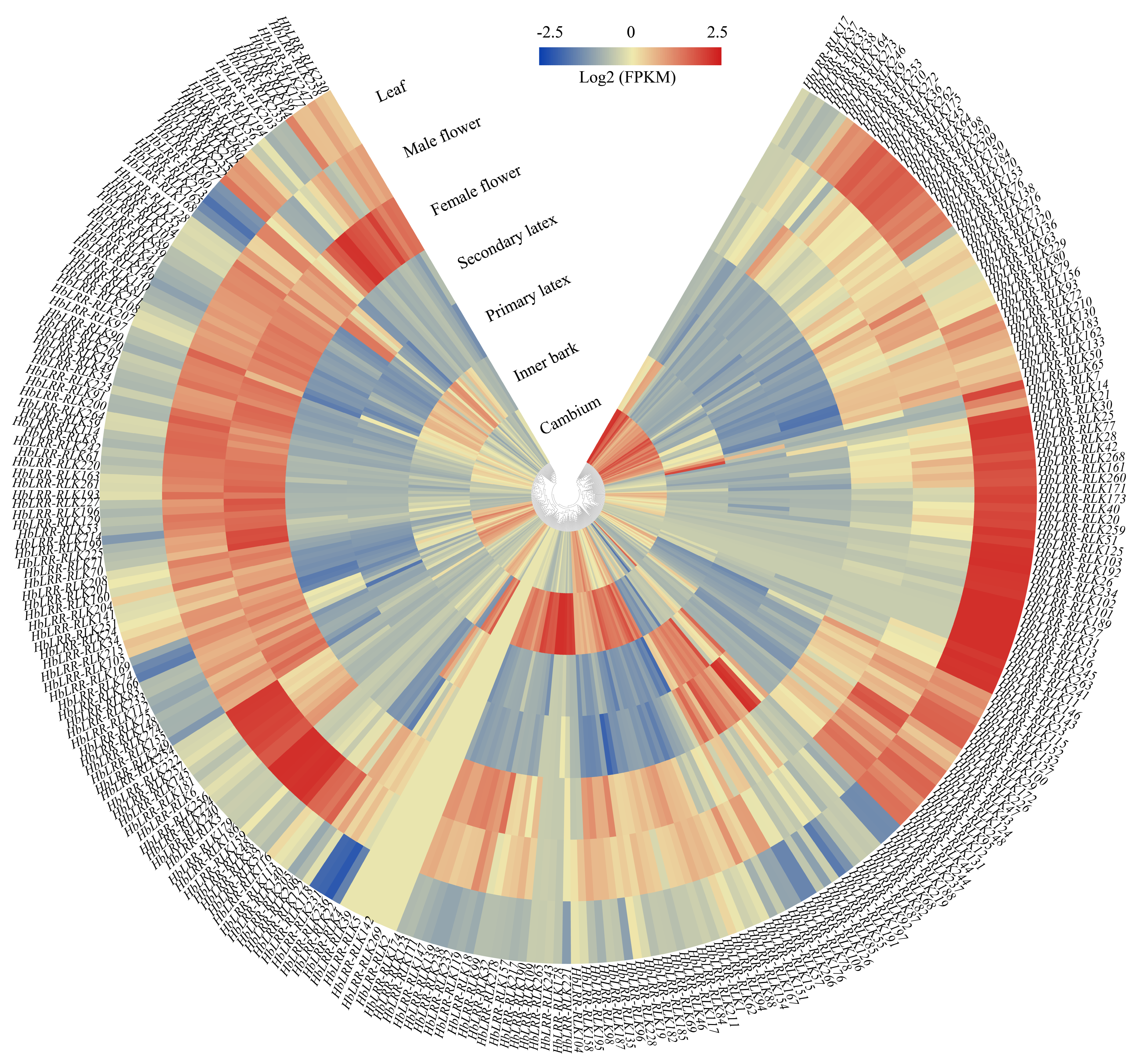
3.5. HbLRR-RLKs Tissue-Specific Expression Patterns

Transcriptome data from seven tissues (cambium, inner bark, primary latex, secondary latex, female flowers, male flowers, and leaves) were analyzed to determine the expression patterns of HbLRR-RLKs. Of the 274 genes, 200 (73.00%) were expressed in at least one tissue, with 30 genes showing constitutive expression across all tissues (Figure 6). Flower tissues contained the highest number of expressed genes (162 in female flowers and 171 in male flowers), while latex tissues showed the lowest (46 in primary latex and 50 in secondary latex). Notably, HbLRR-RLK188, HbLRR-RLK237, and HbLRR-RLK266 were expressed in all seven tissues, whereas HbLRR-RLK85 was expressed in latex only. Comparative analysis revealed that HbLRR-RLK67, HbLRR-RLK165, HbLRR-RLK126, and HbLRR-RLK219 were upregulated in secondary latex compared to primary latex, while HbLRR-RLK155 and HbLRR-RLK205 were downregulated, suggesting their potential roles in latex metabolism.

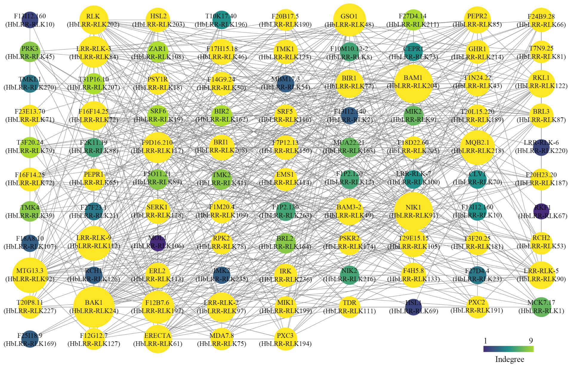
3.6. Prediction of the HbLRR-RLK Protein–Protein Interaction Network

To explore the functional interactions of HbLRR-RLKs, a protein–protein interaction network was constructed using Arabidopsis orthologs in the STRING database, revealing 584 potential interactions (Figure 7, Table S5). Among these, 40 HbLRR-RLKs were predicted to interact with HbLRR-RLK24, a homolog of Arabidopsis brassinosteroid-insensitive 1-associated receptor kinase 1 (BAK1), which is involved in brassinosteroid signaling and immune responses. Additionally, 36 HbLRR-RLKs were predicted to interact with HbLRR-RLK204, a homolog of Arabidopsis BAM1, a key regulator of meristem development. Furthermore, HbLRR-RLK174, a homolog of the phytosulfokine receptor (PSKR), demonstrated potential interactions with 13 HbLRR-RLK proteins, highlighting its role in peptide hormone signaling.

3.7. Subcellular Localization of HbPSKR2

We previously demonstrated that phytosulfokine (PSK) plays a critical role in laticifer differentiation and cloned the HbPSKR2 gene in the rubber tree. To determine its subcellular localization, HbLRR-RLK174 (HbPSKR2) was fused with GFP in the pBin-Pro35S::HbPSKR2:eGFP vector and transiently expressed in tobacco leaves. Cytological observation showed that the GFP signal co-localized with the membrane protein Nα-acetyltransferase (NAA60) fusing with RFP, confirming that HbPSKR2 is located in the cell membrane (Figure 8), which is consistent with its function as a receptor kinase.

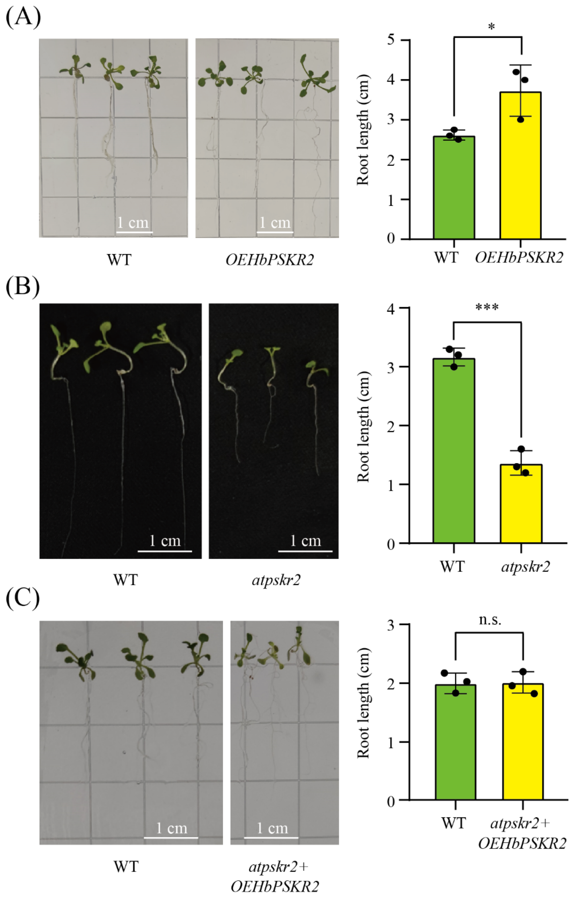
3.8. Functional Identification of HbPSKR2 in Arabidopsis

To further characterize the functional role of HbPSKR2, the gene was overexpressed in Arabidopsis. A total of four transgenic lines were generated, and the overexpressed line 5 (OE-5) with the highest expression level was used for further analysis (Figure S4A). Phenotypic analysis showed that the average root length of the HbPSKR2-overexpressing transgenic line (OEHbPSKR2) was 3.79 ± 0.51 cm, which was 1.5-fold that of the wild type (2.77 ± 0.22 cm) (Figure 9A), suggesting the role of HbPSKR2 in root development. Previous studies have shown that the atpskr2 mutant displays a short-root phenotype [16]. In the present study, the average root length of the atpskr2 mutant was 1.28 ± 0.20 cm, which was only 40% that of the wild type (3.19 ± 0.18 cm) (Figure 9B). To verify whether HbPSKR2 can complement the phenotype, we overexpressed HbPSKR2 in the atpskr2 mutant. A total of three transgenic lines were generated, and the overexpressed line 5 (OE-5) with the highest expression level was used for further analysis (Figure S4B). The phenotypic analysis of root length showed no significant difference between wild-type (2.96 ± 0.12 cm) and transgenic lines (2.97 ± 0.13 cm) (Figure 9C), suggesting that HbPSKR2 overexpression completely restored the atpskr2 mutant. These findings demonstrate the functional conservation of HbPSKR2 in regulating root development and suggest its potential role in rubber tree growth and laticifer differentiation.

4. Discussion

RLKs play a pivotal role in plant developmental regulation and defense responses through their capacity to perceive and transduce extracellular signals [35]. LRR-RLK is the largest RLK subfamily and has been systematically identified in multiple plant species. In the present study, we performed a comprehensive genome-wide analysis of LRR-RLKs in the rubber tree, ultimately identifying 274 HbLRR-RLK members. Comparative analysis revealed that the size of the HbLRR-RLK family exceeds those of Arabidopsis thaliana (225) [36], Gossypium hirsutum L. (234) [37], Vernicia fordii (Hemsl.) Airy Shaw (236) [38], and Broussonetia papyrifera (L.) L'Hér. ex Vent. (236) [39], but is smaller than those of Glycine max (L.) Merr. (467) [40], Triticum aestivum L. (929) [41], and Populus trichocarpa Torr. & Gray (379) [42]. These interspecific variations in gene family size may be attributed to evolutionary mechanisms such as whole-genome duplication events and polyploidization processes.
Tandem and segmental duplications represent two predominant modes of gene family expansion in plants [43]. Previous studies in potato reported that 6.1% (16/246) and 8.1% (20/246) of LRR-RLKs originated from tandem and segmental duplications [44], respectively. Our analysis identified higher proportions in the rubber tree, with 12.77% (35/274) of HbLRR-RLKs resulting from tandem duplication and 29.56% (81/274) from segmental duplication, indicating that both mechanisms significantly contribute to LRR-RLK expansion. Chromosomal localization revealed that tandemly duplicated genes predominantly cluster on chromosome 17 (36.84%, 14/38) and chromosome 9 (26.32%, 10/38). Synteny analysis demonstrated that the chromosome 17 cluster shares extensive orthology with cassava, whereas the chromosome 9 cluster lacks orthologous relationships with other species, suggesting its potential involvement in rubber tree-specific biological processes.
Gene family expansion and contraction represent critical evolutionary drivers [45]. Phylogenetic analysis classified the 274 HbLRR-RLKs into 21 subclades based on Arabidopsis homologs. Consistent with observations in Saccharum [46], Citrus [10], and Solanum lycopersicum [47], we detected marked subclade-specific variation: subclade I exhibited significant contraction (two HbLRR-RLKs vs. 41 AtLRR-RLKs), while subclade XII showed substantial expansion (115 HbLRR-RLKs vs. nine AtLRR-RLKs). MIK2 and PEPR2 are two members of Arabidopsis subclade XII. MIK2 is the receptor for SCOOP peptides regulating multiple processes, including plant growth and development [48], while PEPR2 is a receptor of plants endogenous PEP defense peptides released in response to stresses [49]. Functional annotation identified HbLRR-RLK124 and HbLRR-RLK85 as rubber tree orthologs of MIK2 and PEPR2, respectively. Tissue-specific expression profiling revealed distinct patterns: HbLRR-RLK124 predominated in developmental tissues (cambium, inner bark, flowers, and leaves), while HbLRR-RLK85 displayed latex-specific expression with significant induction via tapping-induced wounding. Protein–protein interaction networks further identified six putative interactors for HbLRR-RLK124 and twelve for HbLRR-RLK85, providing critical entry points for mechanistic investigations.
PSK represents a class of disulfated pentapeptides recognized by the receptor PSKR, which is localized on the plasma membrane [14]. Previous studies have demonstrated that PSKR regulates diverse biological processes, including plant growth and development, cell division and differentiation, and stress responses [15]. In Arabidopsis, root growth is significantly inhibited in the atpskr mutant, but this phenotype can be rescued through AtPSKR overexpression. We previously cloned the HbPSKR2 (HbLRR-RLK174) gene in the rubber tree [50]. In the present study, HbPSKR2 overexpression in Arabidopsis led to significantly elongated root lengths in transgenic lines compared with WT plants. Furthermore, HbPSKR2 restored the short-root phenotype of the atpskr mutant via functional complementation, demonstrating that the previously identified HbPSKR2 is a biologically active gene.
As a plasma membrane-localized receptor, PSKR interacts with other kinases to mediate signal transduction. Using the yeast two-hybrid (Y2H) system, Ding et al. identified CPK28, a calcium-dependent protein kinase, as an interactor of SlPSKR1 that regulates the balance between plant growth and immunity [51]. BAK1, which belongs to the LRR-RLK subclade II, functions as a versatile kinase participating in diverse plant developmental processes [52]. Crystal structure analysis conducted in 2015 demonstrated that PSK facilitates the interaction between the LRR domains of PSKR1 and BAK1, indicating that BAK1 is a co-receptor of PSKR1 in Arabidopsis [15]. We previously identified 12 HbPSKR2-interacting proteins (including two kinases) by screening a cambium Y2H library [50]. In this study, protein–protein interaction analyses revealed 13 LRR-RLKs (including the BAK1 homolog HbLRR-RLK24) as potential interactors of HbPSKR2. These proteins are distributed across subclades II, III, IV, XII, XIII, and XIV, with some exhibiting constitutive expression patterns (HbLRR-RLK122) or tissue-specific expression in laticifers (HbLRR-RLK90) or bark (HbLRR-RLK218), suggesting a complex regulatory network of HbPSKR2 in the rubber tree.

5. Conclusions

Systematic characterization of the LRR-RLK family was performed in the rubber tree in the present study. A total of 274 HbLRR-RLK genes were identified in the rubber tree genome and grouped into 21 subclades based on Arabidopsis homologs. Members within each subclade exhibited conserved protein domains and gene structures, reflecting their evolutionary relationships. Genome-wide duplication analysis revealed 35 tandem and 81 segmental duplication genes, highlighting that both duplications contribute to the expansion of the HbLRR-RLK family. Cis-element analysis of HbLRR-RLK promoters indicated the existence of light, stress, hormone, and development-related elements. Tissue-specific expression profiling showed that 200 HbLRR-RLKs were expressed in at least one tissue. The PPI network identified 584 potential interactions among HbLRR-RLKs. In addition, the function of plasma membrane-localized HbPSKR2 (HbLRR-RLK174) was investigated by complementation of the atpskr2 mutant in Arabidopsis. These comprehensive analyses provide valuable insights for further investigation of LRR-RLKs in the rubber tree.

Supplementary Materials

The following supporting information can be downloaded at: https://www.mdpi.com/article/10.3390/f16030552/s1, Figure S1: Eight conserved motifs in HbLRR-RLKs; Figure S2: Conserved motif (A) and gene structure (B) analysis of HbLRR-RLKs in the rubber tree; Figure S3: Statistics of cis-regulatory elements of HbLRR-RLKs; Figure S4: Overexpression of HbPSKR2 in the wild type (A) and the atpskr2 mutant (B) in Arabidopsis; error bars indicate the mean ± SD and *** represents p < 0.001 significant difference generated by Student’s t-test. Table S1: Identification of LRR-RLKs in the rubber tree genome; Table S2: Subfamily designation and sequence characteristics of the identified HbLRR-RLKs; Table S3: Ka/Ks analysis of segmental and tandem gene duplications of HbLRR-RLKs; Table S4: Cis-regulatory element analysis of HbLRR-RLK promoters; Table S5: Interactions of HbLRR-RLKs; Table S6: Primers used in this study.

Author Contributions

Conceptualization, X.D. (Xiaoyu Du) and J.C.; methodology, X.D. (Xiaomin Deng); software, X.D. (Xiaoyu Du) and J.J.; validation, S.Y., S.W., and M.S.; formal analysis, M.S.; investigation, X.D. (Xiaoyu Du); resources, J.C.; data curation, S.Y.; writing—original draft preparation, X.D. (Xiaoyu Du); writing—review and editing, J.C.; visualization, J.C.; supervision, J.C.; project administration, J.C.; funding acquisition, J.C. All authors have read and agreed to the published version of the manuscript.

Funding

This research was funded by the National Natural Science Foundation of China (32471915), the Project of National Key Laboratory for Tropical Crop Breeding (NKLTCBCXTD20, NKLTCB202309, NKLTCB202331), the Earmarked Fund for China Agriculture Research System (CARS-33-YZ1), and the National Key Research and Development Program (2023YFA0914800).

Data Availability Statement

The transcriptome data presented in this study are openly available in National Genomics Data Center under the BioProject accession number PRJCA004986.

Acknowledgments

The authors thank the Outstanding Talent Team Program of Hainan Province.

Conflicts of Interest

Jie Jin is employed by Wuhan BioRun Biosciences Co., Ltd. His employer’s company was not involved in this study, and there is no relevance between this research and the company.

Abbreviations

The following abbreviations are used in this manuscript:
LRR-RLKLeucine-Rich Repeat Receptor-like Kinase
ECDExtracellular receptor domain
TMTransmembrane domain
KDKinase domain
WAKWall-associated kinase
Duf26Domain of unknown function 26
LysMLysin motif
PSKPhytosulfokine
PSKRPhytosulfokine receptor
NRNatural rubber
NJNeighbor-joining
HbHevea brasiliensis
AtArabidopsis thaliana
PtPopulus trichocarpa
MeManihot esculenta
BAKBrassinosteroid-insensitive 1-associated receptor kinase
NAANα-acetyltransferase
Y2HYeast 2 hybrid

References

  1. Hubbard, S.R.; Miller, W.T. Receptor tyrosine kinases: Mechanisms of activation and signaling. Curr. Opin. Cell Biol. 2007, 19, 117–123. [Google Scholar] [CrossRef] [PubMed]
  2. Liang, X.; Zhou, J.M. Receptor-like cytoplasmic kinases: Central players in plant receptor kinase-mediated signaling. Annu. Rev. Plant Biol. 2018, 69, 267–299. [Google Scholar] [CrossRef] [PubMed]
  3. Walker, J.C.; Zhang, R. Relationship of a putative receptor protein kinase from maize to the S-locus glycoproteins of brassica. Nature 1990, 345, 743–746. [Google Scholar] [CrossRef]
  4. Dievart, A.; Clark, S.E. Using mutant alleles to determine the structure and function of leucine-rich repeat receptor-like kinases. Curr. Opin. Plant Biol. 2003, 6, 507–516. [Google Scholar] [CrossRef]
  5. Gou, X.; He, K.; Yang, H.; Yuan, T.; Lin, H.; Clouse, S.D.; Li, J. Genome-wide cloning and sequence analysis of leucine-rich repeat receptor-like protein kinase genes in Arabidopsis thaliana. BMC Genom. 2010, 11, 19. [Google Scholar] [CrossRef]
  6. Afzal, A.J.; Wood, A.J.; Lightfoot, D.A. Plant receptor-like serine threonine kinases: Roles in signaling and plant defense. Mol. Plant Microbe Interact. 2008, 21, 507–517. [Google Scholar] [CrossRef]
  7. Ngou, B.P.M.; Wyler, M.; Schmid, M.W.; Kadota, Y.; Shirasu, K. Evolutionary trajectory of pattern recognition receptors in plants. Nat. Commun. 2024, 15, 308. [Google Scholar] [CrossRef]
  8. Bella, J.; Hindle, K.L.; McEwan, P.A.; Lovell, S.C. The leucine-rich repeat structure. Cell. Mol. Life Sci. 2008, 65, 2307–2333. [Google Scholar] [CrossRef]
  9. Kajava, A.V. Structural diversity of leucine-rich repeat proteins. J. Mol. Biol. 1998, 277, 519–527. [Google Scholar] [CrossRef]
  10. Magalhães, D.M.; Scholte, L.L.; Silva, N.V.; Oliveira, G.C.; Zipfel, C.; Takita, M.A.; De Souza, A.A. LRR-RLK family from two Citrus species: Genome-wide identification and evolutionary aspects. BMC Genom. 2016, 17, 623. [Google Scholar] [CrossRef]
  11. Guo, Y.; Han, L.; Hymes, M.; Denver, R.; Clark, S.E. CLAVATA2 forms a distinct CLE-binding receptor complex regulating Arabidopsis stem cell specification. Plant J. 2010, 63, 889–900. [Google Scholar] [CrossRef] [PubMed]
  12. Huang, C.; Zhang, R.; Gui, J.; Zhong, Y.; Li, L. The receptor-like kinase AtVRLK1 regulates secondary cell wall thickening. Plant Physiol. 2018, 177, 671–683. [Google Scholar] [CrossRef] [PubMed]
  13. Yue, L.; Zhong, M.; Kang, D.; Qin, H.; Li, H.; Chai, X.; Kang, Y.; Yang, X. Genome-wide identification and characterization of flavonol synthase (FLS) gene family in Brassica vegetables and their roles in response to biotic and abiotic stresses. Sci. Hortic. 2024, 331, 113168. [Google Scholar] [CrossRef]
  14. Matsubayashi, Y.; Sakagami, Y. 120- and 160-kDa receptors for endogenous mitogenic peptide, phytosulfokine-α, in rice plasma membranes. J. Biol. Chem. 2000, 275, 15520–15525. [Google Scholar] [CrossRef]
  15. Wang, J.; Li, H.; Han, Z.; Zhang, H.; Wang, T.; Lin, G.; Chang, J.; Yang, W.; Chai, J. Allosteric receptor activation by the plant peptide hormone phytosulfokine. Nature 2015, 525, 265–268. [Google Scholar] [CrossRef]
  16. Matsubayashi, Y.; Ogawa, M.; Kihara, H.; Niwa, M.; Sakagami, Y. Disruption and overexpression of Arabidopsis phytosulfokine receptor gene affects cellular longevity and potential for growth. Plant Physiol. 2006, 142, 45–53. [Google Scholar] [CrossRef]
  17. Zhang, H.; Hu, Z.; Lei, C.; Zheng, C.; Wang, J.; Shao, S.; Li, X.; Xia, X.; Cai, X.; Zhou, J. A plant phytosulfokine peptide initiates auxin-dependent immunity through cytosolic Ca2+ signaling in tomato. Plant Cell 2018, 30, 652–667. [Google Scholar] [CrossRef] [PubMed]
  18. Nie, Z.; Kang, G.; Yan, D.; Qin, H.; Yang, L.; Zeng, R. Downregulation of HbFPS1 affects rubber biosynthesis of Hevea brasiliensis suffering from tapping panel dryness. Plant J. 2023, 113, 504–520. [Google Scholar] [CrossRef]
  19. Zhang, S.X.; Wu, S.H.; Chao, J.Q.; Yang, S.G.; Bao, J.; Tian, W.M. Genome-wide identification and expression analysis of MYC transcription factor family genes in rubber tree (Hevea brasiliensis Muell. Arg.). Forests 2022, 13, 531. [Google Scholar] [CrossRef]
  20. Yan, Y.; Liang, C.; Liu, X.; Tan, Y.; Lu, Y.; Zhang, Y.; Luo, H.; He, C.; Cao, J.; Tang, C.; et al. Genome-wide association study identifies candidate genes responsible for inorganic phosphorus and sucrose content in rubber tree latex. Trop. Plants 2023, 2, 24. [Google Scholar] [CrossRef]
  21. Wu, S.; Zhang, S.; Chao, J.; Deng, X.; Chen, Y.; Shi, M.; Tian, W.M. Transcriptome analysis of the signalling networks in coronatine-induced secondary laticifer differentiation from vascular cambia in rubber trees. Sci. Rep. 2016, 6, 36384. [Google Scholar] [CrossRef] [PubMed]
  22. Wichaita, W.; Promlok, D.; Sudjaipraparat, N.; Sripraphot, S.; Suteewong, T.; Tangboriboonrat, P. A concise review on design and control of structured natural rubber latex particles as engineering nanocomposites. Eur. Polym. J. 2021, 159, 110740. [Google Scholar] [CrossRef]
  23. Laosombut, T.; Arreewichit, P.; Nirapathpongporn, K.; Traiperm, P.; Kongsawadworakul, P.; Viboonjun, U.; Narangajavana, J. Differential expression of methyl jasmonate-responsive genes correlates with laticifer vessel proliferation in phloem tissue of rubber tree (Hevea brasiliensis). J. Plant Growth Regul. 2016, 35, 1049–1063. [Google Scholar] [CrossRef]
  24. Chao, J.; Wu, S.; Shi, M.; Xu, X.; Gao, Q.; Du, H.; Tian, W.M. Genomic insight into domestication of rubber tree. Nat. Commun. 2023, 14, 4651. [Google Scholar] [CrossRef]
  25. Chen, C.; Wu, Y.; Li, J.; Wang, X.; Zeng, Z.; Xu, J.; Liu, Y.; Feng, J.; Chen, H.; He, Y.; et al. TBtools-II: A “one for all, all for one” bioinformatics platform for biological big-data mining. Mol. Plant 2023, 16, 1733–1742. [Google Scholar] [CrossRef]
  26. Kim, D.; Paggi, J.M.; Park, C.; Bennett, C.; Salzberg, S.L. Graph-based genome alignment and genotyping with HISAT2 and HISAT-genotype. Nat. Biotechnol. 2019, 37, 907–915. [Google Scholar] [CrossRef] [PubMed]
  27. Pertea, M.; Pertea, G.M.; Antonescu, C.M.; Chang, T.C.; Mendell, J.T.; Salzberg, S.L. StringTie enables improved reconstruction of a transcriptome from RNA-seq reads. Nat. Biotechnol. 2015, 33, 290–295. [Google Scholar] [CrossRef]
  28. Linster, E.; Layer, D.; Bienvenut, W.V.; Dinh, T.V.; Weyer, F.A.; Leemhuis, W.; Brünje, A.; Hoffrichter, M.; Miklankova, P.; Kopp, J.; et al. The Arabidopsis Nα-acetyltransferase NAA60 locates to the plasma membrane and is vital for the high salt stress response. New Phytol. 2020, 228, 554–569. [Google Scholar] [CrossRef] [PubMed]
  29. Brown, D.; Feeney, M.; Ahmadi, M.; Lonoce, C.; Sajari, R.; Di Cola, A.; Frigerio, L. Subcellular localization and interactions among rubber particle proteins from Hevea brasiliensis. J. Exp. Bot. 2017, 68, 5045–5055. [Google Scholar] [CrossRef]
  30. Clough, S.J.; Bent, A.F. Floral dip: A simplified method for Agrobacterium-mediated transformation of Arabidopsis thaliana. Plant J. 1998, 16, 735–743. [Google Scholar] [CrossRef]
  31. Bustin, S.A.; Benes, V.; Garson, J.A.; Hellemans, J.; Huggett, J.; Kubista, M.; Mueller, R.; Nolan, T.; Pfaffl, M.W.; Shipley, G.L.; et al. The MIQE guidelines: Minimum information for publication of quantitative real-time PCR experiments. Clin. Chem. 2009, 55, 611–622. [Google Scholar] [CrossRef] [PubMed]
  32. Livak, K.J.; Schmittgen, T.D. Analysis of Relative Gene Expression Data Using Real-Time Quantitative PCR and the 2−ΔΔCT Method. Methods 2001, 25, 402–408. [Google Scholar] [CrossRef]
  33. Liu, W.; Zhou, Y.; Li, X.; Wang, X.; Dong, Y.; Wang, N.; Liu, X.; Chen, H.; Yao, N.; Cui, X.; et al. Tissue-Specific Regulation of Gma-miR396 Family on Coordinating Development and Low Water Availability Responses. Front. Plant Sci. 2017, 8, 1112. [Google Scholar] [CrossRef]
  34. Noman, M.; Jameel, A.; Qiang, W.D.; Ahmad, N.; Liu, W.C.; Wang, F.W.; Li, H.Y. Overexpression of GmCAMTA12 Enhanced Drought Tolerance in Arabidopsis and Soybean. Int. J. Mol. Sci. 2019, 20, 4849. [Google Scholar] [CrossRef]
  35. Soltabayeva, A.; Dauletova, N.; Serik, S.; Sandybek, M.; Omondi, J.O.; Kurmanbayeva, A.; Srivastava, S. Receptor-like kinases (LRR-RLKs) in response of plants to biotic and abiotic stresses. Plants 2022, 11, 2660. [Google Scholar] [CrossRef]
  36. Matsushima, N.; Miyashita, H. Leucine-rich repeat (LRR) domains containing intervening motifs in plants. Biomolecules 2012, 2, 288–311. [Google Scholar] [CrossRef] [PubMed]
  37. Sun, R.; Wang, S.; Ma, D.; Liu, C. Genome-wide analysis of LRR-RLK gene family in four Gossypium species and expression analysis during cotton development and stress responses. Genes 2018, 9, 592. [Google Scholar] [CrossRef] [PubMed]
  38. Zhu, H.; Wang, Y.; Yin, H.; Gao, M.; Zhang, Q.; Chen, Y. Genome-wide identification and characterization of the LRR-RLK gene family in two Vernicia species. Int. J. Genom. 2015, 2015, 823427. [Google Scholar] [CrossRef]
  39. Su, Y.; Peng, X.; Shen, S. Identification of leucine-rich repeat receptor-like protein kinase (LRR-RLK) genes in paper mulberry and their potential roles in response to cold stress. Comput. Biol. Chem. 2022, 97, 107622. [Google Scholar] [CrossRef]
  40. Zhou, F.; Guo, Y.; Qiu, L.J. Genome-wide identification and evolutionary analysis of leucine-rich repeat receptor-like protein kinase genes in soybean. BMC Plant Biol. 2016, 16, 58. [Google Scholar] [CrossRef]
  41. Liu, S.; Lei, J.; Zhang, J.; Liu, H.; Ye, Z.; Yang, J.; Lu, Q.; Liu, P.; Chen, J.; Yang, J. Genome-wide identification and analysis of wheat LRR-RLK family genes following Chinese wheat mosaic virus infection. Front. Plant Sci. 2023, 13, 1109845. [Google Scholar] [CrossRef]
  42. Zan, Y.; Ji, Y.; Zhang, Y.; Yang, S.; Song, Y.; Wang, J. Genome-wide identification, characterization and expression analysis of Populus leucine-rich repeat receptor-like protein kinase genes. BMC Genom. 2013, 14, 318. [Google Scholar] [CrossRef]
  43. Oren, M.; Barela Hudgell, M.A.; D’Allura, B.; Agronin, J.; Gross, A.; Podini, D.; Smith, L.C. Short tandem repeats, segmental duplications, gene deletion, and genomic instability in a rapidly diversified immune gene family. BMC Genom. 2016, 17, 900. [Google Scholar] [CrossRef]
  44. Li, X.; Salman, A.; Guo, C.; Yu, J.; Cao, S.; Gao, X.; Li, W.; Li, H.; Guo, Y. Identification and Characterization of LRR-RLK Family Genes in Potato Reveal Their Involvement in Peptide Signaling of Cell Fate Decisions and Biotic/Abiotic Stress Responses. Cells 2018, 7, 120. [Google Scholar] [CrossRef]
  45. Lespinet, O.; Wolf, Y.I.; Koonin, E.V.; Aravind, L. The role of lineage-specific gene family expansion in the evolution of eukaryotes. Genome Res. 2002, 12, 1048. [Google Scholar] [CrossRef]
  46. Ding, H.; Feng, X.; Yuan, Y.; Wang, B.; Wang, Y.; Zhang, J. Genomic investigation of duplication, functional conservation, and divergence in the LRR-RLK Family of Saccharum. BMC Genom. 2024, 25, 165. [Google Scholar] [CrossRef]
  47. Wei, Z.; Wang, J.; Yang, S.; Song, Y. Identification and expression analysis of the LRR-RLK gene family in tomato (Solanum lycopersicum) Heinz 1706. Genome 2015, 58, 121–134. [Google Scholar] [CrossRef]
  48. Hou, S.; Liu, D.; Huang, S.; Luo, D.; Liu, Z.; Xiang, Q.; Wang, P.; Mu, R.; Han, Z.; Chen, S.; et al. The Arabidopsis MIK2 receptor elicits immunity by sensing a conserved signature from phytocytokines and microbes. Nat. Commun. 2021, 12, 5494. [Google Scholar] [CrossRef]
  49. Song, X.; Li, J.; Lyu, M.; Kong, X.; Hu, S.; Song, Q.; Zuo, K. CALMODULIN-LIKE-38 and PEP1 RECEPTOR 2 integrate nitrate and brassinosteroid signals to regulate root growth. Plant Physiol. 2021, 187, 1779–1794. [Google Scholar] [CrossRef]
  50. Du, X.; Zhao, Y.; Zhang, S.; Tian, W.; Chao, J. Cloning and Interacted Protein Identification of HbPSKR2 Gene from Rubber Tree. Chin. J. Trop. Crops 2024, 45, 653–662. [Google Scholar] [CrossRef]
  51. Ding, S.; Lv, J.; Hu, Z.; Wang, J.; Wang, P.; Yu, J.; Foyer, C.H.; Shi, K. Phytosulfokine peptide optimizes plant growth and defense via glutamine synthetase GS2 phosphorylation in tomato. EMBO J. 2023, 42, e111858. [Google Scholar] [CrossRef] [PubMed]
  52. Li, J.; Wen, J.; Lease, K.A.; Doke, J.T.; Tax, F.E.; Walker, J.C. BAK1, an Arabidopsis LRR receptor-like protein kinase, interacts with BRI1 and modulates brassinosteroid signaling. Cell 2002, 110, 213–222. [Google Scholar] [CrossRef] [PubMed]
Figure 1. Location of HbLRR-RLK genes on the chromosome of rubber tree. The chromosome number is labeled on the left side of each chromosome. The gene density is displayed by the color scale. The leftmost scale represents the chromosome length (Mb).
Forests 16 00552 g001
Figure 2. Phylogenetic analysis conducted on 274 HbLRR-RLKs and 225 AtLRR-RLKs. Different color bands indicate different subclades. The AtLRR-RLKs in each subclade are displayed with a black line.
Forests 16 00552 g002
Figure 3. Conserved motif and gene structure analysis of each HbLRR-RLK subclade in the rubber tree. (A) Phylogenetic relationships and conserved motif of representative HbLRR-RLK proteins. The black triangle represents the subclade that contains more than one HbLRR-RLK members. Eight conserved motifs are displayed with different colors. (B) The number and arrangement of exons and introns of representative HbLRR-RLK coding sequences (CDSs). Coding sequences (CDSs) are shown in yellow and untranslated regions (UTRs) in green.
Forests 16 00552 g003
Figure 4. Analysis of synteny of HbLRR-RLKs in the rubber tree and collinearity of LRR-RLKs families among four species. (A) Synteny of HbLRR-RLKs in the rubber tree. The red lines represent the duplicated gene pairs. (B) Collinearity of LRR-RLKs families among four species (Arabidopsis thaliana, Populus tomentosa, Manihot esculenta, and Hevea brasiliensis). The red lines represent the collinearity of LRR-RLK gene pairs.
Forests 16 00552 g004
Figure 5. Statistics of the cis-acting elements number in HbLRR-RLK promoter region. The number of cis-acting elements is displayed in shades of red.
Forests 16 00552 g005
Figure 6. Expression pattern of HbLRR-RLKs in seven tissues with hierarchical clustering. The color scale represents the log2-based expression values converted from the FPKM values. Colors from red to blue indicate higher and lower levels, respectively.
Forests 16 00552 g006
Figure 7. Protein interaction and structure analysis. Protein–protein interaction networks are constructed using STRING software (version 12.0). The degree of interaction is represented by both the color and the size of each node.
Forests 16 00552 g007
Figure 8. Subcellular localization of HbPSKR2 determined by transient expression in the epidermal cells of tobacco leaves. NAA60 fusing with RFP is used as a membrane location marker. Bar = 20 μm.
Forests 16 00552 g008
Figure 9. Overexpression of HbPSKR2 promoted Arabidopsis root growth. (A) Root length between the wild-type and overexpression lines. (B) Root length between the wild type and the atpskr2 mutant. (C) Root length between the wild-type and functional complementary overexpression lines. WT, wild type; OEHbPSKR2, HbPSKR2 overexpression plants. The numerical value represents the average ± standard deviation of three independent replicates. * p < 0.05, *** p < 0.001, n.s. no significant difference (t test).
Forests 16 00552 g009
Disclaimer/Publisher’s Note: The statements, opinions and data contained in all publications are solely those of the individual author(s) and contributor(s) and not of MDPI and/or the editor(s). MDPI and/or the editor(s) disclaim responsibility for any injury to people or property resulting from any ideas, methods, instructions or products referred to in the content.

