Article

Long-Term Successional Subculture Dynamics and Their Effects on the Proliferation Efficiency, Embryogenic Potential, and Genetic Stability of Embryogenic Tissues in Larix principis-rupprechtii Mayr

1
State Key Laboratory of Tree Genetics and Breeding, State Key Laboratory of Efficient Production of Forest Resources, National Engineering Research Center of Tree Breeding and Ecological Restoration, Key Laboratory of Genetics and Breeding in Forest Trees and Ornamental Plants, Ministry of Education, The Tree and Ornamental Plant Breeding and Biotechnology Laboratory of National Forestry and Grassland Administration, College of Biological Sciences and Biotechnology, Beijing Forestry University, Beijing 100083, China
2
National Key Seed Base of Larch, Hebei Academy of Forestry Science, Shijiazhuang 050061, China
3
Centre for Forest Biology, Department of Biology, University of Victoria, 3800 Finnerty Rd., Victoria, BC V8W 3N5, Canada
*
Author to whom correspondence should be addressed.
These authors contributed equally to this work.
Forests 2024, 15(4), 627; https://doi.org/10.3390/f15040627
Submission received: 7 March 2024 / Revised: 23 March 2024 / Accepted: 28 March 2024 / Published: 29 March 2024
(This article belongs to the Section Genetics and Molecular Biology)

Abstract

:
Larix principis-rupprechtii Mayr, a coniferous species indigenous to Northern China, possesses significant ecological and economic value. Somatic embryogenesis offers a pathway with significant potential for large-scale propagation, long-term germplasm conservation, and genetic transformation in L. principis-rupprechtii Mayr. However, it remains unclear whether significant variations occur in embryogenic tissues during long-term successive subculturing, which could impact the productivity of somatic embryos. This is a pivotal concern that lacks comprehensive understanding. In this study, three embryogenic cell lines were used to explore the dynamics and relationships among proliferation rate, pre-treatment proliferation rate, and embryogenic capabilities across a series of 32 subculturing cycles. Proliferation rate, pre-treatment proliferation rate, and somatic embryo maturation rate showed no significant correlation with subculturing cycles. However, there was a positive correlation between subculturing cycles and pre-treatment proliferation rate and a negative correlation with somatic embryo maturation rate in the BFU1 cell line. In addition, we utilized ten SSR molecular markers to investigate the genetic stability in embryogenic tissues during long-term subculturing. No genomic variations were detected in any of the three embryogenic cell lines, which suggests that the observed phenotypic dynamics during subculturing may not be primarily driven by genomic alterations. This study provides novel insights into the dynamics of the long-term culture of embryogenic tissues, laying a foundation for the optimization and application of somatic embryogenesis techniques in L. principis-rupprechtii Mayr and potentially other coniferous species.

1. Introduction

Prince Rupprecht’s larch (Larix principis-rupprechtii Mayr) is a Pinaceae species native to northern China. Because of its durable qualities, robust resistance to harsh weather conditions, soil retention capability, and wind-resistant traits, this species is valuable for reforestation and afforestation in barren mountains, demonstrating high ecological and economic significance [1]. However, the traditional sexual reproduction methods used for breeding and propagation present several disadvantages. These include a long cycle, low germination rate, and challenges such as the age effect, location effect, and difficulty in obtaining materials [2,3,4,5,6]. Consequently, these factors contribute to the inefficiency of Prince Rupprecht’s larch breeding. The rapid progress in plant somatic embryogenesis has surmounted numerous challenges encountered in conventional sexual reproduction breeding methods, paving the path for the advancement of effective gene transfer technology and widespread propagation of new, high-performing clonal populations [7,8,9,10]. Research on somatic embryogenesis techniques in Pinus species commenced with the initial observation of embryo-like structures in Banks pine (Pinus banksiana) documented in 1976 [11]. Factors impacting diverse stages of somatic embryogenesis (embryogenic tissue induction, proliferation culture, pre-treatment, maturation treatment, etc.) and techniques to enhance culture efficiency across various Pine species have been explored [10,12,13,14,15,16,17,18,19,20]. Furthermore, with the advancement of bioinformatics, researchers are increasingly focusing on the role of genes involved in the various stages of somatic embryogenesis [8,21,22,23]. However, studies have shown that some conifer somatic cells exhibit reduced proliferation efficiency and even loss of maturation capability after long-term ex vivo culture [24,25]. The occurrence of this phenomenon introduces uncertainty regarding the extensive application of somatic embryogenesis in Prince Rupprecht’s larch.
During plant tissue culture, cultivated tissues frequently experience mutations [26]. Some studies have found that the rate of variation in regenerated plants produced through callus tissues is higher than those produced through axillary buds [27]. After callus tissues undergo dedifferentiation and redifferentiation processes, changes in DNA methylation levels occur. By studying the effect of tissue culture on methylation using a series of non-transgenic rice lines regenerated plants and wild-type plants, it was found that all regenerated plants exhibited significantly reduced levels of methylation compared to non-regenerated plants [28]. Physiological changes typically result in non-heritable mutations, while genetic material alterations lead to heritable mutations [29,30]. Variation in somatic embryogenic lines generally originates from two sources: inherent genetic variation in the plant tissue culture material, and variation induced during the tissue culture process, which accumulates with long-term culture cycles. Conventionally, five primary factors are considered to influence somatic cell embryogenic line in plants: (1) Plant genotype directly impacts the quantity and quality of embryogenic line variation. Additionally, substantial variations exist in somatic cell embryogenic line among different plant varieties [31,32]. (2) Regarding explant material, utilizing meristematic tissues like stem tips and axillary buds generally results in a lower likelihood of mutations compared to using highly differentiated plant tissues such as roots and leaves [33,34]. (3) Culture conditions, specifically the composition of the culture medium, influence the cell division mechanism, potentially inducing mutation phenomena [35]. (4) The duration of in vitro culture significantly influences mutation rates [27,36,37]; however, some studies suggest that even short-term plant cultures exhibit genetic variations, implying that culture duration is not the sole determinant of mutation occurrence in plant somatic cell lines [38]. It has been reported that the quality and quantity of maturation of somatic embryos have been observed to decrease with increasing number (and duration) of successions in species of the genus Pinus [39,40,41]. Furthermore, degeneration of embryogenic tissues hindered early embryo formation, potentially exacerbated by manipulations during successive cultures [24]. These findings demonstrate the biochemical impacts of in vitro culture and the adverse effects of aging on these processes.
The objective of this study was to explore the impact of extended culture cycles on the proliferation efficiency, maturation capacity of Prince Rupprecht’s larch embryogenic tissues, and to evaluate genetic variations in the long-term successions using SSR molecular marker technology. This study will lay the foundation for large-scale asexual reproduction and further genetic improvement of Prince Rupprecht’s larch, providing important theoretical guidance for the better application of somatic embryogenesis technology in forestry production.

2. Materials and Methods

2.1. Plant Materials

The open-pollinated cones were collected from the seed orchard of L. principis-rupprechtii Mayr., the location of which, along with the initiation of embryogenic tissues, refers to our previous research results [15]. Based on previous research and observations, the embryogenic tissues of the stable Prince Rupprecht’s larch embryogenic cell lines BFU1, BFU3, and BFU8 established in our laboratory were selected as experimental materials for the study. Each cell line exhibited distinct characteristics in terms of morphology and growth behavior.

2.2. Embryogenic Tissue Proliferation

The embryogenic tissue weighing 1 g was weighed and dispersed in the proliferation medium on day 0, with each series number repeated four times. On the 14th day, the embryogenic tissues in the proliferation medium were weighed, and 4 g of embryogenic tissues was extracted and stored in frozen tubes. After freezing with liquid nitrogen, the tissues were transferred to an ultra-low temperature refrigerator set at −80 °C for DNA extraction. This process was repeated every 14 days to complete a culture cycle. The proliferation in semi-solid medium was 1/2 mLV medium supplemented with 10 g L−1 sucrose, 3 g L−1 phytagel (Sigma, Shanghai, China), 0.4 g L−1 hydrolyzed casein (Sigma, Shanghai, China), 0.5 g L−1 glutamine (Sigma, Shanghai, China), 0.2 mg L−1 2,4-D (Sigma, Shanghai, China), and 0.1 mg L−1 6-BA (Sigma, Shanghai, China).

2.3. Pre-Treatment and Maturation

On the 10th day of the proliferation culture, 0.5 g of embryogenic tissue was weighed and cultured on pre-treatment medium, with each line replicated four times. Subsequently, on the 10th day of pre-treatment, the embryogenic tissues were transferred to the maturation medium, and the count of somatic embryos was conducted after a two-month maturation period. The pre-treatment in semi-solid medium was 1/2 mLV medium supplemented with 10 g L−1 sucrose, 20 g L−1 maltose, 50 g L−1 PEG4000 (Sigma, Shanghai, China), 2 g L−1 activated charcoal (Sigma, Shanghai, China), 0.2 g L−1 hydrolyzed casein (Sigma, Shanghai, China), 0.4 g L−1 glutamine (Sigma, Shanghai, China), and 3 g L−1 phytogel (Sigma, Shanghai, China). The maturation medium was 1/2 mLV medium supplemented with 10 g L−1 sucrose, 30 g L−1 maltose, 50 g L−1 PEG4000 (Sigma, Shanghai, China), 16 mg L−1 ABA (Sigma, Shanghai, China), 0.2 g L−1 hydrolyzed casein (Sigma, Shanghai, China), 0.4 g L−1 glutamine (Sigma, Shanghai, China), and 6 g L−1 phytogel (Sigma, Shanghai, China).

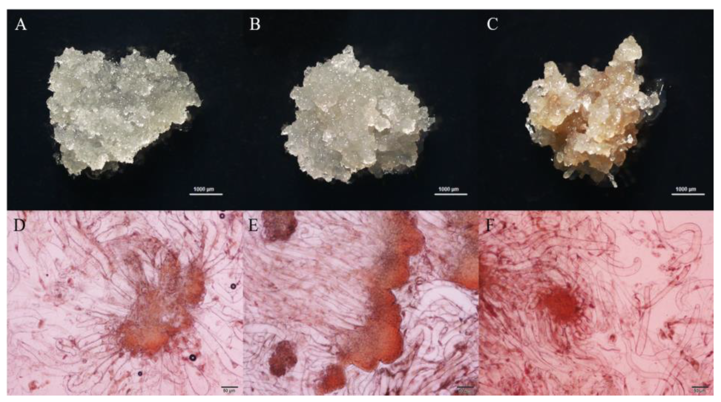
2.4. Staining and Examination of Embryogenic Tissue

Small (~2 mm) diameter clumps of embryogenic tissue were placed on a glass slide, to which a few drops of 2% acetocarmine were added until the callus was completely covered. The callus was dispersed into small pieces using forceps. After 30 s, the slide was washed with deionized water, which was then removed. Slides were examined and photographed using Universal compound microscope fitted with OLYMPUS DP21 (Olympus Corporation, Tokyo, Japan) digital color camera.

2.5. Genomic DNA Extraction

Tissue samples from the BFU1, BFU3, and BFU8 lines at the 1st, 7th, 17th, 25th, and 32nd proliferation cycles, along with fresh embryogenic tissues from the same lines after two years of continuous culture, underwent DNA extraction using the Plant Genomic DNA Extraction Kit (Tiangen, DP360, Beijing, China).

2.6. SSR Identification

Ten pairs of polymorphic primers for Prince Rupprecht’s larch were screened according to previous studies, two pairs were screened from SSR sequences derived from sequencing the transcriptome of Prince Rupprecht’s larch healing tissues by Dong Mingliang et al. [42], three pairs were derived from EST-SSR markers for Pinus species developed by Fan Y. et al. [43], and five pairs were derived from EST-SSR markers for Prince Rupprecht’s larch developed by Dong Mingliang et al. [44]. Information of primers were listed in Table 1.
The electrophoretic profiles were manually interpreted, and the electrophoretic bands produced by each pair of primers were counted for each line number at different periods of time to compare whether polymorphic variation was produced or not.

2.7. Data Analysis

The weight and rate of embryogenic tissue proliferated during proliferation culture, the weight and rate of embryogenic tissue proliferated during pre-treatment, and the capacity of somatic embryogenesis in mature culture were calculated, respectively. The analysis software was Microsoft Excel 2019 and SPSS 27. Pearson correlation coefficient (r) was employed to analyze the correlation between culture period and proliferation efficiency and maturation capacity, and the correlation between proliferation rate of proliferation culture, proliferation rate of pre-treatment, and maturation capacity.
In this study, we introduced three key parameters to evaluate the proliferation and differentiation capabilities of the tissue cultures: Proliferation Weight (PA), Proliferation Rate (PR), and Average Tissue Weight (ATW).
ATW (g) = Weight of tissue and culture medium − Weight of culture medium
Proliferation culture PA (g) = 14 d ATW − 0 d ATW
Proliferation culture PR = (Proliferation culture PA/0 d ATW) × 100%
Pre-treatment culture PA (g) = 10 d ATW − 0 d ATW
Pre-treatment culture PR = (Pre-treatment culture PA/0 d ATW) × 100%
Tissue differentiation capacity is represented by the ratio of the number of somatic embryos differentiated after mature culture to the weight of the cultured tissue after mature culture.

3. Results

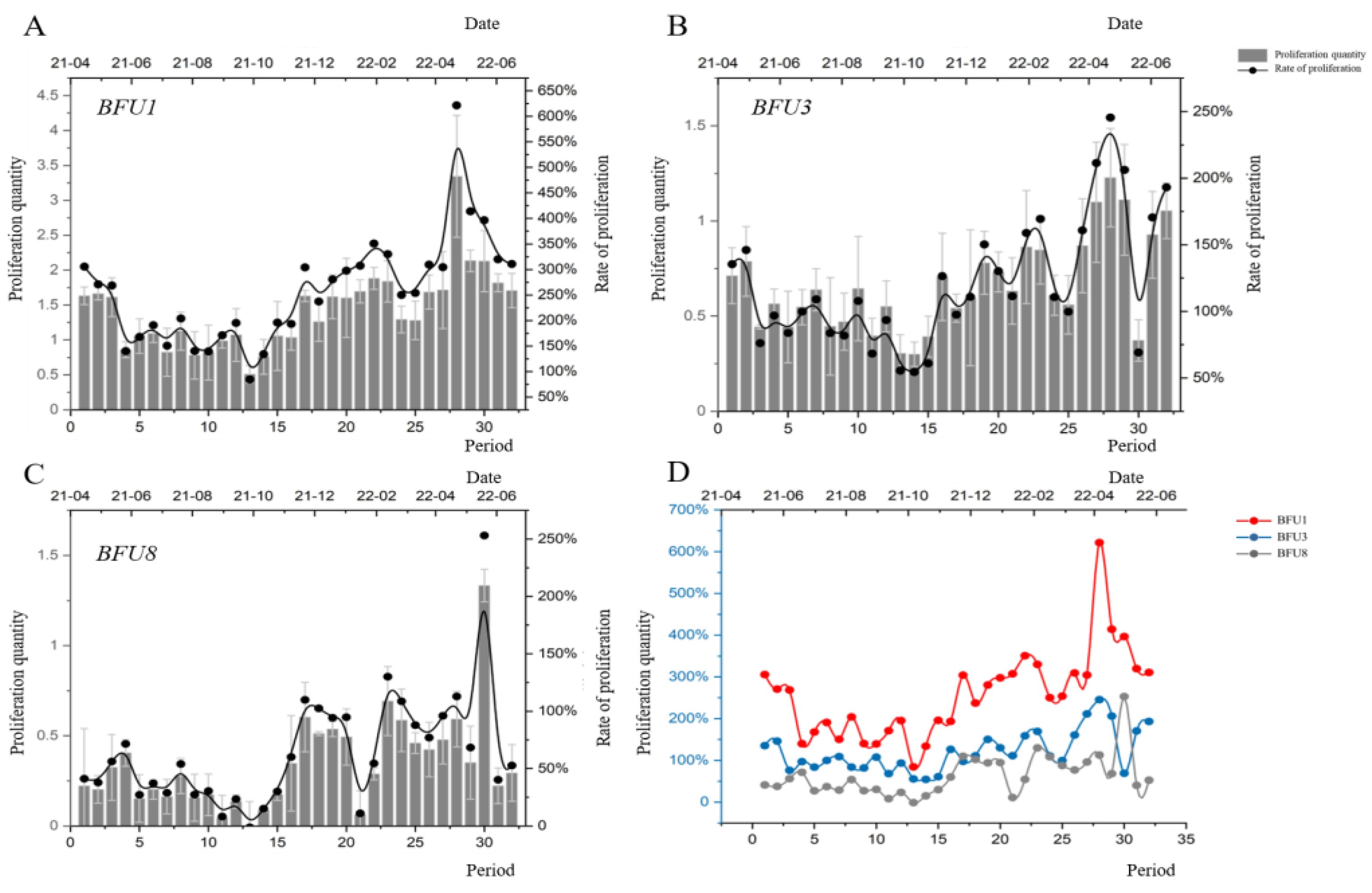
3.1. Proliferation Efficiency of Embryogenic Tissues during Long-Term Successional Subculture

Based on observations, BFU1 cell clusters exhibited a white color, dense and moist tissues, no surface protrusions, the fastest growth rate, and ease of separation in long-term cultures (Figure 1A,D). BFU3 cell clusters displayed a loose structure with filamentous protrusions, fast growth, and easy separability (Figure 1B,E). BFU8 cell-mass presented filamentous projections, stickiness, difficulty separating from surrounding tissues, and exhibited slow growth, internal browning, and a gradual loss of embryogenic properties over time (Figure 1C,F). Under microscopic examination, all three cell lines exhibited proembryogenic mass structures, with the embryogenic head consisting of densely nucleated cell aggregations and the embryogenic stalk consisting of elongated vesicularized cells.
The three embryogenic cell lines exhibited diverse growth patterns across 32 successive cultures. BFU1 demonstrated an average proliferation of 3.95 g, an average proliferation rate of 418%, with a peak average proliferation of 7.93 g and a maximum average proliferation rate of 746% in a single cycle. It also showed a minimum proliferation of 2.46 g with a minimum proliferation rate of 232% (Figure 2A). Both BFU3 and BFU8 displayed notably lower proliferation compared to BFU1. Analyzing data by month and quarter revealed BFU1’s significant decrease in proliferation at the end of the second quarter, stability in the subsequent quarters, and notable increases in the first and second quarters (January and May) (Figure 2A). BFU3 demonstrated decreased proliferation at the end of the second quarter, remaining stable thereafter (Figure 2B), while BFU8 exhibited a decline in June, August, and September, followed by an increase in the fourth quarter and relative stability until the end of the first quarter (Figure 2C). Comparing the three lines throughout the experiment, BFU1 consistently showed higher proliferation weight and rates than BFU3 and BFU8, with BFU1 and BFU3 maintaining robust proliferation abilities even by the 32nd cycle. Conversely, BFU8’s proliferation significantly decreased by the 26th cycle, resulting in a significant reduction in the quantity of embryogenic tissues (Figure 2D). All three lines displayed dynamic changes in proliferation across culture cycles. As succeeding generations increased, the rate of value addition decreased for all three lines, highlighting substantial variability in embryogenic organization among genotypes.

3.2. Proliferation Efficiency of Embryogenic Tissues on Pre-Treatment Medium for Long-Term Successional Subculture

After 10 days of proliferation, embryogenic tissues were transferred to a pre-treatment medium supplemented with 2 g L−1-activated charcoal for 10 days to neutralize hormonal effects and guide tissue development towards embryos. Throughout 32 consecutive culture cycles, the average proliferation at the pre-treatment stage stood at 1.42 g for BFU1, 0.66 g for BFU3, and 0.34 g for BFU8. BFU1 exhibited a maximum average proliferation of 3.34 g, peaking in the 22nd, 23rd, 28th, 29th, and 30th cycles, and notable declines in the 7th, 9th, 10th, 13th, and 14th cycles (Figure 3A). BFU3 and BFU8 also experienced significant proliferation decreases in cycles 11, 13, 14, 15, and 30 (Figure 3B,C). When assessing data by month and quarter, BFU1 showed a declining trend in June, August, and September, followed by an increase in the fourth quarter and stability until the end of the first quarter (Figure 3A). BFU3 displayed decreased proliferation in May, July, and October, an increase in the fourth quarter, and subsequent fluctuations in March and May of the following year (Figure 3B). Similarly, BFU8 exhibited declining trends in June and September, followed by decreases in the fourth quarter, and further fluctuations in March and May of the subsequent year, experiencing an increase in the fourth quarter and subsequent fluctuations in January and April–May of the following year (Figure 3C).
A comprehensive comparison of the three embryogenic cell lines revealed that throughout the experiment, BFU1 exhibited the highest pre-treatment proliferation weight and rate, followed by BFU3 and then BFU8 in descending order (Figure 3D). Notably, BFU1 displayed significantly higher pre-treatment proliferation weight and rates compared to BFU3 and BFU8. The pre-treatment proliferation rates of all three cell lines exhibited noticeable fluctuations, albeit differing in magnitude. However, the trends and monthly fluctuations appeared similar. Furthermore, all three lines showed an inclination towards increased pre-treatment proliferation rates with each successive cycle, notably higher in the final cycles compared to the initial stages of culture. Despite variations in the magnitude of fluctuations, the general trend across months remained alike, indicating an overall tendency for increased pre-treatment proliferation rates with successive cycles, resulting in significantly higher proliferation rates in the final cycles compared to the initial stages of culture.

3.3. Maturation Capacity of Long-Term Successional Subculture

Throughout successive cultures, the maturation capacity of the three embryogenic lines exhibited noticeable fluctuations, generally declining over subsequent cycles (Figure 4A–C). Notably, BFU8 eventually lost its capacity to mature into embryos. In a comprehensive comparison, the maturation capacity across the entire experimental period ranked as BFU1 > BFU3 > BFU8, with BFU1 demonstrating significantly superior maturation capability compared to BFU3 and BFU8 (Figure 4D). BFU1 and BFU3 retained their maturation abilities even in the 32nd cycle, whereas BFU8 lost this capacity starting from the 26th cycle. Despite varying magnitudes, the fluctuations and trends in maturation capacity among the three cell lines appeared to be similar across months, collectively indicating a substantial declining trend in their maturation capabilities.

3.4. Correlation Analysis of the Number of Successive Generations, Proliferation Efficiency during Proliferative Culture, Proliferation Efficiency of Pre-Treatment, and Maturation Capacity

Throughout the continuous relay culture of the three embryogenic cell lines, no significant correlation emerged between the number of relays and proliferation efficiency in the proliferative culture. However, a positive correlation was observed between the number of relays and proliferation efficiency during pre-treatment, BFU1 showing the highest correlation with pre-treatment proliferation rates (Table 2). Additionally, a noteworthy negative correlation was found between the number of substitutions and the maturation capacity. These findings unveil a complex relationship between the number of substitutions and the proliferation efficiency and maturation capacity of cell lines during subculture.
Throughout successive cultures, BFU1 exhibited a positive correlation between its proliferation rate and pre-treatment proliferation rate, while the pre-treatment proliferation rate showed a negative correlation with maturation capacity. Conversely, BFU3 demonstrated no significant correlation between its proliferative rate, pre-treatment proliferation rate, and maturation capacity. On the other hand, BFU8 displayed no notable correlation between its proliferative rate and pre-treatment proliferation rate, but exhibited a positive correlation with maturation capacity. The proliferation rate of BFU8 did not show a significant correlation with the pre-treatment proliferation rate, yet positively correlated with maturation capacity, indicating that higher proliferation rates corresponded to increased maturation capacity in embryogenic tissues across cycles, without a significant relationship between pre-treatment proliferation rate and maturation capacity (Table 3). These findings underscore distinct correlations between proliferation and maturation abilities across various cell lines during secondary culture.

3.5. SSR Genetic Stability Analysis

Long-term cultivation of embryogenic tissues may induce polymorphic variations, leading to detectable changes in electrophoretic bands when the same SSR primer is applied at different stages. Initial electrophoresis outcomes for F42, F1, and SSR11 primers were inconclusive, prompting their exclusion from subsequent experiments, while genetic stability was assessed using seven SSR primer pairs: F94, Lar_eSSR111, Lar_eSSR115, Lar_eSSR78, Lar_eSSR54, Lar_eSSR11, and SSR12. Embryogenic tissues from BFU1, BFU3, and BFU8 lines were sampled at the 1st, 7th, 17th, 25th, and 32nd cycles of consecutive culture. BFU1 and BFU3 lines, which had undergone two years of continuous proliferation, served as control materials. BFU8 embryogenic tissues, due to declining proliferation efficiency, were unavailable for sampling after two years of consecutive proliferation.
The experimental outcomes are illustrated in Figure 5. In the analysis involving seven primer sets, materials from different cultivation cycles of BFU1, BFU3, and BFU8 consistently exhibited distinct and stable bands without the presence of polymorphic fragments. This observation suggests that the genomic regions targeted with SSR molecular markers in embryogenic tissues did not undergo genetic variations during the extended period of successive cultivation, indicating a state of relative genetic stability.

4. Discussion

Embryogenic tissue subculture and proliferation are essential for somatic embryogenesis and scalable propagation of forest trees, especially when combined with bioreactor technologies. This study found significant variations in the proliferation rates of three embryogenic cell lines during the semi-solid cycle, with proliferation coefficients ranging from 2.5 to 7.5. Uniform subculture and proliferation methods showed varying efficiencies across coniferous embryogenic tissue genotypes, including Picea abies [45], Araucaria Angustifolia [46], Pinus taeda L. [47], Pinus pinaster [48], and Pinus pinea L. Most embryogenic tissues exhibited genotype-dependent dynamic stability in proliferation efficiency during long-term culture. The BFU1 and BFU3 cell lines maintained stable proliferative capabilities throughout the 15-month subculture period examined in this study. Conversely, BFU8 showed a marked decline in proliferation efficiency and severe tissue browning by the 12th month, indicating significant physiological changes like rapid aging due to peroxidation. Browning, a common phenomenon in long-term tissue culture [12], particularly affects conifers by gradually reducing embryogenic structures and somatic embryogenesis capacity [49,50]. To address this issue, Breton et al. found that a maltose-based, PGR-free medium could mitigate the loss of early embryogenic potential in maritime pine [51]. Adding glucose, sucrose (5 g/L), and fructose (10 g/L) to the growth medium controlled browning in Taxus brevifolia callus tissues [52]. Therefore, when selecting cell lines for scalable production, it is essential to consider both proliferation efficiency and long-term stability, employing anti-browning measures and other necessary interventions to delay tissue senescence and maintain proliferation stability.
Long-term subculturing leading to the decline or loss of embryogenic tissue’s embryogenesis capacity significantly challenges the somatic embryogenesis of coniferous trees, posing a major barrier to commercial application. In coniferous trees, embryogenic tissue can maintain its potential for embryo development stably over a certain period, which is closely related to the species. In species like Pinus koraiensis [39], Larix kaempferi [53], Pinus pinaster [25], and Pinus massoniana [54], embryogenic tissues start to lose somatic embryogenesis capacity, and mature embryo yield decreases after 5–6 months of proliferation culture, with increased subculture frequency. Conversely, embryogenic tissues in hybrid larch [55], Pinus nigra Arn [56], and Abies hybrids [20] can produce somatic embryos even after 2–3 years of proliferation; Norway spruce can maintain them even longer [57]. This study found Prince Rupprecht’s larch’s embryogenic tissues maintained dynamic stability in embryogenesis capacity for 12 months of subculture. After 12 months, embryogenesis capacity declined, a process dependent on genotype, with the BFU8 cell line losing its ability entirely after this period. Studies indicate somatic embryos are observable in early stages of culture, yet maturation across all genotypes is not guaranteed [58,59]. The PEM to somatic embryo transition is critical, determining mature embryo yield and quality, and thus affecting plant production [60]. Embryogenesis capacity loss may arise from increased embryogenic tissue heterogeneity during subculturing [24]. Excessive culture duration often leads to embryogenesis capacity loss and abnormal embryo production, as aging embryogenic tissues during proliferation diminish somatic embryo yield and quality, reducing germination rates [61]. Currently, a more effective solution involves selecting superior cell lines for cryopreservation at peak embryogenic potential, thus minimizing variations and preserving the developmental potential of embryogenic materials [62,63].
Plant tissue culture, especially somatic embryogenesis, is vital for preserving the stability and uniformity of regenerative plant traits. However, long-term tissue culture can cause genetic, epigenetic, and physiological changes, reducing regenerative capacity and impacting developmental integrity [64]. A study on embryogenic cell lines (ECLs) of Pinus pinaster revealed genetic variations via SSR loci during subculture, with abnormal phenotypes observed in some plants regenerated from somatic embryos [65]. Similarly, high SSR sequence mutation rates were found in Pinus sylvestris embryogenic cell lines and cotyledonary embryos [66]. In contrast, no mutations at SSR loci occurred from PEM to cotyledonary embryos in Picea abies [67,68], mirroring our findings where Prince Rupprecht’s larch’s embryogenic tissues showed no genetic variations at detected loci over extended subculturing, indicating stable genetic characteristics. This genetic stability supports the asexual reproduction of superior Prince Rupprecht’s larch germplasm. Despite the absence of significant genetic variations in long-term cultured embryogenic tissues, the decline in embryogenic capacity might result from multiple factors, with phenotypic variations potentially closely linked to changes in epigenetic modifications. Increasing subculture frequency and duration can augment variations in somatic clonal lines, with DNA methylation variations possibly being a key reason for the loss of differentiation capacity in long-term cultures [69]. In Elaeis guineensis [37] and Quercus alba [70], increased DNA methylation with subculturing leads to embryogenic tissue differentiation loss. Additionally, the reduction and/or loss of somatic embryo production capacity may relate to cell sensitivity to exogenous growth substances or endogenous hormone levels [71]. In Larix sibirica, long-term proliferation culture led to increased IAA and decreased ABA levels, both trends unfavorable for somatic embryo maturation [72]. Long-term proliferation in maritime pine led to lower IAA levels and higher zeatin riboside levels than in newly induced embryogenic tissues [25]. Similarly, long-term proliferation in Picea balfouriana increased endogenous zeatin riboside levels, significantly changing antioxidant enzyme activities and polyamine levels [73]. Future research should delve into these factors’ interrelations and ways to boost embryogenic capacity through culture environment and genetic engineering, aiming to reduce adverse variations and ensure plant germplasm stability. This approach will offer more reliable support for scalable breeding of superior plant germplasm.

5. Conclusions

In summary, during long-term subculture, Prince Rupprecht’s larch embryogenic tissues generally demonstrated reduced proliferation efficiency and maturation capacity. However, despite the long-term culture duration, these tissues retained their capacity to generate embryos. Our analysis of genetic stability in embryogenic cell lines of Prince Rupprecht’s larch revealed no detectable genetic variation over the extended subculture period. This suggests that the observed phenotypic changes during subculture are not primarily attributable to genetic alterations. These findings offer valuable insights into the dynamics of long-term culture of embryogenic tissues in Prince Rupprecht’s larch. They lay a foundation for optimizing somatic embryogenesis techniques in this species and potentially other coniferous species, facilitating large-scale propagation, long-term germplasm conservation, and genetic transformation.

Author Contributions

X.C., C.L. and X.W. contributed to the manuscript writing. Conceived and designed the experiments: J.Z. (Jian Zhao), J.Z. (Jinfeng Zhang), L.K. and X.C. Performed the experiments: X.C. and X.W. Analyzed the data: C.L. and D.Y. Contributed reagents/materials/analysis tools: H.Z. and L.Z. Writing—original draft preparation: X.C. Writing—review and editing: J.Z. (Jian Zhao). All authors have read and agreed to the published version of the manuscript.

Funding

This work was supported by the National Natural Science Foundation of China (No. 32371834), the National Natural Science Foundation of China (No. 32271836), Key R&D Program of Heibei Province, China (20326333D).

Data Availability Statement

Data are contained within the article.

Conflicts of Interest

The authors declare no conflicts of interest.

References

  1. Zhao, J.; Li, H.; Fu, S.; Chen, B.; Sun, W.; Zhang, J.; Zhang, J. An iTRAQ-Based Proteomics Approach to Clarify the Molecular Physiology of Somatic Embryo Development in Prince Rupprecht’s Larch (Larix principis-rupprechtii Mayr). PLoS ONE 2015, 10, e0119987. [Google Scholar] [CrossRef] [PubMed]
  2. Diao, S.; Hou, Y.; Xie, Y.; Sun, X. Age Trends of Genetic Parameters, Early Selection and Family by Site Interactions for Growth Traits in Larix kaempferi Open-Pollinated Families. BMC Genet. 2016, 17, 104. [Google Scholar] [CrossRef] [PubMed]
  3. Fernando, D.D.; Long, S.M.; Sniezko, R.A. Sexual Reproduction and Crossing Barriers in White Pines: The Case between Pinus lambertiana (Sugar Pine) and P. monticola (Western White Pine). Tree Genet. Genomes 2005, 1, 143–150. [Google Scholar] [CrossRef]
  4. Rampart, M. Seed Germination Variation among Crop Years from a Pinus sylvestris Clonal Seed Orchard. Asian J. Res. Agric. For. 2019, 3, 1–14. [Google Scholar] [CrossRef]
  5. Liu, X.; Wang, X.; Wang, D. Assessment of Tree-Ring Mercury Radial Translocation and Age Effect in Masson Pine: Implications for Historical Atmospheric Mercury Reconstruction. J. Environ. Sci. 2024, 138, 266–276. [Google Scholar] [CrossRef]
  6. Rubilar, R.; Bozo, D.; Albaugh, T.; Cook, R.; Campoe, O.; Carter, D.; Allen, H.L.; Álvarez, J.; Pincheira, M.; Zapata, Á. Rotation-Age Effects of Subsoiling, Fertilization, and Weed Control on Radiata Pine Growth at Sites with Contrasting Soil Physical, Nutrient, and Water Limitations. For. Ecol. Manag. 2023, 544, 121213. [Google Scholar] [CrossRef]
  7. Pérez-Oliver, M.A.; González-Mas, M.D.C.; Renau-Morata, B.; Arrillaga, I.; Sales, E. Heat-Priming during Somatic Embryogenesis Increased Resilience to Drought Stress in the Generated Maritime Pine (Pinus pinaster) Plants. Int. J. Mol. Sci. 2023, 24, 9299. [Google Scholar] [CrossRef]
  8. Hassani, S.B.; Trontin, J.-F.; Raschke, J.; Zoglauer, K.; Rupps, A. Constitutive Overexpression of a Conifer WOX2 Homolog Affects Somatic Embryo Development in Pinus pinaster and Promotes Somatic Embryogenesis and Organogenesis in Arabidopsis Seedlings. Front. Plant Sci. 2022, 13, 838421. [Google Scholar] [CrossRef] [PubMed]
  9. Chen, Y.-M.; Fei, Q.; Xia, X.-R.; Ke, X.; Ye, J.-R.; Zhu, L.-H. Pinus massoniana Somatic Embryo Maturation, Mycorrhization of Regenerated Plantlets and Its Resistance to Bursaphelenchus xylophilus. Front. Plant Sci. 2023, 14, 1130471. [Google Scholar] [CrossRef]
  10. Sales, E.; Cañizares, E.; Pereira, C.; Pérez-Oliver, M.A.; Nebauer, S.G.; Pavlović, I.; Novák, O.; Segura, J.; Arrillaga, I. Changing Temperature Conditions during Somatic Embryo Maturation Result in Pinus pinaster Plants with Altered Response to Heat Stress. Int. J. Mol. Sci. 2022, 23, 1318. [Google Scholar] [CrossRef]
  11. Durzan, D.J.; Chalupa, V. Growth and Metabolism of Cells and Tissue of Jack Pine (Pinus banksiana). 3. Growth of Cells in Liquid Suspension Cultures in Light and Darkness. Can. J. Bot. 1976, 54, 456–467. [Google Scholar] [CrossRef]
  12. Arrillaga, I.; Morcillo, M.; Zanón, I.; Lario, F.; Segura, J.; Sales, E. New Approaches to Optimize Somatic Embryogenesis in Maritime Pine. Front. Plant Sci. 2019, 10, 138. [Google Scholar] [CrossRef] [PubMed]
  13. Castander-Olarieta, A.; Montalbán, I.A.; Moncaleán, P. Multi-Strategy Approach towards Optimization of Maturation and Germination in Radiata Pine Somatic Embryogenesis. Plant Cell Tissue Organ Cult. 2023, 153, 173–190. [Google Scholar] [CrossRef]
  14. do Nascimento, A.M.M.; Polesi, L.G.; Back, F.P.; Steiner, N.; Guerra, M.P.; Castander-Olarieta, A.; Moncaleán, P.; Montalbán, I.A. The Chemical Environment at Maturation Stage in Pinus Spp. Somatic Embryogenesis: Implications in the Polyamine Profile of Somatic Embryos and Morphological Characteristics of the Developed Plantlets. Front. Plant Sci. 2021, 12, 771464. [Google Scholar] [CrossRef] [PubMed]
  15. Jiang, S.; Chen, X.; Gao, Y.; Cui, Y.; Kong, L.; Zhao, J.; Zhang, J. Plant Regeneration via Somatic Embryogenesis in Larix principis-rupprechtii Mayr. Forests 2021, 12, 1335. [Google Scholar] [CrossRef]
  16. Li, F.; Yao, J.; Hu, L.; Chen, J.; Shi, J. Multiple Methods Synergistically Promote the Synchronization of Somatic Embryogenesis Through Suspension Culture in the New Hybrid Between Pinus elliottii and Pinus caribaea. Front. Plant Sci. 2022, 13, 857972. [Google Scholar] [CrossRef] [PubMed]
  17. Lineros, Y.; Rojas-Rioseco, M.; Hernández, M.; Ríos, D.; Muñoz, X.; Hasbún, R. Effects of Long-Term Subculture on Maturation Ability and Plant Conversion in Pinus Radiata: Using FT-IR Spectroscopy to Determine Biomarkers of Embryogenic Tissue Aging. Forests 2023, 14, 1446. [Google Scholar] [CrossRef]
  18. Maruyama, T.E.; Hosoi, Y. Progress in Somatic Embryogenesis of Japanese Pines. Front. Plant Sci. 2019, 10, 31. [Google Scholar] [CrossRef] [PubMed]
  19. Peng, C.; Gao, F.; Wang, H.; Nikolaevna Tretyakova, I.; Mikhaylovich Nosov, A.; Shen, H.; Yang, L. Suspension Culture and Somatic Embryogenesis of Korean Pine. Phyton 2022, 91, 223–238. [Google Scholar] [CrossRef]
  20. Salaj, T.; Klubicová, K.; Matusova, R.; Salaj, J. Somatic Embryogenesis in Selected Conifer Trees Pinus nigra Arn. and Abies Hybrids. Front. Plant Sci. 2019, 10, 13. [Google Scholar] [CrossRef]
  21. Alvarez, J.M.; Bueno, N.; Cañas, R.A.; Avila, C.; Cánovas, F.M.; Ordás, R.J. Analysis of the WUSCHEL-RELATED HOMEOBOX Gene Family in Pinus pinaster: New Insights into the Gene Family Evolution. Plant Physiol. Biochem. 2018, 123, 304–318. [Google Scholar] [CrossRef]
  22. Kang, Y.; Li, W.; Zhang, L.; Qi, L. Over-Expression of the Cell-Cycle Gene LaCDKB1;2 Promotes Cell Proliferation and the Formation of Normal Cotyledonary Embryos during Larix kaempferi Somatic Embryogenesis. Genes 2021, 12, 1435. [Google Scholar] [CrossRef]
  23. Peng, C.; Gao, F.; Tretyakova, I.N.; Nosov, A.M.; Shen, H.; Yang, L. Transcriptomic and Metabolomic Analysis of Korean Pine Cell Lines with Different Somatic Embryogenic Potential. Int. J. Mol. Sci. 2022, 23, 13301. [Google Scholar] [CrossRef]
  24. Breton, D.; Harvengt, L.; Trontin, J.F.; Bouvet, A.; Favre, J.M. Long-Term Subculture Randomly Affects Morphology and Subsequent Maturation of Early Somatic Embryos in Maritime Pine. Plant Cell Tissue Organ Cult. 2006, 87, 95–108. [Google Scholar] [CrossRef]
  25. Klimaszewska, K.; Noceda, C.; Pelletier, G.; Label, P.; Rodriguez, R.; Lelu-Walter, M.A. Biological Characterization of Young and Aged Embryogenic Cultures of Pinus pinaster (Ait.). Vitr. Cell. Dev. Biol. Plant 2009, 45, 20–33. [Google Scholar] [CrossRef]
  26. Neelakandan, A.K.; Wang, K. Recent Progress in the Understanding of Tissue Culture-Induced Genome Level Changes in Plants and Potential Applications. Plant Cell Rep. 2012, 31, 597–620. [Google Scholar] [CrossRef]
  27. Zayova, E.; Vassilevska-Ivanova, R.; Stoeva, D.; Kraptchev, B. Somaclonal Variation through Indirect Organogenesis in Eggplant (Solanum melongena L.). Biol. Divers. Conserv. 2010, 3, 1–5. [Google Scholar]
  28. Hume, S.; Bo, D.; Simon, S.A.; Suhua, F.; Maria, B.; Matteo, P.; Guo-Liang, W.; Meyers, B.C.; Jacobsen, S.E. Plants Regenerated from Tissue Culture Contain Stable Epigenome Changes in Rice. eLife 2013, 2, e00354. [Google Scholar]
  29. George, E.F. Plant Propagation by Tissue Culture—Part 1: The Technology; Exegetics Limited: Westbury, UK, 1993. [Google Scholar]
  30. Kaeppler, S.M.; Kaeppler, H.F.; Rhee, Y.; Kaeppler, S.M.; Kaeppler, H.F.; Rhee, Y. Epigenetic Aspects of Somaclonal Variation in Plants. Plant Mol. Biol. 2000, 43, 179–188. [Google Scholar] [CrossRef]
  31. Lee, M.; Phillips, R.L. The Chromosomal Basis of Somaclonal Variation. Plant Mol. Biol. 1988, 39, 413–437. [Google Scholar] [CrossRef]
  32. Etienne, H.; Bertrand, B. Somaclonal Variation in Coffea arabica: Effects of Genotype and Embryogenic Cell Suspension Age on Frequency and Phenotype of Variants. Tree Physiol. 2003, 23, 419–426. [Google Scholar] [CrossRef]
  33. Pijut, P.M.; Beasley, R.R.; Lawson, S.S.; Palla, K.J.; Stevens, M.E.; Wang, Y. In Vitro Propagation of Tropical Hardwood Tree Species—A Review (2001–2011). Propag. Ornam. Plants 2012, 12, 25–51. [Google Scholar]
  34. Sharma, S.K.; Bryan, G.J.; Millam, W.S. Stability of Potato (Solanum tuberosum L.) Plants Regenerated via Somatic Embryos, Axillary Bud Proliferated Shoots, Microtubers and True Potato Seeds: A Comparative Phenotypic, Cytogenetic and Molecular Assessment. Planta 2007, 226, 1449–1458. [Google Scholar] [CrossRef]
  35. Nwauzoma, A.B.; Jaja, E.T. A Review of Somaclonal Variation in Plantain (Musa Spp): Mechanisms and Applications. J. Appl. Biosci. 2013, 67, 5252–5260. [Google Scholar] [CrossRef]
  36. Bairu, M.W.; Fennell, C.W.; van Staden, J. The Effect of Plant Growth Regulators on Somaclonal Variation in Cavendish banana (Musa AAA Cv. ’Zelig’). Sci. Hort. Amst. 2006, 108, 347–351. [Google Scholar] [CrossRef]
  37. Rival, A.; Ilbert, P.; Labeyrie, A.; Torres, E.; Doulbeau, S.; Personne, A.; Dussert, S.; Beulé, T.; Durand-Gasselin, T.; Tregear, J.W. Variations in Genomic DNA Methylation during the Long-Term In Vitro Proliferation of Oil Palm Embryogenic Suspension Cultures. Plant Cell Rep. 2013, 32, 359–368. [Google Scholar] [CrossRef] [PubMed]
  38. Egertsdotter, U. Plant Physiological and Genetical Aspects of the Somatic Embryogenesis Process in Conifers. Scand. J. For. Res. 2019, 34, 360–369. [Google Scholar] [CrossRef]
  39. Peng, C.; Gao, F.; Wang, H.; Shen, H.; Yang, L. Optimization of Maturation Process for Somatic Embryo Production and Cryopreservation of Embryogenic Tissue in Pinus koraiensis. Plant Cell Tissue Organ Cult. 2021, 144, 185–194. [Google Scholar] [CrossRef]
  40. Ree, J.F.; Polesi, L.G.; Back, F.; Bertolazi, A.A.; Guerra, M.P. Aging Peach Palm (Bactris gasipaes Kunth) Cultures Lose Embryogenic Potential and Metabolic Cellular Function Due to Continuous Culture in Hypoxic Environments. Plant Cell Tissue Organ Cult. 2019, 140, 49–67. [Google Scholar] [CrossRef]
  41. Sun, T.; Wang, Y.; Zhu, L.; Liu, X.; Wang, Q.; Ye, J. Evaluation of Somatic Embryo Production during Embryogenic Tissue Proliferation Stage Using Morphology, Maternal Genotype, Proliferation Rate and Tissue Age of Pinus thunbergii Parl. J. For. Res. 2022, 33, 445–454. [Google Scholar] [CrossRef]
  42. Dong, M.; Gao, J.; Sun, W.; Fan, Y.; Yuan, D.; Zhang, J. Genetic Diversity and Population Structure of Elite Larix principis-rupprechtii Mayr in Beijing. J. Plant Genet. Resour. 2016, 17, 616–624. [Google Scholar] [CrossRef]
  43. Fan, Y. Genetic Diversity of Larix principis rupprechtii Revealed by SSR Marker; Beijing Forestry University: Beijing, China, 2014. [Google Scholar]
  44. Dong, M. High-Density Genetic Linkage Map Construction and QTLs Analysis of Phenotypic Traits in Larix principis-rupprechtii Mayr; Beijing Forestry University: Beijing, China, 2020. [Google Scholar]
  45. Businge, E.; Brackmann, K.; Moritz, T.; Egertsdotter, U. Metabolite Profiling Reveals Clear Metabolic Changes during Somatic Embryo Development of Norway Spruce (Picea abies). Tree Physiol. 2012, 32, 232–244. [Google Scholar] [CrossRef]
  46. Leonardo, J.; Dos, S.A.L.W.; Bueno, C.A.; Barbosa, H.R.; Floh, E.I.S. Proteomic Analysis and Polyamines, Ethylene and Reactive Oxygen Species Levels of Araucaria angustifolia (Brazilian pine) Embryogenic Cultures with Different Embryogenic Potential. Tree Physiol. 2014, 34, 94–104. [Google Scholar]
  47. Pullman, G.S.; Namjoshi, K.; Zhang, Y. Somatic Embryogenesis in Loblolly Pine (Pinus taeda L.): Improving Culture Initiation with Abscisic Acid and Silver Nitrate. Plant Cell Rep. 2003, 22, 85–95. [Google Scholar] [CrossRef]
  48. Miguel, C.; Gonçalves, S.; Tereso, S.; Marum, L.; Maroco, J.; Oliveira, M.M. Somatic Embryogenesis from 20 Open-Pollinated Families of Portuguese Plus Trees of Maritime Pine. Plant Cell Tissue Organ Cult. 2004, 76, 121–130. [Google Scholar] [CrossRef]
  49. Quiroz-Figueroa, F.; Méndez-Zeel, M.; Sánchez-Teyer, F.; Rojas-Herrera, R.; Loyola-Vargas, V.M. Differential Gene Expression in Embryogenic and Non-Embryogenic Clusters from Cell Suspension Cultures of Coffea arabica. J. Plant Physiol. 2002, 159, 1267–1270. [Google Scholar] [CrossRef]
  50. Zhang, S.; Zhou, J.; Han, S.; Yang, W.; Li, W.; Wei, H.; Li, X.; Qi, L. Four Abiotic Stress-Induced miRNA Families Differentially Regulated in the Embryogenic and Non-Embryogenic Callus Tissues of Larix leptolepis. Biochem. Biophys. Res. Commun. 2010, 398, 355–360. [Google Scholar] [CrossRef]
  51. Breton, D.; Harvengt, L.; Trontin, J.F.; Bouvet, A.; Favre, J.M. High Subculture Frequency, Maltose-Based and Hormone-Free Medium Sustained Early Development of Somatic Embryos in Maritime Pine. Vitr. Cell. Dev. Biol. Plant 2005, 41, 494–504. [Google Scholar] [CrossRef]
  52. Khosroushahi, A.Y.; Naderi-Manesh, H.; Simonsen, H.T. Effect of Antioxidants and Carbohydrates in Callus Cultures of Taxus brevifolia: Evaluation of Browning, Callus Growth, Total Phenolics and Paclitaxel Production. Bioimpacts Bi 2011, 1, 37. [Google Scholar]
  53. Li, W.F.; Zhang, S.G.; Han, S.Y.; Wu, T.; Zhang, J.H.; Qi, L.W. Regulation of LaMYB33 by miR159 during Maintenance of Embryogenic Potential and Somatic Embryo Maturation in Larix kaempferi (Lamb.) Carr. Plant Cell Tissue Organ Cult. 2013, 113, 131–136. [Google Scholar] [CrossRef]
  54. Xia, X.-R.; Yang, F.; Ke, X.; Chen, Y.-M.; Ye, J.-R.; Zhu, L.-H. Somatic Embryogenesis of Masson Pine (Pinus massoniana): Initiation, Maturation and Genetic Stability Analysis at SSR Loci. Plant Cell Tissue Organ Cult. 2021, 145, 667–677. [Google Scholar] [CrossRef]
  55. Lelu-Walter, M.A.; Paques, L.E. Simplified and Improved Somatic Embryogenesis of Hybrid Larches (Larix × eurolepis and Larix × marschlinsii). Perspectives for Breeding. Ann. For. Sci. 2009, 66, 104. [Google Scholar] [CrossRef]
  56. Klubicová, K.; Uvácková, L.; Danchenko, M.; Nemecek, P.; Skultéty, L.; Salaj, J.; Salaj, T. Insights into the Early Stage of Pinus nigra Arn. Somatic Embryogenesis Using Discovery Proteomics. J. Proteom. 2017, 169, 99–111. [Google Scholar] [CrossRef]
  57. Klimaszewska, K.; Overton, C.; Stewart, D.; Rutledge, R.G. Initiation of Somatic Embryos and Regeneration of Plants from Primordial Shoots of 10-Year-Old Somatic White Spruce and Expression Profiles of 11 Genes Followed during the Tissue Culture Process. Planta 2011, 233, 635–647. [Google Scholar] [CrossRef]
  58. Santos, A.L.W.D.; Steiner, N.; Guerra, M.P.; Zoglauer, K.; Moerschbacher, B.M. Somatic Embryogenesis in Araucaria angustifolia. Biol. Plant. 2008, 52, 195–199. [Google Scholar] [CrossRef]
  59. Santos, A.L.W.D.; Jo, L.; Santa-Catarina, C.; Guerra, M.P.; Floh, E.I.S. Somatic Embryogenesis in Brazilian Pine: Establishment of Biochemical Markers for Selection of Cell Lines with High Embryogenic Potential. In Proceedings of the IUFRO Working Party 2.09.02 Conference “Integrating Vegetative Propagation, Biotechnologies and Genetic Improvement for Tree Production and Sustainable Forest Management”, Brno, Czech Republic, 25–28 June 2012. [Google Scholar]
  60. Bozhkov, P.V.; Filonova, L.H.; von Arnold, S. A Key Developmental Switch during Norway Spruce Somatic Embryogenesis Is Induced by Withdrawal of Growth Regulators and Is Associated with Cell Death and Extracellular Acidification. Biotechnol. Bioeng. 2002, 77, 658–667. [Google Scholar] [CrossRef] [PubMed]
  61. Klimaszewska, K.; Hargreaves, C.; Lelu-Walter, M.A.; Trontin, J.F. Advances in Conifer Somatic Embryogenesis Since Year 2000. In In Vitro Embryogenesis in Higher Plants, Methods in Molecular Biology; Springer Science + Business Media: New York, NY, USA, 2016. [Google Scholar] [CrossRef]
  62. Ahn, Y.E. Propagation and Cryopreservation of Ulleungdo Hemlock (Tsuga ulleungensis) via Somatic Embryogenesis. Trees. Struct. Funct. 2018, 32, 1801–1808. [Google Scholar] [CrossRef]
  63. Varis, S.; Ahola, S.; Jaakola, L.; Aronen, T. Reliable and Practical Methods for Cryopreservation of Embryogenic Cultures and Cold Storage of Somatic Embryos of Norway Spruce. Cryobiology 2017, 76, 8–17. [Google Scholar] [CrossRef] [PubMed]
  64. Brar, D.S.; Jain, S.M. Somaclonal Variation: Mechanism and Applications in Crop Improvement. In Somaclonal Variation and Induced Mutations in Crop Improvement; Springer: Dordrecht, Netherlands, 1998. [Google Scholar] [CrossRef]
  65. Marum, L.; Rocheta, M.; Maroco, J.; Oliveira, M.M.; Miguel, C. Analysis of Genetic Stability at SSR Loci during Somatic Embryogenesis in Maritime Pine (Pinus pinaster). Plant Cell Rep. 2009, 28, 673–682. [Google Scholar] [CrossRef]
  66. Bozhkov, K.B.A.H.P.; Arnold, S. Developmental and Genetic Variation in Nuclear Microsatellite Stability during Somatic Embryogenesis in Pine. J. Exp. Bot. 2007, 58, 687–698. [Google Scholar]
  67. Andreas, H.; Sara, V.A.; Kornel, B.; Bozhkov, P.V. High Stability of Nuclear Microsatellite Loci during the Early Stages of Somatic Embryogenesis in Norway Spruce. Tree Physiol. 2004, 24, 1181–1186. [Google Scholar]
  68. Harvengt, L.; Trontin, J.F.; Reymond, I.; Canlet, F.; Paques, M. Molecular Evidence of True-to-Type Propagation of a 3-Year-Old Norway Spruce through Somatic Embryogenesis. Planta 2001, 213, 828–832. [Google Scholar] [CrossRef]
  69. Valledor, L.; Hasbún, R.; Meijón, M.; Rodríguez, J.L.; Santamaría, E.; Viejo, M.; Berdasco, M.; Feito, I.; Fraga, M.F.; Rodríguez, R.; et al. Involvement of DNA methylation in tree development and development micropropagation. Plant Cell Tissue Organ Cult. 2007, 91, 75–86. [Google Scholar] [CrossRef]
  70. Corredoira, E.; Cano, V.; Bárány, I.; Solís, M.-T.; Rodríguez, H.; Vieitez, A.-M.; Risueño, M.C.; Testillano, P.S. Initiation of Leaf Somatic Embryogenesis Involves High Pectin Esterification, Auxin Accumulation and DNA Demethylation in Quercus alba. J. Plant Physiol. 2017, 213, 42. [Google Scholar] [CrossRef]
  71. Yang, X.; Zhang, X.; Yuan, D.; Jin, F.; Zhang, Y.; Xu, J. Transcript Profiling Reveals Complex Auxin Signalling Pathway and Transcription Regulation Involved in Dedifferentiation and Redifferentiation during Somatic Embryogenesis in Cotton. BMC Plant Biol. 2012, 12, 110. [Google Scholar] [CrossRef]
  72. Tretyakova, I.N.; Kudoyarova, G.R.; Park, M.E.; Kazachenko, A.S.; Shuklina, A.S.; Akhiyarova, G.R.; Korobova, A.V.; Veselov, S.U. Content and Immunohistochemical Localization of Hormones during in Vitro Somatic Embryogenesis in Long-Term Proliferating Larix sibirica Cultures. Plant Cell Tissue Organ Cult. 2019, 136, 511–522. [Google Scholar] [CrossRef]
  73. Li, Q.; Deng, C.; Zhu, T.; Ling, J.; Zhang, H.; Kong, L.; Zhang, S.; Wang, J.; Chen, X. Dynamics of Physiological and miRNA Changes after Long-Term Proliferation in Somatic Embryogenesis of Picea balfouriana. Trees 2019, 33, 469–480. [Google Scholar] [CrossRef]
Figure 1. Embryogenic tissue and staining of three embryogenic lines. (A,D) BFU1, (B,E) BFU3, (C,F) BFU8. Bars (AC) = 1000 µm, (DF) = 50 µm.
Forests 15 00627 g001
Figure 2. Proliferation Analysis and Comparison of BFU1, BFU3, and BFU8 Cell Lines. (AC) Proliferation weight and rate of BFU1, BFU3, and BFU8. (D) Comparison of proliferation rates of BFU1, BFU3, and BFU8.
Forests 15 00627 g002
Figure 3. Pre-treated Proliferation Analysis and Comparison of BFU1, BFU3, and BFU8 Cell Lines. (AC) Pre-treated proliferation weight and rate of BFU1, BFU3, and BFU8. (D) Comparison of pre-treated proliferation rates of BFU1, BFU3, and BFU8.
Forests 15 00627 g003
Figure 4. Maturity Capacity Analysis of BFU1, BFU3, and BFU8 Cell Lines. (AC) Maturity capacity of BFU1, BFU3, and BFU8. (D) Comparison of maturity capacity of BFU1, BFU3, and BFU8.
Forests 15 00627 g004
Figure 5. Separation of different primers in embryogenic tissues of Prince Rupprecht’s larch at Different Stages. (1–6 are tissue sample from the 1st, 7th, 17th, 25th, and 32nd cycles of BFU1 and continuously cultivated for two years; 7–12 are tissue sample from the 1st, 7th, 17th, 25th, and 32nd cycles of BFU3 and continuously cultivated for two years; 13–17 are tissue samples from the 1st, 7th, 17th, 25th, and 32nd cycles of BFU8; M is the marker).
Forests 15 00627 g005
Table 1. Pairs of SSR primers.
NumberPrimer NameSequenceRepetitive UnitTm (°C)Fragment Size (bp)References
1F42F:AACACTTTAATCCCCTCCC(CAGGAA)551190–220[43]
R:CTGGACCTGATACTCCTCTTC
2F94F:GCCGTTGACAACAATTACAT(ACTGG)555150–180[43]
R:AAAGAATAGCAACCCGCAGT
3F1F:AAGGAGGAGGGTCAGGGAA(TCAGGC)555160–190[43]
R:CATGCGGAGGTTGAGTGTG
4SSR11F:ATGGGTTTGACAGCGGATAA(GCA)655240–260[42]
R:CGTGTTCTTGTTTTGGGTGG
5Lar_eSSR111F:GATATCAACTCCCTGCGGAA(CCTGAA)455230[44]
R:AGCTGTGAGCGAGAGAGAGG
6Lar_eSSR115F:TTGTGATGCTTCTTTGACCG(TTGTCT)455239[44]
R:GAGGCAGATAGAGGGCTTCC
7Lar_eSSR78F:CAATCCGATAAAACGCCATC(GTGTCG)455262[44]
R:CAGTAACACTCCCGCCTAGC
8Lar_eSSR54F:GCGCGCTCTTCTTTTCTCT(ACCCGC)655162[44]
R:CGCCGTCGACTGTATAACCT
9Lar_eSSR11F:AATCCAAATTTCTGGACCCC(CAAGGG)456239[44]
R:CCTGCAAAAAGAGGATAGCG
10SSR12F:GCCTTCGCTGATCTGTTT(AC)6(AT)655170–190[42]
R:TTTCCTCTTGACGGACTCTT
Table 2. Correlation between culture period and proliferation efficiency and maturation capacity.
NameVariateCorrelationProliferation RatePre-Treated Proliferation RateMaturation Capacity
BFU1Culture periodpertinence0.2830.622 **−0.409 *
significance0.117<0.0010.020
BFU3pertinence0.2770.562 **−0.427 *
significance0.1250.0010.015
BFU8pertinence−0.3330.511 **−0.621 **
significance0.0630.003<0.001
** At level 0.01 (two-tailed), the correlation was significant. * At level 0.05 (two-tailed), the correlation was significant.
Table 3. The correlation between proliferation rate of proliferation culture, proliferation rate of pre-treatment, and maturation capacity.
NameVariateCorrelationProliferation RatePre-Treated Proliferation RateMaturation Capacity
BFU1Proliferation ratepertinence10.411 *0.025
significance--0.0190.890
Pre-treated
proliferation rate
pertinence0.411 *1−0.513 **
significance0.019--0.003
BFU3Proliferation ratepertinence10.1780.056
significance--0.3300.763
Pre-treated
proliferation rate
pertinence0.1781−0.340
significance0.330--0.057
BFU8Proliferation ratepertinence1−0.1060.404 *
significance--0.5620.022
Pre-treated
proliferation rate
pertinence−0.1061−0.264
significance0.562--0.145
** At level 0.01 (two-tailed), the correlation was significant. * At level 0.05 (two-tailed), the correlation was significant.
Disclaimer/Publisher’s Note: The statements, opinions and data contained in all publications are solely those of the individual author(s) and contributor(s) and not of MDPI and/or the editor(s). MDPI and/or the editor(s) disclaim responsibility for any injury to people or property resulting from any ideas, methods, instructions or products referred to in the content.

