Article

Establishment of a Transient Transformation Protocol in Cinnamomum camphora

1
Zhejiang Provincial Key Laboratory of Forest Aromatic Plants-Based Healthcare Functions, Zhejiang A&F University, Hangzhou 311300, China
2
State Key Laboratory of Subtropical Silviculture, Zhejiang A&F University, Hangzhou 311300, China
3
Tianjin Key Laboratory of Animal and Plant Resistance, College of Life Sciences, Tianjin Normal University, Tianjin 300387, China
*
Author to whom correspondence should be addressed.
Forests 2023, 14(9), 1872; https://doi.org/10.3390/f14091872
Submission received: 25 August 2023 / Revised: 11 September 2023 / Accepted: 13 September 2023 / Published: 14 September 2023

Abstract

:
Cinnamomum camphora is an excellent evergreen broad-leaved tree species with strong stress tolerance, but its molecular character revelation as well as ecological and economic value improvement were limited due to the lack of a genetic transformation system. To establish a simple and efficient transient transformation system for uncovering the molecular mechanism of plant tolerating stresses and promoting the selective breeding of good varieties, the infection method, co-cultivation time, infection solution concentration, and growth density of Agrobacterium tumefaciens containing green fluorescent protein (GFP)-based calmodulin protein 3 gene (GCaMP3) were identified by monitoring the fluorescence emitted from GCaMP3 bound to Ca2+. Meanwhile, the transient transformation effects were evaluated via cytoplasmic Ca2+ concentration variations at high temperatures of 35 °C and 40 °C. When C. camphora leaves were infected with A. tumefaciens containing GCaMP3 via injection and soaking, no significant difference was detected in the fluorescence intensity over 48 h, indicating that the two infection methods had the same transient transformation efficiency. By prolonging the co-cultivation time, the fluorescence intensity gradually increased, reached its strongest at the 48th h, and then gradually declined. For the infection solution concentration, an OD600 of 0.7 led to the strongest fluorescence intensity, with an increase of 42.2%, 13.7%, 4.2%, and 14.2%, respectively, compared to that at OD600 of 0.5, 0.6, 0.8, and 0.9. When A. tumefaciens growth density OD600 was 0.5–0.7, the strongest fluorescence intensity was detected after transient transformation. Combining these optimum conditions, GCaMP3 was transferred into C. camphora, which indicated the variations in cytoplasmic Ca2+ concentration at high temperatures, with the fluorescence intensity at 35 °C and 40 °C increasing by 12.6% and 30.6%, respectively, in contrast to that at 28 °C. Therefore, it should be an efficient transient transformation system for C. camphora, with A. tumefaciens growth density OD600 of 0.5–0.7, infection solution concentration OD600 of 0.7, and co-cultivation time of 48 h by using both injection and soak infection methods, which is beneficial for uncovering the Ca2+ signal transduction in the plant tolerating stresses and promoting its molecular biology development and selective breeding of good varieties.

1. Introduction

Stable plant genetic transformation is a technology that can transfer exogenous genes into receptors via different methods to obtain transgenic plants, which plays an important role in gene function identification and genetic breeding. This technology mainly depends on the plant regeneration ability and gene introduction efficiency [1]. To date, it has been successfully applied to many herbs and crops, such as Miscanthus sinensis, wheat (Triticum aestivum), soybean (Glycine max), and cabbage (Brassica oleracea) [1,2,3,4]. In contrast to these plants, this technology is rarely applied to woody plants, except for Populus, due to its high heterozygosity and long breeding time [5,6].
Compared to stable genetic transformation, transient transformation exhibits a number of advantages, such as short periods, high efficiency, labor, and saving time [7]. There are many types of transient transformation, including Agrobacterium-mediated gene transformation, particle bombardment, electroporation, and poly-ethylene glycol-mediated transformation [8,9]. Particle bombardment can transfer exogenous genes into a broad range of plant species, but it requires expensive and special equipment [10]. For electroporation and poly-ethylene glycol-mediated transformation, high-vitality protoplasts are used as the receptors for exogenous genes. However, protoplast isolation is complicated, and it is hard to obtain a stable yield [11]. As an alternative approach, Agrobacterium-mediated gene transformation is widely used in transient assays with several advantages, such as straightforward operation, low cost, and easily obtaining transformation materials [12,13]. This technology was first developed in tobacco (Nicotiana tabacum) [14] and further applied to Arabidopsis thaliana, rice (Oryza sativa), tomato (Solanum lycopersicum), etc. [15,16,17]. However, its application in woody plants is slower than that in herbs. In recent years, many efforts were made to improve Agrobacterium-mediated gene transformation efficiency, which promoted its application in woody plants, such as Populus, apple (Malus pumila), tea plants (Camellia sinensis), and Citrus [18,19,20,21].
Cinnamomum camphora (L.) Presl. belongs to the genus Cinnamomum in Lauraceae, which is an excellent evergreen broad-leaved tree species with strong stress tolerance in subtropical regions. It not only exhibits ecological value by being used as landscaping tree species but also exhibits great economic value by extracting essential oil and using it as furniture material [22,23]. This species exhibits extensive genetic diversity, and simple sequence repeat (SSR) markers [24], expressed sequence tag-simple sequence repeat (EST-SSR) markers [25], and genome-wide single-nucleotide polymorphism (SNP) [26] have been developed for diversity analysis. However, research on other molecular biology aspects (especially stress tolerance) of the plant is very slow, which limits the improvement of the plant’s ecological and economic value due to genetic breeding without transgenic assistance. Although a cold resistance gene is transferred to the embryogenic calluses of C. camphora, a large number of calluses are false positives, resulting in transgenic calluses not being screened out [27]. C. camphora contained lots of phenols and oxidases, which caused serious browning problems during the culturing of transgenic calluses [28]. Then, it is hard to identify gene functions by using a stable genetic transformation method in plants. Transient transformation is a powerful tool for analyzing the gene expression in the original species, but it has not been established in C. camphora until now. This is not beneficial for promoting the research on C. camphora molecular biology, although plant genome sequencing has already been completed [29].
Green fluorescent protein (GFP)-based calmodulin protein 3 (GCaMP3) contains a circularly permutated enhanced GFP flanked by calmodulin (CaM) and myosin light-chain kinase (M13), which shows green fluorescence in the absence of Ca2+ [30]. To date, this Ca2+ reporter gene GCaMP3 has been used to uncover Ca2+ signal transduction in several herbs, such as A. thaliana and duckweed [31,32]. To establish a simple and efficient Agrobacterium-mediated transient transformation system in C. camphora by using GCaMP3 as the reporter gene, we identified the infection method, co-cultivation time, infection solution concentration and growth density of Agrobacterium tumefaciens GV3101 containing GCaMP3 during transient transformation process in this study. To the best of our knowledge, this is the first report of a transient transformation protocol in plants, which is not only beneficial for promoting the research on Ca2+ signal transduction in C. camphora tolerating stresses but also beneficial for promoting plant molecular biology development and selective breeding of good varieties.

2. Materials and Methods

2.1. Plant Material

Three-year-old C. camphora seedlings with a height of 80 cm were kept in an illumination incubator, and the regime was 16 h light at 28 °C and 8 h dark at 25 °C, with a light intensity of 200 μmol·m−2·s−1. The relative humidity was 65 ± 5%. After adaptation for 15 days, the 3rd to 5th healthy leaves from the top were used for the transient transformation.

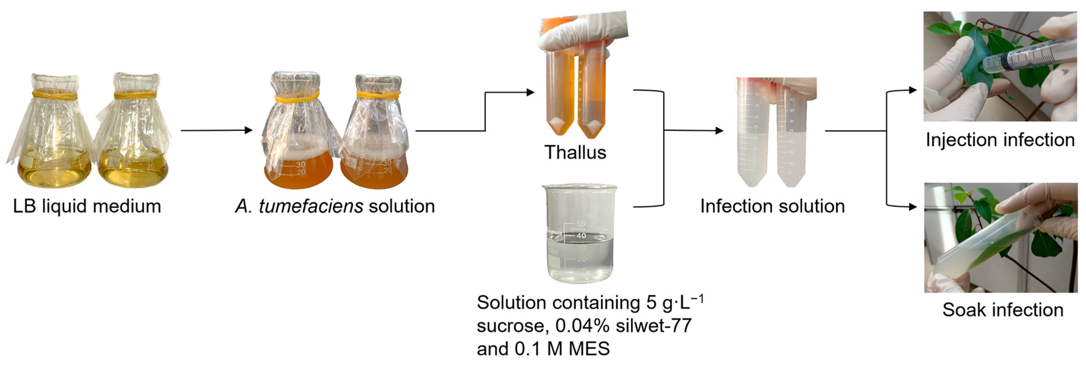
2.2. Preparation for Infection Solution

A. tumefaciens GV3101 containing GCaMP3 was provided by Dr. Lin Yang at Tianjin Normal University, China, and the structure of GCaMP3 plasmid is described in detail by Ren et al. [31]. The bacterium was activated 3 times on a Luria–Bertani (LB) solid plate containing 50 mg·L−1 kanamycin, 25 mg·L−1 rifampicin, and 25 mg·L−1 streptomycin at 28 °C. A single colony was randomly selected and kept in a 10 mL LB liquid medium with the same antibiotics at 28 °C for 18 h. Then, 1 mL of bacterial solution was transferred into a 20 mL of fresh LB liquid medium, and its OD value was recorded every 2 h to draw the growth curve. When the cell density reached a given concentration, they were harvested by centrifugation at 5000× g and used to prepare the infection solution by resuspending them in a solution containing 5 g·L−1 of sucrose, 0.04% of silwet-77, and 0.1 M of 2-morpholinoethanesulfonic acid (MES). After that, the infection solution was used for injection and soak infection (Figure 1), which are the two main methods used in Agrobacterium-mediated transient transformation.

2.3. Infection Methods

2.3.1. Injection Infection

C. camphora leaves were washed twice with sterile water, and the cuticle on the lower surfaces of the leaves was scraped off using a syringe needle without damaging the lower epidermis. The above A. tumefaciens in the logarithmic phase with an OD600 of 0.7 was used to prepare the infection solution, and the solution concentration was indicated by the bacterial cell concentration with an OD600 of 0.8. The infection solution was injected into the leaves through the scraped area by using a syringe without a needle, and the color around the injection site became dark. Then, the infected leaves were covered with a piece of silver paper to maintain darkness, and the plant was kept in an illumination incubator under normal culture conditions. After 12, 24, and 48 h, the fluorescence intensity and GCaMP3 gene in the infected leaves were detected.

2.3.2. Soak Infection

C. camphora leaves were washed with sterile water and the cuticle was scraped off using a blade without damaging the lower epidermis. The infection solution was prepared following the injection method, and the scraped leaves were soaked in the solution for 20 min [31]. After being covered with a piece of silver paper, the plant was kept in an illumination incubator under normal culture conditions, and the fluorescence intensity and GCaMP3 gene in the infected leaves were detected after 12, 24, and 48 h.

2.4. Determination of Optimum Co-Cultivation Time, Infection Solution Concentration, and A. tumefaciens Growth Density

To determine the optimum co-cultivation time, an OD600 infection solution concentration of 0.6 was prepared with the A. tumefaciens OD600 growth density of 0.7. After the soak infection step, C. camphora was kept in an illumination incubator under normal culture conditions, and the fluorescence intensity was measured after 12, 24, 48, 60, and 72 h.
To determine the optimum infection solution concentration, the A. tumefaciens OD600 growth density of 0.6 was used to prepare the infection solution, with the OD600 concentration of 0.5, 0.6, 0.7, 0.8, and 0.9, respectively. After the soak infection step, C. camphora was kept in an illumination incubator for 48 h, and the fluorescence intensity was measured.
To determine the optimum growth density of A. tumefaciens, the OD600 infection solution concentration of 0.7 was separately prepared using the bacterium at OD600 growth densities of 0.3, 0.5, 0.7, 0.9, and 1.1. After the soak infection step, C. camphora was kept in an illumination incubator for 48 h, and the fluorescence intensity was measured.

2.5. Assay of Cytoplasmic Ca2+ Concentration under High Temperature

To evaluate the actual effects of the transient transformation protocol, the OD600 A. tumefaciens with a growth density of 0.5 was used to prepare the infection solution, with an OD600 concentration of 0.7. This solution was used to infect C. camphora leaves via soaking, and then the plants were kept in three illumination incubators under normal culture conditions for 48 h, with 4 plants in each incubator. For high-temperature treatment, the temperature in the two illumination incubators was separately increased to 35 °C and 40 °C for 30 min, while the temperature in the other illumination incubator was maintained at 28 °C as the control. Then, the fluorescence intensity was detected, which indicated the cytoplasmic Ca2+ concentration.

2.6. PCR Analysis

The genomic DNA from C. camphora leaves was extracted following the cetyltrimethylammonium bromide (CTAB) method [33] using a DNA extraction kit (Beijing Kulaibo Technology Co., Ltd., Beijing, China). Polymerase chain reaction (PCR) was carried out using a PCR amplification kit (Takara Biotechnology Co., Ltd., Beijing, China). The cloning system contained 1.25 U Taq polymerase, 100 ng DNA, and 20 μM primers (forward primer: 5′-ATGGGTTCTCATCATCATCATCAT-3′, reverse primer: 5′-TCACTTCGCTGTCATCATTTGTA-3′). The amplification procedure was as follows: 94 °C for 30 s, 55 °C for 30 s, and 72 °C for 90 s. After 30 cycles, the amplified PCR fragments were analyzed via electrophoresis on a 1.5% agarose gel.

2.7. Fluorescence Intensity Measurement

In the successfully infected leaf cells, the expressed protein GCaMP3 bound to cytoplasmic Ca2+ and showed green fluorescence. This fluorescence was observed by using a fluorescence microscope (Olympus BX51, Olympus Corporation, Tokyo, Japan) at an excitation wavelength of 488 nm and an emission wavelength of 507 nm. The fluorescence intensity was analyzed using ImageJ (1.50i) software.

2.8. Statistical Analyses

At least 4 seedlings were used in each treatment, with each as a biological replicate, and 3 leaves were infected in a seedling, with each as a technological replicate. The statistical analyses among the treatments with significant differences at p < 0.05 were carried out by using Origin 8.0 according to the Tukey test in one-way analysis of variance (ANOVA).

3. Results and Discussion

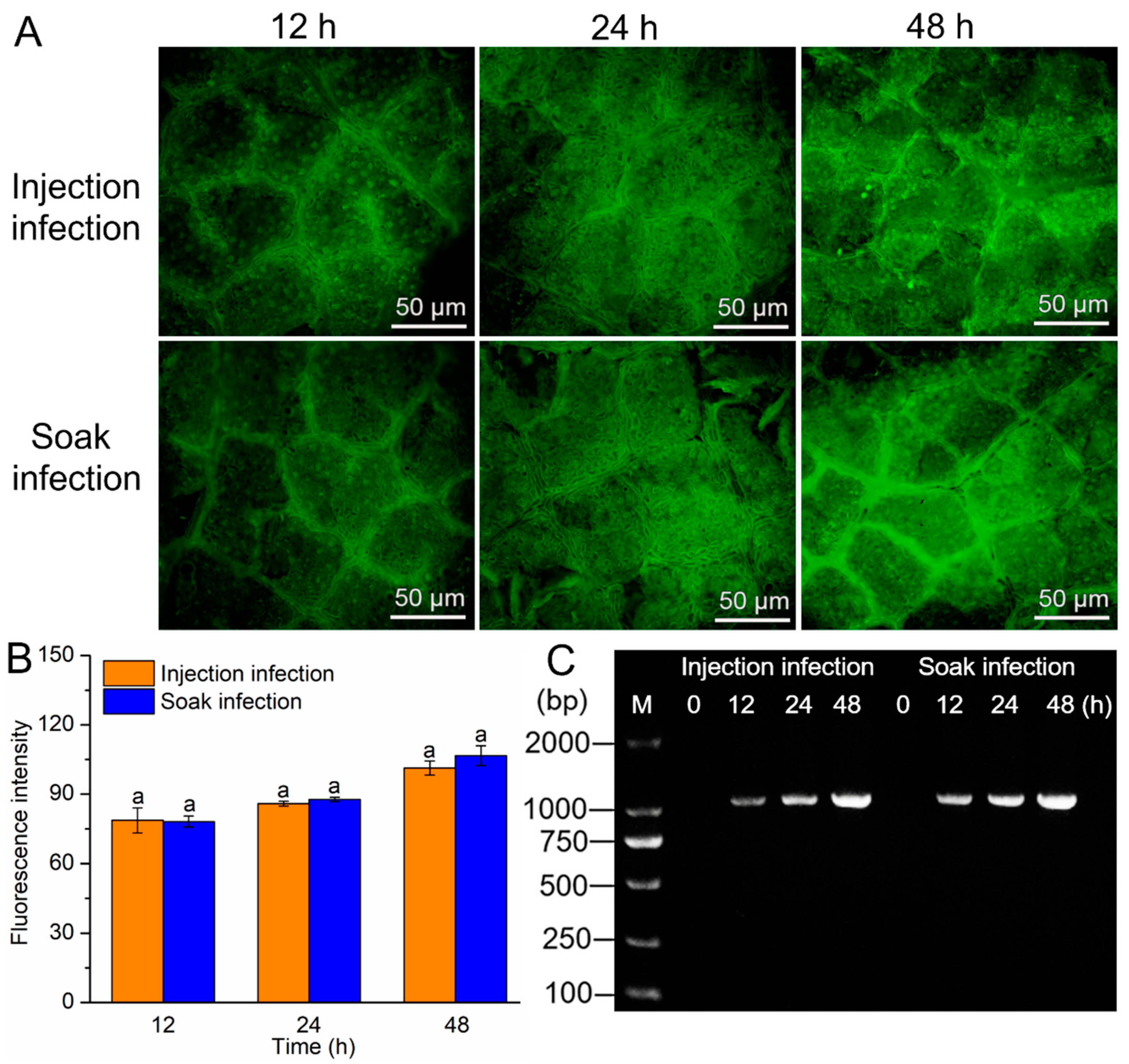
3.1. Effects of Infection Methods on the Transformation Efficiency

In the cells, GCaMP3 is mainly located in the cytoplasm, which combines with Ca2+ to produce a green fluorescence [31,32]. When A. thaliana and duckweed were transformed with GCaMP3 using A. tumefaciens GV3101, green fluorescence was observed in the cells and veins [31,32]. Similar results were also observed in the present study. When C. camphora leaves were transformed with GCaMP3 by injection and soak infection, green fluorescence was detected after 12 h, and the fluorescence intensity gradually increased by prolonging the co-cultivation time (Figure 2A). During the 48 h, there were no significant differences in the fluorescence intensity between the two infection methods (Figure 2B). However, no fluorescence was detected in the leaves infected with A. tumefaciens GV3101 with an empty vector using the two infection methods (Supplementary Figure S1). After PCR amplification using specific primers, the products of GCaMP3 (1321 bp) showed bright electrophoretic bands during co-cultivation for 12, 24, and 48 h after injection and soak infection (Figure 2C). These results indicate that GCaMP3 was successfully transferred into C. camphora leaf cells and the two infection methods had the same transformation efficiency.
Infiltration is the process of substances penetrating from the surface into the depth of plant tissues, which is specially applied in transient transformation mediated by Agrobacterium [34]. This process can be spontaneously performed under normal atmospheric pressure or forcedly performed by generating a pressure difference between the lamina surface and the inside of the leaves. In transient transformation, spontaneous infiltration is considered to be a less efficient manner than forced infiltration [35,36]. In this study, spontaneous infiltration and forced infiltration were carried out by soak infection and injection infection, respectively, but no difference was detected in the infection effects between the two methods (Figure 2). Compared to injection infection, there was a large leaf area in contact with A. tumefaciens during soak infection, which should result in many bacterial cells entering C. camphora leaves to improve infection efficiency. C. camphora produces an abundance of phenolic compounds [37,38], which can facilitate the expression of vir genes in A. tumefaciens to promote the transfer of T-DNA into the leaf cells [39,40]. During soak infection, the large contact area of C. camphora leaves might provide more phenolic compounds to promote the transfer of GCaMP3 into leaf cells, resulting in a high transient transformation efficiency.

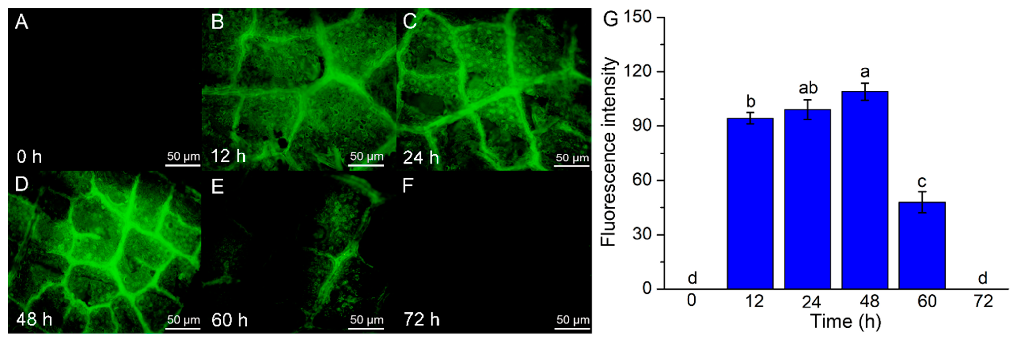
3.2. Effects of Co-Cultivation Time on the Transformation Efficiency

When C. camphora was infected with A. tumefaciens via soak infection and co-cultivated at 28 °C, green fluorescence was detected at the 12th h, and its intensity gradually increased by prolonging the co-cultivation time. The strongest fluorescence intensity was detected at the 48th h, after which it gradually declined with increasing co-cultivation time, and it disappeared at the 72nd h (Figure 3).
The expression of exogenous genes is usually detected within 12 h after transient transformation, and the expression levels gradually increase by prolonging the co-cultivation time due to the gradual increase in exogenous genes entering the plant cells. However, the exogenous genes only exist for a short time, usually 3–5 days, in plant cells, as they are lost when the cells are divided [41,42]. In the present study, the fluorescence intensity in C. camphora disappeared after 72 h (Figure 3), which may have been caused by the loss of the GCaMP3 gene during plant cell division.
The optimum expression time varies in different plants. When pineapple (Ananas comosus) calluses and Chinese birch (Betula platyphylla) leaves were transformed with GFP using A. tumefaciens, the strongest fluorescence intensity was detected on the third day of co-cultivation [43,44]. For apricot (Armeniaca vulgaris) calluses, the strongest fluorescence intensity was detected on the fourth day [45]. When avocado (Persea americana) callus was transformed with EGFP (enhanced green fluorescent gene) and DsRed (red fluorescent gene) using particle bombardment, the maximum fluorescence intensity was detected at the 24th and 72nd h, respectively, suggesting that the exogenous genes might also affect their expression in plant cells [46]. In this study, the strongest fluorescence intensity was detected at the 48th h, indicating that this should be the optimum co-cultivation time (Figure 3).

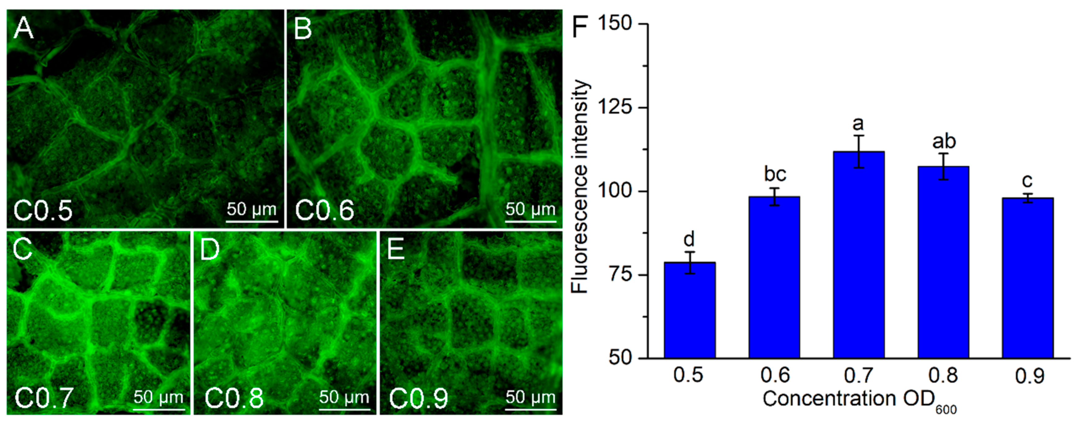
3.3. Effects of Infection Solution Concentration on the Transformation Efficiency

When C. camphora leaves were infected with A. tumefaciens at an OD600 infection solution concentration of 0.5, 0.6, 0.7, 0.8, and 0.9, the fluorescence intensity gradually increased with increasing concentration and reached the highest level at a concentration of 0.7. At this concentration, the fluorescence intensity increased by 42.2% (p < 0.05), 13.7% (p < 0.05), 4.2%, and 14.2% (p < 0.05), respectively, compared to that at OD600 of 0.5, 0.6, 0.8 and 0.9 (Figure 4).
When OD600 infection concentration was 0.2 and 0.6, A. tumefaciens showed maximum transient transformation efficiency in sweetgum (Liquidambar formosana) and Quercus robur, respectively [47,48]. For P. davidiana × P. bollena, the maximum transient transformation efficiency was detected at A. tumefaciens OD600 concentration of 0.8 [49]. These results indicate that the receptor plants need specific Agrobacterium concentrations to obtain the maximum transformation efficiency due to their different sensitivity to the bacterium [50]. High concentrations of Agrobacterium always lower the transient transformation efficiency, as a high density of Agrobacterium may result in inhibition of the recipient cell respiration and even cause death of the bacterial cells for their competition [51,52]. This may be the reason for the low transformation efficiency of high infection concentration (especially OD600 of 0.9) in C. camphora, and OD600 of 0.7 may be the optimum infection concentration for high A. tumefaciens numbers and low adverse effects (Figure 4).

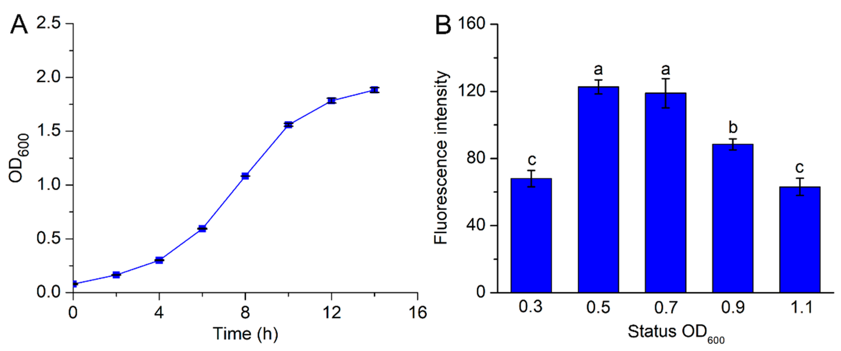
3.4. Effects of A. tumefaciens Growth Density on the Transformation Efficiency

Agrobacterium growth undergoes four growth stages, including lag phase, logarithmic phase, stationary phase, and decline phase [53]. The logarithmic phase is considered the most vigorous period for Agrobacterium, and the bacterium in this period is always used to transfer exogenous genes to plants [54]. For A. rhizogenes R1000, the logarithmic phase was at OD600 = 0.8–1.0 [55]. A. tumefaciens EHA105 and LBA4404 started to enter the logarithmic phase at 0.3 and 0.5, and ended at 2.4 and 2.7, respectively [56,57]. In the present study, A. tumefaciens GV3101 was in the logarithmic phase at an OD600 of 0.5–1.5 (Figure 5A). This indicates that different Agrobacterium species and strains have different growth rates.
Agrobacterium is not suitable for gene transformation during the lag phase due to its low activity. However, it has very high activity in the middle and later logarithmic phases and is still not suitable for gene transformation, as the bacterium produces more toxins to disrupt the plant’s physiological functions and even cause cell death [58,59]. When C. camphora was infected with the infection solution (OD600 of 0.7) separately prepared using A. tumefaciens GV3101 with OD600 growth densities of 0.3, 0.5, 0.7, 0.9, and 1.1, the strongest fluorescence intensity was detected at OD600 of 0.5 and 0.7 without significant differences between them (Figure 5B). At an OD600 growth density of 0.5–0.7, A. tumefaciens GV3101 was in the early logarithmic phase (Figure 5A) and exhibited the strongest infection activity to C. camphora, which might be caused by the bacterium’s high activity and low toxicity to the plant.
In spinach (Spinacia oleracea), A. tumefaciens GV2260 with an OD600 concentration of 1.0 was the optimum growth density for transient transformation [60]. The highest transient transformation efficiency was found in Paeonia lactiflor callus and seedlings infected with A. tumefaciens EHA105 with OD600 of 0.6 and 1.2, respectively [61,62]. In the present study, the optimum OD600 growth density of A. tumefaciens GV3101 was 0.5–0.7 (Figure 5B). This might be caused by differences in the Agrobacterium strains and/or acceptor plants.

3.5. Effects of High Temperature on the Fluorescence Intensity

Ca2+ serves an important signaling function in plants that tolerate high temperatures, and it immediately accumulates in the cytoplasm when plants undergo heat shock; e.g., the cytoplasmic Ca2+ concentration in A. thaliana reached its first peak after heat shock for 10 s, and the second peak after 60 s [63]. Under high temperatures at 37 °C for 30 min, a considerable Ca2+ influx was detected in rice root cells, with a remarkable increase in cytoplasmic Ca2+ concentration [64]. In Ganoderma lucidum, a 2.5-fold increase in the cytoplasmic Ca2+ concentration was found when the plant was treated at a high temperature of 42 °C for 20 min [65].
Combining the optimum transient transformation conditions that were obtained, C. camphor leaves were infected with A. tumefaciens containing GCaMP3 with a bacterial OD600 growth density of 0.5, an OD600 infection solution concentration of 0.7 and a co-cultivation time of 48 h using soak infection. Compared to the control at 28 °C, the fluorescence intensity increased by 12.6% (p < 0.05) and 30.6% (p < 0.05) under high temperatures of 35 °C and 40 °C, respectively, indicating that the quantities of Ca2+ that entered the cytoplasm gradually increased with increasing temperature (Figure 6). This was consistent with the cytoplasmic Ca2+ concentration increase under high temperatures in previous studies [63,64,65], suggesting that this transient transformation system is efficient and can be applied to C. camphor.

4. Conclusions

When GCaMP3 was transferred to C. camphora leaves using A. tumefaciens through injection infection and soak infection, no significant differences were detected in the transient transformation efficiency between the two methods. During the transformation process, the optimum A. tumefaciens growth density, infection solution concentration and co-cultivation time were OD600 of 0.5–0.7, OD600 of 0.7, and 48 h, respectively. This transient transformation protocol combined with these optimum conditions exhibited effective effects that were indicated by the cytoplasmic Ca2+ variations in C. camphora under high temperatures, with a gradual increase in the fluorescence intensity with increasing temperature. This demonstrates that the protocol is suitable for the transient transformation of C. camphora, which is not only used to uncover the Ca2+ signal transduction in C. camphora tolerating stresses but also utilized in plant molecular biology research, promoting selective breeding of good varieties.

Supplementary Materials

The following supporting information can be downloaded at: https://www.mdpi.com/article/10.3390/f14091872/s1, Figure S1: Transformation effects of A. tumefaciens with empty vector and GCaMP3 on C. camphora during 48 h. When C. camphora was infected by A. tumefaciens GV3101 with empty vector (growth density OD600 of 0.5, infection concentration OD600 of 0.7) by using injection and soak infection methods, no fluorescence was detected during 48-h co-cultivation. However, the fluorescence intensity in C. camphora infected by A. tumefaciens with GCaMP3 gradually enhanced with prolonging the co-cultivation time.

Author Contributions

Methodology, L.Y. and Z.Z.; Investigation, S.Z. and R.H.; Writing—original draft, S.Z.; Writing—review & editing, Z.Z.; Supervision, Z.Z.; Funding acquisition, Z.Z. All authors have read and agreed to the published version of the manuscript.

Funding

This research was funded by the National Natural Science Foundation of China (Grant No. 32371830, 31870585), and the Opening Project of Zhejiang Provincial Key Laboratory of Forest Aromatic Plants-based Healthcare Functions (Grant No. 2022E10008).

Data Availability Statement

The data presented in this study are available upon request from the corresponding author.

Conflicts of Interest

The authors declare no conflict of interest.

References

  1. Lian, Z.; Nguyen, C.D.; Liu, L.; Wang, G.; Chen, J.; Wang, S.; Yi, G.; Wilson, S.; Ozias-Akins, P.; Gong, H.; et al. Application of developmental regulators to improve in-planta or in vitro transformation in plants. Plant Biotechnol. J. 2022, 20, 1622. [Google Scholar] [CrossRef] [PubMed]
  2. Wu, Y.F.; Zhou, N.; Ni, X.N.; Okoye, C.; Wang, Y.L.; Li, X.; Gao, L.; Zhou, G.K.; Jiang, J.X. Developing a long-term and powerful in vitro culture and Agrobacterium-mediated transformation system for Miscanthus sinensis (Poaceae). Ind. Crop Prod. 2021, 161, 113190. [Google Scholar] [CrossRef]
  3. Janani, C.; Sundararajan, B.; Kumari, B.D.R. Construction and transformation of peroxisome proliferator activated receptor gamma (RnPPARγ) gene using Agrobacterium tumefaciens into Glycine max L. Merr. Gene Rep. 2019, 16, 100427. [Google Scholar] [CrossRef]
  4. Li, X.N.; Li, H.Y.; Zhao, Y.Z.; Zong, P.X.; Zhan, Z.X.; Piao, Z.Y. Establishment of a simple and efficient Agrobacterium-mediated genetic transformation system to Chinese cabbage (Brassica rapa L. ssp. Pekinensis). Hortic. Plant J. 2021, 7, 117–128. [Google Scholar] [CrossRef]
  5. Giorgio, G.; Ivana, G. Genetic transformation of fruit trees: Current status and remaining challenges. Transgenic Res. 2012, 21, 1163–1181. [Google Scholar]
  6. Van Nocker, S.; Gardiner, S.E. Breeding better cultivars, faster: Applications of new technologies for the rapid deployment of superior horticultural tree crops. Hortic. Res. 2014, 1, 14022. [Google Scholar] [CrossRef]
  7. Pushpa, K.; Rinku, C.; Narendra, T.; Prashant, K. A genotype-independent, simple, effective and efficient in planta Agrobacterium-mediated genetic transformation protocol. Methods Psrotoc. 2022, 5, 69. [Google Scholar]
  8. Franz, L.; Anja, R.; Hannah, T.; Ralph, P. Gene gun-mediated transient gene expression for functional studies in plant immunity. Methods Mol. Biol. 2022, 2523, 63–77. [Google Scholar]
  9. Wang, Y.Y.; Zhang, Y.A.; Dong, Y.X.; Li, D.L.; Shi, S.L.; Li, S.H.; Li, L.Z.; He, Y.J.; Li, J.Y.; Chen, H.Y.; et al. A highly efficient mesophyll protoplast isolation and PEG-mediated transient expression system in eggplant. Sci. Hortic. 2022, 304, 111303. [Google Scholar] [CrossRef]
  10. Yang, Y.; Li, R.; Qi, M. In vivo analysis of plant promoters and transcription factors by agroinfiltration of tobacco leaves. Plant J. 2000, 22, 543–551. [Google Scholar] [CrossRef]
  11. Chen, X.J.; He, S.T.; Jiang, L.; Li, X.Z.; Guo, W.L.; Chen, B.H.; Zhou, J.G.; Skliar, V. An efficient transient transformation system for gene function studies in pumpkin (Cucurbita moschata D.). Sci. Hortic. 2021, 282, 110028. [Google Scholar] [CrossRef]
  12. Zottini, M.; Barizza, E.; Costa, A.; Formentin, E.; Ruberti, C.; Carimi, F.; Schiavo, F.L. Agroinfiltration of grapevine leaves for fast transient assays of gene expression and for long-term production of stable transformed cells. Plant Cell Rep. 2008, 27, 845–853. [Google Scholar] [CrossRef] [PubMed]
  13. Zhong, S.W.; Dong, B.; Zhou, J.; Miao, Y.F.; Yang, L.Y.; Wang, Y.G.; Xiao, Z.; Fang, Q.; Wan, Q.Q.; Zhao, H.B. Highly efficient transient gene expression of three tissues in Osmanthus fragrans mediated by Agrobacterium tumefaciens. Sci. Hortic. 2023, 310, 111725. [Google Scholar] [CrossRef]
  14. Fraley, R.T.; Rogers, S.G.; Horsch, R.B.; Sanders, P.R.; Flick, J.S.; Adams, S.P.; Bittner, M.L.; Brand, L.A.; Fink, C.L.; Fry, J.S.; et al. Expression of bacterial genes in plant cells. Proc. Natl. Acad. Sci. USA 1983, 80, 4803–4807. [Google Scholar] [CrossRef]
  15. Nam, J.; Mysore, K.S.; Zheng, C.; Knue, M.K.; Matthysse, A.G.; Gelvin, S.B. Identification of T-DNA tagged Arabidopsis mutants that are resistant to transformation by Agrobacterium. Mol. Gen. Genet. 1999, 261, 429–438. [Google Scholar] [CrossRef]
  16. Cho, J.H.; Lee, J.Y.; Kim, Y.W.; Lee, M.H.; Choi, S.H. Improvement of transformation efficiencies using Agrobacterium-mediated transformation of Korean rice. Korean J. Crop Sci. 2004, 49, 61–68. [Google Scholar]
  17. Vinoth, S.; Gurusaravanan, P.; Jayabalan, N. Optimization of factors influencing microinjection method for Agrobacterium tumefaciens-mediated transformation of tomato. Appl. Biochem. Biotechnol. 2013, 169, 1173–1187. [Google Scholar] [CrossRef]
  18. Lv, Q.R.; Chen, C.S.; Xu, Y.J.; Hu, S.K.; Wang, L.; Sun, K.; Chen, X.; Li, X.H. Optimization of Agrobacterium tumefaciens-mediated transformation systems in tea plant (Camellia sinensis). Hortic. Plant J. 2017, 3, 105–109. [Google Scholar] [CrossRef]
  19. Lv, Y.D.; Zhang, M.L.; Wu, T.; Wu, T.L.; Zhong, Y. The infiltration efficiency of Agrobacterium-mediated transient transformation in four apple cultivars. Sci. Hortic. 2019, 256, 108597. [Google Scholar] [CrossRef]
  20. Wen, S.S.; Ge, X.L.; Wang, R.; Yang, H.F.; Bai, Y.; Guo, Y.H.; Zhang, J.; Lu, M.Z.; Zhao, S.T.; Wang, L.Q. An efficient Agrobacterium-mediated transformation method for Hybrid Poplar 84K (Populus alba × P. glandulosa) using calli as explants. Int. J. Mol. Sci. 2022, 23, 2216. [Google Scholar] [CrossRef]
  21. Yang, W.H.; Ren, J.Q.; Liu, W.R.; Liu, D.; Xie, K.D.; Zhang, F.; Wang, P.W.; Guo, W.W.; Wu, X.M. An efficient transient gene expression system for protein subcellular localization assay and genome editing in citrus protoplasts. Hortic. Plant J. 2022, 9, 425–436. [Google Scholar] [CrossRef]
  22. Chen, C.H.; Zhong, Y.D.; Yu, F.X.; Xu, M. Deep sequencing identifies miRNAs and their target genes involved in the biosynthesis of terpenoids in Cinnamomum camphora. Ind. Crop. Prod. 2020, 145, 111853. [Google Scholar] [CrossRef]
  23. Ma, R.; Su, P.; Jin, B.; Guo, J.; Tian, M.; Mao, L.Y.; Tang, J.F.; Chen, T.; Lai, C.J.S.; Zeng, W.; et al. Molecular cloning and functional identification of a high-efficiency (+)-borneol dehydrogenase from Cinnamomum camphora (L.) Presl. Plant Physiol. Biochem. 2021, 158, 363–371. [Google Scholar] [CrossRef] [PubMed]
  24. Li, Z.; Zhong, Y.; Yu, F.; Xu, M. Novel SSR marker development and genetic diversity analysis of Cinnamomum camphora based on transcriptome sequencing. Plant Genet. Resour. 2018, 16, 568–571. [Google Scholar] [CrossRef]
  25. Zhong, Y.; Yang, A.; Li, Z.; Zhang, H.; Liu, L.; Wu, Z.; Li, Y.; Liu, T.; Xu, M.; Yu, F. Genetic diversity and population genetic structure of Cinnamomum camphora in South China revealed by EST-SSR markers. Forests 2019, 10, 1019. [Google Scholar] [CrossRef]
  26. Gong, X.; Yang, A.; Wu, Z.; Chen, C.; Li, H.; Liu, Q.; Yu, F.; Zhong, Y. Employing genome-wide SNP discovery to characterize the genetic diversity in Cinnamomum camphora using genotyping by sequencing. Forests 2021, 12, 1511. [Google Scholar] [CrossRef]
  27. Shi, X.P.; Dai, X.G.; Liu, G.F.; Zhang, J.W.; Ning, G.G.; Bao, M.Z. Cyclic secondary somatic embryogenesis and efficient plant regeneration in camphor tree (Cinnamomum camphora L.). In Vitro Cell. Dev. Biol.-Plant 2010, 46, 117–125. [Google Scholar] [CrossRef]
  28. El-Kader, E.M.A.; Serag, A.; Aref, M.S.; Ewais, E.E.A.; Farag, M.A. Metabolomics reveals ionones upregulation in MeJA elicited Cinnamomum camphora (camphor tree) cell culture. Plant Cell Tissue Organ Cult. 2019, 137, 309–318. [Google Scholar] [CrossRef]
  29. Shen, T.F.; Qi, H.R.; Luan, X.Y.; Xu, W.L.; Yu, F.X.; Zhong, Y.D.; Xu, M. The chromosome-level genome sequence of the camphor tree provides insights into Lauraceae evolution and terpene biosynthesis. Plant Biotechnol. J. 2022, 20, 244–246. [Google Scholar] [CrossRef]
  30. Krogman, W.; Sparks, J.A.; Blancaflor, E.B. Cell type-specific imaging of calcium signaling in Arabidopsis thaliana seedling roots using GCaMP3. Int. J. Mol. Sci. 2020, 21, 6385. [Google Scholar] [CrossRef]
  31. Ren, Q.; Xu, Z.; Xue, Y.; Yang, R.; Ma, X.; Sun, J.; Wang, J.; Lin, S.; Wang, W.; Yang, L.; et al. Mechanism of calcium signal response to cadmium stress in duckweed. Plant Signal. Behav. 2022, 17, 2119340. [Google Scholar] [CrossRef] [PubMed]
  32. Toyota, M.; Spencer, D.; Sawai-Toyota, S.; Wang, J.; Zhang, T.; Koo, A.J.; Howe, G.A.; Gilroy, S. Glutamate triggers long-distance, calcium-based plant defense signaling. Science 2018, 361, 1112–1115. [Google Scholar] [CrossRef] [PubMed]
  33. Salehi, Z.; Amirahmadi, A.; Rezaei, A.; Farrokh, P.; Ghasemian, J. Comparison of five DNA extraction methods in three medicinal plants: Peganum harmala L., Tamarix ramosissima Ledeb., and Potentilla reptans L. Mol. Biol. Res. Commun. 2023, 12, 1–61. [Google Scholar]
  34. Chincinska, I.A. Leaf infiltration in plant science: Old method, new possibilities. Plant Methods 2021, 17, 83. [Google Scholar] [CrossRef] [PubMed]
  35. Vonasek, E.; Nitin, N. Influence of vacuum cooling on Escherichia coli O157:H7 infiltration in fresh leafy greens via a multiphoton-imaging approach. Appl. Environ. Microb. 2016, 82, 106–115. [Google Scholar] [CrossRef] [PubMed]
  36. Ranjbaran, M.; Solhtalab, M.; Datta, A.K. Mechanistic modeling of light induced chemotactic infiltration of bacteria into leaf stomata. PLoS Comput. Biol. 2020, 16, e1007841. [Google Scholar] [CrossRef]
  37. Suleiman, W.B. In vitro estimation of superfluid critical extracts of some plants for their antimicrobial potential, phytochemistry, and GC–MS analyses. Ann. Clin. Microbiol. Antimicrob. 2020, 19, 29. [Google Scholar] [CrossRef]
  38. Yan, X.; Liang, S.; Peng, T.; Zhang, G.; Zeng, Z.; Yu, P.; Gong, D.; Deng, S. Influence of phenolic compounds on physicochemical and functional properties of protein isolate from Cinnamomum camphora seed kernel. Food Hydrocolloid 2020, 102, 105612. [Google Scholar] [CrossRef]
  39. Zhao, H.M.; Jia, Y.Q.; Cao, Y.T.; Wang, Y.C. Improving T-DNA transfer to Tamarix hispida by adding chemical compounds during Agrobacterium tumefaciens culture. Front. Plant Sci. 2020, 11, 501358. [Google Scholar] [CrossRef]
  40. Li, S.J.; Xu, B.L.; Niu, X.L.; Lu, X.; Cheng, J.P.; Zhou, M.L.; Hooykaas, P.J.J. JAZ8 interacts with VirE3 attenuating Agrobacterium mediated root tumorigenesis. Front. Plant Sci. 2021, 12, 685533. [Google Scholar] [CrossRef]
  41. Firoozabady, E.; Heckert, M.; Gutterson, N. Transformation and regeneration of pineapple. Plant Cell Tissue Organ 2006, 84, 1–16. [Google Scholar] [CrossRef]
  42. Mo, R.L.; Huang, Y.M.; Yang, S.C.; Zhang, Q.L.; Luo, Z.R. Development of Agrobacterium -mediated transient transformation in persimmon (Diospyros kaki Thunb.). Sci. Hortic. 2015, 192, 29–37. [Google Scholar] [CrossRef]
  43. Zhang, Y.; Wang, Y.C.; Wang, C. Gene overexpression and gene silencing in birch using an Agrobacterium-mediated transient expression system. Mol. Biol. Rep. 2012, 39, 5537–5541. [Google Scholar] [CrossRef]
  44. Priyadarshani, S.V.G.N.; Cai, H.Y.; Zhou, Q.; Liu, Y.H.; Cheng, Y.; Xiong, J.J.; Patson, D.L.; Cao, S.; Zhao, H.; Qin, Y. An efficient Agrobacterium mediated transformation of pineapple with GFP-tagged protein allows easy, non-destructive screening of transgenic pineapple plants. Biomolecules 2019, 9, 617. [Google Scholar] [CrossRef] [PubMed]
  45. Petri, C.; Alburquerque, A.; Garcéa-Castillo, S.; Egea, J.; Burgos, L. Factors affecting gene transfer efficiency to apricot leaves during early Agrobacterium-mediated transformation steps. J. Hortic. Sci. Biotechnol. 2004, 79, 704–712. [Google Scholar] [CrossRef]
  46. Chaparro-Pulido, C.A.; Montiel, M.M.; Palomo-Ríos, E.; Mercado, J.A.; Pliego-Alfaro, F. Development of an efficient transient transformation protocol for avocado (Persea americana Mill.) embryogenic callus. In Vitro Cell. Dev. Biol.-Plant 2014, 50, 292–298. [Google Scholar] [CrossRef]
  47. Mallón, R.; Vidal, A.M.V.N. High-efficiency Agrobacterium-mediated transformation in Quercus robur: Selection by use of a temporary immersion system and assessment by quantitative PCR. Plant Cell Tissue Organ 2013, 114, 171–185. [Google Scholar] [CrossRef]
  48. Jiang, S.F.; Cui, Y.; Zhao, R.R.; Qi, S.Z.; Kong, L.S.; Zhao, J.; Li, S.S.; Zhang, J.F. Agrobacterium tumefaciens-mediated transformation of hybrid sweetgum embryogenic callus. J. Beijing For. Univ. 2021, 43, 9–17, (In Chinese with English abstract). [Google Scholar]
  49. Han, X.; Han, B. Plant regeneration and Agrobacterium-mediated transformation of vacuolar H+-ATPase c subunit gene in hybrid poplar Populus davidiana Dode × P. bollena Lauche. BIO Web Conf. 2017, 8, 03004. [Google Scholar] [CrossRef]
  50. Miao, M.M.; Xu, R.; Zheng, L.J.; Zhou, H.L.; Zhang, Z.D.; Cheng, H. High-efficiency Agrobacterium tumefaciens-mediated transformation of cucumber (Cucumis sativus L.) using stem nodes as explants. J. Hortic. Sci. Biotechnol. 2009, 84, 199–203. [Google Scholar] [CrossRef]
  51. An, D.D.; Danhorn, T.; Fuqua, C.; Parsek, M.R. Quorum sensing and motility mediate interactions between Pseudomonas aeruginosa and Agrobacterium tumefaciens in biofilm cocultures. Proc. Natl. Acad. Sci. USA 2006, 103, 3828–3833. [Google Scholar] [CrossRef] [PubMed]
  52. Sutradhar, M.; Mandal, N. Reasons and riddance of Agrobacterium tumefaciens overgrowth in plant transformation. Transgenic Res. 2023, 32, 33–52. [Google Scholar] [CrossRef] [PubMed]
  53. Kasia, P.; Tanja, S.; Jürgen, T.; Karel, Š.; Karel, K.; Silke, K.; Dietmar, H.P.; Michal, K. Determining lineage-specific bacterial growth curves with a novel approach based on amplicon reads normalization using internal standard (ARNIS). ISME J. 2018, 12, 2640–2654. [Google Scholar]
  54. Benny, A.; Alex, S.; Soni, K.B.; Anith, K.N.; Kiran, A.G.; Viji, M.M. Improved transformation of Agrobacterium assisted by silver nanoparticles. BioTechnologia 2022, 103, 311–317. [Google Scholar] [CrossRef] [PubMed]
  55. Thilip, C.; Raju, C.S.; Varutharaju, K.; Aslam, A.; Shajahan, A. Improved Agrobacterium rhizogenes-mediated hairy root culture system of Withania somnifera (L.) Dunal using sonication and heat treatment. 3 Biotech 2015, 5, 949–956. [Google Scholar] [CrossRef]
  56. Iqbal, A.; Dave, N.; Kant, T. Comparative analysis of different nutrient media for growth of Agrobacterium tumefaciens under small volume cultures. J. Appl. Life Sci. Int. 2021, 24, 27–33. [Google Scholar] [CrossRef]
  57. Irvin, L.; Ortiz, Y.Z.; Rivera, K.R.; Vaidya, B.N.; Sherman, S.H.; Batista, R.A.; Berríos, J.A.N.; Joshee, N.; Arun, A. Micropropagation of rare Scutellaria havanensis Jacq. and preliminary studies on antioxidant capacity and anti-cancer potential. Molecules 2021, 26, 5813. [Google Scholar] [CrossRef]
  58. Ashraf-Dehkordi, E.; Alemzadeh, A.; Tanaka, N.; Razi, H. Effects of vacuum infiltration, Agrobacterium cell density and acetosyringone concentration on Agrobacterium-mediated transformation of bread wheat. J. Consum. Prot. Food Saf. 2021, 16, 59–69. [Google Scholar] [CrossRef]
  59. Brown, P.J.B.; Chang, J.H.; Fuqua, C. Agrobacterium tumefaciens: A transformative agent for fundamental insights into host-microbe interactions, genome biology, chemical signaling, and cell biology. J. Bacteriol. 2023, 205, e00005-23. [Google Scholar] [CrossRef]
  60. Cao, D.V.; Pamplona, R.S.; Kim, J.; Oh, Y.K.; Cho, S.K.; Ahn, J.; Yang, S.W.; Riu, K.Z.; Boo, K.H. Optimization of Agrobacterium-mediated transient expression of heterologous genes in spinach. Plant Biotechnol. Rep. 2017, 11, 397–405. [Google Scholar] [CrossRef]
  61. Duan, S.Y.; Xin, R.J.; Guan, S.X.; Li, X.T.; Fei, R.W.; Cheng, W.; Pan, Q.; Sun, X.M. Optimization of callus induction and proliferation of Paeonia lactiflora Pall. and Agrobacterium-mediated genetic transformation. Front. Plant Sci. 2022, 13, 996690. [Google Scholar] [CrossRef] [PubMed]
  62. Guan, S.; Kang, X.; Ge, J.; Fei, R.; Duan, S.; Sun, X. An efficient Agrobacterium-mediated transient transformation system and its application in gene function elucidation in Paeonia lactiflora Pall. Front. Plant Sci. 2022, 13, 999433. [Google Scholar] [CrossRef] [PubMed]
  63. Li, Z.; Harper, J.F.; Weigand, C.; Hua, J. Resting cytosol Ca2+ level maintained by Ca2+ pumps affect environmental responses in Arabidopsis. Plant Physiol. 2023, 191, 2534–2550. [Google Scholar] [CrossRef] [PubMed]
  64. Wu, H.C.; Luo, D.L.; Vignols, F.; Jinn, T.L. Heat shock-induced biphasic Ca2+ signature and OsCaM1-1 nuclear localization mediate downstream signalling in acquisition of thermotolerance in rice (Oryza sativa L.). Plant Cell Environ. 2012, 35, 1543–1557. [Google Scholar] [CrossRef] [PubMed]
  65. Liu, Y.N.; Lu, X.X.; Ren, A.; Shi, L.; Zhu, J.; Jiang, A.L.; Yu, H.S.; Zhao, M.W. Conversion of phosphatidylinositol (PI) to PI4-phosphate (PI4P) and then to PI (4, 5) P2 is essential for the cytosolic Ca2+ concentration under heat stress in Ganoderma lucidum. Environ. Microbiol. 2018, 20, 2456–2468. [Google Scholar] [CrossRef]
Figure 1. Experimental procedure of transient transformation in C. camphora leaves infected with A. tumefaciens via injection and soak infection.
Forests 14 01872 g001
Figure 2. Transformation effects of injection and soak infection on C. camphora leaves. (A) Fluorescence images; (B) fluorescence intensity; (C) PCR results of GCaMP3 gene in C. camphora leaves. M: DNA marker. 0 h: Before infection. At each time point, different lowercase letters indicate significant differences (p < 0.05) according to the Tukey test in one-way ANOVA. Means ± SE (n = 4).
Forests 14 01872 g002
Figure 3. Effects of co-cultivation time on the transformation efficiency of A. tumefaciens containing GCaMP3 in C. camphora leaves. (AF) The fluorescence images after co-cultivation for 0, 12, 24, 48, 60 and 72 h, respectively. (G) Fluorescence intensity. Different lowercase letters indicate significant differences (p < 0.05) according to the Tukey test in one-way ANOVA. Means ± SE (n ≥ 4).
Forests 14 01872 g003
Figure 4. Effects of infection solution concentration on the transformation efficiency of A. tumefaciens containing GCaMP3 in C. camphora leaves. (AE) The fluorescence images using the infection solution concentration of 0.5 (C0.5), 0.6 (C0.6), 0.7 (C0.7), 0.8 (C0.8), and 0.9 (C0.9), respectively. (F) Fluorescence intensity. Different lowercase letters indicate significant differences (p < 0.05) according to the Tukey test in one-way ANOVA. Means ± SE (n = 4).
Forests 14 01872 g004
Figure 5. Effects of A. tumefaciens growth density on the transformation efficiency in C. camphora leaves. (A) A. tumefaciens growth curve; (B) fluorescence intensity. Different lowercase letters indicate significant differences (p < 0.05) according to the Tukey test in one-way ANOVA. Means ± SE (n = 4).
Forests 14 01872 g005
Figure 6. Effects of high temperatures on the fluorescence in C. camphora leaves transformed with GCaMP3. (AC) Fluorescence images at 28 °C, 35 °C, and 40 °C, respectively; (D) fluorescence intensity. Different lowercase letters indicate significant differences (p < 0.05) according to the Tukey test in one-way ANOVA. Means ± SE (n = 4).
Forests 14 01872 g006
Disclaimer/Publisher’s Note: The statements, opinions and data contained in all publications are solely those of the individual author(s) and contributor(s) and not of MDPI and/or the editor(s). MDPI and/or the editor(s) disclaim responsibility for any injury to people or property resulting from any ideas, methods, instructions or products referred to in the content.

