Article

Comprehensive Analysis of the DNA Methyltransferase Genes and Their Association with Salt Response in Pyrus betulaefolia

1
Co-Innovation Center for Sustainable Forestry in Southern China, College of Biology and the Environment, Nanjing Forestry University, Nanjing 210037, China
2
Institute of Pomology, Jiangsu Academy of Agricultural Sciences, Jiangsu Key Laboratory for Horticultural Crop Genetic Improvement, Nanjing 210014, China
3
Institute of Botany, Jiangsu Province and Chinese Academy of Sciences, Nanjing 210014, China
4
Department of Plant Sciences, University of Tennessee, Knoxville, TN 37996, USA
*
Author to whom correspondence should be addressed.
Forests 2023, 14(9), 1751; https://doi.org/10.3390/f14091751
Submission received: 17 July 2023 / Revised: 16 August 2023 / Accepted: 22 August 2023 / Published: 30 August 2023

Abstract

:
DNA methylation plays an indispensable role in genome stability, regulation of gene expression and plant stress response. It is mediated by DNA methyltransferases (MTases). Twelve putative MTases of P. betulaefolia were identified and were classified into MET1, CMT, DRM2 and Dnmt2 groups based on the organization of various characteristic domains. Three pairs of paralogous genes were identified with the Ka/Ks ratio varied from 0.232 for PbeMET1a and PbeMET1b to 0.251 for PbeCMT2 and PbeCMT3, respectively. In addition, the Ka/Ks ratio for nine pairs of orthologous gene pairs between P. betulaefolia and apple were varied from 0.053 for PbeDRM3 and MD17G1031900 to 0.278 for PbeDnmt2b and MD15G1120500, respectively. All the 12 members of MTase genes were located on nine chromosomes out of 17 P. betulaefolia chromosomes with highly conserved protein sequence structures. The isoelectric points (pI) of MTases ranged from 4.74 to 7.24, while molecular weight varied from 35.99 to 174.32. The expression profiles of MTase and other salt-responsive genes under salt stress treatment revealed their important roles involved in salt response in P. betulaefolia. Furthermore, three selected salt-responsive genes (PbeNHX2.1, PbeCBL2 and PbeAKT2) were found altered in methylation level of promoters (which contain CpG islands) under salt stress. Especially, the PbeAKT2 promoter regions showed high CHG and CHH methylation types. Our study provided a genome-wide survey of the MTase gene family and highlighted their roles in salt response. These results also provided an effective way for the breeding and improvement of salt-tolerant pear varieties.

1. Introduction

Epigenetics refers to modifications in genome structure that do not involve changes in DNA sequence but are heritable and also subject to change gene function [1,2]. DNA methylation is one of the best-characterized epigenetic modifications and is common to all eukaryotes. It is the most stable of all epigenetic modifications and has been reported to play a role in numerous biological processes [3,4]. DNA methylation is catalyzed by enzymes called DNA methyltransferases, with cytosine-5 DNA methyltransferase being one of the main enzymes involved. The methyl group is transferred from a methyl donor molecule called S-adenosylmethionine to a specific location on the DNA molecule, usually to a cytosine residue. This transfer reaction results in the formation of S-adenosylhomocysteine as a byproduct [5]. DNA methylation functions in gene silencing, X chromosome inactivation in females, the maintenance of genomic integrity in eukaryotes and in response to environmental stimuli [6]. DNA methylation in plants, unlike animals, occurs not only at symmetric CG sites but also CHG and CHH sites (where H = A, T, or C), but at a lower percentage, about 1.7% and 6.7%, respectively [7].
In eukaryotes, DNA methylation is dynamically controlled by three DNA methylation mechanisms, de novo DNA methylation, DNA methylation maintenance and DNA demethylation [8]. These processes are mainly performed by three C5-MTase family members, METHYLTRANSFERASE1 (MET1), CHROMOMETHYLTRANSFERASE (CMT) and Domains Rearranged Methyltransferase (DRM). Plants employ specific enzymes, MET1 and CMT, to maintain CpG and CpHpG methylation, respectively. DRM, another crucial enzyme, is responsible for preserving CpHpH methylation and facilitating de novo methylation. These enzymes ensure the stability of DNA methylation patterns, regulate gene expression, defend against transposable elements and maintain genome integrity in plants [8,9]. De novo DNA methylation in all sequence contexts is dependent on the RNA-directed DNA methylation (RdDM) pathway [4]. All C5-MTases contain a methyltransferase domain, but proteins containing a methyltransferase domain are not necessarily C5-MTases. DNA nucleotide methyltransferase (Dnmt2)-type proteins, which include the transfer tRNA MTases [10], are collectively referred to as MTases in the present study.
Several studies have demonstrated that plant development is closely associated with DNA cytosine methylation. There are two main roles that DNA methylation plays in plants. It protects the genome from invading mobile DNA elements (e.g., transgenes, viruses, transposons and retroelements) [11] and also regulates gene expression [12]. MET1 in plants is a homolog of Dnmt1 and plays a similar role in both plants and animals [13]. The function of MET1 has been described in several plant species, including Arabidopsis [14], maize [15], rice [16], Brassica [17] and wheat [18]. For example, MET1-antisense transgenic Arabidopsis plants exhibit abnormal developmental phenotypes, relative to wild-type plants, including small plant size, abnormal leaf size and shape and altered flowering time [14,19]. DNA methylation also plays a crucial role in plant response to environmental stress. Expression of ZmMET1 in maize was associated with a change in the DNA methylation status of cold-stressed quiescent cells [15]. DRMs are another type of methyltransferase. Arabidopsis drm1drm2cmt3 triple mutants exhibited reduced plant size, slow growth and partial sterility [20]. Additionally, DNA cytosine methylation was found to play an important role in fruit ripening. Hypermethylation of the CNR (colorless nonripening) promoter inhibited tomato fruit ripening [21]. CMT principally maintains the methylation of CHH and CHG sequence sites [9] and is a plant-specific methyltransferase. Three CMT genes were identified in Arabidopsis, AtCMT1, AtCMT2 and AtCMT3 [10]. CMT and DRM methyltransferases are partially redundant in Arabidopsis, which is supported by the observation that single mutants of either gene type have no phenotype.
Pear (Pyrus spp. L.) trees are cultivated worldwide and are an important Rosaceae fruit crop grown in temperate climates [22,23]. Salt stress is a major abiotic stress that causes problems in agriculture worldwide, reducing crop yields and quality, including pear. Pear production is limited in some growing areas due to increasing soil salinization. Multiple research has been conducted investigating salt stress in pear trees [24,25]. In China, especially in the northern regions, Pyrus betuleafolia is a native wild species and also has become a popular choice as a rootstock because of several characteristics such as a robust root system, vigorous growth and resilience to various environmental challenges. [26]. As a rootstock, it imparts improved salt tolerance and yield, in saline soils to scion-grafted varieties, and thus provides increased economic efficiency [22,27]. P. betulaefolia reduces the absorption of sodium ion from the soil and limits their transport to shoots [27]. The role of DNA-methylation-related genes in salt response and tolerance remains unclear, though the salt-stress signal transduction pathway has been intensively studied.
Recent evidence indicates that DNA methylation plays an active role in the regulation of gene expression in plants in response to environmental stresses, including salt stress [28]. However, under salt stress, different species participate in the regulation of salt-tolerant genes through DNA methylation; how the MTase gene of P. betulaefolia responds to salt stress and plays a role in epigenetic modification is still unclear. Salinity stress has been reported to alter the level of global DNA methylation in plants [29,30]; thus, MTases may play a crucial role in plant salt tolerance. In the present study, a comprehensive analysis of the MTase gene family in P. betulaefolia was conducted, including a phylogenetic analysis, gene structure analysis, chromosomal localization, evolution analyses and analysis of the expression of these genes in response to salt stress. Additionally, the expression of ten salt-induced genes was monitored, and the promoter regions contained in five genes were also assessed in order to evaluate the methylation level. The data provide basic arguments for future investigations of the role of MTases in salt response and tolerance and functional features of salt-tolerance-related genes in P. betulaefolia.

2. Material and Methods

2.1. Identification of the MTase Genes

For identification of PbeNHX2.1, PbeCBL2 and PbeAKT2 genes in pear, genome sequence and annotation files of Arabidopsis thaliana, Brassica rapa, Glycine max, Malus domestica, Medicago truncatula, Vitis vinifera, Prunus persica and Solanum lycopersicum were downloaded from Phytozome v11.0 (http://www.phytozome.net/ accessed on 1 March 2019). The pear (P. bretschneideri) genome was downloaded from NCBI (https://www.ncbi.nlm.nih.gov/datasets/genome/GCA_007844245.1/ accessed on 1 March 2019, the same as the following websites) [23]. The latest genome sequences of P. betulaefolia [26] were downloaded from BioProject of NCBI (https://www.ncbi.nlm.nih.gov/bioproject/) to construct a local database using the Hidden Markov Model (HMM) profile of the DNA methylase domain (PF00145) obtained from the Pfam website (http://pfam.xfam.org/). The DNA methylase domain was used as a query sequence and e-value cutoff set as 1 × 10⁻⁵ to identify all possible MTases using HMMER (V3.0) software [31]. The Simple Modular Architecture Research Tool (SMART) (http://smart.embl-heidelberg.de/) was used to examine all obtained protein sequences for the presence of the DNA methylase domain. All of the putative MTase proteins were aligned to Arabidopsis MTase proteins to classify them into different groups [32]. The chromosome location images were illustrated using MapInspect software (http://mapinspect.software.informer.com/).

2.2. Sequence Analysis and Phylogenetic Tree Construction

Genomic organization and diagrams of the exon–intron structures were drawn online (GSDS: http://gsds.gao-lab.org/). The GFF3 file was used as input, which contains information about the exon–intron structure of MTase genes. The isoelectric point (pI) and molecular weight (MW) of MTases were predicted using the online tool Compute pI/Mw7 (http://web.expasy.org/compute_pi/). Conserved motifs in the amino acid sequences were predicted using the online tool (http://meme-suite.org/tools/meme) with the following default parameter settings: the width of motifs was set from 6 to 50, the number of motifs was 2 to 15. Gene structure was analyzed by aligning their coding sequences and their corresponding genomic sequences with the online tool GSDS2.0 (http://gsds.cbi.pku.edu.cn/index.php).
Amino acid sequences of the MTases protein sequences from P. betulaefolia, P. bretschneideri, A. thaliana, B. rapa, G. max, M. domestica, M. truncatula, V. vinifera, P. persica and S. lycopersicum were aligned using ClustalX 2.0 [33]. We used the WAG neighbor-joining method and MEGA v7 software for construction of the phylogenetic tree [34], the bootstrap method with 1000 replications and gaps/missing data treatment–pairwise deletion.

2.3. Divergence Analysis and Ks Calculation

Synteny analysis was carried out by mapping the gene sequences to genomes of P. betulaefolia [26] and apple [35]. The ratio between nonsynonymous and synonymous substitutions (Ka/Ks) was evaluated using DnaSP v5 [36] to determine relationships between homologous gene pairs and divergence time of the MTase genes. The estimated divergence time (T) of the MTase homologous gene pairs in P. betulaefolia, apple or between them were calculated based on the formula: T = Ks/2λ (λ = 9.26 × 10⁻⁹) [23]. A syntenic diagram was drawn using Circos software (http://mkweb.bcgsc.ca/circos/tableviewer) [37].

2.4. Stress Treatments and Plant Materials Collection

P. betulaefolia was used for salt stress treatments. P. betulaefolia plants from tissue cultures for one-month seedlings were transferred into soil after roots generated with fixed condition (light/dark cycle: 14 h at 25 °C/10 h at 23 °C; 70% relative humidity). After 45 days (about at the eight leaves stage), the roots were then immersed into a 200 mL solution with 200 mM of NaCl and deionized water as controls. We collected samples including roots, stems and leaves at 0 h (just before the salt treatment) and then at 12 h, 24 h, 48 h and 72 h after the salt treatment. Samples were immediately frozen in liquid nitrogen and stored at −80 °C for RNA preparation with three biological repeats.

2.5. Quantitative Real-Time PCR

The extraction of total RNA from plant samples was conducted using the MoLFarming plant RNA purification kit (RK16-50 T), following the manufacturer’s instructions. To analyze the expression of MTases and salt-related genes, an ABI 7500 real-time PCR system with the SYBR Green Master Mix produced by Takara was utilized. The amplification process consisted of an initial hold at 95 °C for 10 min, than continuous work at 95 °C for 15 s, 60 °C for 15 s and 72 °C for 15 s for 40 cycles. The melting curve stage employed default settings to identify nonspecific products. To ensure accurate normalization, the EF1α gene was employed as an internal control [38,39]. Three technical replicates for each biological replicate were used in the analyses. All of the primers used in this study are listed in Supplementary Table S1.

2.6. Methylation Detection of Candidate Salt-Responsive Genes

A genomic DNA extraction kit Ver.5.0 (Takata No. 9765) was used to extract genomic DNA, And 800 ng DNA samples were prepared and treated with an EpiTect rapid Sodium bisulfite kit (QIAGEN No. 59824). Amplification of DNA by EpiTaq Polymerase chain reaction ™ HS (Takara Code: R110) cloned the PCR product into the pMD19-T vector (Takara Code No. 3271) and sequenced the clone, sequencing more than 20 independent top chain clones from each region from each sample.

3. Results

3.1. Identification and Comprehensive Analysis of PbeMTase Genes

Whole-genome sequence scaffolds of P. betulaefolia [26] were used for the genome-wide identification of the MTase gene family. After extensive searches of the database with the HMM file PF00145 and manual analysis to confirm the PbeMTase genes, a total of 12 MTase genes including nine C5-MTases and two Dnmt2s were identified in the P. betulaefolia genome. All these genes contain the “DNA methylase” domain (Table 1). The other sequences were also obtained for further analysis which included protein, coding and genomic. The 12 PbeMTase genes were all classified according to that in Arabidopsis [14]. The putative PbeMTase genes were characterized and analyzed for their structure (Supplementary Figure S1). Multiple sequence alignments revealed that each MTase protein possessed a DNA methylase domain. The coding sequence of the PbeMTase genes ranged from 945 to 4686 bp, which the length from 314 to 1561 amino acids with the molecular weights ranged from 35.99 to 174.32 kDa proteins were derived by, with isoelectric point values ranging from 4.74 to 7.24 (Table 1).

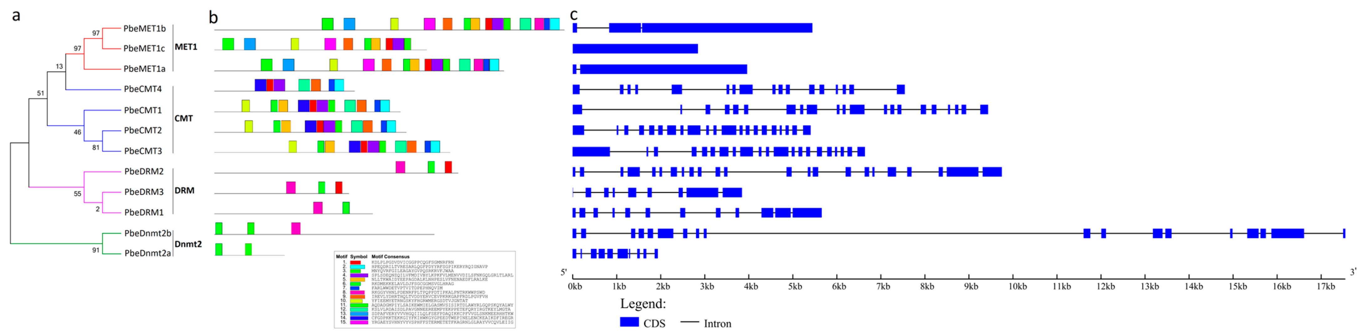
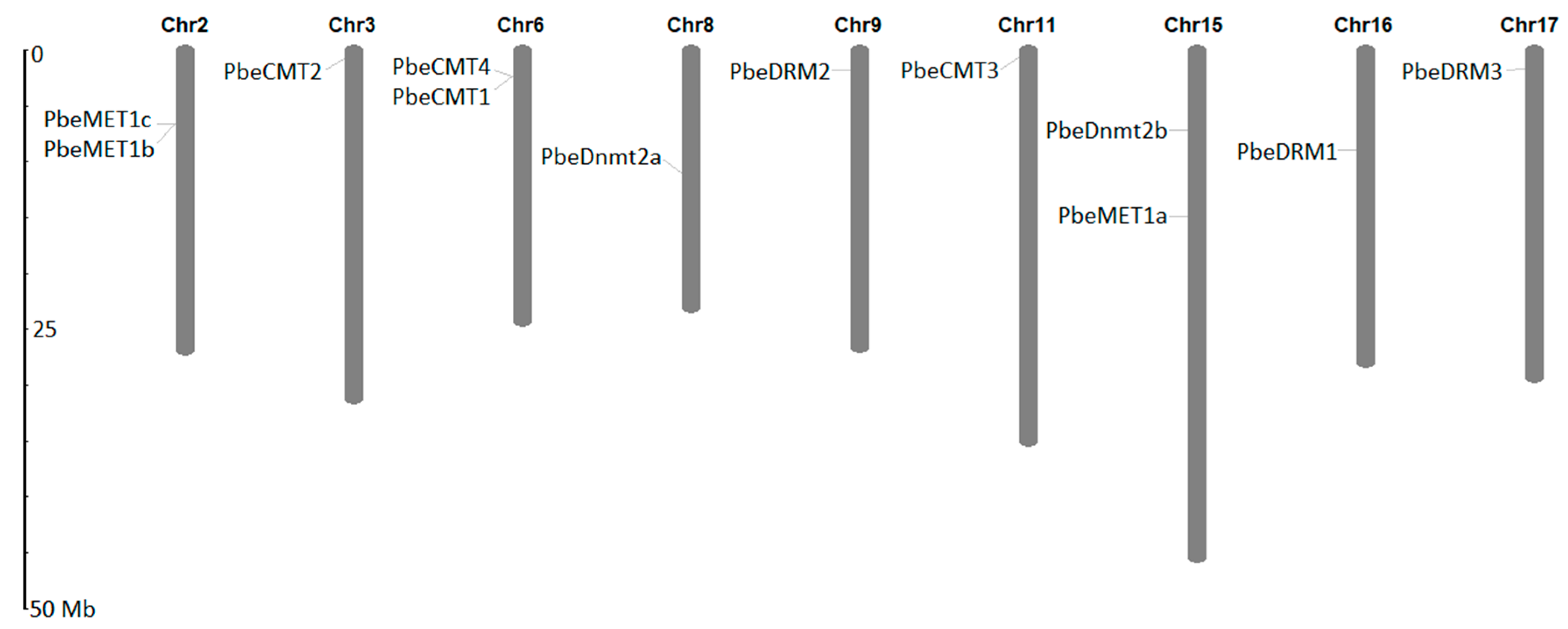
3.2. Gene Structure, Conserved Motif and Genomic Location Analysis of PbeMTases

The conserved motifs and intron–exon distribution of PbeMTase genes were analyzed to better understand their structural features (Figure 1). Fifteen conserved motifs were detected among the different PbeMTases (Figure 1b). The motifs that appear most frequently, and PbeDnmt2 and PbeDRM proteins contained the least number of motifs, two to three, and they also were composed of the least number of amino acids. In contrast, the PbeMET1 contained a greater complexity of motifs but the simplest gene structure.
This indicated that PbeMTases have a relatively complex gene structure by analyzing the structures, with the exception of PbeMET1a and PbeMET1b (Figure 1c). Members in the CMTs group contained 16 to 20 introns, while members in MET1 contained 0 to 2 introns, and the DRM and Dnmt2 groups contained 0 to 22 introns. Interestingly, PbeMET1a/b members contained the most amino acids but exhibited the simplest gene structure (Figure 1c).
The 12 PbeMTase genes were scattered over 9 of the 17 chromosomes of P. betulaefolia as we could examine the genomic distribution of them on chromosomes. Most were distributed individually on different chromosomes, except for PbeMET1a and PbeMET1c on chromosome 2, PbeCMR1 and PbeCMT4 on chromosome 6 and PbeMET1a and PbeDnmt2b on chromosome 15 (Figure 2), suggesting that the gene pairs may have been generated by a gene duplication event. PbeMTase genes comprise a small gene family in plants, including in P. betulaefolia.

3.3. Classification, Organization and Phylogeny of the PbeMTases

We constructed neighbor-joining (N-J) phylogenetic trees to illustrate the evolutionary relationships between the MTase genes. Besides, based on sequence similarities and the composition of the DNA methylase domain, the 12 MTase genes in P. betulaefolia were divided into four groups, MET1, CMT, DRM and Dnmt2, and designated as PbeMET1s, PbeCMTs, PbeDRMs and PbeDnmt2s (Figure 1a). Three members are in the MET1 group (PbeMET1a, PbeMET1b and PbeMET1c), four are in the CMT group (PbeCMT1-4), three are in the DRM group (PbeDRM1-3) and two are in the Dnmt2 group (PbeDnmt2a/b). The DNA methylase domain is a required element for a gene to be an MTase, and each of the 12 PbeMTase genes contained one DNA methylase domain. Other domains are characteristic of the different groups within the MTase gene family. In addition to the methylase domain, the members of the MET1 group also contain two cytosine-specific DNA methyltransferase replication foci domains (DNMT2-RFD, PF12047) and two bromo-adjacent homology (BAH, PF01426) domains with a relatively complex gene structure and the greatest number of amino acids. Members in the CMT group contain one BAH domain and one chromatin organization modifier (CHRO, PF00385) domain. In the DRM group, PbeDRM1 only contained the DNA methylase domain, but PbeDRM2-3 also contained a ubiquitin-associated (UBA, cd14330) domain. The Dnmt2 members only contained one DNA methylase domain and have the lowest number of amino acids (Supplementary Figure S2).
We put P. betulaefolia and nine other species in an unrooted phylogenetic tree that included 102 MTases (Figure 3). The following MTases were contained in the tree: 10 from Arabidopsis thaliana, 10 from Brassica rapa, 7 from Prunus persica, 11 from Malus domestica, 12 from Glycine max, 7 from Vitis vinifera, 14 from Medicago truncatula, 8 from Solanum lycopersicum, 11 from Pyrus bretschneideri and 12 from P. betulaefolia. The results showed that the 102 genes formed four clusters, in line with the previous classification based on domain compositions (Figure 3). Different clades of MTases may indicate diverse functions for the MTase genes in a single species and conserved sequences or functions among species.

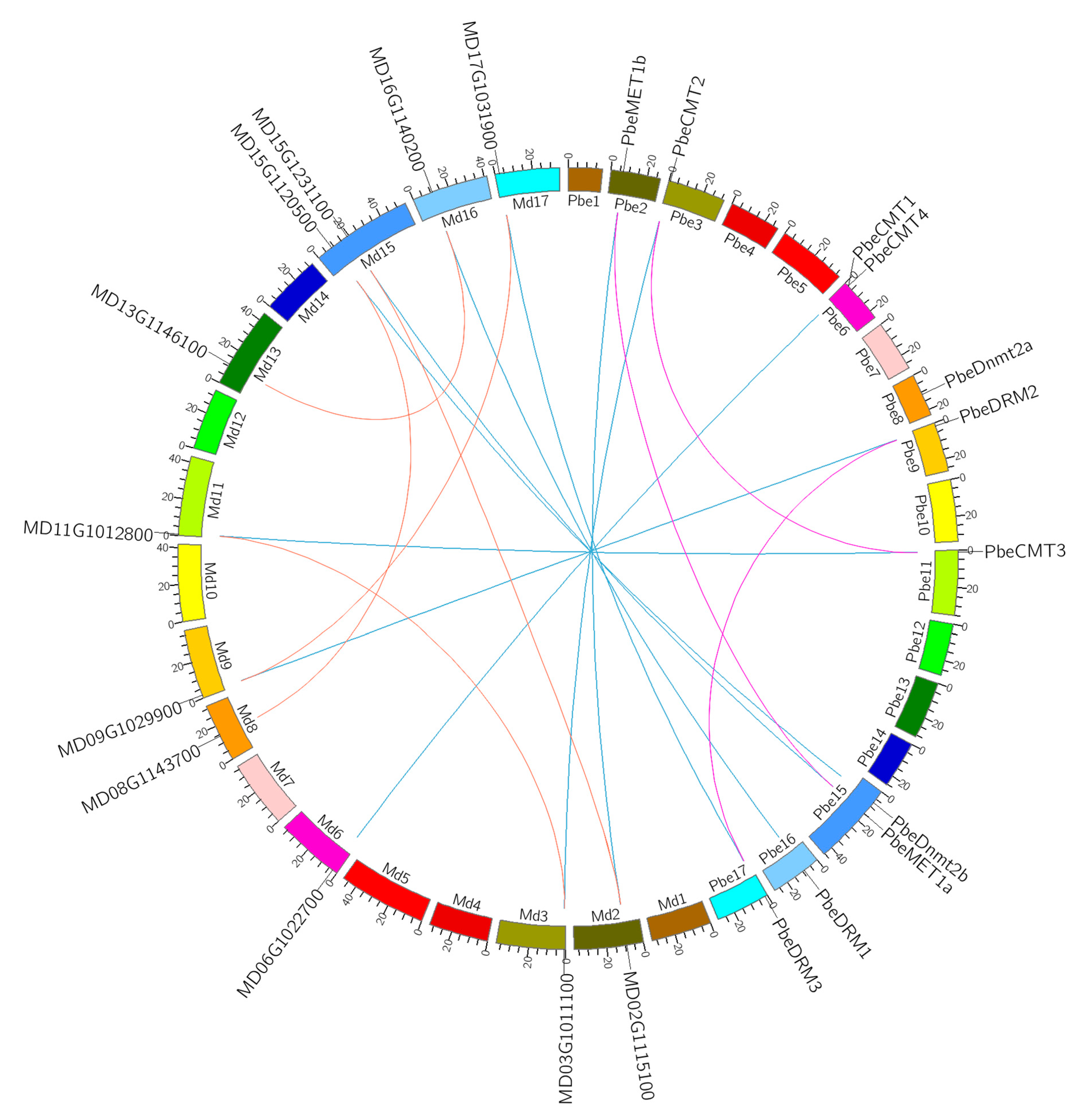
3.4. Evolutionary Clues of MTase Genes in P. betulaefolia and Apple

A comparative analysis was conducted in order to find the relationship between MTase homologous gene pairs across P. betulaefolia and apple, which insinuated the divergence and evolution between these two species (Figure 4). The results showed that there are nine orthologous MTase gene pairs between P. betulaefolia and apple (PbeMET1b and MD02G1115100, PbeMET1a and MD15G1231100, PbeCMT1 and MD06G1022700, PbeCMT2 and MD03G1011100, PbeCMT3 and MD11G1012800, PbeDnmt2b and MD15G1120500, PbeDRM1 and MD16G1140200, PbeDRM2 and MD09G1029900, PbeDRM3 and MD17G1031900), three orthologous MTase gene pairs in P. betulaefolia (PbeMET1a and PbeMET1b, PbeCMT2 and PbeCMT3, PbeDRM2 and PbeDRM3), and five paralogous gene pairs in apple (MD03G1011100 and MD11G1012800, MD15G1120500 and MD08G1143700, MD13G1146100 and MD16G1140200, MD17G1031900 and MD09G1029900, MD02G1115100 and MD15G1231100) (Figure 4 and Supplementary Table S2).
By Ks values, the divergence time between P. betulaefolia and apple was estimated at 2.22~4.28 MYA. This suggests that the orthologous gene pairs between P. betulaefolia and apple diverged after their speciation. Furthermore, the divergence time of MTase paralogous genes within the genomes of P. betulaefolia and apple was also estimated. For P. betulaefolia, the estimated range was 7.82~9.35 MYA, while for apple, it was 7.61~10.81 MYA. Both of these estimated divergence times occurred after the whole-genome duplication (WGD) event that took place in the common ancestor of P. betulaefolia and apple. Therefore, it can be concluded that the MTase genes in both P. betulaefolia and apple have experienced the WGD event. The divergence of orthologous gene pairs and the duplication of MTase genes in these species occurred after their speciation and the WGD event, respectively [26,35].

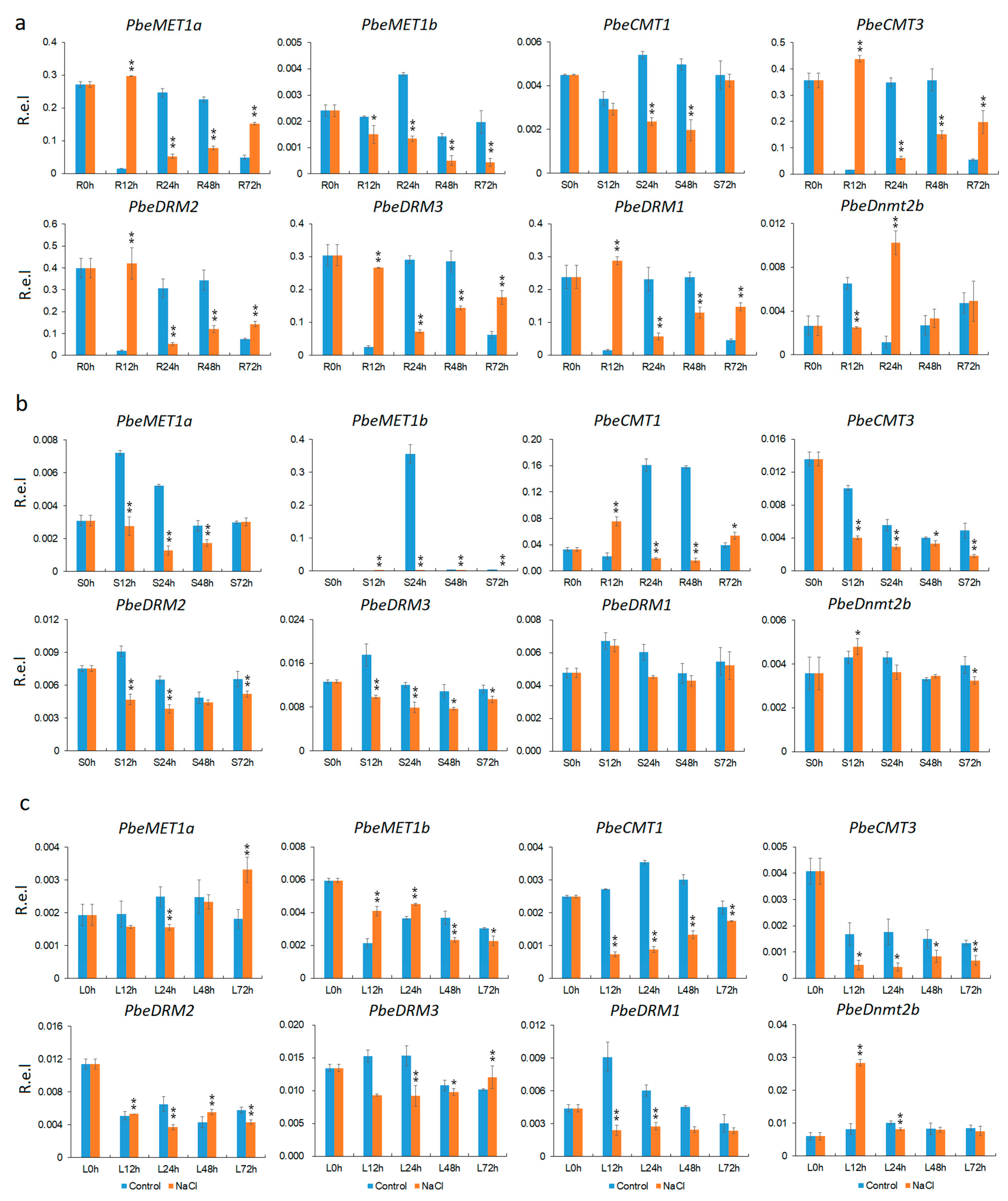
3.5. Tissue and Salt-Stress Expression Analysis

PbeMET1a/b, PbeCMT1-4, PbeDRM1-3 and PbeDnmt2a/b MTase genes were identified in the genome sequence of P. betulaefolia. The expression patterns of these genes in leaves, stems and roots were examined, in order to gain insight into the putative function of them. The results demonstrated the expression of different MTases varied in vegetative organs (Figure 5). Excluding PbeMET1b and PbeDnmt2b, the other six genes (PbeMET1a, PbeCMT2, PbeCMT3, PbeDRM1, PbeDRM2 and PbeDRM3) showed 3 to 140 times higher expression than in leaves and 7 to 87 times higher than in stems. PbeMET1a exhibited the highest level of expression among all of the genes in roots, and its expression level was 87 times greater than in stems and 140 times greater than in leaves. Even the PbeCMT2 had 7–13 times higher expression in roots than in leaves and stems. In contrast, the expression of PbeMET1b and PbeDnmt2 was higher in leaves than in stems and roots, but the difference in expression was not significant. Moreover, relative to the other seven genes, the relative level of expression of PbeMET1b and PbeDnmt2 was much lower (Figure 5). These results suggest that some level of functional divergence may have occurred among these gene family members.
The expression profiles of PbeMTase genes were detected in vegetative organ of the plants growing under both normal conditions and salt stress, with the exception of PbeCMT2, PbeCMT4 and PbeDnmt2a, whose transcripts were not detected in response to salt stress in any tissues or time points. All nine PbeMTase genes, including PbeMET1a/b, PbeCMT1/3, PbeDRM1-3 and PbeDnmt2b, were induced by salt stress at more than one time point. The results demonstrated that the PbeMTase genes were induced in roots quickly after salt treatment but exhibited different repression in leaves and stems (Figure 6). In roots, PbeMET1a, PbeCMT3, PbeDRM1, PbeDRM2 and PbeDRM3 were significantly upregulated at 12 h under the salt stress compared to the controls, and PbeDnmt2b was significantly upregulated at 24 h compared to the controls. PbeMET1a, PbeCMT3, PbeDRM1, PbeDRM2 and PbeDRM3 were also upregulated at 24 h under the salt treatment compared to the controls (Figure 6a). In stems, almost all of the MTase genes were downregulated, relative to the control, in response to the salt stress. PbeDRM1 and PbeDnmt2b, however, were exceptions. There was no difference in the level of expression of PbeDRM1 before or after the salt treatment, and a similar response was observed for PbeDnmt2b (Figure 6b). In leaves, the expression of PbeMET1a was upregulated, relative to the control, at 72 h, PbeMET1b at 12 h and 24 h, PbeDRM2 at 48 h and PbeDnmt2b at 12 h compared to the controls. In contrast, CMT and DRM genes were significantly repressed, especially PbeCMT1, PbeCMT3 and PbeDRM1, compared to the controls (Figure 6c).
The expression pattern of several salt-responsive genes in response to salt stress was also investigated by RT-qPCR in order to gain further insight into their possible function.
In the present study, the expression of the PbeMTase genes was investigated through a salt stress treatment. We analyzed the expression of ten salt-responsive genes (PbeNHX2.1, PbeSOS1, PbeCBL2, PbeCBL4, PbeCBL10, PbeAKT2, PbeAKT6, PbeCIPK8, PbeCIPK16 and PbeCIPK24) in response to the salt stress (Supplementary Figure S3 and Supplementary Table S1) and found that the salt stress used in this study either induced or repressed the salt-responsive genes.

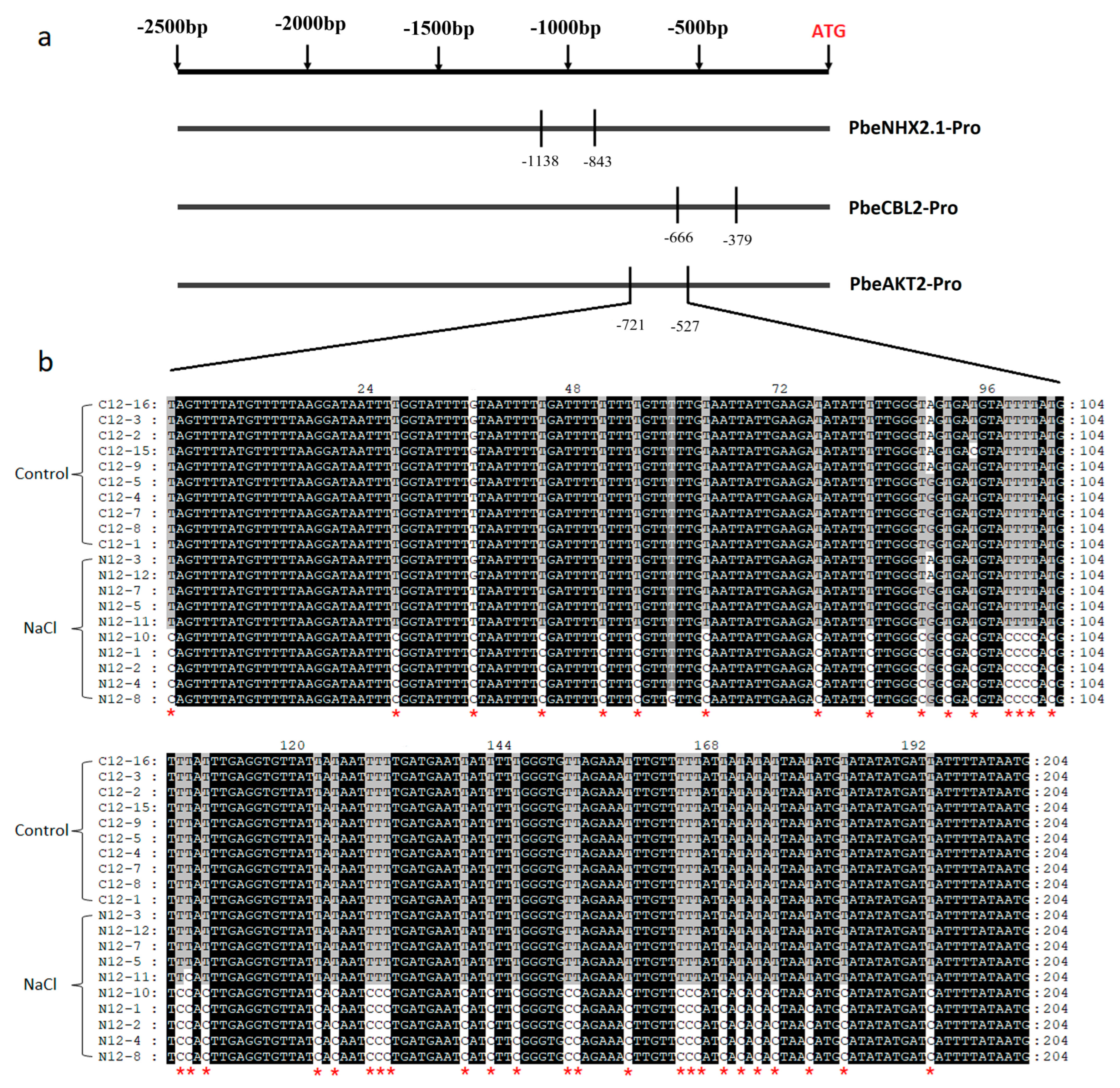
3.6. Methylation Level Changed of Salt-Responsive Genes under Salt Stress

Studies revealed that DNA methylation played important roles in plant response to stress environments [4]. We selected five salt-responsive genes (PbeNHX2.1, PbeCBL2, PbeCBL4, PbeCIPK24 and PbeAKT2) to detect the changes in methylation level in their promoters under salt stress so that we could learn more about how the PbeMTase genes regulate expression (Figure 7 and Supplementary Table S3). The results indicated that two genes, PbeNHX2.1 and PbeCBL2, exhibited a higher level of methylation at CpG sites in response to salt stress, relative to the level observed in the controls (Figure 7a and Supplementary Table S3). Especially for the PbeAKT2 gene, we found a high methylation level in CHG or CHH sites in the tested regions under salt stress treatment (Figure 7b). These genes were repressed in response to salt stress (Supplementary Figure S3), which was consistent with their higher methylation level. For example, the methylation levels of PbeSOS1, PbeNHX2.1 and PbeCBL2 promoter regions were higher in plants subjected to salt stress than those in the untreated controls. The methylation level of PbeCBL4 and PbeCIPK24, however, was barely altered by salt stress (Supplementary Table S3). It was the first identified CBL-CIPK pathway that was salt overly sensitive (SOS), while we have known that PbeSOS1 and PbeCBL2 are involved in it [35]. The results indicated that the expression of PbeMTase genes in P. betulaefolia was responsive to salt stress treatment, as observed in Figure 6. This corresponded with the methylation level of salt-responsive genes, suggesting the involvement of PbeMTase genes in the response to salt stress. Furthermore, it is plausible that other intricate regulatory pathways exist during the salt stress response in P. betulaefolia. Consequently, the findings highlight the significant role of PbeMTase genes in the response to salt stress. These could provide excellent candidates for breeding rootstocks of pear.

4. Discussion

4.1. Conserved Gene Structures May Indicate Specific Functions

Three CMT, two MET1, four DRMs and two Dnmt2s MTase genes in this study were identified in P. betulaefolia. In the phylogenetic analysis, PbeMTase genes belonging to these groups clustered into different clades based on their different domain/motif organization and the protein sequences. The intron–exon structure and domain architecture of the PbeMTase genes were highly conserved within each subfamily (Figure 1 and Supplementary Figures S1 and S2). The PbeMET1 group comprises PbeMET1a and PbeMET1b, which possess the most complex domains, two DNMT1-FRD domains, two BAH domains and one DNA-methylase (Supplementary Figure S2). Similar to other plant species, the PbeCMT group is comprised of four genes (CMT1-4) [10] and each possess three domains (BAH, CHRO, and DNA-methylase). The similar structure of the PbeCMTs to other plant species may indicate a conserved function. A previous study demonstrated that DRM2 in Arabidopsis requires an intact UBA domain during RNA-directed DNA methylation (RdDM) [40]. Some DRM group members, such as PbeDRM1, however, did not possess a UBA domain (Supplementary Figure S2). Formation of a DRM homodimer or heterodimer in seed plants and mammals is required to direct DNA de novo methylation [4]. Formation of a heterodimer between a DRM group member lacking a UBA domain and a DRM member with a UBA domain may enable de novo DNA methylation. The N-terminus of MET contains two BAH domains, potentially facilitating protein–protein interaction [10]. The conserved structure of each group provides the evidence of conserved, while the differences among groups means the diverse functions.

4.2. Roots Are a Key Organ for Plant Response to the Environment

In this study, the expression of PbeMTase genes was examined in different tissues, including leaves, stems and roots. Transcripts of nine PbeMTase genes were detected; however, no expression could be determined for PbeCMT2 and PbeDnmt2a. Interestingly, the detected MTases were predominantly expressed in roots, except for PbeMET1b and PbeDnmt2b (Figure 5). Roots are known to play a key role in plant response to environmental stimuli such as salt stress. Numerous studies have been conducted on salt stress response in many different plant species, including Arabidopsis [41], rice [42], tomato [43], soybean [44], Brassica [45] and cotton [46]. In the present study, hardly any expression of PbeMTase genes could be detected in stems and leaves, except for the expression of PbeMET1b and PbeDnmt2b (Figure 5). These results indicate that roots play a primary role in plant response to environmental stresses, and it appears that MTases play a vital role in root response to salt stress. Thus, roots were selected for further tests to examine their response to salt stress. In regards to PbeMET1b and PbeDnmt2b, their absence of expression in roots and expression in leaves and stems may be the result of the redundant dosage of homologous genes. It should also be noted that the expression level of both these genes was almost two orders of magnitude lower than the level observed for the other seven MTase genes (Figure 5). Perhaps these two MTase genes have some other function that was not reflected in the experimental design of the current study.

4.3. Expression Divergence among Different Group Members of MTases

DNA methylation is an inherited and reversible epigenetic modification which directly affects the regulation of gene expression. Therefore, this process may also affect a plant’s response to salt stress. MTases play an important role in plant development by regulating the level of DNA methylation [4,47]. For example, a dynamic change in the level of DNA methylation in endosperm genome activates the expression of many genes [48].
The comprehensive expression analysis of MTases in different tissues and salt stress treatment has provided evidence for their diverse roles in various aspects of plant development and abiotic stress response. In the present study, two MET1 members were differentially expressed in roots in response to salt stress. The expression level of PbeMET1a was quickly upregulated in roots, relative to the control, in response to a salt treatment and reached a peak at 12 h. In contrast, PbeMET1b was repressed in response to the salt treatment and remained downregulated throughout the time course of the study (Figure 6a). These data suggest that there is a functional divergence between homologous MTase genes within the same subgroup. CMT members also exhibited different expression patterns. CMT genes were induced or repressed in response to the salt treatment at different time points (Figure 6a). Overall, the data indicate that MTase genes within the same subgroup respond differently to salt stress. Some explanations for this differential expression may include maintaining appropriate doses of genes, as well as requirements for interactions and different levels of regulation [49]. In contrast, differential expression response to salt stress was not evident for DRM members. All four DRM members were significantly induced in response to the salt treatment, peaking at 12 h and subsequently decreasing (Figure 6a). DRMs are relatively conserved across plant species and may share the same response to salt stress [50].
Expression of the MTases in stems and leaves could also be subject to some constraints. In that regard, differential patterns of expression were observed for PbeMET1a and PbeMET1b, with a greater difference in expression of the two genes observed in stems (Figure 6b,c). PbeCMT1/3 had a similar pattern of expression in leaves, but they differed in their expression pattern in stems and roots. Interestingly, no difference in the expression of PbeDRM1 was observed between salt stress and control samples of stems (Figure 6b). Therefore, some DRM members may have other specific functions in stems.
Our results indicate that different members of PbeMTase genes play a specific role in different tissues or to different environmental cues. Some PbeMTase genes were induced and some were repressed in response to salt stress, and the change in the level of expression may alter the methylation level of salt-responsive genes. In fact, MTases can have either a positive or negative impact on the expression of salt-responsive genes and also act as epigenetic modules regulating gene expression. This is particularly relevant to immune-responsive genes, whose basal and biotic stress-induced expression needs to be tightly regulated [51]. The perception and transduction of salt-stress signals are crucial for plant survival, growth and reproduction. DNA methylation dynamics have been observed in many plants under salt stress [52,53,54]. Plants have evolved complex and tightly regulated responses to stress that typically involve both the upregulation and downregulation of genes, as was observed in the current study for the expression of salt-responsive genes. The increase in DNA methylation in alfalfa (Medicago spp.) is particularly significant under high salt stress [55]. Further observations showed that transgenic tobacco overexpressing AtROS1 exhibited altered flavonoid biosynthesis and methylation status in the antioxidant enzyme gene promoter region, leading to an increase in gene expression levels and increased tolerance to salt stress [56]. Arabidopsis regulates the salt-induced transcription factor MYB74 through the RdDM pathway under hypertonic conditions [57]. Though the specific mechanism that allows MTase genes to regulate the response of P. betulaefolia to abiotic stress is still unclear, the current study provides new information on the role of MTases in regulating the response to salt stress in Pyrus betulaefolia.

4.4. Conserved Evolution of PbeMTase Genes

A WGD event occurred around ~50 MYA in the ancestor of P. betulaefolia and apple, leading to the retention of 12 MTase genes in both genomes. Paralogous MTase gene pairs diverged earlier than orthologous pairs, suggesting that the WGD event predates the speciation of P. betulaefolia and apple (Figure 4 and Supplementary Table S2). Interestingly, peach, which did not undergo recent WGD, only has seven MTase genes [58,59]. The retained MTase genes likely play crucial roles in growth, development and environmental response in P. betulaefolia and apple. This contrasts with the super ERF gene family, which lost members after WGD events in many plant species [60]. The relationship between homologous MTase gene pairs in P. betulaefolia offers unique insights into Rosaceae plant evolution.
On the other hand, the expression patterns of PbeMTase gene pairs in different organs and in response to salt stress provide clues on the evolution of orthologous and paralogous genes in Pomoideae. One of the paralogous or duplicated gene members is typically lost or shows expression bias based on the gene dosage after a duplication event [61]. The MTase genes in P. betulaefolia, however, retained their expression levels in different organs or in response to salt stress despite the occurrence of a WGD event (Supplementary Figure S4). Possible explanations for this phenomenon include the requirement to maintain adequate gene dosage and interactions and different levels of regulation [61]. For example, three pairs of P. betulaefolia homologous genes exhibited expression bias in different organs or in response to salt stress (Supplementary Figure S4). In regards to the PbeCMT2/3 gene pair, no expression was detected for PbeCMT2 in any of the selected organs or in response to salt stress. Only one of the members of the other gene pairs was dominantly expressed in different organs or at different time points in response to salt stress. This may be the reason that both members of the gene pairs have been retained over the course of domestication.

4.5. Excellent Candidates for P. betulaefolia Salt Tolerance Breeding and Improvement

Under salt stress, the DNA methylation level plays an essential role in plants to regulate the response by dynamically regulating [53,62,63,64]. In the current study, the PbeMTase genes were induced or repressed expression in response to salt stress treatment, and in their promoter regions, methylation occurred differentially (Figure 6 and Figure 7, Supplementary Figure S4 and Supplementary Table S3), resulting in altered expression levels of these downstream genes. It can be said that the MTase gene plays an important role in pear response to salt stress. However, there is no complete correspondence between the expression level of the PbeMTase gene and the methylation level of the selected salt-responsive gene (Figure 6 and Figure 7, Supplementary Figure S4 and Supplementary Table S3). These still indicate that there are more salt-responsive genes or complex signaling pathways in P. betulaefolia, which are involved in salt stress responses. Under the background that the mechanism of the PbeMTase gene in salt stress response is still unclear, our research results provide a new epigenetics perspective for P. betulaefolia and other temperate fruit trees to deal with salt stress. We believe that pretreatment and training of plants with chemicals that change the level of DNA methylation can enhance the salt tolerance of P. betulaefolia. For PbeMTase genes that can actively respond to salt stress, screening varieties containing these genes will be one of the effective ways to select and improve salt-tolerant varieties of P. betulaefolia.
Natural weathering and agricultural activities have led to a continuous increase in high salinity for decades [65]. There are two major threats to plant growth caused by salinity: ions outside the root cause osmotic stress, and ions entering the plant cause ionic stress. The method and success rate in dealing with salt stress in different species pose a large problem [66]. The PbeMET1a, PbeCMT1/3 and PbeDRM1-3 genes were highly or specifically expressed in this study (Figure 5), while these important candidate genes were all responsive to high salinity (Figure 6). They are potential candidates to study the PbeMTase genes and responsive to the environment, and these genes may provide excellent clues to improve the salt tolerance in rootstocks of pear breeding.

5. Conclusions

There were 12 putative MTases in the P. betulaefolia genome that have been identified and analyzed in the present study. They could be classified in MET, CMT, DRM and Dnmt2 groups based on the organization of various characteristic domains. The analysis of gene and protein structures, along with phylogenetic analysis, supported the findings. The PbeMTase genes exhibited a complex structure, and the DRM1 proteins lacked a UBA domain. This suggests that before the P. betulaefolia and apple genomes diverged, the MTase family genes underwent a WGD event in the ancestral ancestor. Expression analysis revealed that MTases and salt-related genes in Pyrus betulaefolia were upregulated in response to high salinity stress. The methylation levels of salt-related genes were also modified in response to salt stress treatment. Furthermore, four selected salt-responsive genes were found altered in methylation level promoters (which contain CpG islands) under salt stress. This study provided a genome-wide survey of the MTase gene family in P. betulaefolia and discussed their roles in P. betulaefolia under salt stress and evidenced it. The whole study provides a basis for apprehending the composite functions of the P. betulaefolia MTase Gene family and is conducive to the epigenetics studies of pear rootstocks’ response to salt stress.

Supplementary Materials

The following supporting information can be downloaded at: https://www.mdpi.com/article/10.3390/f14091751/s1, Figure S1. Alignments of PbeMTase family proteins. Sequences in the red box indicate the DNA methylase domain. Figure S2. Various domains present in PbeMTase proteins. Figure S3. Expression patterns of salt-responsive genes under salt stress in Pyrus betulaefolia. Young P. betulaefolia plants at the eight leaves stage were subjected to 200 mM NaCl stress by watering them with a salt solution. At 0, 12, 24, 48 and 72 h, samples of roots were collected. RT-qPCR analysis was performed to determine the relative expression levels. The 2−∆∆CT method was used to calculate the relative expression. Significance levels of 0.05 and 0.01 were denoted by an asterisk and double asterisks, respectively. R.e.l. represented the relative expression level. Figure S4. Expression profiles of the MTase syntenic gene pairs in response to salt stress treatment. Roots were sampled from P. betulaefolia plants growing under normal conditions. At 0, 12, 24, 48 and 72 h, samples were collected. RT-qPCR analysis was performed to determine the relative expression levels. The 2−∆∆CT method was used to calculate the relative expression. Significance levels of 0.05 and 0.01 were denoted by an asterisk and double asterisks, respectively. R.e.l. represented the relative expression level. Figure S5. The visual picture of a Pyrus betuleafolia. Table S1. Primers used in this paper. Table S2. Orthologous and paralogous gene pairs in the P. betulaefolia and apple MTase family. Table S3. Analysis of cytosine methylation of a 200 bp segment of the promoter region of PbeNHX2.1, PbeCBL2, PbeCBL4 and PbeCIPK24 in Pyrus betulaefolia in response to salt stress.

Author Contributions

C.L. and Y.C. conceived the study and designed the experiments. Y.Z., C.L. and X.X. performed the experiments and analyzed the data. Y.Z. wrote the manuscript. Z.C., Y.C., J.K., H.L. and J.L. revised and proofread the manuscript. All authors have read and agreed to the published version of the manuscript.

Funding

This research was supported by the National Natural Sciences Foundation of China (Nos. 31801827 and 31772287).

Institutional Review Board Statement

Not applicable.

Informed Consent Statement

Not applicable.

Data Availability Statement

All the data used to support the findings of this study are included within the article.

Acknowledgments

All individuals included in this section have consented to the acknowledgement.

Conflicts of Interest

The authors declare no conflict of interest.

Abbreviations

MTase: DNA Methyltransferase; MET1: METHYLTRANSFERASE1; CMT: CHROMOMETHYLTRANSFERASE; DRM: Domains Rearranged Methyltransferase; Dnmt2: DNA nucleotide methyltransferase; Pbe: Pyrus betulaefolia; Ka: nonsynonymous substitutions; Ks: synonymous substitutions; WGD: whole-genome duplication; MYA: million years ago; pI: isoelectric point; MW: molecular weight and qRT-PCR: quantitative Real-Time PCR.

References

  1. Holliday, R. Epigenetics: A historical overview. Epigenetics 2006, 1, 76–80. [Google Scholar] [CrossRef] [PubMed]
  2. Martín-Subero, J.I. How epigenomics brings phenotype into being. Pediatr. Endocrinol. Rev. 2011, 9, 506–510. [Google Scholar] [PubMed]
  3. Yanjun, A.; Shenglin, J.; Zhengnan, C.; Botao, S.; Conghua, X.; Jun, L.; Jun, Z. DNA methylation affects photoperiodic tuberization in potato (Solanum tuberosum L.) by mediating the expression of genes related to the photoperiod and GA pathways. Hortic. Res. 2021, 8, 181. [Google Scholar]
  4. Zhong, X.; Du, J.; Hale, C.J.; Gallego-Bartolome, J.; Feng, S.; Vashisht, A.A.; Chory, J.; Wohlschlegel, J.A.; Patel, D.J.; Jacobsen, S.E. Molecular mechanism of action of plant DRM de novo DNA methyltransferases. Cell 2014, 157, 1050–1060. [Google Scholar] [CrossRef]
  5. Lu, S.C.; Mato, J.M. S-adenosylmethionine in liver health, injury, and cancer. Physiol. Rev. 2012, 92, 1515–1542. [Google Scholar] [CrossRef] [PubMed]
  6. María-Estefanía, L.; David, R.; Claude, B.; Béatrice, D.; Etienne, B. DNA methylation dynamics during stress response in woodland strawberry (Fragaria vesca). Hortic. Res. 2022, 9, uhac174. [Google Scholar]
  7. Cokus, S.J.; Feng, S.; Zhang, X.; Chen, Z.; Merriman, B.; Haudenschild, C.D.; Pradhan, S.; Nelson, S.F.; Pellegrini, M.; Jacobsen, S.E. Shotgun bisulphite sequencing of the Arabidopsis genome reveals DNA methylation patterning. Nature 2008, 452, 215–219. [Google Scholar] [CrossRef]
  8. Law, J.A.; Jacobsen, S.E. Establishing, maintaining and modifying DNA methylation patterns in plants and animals. Nat. Rev. Genet. 2010, 11, 204–220. [Google Scholar] [CrossRef]
  9. Noy-Malka, C.; Yaari, R.; Itzhaki, R.; Mosquna, A.; Auerbach Gershovitz, N.; Katz, A.; Ohad, N. A single CMT methyltransferase homolog is involved in CHG DNA methylation and development of Physcomitrella patens. Plant. Mol. Biol. 2014, 84, 719–773. [Google Scholar] [CrossRef]
  10. Garg, R.; Kumari, R.; Tiwari, S.; Goyal, S. Genomic survey, gene expression analysis and structural modeling suggest diverse roles of DNA methyltransferases in legumes. PLoS ONE 2014, 9, e88947. [Google Scholar] [CrossRef]
  11. Pavlopoulou, A.; Kossida, S. Plant cytosine-5 DNA methyltransferases structure, function, and molecular evolution. Genomics 2007, 90, 530–541. [Google Scholar] [CrossRef] [PubMed]
  12. Kumar, S.; Stecher, G.; Tamura, K. MEGA7 Molecular Evolutionary Genetics Analysis version 70 for bigger datasets. Mol. Biol. Evol. 2016, 33, 1870–1874. [Google Scholar] [CrossRef] [PubMed]
  13. Morris, M.J.; Na, E.S.; Autry, A.E.; Monteggia, L.M. Impact of DNMT1 and DNMT3a forebrain knockout on depressive- and anxiety like behavior in mice. Neurobiol. Learn. Mem. 2016, 135, 139–145. [Google Scholar] [CrossRef] [PubMed]
  14. Finnegan, E.J.; Peacock, W.J.; Dennis, E.S. Reduced DNA methylation in Arabidopsis thaliana results in abnormal plant development. Proc. Natl. Acad. Sci. USA 1996, 9316, 8449–8454. [Google Scholar] [CrossRef]
  15. Steward, N.; Kusano, T.; Sano, H. Expression of ZmMET1, a gene encoding a DNA methyltransferase from maize, is associated not only with DNA replication in actively proliferating cells, but also with altered DNA methylation status in cold-stressed quiescent cells. Nucleic Acids. Res. 2000, 28, 3250–3259. [Google Scholar] [CrossRef] [PubMed]
  16. Teerawanichpan, P.; Chandrasekharan, M.; Jiang, Y.; Narangajavana, J.; Hall, T. Characterization of two rice DNA methyltransferase genes and RNA imediated reactivation of a silenced transgene in rice callus. Planta 2004, 218, 337–349. [Google Scholar]
  17. Fujimoto, R.; Sasaki, T.; Nishio, T. Characterization of DNA methyltransferase genes in Brassica rapa. Genes Genet. Syst. 2006, 81, 235–242. [Google Scholar] [CrossRef]
  18. Thomas, M.; Pingault, L.; Poulet, A.; Duarte, J.; Throude, M.; Faure, S.; Pichon, J.P.; Paux, E.; Probst, A.V.; Tatout, C. Evolutionary history of Methyltransferase 1 genes in hexaploid wheat. BMC Genom. 2014, 15, 922. [Google Scholar] [CrossRef]
  19. Kim, M.; Ohr, H.; Lee, J.W.; Hyun, Y.; Fischer, R.L.; Choi, Y. Temporal and spatial downregulation of Arabidopsis MET1 activity results in global DNA hypomethylation and developmental defects. Mol. Cells 2008, 26, 611–615. [Google Scholar]
  20. Cao, X.F.; Jacobsen, S.E. Locus-specific control of asymmetric and CpNpG methylation by the DRM and CMT3 methyltransferase genes. Proc. Natl. Acad. Sci. USA 2002, 994, 16491–16498. [Google Scholar] [CrossRef]
  21. Manning, K.; Tör, M.; Poole, M.; Hong, Y.; Thompson, A.J.; King, G.J.; Giovannoni, J.J.; Seymour, G.B. A naturally occurring epigenetic mutation in a gene encoding an SBP-box transcription factor inhibits tomato fruit ripening. Nat. Genet. 2006, 38, 948–995. [Google Scholar] [CrossRef] [PubMed]
  22. Chen, M.; Luo, Z.; Zhao, X.; Li, S.; Wu, F.; Chen, J.; Xiang, M. Exogenously applied methyl jasmonate increased the resistance of postharvest pear fruit to blue mold. Fruit Res. 2022, 2, 11. [Google Scholar] [CrossRef]
  23. Wu, J.; Wang, Z.; Shi, Z.; Zhang, S.; Ming, R.; Zhu, S.; Khan, M.A.; Tao, S.; Korban, S.S.; Wang, H.; et al. The genome of the pear (Pyrus bretschneideri Rehd). Genome Res. 2013, 3, 396–408. [Google Scholar] [CrossRef] [PubMed]
  24. Zhang, J.; Yuan, H.; Yang, Q.; Li, M.; Wang, Y.; Li, Y.; Ma, X.; Tan, F.; Wu, R. The genetic architecture of growth traits in Salix matsudana under salt stress. Hortic. Res. 2017, 4, 17024. [Google Scholar] [CrossRef] [PubMed]
  25. Zhu, W.; Cao, H.; Wang, Q.; Niu, Y.; Sadeghnezhad, E.; Han, S.; Liu, M.; Wang, L.; Liu, Z. Transcriptome analysis revealed MAPK and hormone pathway involving in exogenous melatonin-regulated salt tolerance in sour jujube. Fruit Res. 2023, 3, 19. [Google Scholar] [CrossRef]
  26. Dong, X.; Wang, Z.; Tian, L.; Zhang, Y.; Qi, D.; Huo, H.; Xu, J.; Li, Z.; Liao, R.; Shi, M.; et al. De novo assembly of a wild pear (Pyrus betuleafolia) genome. Plant Biotechnol. J. 2019, 18, 581–595. [Google Scholar] [CrossRef]
  27. Matsumoto, K.; Tamura, F.; Chun, J.P.; Ikeda, T.; Imanishi, K.; Tanabe, K. Enhancement in salt tolerance of Japanese pear by using Pyrus betulaefolia rootstock. Hortic. Res. 2007, 6, 47–52. [Google Scholar] [CrossRef]
  28. Zhang, X.; Lii, Y.; Wu, Z.; Polishko, A.; Zhang, H.; Chinnusamy, V.; Lonardi, S.; Zhu, J.K.; Liu, R.; Jin, H. Mechanisms of small RNA generation from cis-NATs in response to environmental and developmental cues. Mol. Plant 2013, 6, 704–715. [Google Scholar] [CrossRef]
  29. Wang, M.; Qin, L.; Xie, C.; Li, W.; Yuan, J.; Kong, L.; Yu, W.; Xia, G.; Liu, S. Induced and constitutive DNA methylation in a salinity-tolerant wheat introgression line. Plant Cell Physiol. 2014, 55, 1354–1365. [Google Scholar] [CrossRef]
  30. Ferreira, L.J.; Azevedo, V.; Maroco, J.; Oliveira, M.M.; Santos, A.P. Salt tolerant and sensitive rice varieties display differential methylome flexibility under salt stress. PLoS ONE 2015, 10, e0124060. [Google Scholar] [CrossRef]
  31. Finn, R.D.; Clements, J.; Eddy, S.R. HMMER web server interactive sequence similarity searching. Nucleic. Acids. Res. 2011, 39, W29–W37. [Google Scholar] [CrossRef]
  32. Huang, J.; Wang, H.; Xie, X.; Zhang, D.; Liu, Y.; Guo, G. Roles of DNA methyltransferases in Arabidopsis development. Afr. J. Biotechnol. 2010, 9, 8506–8514. [Google Scholar]
  33. Larkin, M.A.; Blackshields, G.; Brown, N.P.; Chenna, R.; McGettigan, P.A.; McWilliam, H.; Valentin, F.; Wallace, I.M.; Wilm, A.; Lopez, R.; et al. Clustal W and Clustal X version 20. Bioinformatics 2007, 23, 2947–2948. [Google Scholar] [CrossRef]
  34. Kumar, S.; Cheng, X.; Klimasauskas, S.; Mi, S.; Posfai, J.; Roberts, R.J.; Wilson, G.G. The DNA (cytosine-5) methyltransferases. Nucleic. Acids. Res. 1994, 22, 1–10. [Google Scholar] [CrossRef] [PubMed]
  35. Daccord, N.; Celton, J.M.; Linsmith, G.; Becker, C.; Choisne, N.; Schijlen, E.; van de Geest, H.; Bianco, L.; Micheletti, D.; Velasco, R.; et al. High-quality de novo assembly of the apple genome and methylome dynamics of early fruit development. Nat. Genet. 2017, 49, 1099–1106. [Google Scholar] [CrossRef] [PubMed]
  36. Librado, P.; Rozas, J. DnaSP v5 a software for comprehensive analysis of DNA polymorphism data. Bioinformatics 2009, 25, 1451–1452. [Google Scholar] [CrossRef]
  37. Krzywinski, M.; Schein, J.; Birol, I.; Connors, J.; Gascoyne, R.; Horsman, D.; Jones, S.J.; Marra, M.A. Circos an information aesthetic for comparative genomics. Genome Res. 2009, 19, 1639–1645. [Google Scholar] [CrossRef]
  38. Ma, N.; Liu, C.; Li, H.; Wang, J.; Zhang, B.; Lin, J.; Chang, Y. Genome-wide identification of lectin receptor kinases in pear, Functional characterization of the L-type LecRLK gene PbLRK138. Gene 2018, 661, 11–21. [Google Scholar] [CrossRef]
  39. Hao, X.; Horvath, D.P.; Chao, W.S.; Yang, Y.; Wang, X.; Xiao, B. Identification and Evaluation of Reliable Reference Genes for Quantitative Real-Time PCR Analysis in Tea Plant (Camellia sinensis (L.) O. Kuntze). Int. J. Mol. Sci. 2014, 15, 22155–22172. [Google Scholar] [CrossRef] [PubMed]
  40. Henderson, I.R.; Deleris, A.; Wong, W.; Zhong, X.; Chin, H.G.; Horwitz, G.A.; Kelly, K.A.; Pradhan, S.; Jacobsen, S.E. The de novo cytosine methyltransferase DRM2 requires intact UBA domains and a catalytically mutated paralog DRM3 during RNA directed DNA methylation in Arabidopsis thaliana. PLoS Genet. 2010, 6, e1001182. [Google Scholar] [CrossRef]
  41. Wang, Y.N.; Li, K.X.; Li, X. Auxin redistribution modulates plastic development of root system architecture under salt stress in Arabidopsis thaliana. J. Plant. Physiol. 2009, 166, 1637–1645. [Google Scholar] [CrossRef]
  42. Meng, D.; Fricke, W. Changes in root hydraulic conductivity facilitate the overall hydraulic response of rice (Oryza sativa L.) cultivars to salt and osmotic stress. Plant. Physiol. Biochem. 2017, 113, 64–77. [Google Scholar] [CrossRef]
  43. Mayak, S.; Tirosh, T.; Glick, B.R. Plant growth-promoting bacteria confer resistance in tomato plants to salt stress. Plant. Physiol. Biochem. 2004, 42, 565–572. [Google Scholar] [CrossRef] [PubMed]
  44. Aghaei, K.; Ehsanpour, A.A.; Shah, A.H.; Komatsu, S. Proteome analysis of soybean hypocotyl and root under salt stress. Amino Acids. 2009, 36, 91–98. [Google Scholar] [CrossRef]
  45. Rossi, L.; Zhang, W.; Ma, X. Cerium oxide nanoparticles alter the salt stress tolerance of Brassica napus L. by modifying the formation of root apoplastic barriers. Environ. Pollut. 2017, 229, 132–138. [Google Scholar] [CrossRef] [PubMed]
  46. Irizarry, I.; White, J.F. Application of bacteria from non-cultivated plants to promote growth, alter root architecture and alleviate salt stress of cotton. J. Appl. Microbiol. 2017, 122, 1110–1120. [Google Scholar] [CrossRef]
  47. Eichten, S.R.; Briskine, R.; Song, J.; Li, Q.; Swanson-Wagner, R.; Hermanson, P.J.; Waters, A.J.; Starr, E.; West, P.T.; Tiffin, P.; et al. Epigenetic and genetic influences on DNA methylation variation in maize populations. Plant Cell 2013, 25, 2783–2797. [Google Scholar] [CrossRef] [PubMed]
  48. Wang, P.; Xia, H.; Zhang, Y.; Zhao, S.; Zhao, C.; Hou, L.; Li, C.; Li, A.; Ma, C.; Wang, X. Genome-wide high resolution mapping of DNA methylation identifies epigenetic variation across embryo and endosperm in Maize (Zea may). BMC Genom. 2015, 16, 21. [Google Scholar] [CrossRef]
  49. Heard, E.; Disteche, C.M. Dosage compensation in mammals finetuning the expression of the X chromosome. Genes 2006, 20, 1848–1867. [Google Scholar] [CrossRef]
  50. Birchler, J.A.; Bhadra, U.; Bhadra, M.P.; Auger, D.L. Dosage-dependent gene regulation in multicellular eukaryotes implications for dosage compensation, aneuploid syndromes, and quantitative traits. Dev. Biol. 2001, 234, 275–288. [Google Scholar] [CrossRef]
  51. Deleris, A.; Halter, T.; Navarro, L. DNA Methylation and Demethylation in Plant Immunity. Annu. Rev. Phytopathol. 2016, 54, 579–603. [Google Scholar] [CrossRef]
  52. Li, X.L.; Lin, Z.X.; Nie, Y.C.; Guo, X.P.; Zhang, X.L. MSAP analysis of epigenetic changes in cotton (Gossypium hirsutum L.) under salt stress. Acta Agronom. Sin. 2009, 35, 588–596. [Google Scholar] [CrossRef]
  53. Yaish, M.W.; Abbas, A.L.; Ibtisam, A.H.; Vishwas, P.H. Genome-wide dna methylation analysis in response to salinity in the model plant caliph medic (Medicago truncatula). BMC Genom. 2018, 19, 78. [Google Scholar] [CrossRef] [PubMed]
  54. Wang, W.; Huang, F.; Qin, Q.; Zhao, X.; Li, Z.; Fu, B. Comparative analysis of DNA methylation changes in two rice genotypes under salt stress and subsequent recovery. Biochem. Biophys. Res. Commun. 2015, 465, 790–796. [Google Scholar] [CrossRef] [PubMed]
  55. Al-Lawati, A.; Al-Bahry, S.; Victor, R.; Al-Lawati, A.H.; Yaish, M.W. Salt stress alters dna methylation levels in alfalfa (Medicago spp.). Genet. Mol. Res. 2016, 15, 15018299. [Google Scholar] [CrossRef]
  56. Bharti, P.; Mahajan, M.; Vishwakatma, A.K.; Bhardwaj, J.; Yadav, S.K. Atros1 overexpression provides evidence for epigenetic regulation of genes encoding enzymes of flavonoid biosynthesis and antioxidant pathways during salt stress in transgenic tobacco. J. Exp. Bot. 2015, 66, 5959–5969. [Google Scholar] [CrossRef]
  57. Xu, R.; Wang, Y.; Zheng, H.; Lu, W.; Zheng, C. Salt-induced transcription factor myb74 is regulated by the rna-directed dna methylation pathway in Arabidopsis. J. Exp. Bot. 2015, 66, 5997–6008. [Google Scholar] [CrossRef]
  58. Jiao, Y.; Wickett, N.J.; Ayyampalayam, S.; Chanderbali, A.S.; Landherr, L.; Ralph, P.E.; Tomsho, L.P.; Hu, Y.; Liang, H.; Soltis, P.S.; et al. Ancestral polyploidy in seed plants and angiosperms. Nature 2011, 473, 97–100. [Google Scholar] [CrossRef]
  59. Verde, I.; Abbott, A.G.; Scalabrin, S.; Jung, S.; Shu, S.; Marroni, F.; Zhebentyayeva, T.; Dettori, M.T.; Grimwood, J.; et al.; International Peach Genome Initiative The high-quality draft genome of peach (Prunus persica) identifies unique patterns of genetic diversity, domestication and genome evolution. Nat. Genet. 2013, 45, 487–494. [Google Scholar] [CrossRef]
  60. Liu, C.; Zhang, T. Expansion and stress responses of the AP2/EREBP superfamily in cotton. BMC Genom. 2017, 18, 118. [Google Scholar] [CrossRef]
  61. Riddle, N.C.; Birchler, J.A. Effects of reunited diverged regulatory hierarchies in allopolyploids and species hybrids. Trends Genet. 2003, 19, 597–600. [Google Scholar] [CrossRef]
  62. Garg, R.; Chevala, V.N.; Shankar, R.; Jain, M. Divergent DNA methylation patterns associated with gene expression in rice cultivars with contrasting drought and salinity stress response. Science 2015, 5, 144922. [Google Scholar] [CrossRef]
  63. Zheng, X.; Chen, L.; Li, M.; Lou, Q.; Xia, H.; Wang, P.; Li, T.; Liu, H.; Luo, L. Transgenerational variations in DNA methylation induced by drought stress in two rice varieties with distinguished difference to drought resistance. PLoS ONE 2013, 8, e80253. [Google Scholar] [CrossRef]
  64. Sun, L.; Miao, X.; Cui, J.; Deng, J.; Wang, X.; Wang, Y.; Zhang, Y.; Gao, S.; Yang, K. Genome-wide high-resolution mapping of DNA methylation identifies epigenetic variation across different salt stress in Maize (Zea mays L). Euphytica 2018, 214, 25. [Google Scholar] [CrossRef]
  65. Munns, R.; Tester, M. Mechanisms of salinity tolerance. Annu. Rev. Plant Biol. 2008, 59, 651–681. [Google Scholar] [CrossRef] [PubMed]
  66. Flowers, T.J.; Colmer, T.D. Salinity tolerance in halophytes. New Phytol. 2008, 179, 945–963. [Google Scholar] [CrossRef] [PubMed]
Figure 1. Structural and phylogenetic analysis of the PbeMTase family in P. betulaefolia. (a) The phylogenetic tree of the PbeMTase genes was generated using the neighbor-joining method. (b) Motif analysis was obtained by MEME program; (c) exon–intron structures of PbeMTase proteins.
Forests 14 01751 g001
Figure 2. Chromosomal distributions of PbeMTase genes. Twelve PbeMTase genes were mapped on nine chromosomes. The Roman numerals on top of each chromosome represent the number of the chromosome.
Forests 14 01751 g002
Figure 3. Evolutionary relationships of the MTase gene family in P. betulaefolia and nine other plant species. The evolutionary analysis included the MTase genes from P. betulaefolia and nine other species including pear, Arabidopsis, apple, peach, soybean, grape, tomato, Medicago truncatula and Brassica rapa. Different groups are illustrated in different colors. The phylogenetic tree was inferred using the neighbor-joining method with 1000 bootstraps.
Forests 14 01751 g003
Figure 4. Intra- and interspecific comparisons of MTase genes in P. betulaefolia and apple. All the MTase gene pairs are depicted in the P. betulaefolia chromosomes. The pink and orange lines indicate intraspecific synteny of MTase genes in P. betulaefolia and apple, the blue lines indicate interspecific synteny between P. betulaefolia and apple.
Forests 14 01751 g004
Figure 5. Relative expression of PbeMTase genes in different tissues. Level of MTase gene expression in leaves, stems and roots of Pyrus betulaefolia as determined by RT-qPCR using the 2−∆∆CT method. R.e.l. indicates relative expression level.
Forests 14 01751 g005
Figure 6. Expression profiles of PbeMTase genes under salt stress treatment. Young P. betulaefolia plants at the eight leaves stage were subjected to 200 mM NaCl stress by watering them with a salt solution. At 0, 12, 24, 48 and 72 h, samples of roots (a), stems (b) and leaves (c) were collected. RT-qPCR analysis was performed to determine the relative expression levels. The 2−∆∆CT method was used to calculate the relative expression. Significance levels of 0.05 and 0.01 were denoted by an asterisk and double asterisks, respectively. R.e.l. represented the relative expression level.
Forests 14 01751 g006
Figure 7. Bisulfite sequencing analysis of the salt-responsive genes PbeNHX2.1, PbeCBL2 and PbeAKT2. (a) Diagram of the selected 200 bp regions in the promoters. (b) Methylation details of the PbeAKT2 200 bp segment spanning the promoter. The CG, CHG and CHH methylation types are marked by red stars.
Forests 14 01751 g007
Table 1. The characteristics of MTase family members in pear.
Gene NameGene IDPositionNo. of IntronCDS (bp)Size (aa)MW (kDa)PI
PbeMET1aGWHPAAYT021621Chr15:14910721-14914680 (+)138761291145.035.48
PbeMET1bGWHPAAYT032029Chr2:6616009-6621452 (+)246861561174.325.66
PbeMET1cGWHPAAYT032026Chr2:6586996-6589839 (+)02844947105.725.68
PbeCMT1GWHPAAYT044647Chr6:2315152-2324586 (−)20249383094.886.33
PbeCMT2GWHPAAYT034309Chr3:750623-756027 (−)20257185696.454.94
PbeCMT3GWHPAAYT006172Chr11:703571-710209 (−)2031591052118.245.52
PbeCMT4GWHPAAYT044646Chr6:2305737-2313279 (−)16188162671.367.24
PbeDRM1GWHPAAYT025788Chr16:8967099-8972756 (+)11212470780.145.32
PbeDRM2GWHPAAYT053650Chr9:1746585-1754810 (−)2232641087122.905.58
PbeDRM3GWHPAAYT028091Chr17:1656828-1660671 (−)8180360067.644.74
PbeDnmt2aGWHPAAYT052082Chr8:10976646-10978580 (−)994531435.995.82
PbeDnmt2bGWHPAAYT020534Chr15:7135328-7152877 (−)172946981110.666.20
Disclaimer/Publisher’s Note: The statements, opinions and data contained in all publications are solely those of the individual author(s) and contributor(s) and not of MDPI and/or the editor(s). MDPI and/or the editor(s) disclaim responsibility for any injury to people or property resulting from any ideas, methods, instructions or products referred to in the content.

