Article

Genome-Wide Characterization of HSP90 Gene Family in Malus sieversii and Their Potential Roles in Response to Valsa mali Infection

1
State Key Laboratory of Desert and Oasis Ecology, Xinjiang Institute of Geography and Ecology, Chinese Academy of Sciences, Urumqi 830011, China
2
Turpan Eremophytes Botanical Garden, Chinese Academy of Sciences, Turpan 838008, China
3
University of Chinese Academy of Sciences, Beijing 100049, China
*
Author to whom correspondence should be addressed.
Forests 2021, 12(9), 1232; https://doi.org/10.3390/f12091232
Submission received: 17 July 2021 / Revised: 5 September 2021 / Accepted: 6 September 2021 / Published: 10 September 2021
(This article belongs to the Section Genetics and Molecular Biology)

Abstract

:
Heat shock protein 90 (HSP90) is highly conservative molecular chaperon produced by plants in response to adverse environmental stresses including fungal infection. In China, canker disease, caused by Valsa mali, is the main threat for Malus sieversii, an ancestor of the cultivated apple. In this study, a total of eight HSP90 genes were identified from the M. sieversii genome and randomly distributed on eight chromosomes. According to gene structure and phylogenetic analysis, the MsHSP90s can be divided into five categories. The transcriptome analysis of M. sieversii under V. mali infection showed that the plant pathogen interaction pathway was identified as significantly enriched. RNA-seq data and qRT-PCR analysis demonstrated that the MsHSP90-6a gene was significantly repressed by V. mali infection. We further predicted cis-regulatory elements on the promotor region of MsHSP90 genes and identified canonical SHE motifs. Our results improve our understanding of the HSP90 gene family and provide a foundation for further studies of disease prevention in M. sieversii.

1. Introduction

Heat shock proteins (HSPs) are highly conserved protein families among the fungi, animals, and plants that respond to a wide variety of stress. HSP90s are key molecular chaperons that regulate proteostasis under both physiological and stress conditions in eukaryotic cells [1].
Studies on the biological functions of HSP90s in plants are relatively late than in fungi and mammalian model systems. Recently, a large number of HSP90 genes have been identified in various plant species, including Arabidopsis thaliana (L.) Heynh. [2], Oryza sativa L. [3], Glycine max (L.) Merr. [4], Cucumis sativusi L. [5], Populus trichocarpa Torr. & A.Gray ex Hook. [6], and Nicotiana tabacum L. [7]. The number of HSP90 genes in the plant genome varies from plant to plant. For instance, seven, nine, and fifteen HSP90 genes were identified in Arabidopsis [2], rice [3], and soybean [4], respectively.
Recent studies indicated that HSP90s mediate both abiotic and biotic stress resistances in plants [5,8,9,10], and the expression patterns of HSP90 genes under different biotic and abiotic stress conditions have been widely analysed. HSP90 genes have reported increased expression under heat stress [11] or salt stress [12] in Arabidopsis while being downregulated under cold stress [13] in wheat. It has been shown that heat shock factors (HSFs) are responsible for the activation of HSP genes in plants by binding to the heat shock response element (HSE), which is a specific DNA sequence (nGAAn) observed in HSP gene promoters [14]. Under stress conditions, HSF binds to HSEs in the promoter of HSP90 genes and initiates transcription of HSP90 genes [15,16].
In recent years, it has become apparent that HSP90s mediate disease resistance in plants. Transcriptional response of HSP90 genes against various pathogens are varied in different host plants. Three HSP90 genes were upregulated in Medicago truncatula Gaertn. by Acyrthosiphon pisum (Harris, 1776) infestation [17]. Bacterial pathogen, Ralstonia solani, infection in Nicotiana benthamiana Domin induced HSP90 gene expression [18]; however, a viral pathogen, Stem Grooving Virus, infection in Malus domestica (Suckow) Borkh. supressed the host HSP90 gene expression [18]. Recent studies showed that plant HSP90 genes also display different expression patterns during fungal infection, and it seems plants have evolved pathogen-specific immune response strategies. Stripe rust (Puccinia striiformis) and powdery mildew (Blumeria graminis) are important diseases of wheat (Triticum aestivum L.). The large-scale transcriptome comparison reveals that HSP90 genes were upregulated in response to B. graminis while repressed by infection of P. striiformis [19].
As the primary progenitor of all cultivated apple species, Malus sieversii (Ledeb.) M. Roem. is mainly distributed in central Asia, and featured higher genetic diversity and disease resistance providing genetic materials for the molecular breeding of cultivated apples [20,21]. Valsa canker disease (caused by Valsa mali), however, has been threatening the M. sieversii population [22,23]. Recent studies showed that HSP90s play an essential role in plant R (resistance) protein-mediated immune response. R proteins recognize pathogen virulence proteins, and subsequently trigger host immune response, hypersensitive response, or systemic acquired resistance [24]. However, the characteristic and functional identification of the HSP90 gene family in M. sieversii and transcriptional response upon the infection of V. mali has not yet been reported. Therefore, exploring the biological function of the MsHSP90 gene family provides new insight into the resistance mechanism of M. sieversii response to the infection of V. mali.
In this study, we identified eight HSP90 genes based on M. sieversii genome and transcriptome data. Subsequently, we characterized the gene structure and phylogenetic relationship of HSP90 gene family in M. sieversii. Our work extends our knowledge of the function of MsHSP90 in response to V. mali infection.

2. Materials and Methods

2.1. Plant Material and Pathogen Infection

Six-month-old M. sieversii seedlings were purchased from Nature and Wildlife Conservation Station of Xinyuan County, Xinjiang, China. These seedlings were planted in pots containing soil and vermiculite and grown at 24 °C in 16/8 h light/dark cycles. For the fungal pathogen V. mali, isolate EGI1 [22] was grown on the Potato Dextrose Agar (PDA) medium (Rishui BioTechnologies, China) plate at 25 °C for three days. The plant infection by V. mali was performed as described by Liu et al., 2020 [22]. Briefly, the mycelial plugs (diameter: 5 mm) were excised from the 3-days-cultured isolate EGI1 on PDA medium. The leaves were wounded by a fabric pattern wheel and then inoculated with these mycelial plugs. These inoculated leaves were placed in dishes tapped with parafilm and kept in darkness at 25 °C.

2.2. RNA Isolation and Real Time Quantitative PCR Analysis

Total RNA was isolated from the M. sieversii leaf samples collected at different time points (0 dpi, 1 dpi, 2 dpi, 5 dpi) after V. mali infection by using Plant RNA Kit (Omega, No. R6827, China) according to instructions. cDNA was synthesized with 1 µg of total RNA using PrimeScript RT reagent Kit with gDNA Eraser (Takara, No. RR047Q, China). To validate expression level of MsHSP90s, sequence specific primers (Table S1) were designed using DNAMAN software version 9.0 (Lynnon BioSoft) and synthesized by Sangon Biotech (Shanghai, China). Transcript level of target genes was detected by TB Green Premix Ex Taq II kit (Takara, No. RR820A, China) on the CFX96 Real-Time PCR Detection System (Bio-Rad, CFX96, USA). The thermal profile for qRT-PCR was as follows: preheating at 95 °C for 30 s; cycling stage: 95 °C for 5 s, 58 °C for 30 s, 40 cycles; melt curve analysis was produced to confirm the specificity of amplification. The relative transcription levels of MsHSP90s were analyzed with the 2−ΔΔCT method [25]. The EF-1α (Elongation factor 1-α) gene was used as an internal reference gene [26]. Each sample contained three biological replicates and each biological replicate contained three technical replicates. Statistical analysis was performed by ANOVA test using SPSS v.18 software.

2.3. RNA-Seq Data Analysis

To analyse differentially expressed genes (DEG) of M. sieversii in response to V. mali infection, the PacBio and Illumina transcriptome sequencing data (NCBI BioProject: PRJNA687214) were used [27]. For the sampling of the PacBio and Illumina sequencing, barks of twigs in M. sieverisii near the canker were separately harvested at the time points of 0, 1, 2, and 5 dpi, and each sample contained three biological replicates. The FPKM (fragments per kilobase of transcript per million fragments mapped) value was calculated for each gene. Differential expression analysis of three biological replicates per condition was performed using the DESeq R package (1.18.0). The resulting p-values were adjusted using Benjamini and Hochberg’s approach for controlling the false discovery rate. Transcripts with an adjusted p-value < 0.05 and log2 fold change ≥ 2 were assigned as differentially expressed. The log2 fold change values of the MsHSP90 genes were used for bar plot generation.

2.4. Genome Wide Identification of the MsHSP90 Gens and Gene Sequence Analysis

The amino acid sequences of HSP90 from P. trichocarpa, Vitis vinifera L., O. sativa, and M. domestica were retrieved from the NCBI (https://www.ncbi.nlm.nih.gov/ accessed on 15 May 2021) databases, Arabidopsis thaliana genome data were obtained from TAIR (http://www.arabidopsis.org/, accessed on 15 May 2021) database. These sequences were used as queries for identifying homologous genes in the M. sieversii genome (NCBI genome ID: JAHTLV000000000, unpublished data). Briefly, the local protein BLAST database of M. sieversii was generated with the BLAST 2.2.24 program (http://blast.ncbi.nlm.nih.gov/Blast.cgi, accessed on 15 May 2021), and putative orthologous genes of query were searched using an all-against-all protein BLAST based on the reciprocal best similarity match.
The sequences were aligned by Clustal X2 (http://www.clustal.org/, accessed on 15 May 2021), and the phylogenetic tree was generated using MEGA-X software (https://www.megasoftware.net/, accessed on 15 May 2021) by the neighbor-joining (NJ) method with bootstrap value 1000.
The exon–intron organization of the HSP90 genes were visualized by Gene Structure Display Server 2.0 (http://gsds.cbi.pku.edu.cn/, accessed on 15 May 2021) [28]. The coding sequences were aligned to their corresponding genomic sequences. The alignment was manually adjusted, if necessary.
The corrected transcripts were annotated based on KEGG database (http://www.genome.jp/kegg, accessed on 15 May 2021). For the KEGG enrichment or pathway analysis, the KOBAS3.0 [29] method was used, and visualized by Pathview package of R/Bioconductor.
All databases used in this study shall be subject to the version in May 2021.

2.5. Prediction of Transcription Factor Binding Sites of MsHSP90 Genes Promoter

The promoter sequence (2.5 kb DNA sequence upstream of the start codon) of each HSP90 gene was retrieved from the M. sieversii genome (NCBI genome ID: JAHTLV000000000, unpublished data). Non-redundancy and high-quality transcription factor binding motifs were derived from the promoters of each MsHSP90 genes using the PlantRegMap.3.3 tools [30]. Transcription factor binding sites were scanned from promoters of each MsHSP90 gene sequence, Malsu dom estica served as reference species, and the threshold p-value was adjusted to 1 × 10⁻⁴.
For the prediction of HSEs from promoters of MsHSP90s, we searched for the occurrence of HSE sequences in consensus with [nGAAn/nTTCn] in the promoter regions (within 2500 bp upstream of the coding region) of 8 HSP90 genes in M. sieversii by FIMO 5.3.3 tools Version 5.3.3 [31].

3. Results

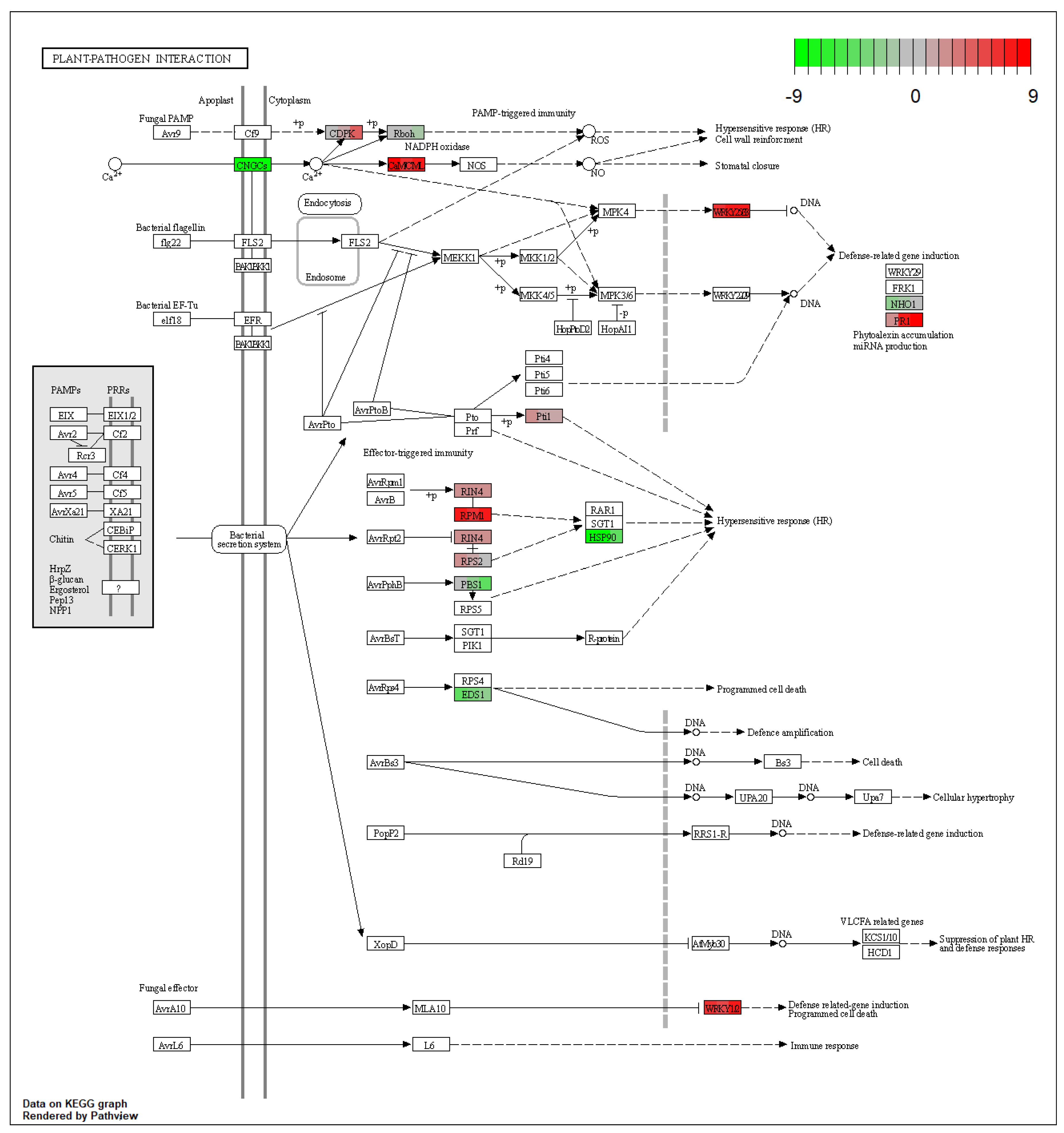
3.1. Analysis of Plant Pathogen Interaction Pathway under V. amli Infection

To understand how M. sieversii responds to the V. mali infection at a transcriptional level, we previously sequenced transcriptome of M. sieversii in response to V. mali infection by PacBio approach [27]. The KEGG pathway enrichment analysis indicated that the plant–pathogen interaction pathway was significantly enriched by the V. mali infection (Figure S1).
Further analysis showed that 73 differentially expressed genes across the time points were involved in plant–pathogen interaction pathways (Figure 1). Infection of V. mali triggered overexpression of the ETI (effector triggered immunity)-related core genes, such as resistance gene RPM1 (resistance to P. syringae pv. maculicola) and RPS2 (resistance to P. syringae) or RIN4 (RPM1interacting protein 4). Interestingly, HSP90 was downregulated while its interacting partners RAR1 and SGT1 remained constant.

3.2. Identification of HSP90 Gene Family in M. sieversii

To accurately identify the members of HSP90 gene family in M. sievesii, we first confirmed the homology of the HSP90 gene family in the M. domestica genome. A total of eight MdHSP90 genes were identified, and were mapped onto different chromosomes (Table 1, Figure 2b). Furthermore, we used a local BLAST search against the M. sievesii genome. The HSP90 proteins from M. domestica, A. thaliana, P. trichocarpa, V. vinifera, and O. sativa were served as a query sequence, and total of eight MsHSP90 genes were identified (Table 1, Figure 2c). To further elucidate the evolutionary relationship of the HSP90 gene family, the phylogenetic tree was constructed using HSP90 genes from different plant species by the neighbor-joining method.
As shown in Figure 2a, the HSP90 genes were classified into seven categories (HSP90-1~7), and the MsHSP90 and MdHSP90 genes were clustered into five different categories (Figure 2d). For the MdHSP90 gene family, each of the MdHSP90-1, MdHSP90-2, and MdHSP90-6 genes has two homologues while MdHSP90-5 and MdHSP90-7 was clustered with single members. For the MsHSP90 gene family, however, each of the MsHSP90-1, MsHSP90-2, and MsHSP90-7 genes has two members while MsHSP90-5 and MsHSP90-6 have single members. We performed exon–intron structure analysis for MsHSP90 and MdHSP90 genes. Our results showed a high degree of similarity in the same branch (Figure 2d).

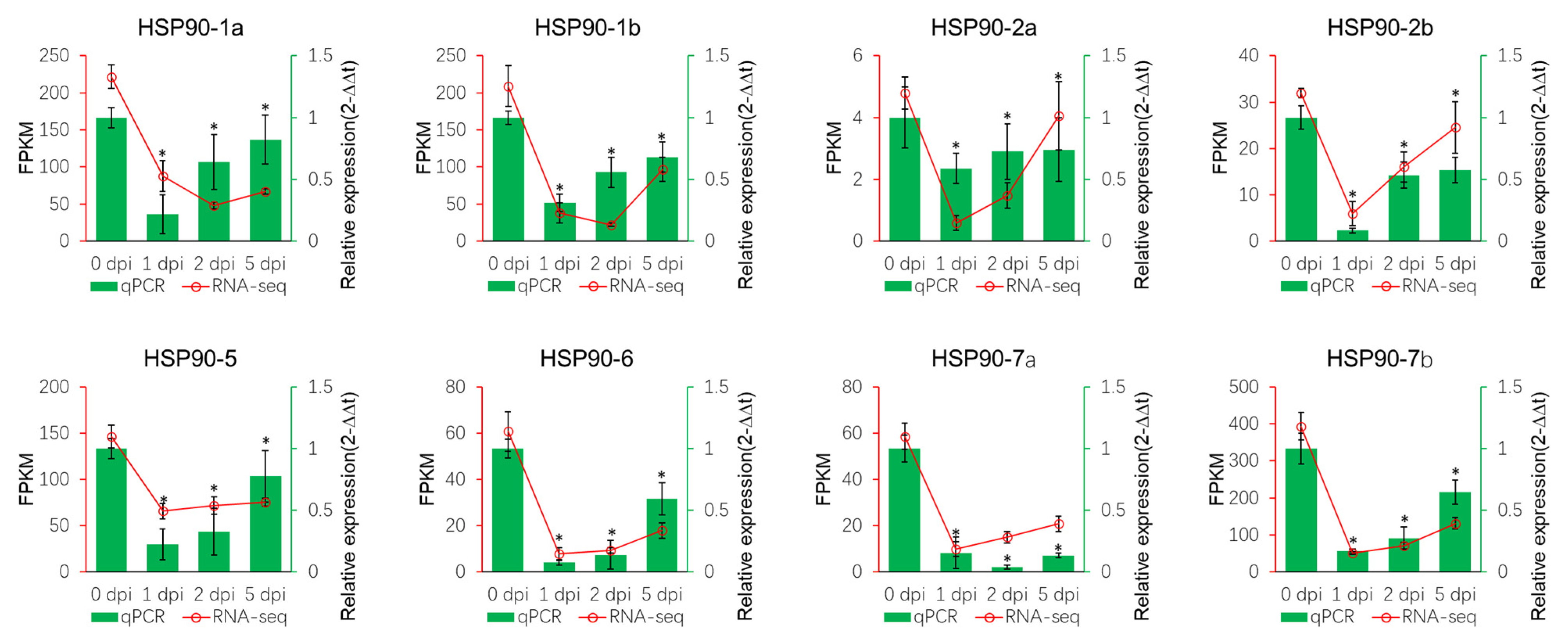
3.3. Expression of MsHSP90 Genes in Response to V.mali

To investigate how MsHSP90 genes respond to a V. mali attack, we analysed M. sieversii transcriptome data under V. mali infection. A total of eight HSP90 transcripts from M. sieversii were subjected to further differential expression analysis through comparisons of foldchange expression. MsHSP90-6 was significantly downregulated across each time point, while MsHSP90-5 did not respond to fungal infection. MsHSP90-2a/b decreased significantly in the early stage but slightly increased at the late stage of infection. To validate the expression pattern of each transcript, we quantified the transcription level of each MsHSP90 gene by qRT-PCR at different time points after infection. Expression analysis revealed that relative expression of all MsHSP90 genes was decreased by infection of V. mali (Figure 3, b). The qRT-PCR results of MsHSP90 transcripts were consistent with the RNA-seq data (Figure 3, a).

3.4. Prediction of Potential Cis-Elements on HSP90s

To explore potential transcriptional mechanism of HSP90 genes, we searched for the occurrence of HSE sequences in the promoter regions (within 2500 bp upstream of the coding region) of 8 MsHSP90 genes. Analysis of the promoter region showed that except MsHSP90-6 other MsHSP90 genes have at least single HSE motif (Table 2), strongly suggesting that MsHSP90 genes are regulated by HSFs. No HSE motif was identified on the promoter region of MsHSP90-6 inferred that MsHSP90-6 gene may be targeted by other transcription factors rather than HSFs.
To investigate whether other transcription factors besides SHF potentially target the MsHSP90 genes, we scan transcription binding sites on the promotor regions of eight MsHSP90 genes by plant regulatory data and analysis platform PlantRegMap.3.3 (http://plantregmap.gao-lab.org/, accessed on 15 May 2021) and a total of 430 binding sites of 145 transcription factors were identified. Binding sites of bZIP, BBR-BPC, ERF, Dof, and MIKC_MADS families were identified as top five abundant binding sites (Figure 4).

4. Discussion

HSP90 is a type of chaperone that is highly conserved throughout biology kingdoms, and widely involved in various biological processes. To date, the HSP90 gene family has been identified in various plants, such as A. thaliana [2], rice [32], and N. tabacum [7]. The apple genome was successfully sequenced and accurate sequence information or annotations are available [33]. However, the genome-wide identification of the HSP90 gene family and their expression patterns under fungal infection has not been conducted in apples, especially wild apples, M. sieversii.
M. sieversii is a wild species native to the mountains of central Asia and eastern China. Recent study showed that M. sieversii in Xinjiang, China is the most primitive and principal ancestor of the domesticated apple and is therefore a globally important genetic resource [20]. In Xinjiang, however, the natural habitat of M. sieversii has declined by expansion and development of agriculture and the population is shrinking by other natural disasters such as canker diseases caused by the necrotrophic pathogen V. mali [22]. Therefore, exploring the disease resistance gene resources in M. sieversii upon the infection of V. mali has great value in disease prevention.
To uncover the potential function of HSP90 genes in M. sieversii under V. mali infection, in the first place we determined eight HSP90 genes in M. sieversii (Table 1). The MsHSP90 genes were mainly distributed on both ends of the chromosome (Figure 2c) which was similar to the distribution of M. domestica (Figure 2b), rice [32], and Nicotiana tabacum [7] HSP90 genes. Different numbers of introns (2 to 19 introns) were identified in different MsHSP90 gene sequences; MsHSP90-6 has the maximum numbers of introns while MsHSP90-2a/b has the minimum number of introns, implying the diverse sensitivity of gene transcription regulation (Figure 2d). Subsequently, the eight transcripts were identified in M. sieversii full-length transcriptome data based on MsHSP90 gene sequences. The MsHSP90 genes shared extremely high similarity in the number of sequence compositions with MdHSP90s, suggesting that M. domestica maintained some of its ancestors’ traits in the respect of HSP90 molecular evolution, implying functional redundancy.
HSP90 plays a crucial role in host fungal defence strategies. To investigate the expression pattern of HSP90 genes in response to fungal infection in plants, the published data of HSP90 gene expression in response to Penicilliumi expansum [21], Alternaria alternate [34], and Phytophthora infestans [35] were collected and fold change expression data were obtained. The expression of HSP90 genes were varied in different host species. The expression of HSP90-7 can be host-specific or pathogen-specific. In M. domestica, for instance, the HSP90-7 gene was repressed by infection of P. expansum while unchanged in the late phase of A. alternata infection (Figure S2). The same expression pattern of HSP90-7 was observed in two different populus species in response to the same pathogen. It seems that different plants employ different HSP90s in response to different fungal pathogens and also implying that each HSP90 genes was regulated by different transcription factors or has extremely distinct transcriptional regulatory elements.
Recent studies have demonstrated that HSP90 proteins mediate signal transduction in plant–pathogen interaction pathways in plants. HSP90 interacts with SGT1 (suppressor of the G-two allele of skp1) and RAR1 (required for Mla12 resistance), and modulates the immune response by regulating R protein stability [36,37,38,39]. Hsp90-SGT1-RAR1complex in Arabidopsis plays a key role in assisting in R protein-mediated disease resistance [37]. Despite the HSP90, RAR1, and SGT1 proteins interacting with each other, the diversity in transcriptional regulation of their encoding genes reflects their distinct functions. In Arabidopsis, for example, transcription level of AtSGT1a and AtSGT1b was elevated by Peronospora parasitica infection [40], but no significant change was observed at the protein level [41].
In this study, transcriptome data analysis indicated that the plant–pathogen interaction pathway in M. sieversii was enriched (Figure S1) and the MsHSP90 gene was downregulated, while SGT1 and RAR1 were unchanged in response to V. mali infection (Figure 1). qRT-PCR analysis of MsHSP90 gene expressions also demonstrated that all members of the MsHSP90 gene family were repressed by V. mali infection (Figure 3), implying that V. mali undermined the M. sieversii immune system by blocking HSP90-mediated signal transduction. In order to investigate how host plant HSP90 gene expression is modulated in response to fungal infection, we collected the published expression data of HSP90 genes from A. thaliana, M. domestica, and tow populus species in response to different fungal pathogens. Only a single gene from Populus deltoids, PaHSP90-4a, was downregulated by Marssonia brunnea infection while PaHSP90-3 was upregulated (Figure S2). In other cases, the HSP90 gene expression showed fluctuation, indicating that it is not common that the pathogen targets host HSP90 to interfere with the immune system so that it successfully evades host immune response such as hypersensitive response.
To understand how MsHSP90 genes were transcriptionally regulated, we analysed the promoter of MsHSP90 genes. We detected canonical HSE (nGAAnnTTCn) in the promotor of MdHSP90 genes, implying that HSE regulatory elements are essential for V. mali response. We further identified various transcription biding elements (Figure 4) on the promotor region of MsHSP90 genes suggesting that each MsHSP90 gene expression may be regulated by different transcription factor. Our results imply the complexity of MsHSP90 gene expression and versatility of HSP90 in distinct biological pathways.
In this study, a total of eight HSP90 genes in M. sieversii were identified and characterized by the bioinformatic analyses. A total of eight HSP90 transcripts in M. sieversii were detected in transcriptome data under V. mali infection, and MsHSP90 genes were repressed in response to V. mali infection. We anticipate that V. mali may facilitate infection by repressing MsHSP90 gene expression so that it diminishes host immune response. Our study also indicated that MsHSP90 may be regulated by various transcription factors in response to different stress. This study could not only provide a scientific foundation for the comprehensive understanding of the apple HSP90 gene family, but could also be helpful in screening candidate genes for canker disease prevention.

Supplementary Materials

The following are available online at https://www.mdpi.com/article/10.3390/f12091232/s1, Figure S1: KEGG pathway enrichment analysis of differentially expressed genes response to V. mali infection in M. sieversii. Figure S2. Expression profiles of HSP90 genes in response to different plant fungal pathogens. Table S1: List of the qRT-PCR primers of the MsHSP90 genes.

Author Contributions

The experimental design, data analyzation, manuscript organization were completed by Y.H., Y.S., X.L. (Xiaojie Liu), Y.D. and X.W. were assistant with RNA quantification. D.Z. and X.L. (Xiaoshuang Li) conceived the project, supervised the analysis, and critically revised the manuscript. G.K. was revised the manuscript. All authors have read and agreed to the published version of the manuscript.

Funding

This work was funded by the Second Tibetan Plateau Scientific Expedition and Research (STEP) program (2019QZKK0502030403), the Science and Technology Plan Project of Xinjiang Uygur Autonomous Region (2016B03040), the Youth Innovation Promotion Association, Chinese Academy of Sciences (No. 2018478) and the China Postdoctoral Science Foundation (2021M693380).

Institutional Review Board Statement

Not applicable.

Informed Consent Statement

Not applicable.

Conflicts of Interest

The authors declare no conflict of interest.

References

  1. Schopf, F.H.; Biebl, M.M.; Buchner, J. The HSP90 chaperone machinery. Nat. Rev. Mol. Cell Biol. 2017, 18, 345–360. [Google Scholar] [CrossRef] [PubMed]
  2. Krishna, P.; Gloor, G. The HSP90 family of proteins in Arabidopsis thaliana. Cell Stress Chaperones 2001, 6, 238–246. [Google Scholar] [CrossRef]
  3. Ye, S.; Yu, S.; Shu, L.; Wu, J.; Wu, A.; Luo, L. Expression profile analysis of 9 heat shock protein genes throughout the life cycle and under abiotic stress in rice. Chin. Sci. Bull. 2012, 57, 336–343. [Google Scholar] [CrossRef] [Green Version]
  4. Xu, J.; Xue, C.; Xue, D.; Zhao, J.; Gai, J.; Guo, N.; Xing, H. Overexpression of GmHSP90s, a heat shock protein 90 (HSP90) gene family cloning from soybean, decrease damage of abiotic stresses in Arabidopsis thaliana. PLoS ONE 2013, 8, e69810. [Google Scholar] [CrossRef]
  5. Zhang, K.; He, S.; Sui, Y.; Gao, Q.; Jia, S.; Lu, X.; Jia, L. Genome-Wide Characterization of HSP90 Gene Family in Cucumber and Their Potential Roles in Response to Abiotic and Biotic Stresses. Front. Genet. 2021, 12, 95. [Google Scholar] [CrossRef]
  6. Zhang, J.; Li, J.; Liu, B.; Zhang, L.; Chen, J.; Lu, M. Genome-wide analysis of the Populus HSP90 gene family reveals differential expression patterns, localization, and heat stress responses. BMC Genom. 2013, 14, 532. [Google Scholar] [CrossRef] [Green Version]
  7. Song, Z.; Pan, F.; Yang, C.; Jia, H.; Jiang, H.; He, F.; Li, N.; Lu, X.; Zhang, H. Genome-wide identification and expression analysis of HSP90 gene family in Nicotiana tabacum. BMC Genet. 2019, 20, 35. [Google Scholar] [CrossRef] [PubMed] [Green Version]
  8. Xu, Z.-S.; Li, Z.-Y.; Chen, Y.; Chen, M.; Li, L.-C.; Ma, Y.-Z. Heat Shock Protein 90 in Plants: Molecular Mechanisms and Roles in Stress Responses. Int. J. Mol. Sci. 2012, 13, 15706–15723. [Google Scholar] [CrossRef]
  9. Corigliano, M.G.; Clemente, M. The Role of Plant 90-kDa Heat Shock Proteins in Plant Immunity. In Heat Shock Proteins and Plants; Asea, A.A.A., Kaur, P., Calderwood, S.K., Eds.; Springer International Publishing: Cham, Switzerland, 2016; pp. 149–167. ISBN 978-3-319-46340-7. [Google Scholar]
  10. Zai, W.S.; Miao, L.X.; Xiong, Z.L.; Zhang, H.L.; Ma, Y.R.; Li, Y.L.; Chen, Y.B.; Ye, S.G. Comprehensive identification and expression analysis of HSP90s gene family in Solanum lycopersicum. Genet. Mol. Res. 2015, 14, 7811–7820. [Google Scholar] [CrossRef]
  11. Prasad, B.D.; Goel, S.; Krishna, P. In silico identification of carboxylate clamp type tetratricopeptide repeat proteins in Arabidopsis and rice as putative co-chaperones of HSP90/HSP70. PLoS ONE 2010, 5, e12761. [Google Scholar] [CrossRef] [PubMed]
  12. Mishra, R.C.; Grover, A. Constitutive over-expression of rice ClpD1 protein enhances tolerance to salt and desiccation stresses in transgenic Arabidopsis plants. Plant Sci. 2016, 250, 69–78. [Google Scholar] [CrossRef] [PubMed]
  13. Vítámvás, P.; Prášil, I.T.; Kosová, K.; Planchon, S.; Renaut, J. Analysis of proteome and frost tolerance in chromosome 5A and 5B reciprocal substitution lines between two winter wheats during long-term cold acclimation. Proteomics 2012, 12, 68–85. [Google Scholar] [CrossRef] [PubMed]
  14. Schöffl, F.; Prändl, R.; Reindl, A. Regulation of the Heat-Shock Response. Plant Physiol. 1998, 117, 1135–1141. [Google Scholar] [CrossRef] [PubMed] [Green Version]
  15. Prasinos, C.; Krampis, K.; Samakovli, D.; Hatzopoulos, P. Tight regulation of expression of two Arabidopsis cytosolic HSP90 genes during embryo development. J. Exp. Bot. 2005, 56, 633–644. [Google Scholar] [CrossRef] [Green Version]
  16. Lohmann, C.; Eggers-Schumacher, G.; Wunderlich, M.; Schöffl, F. Two different heat shock transcription factors regulate immediate early expression of stress genes in Arabidopsis. Mol. Genet. Genom. 2004, 271, 11–21. [Google Scholar] [CrossRef] [PubMed]
  17. Sun, Y.; Guo, H.; Yuan, E.; Ge, F. Elevated CO2 increases R gene-dependent resistance of Medicago truncatula against the pea aphid by up-regulating a heat shock gene. New Phytol. 2018, 217, 1696–1711. [Google Scholar] [CrossRef] [Green Version]
  18. Ito, M.; Ohnishi, K.; Hikichi, Y.; Kiba, A. Molecular chaperons and co-chaperons, HSP90, RAR1, and SGT1 negatively regulate bacterial wilt disease caused by Ralstonia solanacearum in Nicotiana benthamiana. Plant Signal. Behav. 2015, 10, e970410. [Google Scholar] [CrossRef] [Green Version]
  19. Zhang, H.; Yang, Y.; Wang, C.; Liu, M.; Li, H.; Fu, Y.; Wang, Y.; Nie, Y.; Liu, X.; Ji, W. Large-scale transcriptome comparison reveals distinct gene activations in wheat responding to stripe rust and powdery mildew. BMC Genom. 2014, 15, 1–4. [Google Scholar] [CrossRef] [PubMed] [Green Version]
  20. Duan, N.; Bai, Y.; Sun, H.; Wang, N.; Ma, Y.; Li, M.; Wang, X.; Jiao, C.; Legall, N.; Mao, L.; et al. Genome re-sequencing reveals the history of apple and supports a two-stage model for fruit enlargement. Nat. Commun. 2017, 8, 1–11. [Google Scholar] [CrossRef] [PubMed]
  21. Ballester, A.-R.; Norelli, J.; Burchard, E.; Abdelfattah, A.; Levin, E.; González-Candelas, L.; Droby, S.; Wisniewski, M. Transcriptomic Response of Resistant (PI613981–Malus sieversii) and Susceptible (“Royal Gala”) Genotypes of Apple to Blue Mold (Penicillium expansum) Infection. Front. Plant Sci. 2017, 8, 1981. [Google Scholar] [CrossRef]
  22. Liu, X.; Li, X.; Bozorov, T.A.; Ma, R.; Ma, J.; Zhang, Y.; Yang, H.; Li, L.; Zhang, D. Characterization and pathogenicity of six Cytospora strains causing stem canker of wild apple in the Tianshan Forest, China. For. Pathol. 2020, 50, e12587. [Google Scholar] [CrossRef]
  23. Bozorov, T.A.; Luo, Z.; Li, X.; Zhang, D. Agrilus mali Matsumara (Coleoptera: Buprestidae), a new invasive pest of wild apple in western China: DNA barcoding and life cycle. Ecol. Evol. 2019, 9, 1160–1172. [Google Scholar] [CrossRef] [PubMed] [Green Version]
  24. Soosaar, J.L.M.; Burch-Smith, T.M.; Dinesh-Kumar, S.P. Mechanisms of plant resistance to viruses. Nat. Rev. Microbiol. 2005, 3, 789–798. [Google Scholar] [CrossRef] [PubMed]
  25. Livak, K.J.; Schmittgen, T.D. Analysis of relative gene expression data using real-time quantitative PCR and the 2−ΔΔCT Method. Methods 2001, 25, 402–408. [Google Scholar] [CrossRef]
  26. Zhu, L.; Yang, C.; You, Y.; Liang, W.; Wang, N.; MA, F.; Li, C. Validation of reference genes for qRT-PCR analysis in peel and flesh of six apple cultivars (Malus domestica) at diverse stages of fruit development. Sci. Hortic. 2019, 244, 165–171. [Google Scholar] [CrossRef]
  27. Liu, X.; Li, X.; Wen, X.; Zhang, Y.; Ding, Y.; Zhang, Y.; Gao, B.; Zhang, D. PacBio full-length transcriptome of wild apple (Malus sieversii) provides insights into canker disease dynamic response. BMC Genom. 2021, 22, 1–19. [Google Scholar] [CrossRef]
  28. Hu, B.; Jin, J.; Guo, A.-Y.; Zhang, H.; Luo, J.; Gao, G. GSDS 2.0: An upgraded gene feature visualization server. Bioinformatics 2015, 31, 1296–1297. [Google Scholar] [CrossRef] [Green Version]
  29. Bu, D.; Luo, H.; Huo, P.; Wang, Z.; Zhang, S.; He, Z.; Wu, Y.; Zhao, L.; Liu, J.; Guo, J.; et al. KOBAS-i: Intelligent prioritization and exploratory visualization of biological functions for gene enrichment analysis. Nucleic Acids Res. 2021, 49, W317–W325. [Google Scholar] [CrossRef] [PubMed]
  30. Tian, F.; Yang, D.-C.; Meng, Y.-Q.; Jin, J.; Gao, G. PlantRegMap: Charting functional regulatory maps in plants. Nucleic Acids Res. 2019, 48, D1104–D1113. [Google Scholar] [CrossRef]
  31. Grant, C.E.; Bailey, T.L.; Noble, W.S. FIMO: Scanning for occurrences of a given motif. Bioinformatics 2011, 27, 1017–1018. [Google Scholar] [CrossRef] [Green Version]
  32. Hu, W.; Hu, G.; Han, B. Genome-wide survey and expression profiling of heat shock proteins and heat shock factors revealed overlapped and stress specific response under abiotic stresses in rice. Plant Sci. 2009, 176, 583–590. [Google Scholar] [CrossRef]
  33. Daccord, N.; Celton, J.-M.; Linsmith, G.; Becker, C.; Choisne, N.; Schijlen, E.; van de Geest, H.; Bianco, L.; Micheletti, D.; Velasco, R.; et al. High-quality de novo assembly of the apple genome and methylome dynamics of early fruit development. Nat. Genet. 2017, 49, 1099–1106. [Google Scholar] [CrossRef] [PubMed]
  34. Zhu, L.; Weichen, N.; Liu, S.; Cai, B.; Xing, H.; Wang, S. Transcriptomics Analysis of Apple Leaves in Response to Alternaria alternata Apple Pathotype Infection. Front. Plant Sci. 2017, 8, 22. [Google Scholar] [CrossRef] [PubMed] [Green Version]
  35. Swindell, W.R.; Huebner, M.; Weber, A.P. Transcriptional profiling of Arabidopsis heat shock proteins and transcription factors reveals extensive overlap between heat and non-heat stress response pathways. BMC Genom. 2007, 8, 1–5. [Google Scholar] [CrossRef] [Green Version]
  36. Liu, Y.; Burch-Smith, T.; Schiff, M.; Feng, S.; Dinesh-Kumar, S.P. Molecular Chaperone HSP90 Associates with Resistance Protein N and Its Signaling Proteins SGT1 and Rar1 to Modulate an Innate Immune Response in Plants. J. Biol. Chem. 2004, 279, 2101–2108. [Google Scholar] [CrossRef]
  37. Takahashi, A.; Casais, C.; Ichimura, K.; Shirasu, K. HSP90 interacts with RAR1 and SGT1 and is essential for RPS2-mediated disease resistance in Arabidopsis. Proc. Natl. Acad. Sci. USA 2003, 100, 11777–11782. [Google Scholar] [CrossRef] [Green Version]
  38. Qian, L.; Zhao, J.; Du, Y.; Zhao, X.; Han, M.; Liu, Y. HSP90 Interacts With Tm-22 and Is Essential for Tm-22-Mediated Resistance to Tobacco mosaic virus. Front. Plant Sci. 2018, 9, 411. [Google Scholar] [CrossRef] [Green Version]
  39. Shirasu, K. The HSP90-SGT1 Chaperone Complex for NLR Immune Sensors. Annu. Rev. Plant Biol. 2009, 60, 139–164. [Google Scholar] [CrossRef] [PubMed] [Green Version]
  40. Azevedo, C.; Betsuyaku, S.; Peart, J.; Takahashi, A.; Noël, L.; Sadanandom, A.; Casais, C.; Parker, J.; Shirasu, K. Role of SGT1 in resistance protein accumulation in plant immunity. EMBO J. 2006, 25, 2007–2016. [Google Scholar] [CrossRef] [Green Version]
  41. Austin, M.J.; Muskett, P.; Kahn, K.; Feys, B.J.; Jones, J.D.G.; Parker, J.E. Regulatory Role of SGT1 in Early R Gene-Mediated Plant Defenses. Science 2002, 295, 2077–2080. [Google Scholar] [CrossRef] [Green Version]
Figure 1. Visualization of KEGG plant–pathogen interaction pathway. Foldchange value of single genes at 1 dpi, 2 dpi, and 5 dpi in M. sieversii infected with V. mali were plotted using pathview package of R/Bioconductor. The nodes are split between different colors, indicating 1 dpi (left), 2 dpi (middle), and 5 dpi (right) data.
Forests 12 01232 g001
Figure 2. Bioinformatic analysis of HSP90 genes. (a) Phylogenetic tree of HSP90 proteins from different species. Protein sequences were aligned by ClustalW algorism, and phylogenetic tree was generated by using the neighbor-joining method with 1000 bootstrap in MEGA X. Different shapes represent different species. Chromosomal locations of the 8 MdHSP90 (b) and MsHSP90 (c) genes. MG2C online tools were used (http://mg2c.iask.in/mg2c_v2.1/, accessed on 15 May 2021) to depict chromosomal location, and the gene was marked with short lines. (d) Gene structure of MsHSP90 and MdHSP90 genes. GSDS2.0 (http://gsds.cbi.pku.edu.cn/, accessed on 15 May 2021) online tools [28] were used to visualize exon–intron structures.
Forests 12 01232 g002
Figure 3. Expression of HSP90 genes in M. sieversii during V. mali infection. The normalized expression level (FPKM) from the RNA-seq data were shown in red line on right vertical axis and relative expression level from qPCR were shown in green bar on right vertical axis. Verification of the expression of MdHSP90s in M. sieversii under V. mali infection by qRT-PCR. MsEF1-α was used as an internal reference gene. The relative expression data were analyzed by one-way ANOVA method. “*” represented significantly different (p < 0.05, n = 3), and error bars indicate mean ± SE.
Forests 12 01232 g003
Figure 4. Prediction of potential transcription factors that binding sites of MsHSP90 genes. The promoter regions (within 2500 bp upstream of the coding region) of 8 MsHSP90 genes were obtained from the M. sieversii genome and analysed by PlantRegMap.3.3 tools (http://plantregmap.gao-lab.org/binding_site_prediction.php, accessed on 15 May 2021). Each dot represents the number of transcription factor binding sites in the MsHSP90 gene promotor region. Threshold p-value ≤ 1 × 10⁻⁵.
Forests 12 01232 g004
Table 1. HSP90 genes in M. domestica and M. sieversii.
Gene SymbolLength
of CDS (bp)
ChromosomesGenomic CoordinatesStrandNumber of Amino Acids in Protein
MdHSP90-1a21127NC_041795.1: 34410217-34414101+703
MdHSP90-1b21121NC_041789.1: 30385625-30388837+703
MdHSP90-2a210011NC_041799.1: 3210146-3213261699
MdHSP90-2b21003NC_041791.1: 2915642-2918677699
MdHSP90-5238516NC_041804.1: 3808810-3813939794
MdHSP90-6a242117NC_041805.1: 9778825-9785023806
MdHSP90-6b24309NC_041797.1: 9428843-9434534809
MdHSP90-724578NC_041796.1: 866442-871008+818
MsHSP90-1a24909Chr9:35058827-35061327+829
MsHSP90-1b151213Chr13:32200739-32203239+503
MsHSP90-2a21005Chr5:3353408-3350908699
MsHSP90-2b20858Chr8:2754830-2752330694
MsHSP90-523856Chr6:3862971-3860471794
MsHSP90-6241510Chr10:27515958-27518458+804
MsHSP90-7a24571Chr1:622597-625097+818
MsHSP90-7b245715Chr15:32199616-32197116818
Table 2. The list of HSE motifs identified in the promoters of MsHSP90 genes.
Gene NameStartEndp-Valueq-ValueMatched Sequence
MsHSP90-1a40497.01 × 10−50.672--GTTCACGAAC--
MsHSP90-1b213921481.19 × 10−50.232--GGAACGTTCC--
217621852.63 × 10−40.509--AGAACATTCT--
243524442.63 × 10−40.509--AGAAAATTCG--
245724662.63 × 10−40.509--CGAATTTTCT--
MsHSP90-2a1942031.76 × 10−40.509--CGAAAATTCC--
175417631.76 × 10−40.509--AGAACGTTCA--
4454542.63 × 10−40.509--AGAAATTTCG--
144714562.89 × 10−40.672--TTTCATGAAA--
MsHSP90-2b1171261.76 × 10−40.509--GGAATTTTCG--
MsHSP90-5130113102.89 × 10−40.509--TGAATTTTCT--
224422531.76 × 10−40.672--GTTCCAGAAA--
229022991.76 × 10−40.672--TTTCCAGAAG--
MsHSP90-7a154015492.63 × 10−40.509--TGAACTTTCA--
MsHSP90-7b118211911.76 × 10−40.509--GGAAATTTCG--
237223811.76 × 10−40.672--TTTCTGGAAC--
Publisher’s Note: MDPI stays neutral with regard to jurisdictional claims in published maps and institutional affiliations.

