Article

New Species of Tomentella (Thelephorales, Basidiomycota) from Temperate Continental Mountain Climate of China (Xinjiang Region)

1
CAS Key Laboratory of Forest Ecology and Management, Institute of Applied Ecology, Chinese Academy of Sciences, Shenyang 110164, China
2
College of Basic Medical Science, Liaoning He’s Medical University, Shenyang 110163, China
3
University of the Chinese Academy of Sciences, Beijing 100049, China
*
Author to whom correspondence should be addressed.
Forests 2021, 12(11), 1531; https://doi.org/10.3390/f12111531
Submission received: 21 September 2021 / Revised: 1 November 2021 / Accepted: 4 November 2021 / Published: 7 November 2021
(This article belongs to the Special Issue Fungal Diversity in Forest Ecosystems)

Abstract

:
Species in the genus Tomentella are distributed throughout the temperate and tropical regions worldwide, but few studies associated with the taxonomy and phylogeny of this genus had been reported from Northwest China. In this paper, molecular phylogenetic analyses of the nuclear ribosomal ITS (internal transcribed spacer: ITS1-5.8S-ITS2) and LSU (large subunit: 28S) sequences combined with morphological characteristics identified three new species from Xinjiang Autonomous Region in Northwest China, which were named T. aurantispora, T. kanasensis, and T. schrenkiana. Similar macromorphological and anatomical characteristics are shared by these new species: arachnoid basidiocarps; byssoid sterile margins; utriform basidia with a clamp connection at the base; the absence of rhizomorphs and cystidia; and slightly thick-walled, subglobose to globose basidiospores. Among these new species, the color of the hymenophoral surface, the size of the basidiospores, and some other features can be used for species delimitation. The new species and closely related species in the phylogenetic tree were discussed, and a key to the identified species of Tomentella from China was provided.

1. Introduction

Studies on the genus Tomentella Pers. have undergone a long and convoluted history in several regions worldwide. The name Tomentella was used first by Persoon in 1799 [1] and was published as a genus by Patouillard in 1887 [2]. The initial studies on taxonomy of this genus were mainly from North America and Eurasia [3,4], and some species were also reported from West India, Australia, and New Zealand [5,6,7]. In recent years, more species were subsequently reported from many regions worldwide, including Asia, Africa, South America, and so on [8,9,10,11].
Tomentella is closely related to Odontia Pers. and Thelephora Ehrh. ex Willd. They belong to Thelephoraceae Chevall., Thelephorales Corner ex Oberw. of Basidiomycota R.T. Moore [3,4,5]. Odontia is morphologically similar to Tomentella by the resupinate basidiocarps with gray to brown colors and the generative hyphae with simple septa or clamps [3,12]. The basidiocarps of Odontia and Tomentella are often found on fallen branches, leaves, twigs, and decayed wood debris in coniferous and deciduous forests [3,9,13]. The similar morphological features and habitats make it difficult to distinguish these two genera, and species of Odontia were originally recognized as Tomentella. Species of Tomentella had been found to form ectomycorrhizae with jack pine since the 1980s [14]. However, Odontia was declared a non-mycorrhizal genus, verified by stable isotope and phylogenetic analyses, and treated as a separate Tomentella [12,15]. Thelephora was categorized as an ectomycorrhizal genus slightly earlier than Tomentella [16,17], and species in this group often form paraphyletic clades in the phylogenetic trees [9,18]. Thelephora and Tomentella possess obviously different types of basidiocarps, which are epigeous and resupinate, respectively [3,19]. The tomentella-thelephora lineage has been found to be one of the most species-rich ectomycorrhizal (ECM) lineages in forest ecosystems [18,20,21].
ECM fungi have attracted much attention over the years, because they can play an essential role in nutrient cycling [22], seedling establishment [23], tree growth rate and survival [24,25], and biodiversity maintenance [26] in natural ecosystems. ECM fungi can produce conspicuous or inconspicuous basidiocarps, and about 70% of basidiomycete species with inconspicuous basidiocarps are comprised of resupinate tomentelloid fungi [27]. In China, a few studies on ECM fungal diversity and the community structure of Fagaceae, Pinaceae, and Salicaceae revealed that Tomentella was one of the most species-rich genera in some forests [28,29,30,31]. Studies on the ECM fungal communities of Quercus fabri Hance., Q. liaotungensis Koidz. in the family Fagaceae have identified 6 and 33 taxonomic units or species of Tomentella, respectively [29,32,33]. In addition, tomentella-thelephora lineages associated with Fagaceae species from 30 forest sites in China account for 26.4% of the total sequences in 92.8% of the samples [31]. Studies have also reported that the tomentella-thelephora lineage can account for 38.2% of the total ECM fungal reads from the mycorrhizal root tips of Betulaceae, Fagaceae, Pinaceae, and Tiliaceae [34]. These studies revealed that there are a large number of Tomentella species in China, but studies based on basidiocarps of Tomentella species are scarce in Xinjiang Autonomous Region of Northwest China. At present, 48 species distributed in Liaoning, Heilongjiang, and Jilin Provinces of northeastern China have been reported based on the collected specimens [10,11].
In this study, basidiocarps were collected from three regions in Xinjiang Autonomous Region, including the Kanas National Nature Reserve, Tianshan Tianchi National Park, and Tianshan Grand Canyon National Forest Park. The regions belong to the typical temperate continental mountain climate with the obvious temperature fluctuations between day and night. Their average annual temperatures are –0.2 °C, 2.6 °C, and 5 °C, respectively [35,36], with average annual precipitation of 1065, 610, and 600–800 mm [35,36,37]. Kanas National Nature Reserve is the only area characterized by European Siberian flora in China [35], with the domination of Abies sibirica Ledeb., Betula pendula Roth., Larix sibirica Ledeb., Picea obovata Ledeb., Pinus sibirica (Loud.) Mayr, and Populus tremula Linn [36]. Tianshan Tianchi National Park is dominated by the Schrenk spruce, P. schrenkiana Fisch. et Mey., mixed with B. tianschanica Ruprecht, B. pendula, and L. sibirica [38]. P. schrenkiana accounts for more than 91% of the trees in the Tianshan Grand Canyon National Forest Park, with a mixture of Populus davidiana Dode., Betula spp. and some other coniferous and deciduous species [37]. During this study, three undescribed species of Tomentella were detected using morphological and phylogenetic analyses of DNA sequences. The aim of this work is to update the species diversity of Tomentella in China and assess its taxonomic and phylogenetic status.

2. Materials and Methods

2.1. Morphological Studies

The specimens were collected from the Kanas National Nature Reserve (KNNR), Tianshan Tianchi National Park (TTNP), and Tianshan Grand Canyon National Forest Park (TGCNFP) in Xinjiang Autonomous Region of Northwest China and were deposited at the herbarium of the Institute of Applied Ecology, Chinese Academy of Sciences (IFP).
The macromorphological characteristics of the basidiocarps were observed using a stereomicroscope (Nikon SMZ 1000: Tokyo, Japan) at 4× magnification, and the special color terms followed Kornerup [39]. Observations of the microscopic characteristics followed Lu et al. [10], and these were examined on dried specimens rehydrated in 2.5% KOH and stained in cotton blue and Melzer’s reagent. The following abbreviations were used in the text: IKI = Melzer’s reagent; IKI- = neither amyloid nor dextrinoid; KOH = 2.5% potassium hydroxide; CB = cotton blue; CB+ = cyanophilous; CB- = acyanophilous; L = mean spore length (arithmetic average of all spores in lateral face excluding ornamentation); W = mean spore width (arithmetic average of all spores in lateral face excluding ornamentation); Q = variation in the L/W ratios; and n = number of spores measured from every specimen. Micromorphological characteristics were studied at magnifications up to 1000× using a Nikon Eclipse E600 microscope (Tokyo, Japan) with phase contrast illumination. Drawings were made with a drawing tube. The surface morphologies of the basidiospores were investigated by scanning electron microscopy (SEM) (Phenom ProX, Phenom-World, Eindhoven, The Netherlands) at 10 kV accelerating voltage and in a secondary electron detector (SED) imaging mode. A thin layer of gold was sputter coated on the samples to avoid electrostatic charging.

2.2. Molecular Procedures and Phylogenetic Analyses

DNA was extracted from dried basidiocarps using a Thermo Scientific Phire Plant Direct PCR Kit (Thermo Fisher Scientific, Waltham, MA, USA). ITS and LSU regions were amplified with SSU1318-Tom/LSU-Tom4 [40] and LROR/LR7 primer pairs [41], respectively. Protocols for the polymerase chain reaction (PCR) amplifications of ITS and LSU regions were as follows: initial denaturation at 95 °C for 5 min, followed by 39 cycles at 95 °C for 30 s, × °C for 30 s (the annealing temperatures for SSU1318-Tom/LSU-Tom4 and LROR/LR7 were 62 °C and 47.2 °C, respectively) [40,41], 72 °C for 20 s, and a final extension at 72 °C for 1 min. PCR reaction products were confirmed on 1% agarose electrophoresis gels stained with ethidium bromide [42] and sequenced at the Beijing Genomics Institute (BGI) with the same primers. The sequencing data were assembled and manually modified with the software DNAMAN 8.0 (Lynnon Biosoft, San Ramon, CA, USA). All newly generated sequences have been verified [43] and were deposited at GenBank (Table S1). Two hundred and thirteen sequences published in previous studies were downloaded from NCBI [44] and UNITE [45], and six newly generated sequences were used in this study. These sequences were aligned with MAFFT, which was available at http://mafft.cbrc.jp/alignment/server/large.html (accessed on 22 February 2021) [46] with manual adjustment. Output files in the FASTA format were directly loadable in the MAFFT online service and converted into NEXUS and PHYLIP files by ClustalX and EasyCodeMLv1.0, respectively [47]. Maximum likelihood (ML) analysis was conducted in RAxMLGUI1.5b2 with PHYLIP files under the GTRGAMMA model for 100 rapid bootstrap replicates under ML + rapid bootstrap mode [48]. Bayesian analysis was carried out in MrBayes 3.2.6 with NEXUS files [49] based on the Markov chain Monte Carlo (MCMC) technique, and the optimal models (K80 + G for ITS1 + 5.8S + ITS2 and GTR + I + G for LSU) for the ITS1, 5.8S, ITS2 and LSU regions were predicted using jModelTest 2.1.10 according to the corrected Akaike information criterion (AICc) [50]. As the model K80 + G was not available in MrBayes, and HKY + G as the most similar model was run instead of K80 + G. Four Markov chains were executed, starting from random trees and keeping one tree every 1000 generations until the average standard deviation of split frequencies was below 0.01. The initial 25% of trees were removed as burn-in.

3. Results

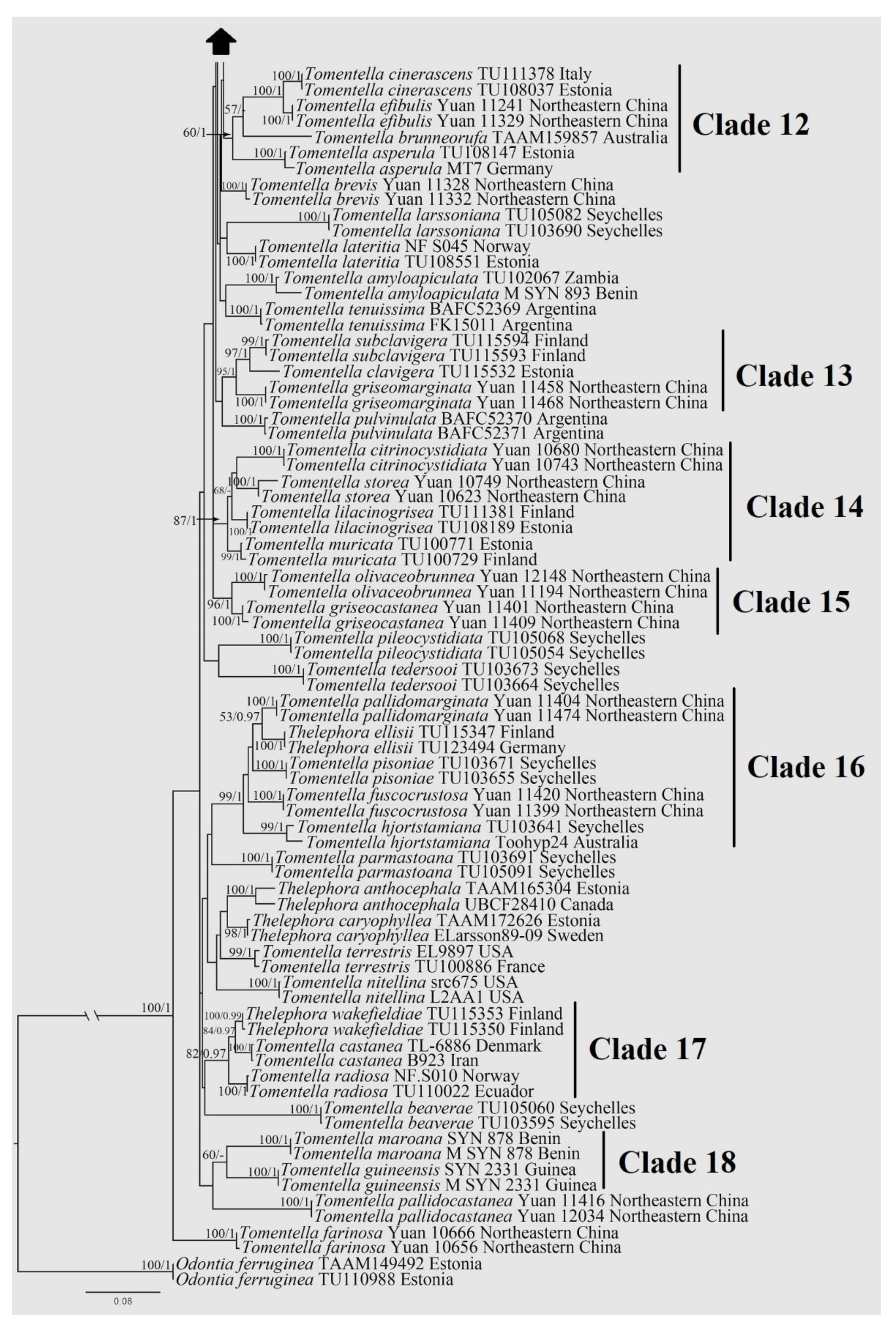
3.1. Phylogenetic Analysis
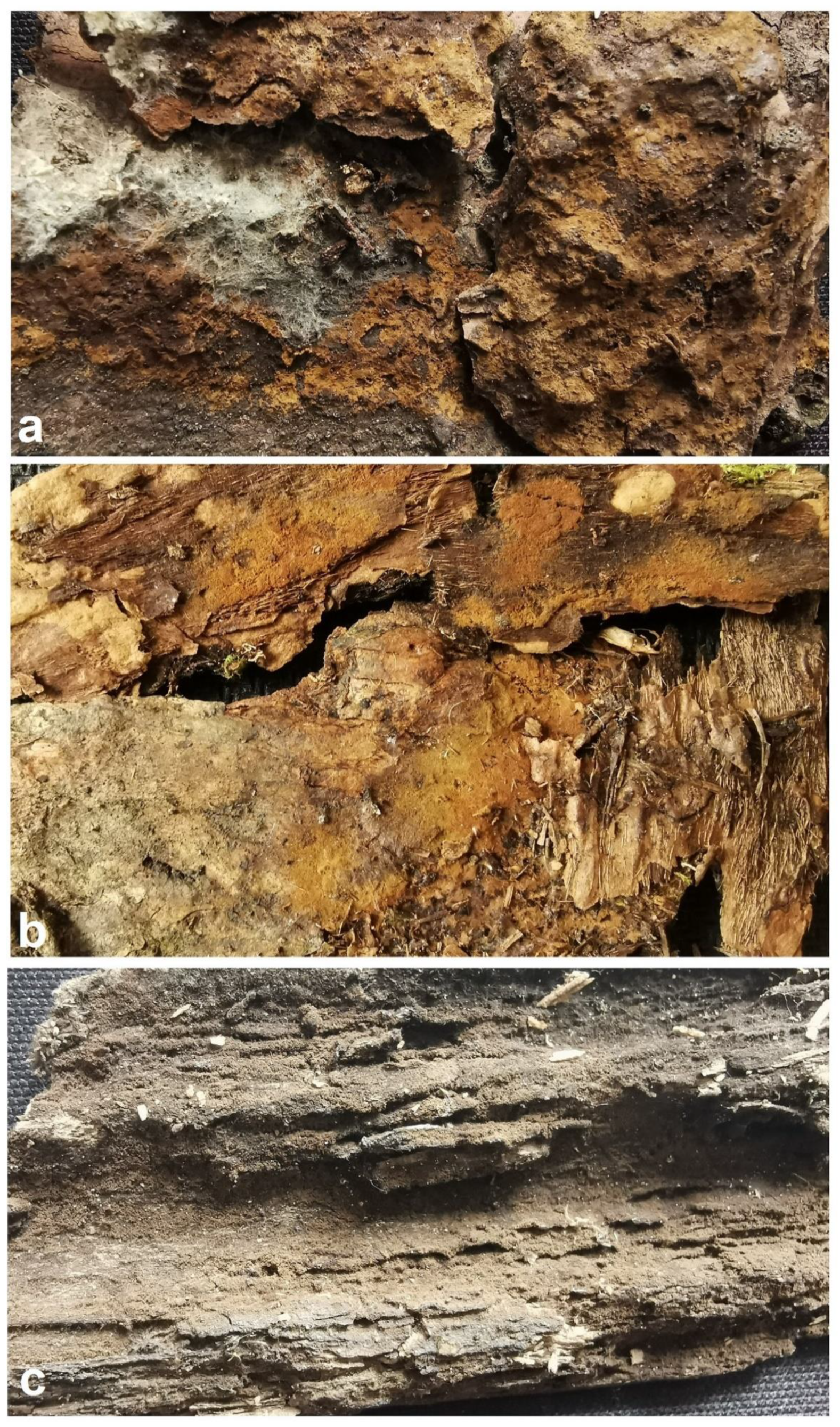
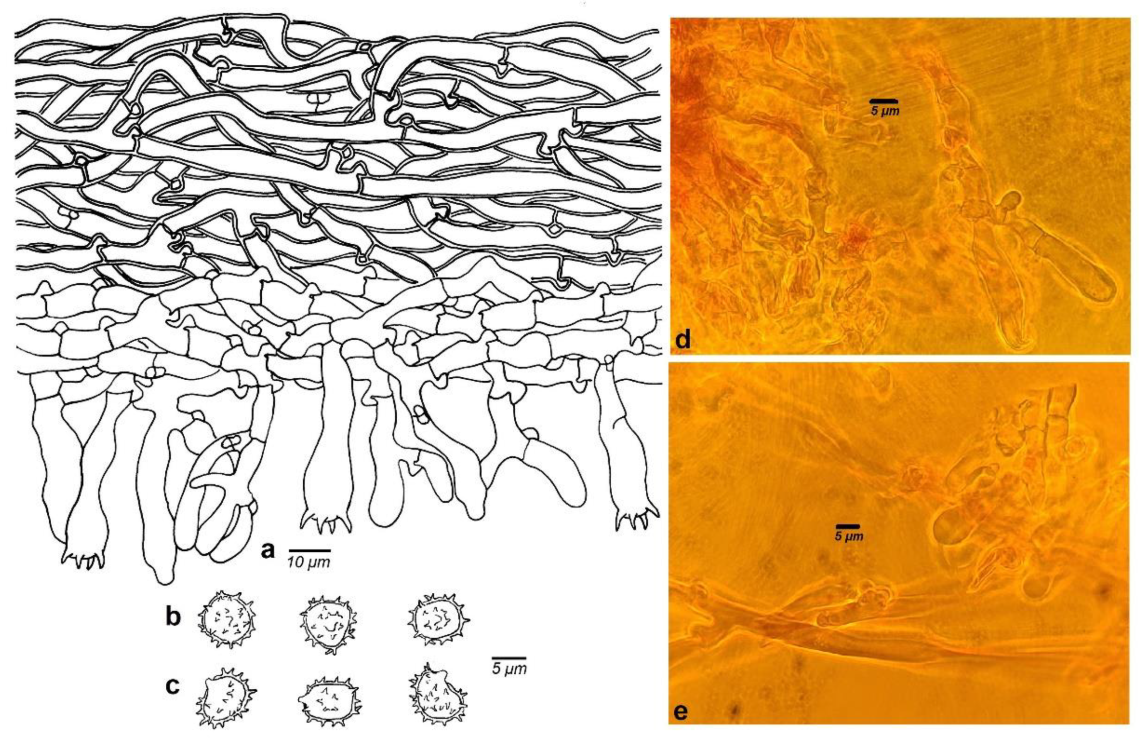
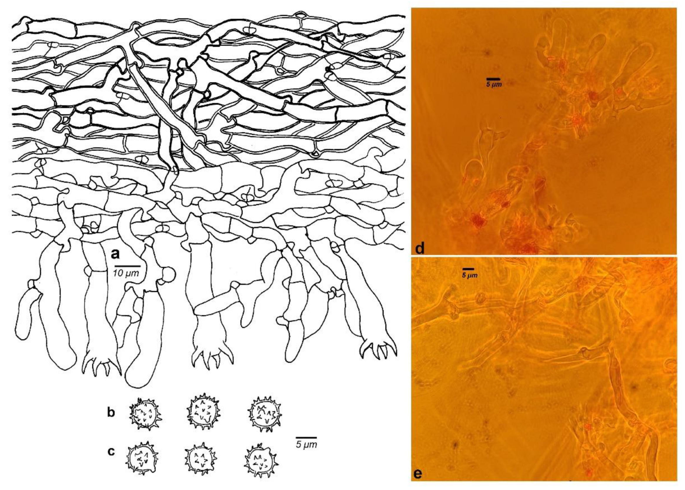
The combined dataset of the 217 sequences had an aligned length of 1675 sites with 669 bp ITS and 1006 bp LSU, which included 1077 constant characters, 87 parsimony-uninformative variable characters, and 511 parsimony-informative positions. A similar topology was obtained using ML and Bayesian analyses with two specimens of Odontia ferruginea as the outgroup [15], and only the ML tree is shown in Figure 1 with the ML bootstrap values and Bayesian posterior probabilities. The Bayesian analysis ran 10 million generations with an average standard deviation of split frequencies = 0.005051. Members of the genus Tomentella from regions in the world have been divided into 18 major clades with moderate support or high support (52 in ML/0.99 BPP for clade 1, 80 in ML/1.00 BPP for clade 2, 62 in ML/1.00 BPP for clade 3, 100 in ML/1.00 BPP for clade 4, 53 in ML for clade 5, 98 in ML/1.00 BPP for clade 6, 81 in ML/1.00 BPP for clade 7, 75 in ML/0.95 BPP for clade 8, 0.96 BPP for clade 9, 84 in ML/1.00 BPP for clade 10, 95 in ML/1.00 BPP for clade 11, 60 in ML/1.00 BPP for clade 12, 95 in ML/1.00 BPP for clade 13, 89 in ML/1.00 BPP for clade 14, 96 in ML/1.00 BPP for clade 15, 99 in ML/1.00 BPP for clade 16, 82 in ML/0.97 BPP for clade 17, and 60 in ML) based on the phylogenetic analyses of combined ITS and LSU sequence data.
The six sampled specimens formed three strongly supported lineages (T. aurantispora, T. kanasensis, and T. schrenkiana: 100 in ML/1.00 BPP for T. aurantispora and T. schrenkiana and 99 in ML/1.00 BPP for T. kanasensis), which clustered together with other species from Northeast China, Estonia, Finland, and Seychelles in clade 1 with moderate support (52 in ML/0.99 BPP). The confirmed distribution range of a new species, T. kanasensis, spans Xinjiang and Jilin Provinces located in the northwest and northeast of China, respectively.

3.2. Taxonomy

Tomentellaaurantispora X. Lu & H.S. Yuan, sp. nov.
MycoBank no. MB 834867
Diagnosis. Basidiocarps: arachnoid. Hymenophoral surface: yellowish brown to brown. Sterile margin: byssoid and brown. Generative hyphae: clamped. Basidia: utriform. Basidiospores: subglobose to globose, slightly thick-walled, echinulate; echinuli up to 1 μm long.
Type: CHINA, Xinjiang Autonomous Region, Urumqi, Tianshan Grand Canyon National Forest Park, 43°49′17″ N, 87°43′59″ E, 1968 m, on fallen branch of Picea schrenkiana, 17.VIII.2019, Yuan 14268 (holotype: IFP 019365, GenBank ITS: MT152155; LSU: MT154238).
Etymology. Aurantispora (Lat.), referring to the orange-yellow basidiospores in distilled water.
Basidiocarps: annual, resupinate, adherent to the substrate, arachnoid, without odor or taste when fresh, 0.2–0.5 mm thick, continuous. Hymenophoral surface: smooth, yellowish brown to brown (5D8–6F8) and turning paler than subiculum. Sterile margin: often determinate, byssoid, darker than hymenophore, brown (6F8).
Rhizomorphs absent.
Subicular hyphae: monomitic; generative hyphae: clamped, thick-walled, 4–6.5 μm diam, without encrustation, grayish yellow in KOH and distilled water, CB+, IKI-. Subhymenial hyphae: clamped, thin-walled, 3–7 μm diam, without encrustation; hyphal cells: short and inflated, grayish yellow in KOH and in distilled water, CB-, IKI-.
Cystidia absent.
Basidia: 20–45 μm long and 4–9 μm diam at apex, 3–5 μm at base, with a clamp connection at base, utriform, not stalked, sinuous, without transverse septa, pale brown in KOH and distilled water, 4-sterigmata; sterigmata: 3–5 μm long and 1–1.5 μm diam at base.
Basidiospores: thick-walled, (6.5–)7–9(–9.5) × (5.5–)6–8(–8.5) μm, L = 7.82 μm, W = 6.95 μm, Q = 1.08–1.15 (n = 60/2), subglobose to globose in frontal and later face, echinulate to aculeate, brown in KOH, orange-yellow in distilled water, CB+, IKI-; echinuli usually grouped in two or more, up to 1 μm long.
Additional specimens (paratype) examined: CHINA, Xinjiang Autonomous Region, Urumqi, Tianshan Grand Canyon National Forest Park, 43°49′17″ N, 87°43′59″ E, 2028 m, on fallen branch of Picea schrenkiana, 17.VIII.2019, Yuan 14276 (paratype: IFP 019366, GenBank ITS: MT152156; LSU: MT154239).
Tomentella kanasensis X. Lu & H.S. Yuan, sp. nov.
MycoBank no. MB 834868
Diagnosis. Basidiocarps: arachnoid. Hymenophoral surface: light brown to dark brown. Sterile margin: byssoid and dark brown. Generative hyphae: clamped and rarely simple-septate. Basidia: utriform. Basidiospores: subglobose to globose, slightly thick-walled, echinulate; echinuli up to 1.5 μm long.
Type: CHINA, Xinjiang Autonomous Region, Buerjin County, Kanas National Nature Reserve, 48°37′23″ N, 87°02′55″ E, 1342 m, on fallen branch of Picea obovata, 6.VIII.2019, Yuan 14034 (holotype: IFP 019362, GenBank ITS: MT152151; LSU: MT154234).
Etymology. Kanasensis (Lat.), referring to the specimens collected from Kanas National Nature Reserve.
Basidiocarps: annual, resupinate, adherent to the substrate, arachnoid, without odor or taste when fresh, 0.3–0.5 mm thick, continuous. Hymenophoral surface: smooth, light brown to dark brown (6D4–6E8) and turning darker than subiculum. Sterile margin: often determinate, byssoid, darker than hymenophore, dark brown (6E8).
Rhizomorphs absent.
Subicular hyphae: monomitic; generative hyphae: clamped and rarely simple-septate, slightly thick- to thick-walled, occasionally collapsed, 4–6 μm diam, without encrustation, light brown in KOH and distilled water, CB+, IKI-. Subhymenial hyphae: clamped and rarely simple-septate, thin-walled, 4–7 μm diam, without encrustation; hyphal cells: short and occasionally inflate up to 10 μm, yellow in KOH and in distilled water, acyanophilous, IKI-.
Cystidia absent.
Basidia: 15–62 μm long and 8–11 μm diam at apex, 4.5–6 μm at base, with a clamp connection at base, utriform, not stalked, sinuous, without transverse septa, yellow in KOH and distilled water, 4-sterigmata; sterigmata: 6–8.5 μm long and 2–3 μm diam at base.
Basidiospores: thick-walled, (7–)7.5–9(–9.5) × (6–)6.5–8(–8.5) μm, L = 8.44 μm, W = 6.98 μm, Q = 1.18–1.24 (n = 60/2), subglobose to globose in frontal and later face, echinulate, light brown in KOH and distilled water, CB+, IKI-; echinuli usually isolated, up to 1.5 μm long.
Additional specimens (paratypes) examined: CHINA, Xinjiang Autonomous Region, Buerjin County, Kanas National Nature Reserve, Wolong Bay, 48°36′58″ N, 87°01′55″ E, 1328 m, on fallen branch of Picea obovata, 6.VIII.2019, Yuan 14029 (IFP 019361, GenBank ITS: MT152152; LSU: MT154235); Jilin Province, Jiaohe City, Qianjin Village, 43°57′52″ N–43°58′02″ N, 127°44′11″ E–127°44′66″ E, 459–517 m, on fallen angiosperm branch, 3.IX.2017, Yuan 12556, 12557, 12565, 12571 & 12579 (paratypes: IFP 019367–IFP 019371, GenBank ITS: MT152157–MT152161; LSU: MT154240–MT154244).
Tomentellaschrenkiana X. Lu & H.S. Yuan, sp. nov.
MycoBank no. MB 834869
Diagnosis. Basidiocarps: arachnoid. Hymenophoral surface: grayish brown to dark brown. Sterile margin: byssoid and grayish brown. Generative hyphae: clamped. Basidia: utriform. Basidiospores: subglobose to globose, slightly thick-walled, echinulate; echinuli up to 1 μm long.
Type: CHINA, Xinjiang Autonomous Region, Fukang City, Tianshan Tianchi National Park, 43°53′51″ N, 88°06′42″ E, 1899 m, on fallen trunk of Picea schrenkiana, 16.VIII.2019, Yuan 14262 (holotype: IFP 019364, GenBank ITS: MT152153; LSU: MT154236).
Etymology. Schrenkiana (Lat.), referring to the substrate of this species.
Basidiocarps: annual, resupinate, adherent to the substrate, arachnoid, without odor or taste when fresh, 0.2–0.5 mm thick, continuous. Hymenophoral surface: smooth, grayish brown to dark brown (7F3–7F6) and concolorous with the subiculum. Sterile margin: often indeterminate, byssoid, concolorous with hymenophore.
Rhizomorphs absent.
Subicular hyphae: monomitic; generative hyphae: clamped, thick-walled, occasionally collapsed, 2.5–5 μm diam, without encrustation, brown in KOH and distilled water, CB+, IKI-. Subhymenial hyphae: clamped, thin- to slightly thick-walled, 2.5–5.5 μm diam, without encrustation; hyphal cells: short and not inflated, grayish brown in KOH and in distilled water, CB+, IKI-.
Cystidia absent.
Basidia: 20–35 μm long and 4–8 μm diam at apex, 3–5 μm at base, with a clamp connection at base, utriform, not stalked, sinuous, without transverse septa, grayish brown in KOH and distilled water, 4-sterigmata; sterigmata: 3–6 μm long and 1.5–2 μm diam at base.
Basidiospores: slightly thick-walled, (5–)5.5–6.5(–7) × (4.5–)5–5.5(–6) μm, L = 6.04 μm, W = 5.2 μm, Q = 1.15–1.18 (n = 60/2), subglobose to globose in frontal and later face, echinulate, brown in KOH and distilled water, CB+, IKI-; echinuli usually grouped in two or more, up to 1 μm long.
Additional specimens (paratype) examined: CHINA, Xinjiang Autonomous Region, Fukang City, Tianshan Tianchi National Park, 43°53′23″ N, 88°06′38″ E, 1854 m, on fallen branch of Picea schrenkiana, 16.VIII.2019, Yuan 14241 (paratype: IFP 019363, GenBank ITS: MT152154; LSU: MT154237).

4. Discussion

Three new Tomentella species distributed in Xinjiang of northwest China were identified by phylogenetic analysis based on a combined ITS and LSU sequence dataset and morphological characteristics. Phylogenetic analyses and morphological features were able to distinguish the three new species from other Tomentella species. The deeper nodes of the tree are weakly supported, which is consistent with previous studies [9]. In the phylogenetic tree, most species from global regions have scattered in the 18 main clades. Species in the clade with moderate support or high support present unique features. Species in clade 1 have basidiocarps with similar color; species in clade 7 and clade 8 have rhizomorphs; species in clade 10 have a simple-septate hyphal system and basidiospores with the same size; species in clade 14 have separable basidiocarps with similar color; species in clade 15 have similar anatomical structure; species in clade 16 have cystidia. In addition, T. brunneocystidiata Yorou & Agerer, T. capitata Yorou & Agerer, T. capitatocystidiata Yuan et al., T. cystidiata Yuan et al., T. gloeocystidiata Yuan et al., and T. pilosa (Burt) Bourdot & Galzin in clade 4 also share common features: the capitate cystidia. T. aureomarginata Yuan et al., T. brunneoflava Yuan et al., and T. separata Yuan et al. in clade 6 also have common features: light brown to brown basidiocarps and the presence of rhizomorphs.
In the phylogenetic tree, T. aurantispora and T. kanasensis are closely related to T. bryophila (Pers.) M.J. Larsen, T. changbaiensis Yuan et al., T. interrupta Yuan et al., T. longiaculeifera Yuan et al., T. megaspora Yuan et al., and T. pallidobrunnea Yuan et al. with strong support (85 in ML and 1.00 BPP), and they formed a highly supported (0.97 BPP) relationship with T. asiae-orientalis Yuan et al., T. lammiensis Lu et al., and T. stipitata Yuan et al. These species share some similar characteristics: clamped hyphal system, globose to subglobose basidiospores, and the absence of rhizomorphs and cystidia [3,11] except for the ellipsoid, triangular, or lobed basidiospores of T. lammiensis and T. stipitata [11,51]. T. aurantispora also presents some other similar morphological characteristics with T. changbaiensis and T. megaspora: byssoid sterile margin, short and inflated subhymenial hyphal cell, and utriform basidia. But T. changbaiensis differs from T. aurantispora by the separable basidiocarps and larger basidiospores with longer echinuli (L = 8.45 μm, W = 7.83 μm, echinuli up to 1.5 μm) [11]. T. megaspora can be distinguished from T. aurantispora based on the larger basidiospores with longer echinuli (L = 9.88 μm, W = 9.08 μm, echinuli up to 2.5 μm) [11]. Morphologically, T. aurantispora and T. asiae-orientalis also share some other common characteristics: arachnoid basidiocarps adherent to the substrate and byssoid sterile margin. Moreover, T. asiae-orientalis can be delimited from T. aurantispora by the more or less uniform subhymenial hyphal cells, clavate basidia, and basidiospores with longer echinuli (echinuli up to 2.5 μm) [11].
Macroscopically, T. kanasensis and T. aurantispora possess the same arachnoid basidiocarps with similar color (yellowish brown to brown and light brown to dark brown) and byssoid sterile margin. But T. aurantispora differs by the length of sterigmata (3–5 μm long) and the hyphal system without simple septa in comparison with T. kanasensis. Some features of T. changbaiensis can be also observed in T. kanasensis, such as the byssoid sterile margin and the equally sized basidiospores. However, T. changbaiensis differs markedly from T. kanasensis by the duplex basidiocarps, pale brown sterile margin, and hyphal system without simple septa [11]. T. bryophila also forms short and inflated subhymenial hyphal cells and utriform basidia similar to those of T. kanasensis. However, the encrusted hyphae of T. bryophila are different from T. kanasensis [3].
The phylogenetic tree shows that T. schrenkiana clusters phylogenetically together with T. dimidiata Yuan et al., T. fuscopelliculosa Yuan et al., T. globospora Yuan et al., T. pertenuis, and T. segregata Yuan et al. with a strong support (0.95 BPP), and they possess the globose to subglobose basidiospores, with the exception of T. dimidiata with lobed basidiospores [3,11,52]. T. schrenkiana and T. fuscopelliculosa also have some other common anatomical structures, such as short subhymenial hyphal cells, utriform basidia, and the absence of rhizomorphs and cystidia. However, T. fuscopelliculosa differs by its pelliculose basidiocarps, inflated subhymenial hyphal cells, and distinctly larger basidiospores with longer echinuli (L = 8.75 μm, W = 8.23 μm, echinuli up to 2 μm) in comparison with T. schrenkiana [11]. T. schrenkiana also shows similar anatomical features to T. globospora, T. pertenuis, and T. segregata, including basidiocarps adherent to the substrate and the absence of rhizomorphs and cystidia. However, the mucedinoid basidiocarps, farinaceous sterile margin, utriform basidia, and slightly larger basidiospores with longer echinuli (L = 7.05 μm, W = 6.35 μm, echinuli up to 1.5 μm) [51] of T. globospora are very different from T. schrenkiana. In comparison with T. schrenkiana, T. segregata can be distinguished in producing discontinuous basidiocarps, a farinaceous sterile margin, and larger basidiospores with isolated and longer echinuli. The continuous basidiocarps and clamped generative hyphae of T. pertenuis also resemble T. schrenkiana, but the farinaceous sterile margin and larger basidiospores (L = 7.99 μm, W = 7.41 μm) [11] of T. pertenuis are good delimitation characteristics.
As Picea spp. constituted the dominant tree distributed in the investigated regions of Xinjiang Autonomous Region, we speculate that T. aurantispora and T. schrenkiana may form ectomycorrhizae with P. schrenkiana. Specimens of T. kanasensis have also been observed in Jilin Province, and the climate of this region is apparently different from Xinjiang. Picea-dominated forests in Xinjiang have quite different tree species in comparison to the mixed broadleaf– conifer forests in Jilin Province dominated by Pinus koraiensis Siebbold & Zucc., Acer pictum subsp. mono (Maxim.) Ohashi, A. mandshuricum Maxim., Tilia amurensis Rupr., and Carpinus cordata Blume mixed with shrub species [53]. The host tree species of T. kanasensis from Xinjiang and Jilin Provinces may be P. obovata and P. koraiensis, respectively. At present, 51 species of Tomentella have been identified based on morphological and molecular characteristics of the Tomentella collections from Northeast China (Liaoning, Heilongjiang, and Jilin Provinces) and Northwest China (Xinjiang Autonomous Region) [10,11]. While several Tomentella spp. of ECM associated with plant roots in Pinaceae and Fagaceae were also reported in previous studies from China, they were not available for identification [29,30,34], and the cryptic or atypical basidiocarps of Tomentella may be easily overlooked in diverse studies. Therefore, we speculate that an increasing number of species in this genus will be found at more sites in China.

5. The Types of Rhizomorphs Used in the Key

Agerer recognized six categories of rhizomorph structure (types A–F) [54], and the dimitic hypal system of rhizomorphs named type G was reported in 2020 [11].

Supplementary Materials

The following are available online at www.mdpi.com/article/10.3390/f12111531/s1, Table S1: GenBank/UNITE accession numbers, voucher numbers, substrates, localities and references for the specimens included in this study. The newly generated sequences are in bold.

Author Contributions

Conceptualization, X.L. and H.-S.Y.; methodology, X.L. and H.-S.Y.; software, X.L.; validation, formal analysis, and investigation, X.L. and H.-S.Y.; resources, H.-S.Y.; data curation, X.L. and H.-S.Y.; writing—original draft preparation, X.L.; writing—review and editing, X.L. and H.-S.Y.; visualization, X.L. and H.-S.Y.; supervision, project administration, and funding acquisition, H.-S.Y. All authors have read and agreed to the published version of the manuscript.

Funding

This research was financed by the National Natural Science Foundation of China (Project Nos. 31970017, 31770028 & 31470148) and the Special Funds for the Young Scholars of Taxonomy of the Chinese Academy of Sciences (Project No. ZSBR-015).

Institutional Review Board Statement

Not applicable.

Informed Consent Statement

Not applicable.

Data Availability Statement

The datasets presented in this study are openly available. All resulting alignments were deposited in TreeBASE (http://www.treebase.org (accessed on 21 September 2021; accession number 28802)). All newly generated sequences were deposited in GenBank (https://www.ncbi.nlm.nih.gov/genbank/ (accessed on 6 March 2020; Table S1)). All new taxa were deposited in Mycobank.

Conflicts of Interest

The authors declare that there is no conflict of interest regarding the design of the study, the writing of the manuscript, the decision to publish the results, and the collection, analyses, or interpretation of data.

Key to Species of Tomentella from China

1. Basidiospores globose to subglobose in frontal view2
1. Basidiospores lobed in frontal view20
2. Rhizomorphs absent3
2. Rhizomorphs absent4
3. Length of subhymenial hyphal cell ≥ 15 μmT. tenuirhizomorpha
3. Length of subhymenial hyphal cell < 15 μmT. brunneogrisea
4. Basidiocarps separable from the substrate5
4. Basidiocarps adherent to the substrate7
5. Sterile margin byssoidT. changbaiensis
5. Sterile margin farinaceous6
6. Subicular hyphae without encrustationT. atrocastanea
6. Subicular hyphae with encrustationT. longiaculeifera
7. Basidiocarps arachnoid8
7. Basidiocarps mucedinoid, crustose, and pelliculose11
8. Basidia clavateT. asiae-orientalis
8. Basidia utriform9
9. Basidiospores L < 7 μm and W < 6 μmT. schrenkiana
9. Basidiospores L ≥ 7 μm and W ≥ 6 μm10
10. Echinuli of basidiospores ≤ 1 μm, sterigmata of basidia ≤ 5 μmT. aurantispora
10. Echinuli of basidiospores > 1 μm, sterigmata of basidia > 5 μmT. kanasensis
11. Basidia clavate12
11. Basidia utriform14
12. Subicular hyphae without encrustationT. globospora
12. Subicular hyphae with encrustation13
13. Basidiocarps pelliculoseT. duplexa
13. Basidiocarps mucedinoidT. griseomarginata
14. Sterigmata of basidia ≥ 10 μmT. conclusa
14. Sterigmata of basidia < 10 μm15
15. Length of subhymenial hyphal cell ≥ 15 μm, not inflatedT. pertenuis
15. Length of subhymenial hyphal cell < 15 μm, inflated16
16. Basidiocarps crustose or pelliculose17
16. Basidiocarps mucedinoid19
17. Basidiocarps crustoseT. griseofusca
17. Basidiocarps pelliculose18
18. Sterigmata of basidia ≤ 5 μm, echinuli of basidiospores > 2 μmT. megaspora
18. Sterigmata of basidia > 5 μm, echinuli of basidiospores ≤ 2 μmT. fuscopelliculosa
19. Echinuli usually isolated, sterile margin farinaceousT. segregata
19. Echinuli usually grouped in 2 or more, sterile margin byssoidT. pallidobrunnea
20. Rhizomorphs present21
20. Rhizomorphs absent43
21. Cystidia present22
21. Cystidia absent25
22. Basidiocarps pelliculoseT. citrinocystidiata
22. Basidiocarps arachnoid23
23. Basidiocarps brown or dark brown24
23. Basidiocarps olive yellow to oliveT. gloeocystidiata
24. Echinuli of basidiospores ≤ 1.5 μmT. cystidiata
24. Echinuli of basidiospores > 1.5 μmT. capitatocystidiata
25. Rhizomorphs of type G26
25. Rhizomorphs of other types29
26. Basidiocarps pelliculose and separable from the substrateT. incrustata
26. Basidiocarps arachnoid and adherent to the substrate27
27. Subicular hyphae without encrustationT. dimidiata
27. Subicular hyphae with encrustation28
28. Rhizomorphs of type C29
28. Rhizomorphs of other types30
29. Basidiocarps mucedinoid and discontinuousT. brevis
29. Basidiocarps arachnoid and continuousT. qingyuanensis
30. Rhizomorphs of type A31
30. Rhizomorphs of type B32
31. Sterile margin of basidiocarps pale yellowT. pallidomarginata
31. Sterile margin of basidiocarps brownT. fuscocrustosa
32. Subicular hyphae with encrustationT. fuscogranulosa
32. Subicular hyphae without encrustation33
33. Basidiocarps discontinuousT. interrupta
33. Basidiocarps continuous34
34. Hymenophoral surface granulose35
34. Hymenophoral surface smooth39
35. Basidiocarps separable from the substrate36
35. Basidiocarps adherent to the substrate37
36. Basidiocarps dark brown, subicular hyphae simple-septateT. efibulata
36. Basidiocarps olive brown, subicular hyphae clampedT. olivacea
37. Hyphae of rhizomorphs simple-septateT. efibulis
37. Hyphae of rhizomorphs clamped38
38. Basidiocarps pelliculoseT. inconspicua
38. Basidiocarps mucedinoidT. parvispora
39. Basidiocarps separable from the substrate40
39. Basidiocarps adherent to the substrate41
40. Basidiocarps mucedinoid, subicular hyphae clampedT. flavidobadia
40. Basidiocarps pelliculose, subicular hyphae clamped and simple-septateT. separata
41. Basidia utriformT. aureomarginata
41. Basidia clavateT. brunneoflava
42. Basidia clavate43
42. Basidia utriform48
43. Subicular hyphae with encrustation44
43. Subicular hyphae without encrustation45
44. Basidiocarps mat-likeT. storea
44. Basidiocarps arachnoidT. farinosa
45. Sterile margin byssoidT. pallidocastanea
45. Sterile margin farinaceous46
46. Basidiocarps brownish grey to olive brownT. olivaceobrunnea
46. Basidiocarps grayish brown to dark blond or brown47
47. Length of subhymenial hyphal cell > 15 μm, not inflatedT. liaoningensis
47. Length of subhymenial hyphal cell ≤ 15 μm, inflatedT. griseocastanea
48. Sterile margin byssoidT. atrobadia
48. Sterile margin farinaceous49
49. Basidiocarps arachnoid or pelliculose50
49. Basidiocarps mucedinoid51
50. Basidiocarps arachnoid, echinuli of basidiospores ≤ 1.5 μmT. fuscofarinosa
50. Basidiocarps pelliculose, echinuli of basidiospores > 1.5 μmT. longiechinuli
51. Basidiocarps clavate, sterigmata of basidia ≤ 5 μmT. coffeae
51. Basidia utriform, sterigmata of basidia > 5 μmT. stipitata

References

  1. Persoon, C.H. Observationes Mycologicae; Gesnerus, Usterius & Wolfius: Leipzig, Germany, 1799; pp. 1–106. [Google Scholar]
  2. Patouillard, N.T. Les Hyménomycètes d’Europe. Anatomie Générale et Classification des Champignons Supérieurs (The Hymenomycetes of Europe. General Anatomy and Classification of the Higher Fungi); Paul Klincksieck: Paris, France, 1887; pp. 1–166. [Google Scholar]
  3. Kõljalg, U. Tomentella (Basidiomycota) and related genera in temperate Eurasia; Synopsis Fungorum 9.; Tõravere Trükikoda and Greif: Tartu, Estonia, 1996; pp. 1–213. [Google Scholar]
  4. Larsen, M.J. A Contribution to the Taxonomy of the Genus Tomentella; Mycologia Memoir 4.; New York Botanical Garden in Collaboration with The Mycological Society of America: New York, NY, USA, 1974; pp. 1–145. [Google Scholar]
  5. Cunningham, G.H. The Thelephoraceae of Australia and New Zealand, 1st ed.; R.E. Owen: New Zealand, UK, 1963; pp. 1–359. [Google Scholar]
  6. Welden, A.L. West Indian Species of dark-spored Thelephoraceae. Sydowia 1968, XXII, 269–273. [Google Scholar]
  7. Wakefield, E.M. Tomentelloideae in the British Isles. Transactions of the British Mycol. Soc. 1969, 53, 161–206. [Google Scholar] [CrossRef]
  8. Yorou, N.S.; Gardt, S.; Guissou, M.L.; Diabaté, M.; Agerer, R. Three new Tomentella species from West Africa identified by anatomical and molecular data. Mycol. Prog. 2012, 11, 449–462. [Google Scholar] [CrossRef]
  9. Kuhar, F.; Barroetaveña, C.; Rajchenberg, M. New species of Tomentella (Thelephorales) from the Patagonian Andes forests. Mycologia 2016, 108, 780–790. [Google Scholar] [CrossRef] [PubMed]
  10. Lu, X.; Mu, Y.H.; Yuan, H.S. Two new species of Tomentella (Thelephorales, Basidiomycota) from Lesser Xingan Mts., northeastern China. Phytotaxa 2018, 369, 080–092. [Google Scholar] [CrossRef]
  11. Yuan, H.S.; Lu, X.; Dai, Y.C.; Hyde, K.D.; Kan, Y.H.; Kušan, I.; He, S.H.; Liu, N.G.; Sarma, V.V.; Zhao, C.L.; et al. Fungal diversity notes 1277–1386: Taxonomic and phylogenetic contributions to fungal taxa. Fungal Divers. 2020, 1, 1–260. [Google Scholar] [CrossRef]
  12. Yuan, Y.; Wu, F.; Dai, Y.C.; Qin, W.M.; Yuan, H.S. Odontia aculeata and O. sparsa, two new species of tomentelloid fungi (Thelephorales, Basidiomycota) from the secondary forests of northeast China. Phytotaxa 2018, 372, 183–192. [Google Scholar] [CrossRef]
  13. Gorjón, S.P.; Greslebin, A.G. Type studies of the species of Odontia described by GH Cunningham. N. Z. J. Bot. 2012, 50, 289–301. [Google Scholar] [CrossRef] [Green Version]
  14. Danielson, R.M.; Visser, S.; Parkinson, D. Microbial activity and mycorrhizal potential of four overburden types used in the reclamation of extracted oil sands. Can. J. Soil Sci. 1983, 63, 363–375. [Google Scholar] [CrossRef] [Green Version]
  15. Tedersoo, L.; Harend, H.; Buegger, F.; Pritsch, K.; Saar, I.; Kõljalg, U. Stable isotope analysis, field observations and synthesis experiments suggest that Odontia is a non-mycorrhizal sister genus of Tomentella and Thelephora. Fungal Ecol. 2014, 11, 80–90. [Google Scholar] [CrossRef]
  16. Marx, D.H.; Bryan, W.C.; Davey, C.B. Influence of temperature on aseptic synthesis of ectomycorrhizae by Thelephora terrestris and Pisolithus tinctorius on loblolly pine. For. Sci. 1970, 16, 424–431. [Google Scholar] [CrossRef]
  17. Erland, S.; Taylor, A.F.S. Resupinate Ectomycorrhizal Fungal Genera. In Ectomycorrhizal Fungi Key Genera in Profile, 1st ed.; Cairney, J.W.G., Chambers, S.M., Eds.; Springer: Berlin/Heidelberg, Germany, 1999; pp. 325–363. [Google Scholar] [CrossRef]
  18. Alvarez-Manjarrez, J.; Villegas-Ríos, M.; Garibay-Orijel, R.; Contreras-Pacheco, M.; Kõljalg, U. Tomentella brunneoincrustata, the first described species of the Pisonieae-associated Neotropical Tomentella clade, and phylogenetic analysis of the genus in Mexico. Mycol. Prog. 2016, 15, 1–11. [Google Scholar] [CrossRef]
  19. Corner, E.J.H. A Monograph of Thelephora (Basidiomycetes). In Beihefte zur Nova Hedwigia; Cramer: Berlin, Germany, 1968; Volume 27, pp. 1–110. [Google Scholar]
  20. Tedersoo, L.; May, T.W.; Smith, M.E. Ectomycorrhizal lifestyle in fungi: Global diversity, distribution, and evolution of phylogenetic lineages. Mycorrhiza 2010, 20, 217–263. [Google Scholar] [CrossRef]
  21. Põlme, S. Ticodendron incognitum and Neea pittieri associated ectomycorrhizal fungi in Neotropical mountain forest. Asian J. Mycol. 2018, 1, 137–145. [Google Scholar] [CrossRef]
  22. Read, D.J.; Perez-Moreno, J. Mycorrhizas and nutrient cycling in ecosystems—A journey towards relevance? New Phytol. 2003, 157, 475–492. [Google Scholar] [CrossRef]
  23. Tedersoo, L.; Gates, G.; Dunk, C.W.; Lebel, T.; May, T.W.; Kõljalg, U.; Jairus, T. Establishment of ectomycorrhizal fungal community on isolated Nothofagus cunninghamii seedlings regenerating on dead wood in Australian wet temperate forests: Does fruit-body type matter? Mycorrhiza 2009, 19, 403–416. [Google Scholar] [CrossRef]
  24. Selosse, M.A.; Bouchard, D.; Martin, F.; Tacon, F.L. Effect of Laccaria bicolor strains inoculated on Douglas-fir (Pseudotsuga menziesii) several years after nursery inoculation. Can. J. For. Res. 2000, 30, 360–371. [Google Scholar] [CrossRef]
  25. Menkis, A.; Vasiliauskas, R.; Taylor, A.F.S.; Stenlid, J.; Finlay, R. Afforestation of abandoned farmland with conifer seedlings inoculated with three ectomycorrhizal fungi—impact on plant performance and ectomycorrhizal community. Mycorrhiza 2007, 17, 337–348. [Google Scholar] [CrossRef]
  26. Simard, S.W.; Molina, R.; Smith, J.E.; Perry, D.A.; Jones, M.D. Shared compatibility of ectomycorrhizae on Pseudotsuga menziesii and Betula papyrifera seedlings grown in mixture in soils from southern British Columbia. Can. J. For. Res. 1997, 27, 331–342. [Google Scholar] [CrossRef]
  27. Kõljalg, U.; Dahlberg, A.; Taylor, A.F.S.; Larsson, E.; Hallenberg, N.; Stenlid, J.; Larsson, K.H.; Fransson, P.M.; Kårén, O.; Jonsson, L. Diversity and abundance of resupinate thelephoroid fungi as ectomycorrhizal symbionts in Swedish boreal forests. Mol. Ecol. 2000, 9, 1985–1996. [Google Scholar] [CrossRef]
  28. Ishida, T.A.; Nara, K.; Ma, S.; Takano, T.; Liu, S.K. Ectomycorrhizal fungal community in alkaline-saline soil in northeastern China. Mycorrhiza 2009, 19, 329–335. [Google Scholar] [CrossRef] [PubMed]
  29. Huang, J.; Nara, K.; Zong, K.; Wang, J.; Xue, S.; Peng, K.; Shen, Z.; Lian, C. Ectomycorrhizal fungal communities associated with masson pine (Pinus massoniana) and white oak (Quercus fabri) in a manganese mining region in Hunan Province, China. Fungal Ecol. 2014, 9, 1–10. [Google Scholar] [CrossRef]
  30. Han, Q.; Huang, J.; Long, D.; Wang, X.; Liu, J. Diversity and community structure of ectomycorrhizal fungi associated with Larix chinensis across the alpine treeline ecotone of Taibai Mountain. Mycorrhiza 2017, 27, 487–497. [Google Scholar] [CrossRef] [PubMed]
  31. Wu, B.W.; Gao, C.; Chen, L.; Buscot, F.; Goldmann, K.; Purahong, W.; Ji, N.N.; Wang, Y.L.; Lü, P.P.; Li, X.C.; et al. Host phylogeny is a major determinant of Fagaceae-associated ectomycorrhizal fungal community assembly at a regional scale. Front. Microbiol. 2018, 9, 1–12. [Google Scholar] [CrossRef]
  32. Wang, Q.; He, X.H.; Guo, L.D. Ectomycorrhizal fungus communities of Quercus liaotungensis Koidz of different ages in a northern China temperate forest. Mycorrhiza 2012, 22, 461–470. [Google Scholar] [CrossRef] [PubMed]
  33. Zhang, J.; Taniguchi, T.; Xu, M.; Du, S.; Liu, G.B.; Yamanaka, N. Ectomycorrhizal fungal communities of Quercus liaotungensis along different successional stands on the Loess Plateau, China. J. For. Res. 2014, 19, 395–403. [Google Scholar] [CrossRef]
  34. Gao, C.; Zhang, Y.; Shi, N.N.; Zheng, Y.; Chen, L.; Wubet, T.; Bruelheide, H.; Both, S.; Buscot, F.; Ding, Q.; et al. Community assembly of ectomycorrhizal fungi along a subtropical secondary forest succession. New Phytol. 2015, 205, 771–785. [Google Scholar] [CrossRef]
  35. Bai, Z.; Wang, W.; Li, H.; Liu, D.; Guo, Z.; Liu, H. Change of forest ecosystem service function and its value in Kanas Nature Reserve. J. Landsc. Res. 2019, 11, 80–83. [Google Scholar] [CrossRef]
  36. Liu, X.; Pan, C. Effects of recovery time after fire and fire severity on stand structure and soil of larch forest in the Kanas National Nature Reserve, Northwest China. J. Arid. Land 2019, 11, 811–823. [Google Scholar] [CrossRef] [Green Version]
  37. Li, Q.; Zhu, J.; Zhang, Y.; Wang, Q.; Li, T.T. Comprehensive evaluation of tourism resources in Xinjiang Tianshan Grand Canyon National Forest Park. For. Eng. 2015, 15, 30–34. [Google Scholar] [CrossRef]
  38. Li, Y.; Zhang, Y.; Ni, J.; Kong, Z.; Wang, L.; Li, Y.; Chen, L. The correlation between airborne Betula pollen content and meteorological factors in the Tianshan Mountains, Xinjiang, China. Chin. Sci. Bull. 2019, 64, 1909–1921. [Google Scholar] [CrossRef] [Green Version]
  39. Kornerup, A.; Wanscher, J. Methuen Handbook of Colour Fletcher, 3rd ed.; Eyre Methuen: Norwich, UK, 1981; pp. 1–252. [Google Scholar]
  40. Taylor, D.L.; McCormick, M.K. Internal transcribed spacer primers and sequences for improved characterization of basidiomycetous orchid mycorrhizas. New Phytol. 2008, 177, 1020–1033. [Google Scholar] [CrossRef] [PubMed]
  41. Vilgalys, R.; Hester, M. Rapid genetic identification and mapping of enzymatically amplified ribosomal DNA from several Cryptococcus species. J. Bacteriol. 1990, 172, 4238–4246. [Google Scholar] [CrossRef] [PubMed] [Green Version]
  42. Li, K.N.; Rouse, D.I.; German, T.L. PCR primers that allow intergeneric differentiation of ascomycetes and their application to Verticillium spp. Appl. Environ. Microbiol. 1994, 60, 4324–4331. [Google Scholar] [CrossRef] [PubMed] [Green Version]
  43. Nilsson, R.H.; Tedersoo, L.; Abarenkov, K.; Ryberg, M.; Kristiansson, E.; Hartmann, M.; Schoch, C.L.; Nylander, J.A.A.; Bergsten, J.; Porter, T.M.; et al. Five simple guidelines for establishing basic authenticity and reliability of newly generated fungal ITS sequences. Mycokeys 2012, 4, 37–63. [Google Scholar] [CrossRef] [Green Version]
  44. Benson, D.A.; Cavanaugh, M.; Clark, K.; Karsch-Mizrachi, I.; Ostell, J.; Pruitt, K.D.; Sayers, E.W. GenBank. Nucleic Acids Res. 2018, 46, D41–D47. [Google Scholar] [CrossRef] [PubMed] [Green Version]
  45. Nilsson, R.H.; Larsson, K.; Taylor, A.F.S.; Bengtsson-Palme, J.; Jeppesen, T.S.; Schigel, D.; Kennedy, P.; Picard, K.; Glöckner, F.O.; Tedersoo, L.; et al. The UNITE database for molecular identification of fungi: Handling dark taxa and parallel taxonomic classifications. Nucleic Acids Res. 2019, 47, D259–D264. [Google Scholar] [CrossRef]
  46. Katoh, K.; Rozewicki, J.; Yamada, K.D. MAFFT online service: Multiple sequence alignment, interactive sequence choice and visualization. Brief. Bioinform. 2019, 20, 1160–1166. [Google Scholar] [CrossRef] [Green Version]
  47. Gao, F.L.; Chen, C.J.; Arab, D.A.; Du, Z.G.; He, Y.H.; Ho, S.Y.W. EasyCodeML: A visual tool for analysis of selection using CodeML. Ecol. Evol. 2019, 9, 1–8. [Google Scholar] [CrossRef] [Green Version]
  48. Silvestro, D.; Michalak, I. raxmlGUI: A graphical front-end for RAxML. Org. Divers. Evol. 2012, 12, 335–337. [Google Scholar] [CrossRef]
  49. Ronquist, F.; Teslenko, M.; Mark, P.V.D.; Ayres, D.L.; Darling, A.; Höhna, S.; Larget, B.; Liu, L.; Suchard, M.A.; Huelsenbeck, J.P. MrBayes 3.2: Efficient Bayesian phylogenetic inference and model choice across a large model space. Syst. Biol. 2012, 61, 539–542. [Google Scholar] [CrossRef] [Green Version]
  50. Darriba, D.; Taboada, G.L.; Doallo, R.; Posada, D. jModelTest 2: More models, new heuristics and parallel computing. Nat. Methods 2012, 9, 772. [Google Scholar] [CrossRef] [PubMed] [Green Version]
  51. Lu, X.; Steffen, K.; Yuan, H.S. Morphological and molecular identification of three new species of Tomentella from Finland. Mycologia 2018, 110, 1–15. [Google Scholar] [CrossRef] [PubMed]
  52. Suvi, T.; Tedersoo, L.; Abarenkov, K.; Beaver, K.; Gerlach, J.; Kõljalg, U. Mycorrhizal symbionts of Pisonia grandis and P. sechellarum in Seychelles: Identification of mycorrhizal fungi and description of new Tomentella species. Mycologia 2010, 102, 522–533. [Google Scholar] [CrossRef] [PubMed]
  53. Zhang, P.; He, H.J.; Fan, C.Y.; Tian, L.Z.; Zhang, C.Y.; Zhao, X.H. Height-diameter models of main tree species in broadleaf-conifer mixed forest in Jiaohe, Jilin, China. For. Res. 2018, 31, 11–18. [Google Scholar] [CrossRef]
  54. Agerer, R. Colour Atlas of Ectomycorrhizae; 1st–9th Delivery; Einhorn: Schwäbisch Gmünd, Germany, 1987–1995; pp. 25i–28i. [Google Scholar]
Figure 1. Maximum likelihood tree illustrating the phylogeny of Tomentella aurantispora, T. kanasensis, T. schrenkiana, and related taxa based on ITS + LSU nuclear rDNA sequences dataset. Branches are labeled with maximum likelihood bootstrap higher than 50% and Bayesian posterior probabilities higher than 0.95. The terminals indicate the regions where they were distributed. New species are in bold (black).
Forests 12 01531 g001aForests 12 01531 g001bForests 12 01531 g001c
Figure 2. Basidiocarps of Tomentella species. (a) T. aurantispora (holotype Yuan 14268); (b) T. kanasensis (holotype Yuan 14034); (c) T. schrenkiana (holotype Yuan 14262).
Forests 12 01531 g002
Figure 3. SEM of basidiospores of Tomentella species. (a,b) T. aurantispora (holotype Yuan 14268); (c,d) T. kanasensis (holotype Yuan 14034); (e,f) T. schrenkiana (holotype Yuan 14262).
Forests 12 01531 g003
Figure 4. Microscopic structures of Tomentella aurantispora (drawn from holotype Yuan 14268). (a) A section through basidiocarp. (b) Basidiospores in frontal view. (c) Basidiospores in lateral view. (d) Optical microscope images of subhymenial hyphae. (e) Optical microscope images of subicular hyphae and basidia.
Forests 12 01531 g004
Figure 5. Microscopic structures of Tomentella kanasensis (drawn from holotype Yuan 14034). (a) A section through basidiocarp. (b) Basidiospores in frontal view. (c) Basidiospores in lateral view. (d) Optical microscope images of subhymenial hyphae and basidia. (e) Optical microscope images of subicular hyphae.
Forests 12 01531 g005
Figure 6. Microscopic structures of Tomentella schrenkiana (drawn from Yuan 14262). (a) A section through basidiocarp. (b) Basidiospores in frontal view. (c) Basidiospores in lateral view. (d) Optical microscope images of subhymenial hyphae and basidia. (e) Optical microscope images of subicular hyphae. (f,g) Optical microscope images of basidia.
Forests 12 01531 g006
Publisher’s Note: MDPI stays neutral with regard to jurisdictional claims in published maps and institutional affiliations.

