Article

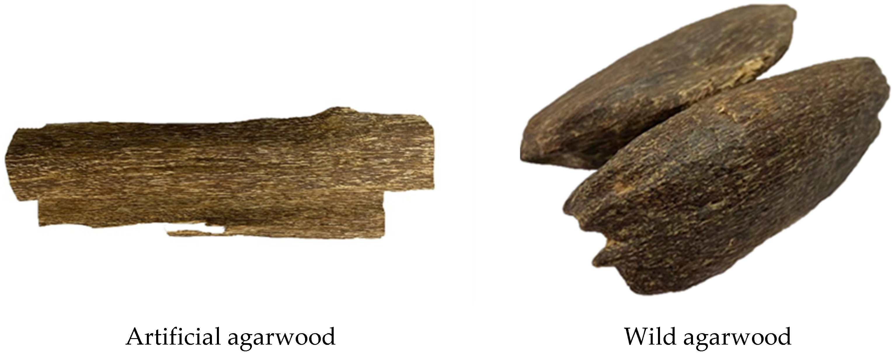
Comparative Analysis of Biological Activity of Artificial and Wild Agarwood

College of Forestry, Guangxi University, Nanning 540004, China
*
Author to whom correspondence should be addressed.
Forests 2021, 12(11), 1532; https://doi.org/10.3390/f12111532
Submission received: 7 October 2021 / Revised: 29 October 2021 / Accepted: 3 November 2021 / Published: 7 November 2021
(This article belongs to the Section Wood Science and Forest Products)

Abstract

:
Agarwood is a highly economically important medicinal herb with widespread uses; however, the difference between the biological activities of artificial and wild agarwood is unclear. In this study, the alcohol-soluble extracts of agarwood produced by fungi and natural agarwood were used to determine the differences between the overall biological activities. The antioxidant ability (the clearance rates of 1,1-diphenyl-2-picrylhydrazyl (DPPH) and 2,2′-azino-bis (3-ethylbenzothiazoline-6-sulfonic acid) (ABTS+) radicals, and total reducing power), anti-acetylcholinesterase, and anti-α-glucosidase activity were determined by ultraviolet-visible spectrophotometry. The results indicated that with 2 mg/mL alcohol-soluble extracts, the scavenging DPPH radical rates of the artificial agarwood samples were 93.74–102.31% of that of the wild agarwood, and the ABTS+ radical clearance rates of the artificial agarwood samples were 75.38–95.52% of that of the natural agarwood. With 3.5 mg/mL alcohol-soluble extracts, the artificial agarwood samples had a total reducing power of 63.07–80.29% of that of the wild agarwood. With 4 mg/mL alcohol-soluble extract, the acetylcholinesterase activity inhibition rates of the artificial agarwood samples were 102.56–109.16% of that of the wild agarwood. With 1 mg/mL alcohol soluble extracts, the α-glucosidase effect inhibitions rates of the artificial agarwood samples were 68.32–100.39% of that of the wild agarwood.

Graphical Abstract

1. Introduction

Agarwood is a medicinal herb produced by Aquilaria of the Thymelaeaceae family. The 21 Aquilaria species are mainly distributed in approximately 20 countries from India to Malaysia to Papua New Guinea [1]. In China, they are mainly distributed in Guangxi, Guangdong, Yunnan and Hainan, among which Aquilaria sinensis and Aquilaria yunnanensis are endemic [2]. According to records, the utilization history of agarwood can be traced back to more than 2000 years ago [3], and its unique components are commonly used in the fields of incense, medicine, and religion [4]. Under natural conditions, it is impossible for a healthy Aquilaria tree to contain agarwood. Only external stress factors, causing a tree to activate a defense response, and the accumulation of secondary metabolic substances lead to the formation of agarwood [5]. Thus, the generation of agarwood is coincidental, and agarwood formation may take decades of processing [6]. Furthermore, over the years, wild agarwood resources have been over-harvested [7], resulting in a scarcity of natural agarwood. To meet the market demand for agarwood, Aquilaria trees have been planted artificially on a large scale in China and Southeast Asian countries. At present, there are more than five hundred million Aquilaria trees [1], and for accelerating the agarwood production in Aquilaria plantations, physical, chemical, and biological methods have been developed to induce agarwood formation [8,9]. Natural agarwood has various biological activities such as antioxidant, hypoglycemic, anti-bacterial, and anti-inflammatory [10,11], making a resource with high potential for application as a natural active ingredient. Thus far, 443 components have been identified in agarwood, of which 197 compounds have been isolated [12]. Many of them have been assayed for their biological activities, such as (6S,7S,8S)-6,7,8-trihydroxyl-2-(3-hydroxyl-4-methoxylphenylethyl)-5,6,7,8-tetrahydro-4H-chromen-4-one, 6-hydroxy-2-(2-phenylethyl)chromone, (5S,7S,9S,10S)-(+)-9-hydroxy-selina-3,11-dien-12-al acetylcholinesterase inhibitory activity [13,14,15], and 6,7-dimethoxy-2-[2-(4-methoxyphenyl)ethyl]chromone having anti-inflammatory and α-glucosidase inhibitory abilities [16,17]. The Chinese Pharmacopeia has several requirements for the quality of agarwood [18], however, currently, it does not include biological activity. Moreover, many components of agarwood have not been identified and isolated, and not all isolated components have been measured for their biological activity capacity. There have been few studies on the overall activity capacity of agarwood, and there is little literature describing the difference between the bioactivity levels of artificial and wild agarwood. Therefore, it is difficult to evaluate the bioactivity of artificial agarwood, which is an important aspect to study.
In this study, a new fungal inducer was injected into Aquilaria plants to stimulate the generation of large agarwood content. According to the Chinese Pharmacopeia, the content of the alcohol-soluble extract of agarwood is an important index [18], and sesquiterpenes and chromones are the main active components of this extract. Therefore, we conducted a study on the overall bioactivity of artificial agarwood by extracting its alcohol-soluble extract, and determined the antioxidant capacity (1,1-diphenyl-2-picrylhydrazyl (DPPH) radical scavenging ability, 2,2′-azino-bis (3-ethylbenzothiazoline-6-sulfonic acid) (ABTS+) radical clearance ability, and total reducing power), acetylcholinesterase activity inhibition ability, and α-glucosidase activity inhibition effect. We also compared the biological activity of the artificial and wild agarwood. The results of this study provide a theoretical basis for the utilization of artificial agarwood in application, such as development of natural antioxidants, Alzheimer’s disease delay, and blood sugar-lowering products, which are of great significance for accelerating the industrialization of artificial agarwood.

2. Materials and Methods

2.1. Materials

Fungal inoculation experiments were conducted on healthy (Figure S1), more than 4 years old and ≥5 cm in diameter A. sinensis and A. crassna trees in Beihai and Pingxiang cities, Guangxi, China, at their breast height (Table 1).
Whole plants were harvested after 6, 12, and 18 months; samples were collected and dried (Figure 1), and the obtained black resin was agarwood (Figure S2). The control group was black wild agarwood (YS) with a high oil content produced by A. sinensis, and it was purchased from the agarwood market in Hainan province, China.

2.2. Methods

2.2.1. Preparation of Alcohol-Soluble Extract

According to the method of the Chinese Pharmacopeia [18], with modification, batches of agarwood samples were crushed and passed through a 40-mesh sieve. Approximately 10 g of the sieved powder was taken and 250 mL of 95% ethanol was added. The mixture was allowed to stand for 1 h, heated at reflux to boiling, kept at a slight boil for 1 h, and filtered. This process was repeated twice and the filtrates were combined, concentrated under reduced pressure, and dried for 24 h to obtain the alcohol-soluble extract of agarwood.

2.2.2. DPPH Free Radical Clearance Capacity

Referring to the method of Yang et al. [19], each alcohol-soluble extract was dissolved in anhydrous ethanol, and sample solutions with concentrations of 0.2, 0.4, 0.6, 1.0, 1.6, and 2.0 mg/mL were prepared. Subsequently, to 2.5 mL of each sample solution, 2.5 mL of a DPPH solution (0.2 mM) was added and completely mixed, and a dark reaction was conducted for 20 min. The absorbance at 517 nm was measured by ultraviolet-visible (UV-Vis) spectrophotometer (GE, Ultrospec 2100 PRO, Amersharm Bioscience, NJ, USA) and was denoted as A1. Subsequently, in the above process, the DPPH solution was replaced by ethanol, keeping all other conditions the same, and the corresponding measured absorbance was denoted as A2. The sample solution was substituted with ethanol, and all other conditions were consistent with those for obtaining A1, and the absorbance was measured. This absorbance was denoted as A0. In accordance with the above conditions, a control test was performed with equal concentrations of ascorbic acid (VC) and wild agarwood.
DPPH   Free   radical   clearance   rate   ( % ) = [ 1 ( A 1 A 2 A 0 ) ] × 100 % .

2.2.3. ABTS+ Free Radical Clearance Ability

According to the method of Li et al. [20], with modifications, solutions of 2,2-azino-bis (3-ethylbenzothiazoline-6-sulfonic acid) (7 mM) and K2S2O8 (4.9 mM) were mixed in equal proportions, and a dark reaction was conducted for 12 h to obtain ABTS+ radicals. Moreover, ethanol was used to dilute them till an absorbance value of 0.7 ± 0.02 was reached. Appropriate amounts of alcohol-soluble extracts were dissolved in anhydrous ethanol and sample solutions of 0.2, 0.4, 0.6, 1.0, 1.6, and 2 mg/mL concentrations were prepared. Subsequently, 0.5 mL of a sample solution was added to 3 mL of the ABTS+ solution, and absorbance A1 was measured at 734 nm after the dark reaction for 15 min. The ABTS+ solution was replaced with ethanol while keeping all other conditions the same as above, and the absorbance was measured, which was denoted as A2. The sample solution was replaced with ethanol while keeping all other conditions consistent with those for obtaining A1, and the absorbance was measured which was denoted as A0. A mixture of equal concentrations of wild agarwood and VC solutions was used as a control.
ABTS + free   radical   clearance   rate   ( % ) = [ 1 ( A 1     A 2 A 0 ) ]   ×   100 % .

2.2.4. Total Reducing Power

Referring to the method of Berker et al. [21], with some modification, the alcohol-soluble extracts were weighed appropriately and dissolved in anhydrous ethanol to prepare sample solutions with concentrations of 0.6, 1.0, 1.6, 2.0, 3.0, and 3.5 mg/L. Subsequently, 800 μL of phosphate working buffer solution (0.2M, pH 6.6), 50 μL of a sample solution, and 1 mL of K3Fe (CN)6 solution (1%) were added to a test tube and heated in a water bath at 50 °C for 20 min, following which 1 mL of trichloroacetic acid solution (10%) was added and thoroughly mixed. Finally, 1 mL of the solution was removed, and 1 mL of water and 200 μL of FeCl3 solution (0.1%) were sequentially added to the removed solution. Absorbance A1 was determined at 700 nm after 10 min. The sample solution was replaced with ethanol while keeping all other steps consistent with the above process, and the absorbance was measured, which was denoted as A0.
Total   reducing   power   A = A 1     A 0 .

2.2.5. Anti-Acetylcholinesterase Activity

The method of Wang et al. was used with modification [22]. Sample solutions with concentrations of 0.5, 1.0, 1.5, 2.0, 3.0, and 4.0 mg/mL were prepared from the alcohol-soluble extracts. In a test tube, 2900 μL of phosphate working buffer solution (0.1M, pH 8.0), 20 μL of acetylcholinesterase solution (0.6 U/mL), and 100 μL of a sample solution were added, fully mixed, and preheated in a water bath at 37 °C for 2 min. Subsequently, 50 μL of 5,5′-dithiobis-2-nitrobenzoic acid (15 mM) solution and 50 μL of acetylthiocholine iodide (15 mM) solution was added sequentially, and the reaction was conducted in a water bath at 37 °C for 20 min. The reaction was terminated by adding 1 mL of 4% sodium dodecyl sulfate solution; the absorbance was rapidly measured at 412 nm and denoted as A1. Due to the alcohol-soluble extract solution having color, the sample background was deducted. Subsequently, 20 μL of phosphate working buffer solution was used instead of the acetylcholinesterase solution, and the absorbance was measured, which was denoted A2. Due to the sample solution being prepared with anhydrous ethanol, which might decrease the enzyme activity, the sample solution was substituted by 100 μL of anhydrous ethanol to serve as the control, and the absorbance was measured, which was denoted as A3. Finally, a blank control group was prepared by replacing the sample solution with 100 μL of the phosphate working buffer solution and the absorbance was measured, which was denoted as A4.
Acetylcholinesterase   activity   inhibition   rate   ( % ) = A 3     ( A 1     A 2 ) A 4   ×   100 % .

2.2.6. Anti-α-Glucosidase Activity

The method of Ting et al. was employed with modifications [23]. Appropriate amounts of the alcohol-soluble extracts were dissolved in ethanol to prepare sample solutions with concentrations of 0.1, 0.2, 0.4, 0.6, 0.8, and 1.0 mg/mL. Subsequently, 1600 μL of phosphate working buffer solution (0.1M pH 8.0), 220 μL of acetylcholinesterase solution (0.6 U/mL), and 10 μL of a sample solution were added to a test tube, mixed well, and preheated at 37 °C for 10 min. Following this, 150 μL of p-nitrophenyl-β-d-galactopyranoside solution (2.5 mM) was added and heated in a water bath at 37 °C for 20 min; finally, 1 mL of Na2CO3 solution (0.2 M) was added to terminate the reaction; the absorbance value was measured rapidly at a wavelength of 405 nm and was denoted as A1. The sample background was deducted using 220 μL of the phosphate working buffer solution instead of the acetylcholinesterase solution, and the absorbance value was measured, which was denoted as A2. Subsequently, 10 μL of anhydrous ethanol was used to replace the sample solution as the control, and the absorbance was measured, which was denoted as A3. Finally, the sample solution was substituted with 10 μL of the phosphate working buffer solution as the control group, and the absorbance value was registered as A4.
A - glucosidase   activity   inhibition   rate   ( % ) = A 3     ( A 1     A 2 ) A 4   ×   100 % .

2.2.7. Data Processing

The experiments were repeated thrice and the results are expressed as mean ± standard deviation. One-way analysis of variation was performed using SPSS software (p < 0.05, significant difference). Moreover, 50% Inhibitory Concentration (IC50) was calculated using GraphPad Prism 8.

3. Results

3.1. Antioxidation Ability

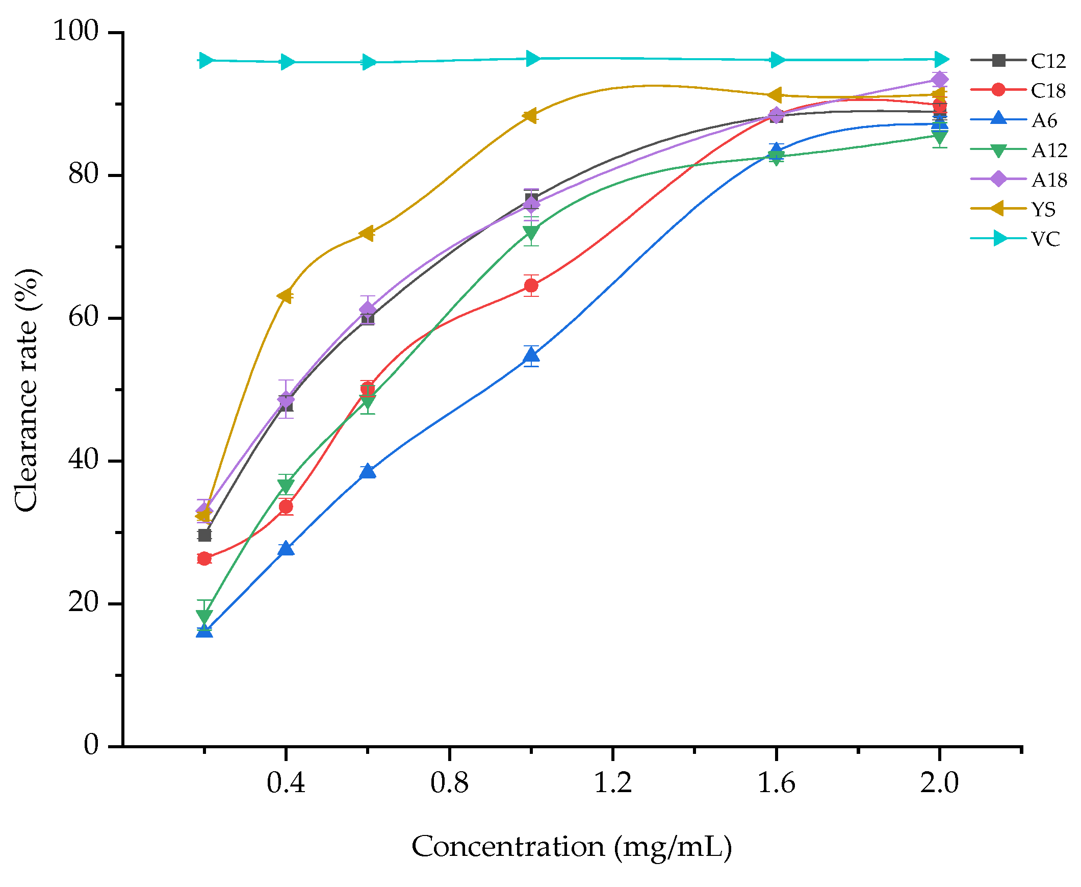
3.1.1. DPPH Free Radical Scavenging Capacity

The agarwood produced by both A. sinensis and A. crassna showed strong elimination of DPPH radicals. The clearance rate of DPPH free radicals tended to increase with increasing alcohol-soluble extract concentrations from 0.2 mg/mL to 2 mg/mL (Figure 2); thus, the scavenging rates showed some dose dependence. The IC50 values of the DPPH free radical by the artificial agarwood samples were 0.4127 mg/mL (C12), 0.5632 mg/mL (C18), 0.7487 mg/mL (A6), 0.5763 mg/mL (A12), 0.3902 mg/mL (A18), and of the wild agarwood, YS, was 0.3070 mg/mL. All batches (five) of the artificial agarwood had excellent DPPH free radical clearance rates. The IC50 value of DPPH radicals for A18 was the smallest among the sample groups, indicating that with 50% scavenging rate, its natural active ingredient was more specific and effective in clearing DPPH radicals than those of the others.
The highest DPPH free radical scavenging rates were achieved when the alcohol-soluble extract concentration was 2 mg/mL. The corresponding rates of the samples and controls (VC and YS) followed the order VC (96.27 ± 0.09%) > A18 (93.45 ± 0.98%) > YS (91.34 ± 0.41%) > C18 (89.88 ± 1.07%) > C12 (88.92 ± 1.15%) > A6 (87.20 ± 1.03%) > A12 (85.62 ± 1.76%) (Table 2), and all sample batches exhibited significant scavenging of DPPH free radicals. Moreover, the effect of VC on scavenging DPPH free radicals was significantly superior to all batches of agarwood (p < 0.05). The DPPH radical clearance rates of C12, C18, A6, A12, and A18 were 97.35%, 98.40%, 95.47%, 93.74%, and 102.31% of that of YS, respectively. The DPPH free radical scavenging effect of C18 was better than that of C12, whereas it was not significantly different from that of YS (p < 0.05). Moreover, the ability of A18 to scavenge DPPH free radicals was significantly higher than those of YS, C12, C18, A6, and A12 (p < 0.05). The results indicated that the fungal inducer not only improved the yield of this artificial agarwood and shortened the agarwood formation time but also made its ability to scavenge DPPH free radicals comparable to that of the wild agarwood. In terms of the inoculation time, the agarwood produced by 18 months of inoculation had a superior DPPH free radical scavenging rate to those produced by 6 and 12 months of inoculation. Interestingly, the DPPH free radical elimination effect of the A. sinensis-generated agarwood was better than that of the A. crassna-produced agarwood generated after 18 months of inoculation, and was even higher than that of the wild agarwood.

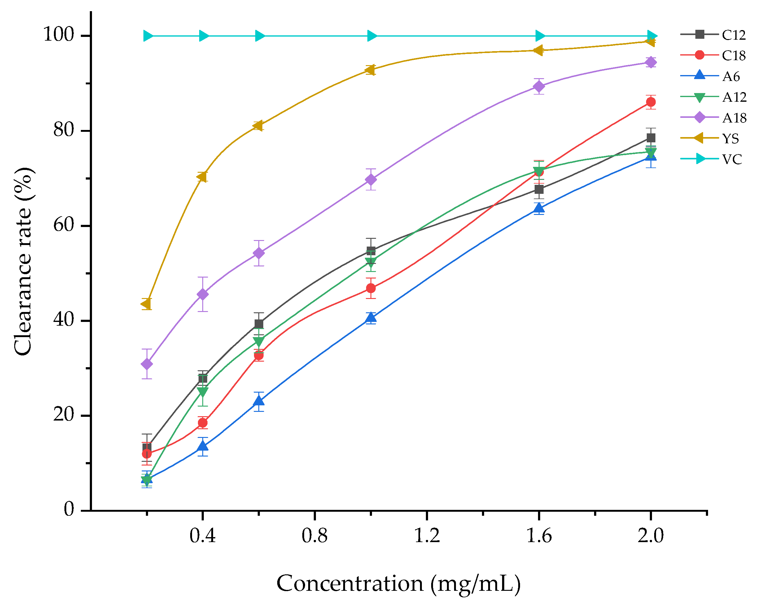
3.1.2. ABTS+ Free Radical Scavenging Capacity

The clearance rate of ABTS+ free radicals increased with increasing mass concentration of the alcohol-soluble extract from 0.2 to 2 mg/mL (Figure 3). Moreover, the scavenging activity showed some dose dependence, with the scavenging rate initially increasing below sample concentration of 1.6 mg/mL and subsequently increasing gradually. The IC50 values of ATBS+ free radical by the artificial (five samples) and wild agarwood were 0.8401 mg/mL (C12), 0.9317 mg/mL (C18), 1.1710 mg/mL (A6), 0.8999 mg/mL (A12), 0.4472 mg/mL (A18), and 0.2368 mg/mL (YS), respectively. The IC50 value of ABTS+ free radicals for A18 was the smallest among the artificial agarwood samples, indicating that 50% scavenging rate contains a natural active ingredient that is more specific than those of the other samples.
When the sample concentration was 2 mg/mL, the maximum ABTS+ free radical scavenging rates of all sample batches (five) and controls followed the order VC (100%) > YS (98.87 ± 0.25%) > A18 (94.44 ± 0.94%) > C18 (86.06 ± 1.47%) > C12 (78.59 ± 2%) > A12 (75.62 ± 1.25% )> A6 (74.53 ± 2.31%) (Table 3). This trend suggests the presence of natural active ingredients with a better clearance effect on ABTS+ free radicals. The ABTS+ radical scavenging rates of C12, C18, A6, A12, and, A18 were 79.49%, 87.93%, 75.38%, 76.48%, and 95.52% of that of YS, respectively; the ability of C18 to clear ABTS+ free radicals was significantly higher than that of C12 (p < 0.05). The ABTS+ free radical clearance rate of A18 was significantly superior to those of C12, C18, A6, and A12 (p < 0.05). Interestingly, in terms of the inoculation time, ABTS+ free radical clearance effects of the agarwood produced by both A. sinensis and A. crassna increased with the inoculation time. This suggests that the agarwood formed after 18 months of inoculation had a better ability to scavenge ABTS+ free radicals than those formed after 6 and 12 months. This was particularly for the agarwood generated by A. sinensis, whose ABTS+ free radical scavenging rate reached 95.52% of that of the wild agarwood, showing that the two rates are almost comparable.

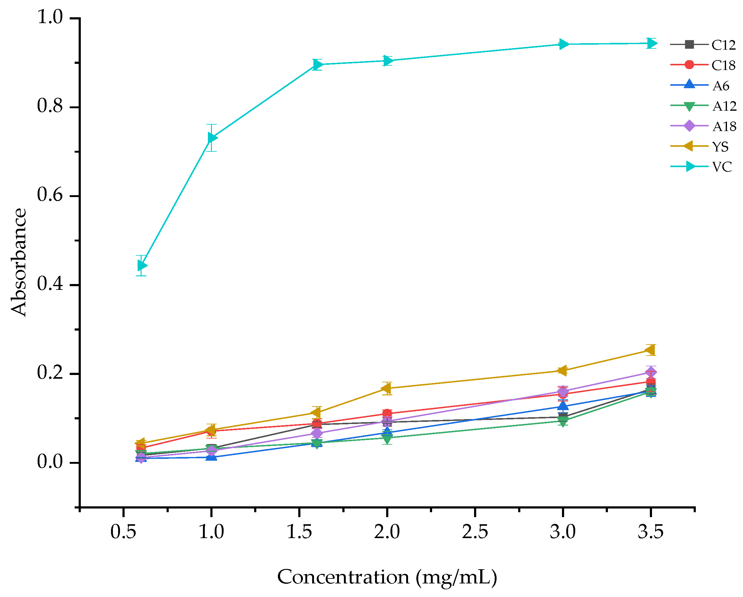
3.1.3. Total Reducing Power

The total reducing capacity of the alcohol-soluble extract increased with the increase in its mass concentration from 0.5 to 3.5 mg/mL (Figure 4), and the total reducing capacity exhibited some dose dependence.
When the sample concentration was 3.5 mg/mL, the total reducing powers of all batches (five) and controls followed the order VC (0.9437 ± 0.0116) > YS (0.2537 ± 0.0127) > A18 (0.2037 ± 0.0140) > C18 (0.1830 ± 0.0230) > C12 (0.1657 ± 0.0115) > A6 (0.1613 ± 0.0121) > A12 (0.1600 ± 0.0085) (Table 4). The total reducing power of C18, C18, A6, A12, and A18 were 65.31%, 72.13%, 63.58%, 63.07%, and 80.29% of that of YS, respectively. The total reducing power of C18 was superior to that of C12; the total reducing power of A18 was greater than that of C18, significantly higher than those of A6, A12, and C12 (p < 0.05), and lower than that of VC. The results indicate that the difference in the total reducing power of the agarwood produced by A. sinensis and A. crassna inoculated for 6 and 12 months was small. In comparison, the total reducing power of the agarwood generated after 18 months of inoculation was the highest. Moreover, for the same period, the agarwood produced by A. sinensis had superior total reducing power to that produced by A. crassna, and even reached 80.29% of that of the wild agarwood.

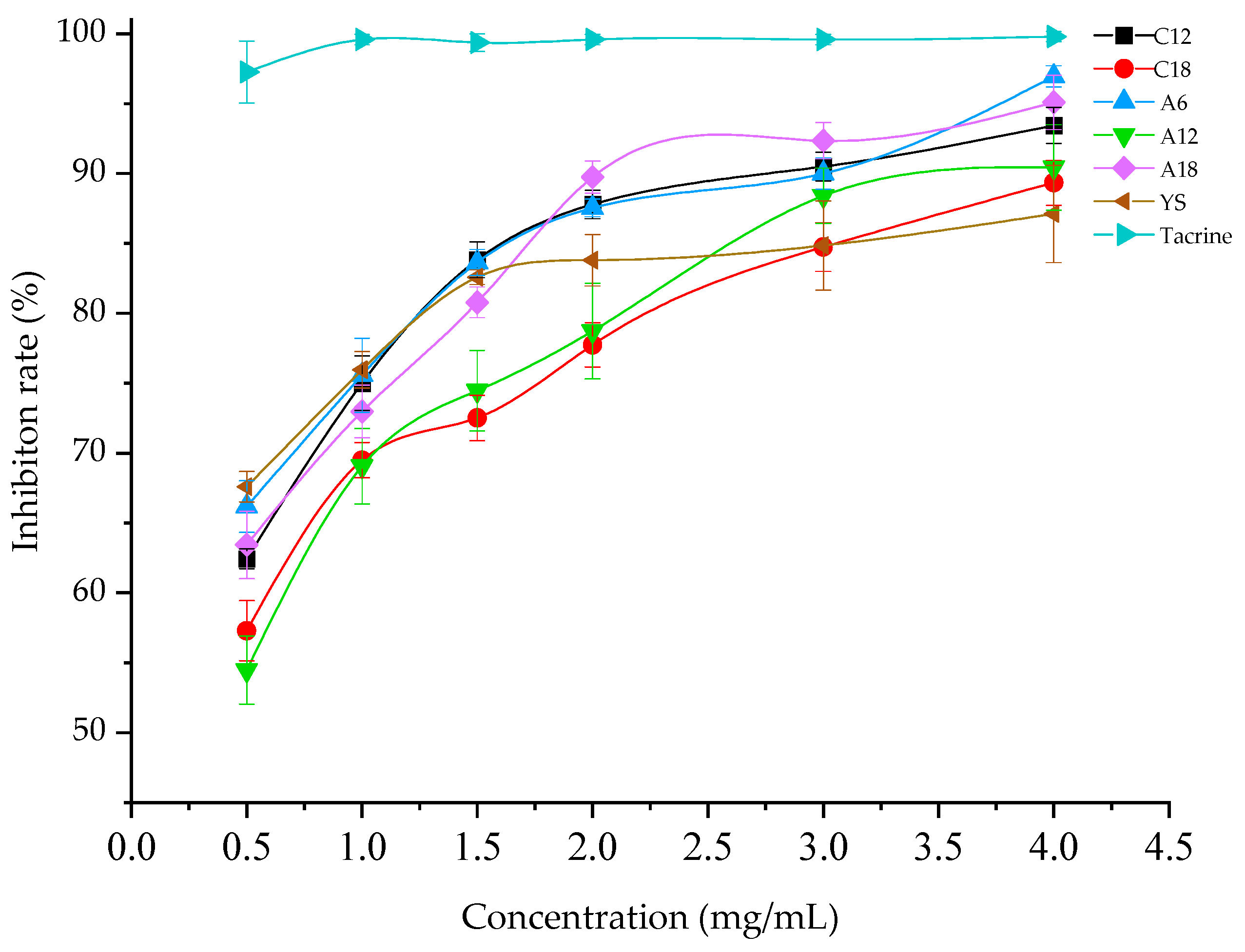
3.2. Acetylcholinesterase Activity Inhibition

The inhibitory ability of the alcohol-soluble extract on acetylcholinesterase activity increased with increasing mass concentration ranging from 0.5 mg/mL to 4 mg/mL (Figure 5). The IC50 values of acetylcholinesterase activity by the artificial (five) and wild agarwood were 0.3105 mg/mL (C12), 0.3600 mg/mL (C18), 0.2654 mg/mL (A6), 0.4274 mg/mL (A12), 0.3200 mg/mL (A18), and 0.1385 mg/mL (YS), respectively. These values indicated that the ability of A6 to inhibit acetylcholinesterase activity reached 50% of required concentration which was lower in the sample groups, and that the natural ingredients of A6 were more specific than those of the others.
The maximum inhibition rates of acetylcholinesterase activity by the samples and controls at the concentration of 4 mg/mL followed the order Tacrine (99.79 ± 0.37%) > A6 (96.95 ± 0.76%) > A18 (95.09 ± 1.95%) > C12 (93.44 ± 1.28%) > A12 (90.44 ± 3.06%) > C18 (89.34 ± 1.61%) > YS (87.11 ± 3.48%) (Table 5). These rates were 107.27%, 102.56%, 111.30%, 103.82%, and 109.16% of that of natural agarwood, respectively. The ability of A6 to inhibit acetylcholinesterase activity was not significantly different from that of the clinical drug, Tacrine (p < 0.05); the acetylcholinesterase inhibitory activity of A6 was 3.51%, 7.61%, 6.51%, 1.86%, and 9.87% higher than that of C12, C18, A12, A18, and YS, respectively. The results indicated that the natural components in all five batches of artificial agarwood inhibited acetylcholinesterase activity efficiently and were superior to those of the wild agarwood, thus indicating their potential application for the development of acetylcholinesterase inhibitors.

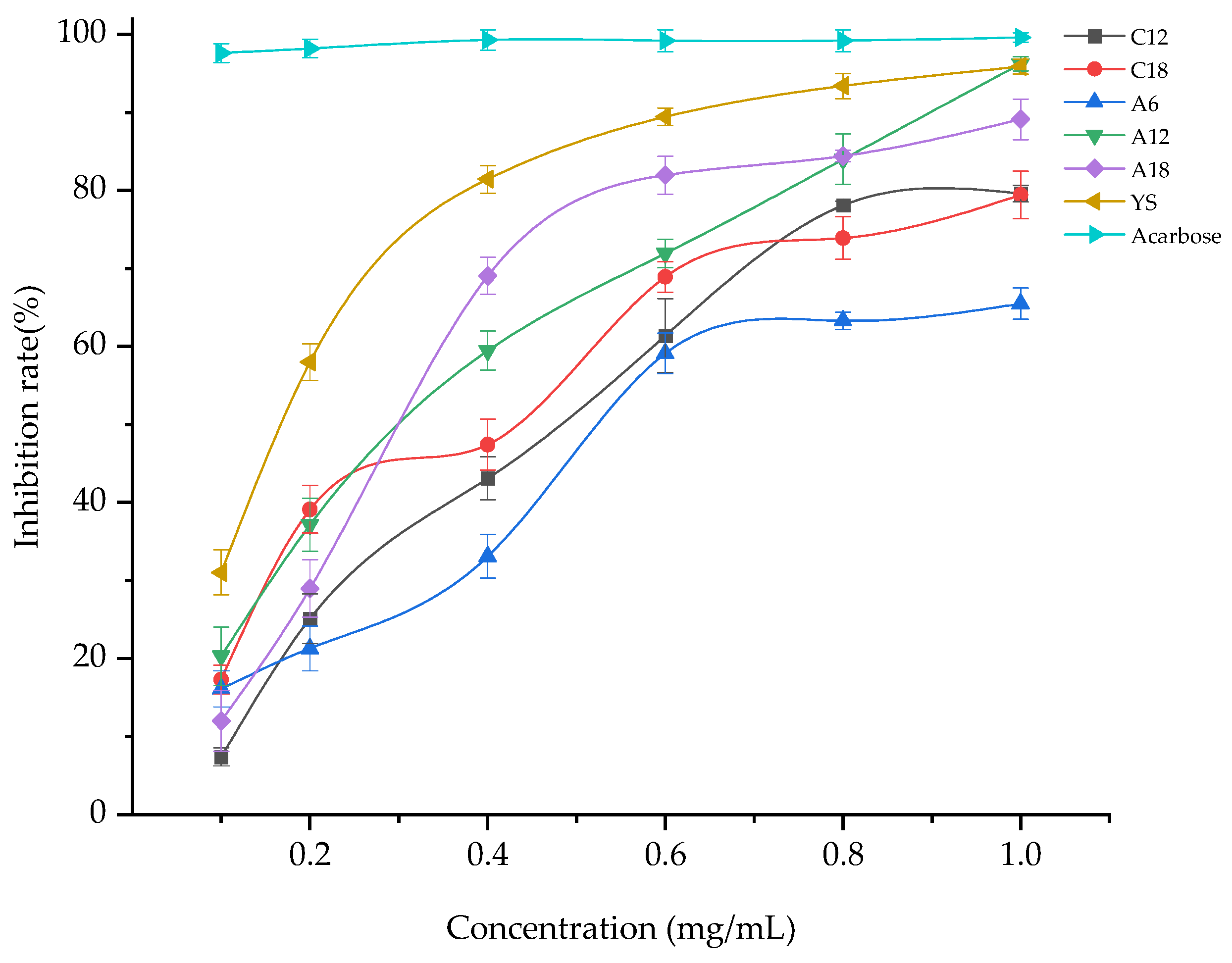
3.3. α-Glucosidase Activity Inhibition

In this study, all sample batches presented excellent inhibition rates of α-glucosidase activity. The inhibition rate of α-glucosidase activity of the alcohol-soluble extract increased with increasing mass concentration from 0.1 mg/mL to 1 mg/mL (Figure 6), and the inhibitory activity displayed a dose-dependent effect. The IC50 values of α-glucosidase activity by the artificial (five samples) and wild agarwood were 0.4326 mg/mL (C12), 0.3430 mg/mL (C18), 0.5468 mg/mL (A6), 0.2824 mg/mL (A12), 0.2920 mg/mL (A18), and 0.1564 mg/mL (YS), respectively. These indicate that the ability of A12 to inhibit acetylcholinesterase activity reached 50% of the required concentration which was lower in artificial agarwood, and that the natural ingredients of A12 were more effective than those of the other samples.
When the sample concentration was 1 mg/mL, the inhibition rates of α-glucosidase activity of the samples and controls followed the order acarbose (99.60 ± 0.60%) > A12 (96.24 ± 0.89%) > YS (95.87 ± 0.94%) > A18 (89.12 ± 2.62%) > C12 (79.60 ± 1.07%) > C18 (79.42 ± 3.05%) > A6 (65.50 ± 2.00%) (Table 6). The inhibition rates of α-glucosidase activity of C12, C18, A6, A12, and A18 were 83.03%, 68.78%, 68.32%, 100.39%, and 92.96% of that of YS, respectively. The inhibition rates of α-glucosidase of A12 and A18 were significantly higher than those of C12 and C18 (p < 0.05). The ability of A12 to inhibit α-glucosidase activity was significantly superior to those of A18, C12, C18, and A6, and was not significantly different from that of YS (p < 0.05). The results indicated that although the artificial agarwood required a short timeframe, its α-glucosidase activity inhibition rate reached more than 68.32% of that of the wild agarwood. Moreover, the inhibition rate of α-glucosidase activity of the agarwood produced by A. sinensis after 12 and 18 months of inoculation reached 100.39% and 92.96% of that of wild agarwood, which shows they are comparable. The inhibition rate of α-glucosidase activity of the agarwood generated by A. sinensis and A. crassna first increased and subsequently decreased with increasing inoculation time. Moreover, the α-glucosidase activity inhibition of the agarwood produced by A. sinensis was superior to that of the agarwood produced by A. crassna at the same inoculation time. Therefore, the best inhibition of α-glucosidase activity was achieved by agarwood from A. sinensis inoculated for 12 months, which were 7.12%, 16.64%, 16.82%, and 30.74% higher than those of A18, C12, C18, and A6, respectively.

4. Discussion

Excessive free radicals such as O2.-, OH., and NO. in the human body may originate from diet, smoking, drugs, inflammation, UV light exposure, air pollutants, stress, and alcohol [24]. Moreover, they can cause damage to biological macromolecules, such as DNA, proteins, and lipids, resulting in aging, allergies, cardiovascular diseases, inflammation, and cancer, which are harmful to human health. Many antioxidants can scavenge free radicals and reduce the damage caused by them to the human body, and several studies have shown that agarwood possesses antioxidant capacity. β-Caryophyllene is a common type of sesquiterpene in agarwood, and it has a remarkable clearance effect on DPPH radicals [7]. Wang et al. (2018) used gas chromatography-mass spectrometry to analyze the essential oil of agarwood and identified various sesquiterpenes and chromones. They also investigated the inhibition of DPPH radicals by the essential oil, indicating that the essential oil of agarwood can eliminate DPPH free radicals, and the IC50 value was obtained as 52.34 mg/mL [25]. Artificial agarwood produced by this fungal inducer, whose IC50 values for the clearance of DPPH free radicals were lower than that of the above mentioned essential oil [25], show that the natural ingredients of these artificial agarwood samples have stronger ability to scavenge DPPH free radicals. Li et al. (2020) demonstrated that several chromones, such as 6,7-dimethoxy-2-(2-phenylethyl)chromone, 6-hydroxy-2-(2-phenylethyl)chromone, and 6,8-dihydroxy-2-(2-phenylethyl)chromone, commonly found in agarwood, can reduce ABTS+ radicals [20].
All sample batches had significant antioxidant capacity, indicating their potential application for the development of oxidation inhibitors. Specifically, the agarwood produced from A. sinensis inoculated for 18 months had the smallest IC50 values of DPPH and ABTS+ radicals as well as possessing the highest reducing power than the remaining four batches of samples, thus its antioxidant capacity was best in the sample groups.
Alzheimer’s disease is a neurological disorder characterized by cognitive dysfunction and behavioral impairment, and with approximately 7.7 million new cases worldwide each year [26], it has become a difficult global problem. Acetylcholinesterase inhibitors can alleviate the symptoms of Alzheimer’s disease; however, the current ones in clinical use are accompanied by various side effects, such as dizziness, fatigue, and cardiac arrhythmias [27,28,29]. Agarwood has been used as a valuable herbal medicine since ancient times that is used to treat allergies, stomach problems, coughs, rheumatism, and gout; promote blood circulation; relieve pain, warm the middle energy and stop vomiting; regulate breathing, and relieve asthma [18]. If anti-acetylcholinesterase-active drugs could be developed from agarwood, it will be possible to avoid the above-mentioned adverse effects, and many studies have confirmed that agarwood contains several active components that can inhibit acetylcholinesterase. Li et al. (2014) tested the anti-acetylcholinesterase activity of 16 chromones isolated from agarwood using an artificial hole-punching induction method and found that 12 chromones inhibited acetylcholinesterase from 10.1 ± 0.9 to 46.1 ± 0.9% [13]. In the study of Liao et al. (2017), agarwood generated by artificial hole (four-year) induction was separated, and the anti-acetylcholinesterase activity was measured, which showed that five tetrahydrochromones and an oxidoagarochromone inhibited acetylcholinesterase activity by 17.5–47.9% [30]. Li et al. (2015) found that in agarwood obtained using the hole-punching method, five sesquiterpenes inhibited acetylcholinesterase activity by 10.3 ± 0.9–20.8 ± 0.9% [15].
However, with a manual hole-punching method, the yield of agarwood is extremely low, primarily near the cut, and the agarwood production cycle is extremely long to meet the needs of industrialization. In this study, fungi were infused into the xylem of Aquilaria trees, and the fungal sap flowed through the entire tree, resulting in whole-tree agarwood formation, which expanded the yield of agarwood, and thus, can solve the current scenario of insufficient yield. If the fungal inoculant induces the production of agarwood as a drug that inhibits acetylcholinesterase activity, then from the perspective of better economy, A. sinensis is selected as the tree species to be inoculated by the fungal inducer. Moreover, 6 months of inoculation is chosen as the better harvesting period than 12 and 18 months.
According to the International Diabetes Federation, in 2015, 415 million people have suffered from diabetes worldwide [31]. α-glucosidase inhibitors are a class of oral hypoglycemic agents; however, the currently used ones, such as acarbose and miglitol can cause gastrointestinal problems [32]. If α-glucosidase inhibitors can be prepared using natural active ingredients, the side effects described above may be avoided. Many studies have revealed that agarwood contains natural active ingredients that can inhibit α-glucosidase. Sukito et al. (2020) demonstrated that the inhibition of the α-glucoside effect reached 63.62% with 100 μg/mL acetone extract of agarwood [33]. Li et al. (2019) proved that agarozizanol E, jinkohol I, jinkohol II, and isokhusenol were sesquiterpenes from agarwood that have significant inhibitory effects on acetylcholine activity [34]. Mi et al. (2021) further established that agarwood has various 2-(2-phenylethyl) chromones, such as 5,8-dihydroxy-2-(2-phenylethyl)chromone, 6-hydroxy-8-methylsulfinyl-2-(2-phenylethyl)chromone, and 8-dihydrox-y-2-[2-(2-hydroxyphenyl)ethyl]chromone, which exhibit excellent inhibition of α-glucoside activity [35]. All artificial agarwood samples presented excellent inhibition activity against α-glucosidase and effectively reduced the decomposition of polysaccharides and sucrose into glucose, so that the absorption of sugar is slowed down accordingly. This suggests that the natural active ingredients of fungus-induced agarwood are important for the prevention and treatment of diabetes, and that the use of agarwood for the preparation of α-glucosidase inhibitors will reduce the incidence of side effects. The IC50 values of α-glucosidase activity of agarwood produced by A. sinensis during the same inoculation period were much lower than those of agarwood produced by A. crassna. If this artificial agarwood is used as a drug for the production of α-glucosidase inhibitors, then from the perspective of better economy, A. sinensis is more suitable as a tree species for inoculation with the fungal inducer. Moreover, 12 months of inoculation for producing artificial agarwood is a better harvesting period than 6 and 18 months.

5. Conclusions

In this paper, the clearance rates of DPPH and ABTS+ radicals, total reducing power, anti-acetylcholinesterase, and anti-α-glucosidase activity were determined for comparing the biological activity of the artificial and wild agarwood. The results indicated that with 2 mg/mL alcohol-soluble extracts, the DPPH free radical scavenging rate of the artificial agarwood samples were 93.74–102.31% of that of the wild agarwood, and the ABTS+ free radical clearance rates of the artificial agarwood samples were 75.38–95.52% of that of the natural agarwood. With 3.5 mg/mL alcohol-soluble extracts, the artificial agarwood samples achieved a total reducing power of 63.07–80.29% of the wild agarwood. With 4 mg/mL alcohol-soluble extracts, the acetylcholinesterase activity inhibition rates of the artificial agarwood samples were 102.56–109.16% of the wild agarwood. With 1 mg/mL alcohol-soluble extracts, the α-glucosidase activity and the inhibition rates of the artificial agarwood samples were 68.32–100.39% of that of the wild agarwood. The agarwood samples produced using A. sinensis inoculated for 6, 12, and 18 months presented the best acetylcholinesterase activity inhibition rate, α-glucosidase activity inhibition rate, and antioxidant activity, respectively. The results suggest that these artificial agarwood samples have the potential to develop products that can fight oxidation, and delay Alzheimer’s disease and treat diabetes.

Supplementary Materials

The following are available online at https://www.mdpi.com/article/10.3390/f12111532/s1, Figure S1: Inoculation of Aquilaria trees and sample collection, Figure S2: The physicochemical chromogenic reactions of samples and standard agarwood.

Author Contributions

Conceptualization, Y.F. and S.M.; methodology, S.M.; software, P.W.; validation, Y.F., S.M. and P.W.; formal analysis, Z.L.; investigation, M.Q.; resources, Y.L.; data curation, Y.L., P.W. and M.Q.; writing—original draft preparation, S.M.; writing-review and editing, P.W.; visualization, S.M.; supervision, P.W.; project administration, Y.F.; funding acquisition, Y.F. All authors have read and agreed to the published version of the manuscript.

Funding

This research was supported by the National Natural Science Foundation of China grant number 31870540.

Institutional Review Board Statement

Not applicable.

Informed Consent Statement

Not applicable.

Data Availability Statement

Data is contained within the article.

Conflicts of Interest

The authors declare no conflict of interest.

References

  1. Azren, P.D.; Azren, P.D.; Lee, S.Y.; Lee, S.Y.; Emang, D.; Emang, D.; Mohamed, R. History and perspectives of induction technology for agarwood production from cultivated Aquilaria in Asia: A review. J. For. Res. 2019, 30, 1–11. [Google Scholar] [CrossRef]
  2. Lee, S.Y.; Mohamed, R. The Origin and Domestication of Aquilaria, an Important Agarwood-Producing Genus; Springer: Singapore, 2016; pp. 1–20. [Google Scholar]
  3. Nor, A.M.; Said, A.A.; Majid, A.; Saidatul, J.; Yasmin, N. Comparison of chemical profile of selected gaharu oils from Peninsular Malaysia. Malays. J. Anal. Sci. 2008, 12, 338–340. [Google Scholar]
  4. López-Sampson, A.; Page, T. History of Use and Trade of Agarwood. Econ. Bot. 2018, 72, 107–129. [Google Scholar] [CrossRef]
  5. Nobuchi, T.; Somkid, S. Preliminary observation of Aquliaria crassna wood associated with the formation of aloeswood bult. Kyoto Univ. For. 1991, 63, 226–235. [Google Scholar]
  6. Faizal, A.; Esyanti, R.R.; Aulianisa, E.N.; Iriawati; Santoso, E.; Turjaman, M. Formation of agarwood from Aquilaria malaccensis in response to inoculation of local strains of Fusarium solani. Trees 2017, 31, 189–197. [Google Scholar] [CrossRef]
  7. Dahham, S.; Tabana, Y.; Iqbal, M.; Ahamed, M.; Ezzat, M.; Majid, A. The Anticancer, Antioxidant and Antimicrobial Properties of the Sesquiterpene β-Caryophyllene from the Essential Oil of Aquilaria crassna. Molecules 2015, 20, 11808–11829. [Google Scholar] [CrossRef]
  8. Liu, Y.; Chen, H.; Yang, Y.; Zhang, Z.; Wei, J.; Meng, H.; Chen, W.; Feng, J.; Gan, B.; Chen, X.; et al. Whole-tree agarwood-inducing technique: An efficient novel technique for producing high-quality agarwood in cultivated Aquilaria sinensis trees. Molecules 2013, 18, 3086–3106. [Google Scholar] [CrossRef] [PubMed]
  9. Rasool, S.; Mohamed, R. Understanding Agarwood Formation and Its Challenges; Springer: Singapore, 2016; pp. 39–56. [Google Scholar]
  10. Chen, H.; Yang, Y.; Xue, J.; Wei, J.; Zhang, Z.; Chen, H. Comparison of Compositions and Antimicrobial Activities of Essential Oils from Chemically Stimulated Agarwood, Wild Agarwood and Healthy Aquilaria sinensis (Lour.) Gilg Trees. Molecules 2011, 16, 4884–4896. [Google Scholar] [CrossRef] [Green Version]
  11. Wang, C.; Wang, S.; Peng, D.; Yu, Z.; Guo, P.; Wei, J. Agarwood Extract Mitigates Intestinal Injury in Fluorouracil-Induced Mice. Biol. Pharm. Bull. 2019, 42, 1112–1119. [Google Scholar] [CrossRef] [Green Version]
  12. Li, W.; Chen, H.; Wang, H.; Mei, W.; Dai, H. Natural products in agarwood and Aquilaria plants: Chemistry, biological activities and biosynthesis. Nat. Prod. Rep. 2021, 38, 528–565. [Google Scholar] [CrossRef]
  13. Li, W.; Cai, C.; Dong, W.; Guo, Z.; Wang, H.; Mei, W.; Dai, H. 2-(2-Phenylethyl)chromone derivatives from Chinese agarwood induced by artificial holing. Fitoterapia 2014, 98, 117–123. [Google Scholar] [CrossRef]
  14. Kuang, T.; Chen, H.; Kong, F.; Cai, C.; Yang, L.; Mei, W.; Dai, H. Three new 2-(2-phenylethyl)chromone derivatives from artificial holing agarwood of Aquilaria sinensis. Phytochem. Lett. 2018, 26, 96–100. [Google Scholar] [CrossRef]
  15. Li, W.; Cai, C.H.; Guo, Z.K.; Wang, H.; Zuo, W.J.; Dong, W.H.; Mei, W.L.; Dai, H.F. Five new eudesmane-type sesquiterpenoids from Chinese agarwood induced by artificial holing. Fitoterapia 2015, 100, 44–49. [Google Scholar] [CrossRef]
  16. Liao, G.; Mei, W.L.; Dong, W.H.; Li, W.; Wang, P.; Kong, F.D.; Gai, C.J.; Song, X.Q.; Dai, H.F. 2-(2-Phenylethyl)chromone derivatives in artificial agarwood from Aquilaria sinensis. Fitoterapia 2016, 110, 38–43. [Google Scholar] [CrossRef]
  17. Wang, S.L.; Liao, H.R.; Cheng, M.J.; Shu, C.W.; Chen, C.L.; Chung, M.I.; Chen, J.J. Four New 2-(2-Phenylethyl)-4H-chromen-4-one Derivatives from the Resinous Wood of Aquilaria sinensis and Their Inhibitory Activities on Neutrophil Pro-Inflammatory Responses. Planta Med. 2018, 84, 1340–1347. [Google Scholar] [CrossRef] [Green Version]
  18. Chinese Pharmacopoeia Commission. Chinese Pharmacopoeia; China Medical Science and Technology Press: Beijing, China, 2015.
  19. Yang, B.; Wang, J.; Zhao, M.; Liu, Y.; Wang, W.; Jiang, Y. Identification of polysaccharides from pericarp tissues of litchi (Litchi chinensis Sonn.) fruit in relation to their antioxidant activities. Carbohydr. Res. 2006, 341, 634–638. [Google Scholar] [CrossRef] [PubMed]
  20. Li, C.; Pan, L.; Han, Z.; Xie, Y.; Hu, H.; Liu, X.; Wu, L.; Yang, L.; Wang, Z. Antioxidative 2-(2-phenylethyl)chromones in Chinese eaglewood from Aquilaria sinensis. J. Asian Nat. Prod. Res. 2020, 22, 639–646. [Google Scholar] [CrossRef] [PubMed]
  21. Berker, K.I.; Demirata, B.; Apak, R. Determination of Total Antioxidant Capacity of Lipophilic and Hydrophilic Antioxidants In the Same Solution by Using Ferric–Ferricyanide Assay. Food Anal. Methods 2012, 5, 1150–1158. [Google Scholar] [CrossRef]
  22. Wang, H.N.; Dong, W.H.; Huang, S.Z.; Li, W.; Kong, F.D.; Wang, H.; Wang, J.; Mei, W.L.; Dai, H.F. Three new sesquiterpenoids from agarwood of Aquilaria crassna. Fitoterapia 2016, 114, 7–11. [Google Scholar] [CrossRef]
  23. Ting, L.I.; Zhang, X.D.; Song, Y.W. A microplate-based screening method for alpha-glucosidase inhibitors. Chin. J. Clin. Pharmacol. Ther. 2005, 10, 1128–1134. [Google Scholar]
  24. Poljšak, B.; Dahmane, R. Free Radicals and Extrinsic Skin Aging. Dermatol. Res. Pract. 2012, 2012, 135206. [Google Scholar] [CrossRef] [PubMed] [Green Version]
  25. Wang, M.; Li, W.; Luo, S.; Zhao, X.; Ma, C.; Liu, S. GC-MS Study of the Chemical Components of Different Aquilaria sinensis (Lour.) Gilgorgans and Agarwood from Different Asian Countries. Molecules 2018, 23, 2168. [Google Scholar] [CrossRef] [Green Version]
  26. Amini, M.; Zayeri, F.; Moghaddam, S.S. Years Lived with Disability due to Alzheimer’s Disease and Other Dementias in Asian and North African Countries: A Trend Analysis. J. Epidemiol. Glob. Health 2019, 9, 29–35. [Google Scholar] [CrossRef] [PubMed] [Green Version]
  27. Celik, I.E.; Acar, B.; Cay, S. An unusual cardiovascular adverse effect of donepezil. Intern. Med. J. 2015, 45, 877–878. [Google Scholar] [CrossRef]
  28. Neely, G.A.; Sabir, S.; Kohli, A.; Neostigmine. StatPearls. 2020. Available online: https://www.ncbi.nlm.nih.gov/books/NBK470596/?report=printable (accessed on 5 March 2021).
  29. Hernandez-Fernandez, F.; Pardal-Fernandez, J.M.; Garcia-Martinez, E.; Segura, T. Respiratory myoclonus, a side effect of galantamine. Farm. Hosp. 2011, 35, 97–99. [Google Scholar] [CrossRef]
  30. Liao, G.; Mei, W.; Kong, F.; Li, W.; Yuan, J.; Dai, H. 5,6,7,8-Tetrahydro-2-(2-phenylethyl)chromones from artificial agarwood of Aquilaria sinensis and their inhibitory activity against acetylcholinesterase. Phytochemistry 2017, 139, 98–108. [Google Scholar] [CrossRef]
  31. Papatheodorou, K.; Banach, M.; Bekiari, E.; Rizzo, M.; Edmonds, M. Complications of Diabetes 2017. J. Diabetes Res. 2018, 2018, 1–4. [Google Scholar] [CrossRef]
  32. Ma, Y.; Zhao, D.; Zhou, A.; Zhang, Y.; Du, Z.; Zhang, K. α-Glucosidase Inhibition and Antihyperglycemic Activity of Phenolics from the Flowers of Edgeworthia gardneri. J. Agric. Food Chem. 2015, 63, 8162–8169. [Google Scholar] [CrossRef]
  33. Sukito, A.; Darmawan, S.; Turjaman, M. Anti-oxidant and anti-diabetes activities of agarwood extracts from Gyrinops versteegii (Gilg.) Domke and their cytotoxicity. IOP Conf. Ser. Earth Environ. Sci. 2020, 415, 012001. [Google Scholar] [CrossRef]
  34. Yang, L.; Yang, Y.L.; Dong, W.H.; Li, W.; Wang, P.; Cao, X.; Yuan, J.Z.; Chen, H.Q.; Mei, W.L.; Dai, H.F. Sesquiterpenoids and 2-(2-phenylethyl)chromones respectively acting as α-glucosidase and tyrosinase inhibitors from agarwood of an Aquilaria plant. J. Enzyme Inhib. Med. Chem. 2019, 34, 853–862. [Google Scholar] [CrossRef] [PubMed] [Green Version]
  35. Mi, C.; Yuan, J.; Zhu, M.; Yang, L.; Wei, Y.; Wang, H.; Long, W.; Mei, W.; Dai, H. 2-(2-Phenylethyl)chromone derivatives: Promising α-glucosidase inhibitors in agarwood from Aquilaria filaria. Phytochemistry 2021, 181, 112578. [Google Scholar] [CrossRef] [PubMed]
Figure 1. Artificial and wild agarwood.
Forests 12 01532 g001
Figure 2. DPPH free radical clearance rate variation with extract concentration (C12 and C18 represented agarwood produced by A. crassna after 12 and 18 months of inoculation, respectively; A6–A18 represented agarwood produced by A. sinensis after 6, 12, and 18 months of inoculation, respectively; YS and VC represented wild agarwood and ascorbic acid, respectively).
Forests 12 01532 g002
Figure 3. ABTS+ free radical clearance rate variation with extract concentration (C12 and C18 represented agarwood produced by A. crassna after 12 and 18 months of inoculation, respectively; A6–A18 represented agarwood produced by A. sinensis after 6, 12, and 18 months of inoculation, respectively; YS and VC represented wild agarwood and ascorbic acid, respectively).
Forests 12 01532 g003
Figure 4. Total reducing power variation with extract concentration (C12 and C18 represented agarwood produced by A. crassna after 12 and 18 months of inoculation, respectively; A6–A18 represented agarwood produced by A. sinensis after 6, 12, and 18 months of inoculation, respectively; YS and VC represented wild agarwood and ascorbic acid, respectively).
Forests 12 01532 g004
Figure 5. Inhibition rate of acetylcholinesterase activity variation with extract concentration (C12 and C18 represented agarwood produced by A. crassna after 12 and 18 months of inoculation, respectively; A6–A18 represented agarwood produced by A. sinensis after 6, 12, and 18 months of inoculation, respectively; YS represented wild agarwood).
Forests 12 01532 g005
Figure 6. Inhibition rate of α-glucosidase activity variation with extract concentration (C12 and C18 represented agarwood produced by A. crassna after 12 and 18 months of inoculation, respectively; A6–A18 represented agarwood produced by A. sinensis after 6, 12, and 18 months of inoculation, respectively; YS represented wild agarwood).
Forests 12 01532 g006
Table 1. Sample formation.
Sample No.Species NameInoculation TimeNumber of Samples
C12A. crassna12 monthsN = 3
C18A. crassna18 monthsN = 3
A6A. sinensis6 monthsN = 3
A12A. sinensis12 monthsN = 3
A18A. sinensis18 monthsN = 3
Table 2. DPPH free radical scavenging capacity of five batches and controls.
SamplesAlcohol-Soluble Extract Solution Concentration (mg/mL)DPPH Free Radical Scavenging
Capacity (%)
C12288.92 ± 1.15 d
C18289.88 ± 1.07 c
A62 87.20 ± 1.03 d,e
A12285.62 ± 1.76 e
A18293.45 ± 0.98 b
YS291.34 ± 0.41 c
VC296.27 ± 0.09 a
Each value represents the mean ± SD (n = 3); SD, standard deviation. At p < 0.05 according to one-way analysis of variation, mean values followed by different letters are significantly different from each other; mean values followed by one letter identical are not significantly different from each other.
Table 3. The ABTS+ free radical scavenging capacity of five batches and controls.
SamplesAlcohol-Soluble Extract Solution Concentration (mg/mL)ABTS+ Free Radical Scavenging
Capacity (%)
C12278.59 ± 2 d
C18286.06 ± 1.47 c
A6274.53 ± 2.31 e
A12275.62 ± 1.25 d,e
A18294.44 ± 0.94 b
YS298.87 ± 0.25 a
VC2100 a
Each value represents the mean ± SD (n = 3); SD, standard deviation. At p < 0.05 according to one-way analysis of variation, mean values followed by different letters are significantly different from each other; mean values followed by one letter identical are not significantly different from each other.
Table 4. The total reducing powers of all five batches and controls.
SamplesAlcohol-Soluble Extract Solution Concentration (mg/mL)Total Reducing Power
C123.50.1657 ± 0.0115 d
C183.5 0.1830 ± 0.0230 c,d
A63.50.1613 ± 0.0121 d
A123.50.1600 ± 0.0085 d
A183.50.2037 ± 0.0140 c
YS3.50.2537 ± 0.0127 b
VC3.50.9437 ± 0.0116 a
Each value represents the mean ± SD (n = 3); SD, standard deviation. At p < 0.05 according to one-way analysis of variation, mean values followed by different letters are significantly different from each other; mean values followed by one letter identical are not significantly different from each other.
Table 5. The inhibition rates of acetylcholinesterase activity in all five batches and controls.
SamplesAlcohol-Soluble Extract Solution Concentration (mg/mL)Acetylcholinesterase Activity
Inhibition (%)
C12493.44 ± 1.28 b,c
C18489.34 ± 1.61 d
A6496.95 ± 0.76 a,b
A12490.44 ± 3.06 c,d
A18495.09 ± 1.95 b
YS487.11 ± 3.48 d
Tacrine499.79 ± 0.37 a
Each value represents the mean ± SD (n = 3); SD, standard deviation. At p < 0.05 according to one-way analysis of variation, mean values followed by different letters are significantly different from each other; mean values followed by one letter identical are not significantly different from each other.
Table 6. The inhibition rates of acetylcholinesterase activity in all five batches and controls.
SamplesAlcohol-Soluble Extract Solution Concentration (mg/mL)α-Glucosidase Activity
Inhibition (%)
C12179.60 ± 1.07 d
C18179.42 ± 3.05 d
A6165.50 ± 2.00 e
A12196.24 ± 0.89 b
A18189.12 ± 2.62 c
YS195.87 ± 0.94 b
Acarbose199.60 ± 0.60 a
Each value represents the mean ± SD (n = 3); SD, standard deviation. At p < 0.05 according to one-way analysis of variation, mean values followed by different letters are significantly different from each other; mean values followed by one letter identical are not significantly different from each other.
Publisher’s Note: MDPI stays neutral with regard to jurisdictional claims in published maps and institutional affiliations.

