Article

Transformation of American Chestnut (Castanea dentata (Marsh.) Borkh) Using RITA® Temporary Immersion Bioreactors and We Vitro Containers

1
Department of Environmental and Forest Biology, State University of New York College of Environmental Science and Forestry, Syracuse, NY 13210, USA
2
Instituto Nacional de Investigação Agrária e Veterinária I.P., Avenida da República, 2780-159 Oeiras, Portugal
3
Green-It Bioresources for Sustainability, ITQB NOVA, Av. Da República, 2780-157 Oeiras, Portugal
*
Author to whom correspondence should be addressed.
Forests 2020, 11(11), 1196; https://doi.org/10.3390/f11111196
Submission received: 20 October 2020 / Revised: 11 November 2020 / Accepted: 12 November 2020 / Published: 13 November 2020
(This article belongs to the Special Issue Application of Tissue Culture in Plant Reproduction)

Abstract

:
American chestnut (Castanea dentata (Marsh.) Borkh) was almost completely wiped out by the fungal pathogen, Cryphonectria parasitica (Murrill) M.E. Barr. Another invasive pathogen, Phytophthora cinnamomi Rands, is devastating American chestnuts in the southern region of the United States. An alternative approach for controlling these pathogens is to use genetic engineering or gene editing. We successfully transformed American chestnut with a detoxifying enzyme, oxalate oxidase, to enhance blight tolerance and more recently with the Cast_Gnk2-like gene, which encodes for an antifungal protein, to be tested for P. cinnamomi putative tolerance. Eight somatic embryo lines were transformed using three methods of selection: semisolid medium in Petri plates, liquid medium in RITA® temporary immersion bioreactors, or liquid medium in We Vitro containers. No significant differences were found between the treatments. These methods will allow for further testing of transgenes and the development of enhanced pathogen resistance in chestnut. It can serve as a model for other tree species threatened by invasive pests and pathogens.

1. Introduction

The American chestnut (Castanea dentata (Marsh.) Borkh) is one of eight Castanea species that include Castanea sativa P. Mill., Castanea mollissima Blume, Castanea crenata Siebold & Zucc., Castanea henryi (Skan.) Rehder. & E.H. Wilson, Castanea seguinii Dode, Castanea pumila (L.) Mill., and Castanea ozarkensis Ashe. As a result of world trade, a fungal pathogen, Cryphonectria parasitica (Murrill) M.E. Barr, which coexisted with the Asian Castanea species, was introduced to native species in North America and Europe with devastating effects [1]. The American chestnut appears to have been the most susceptible species with the loss of over three billion trees within a span of 50 years, leading to significant economic, cultural, and ecological effects, including the extinction of several insect species.
Because of the chestnut’s importance, many attempts have been made to develop trees that are resistant to the blight caused by C. parasitica [2]. Backcross breeding of hybrids to regain ecosystem adaptability has been very complicated due to the quantitative nature of blight resistance as well as the differing characteristics of blight-resistant species and blight-susceptible species that are required in a restoration tree [3,4]. Another complication to restoration is the invasive pathogen Phytophthora cinnamomi Rands and Dryocosmus kuriphilus Yasumatsu, a gall wasp insect pest [2].
An alternative approach for controlling these pathogens and pests is to use genetic engineering or gene editing. From a conservation point of view, these methods may be preferable because of the small genetic changes made compared to hybrid breeding methods and thus, help maintain the genomic integrity and adaptations of the species [5]. Genetic engineering has been successful at developing an American chestnut tree with enhanced blight tolerance by adding a detoxifying enzyme, oxalate oxidase, to counter the major virulence factor of the blight pathogen C. parasitica [6]. For genetic engineering or gene editing to be successful, a robust tissue culture and transformation system needs to be available. This paper describes genetic engineering and tissue culture methods that can be used to develop pest and pathogen resistance in American chestnut and augment restoration efforts. In one experiment, five somatic embryo lines were transformed with an oxalate oxidase detoxifying gene to enhance blight tolerance; using three methods of selection, i.e., semisolid medium in Petri plates, liquid medium in RITA® temporary immersion bioreactors, or liquid medium in We Vitro containers. In a second experiment, four somatic embryo lines were transformed with the Cast_Gnk2-like gene, for possible P. cinnamomi tolerance. It was expected that a liquid selection method would increase transformation efficiency.

2. Materials and Methods

2.1. Plant Material

Embryos for Transformation

American chestnut somatic embryos from clonal lines Alessi T2-1A (AL), AxW3-46B (AxW3), Bass Mountain #5-1B (BM5), Moss Lake 90016 C-21-1C (ML), Spring Hole-4A (SH4), Spring Hole-11D (SH11), and TG-8A (TG) were obtained from Dr. Scott Merkle at the University of Georgia (Athens, GA). AL was from an open pollinated nut collected from Russell, NY; AxW3 was from a controlled pollinated nut between the Adair tree in Kentucky and the Wayne3 tree in Maine; BM5 was from an open pollinated nut collected from Hancock, NY; ML was from an open pollinated nut collected from the White Seed Orchard in Gowanda, NY; SH4 and SH11 were from open pollinated nuts collected from Windsor, NY; and TG was from an open pollinated nut collected from the Thoroughfare Gap tree in Virginia. Clonal line Ellis #1 was established in culture from immature zygotic embryos obtained by ESF from a member of the New York State Chapter of the American Chestnut Foundation located in Windsor, NY. Embryo cultures were established and maintained following the procedure described by Carraway [7] and Maynard et al. [8]. Briefly, immature burs were collected 1 month post-anthesis from wild seed of the American chestnut trees. Nuts were removed from burs, sterilized for 5 min in a 50% bleach solution, and rinsed three times in sterile distilled water. Nuts were cut open and individual zygotic embryos were transferred to 60 mm × 15 mm Petri plates containing embryo initiation medium (E1)—a modified Woody Plant Media composed of WPM salts [9], 109 mg/L Nitsch and Nitsch vitamins [10], 1.0 g/L casein enzymatic hydrolysate, 1.8 µM 2,4-dichlorophenoxyacetic acid (2,4-D), 1.1 µM 6-benzyladenine (BA), 3.0% sucrose (Avantor, Phillipsburg, NJ, USA), and 3.0 g/L Phytagel (Sigma-Aldrich, St. Louis, MO, USA), with the pH adjusted to 5.5 [11]. Unless otherwise stated, all chemicals and media were reagent-grade and purchased from PhytoTech Chemical Company (Shawnee Mission, KS, USA). When the single zygotic embryo from each line began to multiply, the lines were subcultured and transferred to fresh E1 medium every 2–3 weeks.

2.2. Plasmid DNA

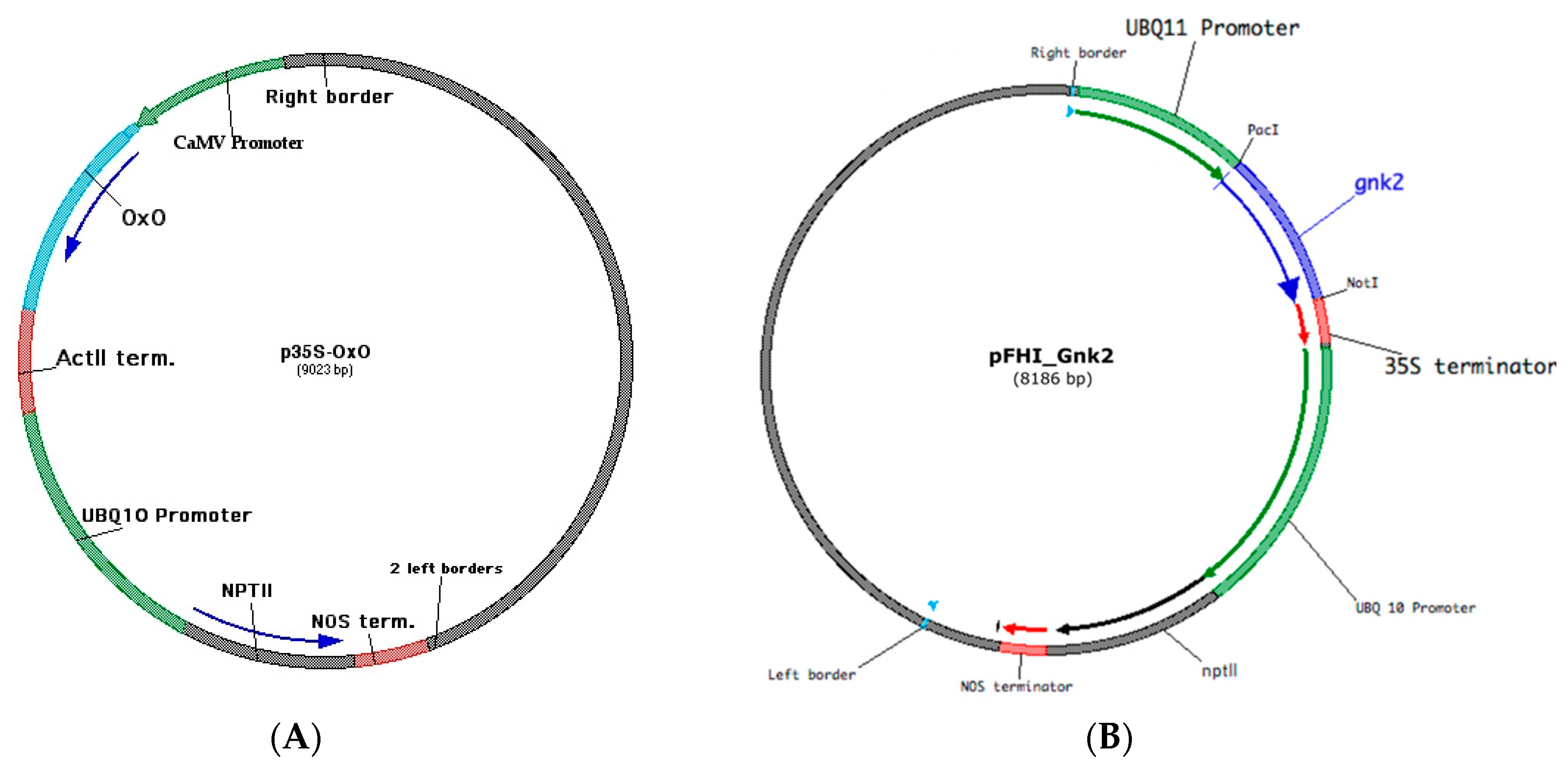
2.2.1. p35S-OxO for Blight Tolerance

The vector used in the transformation for blight tolerance is known as p35S-OxO (Figure 1A) [12]. This vector contains two genes and their associated regulatory sequences, i.e., oxalate oxidase (OxO) for blight tolerance and the selectable marker neomycin phosphotransferase (NPTII). The OxO gene is driven by the constitutive cauliflower mosaic virus (CaMV 35S) promoter [13] and is followed by the Actin2 (ACT2) terminator from Arabidopsis thaliana, which was chosen for efficient termination of transcription and the addition of a poly-A tail to the mRNA. The NPTII selectable marker gene is controlled by the constitutive promoter Ubiquitin 10 (UBQ10), which comes from the Ubiquitin 10 gene from Arabidopsis thaliana. It is followed by the nopaline synthase (NOS) 3′ terminator from Agrobacterium tumefaciens, which was chosen to terminate transcription and to add poly-A tails to the mRNA. Expression of NPTII from Escherichia coli allows plant tissue to survive in the presence of aminoglycoside antibiotics such as kanamycin, neomycin, geneticin (G418), or paromomycin [14,15]. When expressed in American chestnut somatic embryo tissue, this served as a selectable marker, facilitating development and selection of transformed lines in the presence of paromomycin. The full description of p35s-OxO can be found in the Petition for Determination of Nonregulated Status for Blight-Tolerant Darling 58 American Chestnut (Castanea dentata) [16].

2.2.2. pFHI_Gnk2 for Putative Phytophthora-Tolerance

The chestnut gene Cast_Gnk2-like, encoding for an antifungal protein, was isolated from the cDNA of Castanea crenata root tissue inoculated with the ink disease pathogen, Phytophthora cinnamomi. Cast_Gnk2-like was cloned into a pFHI vector (developed by Dr. C. Joseph Nairn, University of Georgia) using the Gibson Assembly Cloning Kit (New England Biolabs, MA, USA). The binary vector pFHI_Gnk2 (Figure 1B) carries the coding sequences of two genes: (i) the Cast_Gnk2-like gene, driven by a Ubiquitin 11 (UBQ11) constitutive promoter and a CaMV 35S terminator and (ii) the NPTII gene as a selectable agent, with a UBQ10 promoter and a NOS terminator. Agrobacterium tumefaciens AGL-1 strain was transformed with pFHI_Gnk2 via electroporation and then used to transform the American chestnut somatic embryo lines.
An Agrobacterium AGL-1, transformed with a pFHI_GFP vector, was used as a positive control during the somatic embryo transformations. The pFHI_GFP vector had the pFHI backbone expressing a green fluorescent protein (GFP) gene instead of the Cast_Gnk2-like gene.

2.3. Transformation Protocol

2.3.1. Transformation of American Chestnut for Blight Tolerance

Transformation followed the protocol described by Maynard et al. [8]. Agrobacterium strain AGL1 containing the p35S-OxO vector was grown overnight in Luria–Bertani broth, Miller Modification [17], containing 50 mg/L kanamycin, at 28 ± 2 °C on an orbital shaker (Lab-Line Instruments, Inc. Melrose Park, IL), set to approximately 150 RPM, until an optical density (OD650) of 0.8–1.0 was reached. The Agrobacterium was centrifuged in a Fisher Scientific AccuSpin™ 400 (Fisher Scientific Company L.L.C., Pittsburgh, PA, USA) for 15 min at 1700× g and the pellet was resuspended in Virulence Induction Media (WPM salts, 1% sucrose, 10 g/L 2-(N-morpholino) ethanesulfonic acid (MES) buffer, and 100 µM acetosyringone). The suspension was incubated at 22 ± 2 °C on a shaker at approximately 75 RPM for 2 h. For each treatment, 16 somatic embryo clumps (2 weeks since last subculture) were transferred to a pre-weighed 14 mL sterile polypropylene Falcon® tube (Corning Life Sciences, Tewksbury, MA, USA) and placed on a Fisher Scientific XT top loading balance (Pittsburgh, PA, USA). The weights of the tissue per tube ranged from 36 to 157 mg. Treatments for the blight-tolerance transformation experiment included four clonal backgrounds; AL, AxW3, BM5, and TG for the first trial and AL, AxW3, ML, and TG for the second trial; three types of vessels were used during selection (Figure 2), i.e., 60 mm × 15 mm Petri plates (Fisher Scientific), We Vitro containers cultivated by Magenta® (We Vitro Inc., 240 Waterloo Ave, Unit 208, Guelph, ON, Canada), and temporary immersion bioreactor systems (RITA® bioreactors, Sigma Aldrich, St. Louis, MO, USA). Semisolid medium was used in the Petri plates, while liquid medium was used in the We Vitro and RITA® containers. After weights were recorded, the clumps were covered with 4 mL of Agrobacterium inoculum, and the tubes were placed on a 360° rotating shaker (Labquake or equivalent, approx. 30–40 RPM). After 1 h at 23 ± 2 °C, the inoculum was removed by carefully pouring into a waste container. The embryo clumps were moved to desiccation plates, sterile 60 mm × 15 mm Petri plates containing sterile 47 mm filter paper (Whatman International Ltd., Maidstone, UK) slightly moistened with 200 µL of sterile distilled water (Figure 2A), and incubated in the dark at 23 ± 2 °C for 2 days. The clumps were transferred to a semisolid Agro-Kill medium (E1 with 50 mg/L cefotaxime and 333 mg/L TIMENTIN) for 2 weeks and then moved to a either RITA® bioreactors, We-Vitro containers, or a small Petri plates containing semisolid medium (Figure 2B–G) and incubated in the dark at 23 ± 2 °C. Embryos in the RITA® bioreactors were intermittently flooded (the immersion period was 2 min every 4 h) with 150 mL of liquid selection medium (E1 with 50 mg/L cefotaxime, 333 mg/L TIMENTIN, and 60 mg/L paromomycin, without a gelling agent). Embryos in the We Vitro containers were first placed in sterile empty tea bags (Finum, Hamburg, Germany) that were trimmed to 8 cm with small notches cut in one side. The tea bags were set to one side of the We Vitro container with a divider (Figure 2C) and 100 mL of liquid selection medium was added. The containers were wrapped in aluminum foil, to keep the tissue in the dark, and then placed on a MicroRocker. The rocker was set as follows: tilt time: 6 s, left angle: 35.00°, right angle: 35.00°, left park time: 4 h, right park time: 3 min, and angle offset: −0.90°. The selection medium was changed every 2 weeks. After 6 weeks, dead tissue was discarded. Any tissue that remained light in color was transferred to semisolid E1 selection medium (same as liquid selection medium with 3.0 g/L Phytagel). The clumps were subcultured and transferred every 2 weeks to fresh selection medium, and after 4 weeks, non-transformed tissue and callus was discarded. The experiment was conducted twice.
Each clump of surviving (light-colored) tissue was considered a separate event, presumably originating from a single transformed cell. Putative transgenic events were multiplied as somatic embryos for at least 1 month and then tested for the presence of OxO via polymerase chain reaction (PCR). Embryos were propagated and monitored for any Agrobacterium tumefaciens growth for more than 6 months before regeneration. Untransformed clumps that escaped the selection process were counted to determine which method was best at selecting transformants.
Once the presence of OxO in the somatic embryos was confirmed, they were multiplied and regenerated as described by Maynard et al. [8]. In previous studies, some of the light-colored sectors on the embryos had originated from multiple independent transformation events, which in turn, led to mixed shoot cultures. To avoid this problem, a single shoot was selected from the regenerated transformed material. This individual shoot was multiplied and all resulting propagated material comprising the event was from this single shoot.

2.3.2. Transformation of American Chestnut for Putative Phytophthora-Tolerance

The protocol for transforming American chestnut embryos with the Cast_Gnk2-like gene for Phytophthora-tolerance was similar to the protocol described in Section 2.3.1, with the following changes. After resuspending the Agrobacterium pellet in virulence induction medium, it was incubated at 22 ± 2 °C on a shaker at approximately 75 RPM for 4 h. Each transformation with pFHI_Gnk2 was carried out in 14 mL sterile polypropylene Falcon® tubes containing 9 somatic embryo clumps of either Ellis #1 (approximately 170–180 mg), AxW3 (144 mg), SH4 (103 mg), or SH11 (113 mg). For the pFHI_GFP positive control, approximately 170 mg of Ellis #1 somatic embryos were used. After mixing the embryos with the Agrobacterium suspension culture, they were transferred to RITA® bioreactors and intermittently flooded for 2.5 min every 4 h with liquid Agro-Kill medium for 1 week and then with liquid selection medium for 5 weeks. The liquid media was changed every week. The transformation efficiency was determined by the number of events per embryo mass. The living tissue of putatively positive events was isolated and transferred to semisolid E1 selection medium for multiplication.

2.4. Multiplication, Rooting, and Acclimatization of Regenerated Shoots Expressing OxO
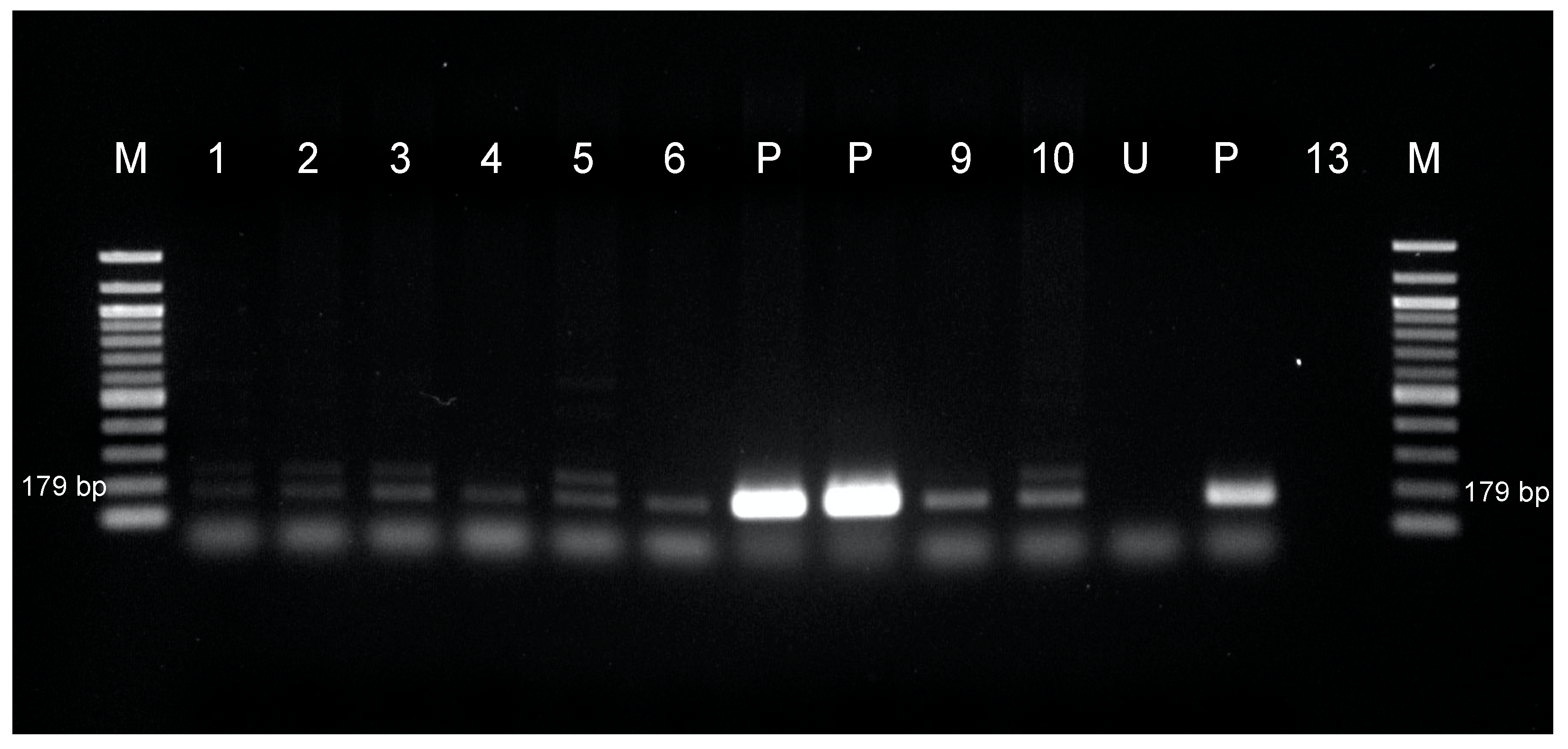
American chestnut shoots were multiplied in ACII medium (WPM salts, Nitsch and Nitsch vitamins, 1.0 µM BA, 10.0 µM indole-3-butyric acid (IBA), 3.0% sucrose, and 7 g/L agar, with the pH adjusted to 5.5 [18]), subculturing every 4 weeks. They were elongated in pre-rooting (PR) medium (WPM salts, Nitsch and Nitsch vitamins, 0.5 µM BA, 500 µM IBA, 3.0% sucrose, 7 g/L agar, with the pH adjusted to 5.5) for 3 weeks and then transferred to fresh PR medium, without cutting the callus base in order to attain long stems (approximately 7 cm) that were thick (>1 mm diameter), for an additional 3 weeks. Ex vitro rooting was done following Oakes et al. methods [19,20]. Briefly, large, well-developed shoots with thick stems were cut while submerged in water and subsequently dipped into Clonex Rooting Gel (Hydrodynamics International, Lansing, MI, USA) before inserting into a water-moistened Jiffy pellet (Jiffy Products, Lorain, OH, USA). The pellets were placed in clear plastic shoe boxes, the plantlets were lightly misted with water, and the box was placed on a light bench with a 16 h photoperiod. Once visible roots emerged from the Jiffy pellet substrate, the “wrapper” around the outside was removed, and the plug was potted (without disturbing the roots) into a very fine germination potting mix that was adjusted to pH 5.5. Gnatrol WDG (Nufarm, Alsip, IL, USA) and Scanmask beneficial nematodes (Hirt’s Gardens, Medina, OH, USA) were used to treat the potting mix both before planting and at subsequent waterings, to prevent fungal gnat infestation. Both Miracle-Gro® Water Soluble Miracid® Acid-Loving Plant Food (The Scotts Company LLC, Marysville, OH, USA) and Jack’s Acid Special 17-6-6 Fertilizer (JR Peters, Allentown, PA, USA) were used to promote growth without nutrient deficiencies. Surface algae overgrowth was treated with 1% ZeroTol applications (BioSafe Systems, Hartford, CT, USA). Potted plantlets were grown in a high humidity growth chamber (Conviron®, Winnipeg, Manitoba Canada) until fresh growth emerged and the plant hardened off, and they were then moved to a greenhouse.

2.5. PCR Analysis

Embryos transformed with p35S-OxO were confirmed by PCR. DNA extraction was carried out using a QIAGEN DNeasy® Plant Mini Kit (QIAGEN, Hilden, Germany) and Lysing Matrix A grinding tubes. Two or three embryos were used in each extraction tube (weight approx. 0.05 g). The individual PCR reactions were assembled using 12.5 µL GoTaq® Green Master Mix (Promega Corporation, Madison, WI, USA), 7.5 µL PCR grade H2O, and 2 µL of the diluted template DNA. The oxalate oxidase-specific PCR primers were (forward: 5′-GCCAACTTGGACGAGAAGAG-3′; reverse: 5′-CCTAGTAGCTGGCCTGTTCG-3′) designed to amplify a 179 bp fragment. Primers were diluted to a 1:10 ratio. Amplification was carried out with an initial cycle at 95 °C for 5 min, 95 °C for 30 s, 58 °C for 30 s, 72 °C for 1 min, followed by 32 cycles at 95 °C for 30 s, and a final extension of 3 min. PCR products were analyzed by gel electrophoresis on a 1.5% agarose gel using 1× TAE buffer, with a 100 bp ladder. The gels were stained with ethidium bromide.
The presence of transgenes NPTII, UBQ11, Cast_Gnk2-like, and NOS in the somatic embryo lines transformed with pFHI_Gnk2 was also verified by PCR analysis. PCR analysis was carried out in a 50 µL volume containing 1× supplied Taq buffer, 3 mM magnesium chloride, dNTP mix (0.2 mM each), 1.0 mM each primer, 1.25 units GoTaq® Flexi DNA Polymerase (Promega Corporation, Madison, WI, USA), and 50–100 mg of genomic DNA. Specific primers were used for NPTII (forward: 5′-ATGATTGAACAAGATGGATTGCAC-3′; reverse: 5′-GAAGAACTCGTCAAGAAGGCG-3′) and the segment UBQ11, Cast_Gnk2-like, and NOS (forward: 5′-TTCCGGAGGTGTTTCTAGTGT-3′; reverse: 5′-CTCAACACATGAGCGAAACCC-3′). The expected sizes of PCR fragments were 791 bp for NPTII and 1768 bp for the other genes. The fragments were amplified by the use of the following programs after the initial activation of the polymerase at 95 °C for 2 min: 30 cycles at 95 °C for 30 s, 59 °C for 15 s, and 72 °C for 1 min for NPTII gene and 35 cycles at 95 °C for 30 s, 62 °C for 15 s, and 72 °C for 2 min for the UBQ11, Cast_Gnk2-like, and NOS genes, respectively. The amplified products were accessed on a 1.2% agarose gel and confirmed as only one single band of the expected size.

2.6. Data Analysis

Linear mixed effect models using the lm function in R (version 3.6.0) [21] were used to build linear models, to examine differences between selection treatment and clonal line on the number of embryo clumps that yielded positive transformants with p35S-OxO via ANOVA. Figures were made using the Summarize function in the FSA package [22] and plotted via the ggplot2 package [23].

3. Results

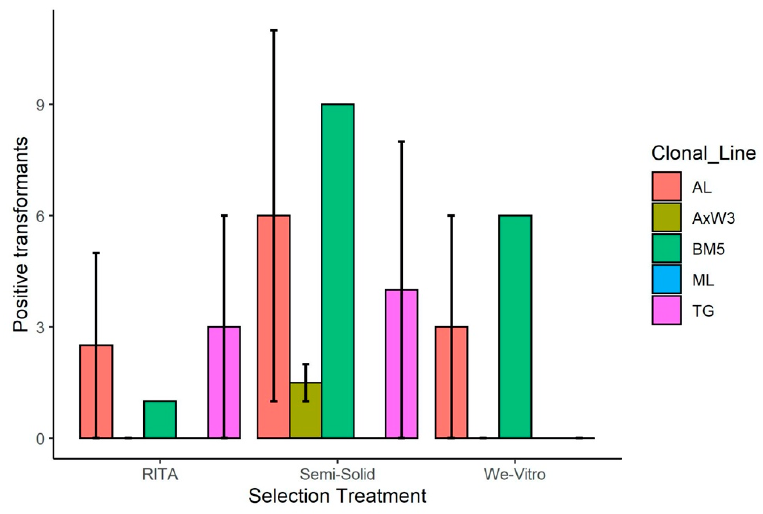
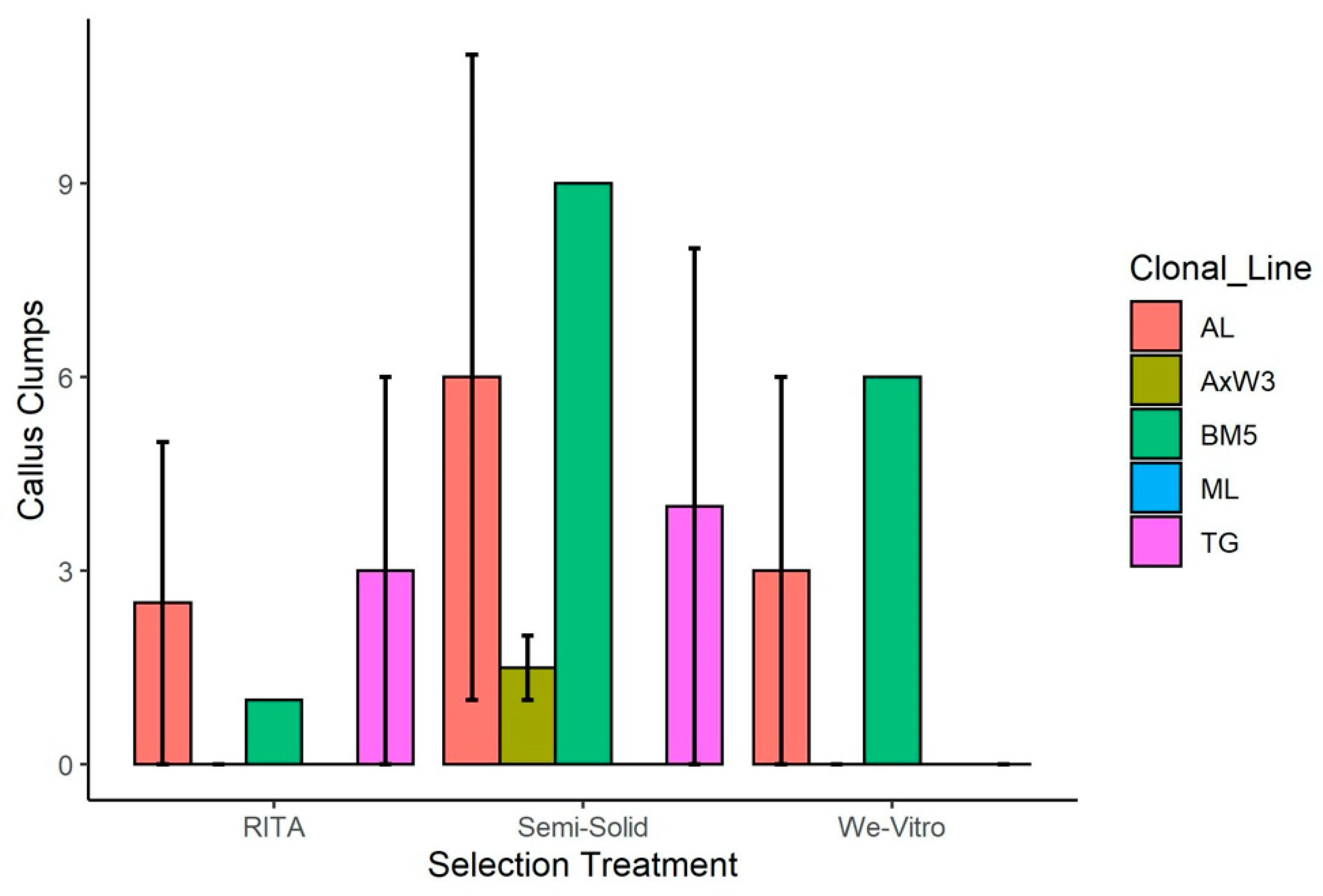
3.1. Transformations

3.1.1. Transformation of American Chestnut for Blight Tolerance

All treatments using the OxO gene produced stably transformed embryogenic lines (Figure 3, Table 1). There was no significant difference between the selection methods (RITA bioreactor, semisolid medium in Petri plates, and We-Vitro container) or between clonal lines (AL, AxW3, BM5, ML, and TG) (Figure 3). There was also no significant difference between the number of non-transformed clumps that escaped the selection process (Figure 4). Due to the lack of any significant general effects, no post hoc analysis methods were performed.
Genomic DNA from putative transformed American chestnut events was extracted and confirmed by PCR (Figure 5). No bands were amplified in the non-transgenic control or the empty (no DNA) lane.

3.1.2. Transformation of American Chestnut for Putative Phytophthora-Tolerance

The transgenic American chestnut somatic embryos carrying the pFHI_GFP construct presented fluorescence under a UV light (Figure 6A) and grew in the presence of paromomycin, demonstrating that the transformation protocol was successful. The transformation efficiency of the American chestnut somatic embryo lines is shown in Table 2. The transformants in all lines produced putative positive events except for SH4. Any tissue that did not turn black or brown was considered to be putatively transformed (Figure 6B).
To check for the presence of the transgenes, putative transgenic lines were analyzed by PCR (Figure 7). Only 3 lines with Ellis #1 background (named “PF-B4GK10, PF-B4GK31, and PF-B4GK32”) were analyzed, as the remaining are still being multiplied. Fragments of 791 and 1768 bp, corresponding to the expected sizes of the NPTII and the segments including UBQ11, Cast_Gnk2-like, and NOS genes, were amplified in the transgenic somatic embryos and the vector (positive control), but not in the untransformed somatic embryos (Figure 7).

4. Discussion

Genetic engineering techniques have made it possible to develop a blight-tolerant American chestnut, as a blight-tolerant tree has yet to be found in the natural population [16]. Today, with the advances in genomics, many resistance candidate genes were narrowed down and are available from transcriptomic studies [24,25,26] and mapping approaches [27,28,29]. The availability of highly-productive embryogenic lines capable of regenerating into plants, combined with the reliable tissue culture protocols described for chestnut transgenics may play an important role as part of an integrated approach to developing pathogen resistance.
Since the first reported regenerated transgenic American chestnut in 2006 [30], multiple transformation events have been produced, including the Darling 58 chestnut, which is currently under review for deregulation [16]. To increase transformation efficiency, several optimization experiments were previously done, including the number of days embryos co-cultivate in desiccation plates (unpublished work), volume of water used to moisten desiccation plates (unpublished work), and plate flooding as an alternative method of transformation [31]. Further optimization experiments were explored in this study with the goal of increasing transformation efficiency, while decreasing explant handling. Five embryo clonal lines were transformed with the oxalate oxidase gene. Transformation of multiple cell lines aids with future breeding by reducing founder effects if only one line is used. Oxalate oxidase detoxifies oxalic acid (oxalate) produced by the fungus Cryphonectria parasitica, preventing the acid from killing the chestnut’s tissue, which can lead to lethal cankers on the tree [16]. During the transformation procedure, selection occurred in one of three types of containers, either in Petri plates with semisolid medium (standard method), in RITA® bioreactors with liquid medium, or in We Vitro containers with liquid medium. It is worth noting that to our knowledge, this is the first study to use We Vitro containers during an Agrobacterium-mediated transformation. In an earlier pilot study, the RITA® bioreactors produced more events than the semisolid medium, yet when done on a larger scale for this experiment, there was no significant difference among treatments. The RITA® bioreactors had the advantage of using a liquid selection medium that surrounded the tissue. By surrounding the embryo clumps, the antibiotic was in direct contact with all surfaces of the tissue and could kill off any non-transformed cells quickly. When using semisolid selection medium, there is a possibility that the cells at the top of the clumps are protected from the antibiotic by the lower cells. The We Vitro containers had the advantage of not needing a pump system or a solenoid, however, it was necessary to keep the embryos in autoclaved tea bags to keep them to one side of the vessel, which was cumbersome.
Andrade et al. [32] found increased transient expression in American chestnut when they co-cultivated the tissue for 3 days instead of 2, however, we saw higher levels of Agrobacterium overgrowth when we increased the time of cocultivation to 3 days. Corredoira et al. [33] were able to prevent Agrobacterium overgrowth in European chestnut by transferring tissue every 2 weeks to proliferation medium supplemented with 300 mg/L carbenicillin, 200 mg/L cefotaxime, and 150 mg/L kanamycin. After a 4-day cocultivation period, Mallon et al. [34] washed Quercus robur embryos for 30 min with sterile distilled water containing 500 mg/L cefotaxime. It is possible that increasing the levels of TIMENTIN in the selection medium or including a TIMENTIN wash before moving the tissue to Agro-Kill medium for our studies will decrease the amount of Agrobacterium overgrowth.
Another concern when transforming plant tissue is the possibility of escapes, untransformed tissue growing on selection medium. We tested whether liquid selection medium would decrease the amount of escapes compared to semisolid selection medium, however, no significant differences were seen. Although not statistically different, the semisolid medium did produce more escapes than the liquid medium. Andrade et al. [32] found that American chestnut selected on semisolid medium produced large numbers of non-transformed material that escaped the process. Therefore, they used liquid selection medium for all their subsequent studies. It is possible that for our study, the amount of paromomycin in the selection medium was not high enough to stop escapes and another kill-curve is needed. Corredoira et al. [33] used semisolid selection with kanamycin in conjunction with GFP to limit the number of escapes. In 2006 [30], we used GFP for transformations, however, with our current protocol, it is no longer necessary to use GFP as a scorable marker except as a control.
Contamination was a problem we encountered early on. Initially, there was large amounts of contamination in the RITA® bioreactors during the transformation process. This problem was solved when the storage location was moved from a wooden cabinet to a metal cabinet and each bioreactor was put into a Ziplock bag (approx. 27 cm × 27 cm), mostly sealed, with only the tubing sticking out. Contamination was not seen in the Petri plates or the We Vitro containers.
We also performed transformations with the Cast_Gnk2-like gene. Ginkbilobin-2 is a protein with an antifungal activity secreted by Ginkgo biloba seeds [35,36] that can activate actin-dependent cell death [37]. Cast_Gnk2-like was identified in chestnut as a putative resistance gene against P. cinnamomi [26]. It appears to be related to the constitutive defense in the coevolved host species Japanese chestnut, and may prevent the pathogen’s growth by chemical properties, or by inducing a hypersensitive-like response cell death [38].
In the transformations with the OxO gene, the transformed cells were selected quickly in the RITA® bioreactors when compared to the other tested techniques. For this reason, the selection for the Cast_Gnk2-like transformants was performed in these containers. From the 14 embryogenic lines putatively expressing the Cast_Gnk2-like, only 3 were confirmed for the T-DNA insertion. The rest of the events will be tested soon and all the transformed lines will be included in the next steps of this research to validate the function of the candidate resistance gene, i.e., (i) the determination of copy number variation to confirm the overexpression of the Cast_Gnk2-like; (ii) regeneration into chestnut lines overexpressing this gene; (iii) and phenotyping after inoculation with P. cinnamomi at key time points of this host-pathogen interaction already characterized at the histological level [39]. In addition, more transformants should be obtained, regenerated, and tested for P. cinnamomi tolerance to guarantee the robustness of the gene’s validation.
Once regenerated, American chestnut shoots need to be multiplied, rooted, and acclimatized. The shoots prefer a relatively low-salt, low-growth regulator medium. The multiplication process as first described by Xing et al. [40] used a medium consisting of WPM salts, Nitsch and Nitsch vitamins, 10 mM Sequestrene, 6 mM calcium chloride, 3 mM magnesium sulfate, 1.0 µM BA, 500 µM IBA, 3.5% sucrose, 5 g/L Phytagel, with the pH adjusted to 5.5. There was a separate shoot elongation medium that included 500 mg/L MES and 500 mg/L polyvinylpyrrolidone 40, which were occasionally incorporated into the multiplication medium as well. Over the years, the multiplication medium has been simplified to WPM salts, Nitsch and Nitsch vitamins, 1.0 µM BA, 10.0 µM IBA, 3.0% sucrose, 7 g/L agar, and the pH adjusted to 5.5, with significant improvement in growth and reduction of vitrification [21]. The original elongation medium developed by Xing et al. [40] had cytokinin (0.22 µM BA) but no auxin (IBA). We have found that using 0.5 µM BA with 0.5 µM IBA reduces horizontal bud break and encourages excellent apical shoot growth.
Original protocols for American chestnut rooting involved inducing roots while still in vitro [40,41]. Much time and effort were spent to improve in vitro rooting methods, including when to add activated charcoal to the medium as well as changing the duration and timing of when plants are put in the dark [42]. Eventually, ex vitro rooting methods were found to be more effective and much less labor intensive for mass production of rooted American chestnut plantlets [22,23]. The full media description, rooting, and acclimatization protocols are described by Oakes et al. [21].

5. Conclusions

The method to deliver the candidate resistance genes into American chestnut was described in this paper. We were able to transform different clonal lines of American chestnut with both the oxalate oxidase gene and the Cast_Gnk2-like gene. All selection methods outlined above, i.e., semisolid medium in Petri plates, liquid medium in RITA® bioreactors, or liquid medium in We Vitro containers, worked to transform different clonal lines of American chestnut somatic embryos with OxO. Although no significant differences were found between treatments, the RITA® bioreactors had the advantage of using a liquid selection medium, which put the antibiotic in direct contact with the tissue to kill off non-transformed cells quickly. The We Vitro containers did not need a pump system or a solenoid, however, it was necessary to keep the embryos in tea bags to keep them on one side of the vessel.
The use of genetic engineering may expedite the characterization of candidate resistance genes for C. parasitica and P. cinnamomi. This will help us understand the molecular mechanisms of these host–pathogen interactions and will contribute to the development of pathogen control strategies. The future road map for the use of the American chestnut genetic transformation techniques may be expanded to enhance tolerance to both pathogens by “stacking” multiple transgenes to broaden the basis of resistance. Theoretically, it would be possible to develop a blight and Phytophthora-tolerant American Chestnut.

Author Contributions

L.M., P.F., and A.O. conceived and designed the studies; L.M., K.S., P.F., and A.O. collected data; A.O. analyzed the data; L.M., K.S., P.F., A.O., and W.P. wrote the paper. All authors have read and agreed to the published version of the manuscript.

Funding

This research was funded in part by The American Chestnut Foundation, by the New York State funding, and the by public donations to the chestnut project. Part of this research was funded by the Fundação para a Ciência e Tecnologia (FCT), Portugal, (PhD grant SFRH/BD/115424/2016) and the Fulbright Portugal under a Fulbright Research Student grant (with the support of FCT) for the 2018/2019 academic year.

Conflicts of Interest

The authors declare no conflict of interest.

References

  1. Rigling, D.; Prospero, S. Cryphonectria parasitica, the Causal Agent of Chestnut Blight: Invasion History, Population Biology and Disease Control. Mol. Plant Pathol. 2018, 19, 7–20. [Google Scholar] [CrossRef] [PubMed] [Green Version]
  2. Anagnostakis, S.L. Chestnut Breeding in the United States for Disease and Insect Resistance. Plant Dis. 2012, 96, 1392–1403. [Google Scholar] [CrossRef] [PubMed]
  3. Steiner, K.C.; Westbrook, J.W.; Hebard, F.V.; Georgi, L.L.; Powell, W.A.; Fitzsimmons, S.F. Rescue of American Chestnut with Extraspecific Genes Following Its Destruction by a Naturalized Pathogen. New For. 2017, 48, 317–336. [Google Scholar] [CrossRef] [Green Version]
  4. Westbrook, J.W.; Zhang, Q.; Mandal, M.K.; Jenkins, E.V.; Barth, L.E.; Jenkins, J.W.; Grimwood, J.; Schmutz, J.; Holliday, J.A. Optimizing Genomic Selection for Blight Resistance in American Chestnut Backcross Populations: A Trade-off with American Chestnut Ancestry Implies Resistance Is Polygenic. Evol. Appl. 2020, 13, 31–47. [Google Scholar] [CrossRef] [PubMed] [Green Version]
  5. Newhouse, A.E.; Powell, W.A. Intentional Introgression of a Blight Tolerance Transgene to Rescue the Remnant Population of American Chestnut. Conserv. Sci. Pract. 2020, in press. [Google Scholar]
  6. Powell, W.A.; Newhouse, A.E.; Coffey, V. Developing Blight-Tolerant American Chestnut Trees. Cold Spring Harb. Perspect. Biol. 2019, 11, a034587. [Google Scholar] [CrossRef] [Green Version]
  7. Carraway, D.T.; Merkle, S.A. Plantlet Regeneration from Somatic Embryos of American Chestnut. Can. J. For. Res. 1997, 27, 1805–1812. [Google Scholar] [CrossRef]
  8. Maynard, C.A.; McGuigan, L.D.; Oakes, A.D.; Zhang, B.; Newhouse, A.E.; Northern, L.C.; Chartrand, A.M.; Will, L.R.; Baier, K.M.; Powell, W.A. Chestnut, American (Castanea dentata (Marsh.) Borkh.). In Agrobacterium Protocols; Methods in Mol Biol; Springer: New York, NY, USA, 2015; pp. 143–161. [Google Scholar] [CrossRef]
  9. Lloyd, G.; McCown, B. Commercially-feasible micropropagation of mountain laurel, Kalmia latifolia, by use of shoot-tip culture. Comb. Proc. Int. Plant Propagators’ Soc. 1980, 30, 421–427. [Google Scholar]
  10. Nitsch, J.P.; Nitsch, C. Haploid Plants from Pollen Grains. Science 1969, 163, 85–87. [Google Scholar] [CrossRef]
  11. Merkle, S.A.; Wiecko, A.T.; Watson-Pauley, B.A. Somatic Embryogenesis in American Chestnut. Can. J. For. Res. 1991, 21, 1698–1701. [Google Scholar] [CrossRef]
  12. Zhang, B.; Oakes, A.D.; Newhouse, A.E.; Baier, K.M.; Maynard, C.A.; Powell, W.A. A Threshold Level of Oxalate Oxidase Transgene Expression Reduces Cryphonectria parasitica-induced Necrosis in a Transgenic American Chestnut (Castanea dentata) Leaf Bioassay. Transgenic Res. 2013, 22, 973–982. [Google Scholar] [CrossRef] [Green Version]
  13. Guilley, H.; Dudley, R.K.; Jonard, G.; Balàzs, E.; Richards, K.E. Transcription of Cauliflower Mosaic Virus DNA: Detection of Promoter Sequences, and Characterization of Transcripts. Cell 1982, 30, 763–773. [Google Scholar] [CrossRef]
  14. Fraley, R.T.; Rogers, S.G.; Horsch, R.B.; Gelvin, S.B. Genetic Transformation in Higher Plants. Crit. Rev. Plant Sci. 1986, 4, 1–46. [Google Scholar] [CrossRef]
  15. Velten, J.; Schell, J. Selection-expression Plasmid Vectors for Use in Genetic Transformation of Higher Plants. Nucleic Acid Res. 1985, 13, 6981–6998. [Google Scholar] [CrossRef] [PubMed] [Green Version]
  16. Newhouse, A.; Coffey, V.; McGuigan, L.; Oakes, A.; Breda, K.; Matthews, D.; Drake, J.; Dougherty, J.; French, J.; Braverman, M.; et al. Petition for Determination of Nonregulated Status for Blight-Tolerant Darling 58 American Chestnut (Castanea dentata). 2020. Available online: https://beta.regulations.gov/document/APHIS-2020-0030-0002 (accessed on 28 September 2020).
  17. Sambrook, J.; Fritsch, E.F.; Maniatis, T. Molecular Cloning. In A Laboratory Manual, 2nd ed.; Cold Spring Harbor Laboratory Press: New York, NY, USA, 1989. [Google Scholar] [CrossRef]
  18. Oakes, A.D.; Pilkey, H.C.; Powell, W.A. Improving Ex Vitro Rooting and Acclimatization Techniques for Micropropagated American Chestnut. J. Environ. Hortic. 2020, in press. [Google Scholar]
  19. Oakes, A.D.; Desmarais, T.; Powell, W.A.; Maynard, C.A. Improving Rooting and Shoot Tip Survival of Micropropagated Transgenic American Chestnut Shoots. HortScience 2016, 51, 171–176. [Google Scholar] [CrossRef]
  20. Oakes, A.D.; Desmarais, T.R.; Powell, W.A.; Maynard, C.A. Ex Vitro Rooting of American Chestnut Improves Acclimatization Survival and Plantlet Quality. J. Environ. Hortic. 2016, 34, 75–79. [Google Scholar] [CrossRef]
  21. R Core Team. R: A Language and Environment for Statistical Computing; R Foundation for Statistical Computing: Vienna, Austria, 2014; Available online: http://www.R-project.org/ (accessed on 5 October 2020).
  22. Ogle, D.H.; Wheeler, P.; Dinno, A. FSA: Fisheries Stock Analysis. R Package Version 0.8.30. Available online: https://github.com/droglenc/FSA (accessed on 5 October 2020).
  23. Wickham, H. ggplot2: Elegant Graphics for Data Analysis, 2nd ed.; Springer: New York, NY, USA, 2016; Available online: https://ggplot2.tidyverse.org (accessed on 5 October 2020).
  24. Barakat, A.; DiLoreto, D.S.; Zhang, Y.; Smith, C.; Baier, K.; Powell, W.A.; Wheeler, N.; Sederoff, R.; Carlson, J.E. Comparison of the Transcriptomes of American Chestnut (Castanea dentata) and Chinese Chestnut (Castanea mollissima) in Response to the Chestnut Blight Infection. BMC Plant Biol. 2009, 9, 51. [Google Scholar] [CrossRef] [Green Version]
  25. Barakat, A.; Staton, M.; Cheng, C.-H.; Park, J.; Yassin, N.B.M.; Ficklin, S.; Yeh, C.-C.; Hebard, F.; Baier, K.; Powell, W.; et al. Chestnut Resistance to the Blight Disease: Insights from Transcriptome Analysis. BMC Plant Biol. 2012, 12, 38. [Google Scholar] [CrossRef] [Green Version]
  26. Serrazina, S.; Santos, C.; Machado, H.; Pesquita, C.; Vicentini, R.; Pais, M.S.; Sebastiana, M.; Costa, R. Castanea Root Transcriptome in Response to Phytophthora cinnamomi Challenge. Tree Genet. Genomes 2015, 11, 6. [Google Scholar] [CrossRef]
  27. Kubisiak, T.; Nelson, C.; Staton, M.; Zhebentyayeva, T.; Smith, C.; Olukolu, B.; Fang, G.-C.; Hebard, F.; Anagnostakis, S.; Wheeler, N.; et al. A Transcriptome-based Genetic Map of Chinese Chestnut (Castanea mollissima) and Identification of Regions of Segmental Homology with Peach (Prunus persica). Tree Genet. Genomes 2013, 9, 557–571. [Google Scholar] [CrossRef] [Green Version]
  28. Santos, C.; Nelson, C.D.; Zhebentyayeva, T.; Machado, H.; Gomes-Laranjo, J.; Costa, R.L. First Interspecific Genetic Linkage Map for Castanea sativa × Castanea crenata Revealed QTLs for Resistance to Phytophthora cinnamomi. PLoS ONE 2017, 12, e0184381. [Google Scholar] [CrossRef] [PubMed] [Green Version]
  29. Zhebentyayeva, T.N.; Sisco, P.H.; Georgi, L.L.; Jeffers, S.N.; Perkins, M.T.; James, J.B.; Hebard, F.V.; Saski, C.; Nelson, C.D.; Abbott, A.G. Dissecting Resistance to Phytophthora cinnamomi in Interspecific Hybrid Chestnut Crosses Using Sequence-based Genotyping and QTL Mapping. Phytopathology 2019, 109, 1594–1604. [Google Scholar] [CrossRef] [PubMed]
  30. Polin, L.D.; Liang, H.; Rothrock, R.E.; Nishii, M.; Diehl, D.L.; Newhouse, A.E.; Joseph Nairn, C.; Powell, W.A.; Maynard, C.A. Agrobacterium-mediated Transformation of American Chestnut (Castanea Dentata (Marsh.) Borkh.) Somatic Embryos. Plant Cell Tissue Organ Cult. 2006, 84, 69–79. [Google Scholar] [CrossRef]
  31. Rothrock, R.E.; Polin-McGuigan, L.D.; Newhouse, A.E.; Powell, W.A.; Maynard, C.A. Plate Flooding as an Alternative Agrobacterium-mediated Transformation Method for American Chestnut Somatic Embryos. Plant Cell Tissue Organ Cult. 2007, 88, 93–99. [Google Scholar] [CrossRef]
  32. Andrade, G.M.; Nairn, C.J.; Le, H.T.; Merkle, S.A. Sexually Mature Transgenic American Chestnut Trees via Embryogenic Suspension-based Transformation. Plant Cell Rep. 2009, 28, 1385–1397. [Google Scholar] [CrossRef]
  33. Corredoira, E.; José, M.C.S.; Vieitez, A.M.; Allona, I.; Aragoncillo, C.; Ballester, A. Agrobacterium -mediated Transformation of European Chestnut Somatic Embryos with a Castanea sativa (Mill.) Endochitinase Gene. New For. 2016, 47, 669–684. [Google Scholar] [CrossRef]
  34. Mallón, R.; Vieitez, A.M.; Vidal, N. High-efficiency Agrobacterium-mediated Transformation in Quercus robur: Selection by Use of a Temporary Immersion System and Assessment by Quantitative PCR. Plant Cell Tissue Organ Cult. 2013, 114, 171–185. [Google Scholar] [CrossRef] [Green Version]
  35. Sawano, Y.; Miyakawa, T.; Yamazaki, H.; Tanokura, M.; Hatano, K. Purification, Characterization, and Molecular Gene Cloning of an Antifungal Protein from Ginkgo biloba Seeds. Biol. Chem. 2007, 388. [Google Scholar] [CrossRef]
  36. Wang, H.; Ng, T.B. Ginkbilobin, a Novel Antifungal Protein from Ginkgo biloba Seeds with Sequence Similarity to Embryo-Abundant Protein. Biochem. Biophys. Res. Commun. 2000, 279, 407–411. [Google Scholar] [CrossRef]
  37. Gao, N.; Wadhwani, P.; Mühlhäuser, P.; Liu, Q.; Riemann, M.; Ulrich, A.S.; Nick, P. An Antifungal Protein from Ginkgo biloba Binds Actin and Can Trigger Cell Death. Protoplasma 2016, 253, 1159–1174. [Google Scholar] [CrossRef] [PubMed]
  38. Santos, C.; Duarte, S.; Tedesco, S.; Fevereiro, P.; Costa, R.L. Expression Profiling of Castanea Genes During Resistant and Susceptible Interactions with the Oomycete Pathogen Phytophthora cinnamomi Reveal Possible Mechanisms of Immunity. Front. Plant Sci. 2017, 8, 515. [Google Scholar] [CrossRef] [PubMed] [Green Version]
  39. Fernandes, P.; Machado, M.H.; Silva, M.C.; Costa, R.L. Histopathological Study Reveals New Insights into Responses of Chestnut (Castanea spp.) to Root Infection by Phytophthora cinnamomi. Phytopathology 2020. [Google Scholar] [CrossRef] [PubMed]
  40. Xing, Z.; Satchwell, M.F.; Powell, W.A.; Maynard, C.A. Micropropagation of American Chestnut: Increasing Rooting Rate and Preventing Shoot-Tip Necrosis. In Vitro Cell. Dev. Biol. Plant 1997, 33, 43–48. [Google Scholar] [CrossRef]
  41. Lapierre, S. Studies in American Chestnut (Castanea Dentata Marsh. (Borhk.)) Micropropagation and Acclimatization. Master’s Thesis, State University of New York College of Environmental Science and Forestry, Syracuse, NY, USA, January 2003. [Google Scholar]
  42. Oakes, A.D.; Powell, W.A.; Maynard, C.A. Doubling Acclimatization Survival of Micropropagated American Chestnuts with Darkness and Shortened Rooting Induction Time. J. Environ. Hortic. 2013, 31, 77–83. [Google Scholar] [CrossRef]
Figure 1. Schematic representation of (A) p35S-OxO binary vector showing the orientation and relative positions of the Oxalate Oxidase (OxO) gene and the neomycin phosphotransferase (NPTII) selectable marker and their respective regulatory sequences: Cauliflower mosaic virus (CaMV 35S) promoter, Ubiquitin 10 (UBQ10) promoter, Actin2 (ACT2) terminator, and nopaline synthase (NOS) terminator. (B) pFHI_Gnk2 binary vector showing the orientation and relative positions of the potential Phytophthora cinnamomi resistance gene Cast_Gnk2-like (indicated as gnk2 in the scheme) and the NPTII selectable marker and their respective regulatory sequences: Ubiquitin 10 and 11 promoters (UBQ10 and UBQ11), respectively; Pac I and Not I restriction sites; CaMV 35S terminator; and NOS terminator.
Forests 11 01196 g001
Figure 2. American chestnut somatic embryos during the transformation process. (A) Somatic embryos on desiccation plates. (B) Selection of embryo clumps on semisolid medium in Petri plates. (CE) Selection of embryo clumps in We Vitro containers with liquid medium. (D) Aluminum foil covers the We Vitro containers. (E) A tea bag is used to keep the embryo clumps from moving into the liquid medium in the We Vitro containers when they are angled up. (F,G) Selection of embryo clumps in a RITA® bioreactor with liquid medium.
Forests 11 01196 g002
Figure 3. Number of American chestnut transformation events with the oxalate oxidase gene after 4 weeks of selection in RITA® bioreactors, semisolid media in Petri plates and We Vitro containers. Error bars indicate standard error of the mean. Two clonal lines, Bass Mountain #5-1B (BM5) and Moss Lake 90016 C-21-1C (ML), were only used in single trials.
Forests 11 01196 g003
Figure 4. American chestnut somatic embryo line escapes (non-transformed tissue surviving on selection medium). Error bars indicate standard error of the mean. Two clonal lines, BM5 and ML, were only used in single trials.
Forests 11 01196 g004
Figure 5. PCR analysis for the amplification of a specific 179 bp fragment of the OxO gene in American chestnut embryo lines transformed with p35S-OxO. Lanes 1, 2, 3, 4, 5, 6, 9, and 10—transformed events derived from embryogenic line TG; Lanes P—OV12, OV6, and T2 020, respectively (positive controls for OxO); Lane U—untransformed embryos (negative control), Lane 13—empty (no DNA negative control); and M—DNA ladder (100 bp).
Forests 11 01196 g005
Figure 6. American chestnut somatic embryos transformed with Agrobacterium AGL-1 strain after 5 weeks in selection medium. (A) Positive control: Ellis #1 transformed with pFHI_GFP observed under ultraviolet light. (B) AxW3 transformed with pFHI_Gnk2. Arrows indicate putative events. Bar = 1 mm.
Forests 11 01196 g006
Figure 7. PCR analysis of American chestnut transgenic lines. (A) PCR amplification using specific primers for the production of a 791 bp NPTII fragment. (B) PCR amplification using specific primers for the production of a 1768 bp UBQ11, Cast_Gnk2-like, and NOS fragment. M—DNA ladder; Lanes 1, 2, and 3—transformed events derived from embryogenic line Ellis #1 (PF-B4GK10, PF-B4GK31, and PF-B4GK32, respectively). P—pFHI-Gnk2 vector (positive control). U—untransformed Ellis #1 (negative control).
Forests 11 01196 g007
Table 1. Number of positive events and escapes per starting mass in transformation using p35S-OxO. American chestnut clonal lines were Alessi T2-1A (AL), AxW3-46B (AxW3), Bass Mountain #5-1B (BM5), Moss Lake 90016 C-21-1C (ML), TG-8A (TG).
TrialClonal LineTreatmentStarting Mass (mg)Positive EventsEscapes
1ALRITA5035
2ALRITA110100
1AxW3RITA3760
2AxW3RITA11310
1BM5RITA9801
2MLRITA14140
1TGRITA8686
2TGRITA13380
1ALSemisolid86811
2ALSemisolid12061
1AxW3Semisolid10862
2AxW3Semisolid13551
1BM5Semisolid6589
2MLSemisolid13660
1TGSemisolid5988
2TGSemisolid9230
1ALWe Vitro6536
2ALWe Vitro12190
1AxW3We Vitro3600
2AxW3We Vitro9860
1BM5We Vitro9466
2MLWe Vitro15730
1TGWe Vitro6340
2TGWe Vitro11320
Table 2. Transformation efficiency of American chestnut somatic embryo lines (AxW3-46B (AxW3), Spring Hole-4A (SH4), Spring Hole-11D (SH11), and Ellis #1) with pFHI_Gnk2 vector and positive control (Ellis #1 transformed with pFHI_GFP), after 5 weeks in selection medium.
Clonal LineVectorStarting Mass (mg)Putative Events
AxW3pFHI_Gnk21445
SH4pFHI_Gnk21030
SH11pFHI_Gnk21132
Ellis #1pFHI_Gnk21682
pFHI_Gnk21805
pFHI_GFP1683
Publisher’s Note: MDPI stays neutral with regard to jurisdictional claims in published maps and institutional affiliations.

