Article

Contactless Vital Sign Monitoring in Emergency Settings: A Factorial Study of Camera Position and Motion Using rPPG

Department of Biomedical Engineering, Keimyung University, Daegu 42601, Republic of Korea
*
Author to whom correspondence should be addressed.
Algorithms 2026, 19(9), 802; https://doi.org/10.3390/a19090802 (registering DOI)
Submission received: 4 August 2026 / Revised: 10 September 2026 / Accepted: 16 September 2026 / Published: 18 September 2026

Abstract

Reliable acquisition of vital signs is essential in prehospital emergency care; however, conventional contact-based sensors may delay assessment, cause patient discomfort, increase cross-contamination risk, and perform poorly during motion. To address these limitations, this study developed a body-worn smart camera system for paramedics incorporating a remote photoplethysmography (rPPG)-based signal-processing algorithm to estimate heart rate (HR), oxygen saturation (SpO2), and blood pressure (BP) in real time from subtle facial blood volume fluctuations. The system was evaluated in a repeated-measures full-factorial experiment with three adults under varying lighting, camera positions, and stabilization conditions. Performance was assessed using mean absolute error (MAE) relative to contact-based reference devices. HR (MAE: 1.20–5.60 bpm) and SpO2 (MAE: 0.93–3.00%) showed relatively small errors relative to the reference measurements across the tested conditions, whereas BP estimation showed larger errors (SYS: 5.00–14.20 mmHg; DIA: 3.87–11.60 mmHg), indicating the need for further algorithm refinement. The head-mounted configuration provided the best performance (HR MAE: 1.96 bpm). These preliminary findings support the technical feasibility of a wearable, contactless rPPG-based vital sign monitoring system under the tested conditions, while identifying BP estimation as the primary target for future improvement. Further validation in larger and more diverse populations is required before clinical or prehospital applicability can be established.

1. Introduction

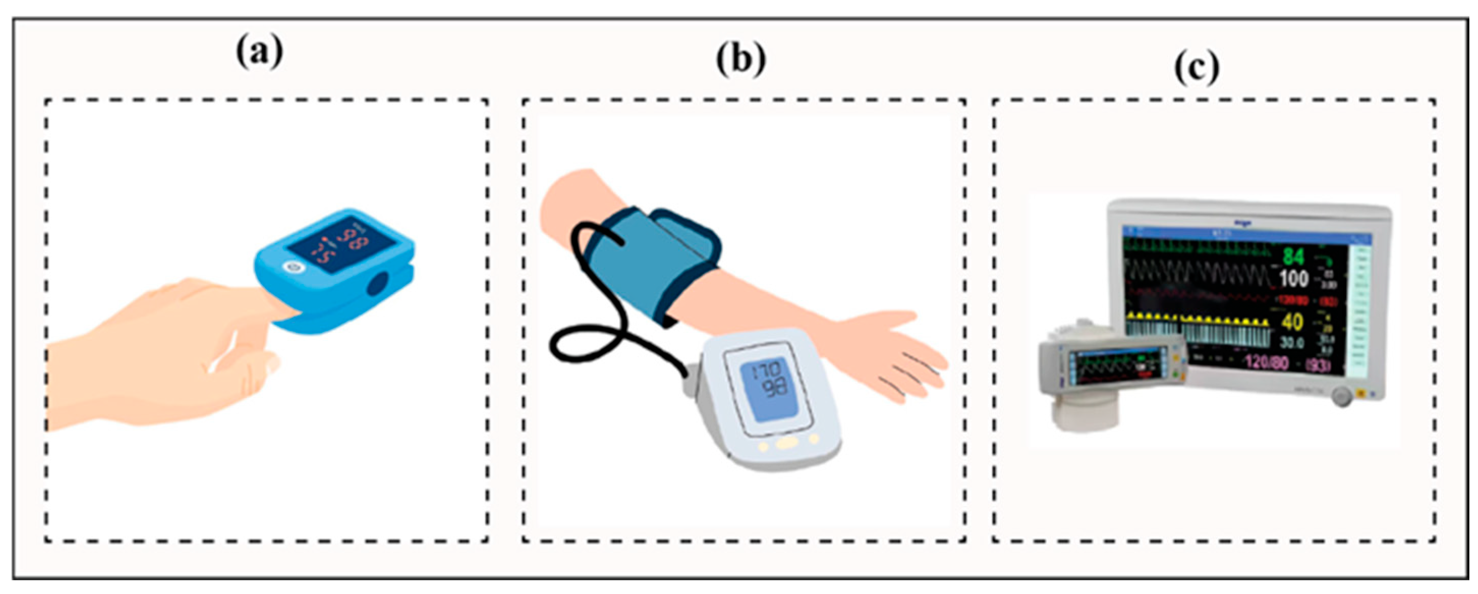
Accurate and timely assessment of vital signs is essential for clinical decision-making, particularly in prehospital and emergency care, where rapid changes in a patient’s physiological condition may require immediate intervention. Heart rate (HR), oxygen saturation (SpO2), blood pressure (BP), and respiratory rate provide critical information regarding cardiovascular and respiratory stability and are routinely used to determine treatment priorities. Conventional contact-based devices, including pulse oximeters, automated blood pressure monitors, and multiparameter patient monitors, are widely used because of their established accuracy and clinical reliability. Current contact devices used for contact-based vital-sign monitoring are shown in Figure 1.
However, despite their clinical utility, contact-based monitoring systems have several limitations in emergency and mobile healthcare environments. Because sensors must be attached directly to the patient, their application may delay the initial assessment, cause discomfort, and increase the risk of cross-contamination, particularly when reusable sensors are employed [1]. Repeated sensor placement, cleaning, and disinfection may also increase the workload of emergency medical personnel and interrupt time-sensitive clinical workflows. The performance of contact-based sensors may also be affected by physiological and environmental factors, including perspiration, skin temperature, body posture, sensor displacement, and patient movement [2,3,4]. These limitations may become more pronounced during emergency transport, where changes in illumination, camera–patient position, vibration, and body movement can contribute to signal distortion or sensor detachment [5,6]. Furthermore, some parameters, particularly blood pressure, are commonly measured intermittently rather than continuously, potentially delaying the detection of sudden hemodynamic deterioration [7]. These challenges highlight the need for monitoring technologies that can rapidly and continuously acquire physiological information without requiring direct sensor attachment.
To address these limitations, several contactless vital-sign monitoring technologies have been investigated. Radar-based systems use reflected electromagnetic waves to detect physiological movements associated with cardiac and respiratory activity. Although these systems can operate without direct contact, their performance may be influenced by the measurement distance, sensor orientation, surrounding structures, signal reflections, metallic objects, and subject motion [8,9,10]. Such factors may restrict their reliability in confined and dynamic environments, including ambulances and emergency treatment areas. Remote photoplethysmography (rPPG) has emerged as a promising camera-based approach for contactless physiological monitoring. This technique estimates cardiovascular signals by analyzing subtle, pulse-related variations in skin color captured using an RGB camera [11]. Because rPPG does not require physical sensor attachment and can be implemented using commercially available imaging devices, it offers potential advantages in terms of rapid deployment, patient comfort, infection control, and operational scalability.
Nevertheless, most previous rPPG studies have been conducted under controlled laboratory conditions. Measurement performance may deteriorate in practical emergency settings because of motion artifacts, vibration, abrupt illumination changes, variations in facial orientation, and inter-individual differences, including skin-tone variation [12,13,14,15,16]. Although methods such as color-space transformation, chrominance-based filtering, signal denoising, and dynamic region-of-interest tracking have been proposed, maintaining stable physiological signal extraction under realistic motion and illumination conditions remains challenging. In particular, limited quantitative evidence is available regarding the influence of wearable-camera mounting position and camera motion on rPPG measurement accuracy in conditions relevant to emergency care.
To address this research gap, the present study developed a smart body-camera system for contactless estimation of HR, SpO2, systolic blood pressure, and diastolic blood pressure from facial video. The system was evaluated using a repeated-measures full-factorial experimental design in which three operational factors were systematically varied: camera mounting position, camera-motion condition, and illumination condition. Camera position was evaluated at the head, shoulder, and chest, while camera motion was classified as fixed, mild, or severe. Measurements were conducted under a well-illuminated laboratory condition and a lower-illumination condition designed to approximate selected lighting characteristics of an ambulance environment.
The objectives of this study were to: (1) quantify differences in measurement performance across camera mounting positions, camera-motion levels, and illumination conditions; (2) identify mounting configurations associated with lower estimation error under the tested conditions; and (3) assess the preliminary feasibility and limitations of body-worn rPPG monitoring for emergency-response applications. This study provides an initial evaluation framework for wearable camera placement and identifies technical priorities for future validation in larger populations and more realistic emergency transport environments.

2. Materials and Methods

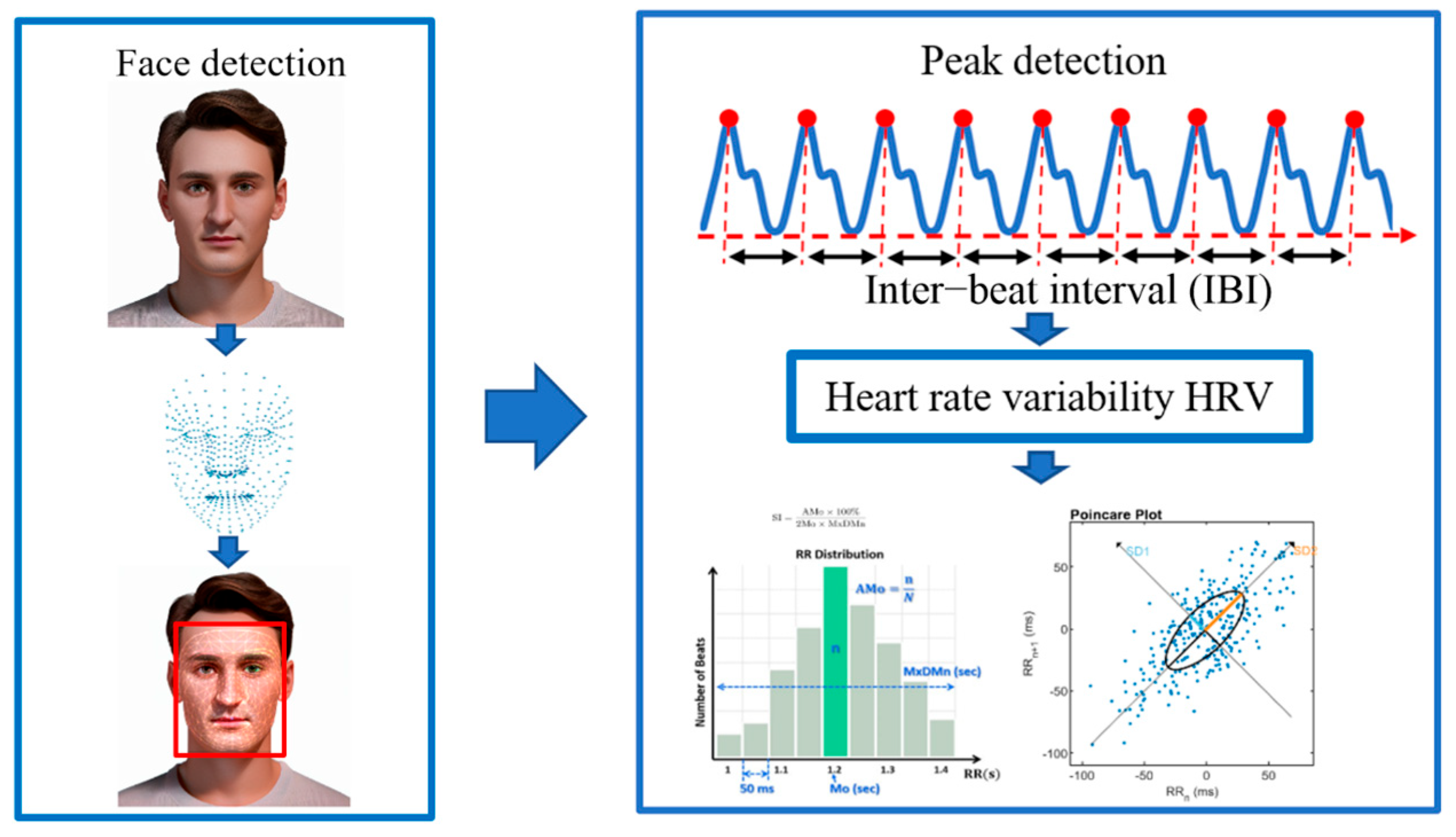
This section describes the architecture of the proposed contactless vital-sign monitoring system, the rPPG signal-processing procedure, and the experimental protocol used for performance evaluation. The system was designed to estimate heart rate, oxygen saturation, and blood pressure from facial video. Its performance was evaluated under different camera mounting positions, motion conditions, and illumination levels using conventional contact-based devices as reference measurements. Figure 2 shows the overall graphical framework of the proposed study.
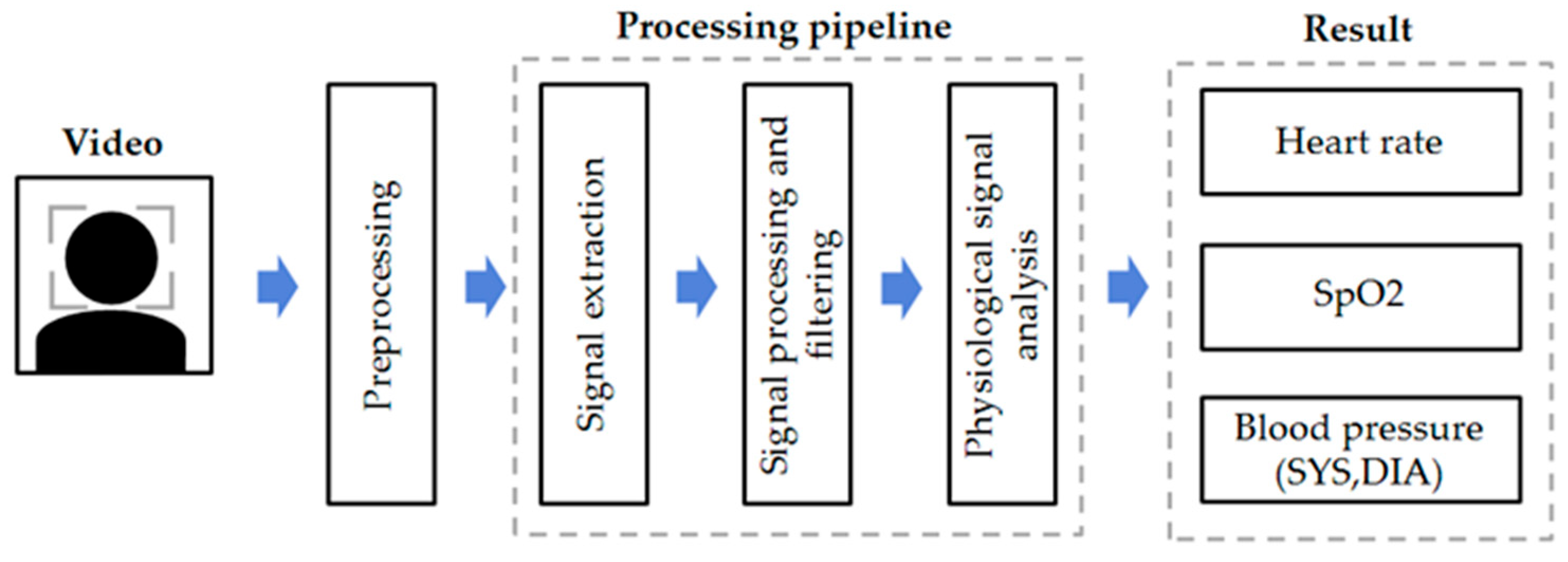
As shown in Figure 3, the facial video was first processed using MediaPipe (0.9.0.1) [17] for face detection and tracking of the facial region of interest (ROI). Eulerian video magnification (EVM) was applied to enhance subtle pulse-related color variations [18]. The mean RGB intensity within the ROI was calculated for each frame to generate temporal color signals. The signals were detrended and filtered using a third-order Butterworth band-pass filter with a frequency range of 0.5–4 Hz. A Savitzky–Golay filter was then applied for waveform smoothing, followed by normalization.
  • Heart rate (HR) was estimated from the preprocessed rPPG signal. The signal was transformed into the frequency domain using the Fast Fourier Transform (FFT), and the dominant spectral peak within the 0.5–4 Hz range was identified. HR was then calculated from the peak frequency.
  • SpO2 was estimated from the red and green color signals extracted from the facial ROI. The pulsatile (AC) and non-pulsatile (DC) components of the red and green signals were used to calculate
    R R = A C R / D C R A C G / D C G
    where A C R and A C G are the pulsatile components of the red and green signals, respectively, and D C R and D C G are the corresponding non-pulsatile components. SpO2 was then calculated as
    S p O 2 = A × R R + B
    where A = 38 and B = 68. These coefficients were obtained by linear regression using the separate five-subject calibration dataset described in Section 2.4 and remained fixed for all participants in the present evaluation.
  • Blood pressure was estimated using the PTT-based method described in [19]. rPPG signals were extracted from the forehead and chin ROIs and processed using the same preprocessing pipeline. Corresponding pulse peaks were detected in the two signals, and PTT was calculated as:
    P T T = t c h i n t f o r e h e a d
    The calculated PTT was then mapped to blood pressure using the model
    BP = aPTT + bHR + c,
    where a, b, and c are fixed model coefficients. Systolic and diastolic blood pressure were estimated separately using their corresponding coefficient sets:
    S B P = a S B P P T T + b S B P H R + c S B P D B P = a D B P P T T + b D B P H R + c D B P
Accordingly, the fixed coefficients (a, b, c) were (−5, −0.0777, 103) for SBP and (−3, −0.02912, 64) for DBP. These coefficients were obtained by fitting the linear PTT–HR model to the separate five-subject calibration dataset described in Section 2.4, using the corresponding reference systolic and diastolic blood pressure measurements.

2.1. Experimental Overview

This study evaluated the feasibility and measurement performance of the proposed smart body camera-based contactless vital-sign monitoring system under controlled conditions designed to reproduce selected challenges encountered in emergency medical settings. A repeated-measures full-factorial design was employed to systematically vary three experimental factors: camera mounting position, illumination condition, and camera-motion level. The camera was positioned at the head, shoulder, or chest. Illumination was evaluated under a laboratory condition of approximately 400 lux or higher and a lower-illumination condition of approximately 150 lux or higher, intended to approximate ambulance lighting. Camera motion was classified as fixed, mild, or severe to represent different levels of movement that may occur during emergency patient transport.
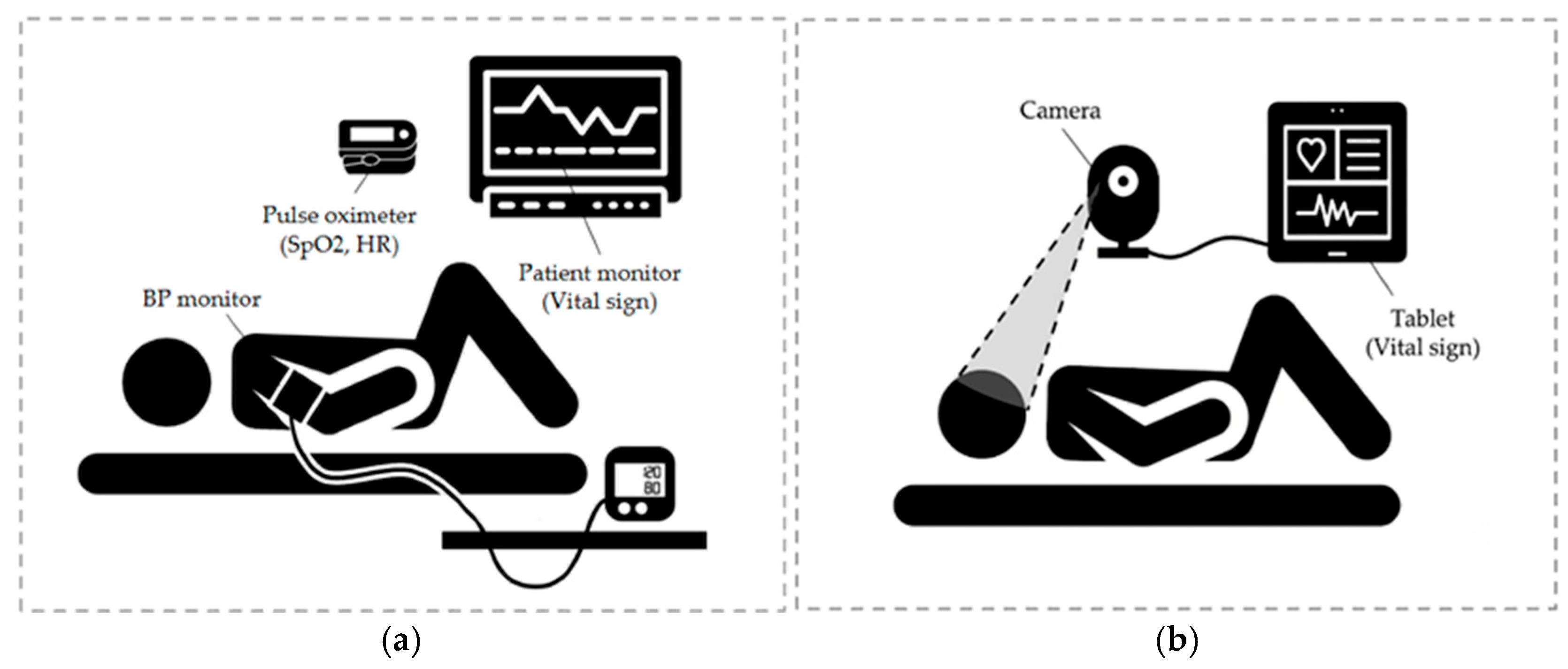
Figure 4 compares the conventional contact-based reference measurement configuration with the proposed camera-based contactless monitoring approach. In the conventional configuration, HR and SpO2 were measured using a pulse oximeter, whereas in the proposed configuration, facial video was acquired using a camera, and the estimated vital signs were displayed in real time without attaching measurement sensors to the participant.
Repeated measurements were performed on three adult participants under each combination of experimental factors. Under each condition, heart rate (HR), oxygen saturation (SpO2), systolic blood pressure (SYS), and diastolic blood pressure (DIA) measured using the contactless vital sign monitoring system were systematically compared with the reference values measured using a conventional contact-based device. This experimental design enables a comprehensive evaluation of the system performance under various environmental conditions and allows identification of camera mounting configurations associated with improved measurement stability.

2.2. System Prototype and Experimental Setup

In this study, we developed a prototype body-camera system consisting of a webcam, a mini-PC, and a display for real-time vital-sign visualization. Facial videos were captured via a Logitech C922 PRO HD webcam (China) connected through a USB interface. The camera supports video resolutions up to 1920 × 1080 pixels at 30 fps and 1280 × 720 pixels at 60 fps, with autofocus and a 78° diagonal field of view. Specific acquisition and implementation parameters, including the exact acquisition resolution and frame rate, video codec, exposure and white-balance settings, detailed mini-PC specifications, complete software and dependency versions, and processing latency, were not systematically recorded during the experiments. The mini-PC processed this footage in real-time using remote photoplethysmography (rPPG) signal extraction and estimation algorithms. The same predefined rPPG processing pipeline and parameter settings were applied to all participants, without subject-specific calibration or tuning using the evaluation data. Experimental measurement values were manually recorded in spreadsheet format during the study. For reference measurements, we used a fingertip pulse oximeter (YK-80A, Yonker, Xuzhou, China) to record HR and SpO2, and an automated blood pressure monitor (HEM-790IT, OMRON Healthcare Co., Ltd., Kyoto, Japan) to measure SYS and DIA simultaneously. The experimental environment consisted of the following two conditions:
  • The laboratory environment (~400+ lux): All ceiling lights were turned on to maintain consistent, bright illumination.
  • Simulated ambulance-related environment (~150+ lux): Instead of using an actual ambulance, we adjusted the lab lighting to a minimum of 150 lux to approximate reduced illumination conditions relevant to an ambulance environment. Note: Vehicle-induced vibration and noise were not independently replicated under the illumination test conditions; instead, the impact of camera motion was evaluated separately using the predefined mild and severe motion scenarios outlined in Section 2.3.1.
In both settings, the camera mounting position was predefined for each experimental condition, while exact camera-to-face distance and viewing angle were not quantitatively documented.

2.3. Experimental Design

2.3.1. Variables and Condition Settings

To systematically evaluate the system’s robustness in emergency-like scenarios, we selected camera mounting position and camera stability as our primary independent variables. Testing was conducted on three adult participants in their 20s (two male, one female) under the two illumination conditions described in Section 2.2. While exact geometric parameters, such as camera-to-face distance and angle, and detailed participant characteristics, including objective skin-tone characterization, medication history, and relevant cardiopulmonary conditions, were not formally recorded in this pilot dataset, the predefined mounting configuration was applied consistently within each experimental condition.
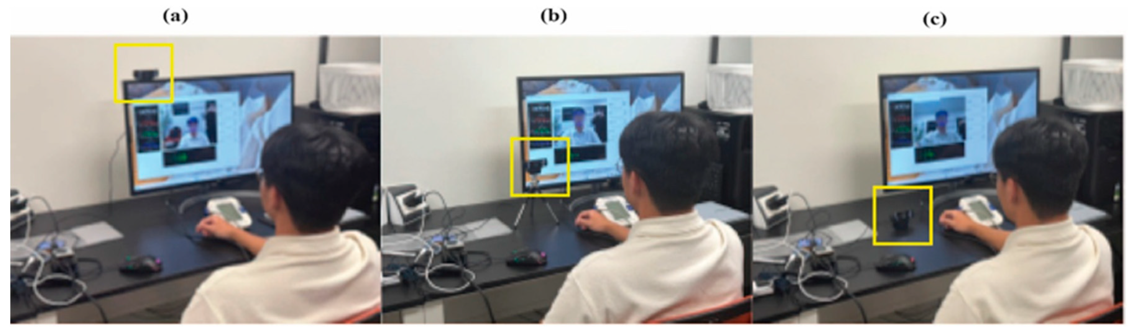
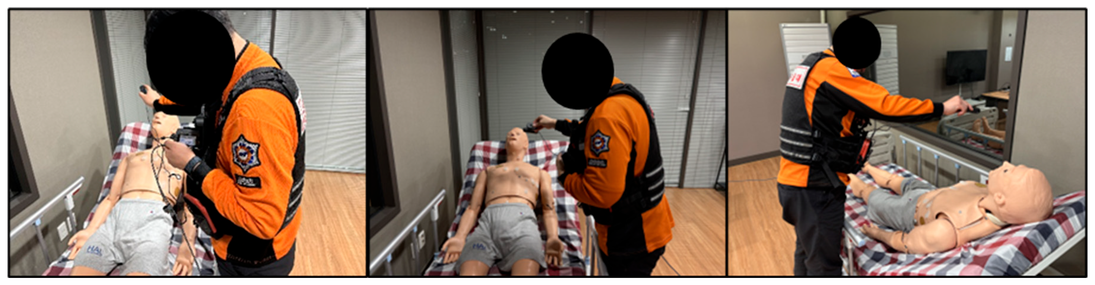
Camera mounting position was tested at three common wearable locations for emergency personnel: the head (using a helmet, glasses, or head-mount), the shoulder, and the chest. Each location inherently differs in facial visibility, field of view, and attachment stability, all factors that can impact rPPG signal quality. By comparing these configurations, we evaluated their effect on the system’s robustness and practicality, considering real-world constraints like ease of attachment and susceptibility to vibration. Figure 5 shows the video acquisition process under fixed camera stability conditions for the three camera mounting positions. Camera Stability was also assessed at three levels:
  • Fixed: The camera was rigidly secured to a tripod or stand, with zero relative movement between the participant and the device.
  • Mild Motion: The camera was worn by the participant to reflect natural body movements and subtle physiological sway (as shown in Figure 6).
  • Severe Motion: The camera remained wearable, but we introduced intentional, regular movements in both vertical and horizontal directions at roughly 1–2 s intervals to represent predefined wearable-camera motion relevant to emergency transport.
We employed a full-factorial design crossing two lighting conditions, three mounting positions, and three stability levels. Five repeated measurements were performed for each condition combination. These repeated measurements were treated as within-participant observations and were used to evaluate measurement consistency and error under each experimental condition, rather than as independent samples representing additional participants. This comprehensive approach enables a detailed assessment of system performance across diverse operational scenarios. To minimize confounding variables, the background and lighting arrangement were kept as uniform as possible throughout all sessions.

2.3.2. Physiological Measurement Indicators

The primary evaluation metrics for this study were heart rate (HR), blood pressure (BP), and oxygen saturation (SpO2). These indicators are clinically essential for assessing patient status in emergency settings, providing critical insights into physiological stability and the risk of acute deterioration.
  • Heart rate: Defined as the number of cardiac cycles per unit time, HR is a sensitive indicator of autonomic nervous system balance, reflecting both sympathetic and parasympathetic activity. It is a fundamental parameter in the emergency assessment of conditions such as shock, arrhythmias, and impending cardiac arrest.
  • Blood pressure: Classified into systolic and diastolic components, BP measures the force exerted by circulating blood against arterial walls. It serves as a key marker of systemic circulatory status and end-organ perfusion, making it a crucial criterion for prediction and acute-phase treatment decisions.
  • Oxygen saturation: Representing the fraction of oxygen-saturated hemoglobin in arterial blood, SpO2 indicates the adequacy of oxygen delivery and the functional integrity of the cardiopulmonary system. A reduction below clinically defined thresholds necessitates immediate oxygen supplementation and airway intervention, underscoring its vital clinical significance.
The primary endpoint of this study was the measurement accuracy of the proposed rPPG system relative to contact-based reference devices, quantified using the mean absolute error (MAE) for heart rate (HR), oxygen saturation (SpO2), systolic blood pressure (SYS), and diastolic blood pressure (DIA). Secondary endpoints included evaluating the effects of camera motion, illumination conditions, and mounting positions on measurement performance, as well as identifying the mounting configuration associated with the lowest estimation error.

2.4. Data Acquisition

A separate calibration dataset comprising five subjects was collected before the present factorial evaluation under controlled laboratory illumination (~400+ lux). Reference SpO2 and blood pressure values were recorded simultaneously using the fingertip pulse oximeter and automated blood pressure monitor, respectively. For SpO2, the paired ratio-of-ratios (RR) and reference SpO2 measurements were used to determine the calibration coefficients A and B by linear regression. The same calibration dataset was also used to determine the fixed coefficients of the PTT-based systolic and diastolic blood pressure estimation equations. These calibration data were not included in the three-subject factorial analysis. After the calibration coefficients had been determined and fixed, the main three-participant factorial evaluation was conducted as described below.
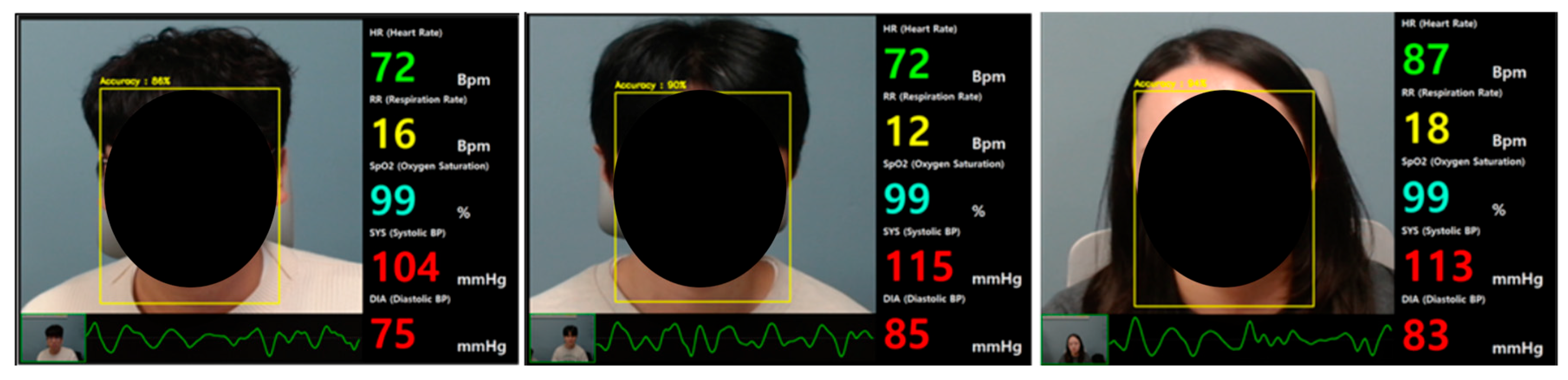
Data acquisition was performed under two illumination conditions: a laboratory setting (~400+ lux) and a simulated ambulance-related environment (~150+ lux). For each experimental trial, continuous facial video recordings of at least 30 s were captured using the camera. In accordance with the full-factorial experimental design, measurements were repeated five times for every combination of mounting position, camera stability, and lighting condition. Participants were evaluated in either a seated or supine position and instructed to maintain a comfortable, stationary posture during measurements. No specific breathing protocols were imposed. Prior to each recording, participants underwent a resting period of approximately 30–60 s. No skin preparation was performed, and the use of facial cosmetics or eyewear was unrestricted. Facial video streams were processed in real time without recording or storing video files. Environmental background and illumination conditions were maintained as consistently as possible across all test sessions. HR, SpO2, and BP reference measurements were acquired concurrently with the rPPG recording. For HR and SpO2, the values displayed by the fingertip pulse oximeter were recorded at the same time as the corresponding rPPG-derived values. For BP, the automated cuff measurement was initiated during the video recording, and the value displayed after signal stabilization was recorded together with the corresponding rPPG estimate. Furthermore, the rPPG-derived waveforms and estimated signals were visualized in real time on the system display in Figure 7.
To quantitatively evaluate the agreement between rPPG-based estimates and contact-based reference measurements, the following statistical metrics were used:
Mean Absolute Error (MAE) represents the average absolute deviation between rPPG-estimated values and reference measurements, and it retains the same unit as the measured variable.
M A E = 1 N i = 1 N y ^ i y i
Here, N denotes the total number of samples (measurements), y ^ i represents the value estimated by the rPPG algorithm, and y i represents the actual reference value obtained from the contact-based device.
For each experimental condition, MAE was calculated to quantitatively compare measurement performance across varying camera mounting positions, lighting conditions, and camera stability levels. This study was conducted as an internal pilot investigation involving members of the research team, in strict accordance with the ethical guidelines of Keimyung University. All participants provided written informed consent prior to enrollment. Furthermore, all facial video was processed in real time without video-file storage, and physiological data used for analysis were anonymized to protect participant privacy and data confidentiality.

3. Results

This section presents the quantitative performance of the proposed contactless vital sign monitoring system across all experimental conditions. We first provide an overview of the overall measurement accuracy and robustness. We systematically evaluate the influence of camera stability on rPPG-derived heart rate and oxygen saturation estimates and analyze the optimal camera mounting position to identify the configuration yielding the most stable and accurate performance.

3.1. Baseline Parameter Specific Performance

Under both laboratory (~400+ lux) and simulated ambulance-related (~150+ lux) conditions, the proposed system showed relatively stable estimation of heart rate (HR) and oxygen saturation (SpO2) across the tested lighting, camera-stability, and mounting conditions. As summarized in Table 1, the mean absolute error (MAE) for HR ranged from 1.20 to 5.60 bpm, indicating strong agreement with reference measurements. Similarly, SpO2 estimations exhibited minimal deviation from the reference measurements (MAE: 0.93–3.00%) across the tested conditions. In contrast, blood pressure (BP) estimation yielded substantially larger errors and higher sensitivity to experimental variations. The MAE for systolic BP ranged from 5.00 to 14.20 mmHg, while diastolic BP ranged from 3.87 to 11.60 mmHg. Notably, BP estimation errors increased under motion and suboptimal illumination, whereas HR and SpO2 exhibited relatively small variations across the tested motion conditions. These results underscore the system’s comparative reliability for cardiorespiratory monitoring, while highlighting the inherent challenges of contactless BP estimation.

3.2. Influence of Camera Stability on rPPG Signal Fidelity

To evaluate motion robustness, system performance was analyzed across three stability levels: fixed, mild motion, and severe motion. Table 2 presents the estimated and reference value ranges for all vital signs under each condition.

3.2.1. Cardiorespiratory Parameters (HR and SpO2)

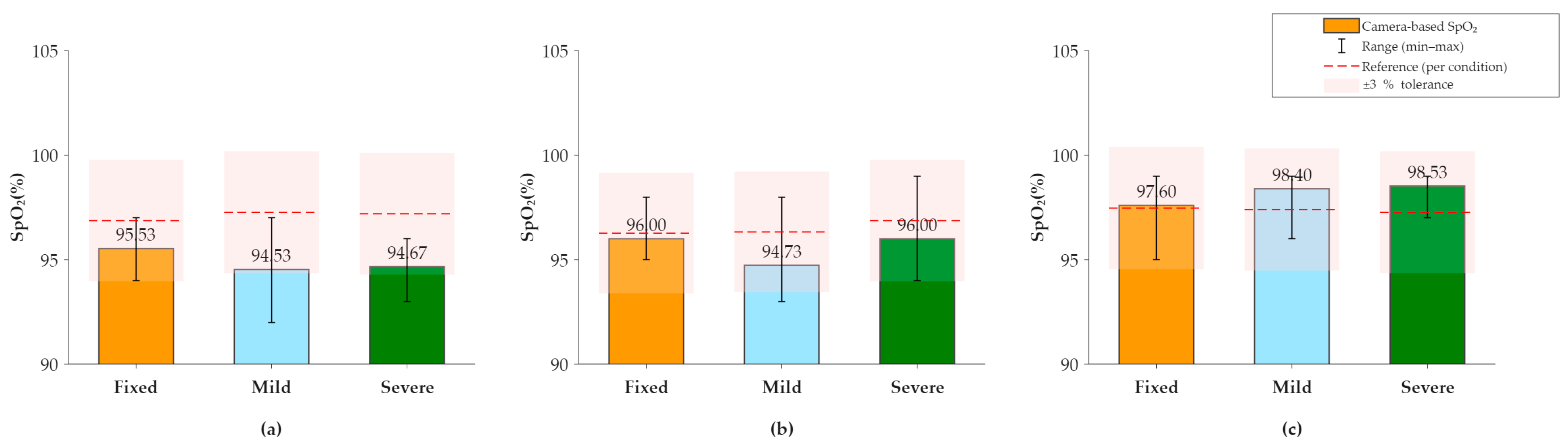
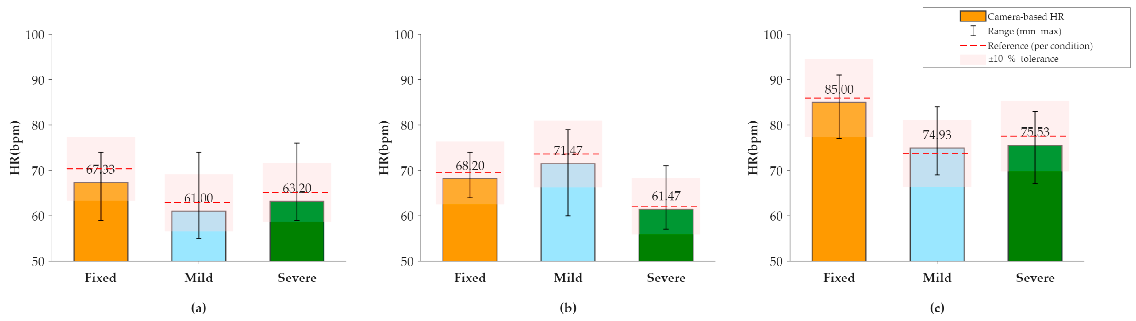
Estimated HR and SpO2 values exhibited strong alignment with reference measurements across all stability levels. Under severe motion, the estimated HR range (61.5–75.5 bpm) closely tracked the reference range (62.1–79.1 bpm), demonstrating minimal motion-induced degradation (Figure 8). Likewise, SpO2 estimates under severe motion ranged from 94.7% to 98.5% and showed relatively small deviations from the corresponding reference range of 96.9% to 97.3% (Figure 9). No consistent increase in HR or SpO2 estimation error was observed across the predefined camera-motion conditions.

3.2.2. Oxygen Saturation (SpO2) Measurement Performance

The mean oxygen saturation values estimated by the proposed system were compared with reference measurements across the three camera stability conditions. As presented in Table 2, the estimated SpO2 ranges under fixed, mild, and severe motion conditions were 95.5–97.6%, 94.5–98.4%, and 94.7–98.5%, respectively. These estimates remained highly consistent with the corresponding reference ranges (96.3–97.5%, 96.3–97.4%, and 96.9–97.3%), indicating relatively close agreement across the tested motion conditions (Figure 9). Overall, the estimated SpO2 values showed close agreement with the reference measurements across all camera stability levels, with relatively small deviations under the tested conditions.

3.2.3. Results of the Systolic Blood Pressure Measurement

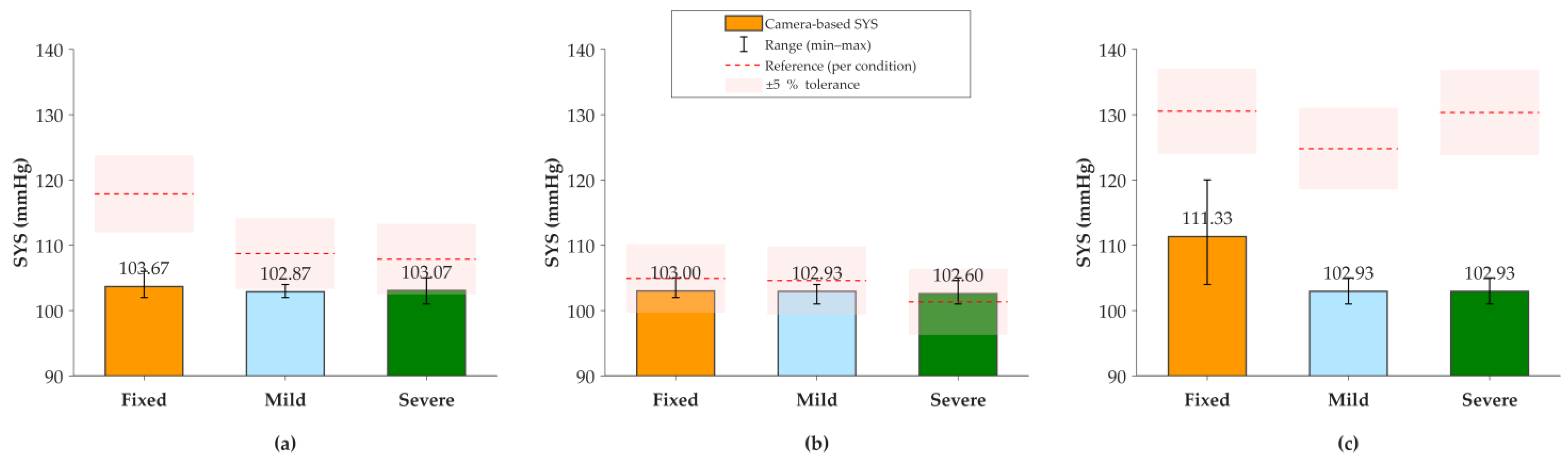
The systolic blood pressure estimates derived from the proposed system were compared with reference measurements under each camera stability condition. Under fixed conditions, estimated systolic values ranged from 103.0 to 111.3 mmHg, whereas the corresponding reference values spanned a substantially wider range of 104.9 to 130.5 mmHg. This disparity persisted under severe motion, where estimates narrowed to 102.6–103.1 mmHg, markedly underestimating the broad physiological variation observed in the reference range (101.3–130.3 mmHg) (Figure 10). These results indicate that while the system tracks average trends, its dynamic range for systolic BP estimation is constrained under the tested motion conditions.

3.2.4. Diastolic Blood Pressure Measurement Results

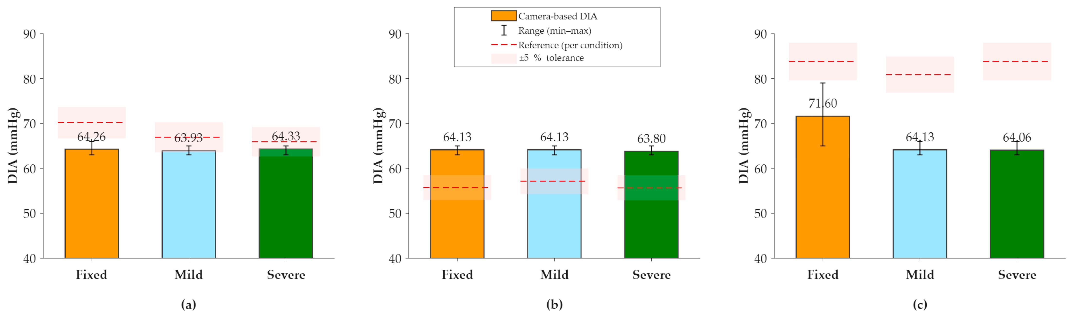
The diastolic blood pressure values measured by the proposed system were compared with the corresponding reference values under fixed, mild motion, and severe motion conditions. Under fixed conditions, the proposed system-derived mean diastolic blood pressure values ranged from 63.0 to 71.6 mmHg, while the corresponding reference values ranged from 55.7 to 83.8 mmHg. Under mild motion conditions, derived diastolic blood pressure values ranged from 63.9 to 64.1 mmHg, compared with reference values ranging from 57.1 to 80.7 mmHg. Under severe motion conditions, derived diastolic blood pressure values ranged from 64.0 to 64.3 mmHg, whereas reference values ranged from 55.7 to 82.8 mmHg (Figure 11).

3.3. Comparative Analysis of the Optimal Mounting Position for the rPPG Measurement

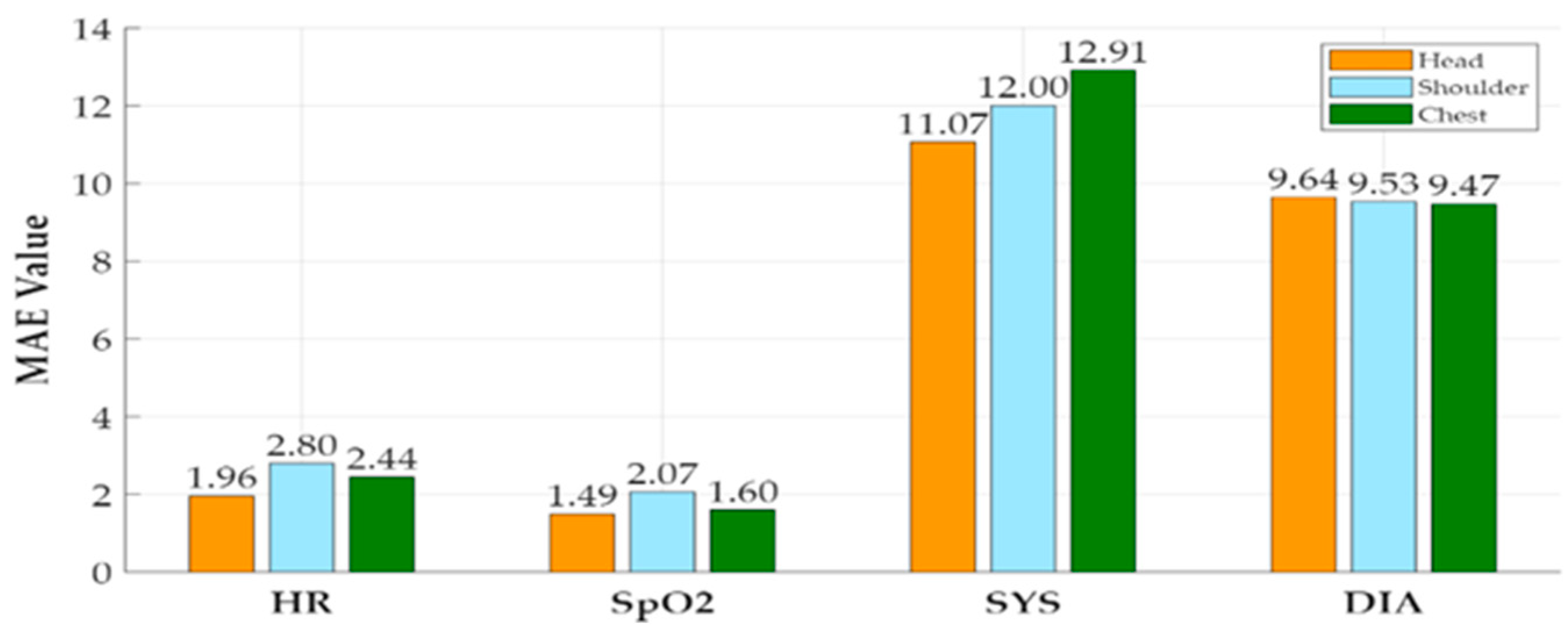
To identify the most robust wearable configuration, measurement performance was compared across three practical mounting positions for emergency personnel: head, shoulder, and chest. The comparison was conducted using the MAE between the contactless estimates and reference values, as summarized in Table 3 and visualized in Figure 12. The head-mounted configuration consistently yielded the lowest MAE for HR (1.96 bpm), SpO2 (1.49%), and SYS (11.07 mmHg), indicating superior signal fidelity. The shoulder position exhibited moderate errors, whereas the chest-mounted configuration demonstrated the highest MAE for SYS (12.91 mmHg). However, for DIA alone, the chest-mounted position produced the lowest MAE (9.47 mmHg), compared with 9.64 mmHg for the head and 9.53 mmHg for the shoulder. Overall, the head-mounted configuration offers the most stable and reliable performance for cardiorespiratory measurements, likely due to the unobstructed and consistent framing of the facial region. Nevertheless, the variability in DIA results highlights that the optimal mounting position may be parameter-specific, a consideration worth further investigation.

4. Discussion

This study evaluated the feasibility of a wearable body camera-based contactless vital sign monitoring system under simulated ambulance-related conditions incorporating reduced illumination and predefined wearable-camera motion. The proposed system demonstrated reliable performance for heart rate (HR) and oxygen saturation (SpO2) estimation across different camera stability conditions. As shown in Figure 8 and Figure 9, HR and SpO2 measurements remained relatively stable despite increasing camera motion, indicating that the proposed rPPG framework maintained relatively stable performance under the tested wearable motion conditions. In contrast, blood pressure estimation was more sensitive to camera instability, resulting in larger errors for both systolic and diastolic blood pressure (Figure 10 and Figure 11). This observation is consistent with previous studies, which reported that cuffless blood pressure estimation based on pulse-derived features is more susceptible to motion artifacts and physiological variability than HR estimation. Consequently, improved motion compensation and subject-specific calibration are required to enhance blood pressure estimation accuracy.
Table 4 compares the proposed system with representative contactless vital sign monitoring methods. Compared with the chrominance-based rPPG method of de Haan et al. [12] and the DistancePPG framework proposed by Kumar et al. [20], the proposed wearable system achieved comparable HR estimation accuracy while operating under more challenging wearable and motion conditions. Furthermore, unlike previous studies that primarily focused on HR estimation, the proposed system simultaneously estimated HR, SpO2, systolic blood pressure, and diastolic blood pressure using a single wearable camera. Although the blood pressure errors remain higher than those reported for dedicated vision-based blood pressure estimation methods such as Luo et al. [21], the proposed approach supports the preliminary feasibility of multimodal vital sign monitoring, with BP estimation remaining an area for further refinement and validation. Camera mounting position also had an impact on measurement performance. Among the evaluated configurations, the head-mounted position achieved the lowest errors for most physiological parameters. This improvement is attributed to a more stable facial field of view, reduced occlusion, and improved rPPG signal quality. In comparison, the shoulder- and chest-mounted configurations exhibited higher measurement errors due to increased camera motion, viewing angle variations, and partial facial occlusion. These findings indicate that camera placement is a critical design consideration for wearable contactless vital sign monitoring systems.

5. Conclusions

This preliminary study evaluated the technical feasibility of a wearable body-camera system for contactless rPPG-based vital sign monitoring under controlled conditions relevant to emergency environments. Across the tested experimental configurations, heart rate and SpO2 estimates showed relatively small errors compared with the contact-based reference measurements (HR MAE: 1.20–5.60 bpm; SpO2 MAE: 0.93–3.00%) across the tested motion and lighting conditions. In contrast, blood pressure estimation showed larger errors relative to the reference measurements (SYS: 5.00–14.20 mmHg; DIA: 3.87–11.60 mmHg). This performance gap suggests that the underlying pulse transit time (PTT) model may be susceptible to mechanical instability, subject-specific vascular compliance, and motion-induced signal noise under dynamic emergency conditions.
The head-mounted configuration consistently outperformed shoulder- and chest-mounted alternatives overall, showing the most favorable MAE profile across the evaluated parameters (HR: 1.96 bpm; SpO2: 1.49%; SYS: 11.07 mmHg; DIA: 9.64 mmHg). This performance advantage may be related to unobstructed facial ROI tracking, which may contribute to more stable signal acquisition. Conversely, chest and shoulder placements suffered from respiratory interference, upper-body occlusion, and suboptimal field-of-view, leading to signal degradation and increased error rates. The present findings support the technical potential of the proposed approach for contactless cardiorespiratory monitoring; however, further validation is required before its applicability in prehospital or emergency-care settings can be established. BP estimation remains a critical limitation requiring advanced motion-compensated filtering, adaptive noise reduction, and personalized calibration models.
This study was designed as a preliminary technical feasibility assessment using a small and relatively homogeneous cohort. Accordingly, the present findings are intended to characterize the performance of the proposed system under the tested experimental conditions rather than to establish population-level generalizability or clinical performance. Future studies with larger and more diverse cohorts, including clinical populations and participants with abnormal physiological conditions relevant to emergency care, will be required to further evaluate the robustness, generalizability, and practical applicability of the proposed system.

Author Contributions

Conceptualization, J.-H.L.; methodology, T.-E.K.; validation, T.-E.K., S.-H.K. and J.-H.M.; formal analysis, S.-H.K.; investigation, T.-E.K., S.-H.K. and J.-H.M.; data curation, J.-H.M.; writing—original draft preparation, T.-E.K., S.-H.K. and J.-H.M.; writing—review and editing, T.-E.K., S.A. and M.K.; visualization, T.-E.K., S.-H.K. and J.-H.M.; supervision, J.-H.L.; project administration, T.-E.K. All authors have read and agreed to the published version of the manuscript.

Funding

This research was supported by the Korea Health Technology R&D Project through the Korea Health Industry Development Institute (KHIDI), funded by the Ministry of Health & Welfare, Republic of Korea (Grant No. RS-2023-KH136789); the Collabo R&D Program between Industry, University, and Research Institute funded by the Ministry of SMEs and Startups (MSS), Republic of Korea (Grant No. RS-2026-25533076); the Regional Innovation System & Education (RISE) Program through the Daegu RISE Center, funded by the Ministry of Education (MOE) and Daegu Metropolitan City, Republic of Korea (Grant No. 2025-RISE-03-002); and the Commercialization Promotion Agency for R&D Outcomes (COMPA), funded by the Ministry of Science and ICT (MSIT), Republic of Korea (Grant No. RS-2025-02413013, IP Enhancement and Commercialization to Promote the Market Adoption of Vision Intelligence-Based Multi-Biosignal Measurement Technology).

Institutional Review Board Statement

The study was conducted in accordance with the Declaration of Helsinki, and approved by the Institutional Review Board of Keimyung University Dongsan Hospital (protocol code DSMC 2024-01-010-003, approval date: 27 February 2024).

Informed Consent Statement

Informed consent was obtained from all subjects involved in the study.

Data Availability Statement

The data supporting the findings of this study are not publicly available due to privacy and ethical restrictions.

Conflicts of Interest

The authors declare no conflicts of interest.

Abbreviations

The following abbreviations are used in this manuscript:
rPPGRemote photoplethysmography
HRVHeart rate variability
MAEMean absolute error
ROIRegion of interest
SDStandard deviation
SpO2Peripheral oxygen saturation
SYSSystolic blood pressure
PPGPhotoplethysmography
PTTPulse transit time
RMSERoot mean square error
HDHigh-definition
HRHeart rate

References

  1. Nardelli, M.; Bailón, R. Advances in wearable photoplethysmography applications in health monitoring. Sensors 2023, 23, 7064. [Google Scholar] [CrossRef] [Scilit]
  2. Yang, H.; Ji, S.; Chaturvedi, I.; Xia, H.; Wang, T.; Chen, G.; Pan, L.; Wan, C.; Qi, D.; Ong, Y.S.; et al. Adhesive biocomposite electrodes on sweaty skin for long-term continuous electrophysiological monitoring. ACS Mater. Lett. 2020, 2, 478–484. [Google Scholar] [CrossRef] [Scilit]
  3. Fine, J.; Branan, K.L.; Rodriguez, A.J.; Boonya-ananta, T.; Ajmal; Ramella-Roman, J.C.; McShane, M.J.; Coté, G.L. Sources of inaccuracy in photoplethysmography for continuous cardiovascular monitoring. Biosensors 2021, 11, 126. [Google Scholar] [CrossRef] [Scilit]
  4. Xu, L.; Pan, C.; Xu, B.; Sun, Z.; Zhou, K.; Yan, R.; Weng, K.; Wu, Y.; Li, H. A Qualitative Exploration of a User-Centered Model for Smartwatch Comfort Using Grounded Theory. Int. J. Hum.-Comput. Interact. 2025, 41, 1091–1106. [Google Scholar] [CrossRef] [Scilit]
  5. Amar, D.; Neidzwski, J.; Wald, A.; Finck, A.D. Fluorescent light interferes with pulse oximetry. J. Clin. Monit. 1989, 5, 135–136. [Google Scholar] [CrossRef] [Scilit] [PubMed]
  6. Tricedi, N.S.; Ghouri, A.F.; Shah, N.K.; Lai, E.; Barker, S.J. Effects of motion, ambient light, and hypoperfusion on pulse oximeter function. J. Clin. Anesth. 1997, 9, 179–183. [Google Scholar] [CrossRef] [Scilit] [PubMed]
  7. Wijinberge, M.; van der Ster, B.; Vlaar, A.P.J.; Hollmann, M.W.; Geerts, B.F.; Veelo, D.P. The effect of intermittent versus continuous non-invasive blood pressure monitoring on the detection of intraoperative hypotension: A sub-study. J. Clin. Med. 2022, 11, 4083. [Google Scholar] [CrossRef] [Scilit]
  8. Kebe, M.; Gadhafi, R.; Mohammad, B.; Sanduleanu, M.; Saleh, H.; Al-Qutayri, M. Human vital signs detection methods and potential using radars: A review. Sensors 2020, 20, 1454. [Google Scholar] [CrossRef] [Scilit]
  9. Hao, Z.; Wang, Y.; Li, F.; Ding, G.; Gao, Y. mmWave-RM: A respiration monitoring and pattern classification system based on mmWave radar. Sensors 2024, 24, 4315. [Google Scholar] [CrossRef] [Scilit]
  10. Wang, Y.; Wang, Z.; Zhang, J.A.; Zhang, H.; Xu, M. Vital sign monitoring in dynamic environment via mmWave radar and camera fusion. IEEE Trans. Mob. Comput. 2024, 23, 4163–4180. [Google Scholar] [CrossRef] [Scilit]
  11. Verkruysse, W.; Svaasand, L.O.; Nelson, J.S. Remote plethysmographic imaging using ambient light. Opt. Express 2008, 16, 21434–21445. [Google Scholar] [CrossRef] [Scilit] [PubMed]
  12. De Haan, G.; Jeanne, V. Robust pulse rate from chrominance-based rPPG. IEEE Trans. Biomed. Eng. 2013, 60, 2878–2886. [Google Scholar] [CrossRef] [Scilit] [PubMed]
  13. Yang, Y.; Liu, C.; Yu, H.; Shao, D.; Tsow, F.; Tao, N. Motion-robust remote photoplethysmography in CIELab color space. J. Biomed. Opt. 2016, 21, 117001. [Google Scholar] [CrossRef] [Scilit] [PubMed]
  14. Allado, E.; Poussel, M.; Renno, J.; Moussu, A.; Hily, O.; Temperelli, M.; Albuisson, E.; Chenuel, B. Remote photoplethysmography is an accurate method to remotely measure respiratory rate. J. Clin. Med. 2022, 11, 3647. [Google Scholar] [CrossRef] [Scilit]
  15. Allado, E.; Poussel, M.; Moussu, A.; Hily, O.; Temperelli, M.; Cherifi, A.; Saunier, V.; Bernard, Y.; Albuisson, E.; Chenuel, B. Accurate and reliable assessment of heart rate in real-life clinical settings using imaging photoplethysmography. J. Clin. Med. 2022, 11, 6101. [Google Scholar] [CrossRef] [Scilit]
  16. Castaneda, D.; Esparza, A.; Ghamari, M.; Soltanpur, C.; Nazeran, H. A review on wearable photoplethysmography sensors and their potential future application in health care. Int. J. Biosens. Bioelectron. 2018, 4, 195–202. [Google Scholar] [CrossRef] [Scilit] [PubMed]
  17. Lugaresi, C.; Tang, J.; Nash, H.; McClanahan, C.; Uboweja, E.; Hays, M.; Zhang, F.; Chang, C.; Yong, M.; Lee, J.; et al. Mediapipe: A framework for building perception pipelines. arXiv 2019, arXiv:1906.08172. [Google Scholar]
  18. Wu, H.Y.; Rubinstein, M.; Shih, E.; Guttag, J.; Durand, F.; Freeman, W. Eulerian video magnification for revealing subtle changes in the world. ACM Trans. Graph. (TOG) 2012, 31, 65. [Google Scholar] [CrossRef]
  19. Khomidov, M.; Lee, D.; Lee, J.-H. A Novel Contactless Blood Pressure Measurement System and Algorithm Based on Vision Intelligence. Electronics 2023, 12, 4898. [Google Scholar] [CrossRef] [Scilit]
  20. Kumar, M.; Veeraraghavan, A.; Sabharwal, A. DistancePPG: Robust non-contact vital signs monitoring using a camera. Biomed. Opt. Express 2015, 6, 1565–1588. [Google Scholar] [CrossRef] [Scilit] [PubMed]
  21. Luo, H.; Yang, D.; Barszczyk, A.; Vempala, N.; Wei, J.; Wu, S.J.; Zheng, P.P.; Fu, G.; Lee, K.; Feng, Z.P. Smartphone-based blood pressure measurement using transdermal optical imaging. Circ. Cardiovasc. Imaging 2019, 12, e008857. [Google Scholar] [CrossRef] [Scilit] [PubMed]
Figure 1. Current contact devices used for routine vital-sign monitoring: (a) pulse oximeter, (b) automated blood pressure monitor, and (c) patient monitoring system.
Algorithms 19 00802 g001
Figure 2. Process of remote photoplethysmography signal extraction and analysis of heart rate variability through facial detection.
Algorithms 19 00802 g002
Figure 3. Flowchart of contactless remote photoplethysmography signal processing and vital sign analysis.
Algorithms 19 00802 g003
Figure 4. Comparison between conventional contact-based measurement and proposed contactless method: (a) Conventional contact-based method. (b) Proposed contactless method.
Algorithms 19 00802 g004
Figure 5. Video acquisition process under fixed camera stability conditions by camera attachment position. (a) Head, (b) shoulder, and (c) chest.
Algorithms 19 00802 g005
Figure 6. Representation of facial video acquisition under mild camera movement conditions to assess camera stability settings.
Algorithms 19 00802 g006
Figure 7. Real-time display output of the rPPG-based contactless vital sign monitoring system.
Algorithms 19 00802 g007
Figure 8. Heart rate results by participants for each condition. (a) Participant 1, (b) Participant 2, (c) Participant 3.
Algorithms 19 00802 g008
Figure 9. Oxygen saturation measurement results by condition for each participant. (a) Participant 1, (b) Participant 2, (c) Participant 3.
Algorithms 19 00802 g009
Figure 10. Systolic blood pressure measurement results by condition for each participant. (a) Participant 1, (b) Participant 2, (c) Participant 3.
Algorithms 19 00802 g010
Figure 11. Diastolic blood pressure results by condition for each participant. (a) Participant 1, (b) Participant 2, (c) Participant 3.
Algorithms 19 00802 g011
Figure 12. Comparison of the mean absolute error according to the camera mounting position.
Algorithms 19 00802 g012
Table 1. Summary of the mean absolute error (MAE) across all experimental conditions.
Vital SignMAE RangeUnit
Heart Rate (HR)1.20–5.60bpm
Oxygen Saturation (SpO2)0.93–3.00%
Systolic BP (SYS)5.00–14.20mmHg
Diastolic BP (DIA)3.87–11.60mmHg
Table 2. Comparison of estimated versus reference vital sign ranges across camera stability levels.
Stability LevelHR (bpm)SpO2 (%)SYS (mmHg)DIA (mmHg)
FixedEst: 67.3–85.9
Ref: 69.5–85.9
Est: 95.5–97.6
Ref: 96.3–97.5
Est: 103.0–111.3
Ref: 104.9–130.5
Est: 63.0–71.6
Ref: 55.7–83.8
Mild MotionEst: 61.0–74.9
Ref: 62.9–73.7
Est: 94.5–98.4
Ref: 96.3–97.4
Est: 102.9
Ref: 104.6–124.8
Est: 63.9–64.1
Ref: 57.1–80.7
Severe MotionEst: 61.5–75.5
Ref: 62.1–79.1
Est: 94.7–98.5
Ref: 96.9–97.3
Est: 102.6–103.1
Ref: 101.3–130.3
Est: 64.0–64.3
Ref: 55.7–82.8
Note: Est = rPPG-estimated value; Ref = contact-based reference value.
Table 3. Participant-averaged MAE by camera mounting position under the simulated ambulance-related condition.
Vital SignHeadShoulderChest
HR1.96 bpm2.80 bpm2.44 bpm
SpO21.49%2.07%1.60%
SYS11.07 mmHg12.00 mmHg12.91 mmHg
DIA9.64 mmHg9.53 mmHg9.47 mmHg
Table 4. Comparison of the proposed system’s measurement accuracy with previously reported contactless vital sign studies.
ResearchCamera TypeVital SignsHR MAE (bpm)SpO2 MAE (%)SYS MAE (mmHg)DIA MAE (mmHg)
Verkruysse et al. [11]RGB HRAlgorithms 19 00802 i001Algorithms 19 00802 i001Algorithms 19 00802 i001Algorithms 19 00802 i001
de Haan et al. [12]RGB HR1.96Algorithms 19 00802 i001Algorithms 19 00802 i001Algorithms 19 00802 i001
Kumar et al. [20]RGB HR, RR1.30Algorithms 19 00802 i001Algorithms 19 00802 i001Algorithms 19 00802 i001
Luo et al. [21]RGB SYS, DIAAlgorithms 19 00802 i001Algorithms 19 00802 i00112.138.31
Proposed MethodWearable
Body
HR, SpO2,
SYS, DIA
1.961.4911.079.64
Note: “Algorithms 19 00802 i001” indicates that the corresponding MAE value was not reported in the cited study.
Disclaimer/Publisher’s Note: The statements, opinions and data contained in all publications are solely those of the individual author(s) and contributor(s) and not of MDPI and/or the editor(s). MDPI and/or the editor(s) disclaim responsibility for any injury to people or property resulting from any ideas, methods, instructions or products referred to in the content.