Article

MSA-Net: A Deep Learning Network with Multi-Axial Hadamard Attention and Pyramid Pooling for Stroke Microwave Imaging

1
School of Computer and Information Engineering, Fuyang Normal University, Fuyang 236037, China
2
Anhui Engineering Research Center for Intelligent Computing and Information Innovation, Fuyang Normal University, Fuyang 236037, China
*
Author to whom correspondence should be addressed.
Algorithms 2026, 19(4), 276; https://doi.org/10.3390/a19040276
Submission received: 30 January 2026 / Revised: 23 March 2026 / Accepted: 25 March 2026 / Published: 2 April 2026
(This article belongs to the Special Issue Algorithms for Computer Aided Diagnosis: 3rd Edition)

Abstract

Microwave imaging is emerging as an alternative to conventional medical diagnostic techniques. Traditional analytical and numerical methods fail to adequately address these fundamental challenges: they often rely on strict linear approximations or simplified physical models, leading to low reconstruction accuracy, poor robustness, and limited generalization ability in complex clinical scenarios. As a result, they cannot meet the high-precision requirements of practical stroke microwave imaging. To further improve the accuracy of microwave imaging algorithms in recognizing stroke regions and solving the backscattering problem, this study employs a combination of methods with deep learning. It presents the Multi-Scale Attention Network (MSA-Net) for microwave imaging. The network is based on the EGE-UNet network structure with improved multi-axis Hadamard attention, incorporating null-space pyramid pooling and introducing a deep supervisory mechanism to improve the network performance further. To combine microwave imaging with deep learning, firstly, a large amount of microwave data need to be simulated with HFSS, in which the simulation model is a human brain stroke model constructed by an HFSS simulation system. Secondly, the microwave data obtained from the simulation are converted into a tensor format. Then, the tensor data are input into the MSA-Net neural network, which generates a binary mask image that can be used to detect the size and location of the stroke. This study also prompts the model to converge faster by sparsifying the microwave data to improve training efficiency. The method has been tested using simulation data, and based on the comparison experiments with other networks, MSA-Net is more accurate in detecting the location and the bleed size. The experimental results show that the proposed method is superior for stroke imaging. The experimental results show that the proposed model achieves a 1.08 improvement in peak signal-to-noise ratio and a 0.017 reduction in learned perceptual image block similarity, fully validating the effectiveness of the structural optimization strategy proposed in this paper.

1. Introduction

In recent years, researchers have proposed various improvement methods to address the ill-posedness and nonlinearity issues in microwave imaging. On the one hand, traditional iterative optimization and regularization techniques are employed to mitigate the instability of inverse problems, enhancing the reliability and robustness of imaging [1,2,3,4]. On the other hand, with the advancement of deep learning, data-driven methods based on neural networks have gradually become a research hotspot. By training convolutional neural networks or improved U-Net models on simulated microwave data, images can be directly reconstructed from raw microwave signals, which refer specifically to the complex-valued scattering measurements from antenna arrays—i.e., the complex phasor data (amplitude and phase) of the received responses for each transmitter–receiver pair—thus significantly reducing reliance on complex physical modeling [5,6]. Meanwhile, attention mechanisms and multi-scale feature fusion are utilized to enhance the reconstruction ability of key areas [7]. These studies indicate that combining traditional physical models with deep learning methods can effectively alleviate the core challenges in microwave imaging and achieve high-precision image reconstruction in complex scenarios, as shown in Table 1.
Existing research has shown that the generation of simulated data and the training of deep neural networks can effectively alleviate the ill-posedness issues in traditional methods. For instance, utilizing High-Frequency Structure Simulation Software (HFSS, Version 2023 R2) to generate microwave data and converting it into tensor inputs suitable for neural network processing can simplify the data preprocessing process and enhance imaging efficiency [8]. In terms of network architecture design, combining the multi-axis Hadamard attention mechanism (MAHAM) with the channel-wise and spatial attention module (CBAM) has become an effective means to improve feature extraction capabilities and detail reconstruction quality [9]. MAHAM captures nonlinear correlations through multi-axis feature interaction, achieving the integration of global information, while CBAM enhances local feature representation by independently modeling channels and spatial dimensions. Furthermore, the introduction of deep supervision mechanisms can provide additional training signals in multi-level features, facilitating network convergence and mitigating the vanishing gradient problem [10].
Overall, the existing literature primarily focuses on the following directions: first, improving the physical models and algorithms of microwave imaging to enhance imaging accuracy; second, combining deep learning methods with attention mechanisms and multi-scale feature fusion to enhance reconstruction capabilities in complex scenarios; third, utilizing simulated data or multi-modal data to enhance the quality of the training set. However, these methods still have certain limitations when dealing with heterogeneous objects, extremely low signal-to-noise ratios, or complex electromagnetic environments. Therefore, how to fully utilize the nonlinear fitting capabilities of deep learning combined with the physical characteristics of microwave imaging remains a key issue in current research.
In summary, the method proposed in this study makes three key contributions: Firstly, it successfully integrates the nonlinear fitting capability of deep learning with the physical characteristics of microwave imaging, effectively breaking through the bottleneck of traditional microwave imaging methods that struggle to balance fitting accuracy and physical rationality. Secondly, addressing core challenges in microwave imaging, such as insufficient reconstruction accuracy and poor robustness in complex environments, the method proposes precise and efficient solutions, significantly enhancing the quality and effectiveness of image reconstruction. Finally, fully validated by experiments, the proposed method demonstrates excellent accuracy and robustness in image reconstruction tasks under complex scenarios, laying important technical support and a solid theoretical foundation for the future development of microwave imaging technology.

2. Methods

2.1. MSA-Net Methodology

EGE-UNet is a variant network that was improved based on the U-Net architecture, which was initially designed for medical image segmentation tasks. Based on U-Net, it introduces two core modules: the grouped Hadamard product attention (GHPA) module and the group aggregation bridge (GAB) module [9]. Among them, the GHPA module performs Hadamard product operations on input features through learnable weights to enhance cross-dimensional feature modeling capability; the GAB module achieves effective fusion of high-level and low-level features based on a grouped aggregation strategy. This structure is, to a certain extent, consistent with the requirements of multi-scale information extraction and target-region modeling in the stroke microwave imaging task.
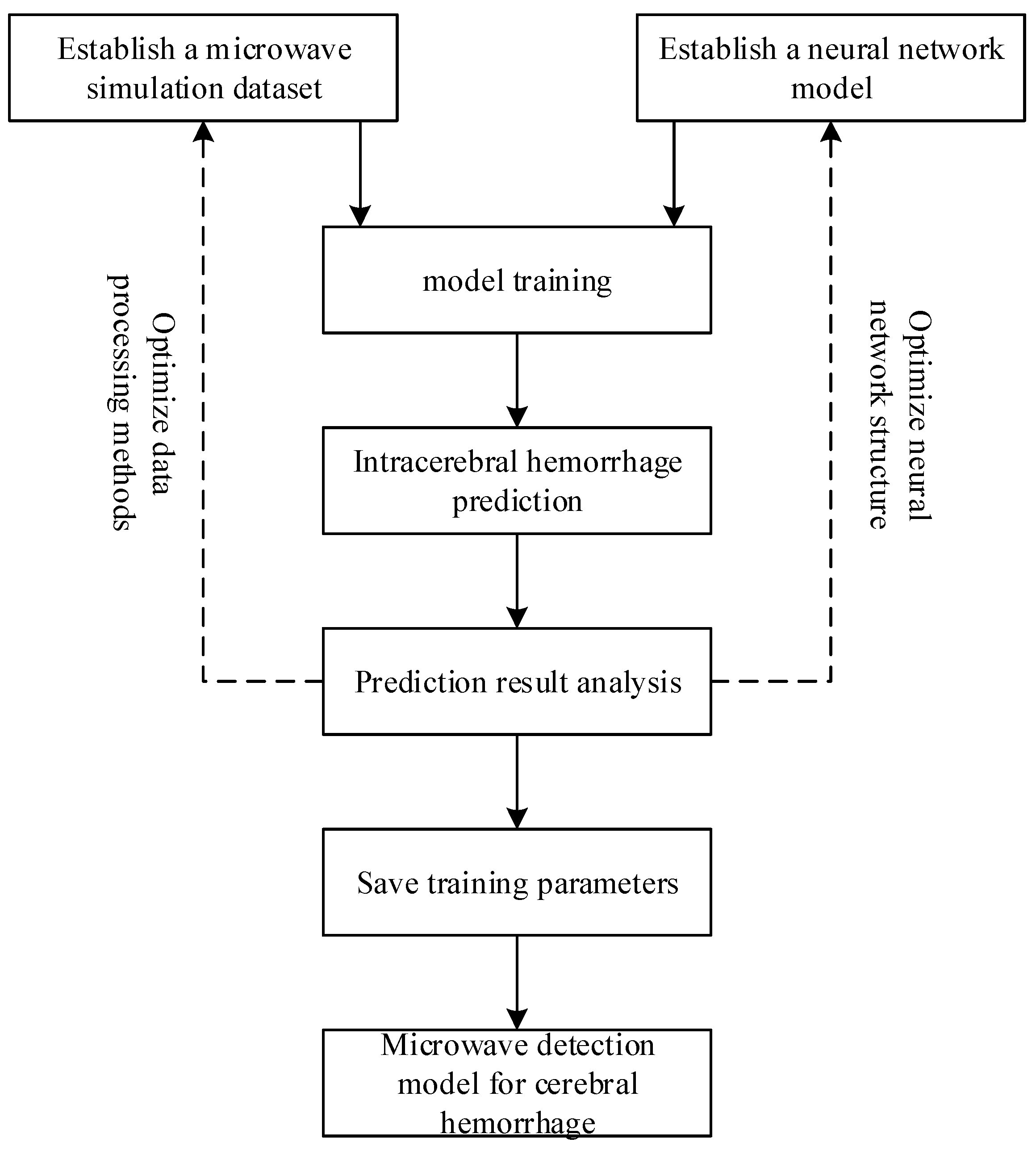
As illustrated in Figure 1, the proposed study follows a complete workflow for microwave-based intracerebral hemorrhage detection. First, a microwave simulation dataset is established using the HFSS electromagnetic simulation system. In this system, an 8-element antenna array is employed, where one antenna transmits microwave signals and the remaining seven antennas receive the corresponding scattered responses. To enhance the diversity and representativeness of the dataset, the constructed microwave simulation dataset includes hemorrhagic lesions with different locations and sizes. Based on the acquired microwave data, a neural network model is then constructed for intracerebral hemorrhage detection.
Subsequently, the collected microwave data are preprocessed and organized into tensor representations of 64 × 64 × 1 × 1 before being fed into the network for model training. During this process, both the data processing methods and the neural network structure are continuously optimized to improve prediction performance. After training, the model is used to perform intracerebral hemorrhage prediction, and the prediction results are further analyzed to evaluate the effectiveness of the proposed method. Finally, the trained parameters are saved, and a microwave detection model for cerebral hemorrhage is obtained.
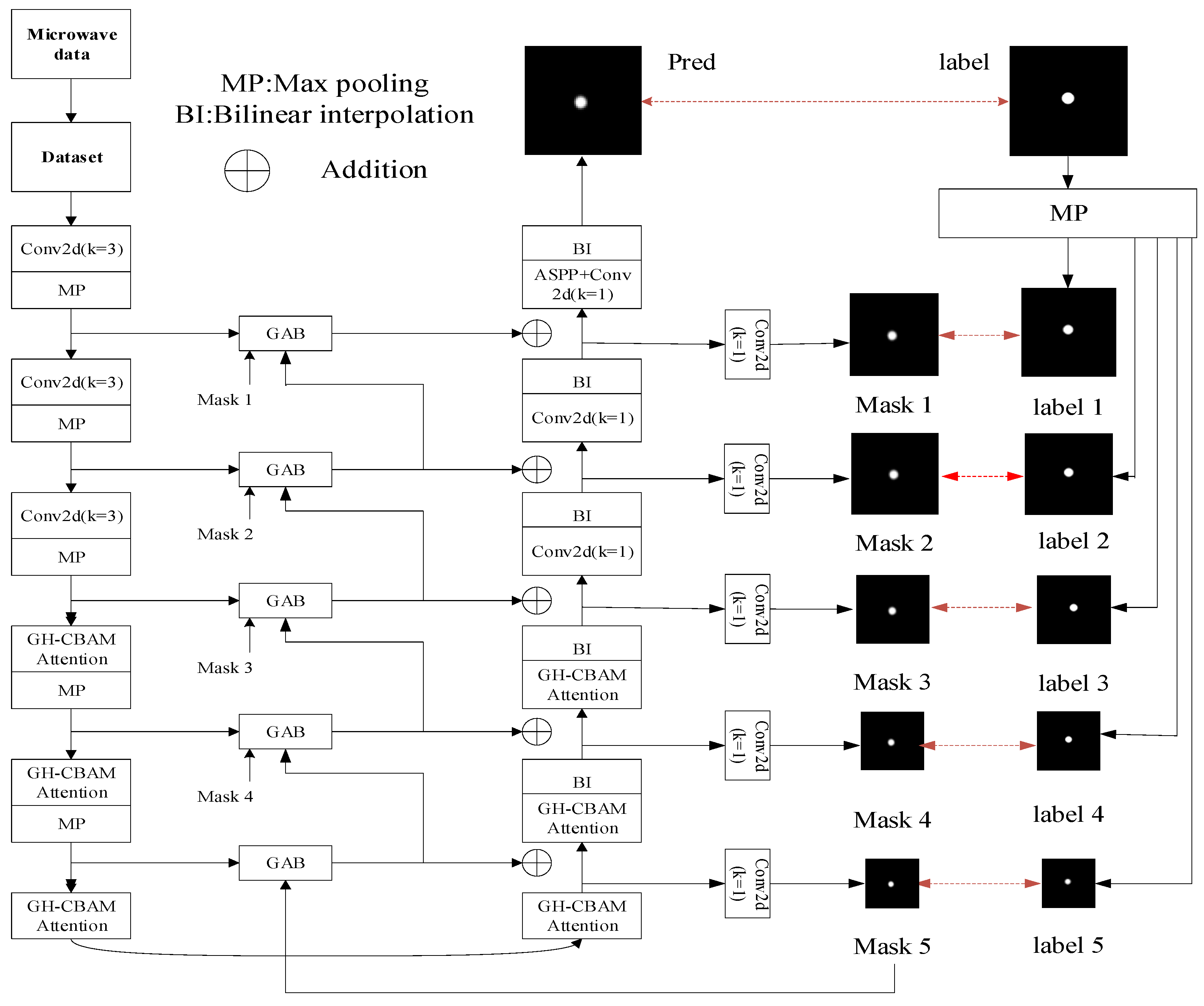
In the network implementation, the encoder performs five downsampling stages. In the first three stages, the input features are extracted using a 3 × 3 convolution with 8 channels, followed by group normalization and 2 × 2 pooling for downsampling. In the fourth and fifth stages, the feature maps are further enhanced by the grouped Hadamard-channel spatial attention module (GH-CBAM) before normalization and pooling, thereby strengthening the representation capability of deep features in the target region.
However, when EGE-UNet is directly applied to the stroke microwave imaging task, there are still deficiencies in target-edge detail recovery and key-region focusing. Specifically, the network lacks direct constraints on features of different depths, which easily leads to a decrease in the reconstruction accuracy of edge regions; at the same time, although GHPA can enhance context modeling capability, its selective attention to target salient regions is still insufficient, and there are limitations in the joint modeling of channel and spatial dimensions. To address the above problems, this study introduces a channel spatial attention module on the basis of GHPA to construct the grouped Hadamard-channel spatial attention module (GH-CBAM), so as to further fuse the saliency information of channel and spatial dimensions, enhance the network’s ability to focus on key regions and suppress background interference.
On this basis, considering that problems such as insufficient gradient propagation and slow convergence may still exist in the training of deep networks, this study further introduces a deep supervision mechanism, adding auxiliary supervision signals in the middle layers to promote the effective learning of deep features, improve training stability and accelerate network convergence. At the same time, to enhance the model’s representation capability for lesion regions of different sizes, an atrous spatial pyramid pooling (ASPP) module is introduced in the network bottleneck layer, which extracts multi-scale contextual information through parallel atrous convolutions, thereby improving the network’s reconstruction capability for stroke lesions of different sizes [11].
In summary, the introduction of the grouped Hadamard-channel spatial attention module, deep supervision mechanism and ASPP module enables the network to more effectively complete multi-scale feature extraction, key region enhancement and deep feature optimization in the microwave imaging task. It not only improves the stability and convergence speed of model training but also enhances the reconstruction quality of target edges and detail regions, thereby improving the overall reconstruction accuracy and robustness. The network structure is shown in Figure 2.

2.2. GH-CBAM

The grouped Hadamard-channel spatial attention module combines the grouped multi-axis Hadamard product attention module (GHPA) and the convolutional block attention module (CBAM), aiming to enhance the neural network’s attention to key feature regions, especially in microwave image generation tasks, to improve the processing effect on image details and edges (see Figure 3). Firstly, the multi-axis Hadamard product attention module divides the input feature maps into four groups and performs Hadamard product operations on the first three groups in the dimensions of height width, channel height, and channel width, respectively. Only deep convolution operations are performed on the last set of feature maps. Subsequently, these four sets of data are fused along the channel dimension, and different dimensions of information are integrated through another deep convolution [12]. Then, the fused data are input into the convolutional block attention module, which optimizes the attention to key features through channel attention and spatial attention mechanisms—channel attention captures the importance of feature channels through global pooling, while spatial attention highlights key spatial regions through global pooling [13,14,15,16,17].
The combination of the grouped multi-axis Hadamard product attention module and the convolutional block attention module enables the grouped Hadamard-channel spatial attention module to efficiently capture complex feature dependencies, accurately focus on target areas in microwave images, suppress irrelevant background information, and thus improve the accuracy and robustness of generated images. This module significantly improves the performance of the model in microwave image generation tasks by increasing the sensitivity of the network to different scales and features, improving the reconstruction effect of image details and edges.

2.3. GAB

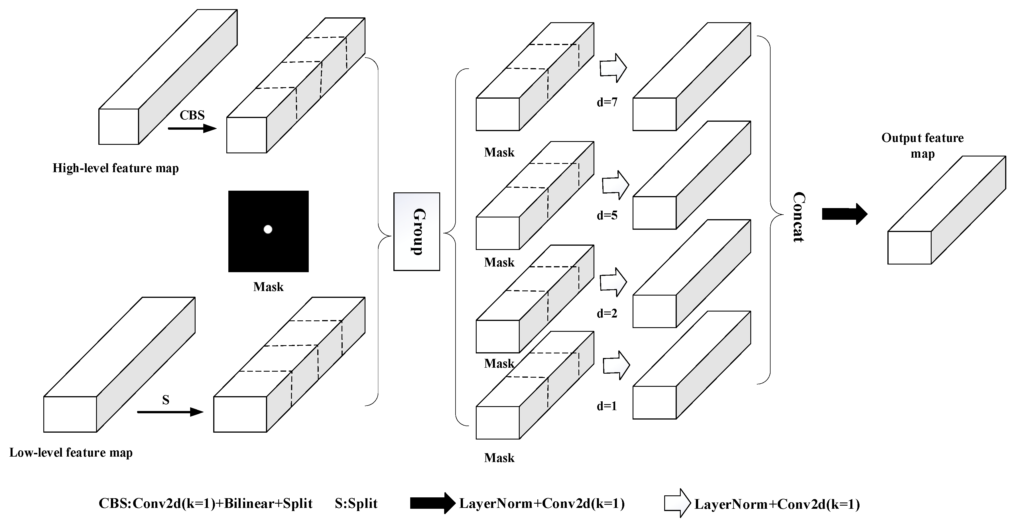
For high-density generation tasks such as microwave image generation, obtaining multi-scale information is crucial. Therefore, this study introduces the Group Aggregation Bridge (GAB) module, which requires three inputs: low-level features, high-level features, and mask maps. Firstly, the high-level features are adjusted to the same size as the low-level features through depthwise separable convolution and bilinear interpolation (see Figure 4). Subsequently, the two feature maps are divided into four groups along the channel dimension, with each group consisting of low-level features concatenated with a set of high-level features to obtain four sets of fused features. Finally, a depthwise separable convolution operation is performed on the concatenated results of each fused feature and mask image. The convolution process uses different dilation rates to gradually expand the receptive field in order to capture feature information at different scales.
Specifically, each set of fused features is processed through four convolutional branches with different dilation rates to generate locally enhanced feature maps. Next, the feature maps output by the four branches are concatenated along the channel dimension to form a fused feature map containing multi-scale information. Finally, the fused feature map is channel-compressed and projected using a 1 × 1 convolution at the tail to generate the final output features. This design can fully utilize the complementary information between high- and low-layer features, while combining mask maps to focus on specific areas, thereby achieving better generation quality and detail representation in high-density generation tasks such as microwave image generation [18,19,20]. The structure is shown in Figure 4.

2.4. ASPP

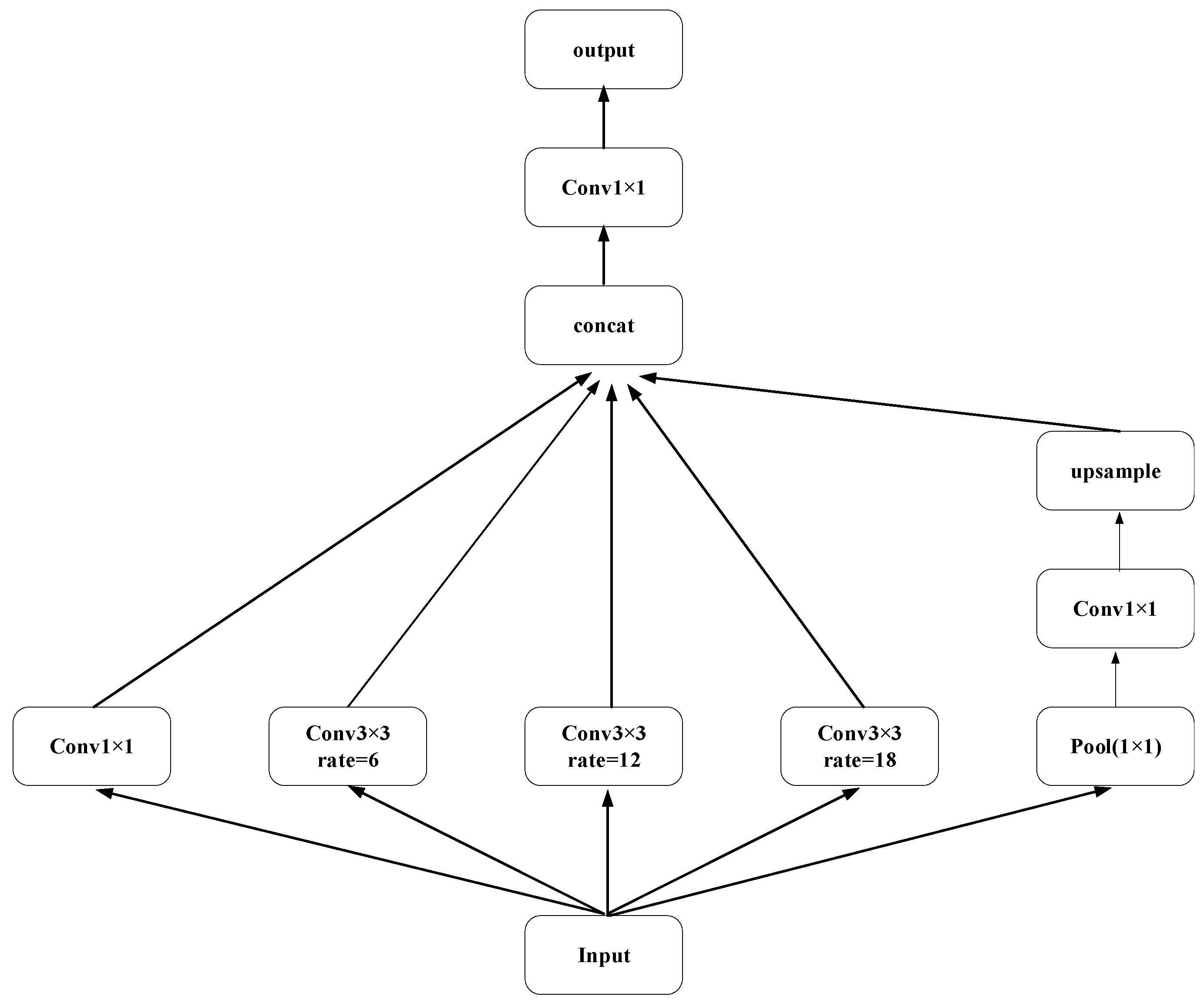
The hollow space pyramid pooling module captures multi-scale contextual information by using different dilation rates in different hollow convolution branches. This module contains multiple parallel dilated convolution branches, each with different dilation rates (1, 6, 12, 18), to achieve feature extraction at different scales. Hollow convolution can expand the receptive field and obtain a wider range of contextual information while maintaining high spatial resolution. In addition, the module also includes a global average pooling branch to capture global contextual information and convert it into feature maps of the same size as other branches through 1 × 1 convolution. Finally, the hollow space pyramid pooling module concatenates the outputs of each branch and integrates them through a 1 × 1 convolution to output a multi-scale fused feature map. This process effectively enhances the network’s perception ability of information at different scales, improves segmentation accuracy, and can better capture detailed features, especially when processing images containing objects of different sizes [21,22,23]. The structure is shown in Figure 5.

2.5. Loss Function

The loss function evaluates the degree of discrepancy between the predictions generated by the model after processing the sample inputs and the true labels of the samples. By minimizing the value of the loss function, the model parameters can be effectively adjusted to improve the model’s ability to fit the training data and thus achieve more accurate predictions. In the training phase, to optimize the model performance, this study chooses the Binary Cross-Entropy Loss (BCELoss) as the objective function of the study [24], and the prediction results obtained from MSA-Net are calculated with the labels to obtain the loss results. The calculation function of this loss function is shown in Equation (1):
B C E L o s s = 1 N i = 1 N y i × log p y i + 1 y i × log 1 p y i ,
where N denotes the total number of samples in a batch, y i represents the ground-truth label of the i-th sample (0 for negative and 1 for positive), and p( y i ) is the probability predicted by the model that the i-th sample belongs to the positive class. The term 1 N 1 N x computes the average loss over all samples, and the negative sign ensures the loss value is positive, such that the loss decreases as the prediction becomes more accurate.

2.6. Training Protocol

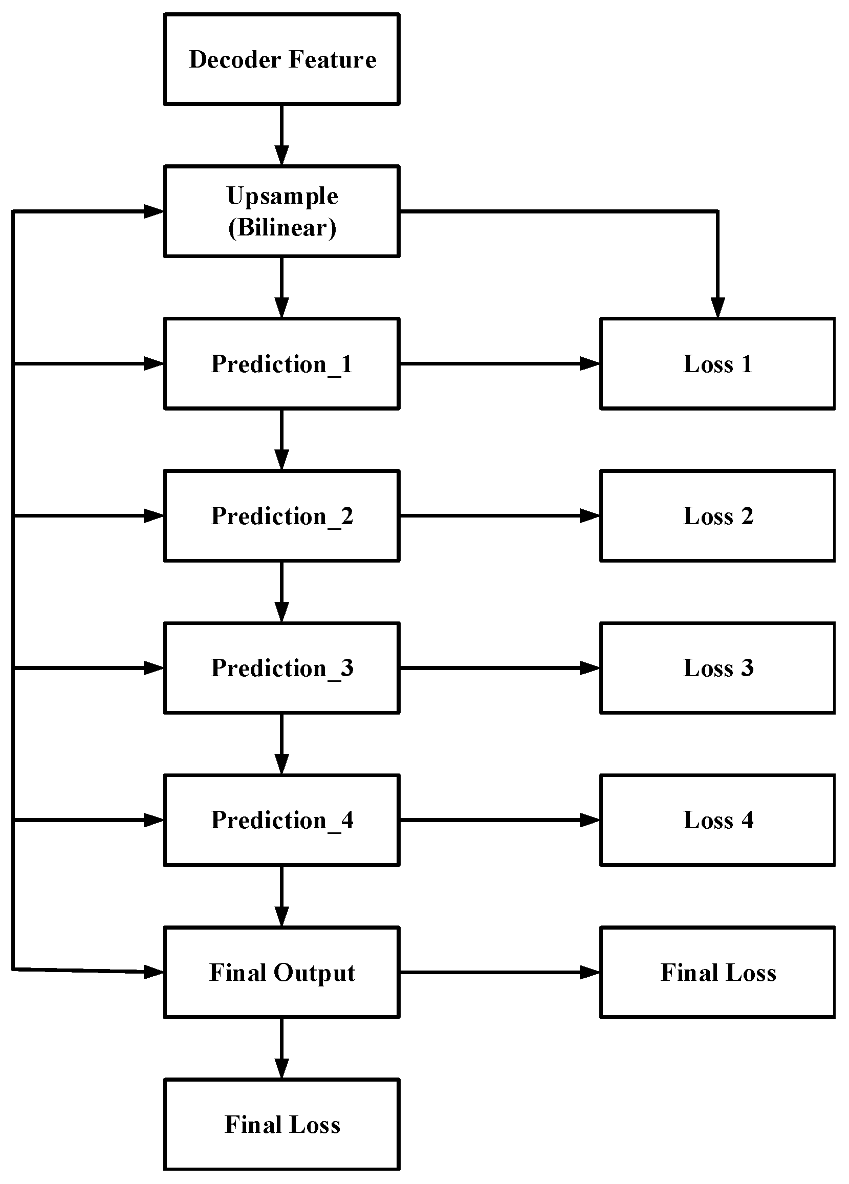
In the microwave imaging task of stroke, the network needs to predict the pixel-level reconstruction and segmentation results of the stroke area from the input microwave data, and the output is usually a probability map or binary mask of the foreground (stroke lesion) and background tissue. Due to the weak difference in microwave scattering signals between the lesion area and normal brain tissue, blurred boundaries, and significant differences in lesion morphology in microwave imaging of stroke, imaging learning is easily affected by noise, signal attenuation, and sample differences. If only supervision is applied to the final output, the gradient signal needs to propagate across multiple layers during the training process, which can easily lead to problems such as gradient attenuation and slow convergence, resulting in insufficient learning of deep semantic features and affecting the consistency of stroke area localization and the quality of boundary detail characterization. To this end, this article introduces a deep supervision mechanism at multiple output terminals of the decoder, sets up auxiliary prediction branches, and applies label constraints at different resolution levels, so that each level of features can obtain more direct optimization directions and more sufficient gradient feedback during the training stage, thereby accelerating network convergence, improving training stability, and further improving the overall consistency of imaging results and the prediction effect of boundary regions, especially in scenarios with limited sample size or obvious boundary blurring. The structure is shown in Figure 6. The overall loss function is defined as the weighted sum of the main output loss and the auxiliary output loss at each scale:
L total = L y 0 ^ , y + λ i = 1 5 L y i ^ , y
where L total denotes the overall loss function, L ( y 0 ^ , y ) is the loss between the main prediction y 0 ^ and the ground truth y , L ( y i ^ , y ) is the loss of the i -th auxiliary output, λ i denotes the weighting factor of the corresponding auxiliary loss, and N represents the total number of auxiliary outputs.

3. Results

3.1. Microwave System Configuration

This study is based on the HFSS electromagnetic simulation platform and constructs a three-dimensional head simulation model of stroke (cerebral hemorrhage). The geometric structure and physical field definition are strictly determined according to the physiological structure of the human body, and the core adopts a three-layer spherical head model of skin, skull, and brain tissue. There are significant differences in the relative dielectric constant and conductivity of different levels of biological tissues, and these two key parameters directly determine the reflection characteristics, penetration ability, and attenuation law of microwaves inside the head tissue. Therefore, in the HFSS simulation environment, based on actual physiological measurement data of the human body, the relative dielectric constant and conductivity of each layer of tissue are accurately set to achieve a precise simulation of microwave propagation in real head tissue. To ensure the consistency of simulation conditions and experimental reproducibility, the dielectric parameters of each organization are standardized according to Table 2; at the same time, the distance between the antenna array and the surface of the head model is fixed at 2 mm [25] to maintain a stable electromagnetic coupling state and effectively reduce the interference of geometric parameter fluctuations on the measurement results.
This model uses eight antennas to construct a uniform circular array, with one antenna selected as the transmitting end and the remaining seven antennas as the receiving end each time. Each antenna is used as the source, and the operating frequency is set to 2–4 GHz. Sweep frequency measurements are performed within this frequency band to obtain scattering data containing multi-frequency information. Multi-frequency scattering responses can reflect the variation in tissue dielectric properties with frequency, while enhancing the ability to distinguish different tissue types and lesion structures, providing more comprehensive frequency domain feature support for subsequent end-to-end learning-based reconstruction, and improving the network’s localization and scale representation capabilities for target areas. In addition, multi-frequency measurement can improve the accuracy of the network in target area localization, lesion scale characterization, and boundary recovery, laying the foundation for the robustness and reliability of microwave imaging reconstruction [25]. The broadband electromagnetic wave penetrates different dielectric layers of the head and reaches the bleeding area. Due to the significant difference in dielectric constant between blood and surrounding brain tissue, the electromagnetic wave reflects at the bleeding point and generates scattered echoes, which are then collected by other receiving antennas. The microwave scattering data obtained from different angles are the core foundation for achieving precise three-dimensional positioning and quantitative analysis of bleeding points.

3.2. Experimental Platforms and Datasets

The experimental environment of this study is configured as follows: based on the PyTorch 1.12.0 deep learning framework, Python 3.11 is used as the development language. In terms of hardware configuration, an Intel Core i7-11700K processor, 32 GB DDR4 RAM, and an NVIDIA RTX 3070 GPU are used to achieve GPU acceleration with NVIDIA CUDA 11.6, which significantly improves the efficiency of model training. In the model optimization strategy, the RMSprop optimizer with adaptive learning rate adjustment is introduced, and the initial learning rate is set to 0.00001 to ensure the stability and convergence speed of the training process. The input dataset is generated using the HFSS electromagnetic simulation platform to simulate stroke-related microwave signals. During data acquisition, the antenna array is rotated to two angles (0° and 30°) around the center, with data collected from 56 channels at each angle. The dataset is stored in CSV format with 56 columns and 101 rows, recording the S-parameters of each transmit–receive channel, including both magnitude and phase information. These parameters characterize the electromagnetic responses caused by permittivity differences among brain tissue, skull, and hemorrhage, reflecting changes in transmission and reflection characteristics. To enhance the discriminability of hemorrhage-related features and suppress background clutter, differential processing is applied between two groups of data to extract fine-grained information. The CSV data are then read and parsed into numerical matrices, followed by transposition to match the model input dimensions. Considering variations in numerical scales across samples, the data are reshaped into matrices of size [x, 56 × 56] and normalized to the range [0, 1] using the min–max normalization method to improve training stability. Finally, the normalized data are converted into 64 × 64 × 1 × 1 PyTorch tensors, which preserve detailed information and further facilitate effective feature extraction by the model. After several experiments, the loss function stabilizes when the training reaches the 800th round. Therefore, the number of training iterations of the model is reasonably set to 800 to achieve the optimal balance between performance and efficiency.
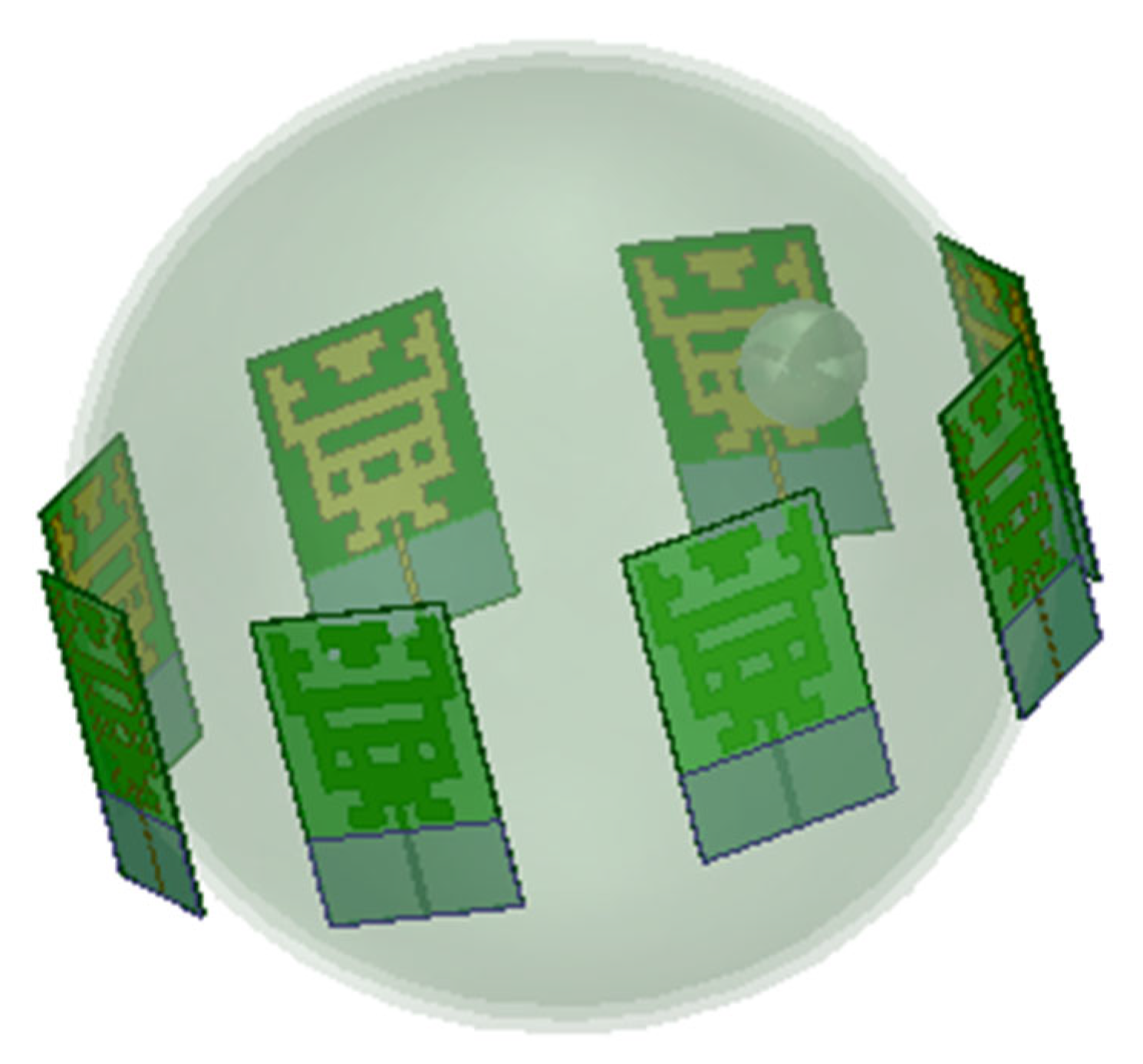
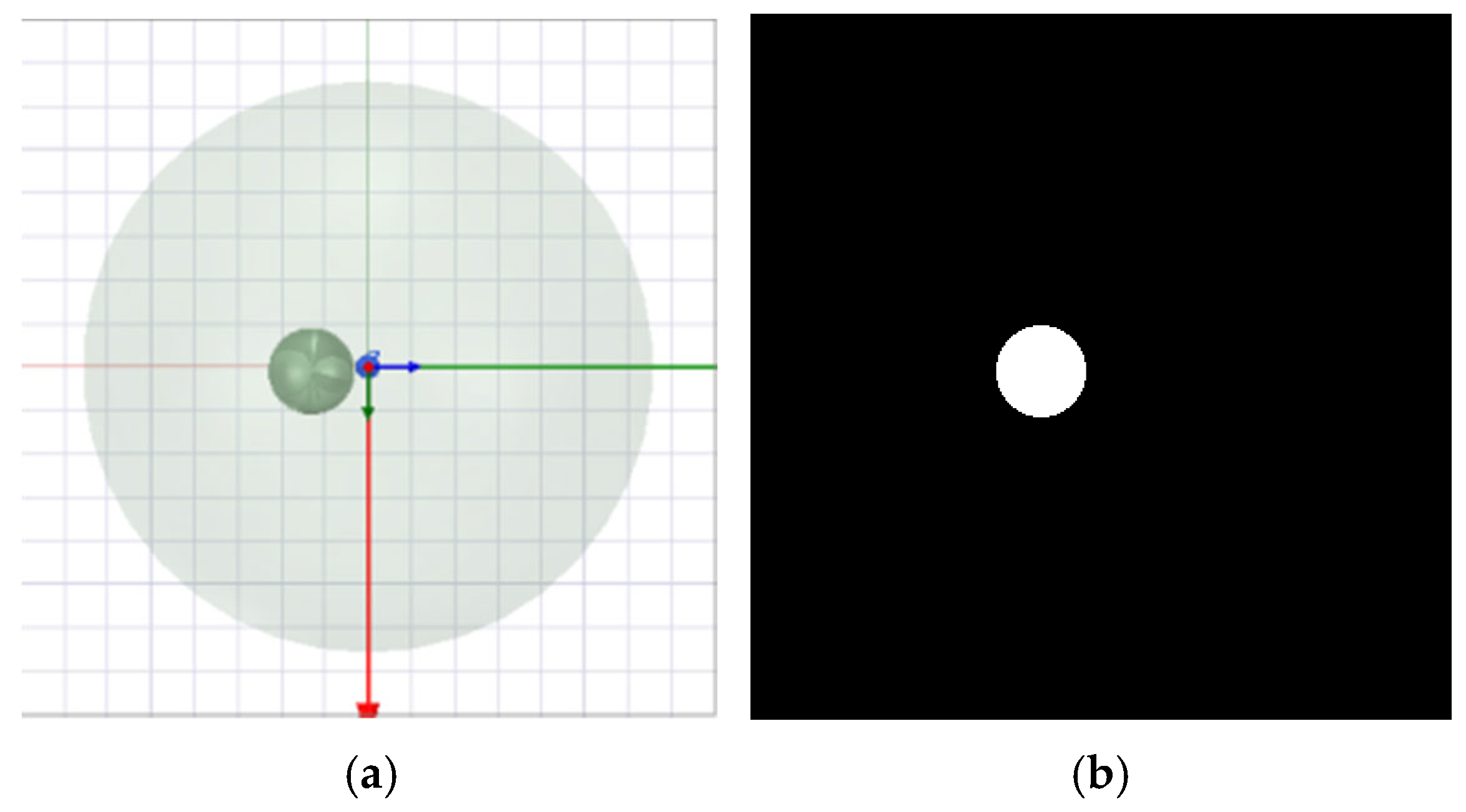
This constructed microwave dataset consists of 624 samples in total, which are split into a training set of 594 samples and a test set of 30 samples. One of the simulation models is a human brain hemorrhage model constructed by the HFSS simulation system [25]; the model mainly includes four modules of plasma, skin, skull, and tissue, while eight antenna arrays are set up at the periphery of the model, in which the innermost tissue module is a 66 mm sphere, as shown in Figure 7. To simulate the actual situation of human brain hemorrhage, the coordinates of the hemorrhage block are randomly generated, where the x, y, and z coordinates represent the different spatial positions of the hemorrhage block in the 3D simulation model, and all three-dimensional coordinates (x, y, z) of the hemorrhage block are between ±30 mm. To ensure that the hemorrhage range is inside the tissue module, the radius size of the hemorrhage block is between 0 and 9 mm. The microwave simulation experiments were carried out through the HFSS simulation system for the bleeding cases with different coordinates and radii, in which the two sets of CSV data simulated by the antenna at the zero-degree angle and the thirty-degree angle, respectively, were subtracted as the input data of the MSA-Net network, and at the same time, the screenshots of the x-y planes corresponding to the three-dimensional model were used as the labeling data of the model, as shown in Figure 8. As can be seen from Figure 8, the labeled images are color images with both hemorrhagic blocks and human brain tissues. In reality, only the size and position of the hemorrhagic blocks change. To eliminate the noise of the label data, the label data annotation tool is used to label the pictures with data to obtain the grayscale map. To meet the requirements of deep learning models for input data format and numerical distribution, it is necessary to perform normalized preprocessing on the original simulation data. First, the CSV file is read and parsed into a numerical matrix. The data matrix is transposed to match the dimensional organization of the network input and then converted into a PyTorch tensor as the network input. Considering the numerical scale differences in microwave measurement data among different samples, this paper adopts the min–max normalization method to uniformly map the input data to the [0, 1] interval in order to reduce the impact of dimensional differences on the stability of model training.

3.3. Comparative Experiment and Ablation Experiment

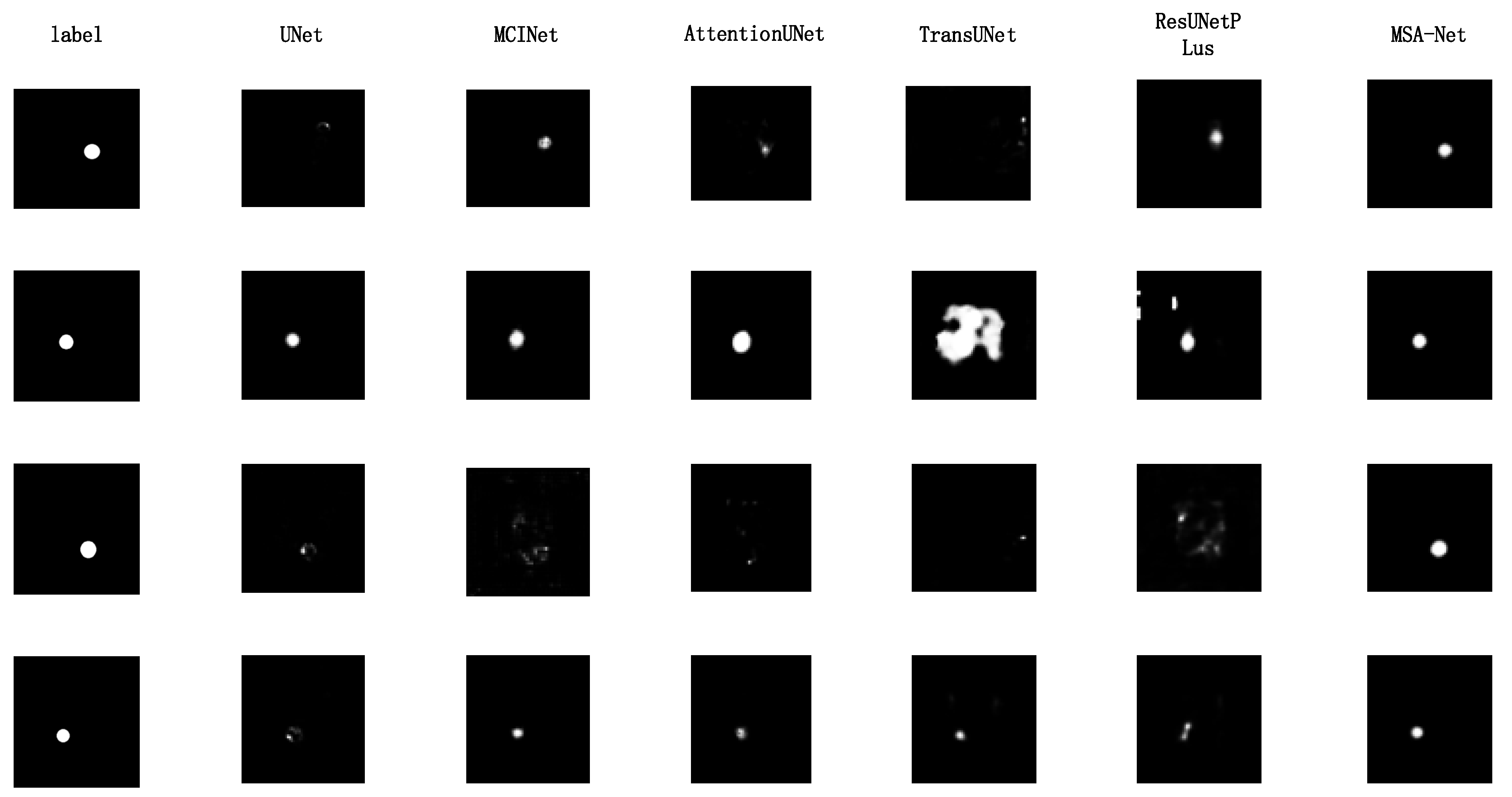
To verify the effectiveness of the MSA-Net network in the microwave data reconstruction task, this study uses UNet, ResUNetPlus, TransUNet, AttentionUNet, and MCINet to test the same dataset [26,27,28,29]. It compares the reconstruction results of these networks with those of MSA-Net, as shown in Figure 9. In these six sets of experiments, the Binary Cross-Entropy Loss (BCELoss) was used as the loss function of the reconstructed networks in this study, ensuring the experimental results’ fairness and comparability. In addition, the RMSprop optimizer was chosen as the optimizer, which has better robustness in dealing with non-smooth targets and gradient oscillations, which helps the network converge quickly and improves training stability. Meanwhile, to further ensure the scientific validity of the experiments and the credibility of the results, this study trains all network models under the same hyperparameter settings. It strictly controls the consistency of the training, validation, and test data. During the training process, each network is trained with the same initial learning rate and weight initialization method, which minimizes the influence of differences in training parameters.
The comparison between the images generated by different networks and the gold standard is shown in Figure 9. From the figure, it can be observed that the MSA-Net proposed in this study outperforms other networks (e.g., TransUNet, AttentionUNet, UNet, ResUNetPlus, and MCINet) in terms of boundary clarity of the stroke region, detail restoration ability, and background noise suppression effect. The images generated by the MSA-Net can more accurately reproduce the brightness distribution and structural features of the target region, with better agreement with the gold standard, while significantly reducing artifacts and enhancing the contrast between the stroke region and the background, further validating its excellent performance in microwave medical imaging tasks.
To quantitatively evaluate the differences in perceptual similarity, structural consistency, and pixel fidelity between microwave imaging reconstruction results and reference images, this study selected Learned Perceptual Image Patch Similarity (LPIPS) and peak signal-to-noise ratio (PSNR) as performance evaluation indicators. Among them, LPIPS is mainly used to measure the difference between reconstructed images and reference images in high-dimensional perceptual feature space, while PSNR is used to reflect the reconstruction error size of the two at the pixel intensity level. The combination of the two can provide a more comprehensive evaluation of microwave imaging reconstruction quality from the perspectives of perceptual features and pixel fidelity.
LPIPS is based on the construction of multi-layer feature representation using pretrained convolutional neural networks, which can simulate the perception mechanism of the human visual system to a certain extent [30]. It not only focuses on the differences in pixel values of images, but also characterizes the consistency of images in terms of contour, boundary, shape, and local structure. This is particularly important in microwave imaging reconstruction tasks, as the core goal of this task is to accurately restore the shape, boundary position, and scale range of the target area, which are often difficult to fully reflect through traditional pixel-level error indicators. In contrast, PSNR is calculated based on the mean square error between the reconstructed image and the reference image, which can measure the overall distortion of the image from a pixel-level perspective [31]. The higher the PSNR value, the closer the reconstructed result is to the reference image. Therefore, LPIPS and PSNR have strong complementarity, with the former focusing more on perceptual consistency and structural details, while the latter emphasizes reconstruction accuracy at the pixel-level.
Specifically, LPIPS extracts feature maps from multiple convolutional layers of a pre-trained network, calculates the differences in corresponding feature blocks between the reconstructed image and the reference image, and weights and fuses the feature distances of each layer to obtain the perceived similarity score. This indicator can sensitively reflect structural differences such as slight deformation of the target contour, boundary position deviation, and scale range changes. The lower the LPIPS value, the higher the consistency between the reconstructed image and the reference image in terms of perceptual features and structural details. PSNR supplements the overall fidelity of the reconstructed results at the pixel level, with higher values indicating smaller image distortion and better reconstruction quality.
This study calculates LPIPS and PSNR for all samples in the test set and takes their average as the final evaluation result to comprehensively and objectively reflect the performance of the proposed method in microwave imaging reconstruction tasks. The calculation formulas for LPIPS and PSNR are shown in Equations (3) and (4), respectively.
L P I P S ( x , y ) = l 1 H l W l h = 1 H l w = 1 W l | | ω l ( l x h , w y h , w ) | | 2 2 ,
where l denotes the layer index of feature extraction, represents the overall luminance distribution of the image; H l and W l denote the height and width of the feature map at the l-th layer, respectively; W l denotes the learnable channel weight vector of the l-th layer, which is optimized during training to adjust the perceptual importance of each feature channel; ⊙ denotes the element-wise multiplication operation; and | | · | | 2 2 denotes the squared L2 norm, which is employed to quantify the magnitude of feature differences.
PSNR = 10 log 10 MAX 2 1 HW i = 1 H j = 1 W I i , j K i , j 2 ,
where I represents the reference image; K represents the reconstructed image; H and W are the height and width of the image, respectively; and MAX represents the maximum possible value of a pixel.
Based on the results in Table 3, MSA-Net demonstrates the best overall performance on the HFSS simulated microwave dataset. In terms of LPIPS, MSA-Net achieves a score of 0.093, which is lower than those of EGE-UNet, TransUNet, AttentionUNet, UNet, ResUNetPlus, and MCINet by 0.031, 0.164, 0.100, 0.085, 0.085, and 0.077, respectively. In terms of PSNR, MSA-Net reaches 23.35 dB, exceeding EGE-UNet, TransUNet, AttentionUNet, UNet, ResUNetPlus, and MCINet by 1.08 dB, 0.49 dB, 0.28 dB, 1.03 dB, 0.41 dB, and 1.22 dB, respectively. These results indicate that MSA-Net is more effective in preserving perceptual similarity and improving pixel-level fidelity, enabling it to reconstruct grayscale images that better reflect the underlying microwave imaging information. The superior performance of MSA-Net can be attributed to its enhanced structural design, including the introduction of the multi-axis Hadamard attention mechanism, the ASPP module, and the deep supervision strategy. Together, these components strengthen multi-scale feature modeling, improve detail recovery, and enhance optimization during training, thereby allowing MSA-Net to outperform the comparison networks in stroke microwave imaging reconstruction.
To evaluate the contribution of each module to overall performance, this article conducted ablation experiments and analyzed the role of each module using a “one-by-one removal” approach by taking the MSA-Net model as the baseline, removing the CBAM, ASPP module, and deep supervision module, respectively, and comparing the changes in model performance under the same training settings and hyperparameter conditions. In the experiment, the reconstruction quality is mainly quantified through two indicators: perceptual loss similarity (LPIPS) and peak signal-to-noise ratio (PSNR), which evaluate the impact of different module stacking on perceptual consistency and pixel fidelity. By analyzing the changes in indicators after removing each module, the specific contributions of each module to perceptual consistency, pixel accuracy, and overall reconstruction performance can be clarified, providing empirical evidence for subsequent model optimization and module design.
According to the ablation experiment results in Table 4, it can be seen that in the task of microwave imaging reconstruction for stroke, each improved module has played a positive role in improving the performance of the model. The baseline model without any modules had an LPIPS of 0.124 and a PSNR of 22.27 dB. After introducing the CBAM attention mechanism, the LPIPS decreased to 0.116 and the PSNR increased to 22.91 dB, indicating that CBAM can enhance the model’s ability to focus on bleeding lesion-related features, suppress background stray information interference, and improve reconstruction quality. After further adding the ASPP module, LPIPS continued to decrease to 0.107, and PSNR increased to 23.10 dB, indicating that ASPP helps enhance the modeling ability of the model for multi-scale lesion information, enabling the network to more fully extract the bleeding features at different scales. Finally, after adding DSM (deep supervision mechanism) on this basis, the model performance reached its optimal level, with LPIPS further reduced to 0.093 and PSNR increased to 23.35 dB. This indicates that the deep supervision mechanism can strengthen the constraint effect in the feature learning process of each layer of the network, promote effective collaboration between shallow and deep features, and thus improve the stability of the model training and reconstruction accuracy. At the same time, the single-round training time of the model has been reduced from 28 s to 24 s, a decrease of 4 s or about 14.29%, indicating that the introduced module can improve reconstruction performance while still maintaining a certain level of training efficiency. Overall, the synergistic effect of CBAM, ASPP, and DSM can effectively improve the lesion localization ability and image reconstruction quality in microwave imaging tasks for stroke, verifying the effectiveness of the proposed improvement strategy.

4. Concluding Remarks

The MSA-Net network proposed in this study, which is based on deep learning, has achieved optimal experimental results in the task of microwave imaging for cerebral stroke. Its imaging performance comprehensively surpasses existing traditional microwave imaging methods and related deep learning approaches. Experimental verification demonstrates that this network effectively achieves precise mapping from microwave data to cerebral stroke images by combining the multi-axis Hadamard attention mechanism (MAHAM), CBAM, ASPP module, and deep supervision mechanism. It not only significantly enhances imaging resolution and the ability to capture subtle lesions but also greatly improves imaging speed and stability. Compared to traditional methods, this approach omits the complex inverse problem-solving process, thoroughly addressing the main points of high computational complexity and unstable imaging results in traditional microwave imaging. It exhibits significant advantages in the accuracy and detail-integrity of cerebral stroke lesion reconstruction. Specifically, it improves the peak signal-to-noise ratio by 0.98, and the learned perceptual image block similarity decreases by 0.017, fully demonstrating the effectiveness and superiority of the MSA-Net network in microwave imaging for cerebral stroke.
The MSA-Net network method proposed in this study possesses significant advantages, but it also has certain limitations. Its core strength lies in the integration of the technical advantages of deep learning, microwave data processing, and image generation, constructing an efficient solution from microwave data to cerebral stroke image reconstruction. Through the improvement of key modules, it achieves simultaneous enhancement in imaging accuracy, speed, and robustness, without requiring complex inverse problem solving, adapting to the practical needs of the early detection of cerebral stroke. Simultaneously, this method provides a feasible approach for the application of deep learning technology in the field of microwave imaging by converting microwave data into a tensor format, exhibiting strong technological innovation. However, this method also has several limitations. First, the model performance is highly dependent on high-quality microwave simulation datasets and clinical data support, and the currently limited data scale may affect its generalization ability in different clinical scenarios. Second, although introducing multiple improved modules enhances imaging performance, it also increases the model’s parameter size and training cost, requiring considerable hardware computing resources. Third, the model performance may degrade to some extent due to domain shift when the background geometry varies, such as slight differences in brain size; thus, retraining or fine-tuning the network on data with new geometries is generally required to maintain stable and accurate reconstruction performance.
To address the limitations of this study, further research efforts should focus on three major directions: data augmentation, model optimization, and clinical implementation. Firstly, the scale of the microwave simulation dataset should be expanded, and more clinical stroke microwave detection data should be collected. By utilizing data augmentation techniques, the diversity and representativeness of the dataset will be enhanced, improving the model’s generalization ability and adapting it to different types and degrees of stroke lesion imaging needs. Secondly, the MSA-Net network structure will be further optimized. While ensuring imaging performance, network parameters will be simplified, training costs will be reduced, and the model’s lightweight level will be improved, making it more suitable for clinical practical application scenarios. Finally, the clinical validation and transformation of this method will be promoted. Clinical trials will be conducted in collaboration with medical institutions to optimize the imaging process and improve the technical scheme. The aim is to apply the MSA-Net network to the clinical diagnosis and adjuvant therapy of early stroke detection, providing more efficient and precise technical support for stroke diagnosis and treatment and promoting the further development of microwave imaging technology in the field of neurological disease diagnosis.

Author Contributions

Conceptualization, methodology, investigation, visualization, and writing—original draft, B.H. and X.Z.; software, validation, formal analysis, data curation, and writing—review, D.L., M.Z. and P.L.; supervision and project administration, B.H. All authors have read and agreed to the published version of the manuscript.

Funding

This work was supported by the Anhui Provincial Natural Science Foundation under Grant No. 2008085MF215, and the Major Scientific Research Project of Colleges and Universities in Anhui Province under Grant No. 2022AH040198.

Institutional Review Board Statement

Not applicable.

Informed Consent Statement

Not applicable.

Data Availability Statement

The original contributions presented in this study are included in the article. Further inquiries can be directed to the corresponding author.

Conflicts of Interest

The authors declare no conflicts of interest.

References

  1. Guo, L.; Nguyen-Trong, N.; AI-Saffar, A.; Stancombe, A.; Bialkowski, K.; Abbosh, A. Calibrated Frequency-Division Distorted Born Iterative Tomography for Real-Life Head Imaging. IEEE Trans. Med. Imaging 2022, 41, 1087–1103. [Google Scholar] [CrossRef] [Scilit] [PubMed]
  2. Semenov, S.Y.; Corfield, D.R. Microwave tomography for brain imaging: Feasibility assessment for stroke detection. Int. J. Antennas Propag. 2008, 2008, 254830. [Google Scholar] [CrossRef] [Scilit]
  3. Pastorino, M.; Randazzo, A. Microwave Imaging Methods and Applications; Artech House: Norwood, MA, USA, 2018. [Google Scholar]
  4. Alkhodari, M.; Zakaria, A.; Qaddoumi, N. Using prior information to enhance microwave tomography images in bone health assessment. Biomed. Eng. OnLine 2022, 21, 8. [Google Scholar] [CrossRef] [Scilit] [PubMed]
  5. Pastorino, M. Microwave Imaging; John Wiley & Sons, Ltd.: Hoboken, NJ, USA, 2010; ISBN 9780470602492. [Google Scholar]
  6. Ahmed, S.S.; Schiessl, A.; Gumbmann, F.; Tiebout, M.; Methfessel, S.; Schmidt, L.-P. Advanced Microwave Imaging. IEEE Microw. Mag. 2012, 13, 26–43. [Google Scholar] [CrossRef] [Scilit]
  7. Shao, W.; Du, Y. Microwave Imaging by Deep Learning Network: Feasibility and Training Method. IEEE Trans. Antennas Propag. 2020, 68, 5626–5635. [Google Scholar] [CrossRef] [Scilit] [PubMed]
  8. Xu, K.; Wu, L.; Ye, X.; Chen, X. Deep Learning-Based Inversion Methods for Solving Inverse Scattering Problems with Phaseless Data. IEEE Trans. Antennas Propag. 2020, 68, 7457–7470. [Google Scholar] [CrossRef] [Scilit]
  9. Zhang, J.; Sharma, R.; Zhang, J.; Babar Abbasi, M.A.; Yurduseven, O. Learning Assisted Sensing Matrix Calculation in Computational Microwave Imaging. In Proceedings of the 2025 19th European Conference on Antennas and Propagation (EuCAP); IEEE: New York, NY, USA, 2025; pp. 1–5. [Google Scholar]
  10. Woo, S.; Park, J.; Lee, J.-Y.; Kweon, I.S. CBAM: Convolutional Block Attention Module. In Proceedings of the Computer Vision—2018; Ferrari, V., Hebert, M., Sminchisescu, C., Weiss, Y., Eds.; Springer International Publishing: Cham, Switzerland, 2018; pp. 3–19. [Google Scholar]
  11. Sunandini, G.; Sivanpillai, R.; Sowmya, V.; Sajith Variyar, V.V. Significance of Atrous Spatial Pyramid Pooling (ASPP) in Deeplabv3+ for Water Body Segmentation. In Proceedings of the 2023 10th International Conference on Signal Processing and Integrated Networks (SPIN); IEEE: New York, NY, USA, 2023; pp. 744–749. [Google Scholar]
  12. Chen, F.; Li, S.; Han, J.; Ren, F.; Yang, Z. Review of Lightweight Deep Convolutional Neural Networks. Arch. Comput. Methods Eng. 2024, 31, 1915–1937. [Google Scholar] [CrossRef] [Scilit]
  13. Wang, B.; Huang, G.; Li, H.; Chen, X.; Zhang, L.; Gao, X. Hybrid CBAM-EfficientNetV2 Fire Image Recognition Method with Label Smoothing in Detecting Tiny Targets. Mach. Intell. Res. 2024, 21, 1145–1161. [Google Scholar] [CrossRef] [Scilit]
  14. Ling, Y.; Wang, Y.; Dai, W.; Yu, J.; Liang, P.; Kong, D. Mtanet: Multi-task attention network for automatic medical image segmentation and classification. IEEE Trans. Med. Imaging 2023, 43, 674–685. [Google Scholar] [CrossRef] [Scilit] [PubMed]
  15. Ruan, J.; Xie, M.; Gao, J.; Liu, T.; Fu, Y. EGE-UNet: An Efficient Group Enhanced UNet for Skin Lesion Segmentation. In Proceedings of the Medical Image Computing and Computer Assisted Intervention—MICCAI 2023; Greenspan, H., Madabhushi, A., Mousavi, P., Salcudean, S., Duncan, J., Syeda-Mahmood, T., Taylor, R., Eds.; Springer Nature: Cham, Switzerland, 2023; pp. 481–490. [Google Scholar]
  16. Du, Y.; Hou, S.; Li, G. Detection Method of Maize Kernel Impurity Based on Deep Learning. J. Agric. Mech. Res. 2025, 47, 1–8. [Google Scholar] [CrossRef]
  17. Wang, Q.; Wu, B.; Zhu, P.; Li, P.; Zuo, W.; Hu, Q. ECA-Net: Efficient channel attention for deep convolutional neural networks. In Proceedings of the IEEE/CVF Conference on Computer Vision and Pattern Recognition(CVPR); IEEE: New York, NY, USA, 2020; pp. 11534–11542. [Google Scholar]
  18. Chollet, F. Xception: Deep Learning with Depthwise Separable Convolutions. In Proceedings of the 2017 IEEE Conference on Computer Vision and Pattern Recognition (CVPR); IEEE: New York, NY, USA, 2017; pp. 1800–1807. [Google Scholar]
  19. Wang, Y.; Zhang, H.; Fu, J.; Tian, H. MSCB-unet: Elevating skin lesion segmentation performance with multi-scale spatial-channel bridging network. Biomed. Signal Process. Control. 2025, 110, 107986. [Google Scholar] [CrossRef] [Scilit]
  20. Liu, J.; Liang, T.; Peng, Y.; Peng, G.; Sun, L.; Li, L.; Dong, H. Segmentation of Acetowhite Region in Uterine Cervical Image Based on Deep Learning. Technol. Health Care 2022, 30, 469–482. [Google Scholar] [CrossRef] [Scilit] [PubMed]
  21. Cheng, B.; Misra, I.; Schwing, A.G.; Kirillov, A.; Girdhar, R. Masked-Attention Mask Transformer for Universal Image Segmentation. In Proceedings of the 2022 IEEE/CVF Conference on Computer Vision and Pattern Recognition (CVPR); IEEE: New York, NY, USA, 2022; pp. 1280–1289. [Google Scholar]
  22. Pan, X.; Ge, C.; Lu, R.; Song, S.; Chen, G.; Huang, Z.; Huang, G. On the Integration of Self-Attention and Convolution. In Proceedings of the 2022 IEEE/CVF Conference on Computer Vision and Pattern Recognition (CVPR); IEEE: New York, NY, USA, 2022; pp. 805–815. [Google Scholar]
  23. Ma, Q.; Xie, L.; Peng, L. Application of Improved Convolutional Neural Network in Medical Image Segmentation. Laser Optoelectron. Prog. 2020, 57, 141020. [Google Scholar] [CrossRef] [Scilit]
  24. Han, B.; Wang, S.; Shi, X. Design of Compact Fragment-Type Antenna Array for Microwave-Based Head Imaging Application. J. Electromagn. Eng. Sci 2023, 23, 344–350. [Google Scholar] [CrossRef] [Scilit]
  25. Ronneberger, O.; Fischer, P.; Brox, T. U-Net: Convolutional Networks for Biomedical Image Segmentation. In Proceedings of the Medical Image Computing and Computer-Assisted Intervention—MICCAI 2015; Navab, N., Hornegger, J., Wells, W.M., Frangi, A.F., Eds.; Springer International Publishing: Cham, Switzerland, 2015; pp. 234–241. [Google Scholar]
  26. Jha, D.; Smedsrud, P.H.; Riegler, M.A.; Johansen, D.; Lange, T.D.; Halvorsen, P.; Johansen, H.D. ResUNet++: An Advanced Architecture for Medical Image Segmentation. In Proceedings of the 2019 IEEE International Symposium on Multimedia (ISM); IEEE: New York, NY, USA, 2019; pp. 225–2255. [Google Scholar]
  27. Chen, J.; Lu, Y.; Yu, Q.; Luo, X.; Adeli, E.; Wang, Y.; Lu, L.; Yuille, A.L.; Zhou, Y. TransUNet: Transformers Make Strong Encoders for Medical Image Segmentation. arXiv 2021, arXiv:2102.04306. [Google Scholar] [CrossRef] [Scilit]
  28. Oktay, O.; Schlemper, J.; Folgoc, L.L.; Lee, M.J.; Heinrich, M.P.; Misawa, K.; Mori, K.; McDonagh, S.G.; Hammerla, N.Y.; Kainz, B.; et al. Attention U-Net: Learning Where to Look for the Pancreas. arXiv 2018. [Google Scholar] [CrossRef] [Scilit]
  29. Xie, X.; Pan, X.; Shao, F.; Zhang, W.; An, J. MCI-Net: Multi-Scale Context Integrated Network for Liver CT Image Segmentation. Comput. Electr. Eng. 2022, 101, 108085. [Google Scholar] [CrossRef] [Scilit]
  30. Wu, J.; Zhang, G.; Fan, Y. LM-CycleGAN: Improving underwater image quality through learned perceptual image patch similarity and multi-scale adaptive fusion attention. Sensors 2024, 24, 7425. [Google Scholar] [CrossRef] [Scilit] [PubMed]
  31. Sara, U.; Akter, M.; Uddin, M.S. Image quality assessment through FSIM, SSIM, MSE and PSNR—A coMPArative study. J. Comput. Commun. 2019, 7, 8–18. [Google Scholar] [CrossRef]
Figure 1. Technical flowchart.
Algorithms 19 00276 g001
Figure 2. MSA-Net network structure diagram.
Algorithms 19 00276 g002
Figure 3. GH-CBAM attention.
Algorithms 19 00276 g003
Figure 4. Group aggregation bridge module.
Algorithms 19 00276 g004
Figure 5. ASPP module structure diagram.
Algorithms 19 00276 g005
Figure 6. Deep supervision mechanism.
Algorithms 19 00276 g006
Figure 7. HFSS simulation system to build a 3D simulation model of human brain stroke [25].
Algorithms 19 00276 g007
Figure 8. Dataset mask map: (a) is a color image of the dataset mask map with stroke regions and human brain tissue; (b) is a grayscale image of the dataset mask map intended to remove noise from the labeled data.
Algorithms 19 00276 g008
Figure 9. Comparison of stroke images reconstructed by different networks.
Algorithms 19 00276 g009
Table 1. Review table.
Research FocusMethodKey FeaturesAdvantagesLimitationsReferences
Microwave imagingTomographic imagingModels as an inverse problemMature, widely usedIll-posedness; sensitive to noise[1]
Ill-posednessIterative/regularizationMitigates instabilityImproves robustnessHigh computation[2,3,4]
Data-driven reconstructionDeep learning (HFSS data)Maps measurements to imagesFast, handles nonlinearityNeeds high-quality data[5,6]
Microwave imaging based on deep learningAttention networksUsing attention mechanism to model the correlation between featuresEnhance the expression ability of key features and improve the accuracy of object detection and segmentationIncreasing model complexity may result in computational overhead and training difficulty[7,8]
Deep supervisionMulti-level lossExtra training signalFaster convergence, stableExtra computation[9]
Table 2. Dielectric properties of head model tissues at 2–4 GHz.
TissueRelative Permittivit (εr)Conductivity (σ)
Skin241.6
Skull120.16
Brain5.20.15
Blood611.58
Conventional10
Table 3. Comparison of different networks.
ModelLPIPSPSNR
UNet0.17822.32
MCINet0.17022.13
Attention0.19323.07
TransUNet0.25722.86
ResUNetPLus0.17822.94
EGE-UNet0.12422.27
MSA-Net0.09323.35
Table 4. The impact of different modules on model performance.
CBAMASPPDSMLPIPSPSNR
×××0.12422.27
××0.11622.91
×0.10723.10
0.09323.35
Disclaimer/Publisher’s Note: The statements, opinions and data contained in all publications are solely those of the individual author(s) and contributor(s) and not of MDPI and/or the editor(s). MDPI and/or the editor(s) disclaim responsibility for any injury to people or property resulting from any ideas, methods, instructions or products referred to in the content.