Communication

Tunable Emission Peak Position and Enhanced Thermal Stability of CsPbBr3 Quantum Dots via TMCS Ligand Exchange


State Key Laboratory of Advanced Technology for Materials Synthesis and Processing, International School of Materials Science and Engineering (ISMSE), Wuhan University of Technology, Wuhan 430070, China
*
Author to whom correspondence should be addressed.
These authors contributed equally to this work.
Materials 2026, 19(9), 1860; https://doi.org/10.3390/ma19091860
Submission received: 21 March 2026 / Revised: 22 April 2026 / Accepted: 29 April 2026 / Published: 1 May 2026

Abstract

All-inorganic lead halide perovskite quantum dots (QDs), featuring high photoluminescence quantum yield, narrow full width at half maximum, and solution processability, show great promise for high-color-purity displays and optoelectronic devices. Their emission peak position and stability are highly dependent on the surface coordination environment, and achieving controllable color tuning while maintaining stability without altering the primary synthetic route remains a critical challenge. Herein, we propose a facile solution-phase post-treatment strategy using TMCS, which can react with the oleate ligands on the CsPbBr3 QD surface while providing abundant Cl ions, thereby leading to partial halide exchange, achieving continuous tuning of the emission wavelength from 499 nm to 473 nm. The appearance of new absorption peaks in the FTIR spectra indicated the successful introduction of TMCS and the in situ generation of HCl, which led to surface etching and passivation. After being heated at 40 °C for 6 h, the TMCS-50 sample retained 39% of its initial photoluminescence intensity, while the pristine CsPbBr3 QD sample retained only 8%, demonstrating that TMCS treatment significantly improves the thermal stability of the CsPbBr3 QDs.

1. Introduction

All-inorganic lead halide perovskite quantum dots (QDs) have attracted extensive interest for displays [1,2,3], lighting [4,5,6], and photodetectors [7,8,9] due to their high PLQY, narrow full width at half maximum (FWHM), and solution processability [10]. Compared with conventional II-VI quantum dots, CsPbBr3 QDs can deliver “purer” visible emission and broader color-gamut coverage [11]. Meanwhile, influenced by nanoscale quantum-confinement effects and surface chemistry, their emission performance and peak position are highly sensitive to particle size, composition, and the surface ligand environment [12,13,14,15]. Developing a simple and controllable approach to tune the emission wavelength without changing the primary synthetic route, while simultaneously improving stability, remains a key challenge for practical applications of perovskite quantum dots.
At present, emission-wavelength tuning of CsPbBr3 QDs is mainly achieved through halide-composition engineering (Br/Cl or Br/I exchange) [16], size regulation by controlling reaction temperature and time, or post-treatment-induced surface reconstruction via ligand introduction [17,18]. Among these strategies, post-treatment has received growing attention because it is mild, causes minimal perturbation to the original system, and is amenable to scale-up and process integration [19]. Ligand exchange-based surface passivation can directly modulate the surface coordination environment [20,21], mitigate defect states and local lattice strain [22,23], and thereby alter the band structure and exciton recombination processes, which ultimately manifests as coupled changes in emission peak position and stability [24,25,26]. However, commonly used long-chain amine ligands [27,28], although beneficial for colloidal stability [29], hinder charge transport and suffer from dynamic desorption [30,31]. Trivalent metal chloride doping has been confirmed to effectively passivate the perovskite surface and enhance its stability [32]. Accordingly, introducing post-treatment ligands with higher reactivity and stronger surface-regulation capability remains of research value.
On this basis, we propose a simple solution-phase post-treatment strategy that broadens the tunable emission range while enhancing stability. With the gradual increase in the amount of TMCS dilute solution added, continuous tuning of the emission wavelength was achieved from 499 nm (TMCS-0) to 480 (TMCS-25), 477 (TMCS-50), 475 (TMCS-75), and 473 nm (TMCS-100). Subsequent analyses by XRD and transmission electron microscopy demonstrated that the morphology and crystal structure of the quantum dots remained unchanged after TMCS treatment. In addition, the appearance of new absorption bands in the FTIR spectra indicated the successful introduction of TMCS and the in situ generation of HCl, which resulted in the etching and passivation of oleic acid ligands on the quantum dot surface. Thermal stability tests further confirmed that TMCS treatment can greatly enhance the thermal stability of the quantum dots.

2. Materials and Methods

2.1. Methods

2.1.1. Chemicals

Cesium carbonate (Cs2CO3; Aladdin; 99.9%), lead (II) bromide (PbBr2; Aladdin; 99.99%), zinc bromide (ZnBr2; Aladdin; 99.99%), trimethylchlorosilane (TMCS; Aladdin; 99%), oleic acid (OA; Aladdin; AR), oleylamine (OAm; Aladdin; 80–90%), 1-octadecene (ODE; Aladdin; >90%), and n-hexane (Aladdin; >99%) were used as received. Unless otherwise specified, reagents labeled “Aladdin” were purchased from Aladdin Reagent (Shanghai) Co., Ltd. (Shanghai, China), and reagents labeled “Macklin” were obtained from Shanghai Macklin Biochemical Co., Ltd. (Shanghai, China).

2.1.2. CsPbBr3 Synthesis and Post-Synthetic Ligand Exchange

PbBr2 (0.2760 g) and ZnBr2 (0.6712 g) were dissolved in a mixed solution containing 30 mL of ODE, 6 mL of OA, and 6 mL of OAm. The resulting solution was degassed under vacuum at 120 °C for 30 min, then switched to a nitrogen atmosphere and heated to 170 °C. A total of 1.6 mL Cs-OA precursor solution was rapidly injected, and the reaction was allowed to proceed for 30 s. The mixture was then quenched by an ice-water bath and cooled to room temperature. The crude nanocrystal dispersion was centrifuged at 10,000 rpm for 10 min, and the supernatant was collected. Excess acetone was added to the supernatant to induce precipitation, followed by centrifugation at 10,000 rpm for 10 min. The precipitate was collected, redispersed in 2 mL of n-hexane, and aliquoted equally into five vials for subsequent ligand exchange. For ligand exchange, a TMCS stock solution was prepared by diluting 0.5 mL of TMCS in 4.5 mL of ODE. Aliquots of 25, 50, 75, and 100 μL of the diluted TMCS solution were added to separate vials of the CsPbBr3 QD dispersion and stirred at room temperature for 2 min to complete the ligand exchange process.

2.2. Characterization

2.2.1. Transmission Electron Microscopy (TEM)

TEM images were collected using a JEOL JEM-1400 Plus transmission electron microscope (Tokyo, Japan) operated at 120 kV. Samples were prepared by drop-casting a diluted n-hexane suspension onto carbon-coated copper grids.

2.2.2. X-Ray Diffraction (XRD)

XRD patterns were collected on a Bruker D8 ADVANCE diffractometer (Karlsruhe, Germany) using Cu Kα radiation (λ = 1.54056 Å) at 40 kV and 40 mA. Samples were prepared by drop-casting a concentrated n-hexane suspension onto SiO2/Si substrates.

2.2.3. Fourier Transform Infrared (FTIR) Spectroscopy

FTIR spectra were recorded at room temperature using a Nicolet 6700 FTIR spectrometer (Thermo Fisher Scientific, Madison, WI, USA). Measurements were collected over 500–4000 cm−1. Samples were prepared by drop-casting a highly concentrated n-hexane dispersion of CsPbBr3 nanocrystals onto a glass slide, drying under ambient conditions, and then measuring.

2.2.4. Fluorescence Spectrum Measurements

UV–vis absorption spectra were recorded on a Shimadzu UV-1800 spectrophotometer (Kyoto, Japan). Photoluminescence (PL) spectra were measured using a Shimadzu RF-6000 spectrofluorometer (Kyoto, Japan) with excitation wavelengths of 410 nm.

3. Results

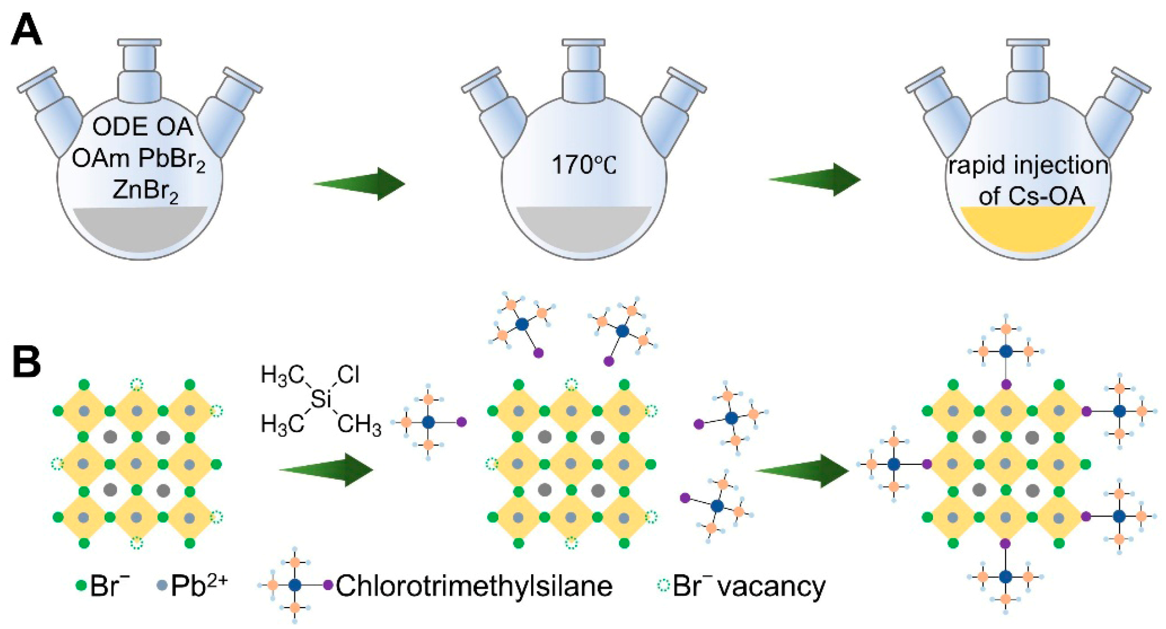
CsPbBr3 QDs were synthesized using a hot-injection method (Figure 1A). During the synthesis, excess ZnBr2 was introduced to create a halide-rich environment, thereby regulating the size and improving the synthetic quality of the quantum dots. In the hot-injection process, [PbBr6]4− octahedra are first formed in solution. After the Cs precursor is injected, Cs+ ions rapidly coordinate with the [PbBr6]4− octahedra to generate the CsPbBr3 structure. When the halide content in the solution is only three times that of Pb2+, the formation of [PbBr6]4− octahedra may be insufficient, which can result in halide vacancies. By increasing the halide content in the solution, the formation of [PbBr6]4− octahedra is promoted, thus improving the synthesis quality of the CsPbBr3 QDs.
To enable controllable post-synthetic tuning of the emission properties, we further implemented a solution-phase TMCS treatment for ligand exchange modification (Figure 1B). Briefly, TMCS was first diluted with ODE. Different volumes of the diluted TMCS solution were then added to the CsPbBr3 QD dispersion in n-hexane, followed by stirring at room temperature. The samples were denoted as TMCS-0, TMCS-25, TMCS-50, TMCS-75, and TMCS-100 according to the amount of TMCS solution added. After treatment, the dispersions were stored in sealed vials for subsequent structural and optical characterization as well as thermal-stability measurements.
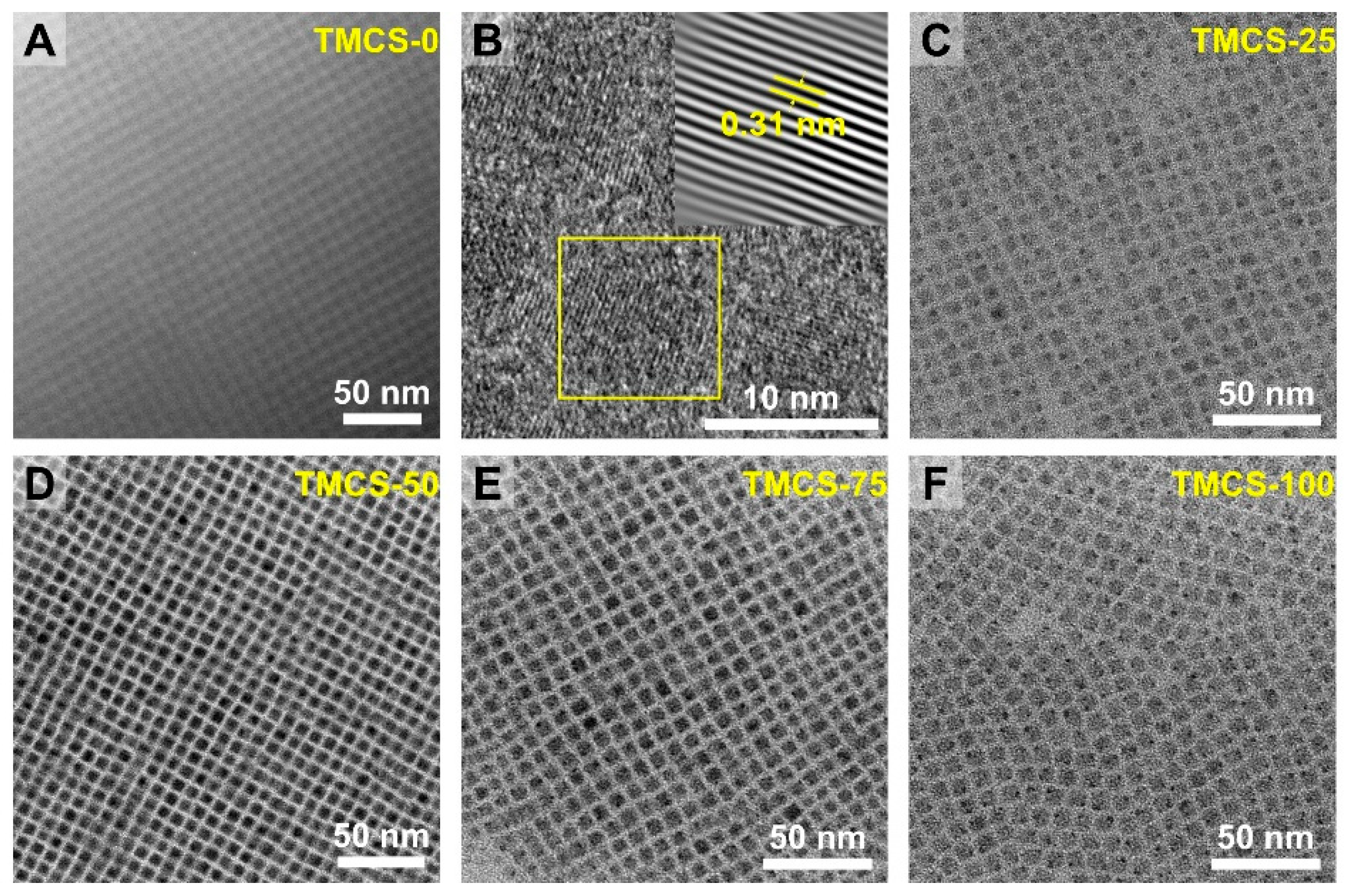
Figure 2 shows the TEM images of the samples before and after treatment. All samples exhibit a cubic structure, and no obvious morphological change is observed after TMCS treatment. Furthermore, we analyzed the size distribution of all samples and found a slight increase in size after TMCS treatment (Figure S1). In particular, the average size of TMCS-75 (Figure S1D) is about 0.4 nm larger than that of the untreated quantum dots, which may be attributed to the passivation of surface defects by TMCS, leading to an increase in the surface thickness.
In the high-resolution image of the TMCS-0 sample, typical features of the cubic phase are observed, with an interplanar spacing of 0.31 nm. In addition, varying degrees of ordered packing can be seen, where QDs in certain local regions form quasi-2D and 3D periodic assemblies. Such periodic packing corresponds to self-assembled superstructures (often referred to as superlattices), which can form when a high-concentration QD dispersion is dried at room temperature as solvent evaporation and interparticle interactions drive organization. Various superlattice configurations of perovskite nanocrystals have been reported, and their formation is closely related to factors such as particle size distribution, the thickness and flexibility of the surface ligand layer, and concentration.
Notably, higher packing regularity is visible in Figure 2A,D,E, appearing brighter and more periodic than in Figure 2C,F. This difference can arise from multiple factors. First, the self-assembly process is inherently stochastic; variations in local concentration and solvent evaporation rate across different regions can lead to fluctuations in ordering. Second, the uniformity of the QD size distribution strongly affects assembly quality, and more monodisperse particles more readily form long-range ordered arrays. Third, an increased stacking thickness along the electron-beam direction enhances projection contrast, making the periodicity appear more ordered. The dark dots in panels C and F are attributed to Pb precipitation induced by prolonged electron-beam irradiation. It should be emphasized that the differences in ordering observed here mainly reflect local self-assembly behavior and are not directly associated with changes in the parent crystal phase. Considering that all samples retain a cubic morphology and good dispersibility, we conclude that the effect of TMCS post-treatment on CsPbBr3 QDs primarily occurs at the surface ligand level rather than causing pronounced morphological or structural damage. Because of the small size of the quantum dots, the diffraction peaks are relatively broad (Figure S2A). The peak located at 32.97° is indexed to the (002) plane. After TMCS treatment, partial halide exchange from Br to Cl takes place. Owing to the smaller ionic radius of Cl, the interplanar spacing is reduced, causing the diffraction peak to shift to a higher diffraction angle (Figure S2B).
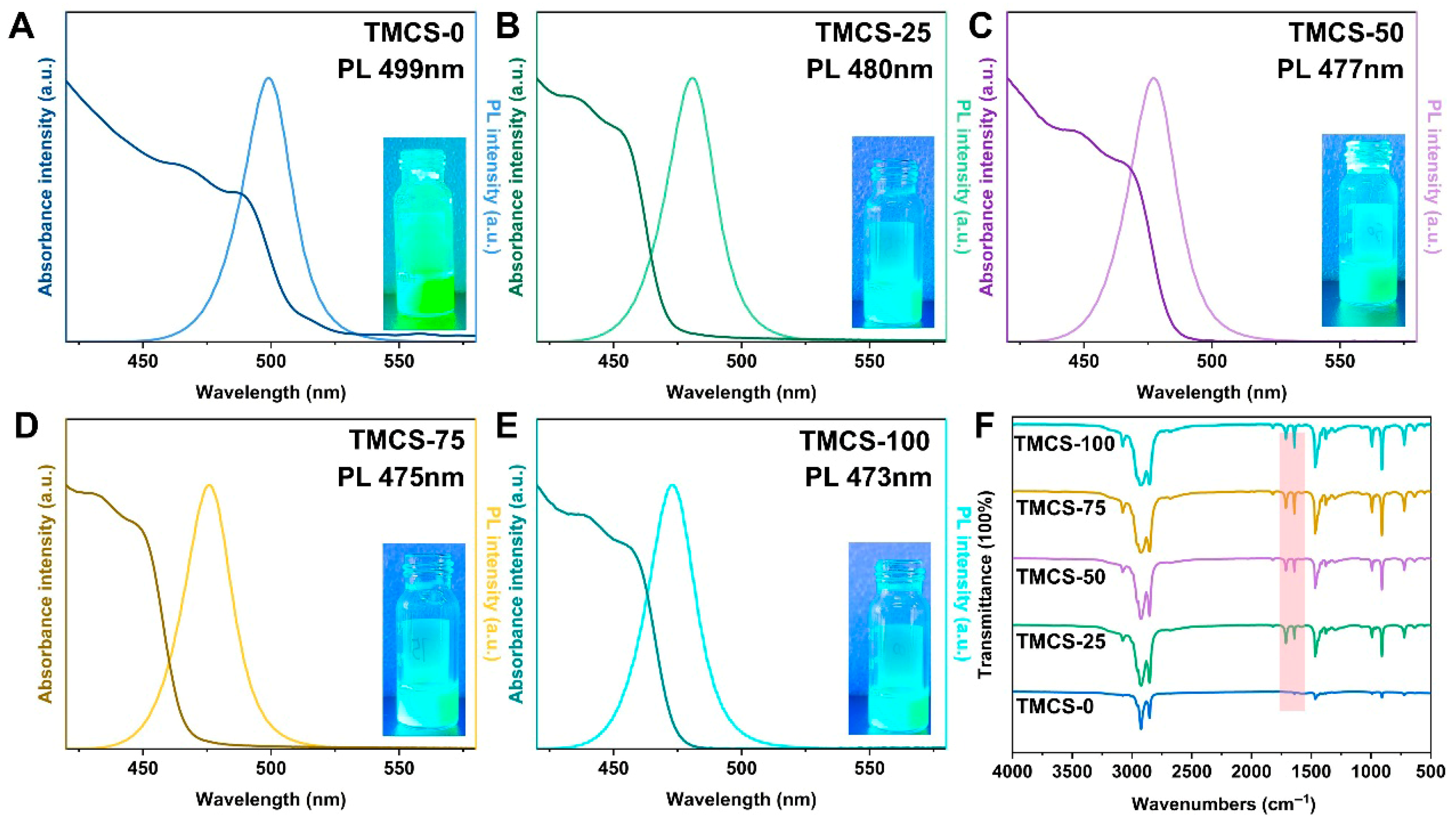
All samples exhibit narrow PL and UV–vis absorption features, indicative of small emission linewidths and thus high color purity (Figure 3A–E). With increasing ligand dosage, the emission peak shows a clear ligand-molarity dependence: the PL maximum continuously shifts from 499 nm to 473 nm as the TMCS amount increases, demonstrating a systematic and monotonic wavelength modulation. The inset photograph shows the corresponding quantum dot solutions under UV illumination, in good agreement with the PL emission behavior. The UV–vis absorption peak of the TMCS-0 sample is centered at 489 nm, with a Stokes shift of 10 nm. Following TMCS treatment, a blue shift of the absorption peak is observed, together with a further increase in the Stokes shift. These changes may be related to halide exchange or surface dielectric modification; however, the detailed mechanism is still not fully understood.
To further verify the surface-chemical changes induced by TMCS post-treatment, FTIR spectroscopy was performed on samples prepared with different TMCS dosages (Figure 3F). Compared with the pristine sample (TMCS-0), the treated samples display new absorption bands primarily located at 1820 cm−1, 1260 cm−1 and 850 cm−1, and their intensities progressively increase with increasing TMCS addition. The absorption peak at 1820 cm−1 lies in the characteristic region of acyl chlorides and is assigned to RC(=O)Cl (1800–1850 cm−1), indicating that TMCS reacts with oleic acid to generate HCl in situ (Figure 3F). Owing to the low TMCS content in the samples, the characteristic Si-CH3-related absorption bands are much weaker than those of oleic acid and oleylamine and can only be clearly identified in the enlarged spectrum (Figure S3). The peaks at 1260 cm−1 and 850 cm−1 (Figure S3) are attributed to the symmetric deformation vibration of Si-CH3 and the Si-C or Si-CH3 rocking/stretching-related vibrations (850–750 cm−1), respectively, further confirming the successful introduction of TMCS. Moreover, the intensities of these peaks gradually increase with increasing TMCS content.
Meanwhile, characteristic bands at 2853 cm−1, 1641 cm−1, 1467 cm−1, 992 cm−1, 909 cm−1, and 721 cm−1 become stronger overall. Specifically, the bands at 2853 cm−1 and 1467 cm−1 correspond to the stretching and bending vibrations of long-chain alkyl CH2 groups, respectively, and the band at 721 cm−1 is assigned to the rocking vibration of long-chain (CH2) segments. The bands at 1641 cm−1, 992 cm−1, and 909 cm−1 are associated with vibrations of unsaturated bonds (C=C) and =C-H modes. Collectively, these spectral changes indicate that TMCS post-treatment markedly alters the organic coordination environment at the quantum dot surface, consistent with ligand exchange, re-coordination, and reconstruction of the surface organic layer. Notably, at higher TMCS dosages, stronger byproduct-related signatures may emerge, which could be detrimental to surface passivation.
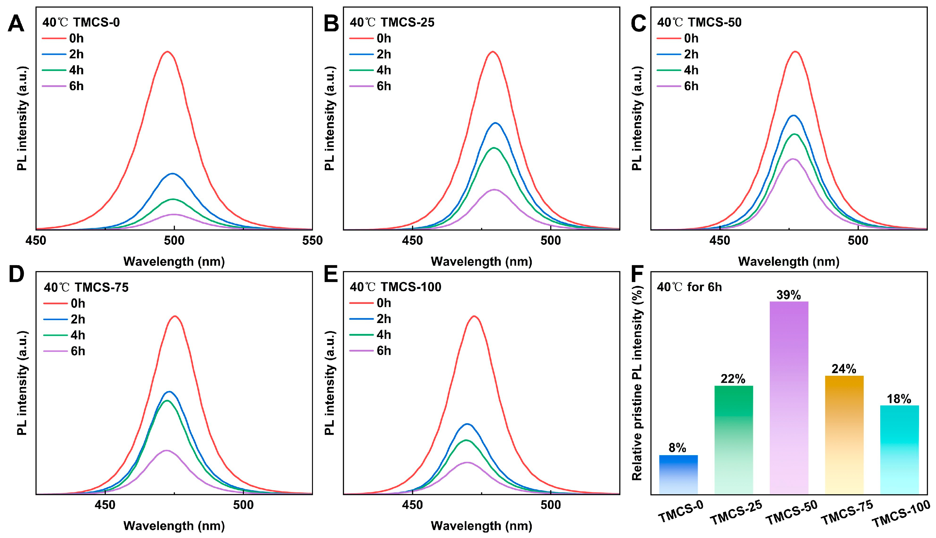
To evaluate the influence of TMCS post-treatment on the thermal stability of the samples, the QDs were dispersed in n-hexane and maintained on a heating plate at 40 °C for thermal-stability testing (Figure 4). With prolonged heating time, the PL intensities of all samples showed a pronounced decline. After heating for 6 h, the PL intensity of the untreated sample remained at only 8% of its initial value, whereas all TMCS-treated samples exhibited markedly improved PL intensity retention (Figure 4F). Notably, TMCS-50 retained 39% of its initial PL intensity after 6 h of heating, indicating the best thermal stability among all samples. Additionally, the quantum dot solutions were spin-coated onto silicon substrates, and photographs of their luminescence under 365 nm UV irradiation at different temperatures were recorded (Figure S4). The untreated sample showed no visible emission at 140 °C, whereas the TMCS-treated samples retained bright luminescence at higher temperatures. In particular, the TMCS-25, TMCS-50, and TMCS-75 samples still exhibited weak emission at 180 °C. Notably, the thermal stability of TMCS-100 was lower than that of TMCS-50, in good agreement with the quantitative results in Figure 4. This behavior may be associated with an excessive amount of TMCS. FTIR results suggest that TMCS can react with oleic acid to generate HCl in situ, and excess HCl may further deteriorate the quantum dots. However, the exact mechanism remains unclear and requires further detailed characterization. Overall, an appropriate TMCS treatment significantly enhances the thermal stability of the QDs.

4. Discussion

In this work, a TMCS solution post-treatment strategy was developed to modify the surface of CsPbBr3 QDs using reactive ligands, thereby achieving the synergistic enhancement of their photoluminescent properties and thermal stability. TMCS can react with oleic acid to generate HCl in situ, which subsequently etches and passivates the quantum dot surface. This observation further suggests that TMBS and TMIS may also induce similar surface etching and passivation effects. When the halide species introduced by the ligand is the same as that originally present in the quantum dots, such post-treatment ligand exchange is expected to passivate surface defects and improve the stability of the quantum dots. By contrast, when the halide species in the added ligand differs from that of the quantum dots, partial halide exchange can be achieved. In addition, the newly introduced halide is closely associated with the ligand, which may further enhance the phase stability of mixed-halide quantum dots and suppress phase segregation under operational conditions.

Supplementary Materials

The following supporting information can be downloaded at: https://www.mdpi.com/article/10.3390/ma19091860/s1, Figure S1: Size distribution statistics for (A) TMCS-0, (B) TMCS-25, (C) TMCS-50, (D) TMCS-75, and (E) TMCS-100; Figure S2: XRD patterns of the quantum dots; Figure S3: Enlarged view of the FTIR spectra; Figure S4: Photographs of the QDs emitting under UV illumination at different temperatures.

Author Contributions

Y.L. (Yong Liu) conceived the project and material synthetic design. C.P., Y.F., Z.S. and Z.P. performed the experiments, sample testing, data collection, and analysis. S.R., X.W., Y.L. (Yingfei Liu), J.Q., K.H. and X.H. jointly completed the methodological design, software analysis, and visualization analysis. C.P. discussed the data and performed formal analyses. C.P. and Y.L. (Yong Liu) wrote, reviewed, and edited the manuscript. All authors have read and agreed to the published version of the manuscript.

Funding

This work is financially supported by the Major Program of the National Natural Science Foundation of China (22293021, Y.L.).

Data Availability Statement

The original contributions presented in this study are included in the article/Supplementary Materials. Further inquiries can be directed to the corresponding author.

Acknowledgments

We are grateful to the State Key Laboratory of Advanced Technology for Materials Synthesis and Processing for providing UV–vis and PL characterization and to the Center for Materials Research and Analysis of Wuhan University of Technology Materials for FTIR testing.

Conflicts of Interest

The authors declare no conflicts of interest.

References

  1. Lv, X.; Su, Y.; Xiang, H.; Yang, L.; Chen, X.; Wang, Y.; Zhang, K.; Tang, J.; Ye, Y.; Cai, B.; et al. TOP-Zn Steric Hindrance Effect Enables Ultra-Uniform CsPbX3 Quantum Dots for Wide-Color Gamut Displays. Adv. Mater. 2025, 37, 2409308. [Google Scholar] [CrossRef]
  2. Lin, J.; Lu, Y.; Li, X.; Huang, F.; Yang, C.; Liu, M.; Jiang, N.; Chen, D. Perovskite Quantum Dots Glasses Based Backlit Displays. ACS Energy Lett. 2021, 6, 519–528. [Google Scholar] [CrossRef]
  3. Fan, X.; Wang, S.; Yang, X.; Zhong, C.; Chen, G.; Yu, C.; Chen, Y.; Wu, T.; Kuo, H.-C.; Lin, Y.; et al. Brightened Bicomponent Perovskite Nanocomposite Based on Förster Resonance Energy Transfer for Micro-LED Displays. Adv. Mater. 2023, 35, 2300834. [Google Scholar] [CrossRef]
  4. Chen, J.; Liu, X.; Cai, B.; Cheng, Y.; Wen, H.; Zhu, D.; Xiong, Y.; Dai, L.; Yan, X.; Xiang, B.; et al. Lattice-matched molecular-anchor design for high-performance perovskite quantum dot light-emitting diodes. Nat. Commun. 2025, 16, 8201. [Google Scholar] [CrossRef]
  5. Wei, K.; Zhou, T.; Jiang, Y.; Sun, C.; Liu, Y.; Li, S.; Liu, S.; Fu, X.; Hu, C.; Tian, S.; et al. Perovskite heteroepitaxy for high-efficiency and stable pure-red LEDs. Nature 2025, 638, 949–956. [Google Scholar] [CrossRef]
  6. Xu, Y.; Guo, W.; Yao, J.; Xu, L.; Wang, J.; Yang, Z.; Wang, S.; Song, J. Multisite Cross-Linked Ligand Suppressing Ion Migration for Efficient and Stable CsPbBr3 Perovskite Quantum Dot-Based Light-Emitting Diodes. Angew. Chem. Int. Ed. 2025, 64, e202422823. [Google Scholar] [CrossRef]
  7. Gao, D.; Yang, Y.; Zhou, X.; Sun, Y.; Miao, W.; Liu, D.; Li, W.; Wang, T. CsPbI2Br quantum dots integration for high performance organic photovoltaics and photodetectors. Energy Environ. Sci. 2025, 18, 3288–3295. [Google Scholar] [CrossRef]
  8. Wang, Y.; Xu, B.-T.; Song, Z.-Y.; Liu, W.-Z.; Shen, W.-S.; Zhou, D.-Y.; Wang, Y.-K.; Liao, L.-S. Dipole Regulation Enables Efficient Near-Infrared Perovskite Quantum Dots Photodetector. Adv. Funct. Mater. 2026, 36, e14588. [Google Scholar] [CrossRef]
  9. Sun, H.; Li, C.; Li, L.; Zhang, X.; Luo, J.; Ahmad Rasid, N.N.b.; Mohamad Fadil, N.W.S.b.; Vasilopoulou, M.; Rashid bin Mohd Yusoff, A. The development of customized perovskite photodetectors. Nat. Electron. 2025, 8, 1170–1181. [Google Scholar] [CrossRef]
  10. Feng, Y.; Li, H.; Zhu, M.; Gao, Y.; Cai, Q.; Lu, G.; Dai, X.; Ye, Z.; He, H. Nucleophilic Reaction-Enabled Chloride Modification on CsPbI3 Quantum Dots for Pure Red Light-Emitting Diodes with Efficiency Exceeding 26 %. Angew. Chem. Int. Ed. 2024, 63, e202318777. [Google Scholar] [CrossRef]
  11. Li, P.; Xie, W.; Mao, W.; Tian, Y.; Huang, F.; Xu, S.; Zhang, J. A new whole family perovskites quantum dots (CsPbX3, X=Cl, Br, I) phosphate glasses with full spectral emissions. J. Alloys Compd. 2020, 817, 153338. [Google Scholar] [CrossRef]
  12. Zhang, H.; Lv, Y.; Chang, Y.; Guo, X.; Zou, D.; Sun, Y.; Zeng, Q.; Liu, X. Ultra small size, highly efficient and stable CsPbBr3 quantum dots synthesized by using a cesium-dodecyl benzene sulfonic acid solution. Chem. Eng. J. 2023, 473, 145213. [Google Scholar] [CrossRef]
  13. Bai, Y.; Hao, M.; Ding, S.; Chen, P.; Wang, L. Surface Chemistry Engineering of Perovskite Quantum Dots: Strategies, Applications, and Perspectives. Adv. Mater. 2022, 34, 2105958. [Google Scholar] [CrossRef]
  14. Zhang, X.; Huang, H.; Zhao, C.; Yuan, J. Surface chemistry-engineered perovskite quantum dot photovoltaics. Chem. Soc. Rev. 2025, 54, 3017–3060. [Google Scholar] [CrossRef]
  15. Jiang, Y.; Sun, C.; Xu, J.; Li, S.; Cui, M.; Fu, X.; Liu, T.; Liu, Y.; Wan, H.; Wei, K.; et al. Synthesis on substrate of quantum dot solids. Nature 2022, 612, 679–684. [Google Scholar] [CrossRef]
  16. Wright, A.; Patel, J.; Johnston, M.; Herz, L. Temperature Dependent Reversal of Phase Segregation in Mixed-Halide Perovskites. Adv. Mater. 2023, 35, 2210834. [Google Scholar] [CrossRef]
  17. Dong, Y.; Qiao, T.; Kim, D.; Parobek, D.; Rossi, D.; Son, D. Precise Control of Quantum Confinement in Cesium Lead Halide Perovskite Quantum Dots via Thermodynamic Equilibrium. Nano Lett. 2018, 18, 3716–3722. [Google Scholar] [CrossRef]
  18. Qin, N.; Han, H.; Guan, G.; Han, M.-Y. Structurally altered size, composition, shape and interface-dependent optical properties of quantized nanomaterials. Nano Res. 2024, 17, 10543–10569. [Google Scholar] [CrossRef]
  19. Zhao, F.; Duan, H.-W.; Li, S.-N.; Pan, J.-L.; Shen, W.-S.; Li, S.-M.; Zhang, Q.; Wang, Y.-K.; Liao, L.-S. Iodotrimethylsilane as a Reactive Ligand for Surface Etching and Passivation of Perovskite Nanocrystals toward Efficient Pure-red to Deep-red LEDs. Angew. Chem. Int. Ed. 2023, 62, e202311089. [Google Scholar] [CrossRef]
  20. Pan, Q.; Zhao, Q.; Wei, P.; Li, G. Surface Ligands for Perovskite Quantum Dots. ChemSusChem 2025, 18, e202401875. [Google Scholar] [CrossRef]
  21. Yan, Z.; Zhang, Z.; Nie, R.; Chen, G.; Ye, L.; Sun, J.; Yang, Y. Strongly Bonded Organic Acids Passivate Perovskite Quantum Dot Surfaces to Achieve High Brightness and Stability. ACS Appl. Mater. Interfaces. 2025, 17, 58585–58594. [Google Scholar] [CrossRef]
  22. Cao, S.; Bi, Z.; Zheng, T.; Luo, S.; Gutsev, L.; Ramachandran, B.; Ozerova, V.; Emelianov, N.; Slesarenko, N.; Zheng, Y.; et al. Revealing Interaction of Fluorinated Propylamine Hydrochloride with Precursor and Defect States of Perovskite Films Toward Efficient Flexible Solar Cells. Adv. Funct. Mater. 2024, 34, 2405078. [Google Scholar] [CrossRef]
  23. Xie, J.; Li, D.; Li, H.; Peng, B.; Bao, Q.; Jiang, J.; Li, B.; Liu, W. Surface and Bulk Defect Passivation in MAPbI3 Perovskites with Daminozide: Effects on Carrier Dynamics and Mobility. Adv. Sci. 2025, 12, 2500530. [Google Scholar] [CrossRef]
  24. Cao, Q.; Cai, Q.; Feng, Y.; Wang, X.; Zhang, D.; Gao, Y.; Zhang, H.; Zhu, M.; He, Y.; He, H.; et al. Surface-ligand-triggered synthetic control of defects in nanocrystals toward high-efficiency blue electroluminescence. Innovation 2026, 7, 101273. [Google Scholar] [CrossRef]
  25. Wang, Y.-K.; Singh, K.; Li, J.-Y.; Dong, Y.; Wang, X.-Q.; Pina, J.; Yu, Y.-J.; Sabatini, R.; Liu, Y.; Ma, D.; et al. In Situ Inorganic Ligand Replenishment Enables Bandgap Stability in Mixed-Halide Perovskite Quantum Dot Solids. Adv. Mater. 2022, 34, 2200854. [Google Scholar] [CrossRef]
  26. Hujiabudula, M.; Ågren, H.; Chen, G. Multifunctional ligand engineering enables high-performance CsPb(Br/Cl)3 nanocrystals toward efficient and stable pure-blue perovskite LEDs. Light Sci. Appl. 2026, 15, 135. [Google Scholar]
  27. Pan, J.; Shang, Y.; Yin, J.; Bastiani, M.; Peng, W.; Dursun, I.; Sinatra, L.; El-Zohry, A.; Hedhili, M.; Emwas, A.; et al. Bidentate Ligand-Passivated CsPbI3 Perovskite Nanocrystals for Stable Near-Unity Photoluminescence Quantum Yield and Efficient Red Light-Emitting Diodes. J. Am. Chem. Soc. 2018, 140, 562–565. [Google Scholar] [CrossRef]
  28. Dong, H.; Xi, J.; Zuo, L.; Li, J.; Yang, Y.; Wang, D.; Yu, Y.; Ma, L.; Ran, C.; Gao, W.; et al. Conjugated Molecules “Bridge”: Functional Ligand toward Highly Efficient and Long-Term Stable Perovskite Solar Cell. Adv. Funct. Mater. 2019, 29, 1808119. [Google Scholar] [CrossRef]
  29. Maxwell, A.; Chen, H.; Grater, L.; Li, C.; Teale, S.; Wang, J.; Zeng, L.; Wang, Z.; Park, S.; Vafaie, M.; et al. All-Perovskite Tandems Enabled by Surface Anchoring of Long-Chain Amphiphilic Ligands. ACS Energy Lett. 2024, 9, 520–527. [Google Scholar] [CrossRef]
  30. Zhao, C.; Zhu, C.; Xu, Z.; Ma, Q.; Yuan, F.; Li, J.; Xi, J.; Li, L.; Wang, S.; Jiao, B.; et al. Boosting carrier transport via functionalized short-chain conjugated ligands enables efficient green perovskite quantum dot light-emitting diodes. Chem. Eng. J. 2024, 501, 157596. [Google Scholar] [CrossRef]
  31. Wang, L.; Wu, W.; Yang, J.; Nughays, R.; Zhou, Y.; Ugur, E.; Zhang, X.; Shao, B.; Wang, J.-W.; Yin, J.; et al. Real-space imaging of photo-generated surface carrier transport in 2D perovskites. Light Sci. Appl. 2025, 14, 124. [Google Scholar] [CrossRef]
  32. Park, Y.; Nishikubo, R.; Pylnev, M.; Shimomura, R.; Saeki, A. Trivalent Metal Chloride Doping for Interfacial Passivation and Enhanced Charge Transfer in Wide Bandgap Perovskite Solar Cells. ACS Appl. Energy Mater. 2024, 7, 11818–11826. [Google Scholar] [CrossRef]
Figure 1. Schematic illustration of CsPbBr3 QD synthesis and TMCS ligand exchange. (A) Synthesis of CsPbBr3 QDs via the hot-injection method. (B) Post-treatment ligand exchange.
Materials 19 01860 g001
Figure 2. TEM images of the quantum dots for (A,B) TMCS-0, (C) TMCS-25, (D) TMCS-50, (E) TMCS-75, and (F) TMCS-100.
Materials 19 01860 g002
Figure 3. Photoluminescence (PL), UV–vis absorption spectra and FTIR spectra for (A) TMCS-0, (B) TMCS-25, (C) TMCS-50, (D) TMCS-75, and (E) TMCS-100. (F) FTIR spectra.
Materials 19 01860 g003
Figure 4. Thermal stability test at 40 °C for 2, 4, and 6 h for (A) TMCS-0, (B) TMCS-25, (C) TMCS-50, (D) TMCS-75, and (E) TMCS-100. (F) Comparison of the PL intensities of all samples after heating at 40 °C for 6 h relative to their initial values.
Materials 19 01860 g004
Disclaimer/Publisher’s Note: The statements, opinions and data contained in all publications are solely those of the individual author(s) and contributor(s) and not of MDPI and/or the editor(s). MDPI and/or the editor(s) disclaim responsibility for any injury to people or property resulting from any ideas, methods, instructions or products referred to in the content.