Correction: Ma et al. 3D Printing of Conductive Tissue Engineering Scaffolds Containing Polypyrrole Nanoparticles with Different Morphologies and Concentrations. Materials 2019, 12, 2491


 and
and Figure Legend
- Figure 1. SEM images (a) and TEM images (b) of tubular PPy nanoparticles (T-PPy), SEM images (c) and TEM images (d) of spherical PPy nanoparticles (S-PPy). Scale bar is 200 nm.
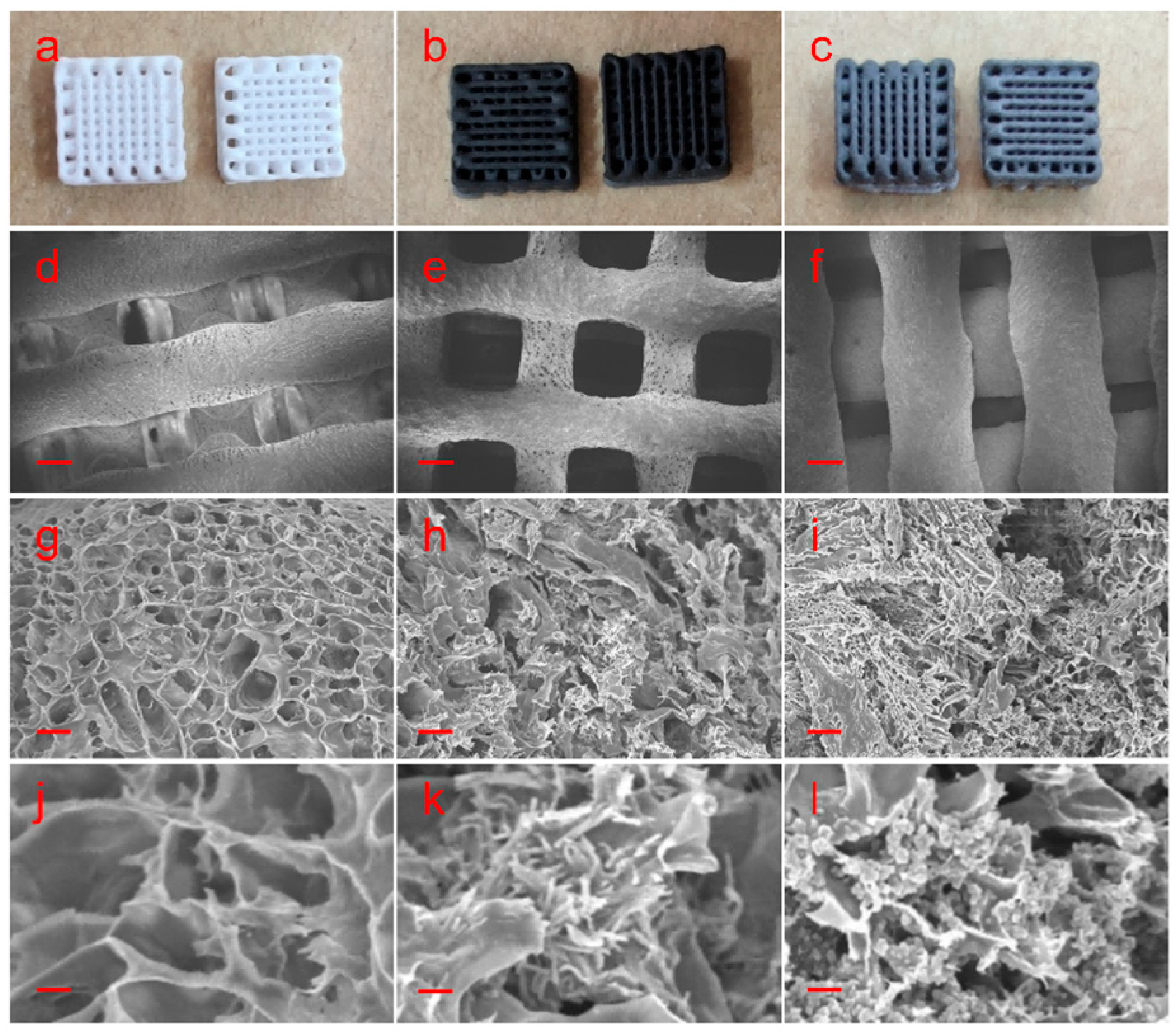
- Figure 6. SEM images of pure poly-l-lactide (PLLA) film and films containing 10 wt% T-PPy and S-PPy, (a–c) physical images of three films, scale bar is 1 cm (pure, T-PPy, and S-PPy, in turn)), (d–f) SEM images of three films at 104 magnification, scale bar is 200 μm (10 wt% PPy, T-PPy, and S-PPy, in turn), (g–i) SEM images of three films at 10,000 magnification, and scale bar is 1 μm (10 wt% PPy, T-PPy, and S-PPy, in turn).
Error in Figure
Reference
- Ma, C.; Jiang, L.; Wang, Y.; Gang, F.; Xu, N.; Li, T.; Liu, Z.; Chi, Y.; Wang, X.; Zhao, L.; et al. 3D Printing of Conductive Tissue Engineering Scaffolds Containing Polypyrrole Nanoparticles with Different Morphologies and Concentrations. Materials 2019, 12, 2491. [Google Scholar] [CrossRef] [PubMed]

Disclaimer/Publisher’s Note: The statements, opinions and data contained in all publications are solely those of the individual author(s) and contributor(s) and not of MDPI and/or the editor(s). MDPI and/or the editor(s) disclaim responsibility for any injury to people or property resulting from any ideas, methods, instructions or products referred to in the content. |
© 2026 by the authors. Licensee MDPI, Basel, Switzerland. This article is an open access article distributed under the terms and conditions of the Creative Commons Attribution (CC BY) license.
Share and Cite
Ma, C.; Jiang, L.; Wang, Y.; Gang, F.; Xu, N.; Li, T.; Liu, Z.; Chi, Y.; Wang, X.; Zhao, L.; et al. Correction: Ma et al. 3D Printing of Conductive Tissue Engineering Scaffolds Containing Polypyrrole Nanoparticles with Different Morphologies and Concentrations. Materials 2019, 12, 2491. Materials 2026, 19, 3062. https://doi.org/10.3390/ma19143062
Ma C, Jiang L, Wang Y, Gang F, Xu N, Li T, Liu Z, Chi Y, Wang X, Zhao L, et al. Correction: Ma et al. 3D Printing of Conductive Tissue Engineering Scaffolds Containing Polypyrrole Nanoparticles with Different Morphologies and Concentrations. Materials 2019, 12, 2491. Materials. 2026; 19(14):3062. https://doi.org/10.3390/ma19143062
Chicago/Turabian StyleMa, Chunyang, Le Jiang, Yingjin Wang, Fangli Gang, Nan Xu, Ting Li, Zhongqun Liu, Yongjie Chi, Xiumei Wang, Lingyun Zhao, and et al. 2026. "Correction: Ma et al. 3D Printing of Conductive Tissue Engineering Scaffolds Containing Polypyrrole Nanoparticles with Different Morphologies and Concentrations. Materials 2019, 12, 2491" Materials 19, no. 14: 3062. https://doi.org/10.3390/ma19143062
APA StyleMa, C., Jiang, L., Wang, Y., Gang, F., Xu, N., Li, T., Liu, Z., Chi, Y., Wang, X., Zhao, L., Feng, Q., & Sun, X. (2026). Correction: Ma et al. 3D Printing of Conductive Tissue Engineering Scaffolds Containing Polypyrrole Nanoparticles with Different Morphologies and Concentrations. Materials 2019, 12, 2491. Materials, 19(14), 3062. https://doi.org/10.3390/ma19143062