Article

Green Chitosan Bioplastics: How the Filler Impacts the Biological Activity and the Biodegradability?


by
Natalia Wrońska
1,*,
Mohamed Amine Benzaouia
2,
Beata Bielska
3,4,
Agata Majkut
3,
Maria Bryszewska
3,
Katarzyna Miłowska
3,
Abdelkrim El Kadib
2 and
Katarzyna Lisowska
1
1
Department of Industrial Microbiology and Biotechnology, Faculty of Biology and Environmental Protection, University of Lodz, 12/16 Banacha Street, 90-236 Lodz, Poland
2
Euromed Research Center, Engineering Division, Euromed University of Fès (UEMF), Route de Meknès, Rond-Point de Bensouda, Fès 30070, Morocco
3
Department of General Biophysics, Faculty of Biology and Environmental Protection, University of Lodz, 90-236 Lodz, Poland
4
Doctoral School of Exact and Natural Sciences, University of Lodz, 90-237 Lodz, Poland
*
Author to whom correspondence should be addressed.
Materials 2026, 19(10), 2167; https://doi.org/10.3390/ma19102167
Submission received: 24 April 2026 / Revised: 15 May 2026 / Accepted: 19 May 2026 / Published: 21 May 2026
(This article belongs to the Section Green Materials)

Highlights

  • Chitosan presents a biodegradable alternative to synthetic plastic.
  • Filler incorporation enhanced the functional properties of chitosan films.
  • Biodegradable chitosan nanocomposites with antibacterial activity and low cytotoxicity.

Abstract
The growing environmental plastic pollution triggered research for biodegradable and safe materials, among which biopolymer-based films stand as the most promising. Among these, chitosan has gained significant attention due to its biocompatibility, film-forming ability, and inherent antimicrobial properties. In this context, the use of fillers to design chitosan nanocomposite films has been shown to enhance the mechanical, barrier, thermal, optical, and antimicrobial properties of the resulting bioplastics. However, the fate and destiny of these fillers, as well as their impact on the biological properties and biodegradability of chitosan films, remain underexplored. We herein report a more comprehensive screening of a set of fillers, encompassing three clay variants (montmorillonite, sepiolite, and halloysite) and microcrystalline chitin. The films were systematically characterized to assess their antibacterial performance, cytocompatibility, hemocompatibility, and biodegradability. The highest antibacterial activity was observed for CS@MMT-f film towards Staphylococcus aureus and Escherichia coli. Importantly, all developed films demonstrated negligible hemolytic activity and low cytotoxicity, indicating their safety for potential biomedical or food-contact applications. Moreover, the selected films completely degrade within four to six weeks under soil burial conditions, demonstrating their potential as environmentally friendly packaging materials.

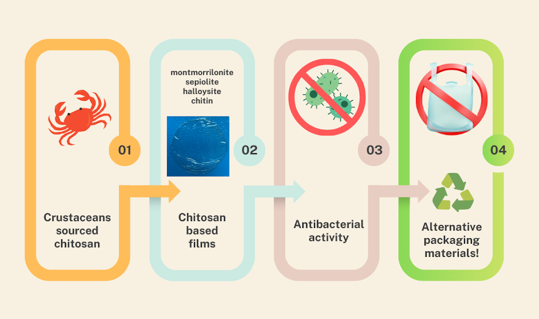
Graphical Abstract

1. Introduction

In recent years, biopolymers have garnered increasing attention as a sustainable alternative to synthetic polymers, driven by growing environmental concerns and the urgent need to limit plastic waste generation. Petroleum-based plastics suffer from slow degradation, and even worse, their disintegration leads to the so-called micro- and nanoplastics, two variants that penetrate human bodies and the surrounding environment [1]. An ideal plastic requires at least 90% disintegration after twelve weeks, and 90% biodegradation involving CO2 in only six months, according to the European bioplastics industry standards for biodegradable plastics. Biopolymers, which are extracted from renewable biological sources such as plants, animals, and microorganisms, offer several advantages over petroleum-based plastics, including sustainability, availability, biodegradability, and a lower carbon footprint [2,3]. Biopolymer-based materials include polysaccharides (cellulose, chitosan, starch, etc.), proteins (soy protein, casein, gelatin, etc.) and lipids (beeswax, carnauba wax, etc.) [4,5,6,7]. From a technological standpoint (low cost, easy acquisition and modification), chitosan is an excellent biopolymer, which, due to its biological properties, can be used in various industries [8]. Chitosan (CS) is derived from chitin, which, combined with calcium carbonate, constitutes the exoskeletons of crustaceans such as shrimp, crabs, and lobsters [9,10]. It is produced through the deacetylation of chitin and is mostly composed of (1–4)-linked 2-amino-2-deoxy-β-D-glucose monomers. Chitosan and its derivatives have gained considerable interest in various fields, including biomedical applications [11,12,13], food packaging [14,15,16], agriculture [17,18], water treatment [19,20], and the pharmaceutical industry [21,22,23]. Despite its many beneficial properties, the practical applications of chitosan are limited. Its poor solubility in neutral and alkaline pH environments limits its use in some biological and industrial processes [24]. Furthermore, chitosan’s mechanical strength and thermal stability are relatively low compared to synthetic polymers. It has been shown that CS films can be chemically modified, blended with other materials, or reinforced with functional compounds to enhance their chemical and thermal stability, mechanical properties, and even biological activity [25,26,27,28,29,30]. Research indicates that carbon-based materials, metal (oxide) nanoparticles, clay nanoparticles, bio-ceramics, and other well-shaped particulates are excellent reinforcements for nanocomposites in a chitosan matrix. More recently, bio-based fillers derived from natural resources have also emerged as greener alternatives, thereby expanding the library of the crafted bioplastics towards more sustainable variants [31]. Different type of fillers, the origin of their reinforcing properties, and their mechanisms of action are gathered in Supplementary Table S1 to provide an overview of their use in the field of bioplastic coating and packaging. Schnabl et al. (2024) investigated the effect of a set of chemical additives, commonly used in the plastic industry, on chitosan films, with a special emphasis on their long thermal stability, weight loss, and film disintegration depending on aging and storing conditions [32]. This study concluded on the excellent properties of citric acid, and to a lesser extent, lactic acid for conceiving practical chitosan-based films [32]. Comparatively, few related data are available concerning the impact of the fillers, where it is not yet clear whether they compromise the biocompatibility of the conceived nanocomposite films and to what extent they could hinder or delay their biodegradability. Given the importance of adding both fillers and plasticizers, among other functionalities, gathering complementary knowledge in the two directions could indeed help promoting the use of chitosan in the realm of the plastic industry.
Clay materials are used because of their large specific surface area, ion exchange capacity and layered charges [33]. Moreover, their excellent mechanical and rheological properties give them a wide range of applications as auxiliary substances to improve technological properties, e.g., in the packaging industry. In pharmacy, clays are used in drug delivery systems. They can increase the solubility of the drug, control its release and prevent side effects [34]. They are added as reinforcement to green composites in which the matrix and/or reinforcement are made from biodegradable and renewable resources [35].
The purpose of this work was to comprehensively evaluate biopolymer-based composite films containing selected natural fillers, including montmorillonite, sepiolite, halloysite, and microcrystalline chitin, with particular emphasis on the influence of the filler type on the biological and environmental properties of the materials. The study aimed to assess the antibacterial activity, cytocompatibility, and hemocompatibility of the developed composites, as well as to determine how the incorporation of different fillers affects the biodegradation behavior of the films in a soil environment.

2. Materials and Methods

2.1. Materials

Chitosan of medium molecular weight and 85% deacetylation degree was purchased from Sigma-Aldrich (Hamburg, Germany). Microcrystalline chitin was provided as a dry powder by France Chitine (Orange, France). Cellulose nanocrystals (LNCC = 130 50 nm; DNCC = 25 15 nm) were prepared from microcrystalline cellulose in H2SO4 (65 wt%) at 318 K for 60 min, following procedures described elsewhere [36]. Montmorillonite (denoted herein as MMT) was purchased from Alfa Aesar (Karlsruhe, Germany). Sepiolite is built from slightly aggregated fibers with a length of a few micrometers. Halloysite nanotubes (high purity: 99%; 1% of Fe2O3 and TiO2 and CaCO3 impurities), referred herein as HNT, with the following parameters (length: 0.2–2 m; outside diameter 50–70 nm; inside diameter 15–45 nm) was made available by Dragonite company (Eureka, UT, USA). Human erythrocytes were obtained from buffy coats provided by the Regional Blood Donation and Blood Treatment Center in Lodz.
The human skin fibroblast cell line BJ (CRL-2522) was purchased from the American Type Culture Collection ATCC (Manassas, VA, USA). Reagents used for cell culture, such as Dulbecco’s Modified Eagle Medium (DMEM), fetal bovine serum (FBS), and antibiotics (streptomycin and penicillin), were from Gibco, a Thermo Fisher Scientific company (Waltham, MA, USA). Dimethylsulfoxide (DMSO), phosphate-buffered saline (PBS) tablets, trypan blue and trypsin were supplied by Sigma-Aldrich (Saint Louis, MO, USA).

2.2. Preparation of Pristine and Filled Chitosan Bioplastic Films

Pristine chitosan film. An amount of 50 mg of chitosan was solubilized in 4 mL of acidified water (2 mL of acetic acid in 98 mL of water) for 4 h. The resulting colloidal gelling solution was poured into Petri dishes, resulting, after solvent evaporation, on pristine chitosan film.
CS@ChitMC film. Chitosan in the amount of 50 mg was solubilized in 4 mL acidified water (2 mL of acetic acid in 98 mL of water) for 4 h until the formation of the gel. A total of 1.5 mg of chitin micro crystals was dispersed in 2 mL of 1% (v/v) aqueous acetic acid solution. The filler dispersion was gradually added to the CS solution, and the resulting mixture was stirred for further 4 h. The resulting solution was cast into plastic Petri dishes, allowing solvent removal and film formation after drying.
CS@MMT film. A total of 50 mg of chitosan was solubilized in 4 mL acidified water (2 mL of acetic acid in 98 mL of water) for 4 h until the formation of the gel. Then, 1.5 mg of MMT, previously dispersed in 2 mL of water, was added. The mixture was stirred for 4 h at room temperature and then introduced into a Petri dish under static conditions. Upon water removal and gel drying, good quality, easy-to-handle, crack-free films were obtained.
CS@Sep film. 50 mg of chitosan was solubilized in 4 mL acidified water (2 mL of acetic acid in 98 mL of water) for 4 h until the formation of the gel. Then, 1.5 mg of Sepiolite, previously dispersed in 2 mL of water, was added. The mixture was stirred during 4 h at room temperature and then introduced in a Petri dish under static conditions. Upon water removal and gel drying, good quality, easy-to-handle, crack-free films were obtained.
CS@HNT film. A total of 50 mg of chitosan was solubilized in 4 mL acidified water (2 mL of acetic acid in 98 mL of water) for 4 h until the formation of the gel. Then, 1.5 mg of HNT previously dispersed in 2 mL of water was added. The mixture was stirred during 4 h at room temperature and then introduced in a Petri dish under static conditions. Upon water removal and gel drying, good quality, easy-to-handle, crack-free films were obtained.

2.3. Determination of Antimicrobial Activity

The antimicrobial activity of chitosan nanocomposite films against Gram-positive bacteria: Staphylococcus aureus ATCC 6538, and Gram-negative bacteria: Escherichia coli ATCC 25922 was evaluated using the Japanese Industrial Standards JIS Z 2801:2000, with modification. The bacterial strains were cultured on Luria–Bertani (LB) medium at 37 °C on a rotary shaker. After the incubation, the inoculum of tested bacteria containing 1 × 10⁵ colony-forming units (CFU per mL) in 500-fold diluted LB medium was prepared. Next, the suspension of bacteria was applied to chitosan films of 2 cm × 2 cm. Native chitosan films were analyzed as control samples. After dripping the suspension of the tested bacterial strains on the films, each sample was covered with a sterile film (1.7 cm × 1.7 cm). The samples were incubated in the moist chamber in the dark for 24 h at 37 °C. After incubation, the samples were put in aseptic Falcon tubes containing phosphate buffer and vortexed. Next, samples were removed from the Falcon tubes, and the remaining solution was used to perform a serial dilution in phosphate buffer. Out of each dilution, 100 µL of bacterial suspension was seeded on an agar plate and incubated for 24 h at 37 °C. After incubation, CFU (Colony-Forming Unit) was calculated by counting the visible colonies on a plate. Each type of tested nanocomposite films was examined in triplicate and analyzed individually in four independent experiments. The antimicrobial activity of the tested films was calculated as the percentage of bacterial growth inhibition (SD) towards the control (native chitosan film).

2.4. Hemolysis Assay

Red blood cells (RBCs) were separated from the buffy coat by centrifugation at 3000 rpm for 10 min at 4 °C. Following isolation, the erythrocytes were washed three times with phosphate-buffered saline (PBS, pH 7.4) to remove residual plasma and leukocytes. Experimental conditions were established by suspending erythrocytes at a hematocrit of 2% in the presence of square-shaped chitosan films (0.5 × 0.5 cm2). These preparations were incubated at 37 °C for 24 h.
Control samples consisted of erythrocytes suspended in PBS without the chitosan film, while a positive control for 100% of complete hemolysis was prepared by suspending erythrocytes in distilled water. After incubation, all samples were subjected to a centrifugation step under the same conditions (3000 rpm, 10 min). The supernatants were collected and transferred to a 96-well microplate for spectrophotometric analysis. Absorbance was measured at 540 nm using a BioTek Synergy HTX microplate reader (Winooski, VT, USA). Hemolysis was quantified by determining the concentration of hemoglobin released into the supernatant, using the following formula:
h e m o l y s i s   % = A s A 100 %   o f   h e m o l y s i s × 100 %
As—absorbance of the sample
A100% of hemolysis—absorbance of the sample with water (100% of hemolysis).

2.5. Assessment of Hemoglobin Adsorption by Chitosan-Based Composites

To evaluate the potential of the tested chitosan-based composites to adsorb hemoglobin from the surrounding medium, square-shaped chitosan films (0.5 × 0.5 cm2) were immersed in hemoglobin solutions at a concentration of 0.1% v/v. The samples were incubated at 37 °C for 24 h to allow interaction between the film and the protein. Following incubation, the mixtures were centrifuged, and the residual hemoglobin concentration in the supernatant was determined spectrophotometrically at 540 nm. The percentage of hemoglobin adsorption was calculated based on the decrease in absorbance, using the following formula:
A d s o r p t i o n   o f   H b % = 100 % ( A s A c × 100 % )
where
As—absorbance of the sample containing the chitosan composites
Ac—absorbance of the control (hemoglobin without chitosan composites).

2.6. Spectrophotometric Determination of Methemoglobin Content

The oxidation of hemoglobin to methemoglobin triggered by chitosan films was investigated using spectrophotometry. Erythrocyte suspensions were incubated with films (0.5 × 0.5 cm2) for 24 h at 37 °C.
The proportion of methemoglobin (metHb) relative to total hemoglobin (Hb) following 24 h incubation with the composite materials was calculated based on absorbance values at 630 nm and 700 nm. Hemoglobin oxidized with potassium ferricyanide, representing 100% metHb, was used as the positive control.
% m e t H b = A 630 A 700 A 630 # A 700 # × 100 %
where
A630 i A700—absorbance of the sample at 630 and 700 nm
A 630 # i A 700 # —absorbance of the sample with potassium ferricyanide at 630 and 700 nm.

2.7. Cell Culture

BJ cells were grown as a monolayer in DMEM supplemented with 10% fetal bovine serum (FBS) and 1% streptomycin. The cultures were incubated at 37 °C in an atmosphere of 5% CO2. Cells were split for subcultures every 2 days and were kept in log phase by regular passage according to the procedure described previously [37].

2.8. Cell Viability (MTT Test)

Cell viability was evaluated using the MTT assay following a 24 h exposure to chitosan-based nanocomposites. Cells were seeded at a density of 5 × 10⁴ per well into a 24-well culture plate and incubated overnight at 37 °C in a humidified atmosphere containing 5% CO2. The next day, the culture medium was replaced with fresh medium and chitosan nanocomposites were added to the samples.
After 24 h of incubation, the cells were washed with phosphate-buffered saline (PBS) and subsequently treated with MTT reagent (0.5 mg/mL; 3-(4,5-dimethylthiazol-2-yl)-2,5-diphenyl tetrazolium bromide) for 3 h at 37 °C. Following incubation, the MTT solution was removed, and dimethyl sulfoxide (DMSO) was added to solubilize the formazan crystals formed by metabolically active cells, as described by Mossman [38].
Absorbance was measured at 570 nm using a BioTek Synergy HT microplate reader (BioTek Instruments, Winooski, VT, USA). Cell viability was expressed as a percentage relative to the untreated control group, calculated using the following formula:
V i a b i l i t y   % = A s A c × 100 %
where
As—absorbance of the sample
Ac—absorbance of the control (untreated cells).

2.9. Biodegradability of Chitosan Films in Soil

The study presents evaluating the biodegradability of chitosan films via determinating the degradability of the CS films by soil microorganisms. The soil samples were collected in the garden of the Faculty of Biology and Environmental Protection University of Lodz, Poland, and used as an incubation medium. The soil was maintained at room temperature, 45% moisture, and pH equal to 6. The soil was placed in a special container and sterile samples were buried at a depth of approximately 10 cm (each sample in a separate container) and incubated at room temperature. The buried samples were dug out at different periods of time (1, 2, 4 and 6 weeks of incubation) and rubbed gently with brush to remove soil residues. Then, the samples were rinsed thoroughly with distilled water, dried at 50 °C for 4 h, desiccated for 24 h at room temperature, weighted and the percent of weight loss of samples was determined. We analyzed 5 films: chitosan (CS-f), chitosan–chitin (CS@ChitMC-f), chitosan–MMT (CS@MMT-f), chitosan–sepiolite (CS@Sep-f), chitosan–HNT (CS@HNT-f). The obtained images of CS films were compared with images after 1-, 2-, or 4-week period incubation.

2.10. Statistical Analysis

Data are presented as the mean ± SD from a minimum of 3 sets of measurements. Statistical differences between the control and treatment groups were analyzed by one-way ANOVA followed by Tukey’s analysis. p < 0.05 was taken as statistically significant.

3. Results

3.1. Characterization of Filled Chitosan Nanocomposites

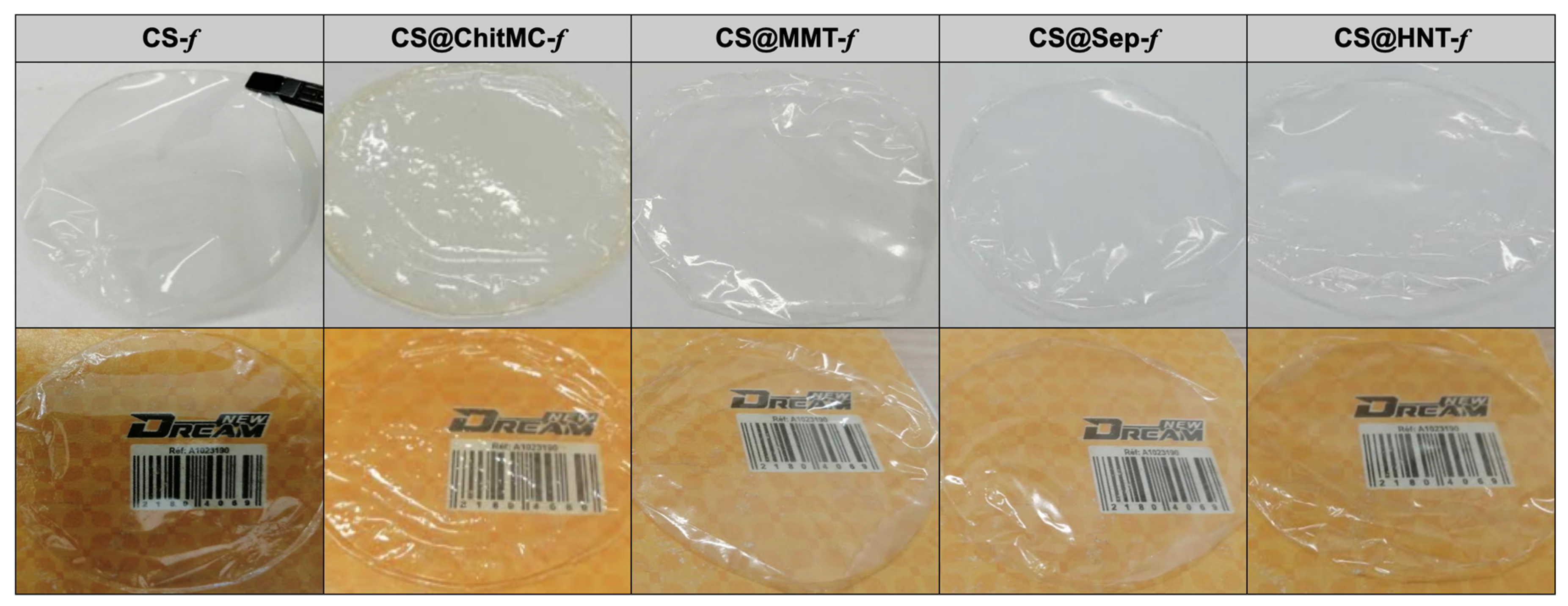
Regardless of the nature of the filler, a homogenous chitosan aqueous acidic solution containing 3 wt% of dispersed filler was prepared. Evaporative-induced assembly of the resulting solutions provides high-quality, crack-free, flexible and easy-to-handle chitosan-based bioplastic films. Their photos are presented below along with pristine chitosan films prepared for comparison (Figure 1).
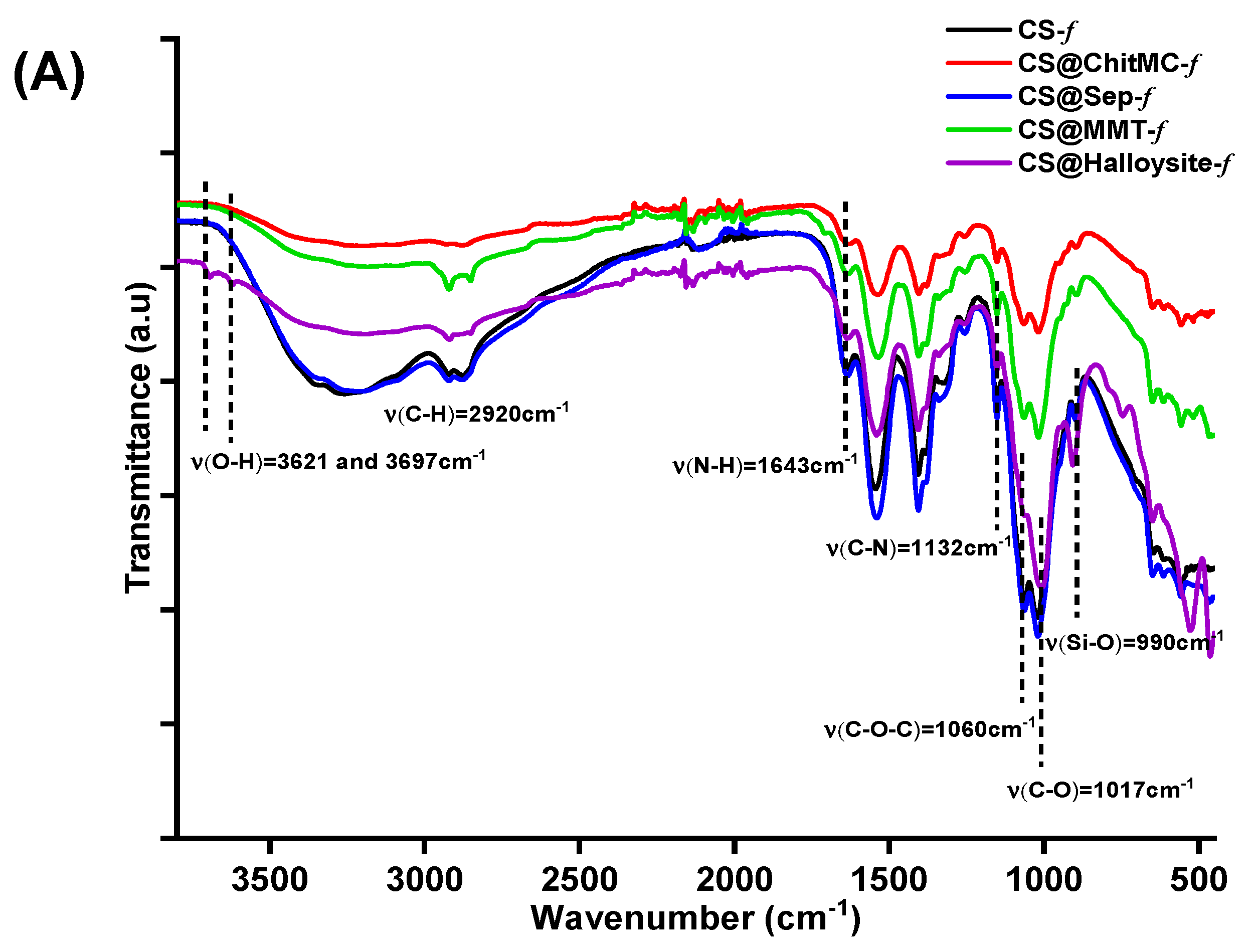
The FTIR spectrum of the prepared films shows primarily the fingerprint signature of chitosan polymer, the latter being more abundant compared to 3 wt.% of the filler inserted (Figure 2A). The common bands attributed to C–O, C–O–C, C–N, N–H, acetamide C–H, and O–H vibrations appear at 1017, 1060, 1160, 1332, 1544, 1643, 2870, 2920, and 3200–3400 cm−1. The increase in the band around 990 cm−1, assigned to Si–O stretching, confirms the presence of clay minerals (montmorillonite and halloysite) within the chitosan matrix. Furthermore, two additional bands observed at 3621 and 3697 cm−1 are attributed to the hydroxyl groups located on the internal and external surfaces of halloysite nanotubes (HNTs), providing further evidence for the successful incorporation of clays into the chitosan films.
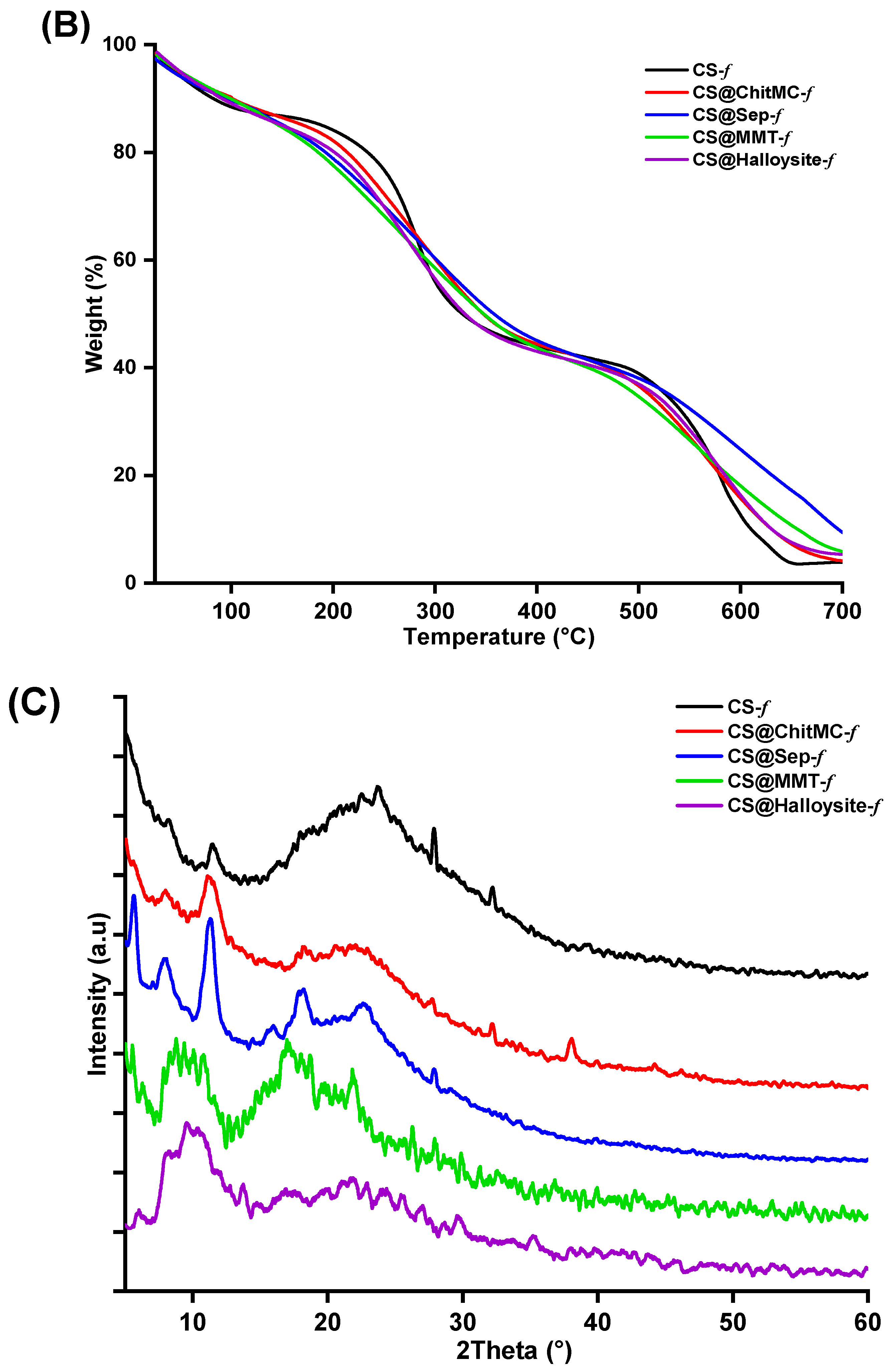
Thermogravimetric analysis reveals that unmodified chitosan film exhibits the highest thermal stability at lower temperatures (Figure 2B). However, this trend is reversed at higher temperatures. In particular, the chitosan film reinforced with sepiolite shows the highest char residue (10%) and the highest T50 value (360 °C), indicating improved thermal resistance due to the presence of the clay filler.
The X-ray diffraction (XRD) pattern of native chitosan films shows two characteristic peaks at 2θ ≈ 10° and 20°, corresponding to the semi-crystalline structure of chitosan (Figure 2C). After the incorporation of chitin (CS@ChitMC-f), a slight increase in peak intensity is observed, indicating the contribution of the crystalline domains of chitin within the chitosan matrix. The pattern of the composite films was significantly modified with the incorporation of clay particulates, indicating that secondary chitosan organization has been altered by the entanglement of the filler particulates within the polysaccharide network. This profile is indeed indicative of the occurrence of strong interactions between chitosan chains and dispersed clay particles, as it has been already reported for similar systems [39,40].

3.2. Biological Activity of Chitosan Nanocomposite Films

3.2.1. Antimicrobial Activity

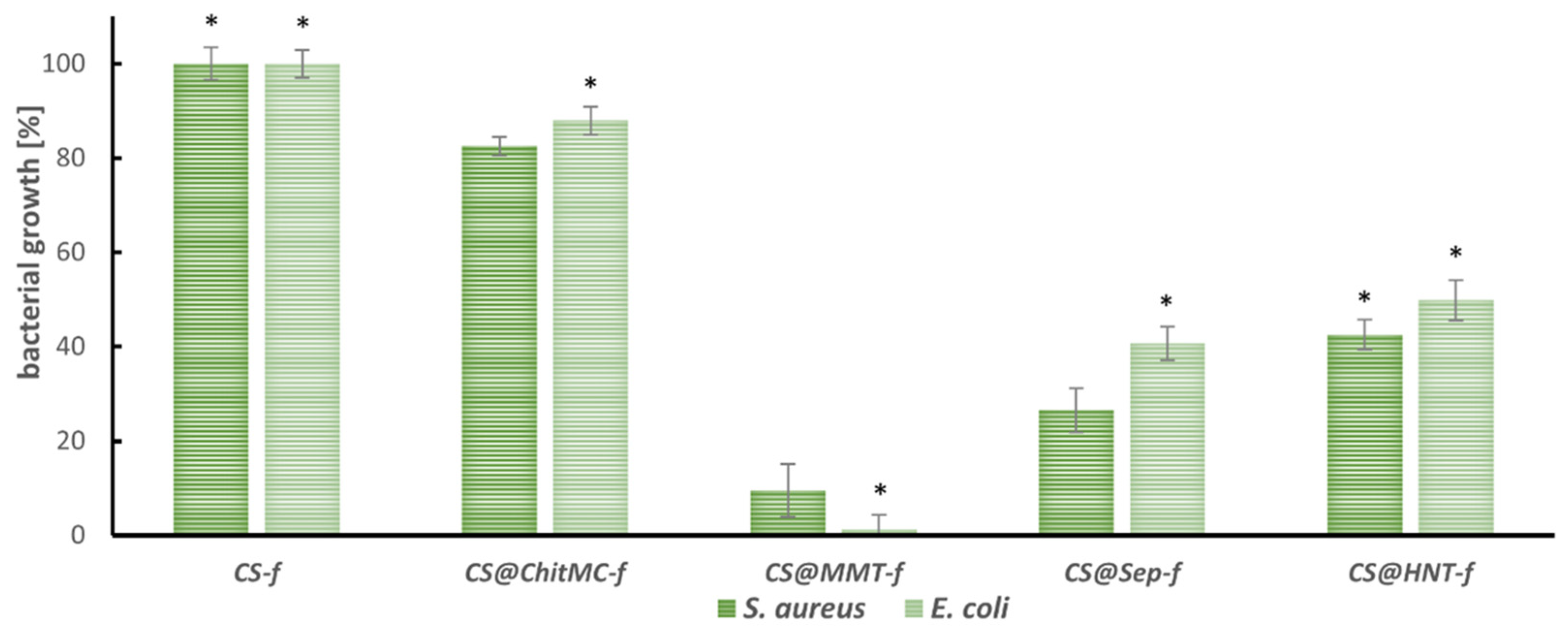
The antimicrobial activity of chitosan nanocomposite films: chitosan (CS-f), chitosan–chitin (CS@ChitMC-f), chitosan–montmorillonite (CS@MMT-f), chitosan–sepiolite (CS@Sep-f), chitosan–nanotubular halloysite (CS@HNT-f), was assessed using Staphylococcus aureus ATCC 6538 and Escherichia coli ATCC 25922 as model bacteria and taking native chitosan film CS-f as a reference (Figure 3).
All filler-containing chitosan nanocomposites limited the growth of the tested microorganisms compared to the native chitosan coating. In the case of the combination of chitosan and chitin (CS@ChitMC-f), the antibacterial activity was low. Only 10–17% growth inhibition of the tested strains was observed. The highest antibacterial activity was observed for the CS-MMT (CS@MMT-f) film. The chitosan-reinforced montmorillonite nanocomposites caused almost 100% inhibition of S. aureus growth and 91% inhibition of E. coli. In the case of the other two composites enriched with clay minerals, chitosan–sepiolite showed better activity against the tested bacteria (70% inhibition of S. aureus growth) compared to CS–halloysite (CS@HNT-f). The addition of sepiolite or halloysite demonstrated lower growth inhibition of the Gram-negative E. coli strain by 60% and 50%, respectively.

3.2.2. Hemolysis and Hb Adsorption

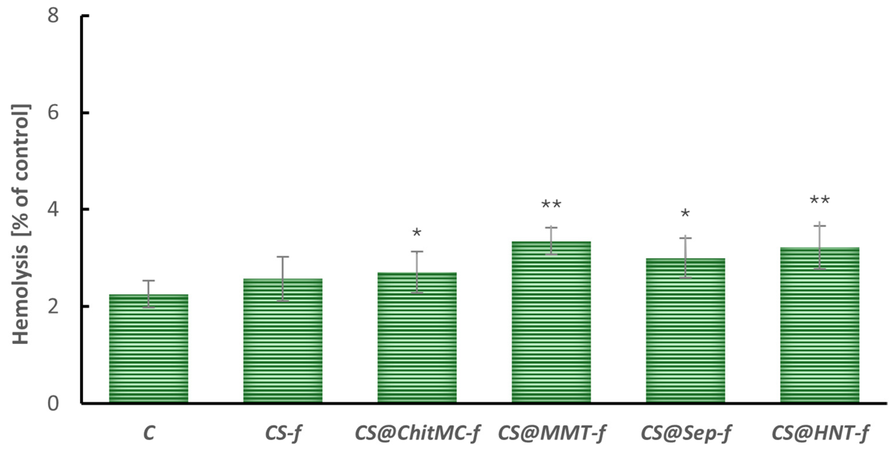
The hemolysis test results, presented in Figure 4, indicate a statistically significant increase in the degree of erythrocyte membrane damage in the tested chitosan-based materials (excluding unmodified chitosan). The control sample (C) showed a baseline level of hemolysis of approximately 2%, providing a reference point for assessing the biocompatibility of the modified films. It was demonstrated that despite the statistically significant changes, the increase in hemolysis was small, and the percentage of hemolysis after 24 h of incubation with the nanocomposites was below 5%.
Previous studies have demonstrated that chitosan-based materials possess the ability to adsorb hemoglobin onto their surface, potentially influencing the accuracy of hemolysis measurements [41,42].
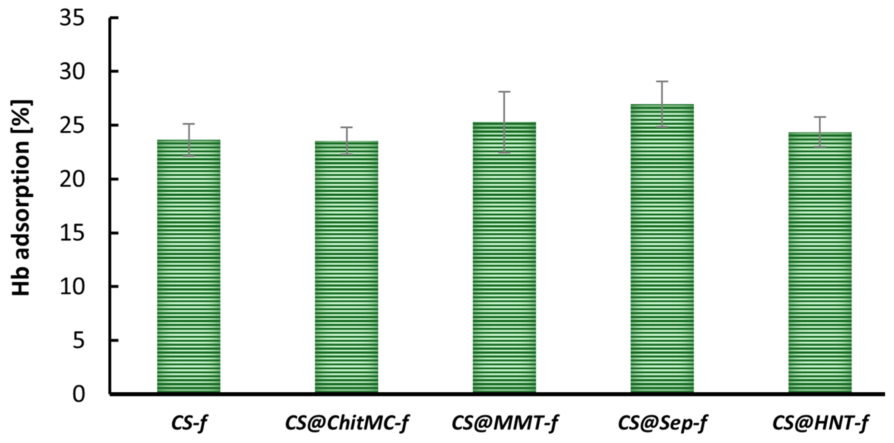
In the present study, the hemoglobin adsorption capacity of various chitosan-based films was also evaluated (Figure 5). As illustrated in Figure 5, the unmodified chitosan film (CS-f) adsorbed approximately 25% of the available hemoglobin, serving as a reference for comparative analysis across the tested materials.
Most of the materials tested exhibited Hb adsorption levels ranging from approximately 20% to 30%, indicating moderate affinity for hemoglobin. Among the samples, CS@Sep-f demonstrated the highest adsorption capacity, with values slightly exceeding 30%, suggesting a strong interaction between the film surface and hemoglobin molecules.

3.2.3. Methemoglobin

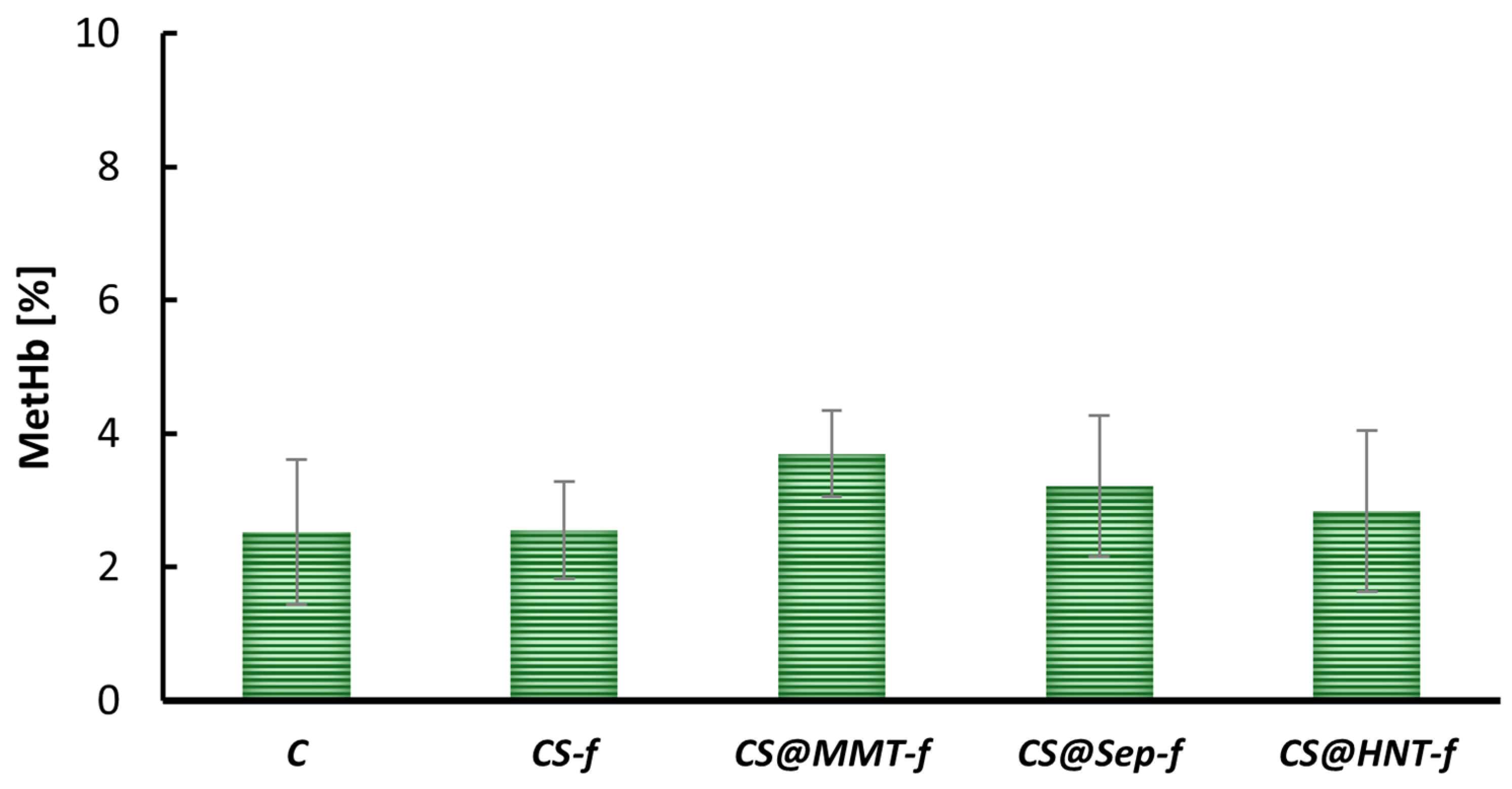
Methemoglobin (MetHb) formation was assessed to evaluate the oxidative stress induced by various chitosan-based films. As shown in Figure 6, the control sample (C) had a baseline MetHb level of approximately 2.5%. All tested materials had MetHb levels similar to the control, with values ranging from approximately 2.5% to 4%.
These results indicate that the chemical modifications used do not cause oxidative stress in erythrocytes, as no significant changes in MetHb levels were observed compared to the control.

3.2.4. Cytotoxicity

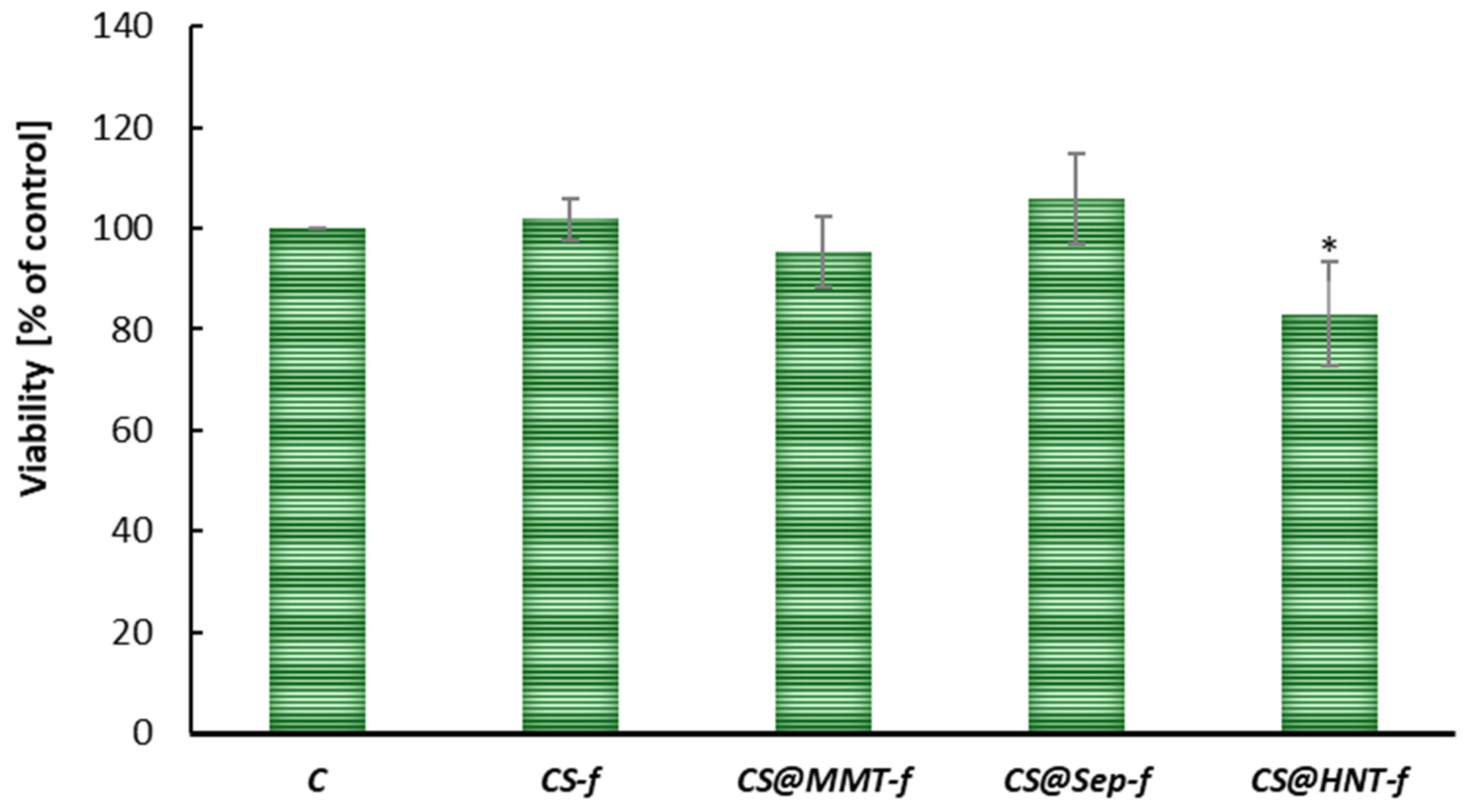
The cytotoxicity of chitosan-based films was assessed by analyzing the viability of human skin fibroblasts (BJ) after 24 h of exposure. As shown in Figure 7, the control group (C) was designated as 100% viability, serving as a reference for untreated cells.
Most tested films maintained cell viability close to or above the control level, indicating good biocompatibility. The samples CS-f, CS@ChitMC-f, CS@MMT-f, CS@Sep-f showed viability levels ranging from approximately 90% to 106%, suggesting that these formulations do not exert cytotoxic effects and may even promote cell proliferation in some cases.
In contrast, sample CS@HNT-f demonstrated a statistically significant reduction in cell viability (p < 0.05), indicating potential cytotoxicity or adverse interactions with fibroblast cells. However, the observed reduction in cell viability is relatively minor, not exceeding 20%, which suggests that the tested films can also be considered biocompatible with skin cells.

3.2.5. Biodegradability of Chitosan Films in Soil

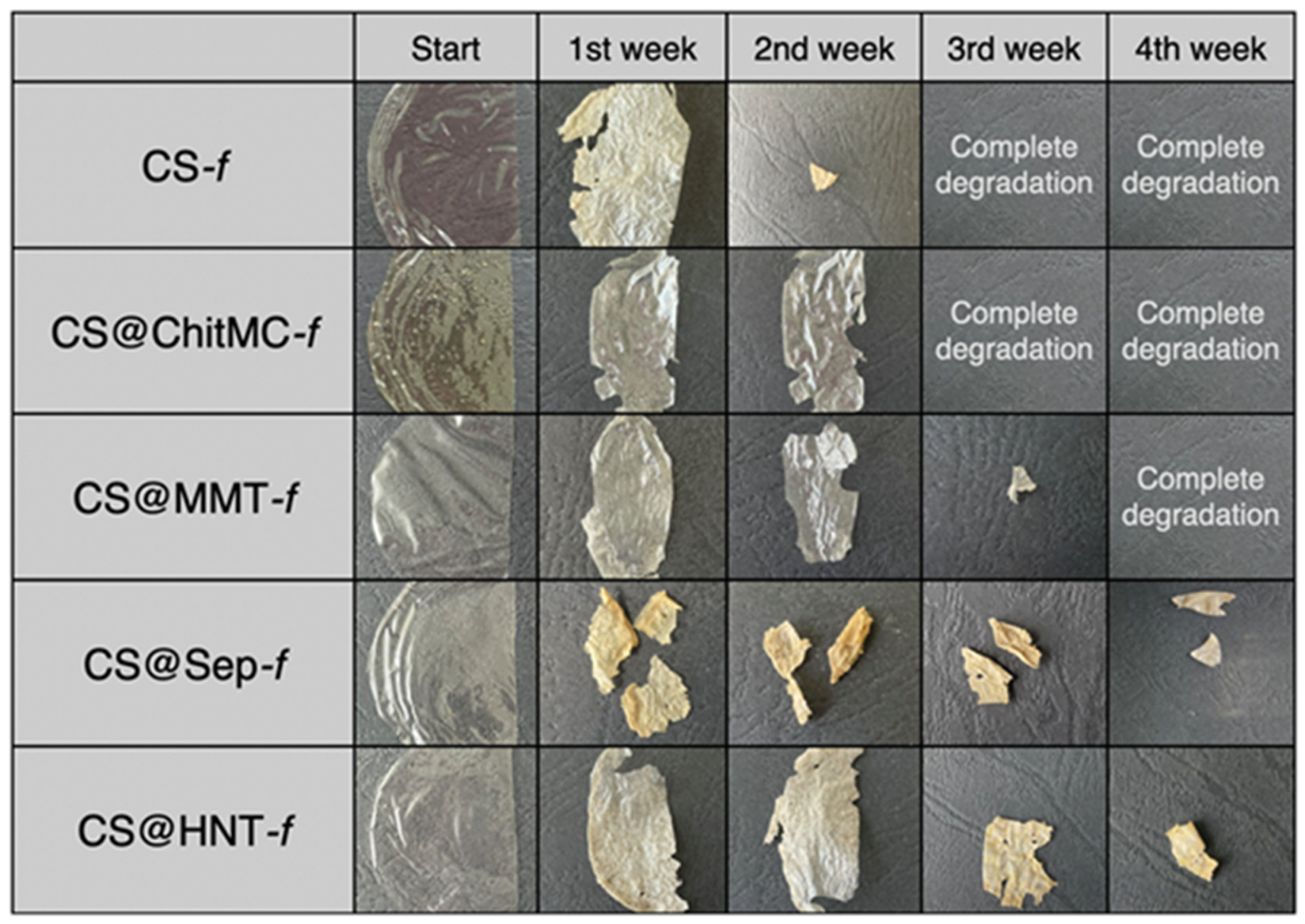
All tested films were homogeneous and were stored at room temperature. Each film was halved and photographed before being placed in the soil. The biodegradation of these films in a soil environment was examined and compared with that of pure chitosan films. The photographs presented in Figure 8 document the results obtained during the incubation of nanocomposites in the soil environment. All tested films are presented in Table 1 (Section 2).
The biodegradation of those films was studied and compared with a pure chitosan film. The results were recorded with photographs, which are illustrated in Figure 7. In soil, placed in separate containers and incubated at room temperature, CS-f, CS@ChitMC-f, and CS@MMT-f were degraded after 4 weeks of incubation. The results clearly showed that chitosan modifications can significantly affect the decomposition of the tested materials.
After 2 weeks of incubation, almost complete degradation of the pure chitosan film (CS-f) was observed, along with a significant loss in the CS@Sep-f sample. The other films were wrinkled, pliable with a slight loss of composite mass (Figure 8). The greatest changes were noted after 3 weeks of incubation of the samples in the soil environment. Complete biodegradation of the CS-f, CS@ChitMC-f and CS@MMT-f films occurred. Furthermore, a large mass loss was observed in the CS@HNT-f (Figure 8). After 4 weeks of incubation, complete biodegradation of CS@MMT-f was observed, while after 6 weeks, no CS@Sep-f or CS@HNT-f films were observed in the soil.
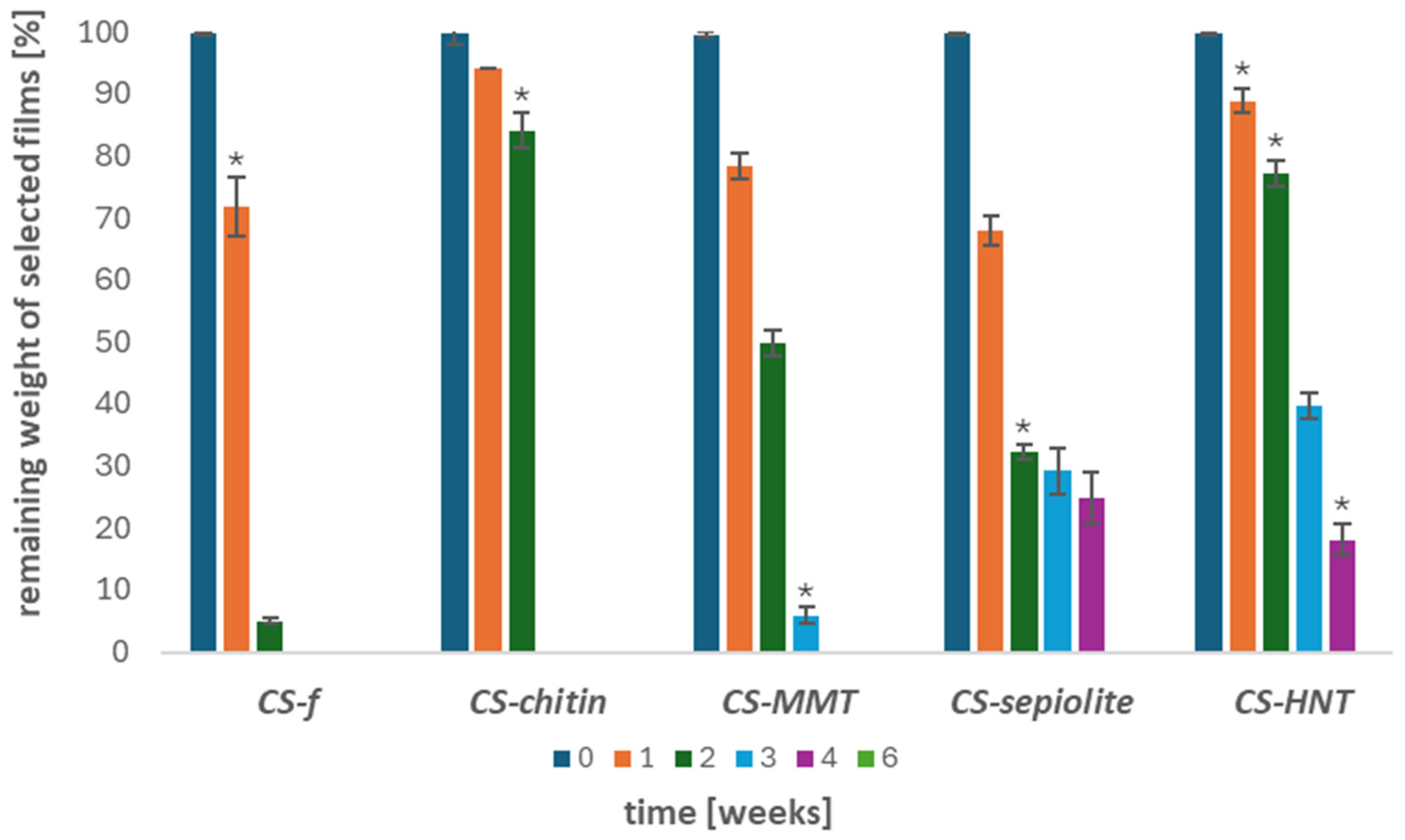
Moreover, the extent of weight loss of the tested materials was studied. The results are presented in Figure 9.
Figure 9 shows the changes in the weight ratio of the tested chitosan films over 1, 2, 3, 4, and 6 weeks of incubation. After the first week, approximately 30% weight loss was observed for both CS-f and CS@Sep-f films. By the second week, the CS-f film had nearly completely degraded, while the CS@MMT-f and CS@Sep-f samples exhibited significant weight loss. After 3 weeks of incubation, complete (100%) weight loss was observed for CS-f and CS@ChitMC-f, whereas for CS@MMT-f this occurred after 4 weeks. The remaining samples reached total degradation after 6 weeks of incubation in the soil environment.

4. Discussion

The incorporation of mineral fillers into chitosan-based bioplastic films significantly modulates both their biological activity and biodegradability, largely through changes in structural organization, interfacial interactions, and physicochemical properties. In this study, the nature of the filler, including its chemical composition, morphology, and surface functional groups, emerged as a factor governing performance. CS@MMT-f composites exhibited superior antibacterial activity towards Staphylococcus aureus and Escherichia coli. In this case, almost complete inhibition of E. coli growth and 90% inhibition of S. aureus growth were observed. Similar results were obtained by Liu et al. (2025), demonstrating a broad spectrum of antimicrobial activity of the montmorillonite-chitosan hydrogel system, especially against E. coli and S. aureus [43]. In other studies, the CS-MMT composite was tested as an anticancer drug carrier. The incorporation of MMT into the CS matrix reduced the release rate of 5-FU by entrapping it within the clay layers, thereby enhancing the antibacterial activity of the CS–clay/drug nanocomposite film against S. aureus and E. coli. [44]. CS–sepiolite (CS@Sep-f) also showed promising antimicrobial effects, likely due to its fibrous structure and surface chemistry. Dutta et al. (2021) investigated that the CS-SEP films showed better antibacterial activity than chitosan alone against both B. subtilis and E. coli [45]. It was revealed that the addition of sepiolite to chitosan–silver composites, apart from enhancing its stability and antimicrobial activity, also allowed for a reduction in cytotoxicity (compared to silver alone). They report average widths of inhibition zones (disk diffusion) of ≈51.8 mm against S. aureus, ≈31.8 mm vs. E. coli, and ≈44.7 mm vs. Aspergillus niger [46]. Several studies have shown that combining chitosan with halloysite nanotubes creates a nanocomposite that often exhibits superior antibacterial properties compared to chitosan alone [47,48,49]. The results of our study clearly showed that the chitosan–HNT (CS@HNT-f) composite showed higher antibacterial activity compared to the pure chitosan film. The enhanced antibacterial action of the chitosan–halloysite nanotube (HNT) composite is generally attributed to a combination of electrostatic interactions, structural effects, and improved availability of active groups [50]. Chitosan’s positively charged amino groups can bind to negatively charged bacterial cell surfaces, disrupting membrane integrity and increasing permeability [51]. When HNTs are incorporated, their high surface area and tubular structure promote interaction with bacterial cells [52]. Additionally, HNTs may facilitate a more uniform distribution and exposure of chitosan’s active sites, leading to more efficient interaction with microbial membranes. The mechanism of antibacterial action of CS-HNT films enhances bacterial membrane disintegration, disrupts cellular metabolism, and ultimately inhibits bacterial growth more effectively than chitosan alone.
Hemolysis and hemoglobin adsorption results suggest that while chitosan’s amino groups facilitate protein binding, excessive adsorption may underestimate hemolysis levels. Hemoglobin released from lysed erythrocytes can bind to the film surface rather than remaining in the supernatant, resulting in lower absorbance readings during spectrophotometric analysis. Consequently, the actual extent of hemolysis induced by the tested materials may be higher than indicated by conventional methods. This phenomenon is consistent with previous reports describing the strong affinity of chitosan for blood components, particularly proteins such as hemoglobin [42].
The hemoglobin adsorption capacity of chitosan-based films is strongly influenced by their chemical structure. Chitosan’s amino groups play a central role in protein binding, facilitating interactions with hemoglobin and other biomolecules. Chemical modifications that block or alter these functional groups, such as crosslinking or incorporation of bulky substituents, can significantly reduce the material’s ability to bind hemoglobin. This tunable adsorption behavior is particularly relevant in the design of chitosan-based materials for biomedical and environmental applications, such as hemostatic dressings, filtration systems and packaging materials, where controlled protein interaction is essential [53]. These findings emphasize the need for careful interpretation of hemolysis data when evaluating the blood compatibility of chitosan-based biomaterials, especially in applications where accurate assessment of erythrocyte integrity is critical.
Cytotoxicity assays confirmed the biocompatibility of most films, with CS-MMT and CS–sepiolite potentially promoting cell proliferation. Slight reductions in viability for CS-HNT warrant further optimization.
Overall, the majority of chitosan-based films tested were well tolerated by BJ fibroblasts, supporting their potential use in skin-contact applications. However, the reduced viability observed in select samples highlights the importance of careful material design and screening for cytotoxic effects.
There are several papers in the literature describing the degradation of pure chitosan membranes [54,55,56,57]. Unfortunately, although the biopolymer material itself is biodegradable, its enrichment with active compounds may disturb the biodegradation process by natural soil microflora. Therefore, one of the aims of the presented work was to assess the impact of the filler on the biodegradability of composites in the soil environment. Due to the current environmental pollution, films that can potentially be used in the medical or packaging industry must be completely biodegradable.
The most desirable effect of our research was to obtain a composite variant with excellent mechanical and physicochemical properties, high antimicrobial potential and no cytotoxicity. Given the growing role of chitosan in sectors such as food packaging, medicine, cosmetics and agriculture, it is extremely important to prove that chitosan films are safe for humans. However, the future of sustainable materials depends not only on their safety for human health but also on their environmental friendliness. Replacing environmentally harmful synthetic polymers with biopolymers is highly desirable and is expected to play a central role in strategies aimed at eliminating the risks posed by micro- and nanoplastics. Therefore, it is important to assess the biodegradability of chitosan-based films. Literature data describe the chitosan degradation process as several stages leading to the disintegration of polymer chains. The first stage is the swelling of the membranes initiated by water. This hydration step facilitates the diffusion of enzymes and microorganisms into the film matrix. Enzymes such as chitosanase, lysozyme, and proteases target the glycosidic bonds in chitosan, breaking them down into smaller fragments. The initial stage of the proposed chitosan degradation mechanism involves depolymerization, characterized by the random cleavage of β-1,4-glycosidic bonds, followed by deacetylation through hydrolysis of N–acetyl bonds. As a consequence of these processes, the degree of deacetylation increases and the molecular weight decreases [58]. The cleavage of chitosan functional groups (amine, carbonyl, amide and hydroxyl) may occur simultaneously, depending on chemical and/or enzymatic conditions. Chitosanase is a key enzyme in the initial stages of degradation, cleaving chitosan chains at random points [59]. Thanks to enzymatic hydrolysis, the chitosan film is gradually broken down into oligosaccharides and ultimately into single monomers such as glucosamine (D-glucosamine or N-acetylglucosamine), which can be further used by microorganisms [58]. We observed the fastest degradation of chitosan (CS-f), chitosan–chitin (CS@ChitMC-f), and chitosan–MMT (CS@MMT-f) films. Films with the addition of sepiolite and halloysite nanotubes remained in the soil environment longer. A study analyzing the degradation of chitosan films in commercial composts, garden soil, and vineyard soil showed that after 10 days, the chitosan films were completely degraded [60]. Milbreta et al. (2024) showed that chitosan or alginate films with the addition of nanofibrillated cellulose were completely decomposed within 3 weeks in the soil environment [61]. The high cross-linking capacity of chitosan films with mineral clays contributed to their greater stability and resistance to biodegradation factors present in the soil. However, after 6 weeks of incubation, 100% biodegradability was observed.
Taking into account the acquisition of a stable composite with high antibacterial activity, non-cytotoxic and completely biodegradable in the soil environment after 4 weeks, films CS-MMT and CS–sepiolite with high potential for industrial use could be selected. These characteristics make them strong candidates for applications in packaging, medicine, and other sustainable material technologies

5. Conclusions

The results of this study confirm that the incorporation of specific fillers into chitosan films can significantly influence their biological activity and biodegradation profile. Among the tested nanocomposites, films such as CS–montmorillonite (CS@MMT-f) and CS–sepiolite (CS@Sep-f) demonstrated a well-balanced combination of desirable properties, including strong antibacterial activity, non-cytotoxicity, and complete biodegradability within 4 to 6 weeks in a soil environment. Notably, CS@MMT-f film exhibited the highest antibacterial effectiveness against Staphylococcus aureus and Escherichia coli. These findings highlight the potential of chitosan-based nanocomposite films as sustainable alternatives to conventional plastic materials, particularly in applications where both antimicrobial performance and environmental safety are essential.

Supplementary Materials

The following supporting information can be downloaded at https://www.mdpi.com/article/10.3390/ma19102167/s1, Table S1: Selected types of fillers in the chitosan matrix and their mechanisms of action [41,62,63,64,65,66,67,68,69,70,71,72,73,74,75,76,77,78,79,80,81,82,83,84,85,86,87,88,89,90,91,92,93].

Author Contributions

Conceptualization, N.W., A.E.K. and K.L.; methodology, N.W., M.A.B., K.M. and A.E.K.; investigation, N.W., M.A.B., B.B., A.M. and K.M.; data curation, N.W. and A.E.K.; writing—original draft preparation, N.W. and K.M.; writing—review and editing, M.B. and A.E.K.; project administration, K.L.; funding acquisition, K.L. All authors have read and agreed to the published version of the manuscript.

Funding

This research was funded by National Science Center (NCN) Poland no. 2017/25/B/NZ9/02900.

Data Availability Statement

The original contributions presented in this study are included in the article/Supplementary Material. Further inquiries can be directed to the corresponding author.

Conflicts of Interest

The authors declare no conflicts of interest.

References

  1. Yee, M.S.-L.; Hii, L.-W.; Looi, C.K.; Lim, W.-M.; Wong, S.-F.; Kok, Y.-Y.; Tan, B.-K.; Wong, C.-Y.; Leong, C.-O. Impact of Microplastics and Nanoplastics on Human Health. Nanomaterials 2021, 11, 496. [Google Scholar] [CrossRef]
  2. Mohanrasu, K.; Manivannan, A.C.; Rengarajan, H.J.R.; Kandaiah, R.; Ravindran, A.; Panneerselvan, L.; Palanisami, T.; Sathish, C.I. Eco-Friendly Biopolymers and Composites: A Sustainable Development of Adsorbents for the Removal of Pollutants from Wastewater. npj Mater. Sustain. 2025, 3, 13. [Google Scholar] [CrossRef]
  3. Zhao, X.; Wang, Y.; Chen, X.; Yu, X.; Li, W.; Zhang, S.; Meng, X.; Zhao, Z.-M.; Dong, T.; Anderson, A.; et al. Sustainable Bioplastics Derived from Renewable Natural Resources for Food Packaging. Matter 2023, 6, 97–127. [Google Scholar] [CrossRef]
  4. Pires, J.; Paula, C.D.D.; Souza, V.G.L.; Fernando, A.L.; Coelhoso, I. Understanding the Barrier and Mechanical Behavior of Different Nanofillers in Chitosan Films for Food Packaging. Polymers 2021, 13, 721. [Google Scholar] [CrossRef] [PubMed]
  5. Oladzadabbasabadi, N.; Mohammadi Nafchi, A.; Ariffin, F.; Wijekoon, M.M.J.O.; Al-Hassan, A.A.; Dheyab, M.A.; Ghasemlou, M. Recent Advances in Extraction, Modification, and Application of Chitosan in Packaging Industry. Carbohydr. Polym. 2022, 277, 118876. [Google Scholar] [CrossRef] [PubMed]
  6. Edo, G.I.; Ndudi, W.; Ali, A.M.; Yousif, E.; Jikah, A.N.; Isoje, E.F.; Igbuku, U.A.; Mafe, A.N.; Opiti, R.A.; Madueke, C.J.; et al. Biopolymers: An Inclusive Review. Hybrid Adv. 2025, 9, 100418. [Google Scholar] [CrossRef]
  7. Cheng, J.; Gao, R.; Zhu, Y.; Lin, Q. Applications of Biodegradable Materials in Food Packaging: A Review. Alex. Eng. J. 2024, 91, 70–83. [Google Scholar] [CrossRef]
  8. Koumentakou, I.; Meretoudi, A.; Emmanouil, C.; Kyzas, G.Z. Environmental Toxicity and Biodegradation of Chitosan Derivatives: A Comprehensive Review. J. Ind. Eng. Chem. 2025, 146, 70–86. [Google Scholar] [CrossRef]
  9. Izadi, H.; Asadi, H.; Bemani, M. Chitin: A Comparison between Its Main Sources. Front. Mater. 2025, 12, 1537067. [Google Scholar] [CrossRef]
  10. Ngasotter, S.; Xavier, K.A.M.; Meitei, M.M.; Waikhom, D.; Madhulika; Pathak, J.; Singh, S.K. Crustacean Shell Waste Derived Chitin and Chitin Nanomaterials for Application in Agriculture, Food, and Health–A Review. Carbohydr. Polym. Technol. Appl. 2023, 6, 100349. [Google Scholar] [CrossRef]
  11. Mu, L.; Wu, L.; Wu, S.; Ye, Q.; Zhong, Z. Progress in Chitin/Chitosan and Their Derivatives for Biomedical Applications: Where We Stand. Carbohydr. Polym. 2024, 343, 122233. [Google Scholar] [CrossRef] [PubMed]
  12. Hamdi, M.; Sun, H.; Pan, L.; Wang, D.; Sun, M.; Zeng, Z.; Li, S.; Dong, Q.; Su, F. Chitosan and Its Derivatives as Potential Biomaterials for Biomedical and Pharmaceutical Applications: A Comprehensive Review on Green Extraction Approaches, Recent Progresses, and Perspectives. Eur. Polym. J. 2025, 229, 113882. [Google Scholar] [CrossRef]
  13. Medeiros Borsagli, F.G.L. Carboxymethyl Chitosan and Its Derivatives in Tissue Engineering. In Multifaceted Carboxymethyl Chitosan Derivatives: Properties and Biomedical Applications; Jayakumar, R., Ed.; Advances in Polymer Science; Springer Nature: Cham, Switzerland, 2023; Volume 292, pp. 257–280. ISBN 978-3-031-44099-1. [Google Scholar]
  14. Mujtaba, M.; Morsi, R.E.; Kerch, G.; Elsabee, M.Z.; Kaya, M.; Labidi, J.; Khawar, K.M. Current Advancements in Chitosan-Based Film Production for Food Technology; A Review. Int. J. Biol. Macromol. 2019, 121, 889–904. [Google Scholar] [CrossRef] [PubMed]
  15. Jiang, A.; Patel, R.; Padhan, B.; Palimkar, S.; Galgali, P.; Adhikari, A.; Varga, I.; Patel, M. Chitosan Based Biodegradable Composite for Antibacterial Food Packaging Application. Polymers 2023, 15, 2235. [Google Scholar] [CrossRef]
  16. Kumar, S.; Mukherjee, A.; Dutta, J. Chitosan Based Nanocomposite Films and Coatings: Emerging Antimicrobial Food Packaging Alternatives. Trends Food Sci. Technol. 2020, 97, 196–209. [Google Scholar] [CrossRef]
  17. Othman, S.H.; Ronzi, N.D.A.; Shapi’i, R.A.; Dun, M.; Ariffin, S.H.; Mohammed, M.A.P. Biodegradability of Starch Nanocomposite Films Containing Different Concentrations of Chitosan Nanoparticles in Compost and Planting Soils. Coatings 2023, 13, 777. [Google Scholar] [CrossRef]
  18. Rojas-Pirela, M.; Carillo, P.; Lárez-Velásquez, C.; Romanazzi, G. Effects of Chitosan on Plant Growth under Stress Conditions: Similarities with Plant Growth Promoting Bacteria. Front. Plant Sci. 2024, 15, 1423949. [Google Scholar] [CrossRef] [PubMed]
  19. Algethami, J.S.; Alhamami, M.A.M.; Alqadami, A.A.; Melhi, S.; Seliem, A.F. Magnetic Hydrochar Grafted-Chitosan for Enhanced Efficient Adsorption of Malachite Green Dye from Aqueous Solutions: Modeling, Adsorption Behavior, and Mechanism Analysis. Int. J. Biol. Macromol. 2024, 254, 127767. [Google Scholar] [CrossRef]
  20. Saiyad, M.; Shah, N.; Joshipura, M.; Dwivedi, A.; Pillai, S. Chitosan and Its Derivatives in Wastewater Treatment Application. Mater. Today Proc. 2024, 99, 190–194. [Google Scholar] [CrossRef]
  21. Kedir, W.M.; Abdi, G.F.; Goro, M.M.; Tolesa, L.D. Pharmaceutical and Drug Delivery Applications of Chitosan Biopolymer and Its Modified Nanocomposite: A Review. Heliyon 2022, 8, e10196. [Google Scholar] [CrossRef]
  22. Ong, T.H.; Chitra, E.; Ramamurthy, S.; Siddalingam, R.P.; Yuen, K.H.; Ambu, S.P.; Davamani, F. Chitosan-Propolis Nanoparticle Formulation Demonstrates Anti-Bacterial Activity against Enterococcus Faecalis Biofilms. PLoS ONE 2017, 12, e0174888. [Google Scholar] [CrossRef]
  23. Amasya, G.; Tuba Sengel-Turk, C.; Basak Erol, H.; Kaskatepe, B.; Oncu, A.; Guney-Eskiler, G.; Celikten, B. Rational Design of a Nano-Antibiotic Chitosan Hydrogel for the Bacterial Infection Therapy: In Vitro & Ex Vivo Assessments. Int. J. Pharm. 2024, 665, 124692. [Google Scholar] [CrossRef]
  24. Suryani, S.; Chaerunisaa, A.; Joni, I.M.; Ruslin, R.; Aspadiah, V.; Anton, A.; Sartinah, A.; Ramadhan, L.O.A. The Chemical Modification to Improve Solubility of Chitosan and Its Derivatives Application, Preparation Method, Toxicity as a Nanoparticles. Nanotechnol. Sci. Appl. 2024, 17, 41–57. [Google Scholar] [CrossRef]
  25. Mourak, A.; Hajjaji, M.; Alagui, A.; Martin, P.; Joly, N. Effects of Geomaterial-Originated Fillers on Microstructure and Mechanical/Physical Properties of α- and β-Chitosan-Based Films. Molecules 2021, 26, 7514. [Google Scholar] [CrossRef] [PubMed]
  26. Chabbi, J.; Aqil, A.; Katir, N.; Vertruyen, B.; Jerôme, C.; Lahcini, M.; El Kadib, A. Aldehyde-Conjugated Chitosan-Graphene Oxide Glucodynamers: Ternary Cooperative Assembly and Controlled Chemical Release. Carbohydr. Polym. 2020, 230, 115634. [Google Scholar] [CrossRef]
  27. Zhou, W.; Yu, J.; Zhao, L.; Wang, K.; Hu, Z.; Wu, J.-Y.; Liu, X. Enhancement of Chitosan-Based Film Physicochemical and Storage Properties by Interaction with Proanthocyanidin and Natural Deep Eutectic Solvent. Int. J. Biol. Macromol. 2024, 278, 134611. [Google Scholar] [CrossRef]
  28. Hu, F.; Sun, T.; Xie, J.; Xue, B.; Li, X.; Gan, J.; Li, L.; Bian, X.; Shao, Z. Functional Properties of Chitosan Films with Conjugated or Incorporated Salicylic Acid. J. Mol. Struct. 2021, 1223, 129237. [Google Scholar] [CrossRef]
  29. Wrońska, N.; Katir, N.; Miłowska, K.; Hammi, N.; Nowak, M.; Kędzierska, M.; Anouar, A.; Zawadzka, K.; Bryszewska, M.; El Kadib, A.; et al. Antimicrobial Effect of Chitosan Films on Food Spoilage Bacteria. Int. J. Mol. Sci. 2021, 22, 5839. [Google Scholar] [CrossRef]
  30. Schnabl, K.B.; Mandemaker, L.D.B.; Nierop, K.G.J.; Deen, O.V.B.; Eefting, D.D.; Vollmer, I.; Weckhuysen, B.M. Green Additives in Chitosan-Based Bioplastic Films: Physical, Mechanical, and Chemical Properties. ChemSusChem 2023, 16, e202300585. [Google Scholar] [CrossRef]
  31. Gonçalves, R.; Serra, J.; Reizabal, A.; Correia, D.M.; Fernandes, L.C.; Brito-Pereira, R.; Lizundia, E.; Costa, C.M.; Lanceros-Méndez, S. Biobased Polymers for Advanced Applications: Towards a Sustainable Future. Prog. Polym. Sci. 2025, 162, 101934. [Google Scholar] [CrossRef]
  32. Schnabl, K.B.; Mandemaker, L.D.B.; Ganjkhanlou, Y.; Vollmer, I.; Weckhuysen, B.M. Green Additives in Chitosan-based Bioplastic Films: Long-term Stability Assessment and Aging Effects. ChemSusChem 2024, 17, e202301426. [Google Scholar] [CrossRef]
  33. Krajišnik, D.; Uskoković-Marković, S.; Daković, A. Chitosan–Clay Mineral Nanocomposites with Antibacterial Activity for Biomedical Application: Advantages and Future Perspectives. Int. J. Mol. Sci. 2024, 25, 10377. [Google Scholar] [CrossRef]
  34. Nomicisio, C.; Ruggeri, M.; Bianchi, E.; Vigani, B.; Valentino, C.; Aguzzi, C.; Viseras, C.; Rossi, S.; Sandri, G. Natural and Synthetic Clay Minerals in the Pharmaceutical and Biomedical Fields. Pharmaceutics 2023, 15, 1368. [Google Scholar] [CrossRef]
  35. Ramesh, D.; Karthikeyan, S.; Subramanian, P.; Ammayappan, L.; Anand, K.V. Green Composites from Renewable Feedstocks. In Encyclopedia of Green Materials; Baskar, C., Ramakrishna, S., Rosa, A.D.L., Eds.; Springer Nature: Singapore, 2025; pp. 723–731. ISBN 978-981-9746-17-0. [Google Scholar]
  36. Blilid, S.; Katir, N.; El Kadib, A.; Lahcini, M.; Flaud, V.; Alonso, B.; Belamie, E. Phosphorylated Chitin and Cellulose Nanocrystals as Colloidal Bio-Templates towards Mesoporous Aluminophosphates. New J. Chem. 2025, 49, 6469–6483. [Google Scholar] [CrossRef]
  37. Kędzierska, M.; Blilid, S.; Miłowska, K.; Kołodziejczyk-Czepas, J.; Katir, N.; Lahcini, M.; El Kadib, A.; Bryszewska, M. Insight into Factors Influencing Wound Healing Using Phosphorylated Cellulose-Filled-Chitosan Nanocomposite Films. Int. J. Mol. Sci. 2021, 22, 11386. [Google Scholar] [CrossRef] [PubMed]
  38. Mosmann, T. Rapid Colorimetric Assay for Cellular Growth and Survival: Application to Proliferation and Cytotoxicity Assays. J. Immunol. Methods 1983, 65, 55–63. [Google Scholar] [CrossRef]
  39. Darder, M.; Colilla, M.; Ruiz-Hitzky, E. Biopolymer–Clay Nanocomposites Based on Chitosan Intercalated in Montmorillonite. Chem. Mater. 2003, 15, 3774–3780. [Google Scholar] [CrossRef]
  40. Chabbi, J.; Jennah, O.; Katir, N.; Lahcini, M.; Bousmina, M.; El Kadib, A. Aldehyde-Functionalized Chitosan-Montmorillonite Films as Dynamically-Assembled, Switchable-Chemical Release Bioplastics. Carbohydr. Polym. 2018, 183, 287–293. [Google Scholar] [CrossRef]
  41. Wrońska, N.; Anouar, A.; El Achaby, M.; Zawadzka, K.; Kędzierska, M.; Miłowska, K.; Katir, N.; Draoui, K.; Różalska, S.; Piwoński, I.; et al. Chitosan-Functionalized Graphene Nanocomposite Films: Interfacial Interplay and Biological Activity. Materials 2020, 13, 998. [Google Scholar] [CrossRef] [PubMed]
  42. Blilid, S.; Kędzierska, M.; Miłowska, K.; Wrońska, N.; El Achaby, M.; Katir, N.; Belamie, E.; Alonso, B.; Lisowska, K.; Lahcini, M.; et al. Phosphorylated Micro- and Nanocellulose-Filled Chitosan Nanocomposites as Fully Sustainable, Biologically Active Bioplastics. ACS Sustain. Chem. Eng. 2020, 8, 18354–18365. [Google Scholar] [CrossRef]
  43. Liu, X.; Hong, Z.; Li, R.; Hou, S.; Shi, X.; Yang, G.; Wang, Y.; Wang, Y.; Tao, Z.; Ge, J.; et al. Antimicrobial Mechanism Study of NIR-Activated Chitosan-Montmorillonite Hydrogel and Their Anti-MDR S. aureus Properties in Vitro Pigskin Model. J. Drug Deliv. Sci. Technol. 2025, 107, 106839. [Google Scholar] [CrossRef]
  44. Cardoso, H.P.; Bacalhau Rodrigues, J.F.; Nunes Da Silva, H.; Galdino, T.P.; Luna, C.B.B.; Fook, M.V.L.; Montazerian, M.; Baino, F.; De Lima Silva, S.M. Chitosan/Montmorillonite Nanocomposite Film as Anticancer Drug Carrier: A Promising Biomaterial to Treat Skin Cancers. Ceram. Int. 2024, 50, 18528–18539. [Google Scholar] [CrossRef]
  45. Dutta, J.; Devi, N. Preparation, Optimization, and Characterization of Chitosan-Sepiolite Nanocomposite Films for Wound Healing. Int. J. Biol. Macromol. 2021, 186, 244–254. [Google Scholar] [CrossRef] [PubMed]
  46. Li, D.; Gao, X.; Huang, X.; Liu, P.; Xiong, W.; Wu, S.; Hao, F.; Luo, H. Preparation of Organic-Inorganic Chitosan@silver/Sepiolite Composites with High Synergistic Antibacterial Activity and Stability. Carbohydr. Polym. 2020, 249, 116858. [Google Scholar] [CrossRef]
  47. Hernández-Rangel, A.; Silva-Bermudez, P.; Almaguer-Flores, A.; García, V.I.; Esparza, R.; Luna-Bárcenas, G.; Velasquillo, C. Development and Characterization of Three-Dimensional Antibacterial Nanocomposite Sponges of Chitosan, Silver Nanoparticles and Halloysite Nanotubes. RSC Adv. 2024, 14, 24910–24927. [Google Scholar] [CrossRef]
  48. Khan, S.; Kumar, V.; Roy, P.; Kundu, P.P. TiO2 Doped Chitosan/Hydroxyapatite/Halloysite Nanotube Membranes with Enhanced Mechanical Properties and Osteoblast-like Cell Response for Application in Bone Tissue Engineering. RSC Adv. 2019, 9, 39768–39779. [Google Scholar] [CrossRef]
  49. Roy, S.; Rhim, J.-W. Effect of Chitosan Modified Halloysite on the Physical and Functional Properties of Pullulan/Chitosan Biofilm Integrated with Rutin. Appl. Clay Sci. 2021, 211, 106205. [Google Scholar] [CrossRef]
  50. Boraei, S.B.A.; Eshghabadi, F.; Hosseinpour, R.; Zare, Y.; Munir, M.T.; Rhee, K.Y. Halloysite Nanotubes in Biomedical Applications: Recent Approaches and Future Trends. Appl. Clay Sci. 2024, 253, 107346. [Google Scholar] [CrossRef]
  51. Dhlamini, K.S.; Selepe, C.T.; Ramalapa, B.; Tshweu, L.; Ray, S.S. Reimagining Chitosan-Based Antimicrobial Biomaterials to Mitigate Antibiotic Resistance and Alleviate Antibiotic Overuse: A Review. Macro Mater. Eng. 2024, 309, 2400018. [Google Scholar] [CrossRef]
  52. Farzaneh, A.; Babazadeh, M.; Ehsani, A.; Ganjidoust, H.; Hadi, M. Batch Experimental Studies on Acid Blue 25 Dye Removal by Synthesized Chitosan Containing Sodium Alginate and Halloysite Nanotubes. Sci. Rep. 2025, 15, 32718. [Google Scholar] [CrossRef]
  53. Gheorghiță, D.; Moldovan, H.; Robu, A.; Bița, A.-I.; Grosu, E.; Antoniac, A.; Corneschi, I.; Antoniac, I.; Bodog, A.D.; Băcilă, C.I. Chitosan-Based Biomaterials for Hemostatic Applications: A Review of Recent Advances. Int. J. Mol. Sci. 2023, 24, 10540. [Google Scholar] [CrossRef] [PubMed]
  54. De Souza, A.M.N.; Avila, L.B.; Contessa, C.R.; Valério Filho, A.; De Rosa, G.S.; Moraes, C.C. Biodegradation Study of Food Packaging Materials: Assessment of the Impact of the Use of Different Biopolymers and Soil Characteristics. Polymers 2024, 16, 2940. [Google Scholar] [CrossRef]
  55. Fiallos-Núñez, J.; Cardero, Y.; Cabrera-Barjas, G.; García-Herrera, C.M.; Inostroza, M.; Estevez, M.; España-Sánchez, B.L.; Valenzuela, L.M. Eco-Friendly Design of Chitosan-Based Films with Biodegradable Properties as an Alternative to Low-Density Polyethylene Packaging. Polymers 2024, 16, 2471. [Google Scholar] [CrossRef]
  56. Altun, E.; Çelik, E.; Yavuz Ersan, H. Biodegradation Behavior of Two Different Chitosan Films under Controlled Composting Environment. Environ. Res. Technol. 2019, 2, 80–84. [Google Scholar] [CrossRef]
  57. Indumathi, M.P.; Saral Sarojini, K.; Rajarajeswari, G.R. Antimicrobial and Biodegradable Chitosan/Cellulose Acetate Phthalate/ZnO Nano Composite Films with Optimal Oxygen Permeability and Hydrophobicity for Extending the Shelf Life of Black Grape Fruits. Int. J. Biol. Macromol. 2019, 132, 1112–1120. [Google Scholar] [CrossRef]
  58. Elieh-Ali-Komi, D.; Hamblin, M.R. Chitin and Chitosan: Production and Application of Versatile Biomedical Nanomaterials. Int. J. Adv. Res. 2016, 4, 411–427. [Google Scholar]
  59. Thadathil, N.; Velappan, S.P. Recent Developments in Chitosanase Research and Its Biotechnological Applications: A Review. Food Chem. 2014, 150, 392–399. [Google Scholar] [CrossRef] [PubMed]
  60. Oberlintner, A.; Bajić, M.; Kalčíková, G.; Likozar, B.; Novak, U. Biodegradability Study of Active Chitosan Biopolymer Films Enriched with Quercus Polyphenol Extract in Different Soil Types. Environ. Technol. Innov. 2021, 21, 101318. [Google Scholar] [CrossRef]
  61. Milbreta, U.; Andze, L.; Filipova, I.; Dortins, E. Effect of Nanofibrillated Cellulose on Alginate and Chitosan Film Properties as Potential Barrier Coatings for Paper Food Packaging. BioRes 2024, 19, 3375–3389. [Google Scholar] [CrossRef]
  62. Lessa, E.F.; Gerhardt, R.; Arabidian, V.; Da Silva, K.A.; Da Silveira Junior, N.; Jaeschke, D.P.; Cadaval Junior, T.R.S.; Pinto, L.A.D.A. Synthesis of Chitosan/Carbon Nanotubes Composite Films as Potential Removal of Anionic and Cationic Dyes in Aqueous Solutions. ACS Omega 2025, 10, 31840–31853. [Google Scholar] [CrossRef]
  63. Demirel, A.; Yılmaz, E.; Türk, S.; Çalışkan, F. Preparation and Investigation of Porous Chitosan/Carbon Nanotube Biocomposite Coating by Space Holder Method. Diam. Relat. Mater. 2023, 138, 110217. [Google Scholar] [CrossRef]
  64. Magallanes-Vallejo, A.G.; López-Oyama, A.B.; González, E.R.; Del Angel-López, D.; Pulido-Barragán, E.U.; García-Guendulain, C.; Madera-Santana, T.J.; Rodríguez-Beas, C.; Gámez-Corrales, R. Study of the Influence of Chitosan-Wrapped Carbon Nanotubes on Biopolymer Film Properties. Polymers 2025, 1, 889. [Google Scholar] [CrossRef]
  65. Wen, F.; Li, P.; Yan, H.; Su, W. Turmeric Carbon Quantum Dots Enhanced Chitosan Nanocomposite Films Based on Photodynamic Inactivation Technology for Antibacterial Food Packaging. Carbohydr. Polym. 2023, 311, 120784. [Google Scholar] [CrossRef]
  66. Fu, B.; Liu, Q.; Liu, M.; Chen, X.; Lin, H.; Zheng, Z.; Zhu, J.; Dai, C.; Dong, X.; Yang, D.-P. Carbon Dots Enhanced Gelatin/Chitosan Bio-Nanocomposite Packaging Film for Perishable Foods. Chin. Chem. Lett. 2022, 33, 4577–4582. [Google Scholar] [CrossRef]
  67. Jin, L.; Chen, Q.; Hu, X.; Chen, H.; Lu, Y.; Zhang, Y.; Zhou, H.; Bai, Y. Enhanced Mechanical Strength and Antibacterial Properties of Chitosan/Graphene Oxide Composite Fibres. Cellulose 2022, 29, 3889–3900. [Google Scholar] [CrossRef]
  68. Ramezani Farani, M.; Zare, I.; Mirshafiei, M.; Gholami, A.; Zhang, M.; Pishbin, E.; Ahn, J.E.; Mohammadi, A.; Imani, M.; Lak, M.; et al. Graphene Oxide-Engineered Chitosan Nanoparticles: Synthesis, Properties, and Antibacterial Activity for Tissue Engineering and Regenerative Medicine. Chem. Eng. J. 2025, 509, 160852. [Google Scholar] [CrossRef]
  69. Harun-Ur-Rashid, M.; Foyez, T.; Krishna, S.B.N.; Poda, S.; Imran, A.B. Recent Advances of Silver Nanoparticle-Based Polymer Nanocomposites for Biomedical Applications. RSC Adv. 2025, 15, 8480–8505. [Google Scholar] [CrossRef]
  70. Ali, S.; Bahadur, A.; Hassan, A.; Ahmad, S.; Shah, W.; Iqbal, S. Optimized Silver Nanostructures for Enhanced Antibacterial Potential: Recent Trends and Challenges in the Development of Metallo-Antimicrobials. Chem. Eng. J. 2025, 507, 160470. [Google Scholar] [CrossRef]
  71. Tripathi, N.; Goshisht, M.K. Recent Advances and Mechanistic Insights into Antibacterial Activity, Antibiofilm Activity, and Cytotoxicity of Silver Nanoparticles. ACS Appl. Bio Mater. 2022, 5, 1391–1463. [Google Scholar] [CrossRef]
  72. Mujeeb Rahman, P.; Abdul Mujeeb, V.M.; Muraleedharan, K.; Thomas, S.K. Chitosan/Nano ZnO Composite Films: Enhanced Mechanical, Antimicrobial and Dielectric Properties. Arab. J. Chem. 2018, 11, 120–127. [Google Scholar] [CrossRef]
  73. Wang, Y.; Liu, J.; Wang, T.; Liu, L.-Z.; Tian, C.; Cui, Y.; Shao, W.; Hua, X.; Shi, Y.; Wang, Y. Antibacterial Properties and Mechanism of Nanometer Zinc Oxide Composites. Food Packag. Shelf Life 2023, 40, 101167. [Google Scholar] [CrossRef]
  74. Winnicki, M.; Łatka, L.; Jasiorski, M.; Baszczuk, A. Mechanical Properties of TiO2 Coatings Deposited by Low Pressure Cold Spraying. Surf. Coat. Technol. 2021, 405, 126516. [Google Scholar] [CrossRef]
  75. Serov, D.A.; Gritsaeva, A.V.; Yanbaev, F.M.; Simakin, A.V.; Gudkov, S.V. Review of Antimicrobial Properties of Titanium Dioxide Nanoparticles. Int. J. Mol. Sci. 2024, 25, 10519. [Google Scholar] [CrossRef]
  76. Jemai, R.; Chalghaf, R.; Boubakri, S.; Amine Djebbi, M.; Naamen, S.; Ben Rhaiem, H.; Ben Haj Amara, A. Montmorillonite: Properties, Characteristics, and Its Harnessing in Environmental Applications. In Recent Advances in Montmorillonite; Morari Do Nascimento, G., Ed.; IntechOpen: London, UK, 2024. [Google Scholar] [CrossRef]
  77. Qian, Y.; Huang, Z.; Zhou, G.; Chen, C.; Sang, Y.; Yu, Z.; Jiang, L.; Mei, Y.; Wei, Y. Preparation and Properties of Organically Modified Na-Montmorillonite. Materials 2023, 16, 3184. [Google Scholar] [CrossRef]
  78. Gao, H.; Soto, M.A.; Szymkowiak, J.K.; Andrew, L.J.; Hamad, W.Y.; MacLachlan, M.J. Halloysite Nanotubes Enhance the Mechanical Properties and Thermal Stability of Iridescent Cellulose Nanocrystal Films. Dalton Trans. 2023, 52, 7136–7142. [Google Scholar] [CrossRef]
  79. Dashti Rahmatabadi, M.; Eslami, H.; Shafiei, S.S.; Ansari, M. Biological and Mechanical Effects of Halloysite Nanotubes and Erythropoietin Release in Brushite-Based Bone Cements. Mater. Chem. Phys. 2025, 346, 131371. [Google Scholar] [CrossRef]
  80. Biddeci, G.; Spinelli, G.; Colomba, P.; Di Blasi, F. Nanomaterials: A Review about Halloysite Nanotubes, Properties, and Application in the Biological Field. Int. J. Mol. Sci. 2022, 23, 11518. [Google Scholar] [CrossRef]
  81. Biddeci, G.; Spinelli, G.; Colomba, P.; Di Blasi, F. Halloysite Nanotubes and Sepiolite for Health Applications. Int. J. Mol. Sci. 2023, 24, 4801. [Google Scholar] [CrossRef]
  82. Jiang, Y.; Wang, L.; Qi, W.; Yin, P.; Liao, X.; Luo, Y.; Ding, Y. Antibacterial and Self-Healing Sepiolite-Based Hybrid Hydrogel for Hemostasis and Wound Healing. Biomater. Adv. 2024, 159, 213838. [Google Scholar] [CrossRef]
  83. Iqbal Khan, Z.; Habib, U.; Binti Mohamad, Z.; Razak Bin Rahmat, A.; Amira Sahirah Binti Abdullah, N. Mechanical and Thermal Properties of Sepiolite Strengthened Thermoplastic Polymer Nanocomposites: A Comprehensive Review. Alex. Eng. J. 2022, 61, 975–990. [Google Scholar] [CrossRef]
  84. Hassanain, M.; Abdel-Ghafar, H.M.; Hamouda, H.I.; El-Hosiny, F.I.; Ewais, E.M.M. Enhanced Antimicrobial Efficacy of Hydroxyapatite-Based Composites for Healthcare Applications. Sci. Rep. 2024, 14, 26426. [Google Scholar] [CrossRef]
  85. Mondal, S.; Park, S.; Choi, J.; Vu, T.T.H.; Doan, V.H.M.; Vo, T.T.; Lee, B.; Oh, J. Hydroxyapatite: A Journey from Biomaterials to Advanced Functional Materials. Adv. Colloid Interface Sci. 2023, 321, 103013. [Google Scholar] [CrossRef] [PubMed]
  86. Marta, H.; Rizki, D.I.; Mardawati, E.; Djali, M.; Mohammad, M.; Cahyana, Y. Starch Nanoparticles: Preparation, Properties and Applications. Polymers 2023, 15, 1167. [Google Scholar] [CrossRef]
  87. Hou, X.; Wang, H.; Shi, Y.; Yue, Z. Recent Advances of Antibacterial Starch-Based Materials. Carbohydr. Polym. 2023, 302, 120392. [Google Scholar] [CrossRef]
  88. Shi, A.; Wang, L.; Li, D.; Adhikari, B. Characterization of Starch Films Containing Starch Nanoparticles. Carbohydr. Polym. 2013, 96, 593–601. [Google Scholar] [CrossRef]
  89. Norrrahim, M.N.F.; Nurazzi, N.M.; Jenol, M.A.; Farid, M.A.A.; Janudin, N.; Ujang, F.A.; Yasim-Anuar, T.A.T.; Syed Najmuddin, S.U.F.; Ilyas, R.A. Emerging Development of Nanocellulose as an Antimicrobial Material: An Overview. Mater. Adv. 2021, 2, 3538–3551. [Google Scholar] [CrossRef]
  90. Deng, N.; Li, Q.; Wang, W. Design and Fabrication of Nanocellulose-Chitosan Composite Hydrogels with Enhanced Mechanical and Antibacterial Properties. Langmuir 2025, 41, 13604–13610. [Google Scholar] [CrossRef]
  91. Panackal Shibu, R.; Jafari, M.; Sagala, S.L.; Shamshina, J.L. Chitin Nanowhiskers: A Review of Manufacturing, Processing, and the Influence of Content on Composite Reinforcement and Property Enhancement. RSC Appl. Polym. 2025, 3, 1031–1123. [Google Scholar] [CrossRef]
  92. Qin, Y.; Zhang, S.; Yu, J.; Yang, J.; Xiong, L.; Sun, Q. Effects of Chitin Nano-Whiskers on the Antibacterial and Physicochemical Properties of Maize Starch Films. Carbohydr. Polym. 2016, 147, 372–378. [Google Scholar] [CrossRef] [PubMed]
  93. Abdelrahman, R.M.; Abdel-Mohsen, A.M.; Zboncak, M.; Frankova, J.; Lepcio, P.; Kobera, L.; Steinhart, M.; Pavlinak, D.; Spotaz, Z.; Sklenářévá, R.; et al. Hyaluronan Biofilms Reinforced with Partially Deacetylated Chitin Nanowhiskers: Extraction, Fabrication, in-Vitro and Antibacterial Properties of Advanced Nanocomposites. Carbohydr. Polym. 2020, 235, 115951. [Google Scholar] [CrossRef]
Figure 1. Images of the visual appearance of the chitosan (CS-f), chitosan–chitin (CS@ChitMC-f), chitosan–montmorillonite (CS@MMT-f), chitosan–sepiolite (CS@Sep-f), and chitosan–HNT (CS@HNT-f).
Materials 19 02167 g001
Figure 2. Characterization of chitosan nanocomposites: (A) FTIR analysis, (B) TGA profile of tested films, (C) XRD analysis.
Materials 19 02167 g002aMaterials 19 02167 g002b
Figure 3. Antibacterial activity of the nanocomposite films, bacterial growth [%] after 24 h incubation (n = 6, * p < 0.05).
Materials 19 02167 g003
Figure 4. Hemolysis of human red blood cells after incubation (24 h) with chitosan-modified films (n = 6, * p < 0.05, ** p < 0.01).
Materials 19 02167 g004
Figure 5. Adsorption of hemoglobin to chitosan films (n = 6, p < 0.05).
Materials 19 02167 g005
Figure 6. Methemoglobin content in human erythrocytes exposed to chitosan films after 24 h of incubation at 37 °C (n = 6).
Materials 19 02167 g006
Figure 7. Viability of human skin fibroblasts (BJ) treated with chitosan films for 24 h at 37 °C (n = 6, * p < 0.05).
Materials 19 02167 g007
Figure 8. Images of the visual appearance of the chitosan (CS-f), chitosan–chitin (CS@ChitMC-f), chitosan–MMT (CS@MMT-f), chitosan–sepiolite (CS@Sep-f), chitosan–NT (CS@HNT-f)—during incubation in the soil. Time incubation, t = 0, 1, 2, 3, 4 weeks.
Materials 19 02167 g008
Figure 9. Change in the weight ratio in chitosan (CS-f), chitosan–chitin (CS@ChitMC-f), chitosan–MMT (CS@MMT-f), chitosan–sepiolite (CS@Sep-f), and chitosan–HNT (CS@HNT-f) films exposed to the soil environment in 1, 2, 3, 4 and 6 weeks of incubation time (n = 3, * p < 0.05).
Materials 19 02167 g009
Table 1. Chemical composition of filled chitosan nanocomposite films.
CompositionCode
ChitosanCS-f
Chitosan–ChitineCS@ChitMC-f
Chitosan–MMTCS@MMT-f
Chitosan–SepioliteCS@Sep-f
Chitosan–HNTCS@HNT-f
Disclaimer/Publisher’s Note: The statements, opinions and data contained in all publications are solely those of the individual author(s) and contributor(s) and not of MDPI and/or the editor(s). MDPI and/or the editor(s) disclaim responsibility for any injury to people or property resulting from any ideas, methods, instructions or products referred to in the content.